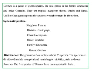
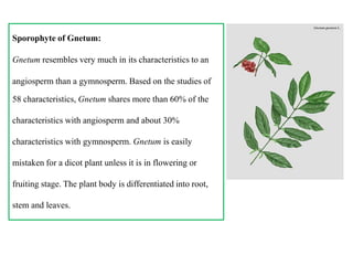
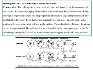
- Gnetum is a genus of gymnosperms that are tropical evergreen trees, shrubs, and lianas. Unlike other gymnosperms, they possess vessel elements in their xylem.
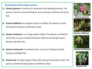
- The genus includes about 35 species distributed mainly in tropical regions of Africa, Asia, and South America. Five species are reported in India.
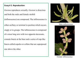
- Gnetum reproduces sexually as a dioecious plant. The male and female strobili are compound inflorescences composed of cup-like bracts. Male flowers each contain two anthers and female flowers each contain 4-10 ovules.
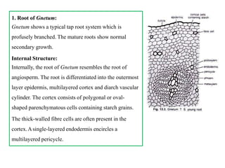
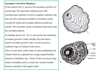
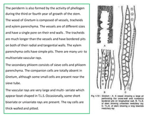
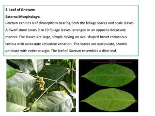
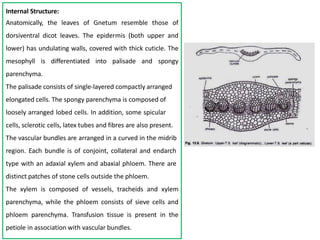


![Cordaitales - Yudhvir Singh Checked[1].pptx gymnosperms](https://cdn.slidesharecdn.com/ss_thumbnails/cordaitales-yudhvirsinghchecked1-250516114347-93347ab7-thumbnail.jpg?width=640&height=640&fit=bounds)