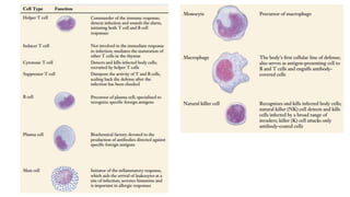
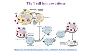
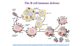
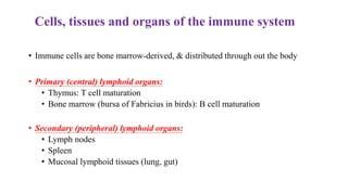
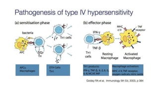
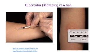
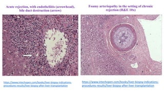
IMMUNOPATHOLOGY.
COMPENSATORY-ADAPTIVE
PROCESSES
Azerbaijan Medical University
Department of Pathological Anatomy
Lecture 3
Introduction
• We live in a world attackers, with too tiny to see with
the naked eye, and no vertebrate could long withstand
their attacks unprotected.
• We survive because we have evolved a variety of very
effective defenses against this constant attack.
• Immune reactions. The main goal of the immune system is to protect the host from pathogenic
microorganisms, but immune reactions can cause damage to cells and tissues. Reactions to damage
to one's own endogenous autoantigens underlie several autoimmune diseases. Immune reactions to
many external factors, such as microorganisms or environmental substances, can also cause cell
and tissue damage.
Many of the body’s most effective defenses are nonspecific
• The vertebrate is defended from infection the same way knights defended
medieval cities.
• “Walls and moats” make entry difficult; “roaming patrols” attack strangers;
and “sentries” challenge anyone wandering about and call patrols if a proper
“ID” is not presented.
The First Line of Defense
Walls and moats.
• The outermost layer of the vertebrate body, the skin, is the first barrier to
penetration by microbes.
• Mucous membranes in the respiratory and digestive tracts are also important
barriers that protect the body from invasion.
Staphylococcus aureus bacteria (yellow) sticking to the
mucus (blue) on the hair-like cilia
https://www.pinterest.com/pin/493214596671881001/
The Second Line of Defense: Cellular Counterattack
Roaming patrols.
• If the first line of defense is penetrated, the response of the body is to mount a
cellular counterattack, using a battery of cells and chemicals that kill microbes.
• These defenses act very rapidly after the onset of infection – macrophages,
neutrophils and natural killers.
Macrophages (“big eaters”) are large, irregularly shaped cells that kill microbes by
ingesting them through phagocytosis, much as an amoeba ingests a food particle.
Neutrophils are leukocytes that, like macrophages, ingest and kill bacteria by
phagocytosis.
Natural killer cells do not attack invading microbes directly.
• Instead, they kill cells of the body that have been infected with viruses.
• They kill not by phagocytosis, but rather by creating a hole in the plasma membrane
of the target cell.
Macrophage engulfing TB bacteria.
Colored SEM.
A macrophage in action
https://slideplayer.com/slide/3862267/
How natural killer cells kill target cells?
https://www.semanticscholar.org/paper/Cellular-Counterattack-%3A-The-Second-Line-of/
The Inflammatory Response
• The inflammatory response is a localized, nonspecific response to
infection.
• Infected or injured cells release chemical alarm signals-histamine and
prostaglandins.
• These chemicals promote the dilation of local blood vessels, which
increases the blood inflow to the site of infection or injury and causes
the area to become red and warm.
• They also increase the permeability of capillaries in the area,
producing the edema (tissue swelling) so often associated with
infection.
The Inflammatory Response
• The more permeable capillaries allow phagocytes (monocytes and
neutrophils) to migrate from the blood to the extracellular fluid, where
they can attack bacteria.
• Neutrophils arrive first, spilling out chemicals that kill the bacteria in
the vicinity (as well as tissue cells and themselves); the pus associated
with some infections is a mixture of dead or dying pathogens, tissue
cells, and neutrophils.
• Monocytes follow, become macrophages and engulf pathogens and the
remains of the dead cells.
The Third Line of Defense: The Immune Response
• Few of us pass through childhood without contracting some sort of infection.
• Chicken pox, for example, is an illness that many of us experience before we
reach our teens.
• It is a disease of childhood, because most of us contract it as children and
never catch it again.
• Once you have had the disease, you are usually immune to it.
• Specific immune defense mechanisms provide this immunity.
• Immune reactions. The main goal of the immune system is to protect the
host from pathogenic microorganisms, but immune reactions can cause
damage to cells and tissues. Reactions to damage to one's own endogenous
autoantigens underlie several autoimmune diseases. Immune reactions to
many external factors, such as microorganisms or environmental substances,
can also cause cell and tissue damage.
Immune System
• The immune system is a complex system that protects the organism
against influence of the external and internal proteinous factors, especially
microbial infections and other genetically foreign cells and proteins
(antigens).
• Lymphocytes called B cells respond to antigens by producing proteins
called antibodies.
• Antibody proteins are secreted into the blood and other body fluids and
thus provide humoral immunity.
• Other lymphocytes called T cells do not secrete antibodies but instead
directly attack the cells that carry the specific antigens.
• These cells are thus described as producing cell-mediated immunity.
Immune System
• The specific immune responses protect the body in two ways.
1) An individual can gain immunity by being exposed to a pathogen (disease-causing
agent) and perhaps getting the disease.
• This is acquired immunity, such as the resistance to the chicken pox that you acquire
after having the disease in childhood.
• Another term for this process is active immunity.
2) An individual can gain immunity by obtaining the antibodies from another individual.
• This happened to you before you were born, with antibodies made by your mother
being transferred to you across the placenta.
• Immunity gained in this way is called passive immunity.
The T cell immune defense
https://www.semanticscholar.org/paper/Cellular-Counterattack-%3A-The-Second-Line-of/
The B cell immune defense
https://www.semanticscholar.org/paper/Cellular-Counterattack-%3A-The-Second-Line-of/
Cells, tissues and organs of the immune system
• Immune cells are bone marrow-derived, & distributed through out the body
• Primary (central) lymphoid organs:
• Thymus: T cell maturation
• Bone marrow (bursa of Fabricius in birds): B cell maturation
• Secondary (peripheral) lymphoid organs:
• Lymph nodes
• Spleen
• Mucosal lymphoid tissues (lung, gut)
Morphology of the Thymus
Thymic age involution
• One of the major characteristics of vertebrate immunology
is thymic involution, the shrinking of the thymus with age, resulting in
changes in the architecture of the thymus and a decrease in tissue mass.
• After 2 years-old the volume of thymus increases and the weight
changes from 10-15 grams to 30-40 grams.
• After 7-8 years-old the volume of the thymus gradually decreases, its
functions are weakened and the parenchyma of the thymus is being
replaced by the fatty tissue.
• This process is continue until senescence.
Accidental involution of the thymus
• Causes by the action of the abundant glucocorticoids released according
the long-time and massive antigen stimulation during the variable
infectious diseases, intoxications, traumas and etc.
• Usually occurs in children.
• Thymus is shrinking in size, weight decreases and lymphocytes in
parenchyma phagocytosis by macrophages.
• The process is reversible, sometimes results in thymic atrophy:
sclerosing, dystrophic calcinosis and replacing by fatty tissue.
Thymic hyperplasia
• There are two distinct histologic types of thymic hyperplasia: true thymic hyperplasia and lymphoid
hyperplasia.
• True thymic hyperplasia is defined as enlargement of the thymus, which remains normally
organized, beyond the upper limit of normal for a given patient age.
• This disease entity is seen when a patient is recovering from some recent stress, such as chemotherapy
for neoplasm, corticosteroid therapy, irradiation, or thermal burns.
• The thymus sometimes grows to an even larger size after such stress, a phenomenon known as
rebound hyperplasia.
• Lymphoid hyperplasia of the thymus refers to the presence of an increased number of lymphoid
follicles.
• This condition is most commonly associated with myasthenia gravis, being seen in up to 65% of
cases.
• Lymphoid hyperplasia of the thymus is observed in a number of immunologically mediated disorders,
including SLE, rheumatoid arthritis, scleroderma, vasculitis, thyrotoxicosis, and Graves disease.
Thymic hypoplasia
• Synonyms: CATCH 22, congenital aplasia of thymus, pharyngeal pouch syndrome)
• Thymic hypoplasia is a condition where the thymus is underdeveloped or involuted.
• Calcium levels can be used to distinguish between the following two conditions associated
with thymic hypoplasia.
• The DiGeorge syndrome is an example of a selective T-cell deficiency caused by the
failure of development of the third and fourth pharyngeal pouches.
• The DiGeorge syndrome results in the absence or underdevelopment of the thymus and
parathyroid gland, associated with abnormalities of the outflow tract of the heart,
distinctive facies, hypoparathyroidism, hypocalcemia with tetany, and deficiency in T-cell
immunity.
Hypersensitivity
• Hypersensitivity (also called hypersensitivity reaction or intolerance) is a set of
undesirable reactions produced by the normal immune system, including allergies and
autoimmunity.
• These reactions may be damaging, uncomfortable, or occasionally fatal.
• Hypersensitivity reactions require a pre-sensitized (immune) state of the host.
• There are the following types of hypersencivity reactions: :
Type I - immediate (atopic, or anaphylactic)
Type II - antibody-dependent
Type III - immune complex
Type IV - cell-mediated or delayed
Type I hypersensitivity reactions – immediate
(or atopic, or anaphylactic)
• Type I hypersensitivity is an allergic reaction provoked by re-exposure to a specific antigen.
• Exposure may be by ingestion, inhalation, injection, or direct contact.
• The reaction is mediated by IgE antibodies and produced by the immediate release of histamine,
tryptase, arachidonate and derivatives by basophils and mast cells.
• This causes an inflammatory response leading to an immediate (within seconds to minutes) reaction.
• The reaction may be either local or systemic.
• Symptoms vary from mild irritation to sudden death from anaphylactic shock.
• Treatment usually involves epinephrine, antihistamines, and corticosteroids.
• Some examples: Allergic asthma, Allergic conjunctivitis, Allergic rhinitis ("hay fever“-also called
allergic rhinitis), Anaphylaxis, Angioedema, Urticaria (hives).
Type II hypersensitivity reactions - antibody-mediated
• In type II hypersensitivity, the antibodies produced by the immune response bind
to antigens on the patient's own cell surfaces.
• The antigens recognized in this way may either be intrinsic ("self" antigen,
innately part of the patient's cells) or extrinsic (absorbed onto the cells during
exposure to some foreign antigen, possibly as part of infection with a pathogen).
• IgG and IgM antibodies bind to these antigens to form complexes that activate
the classical pathway of complement activation for eliminating cells presenting
foreign antigens (which are usually, but not in this case, pathogens).
• As a result mediators of acute inflammation are generated at the site and
membrane attack complexes cause cell lysis and death.
• The reaction takes hours to a day.
• Examples: Autoimmune hemolytic anemia, Pernicious anemia, Immune
thrombocytopenia, Transfusion reactions, Hashimoto's thyroiditis, Graves'
disease, Myasthenia gravis, Farmer's Lung, Hemolytic disease of the newborn.
Type III hypersensitivity reactions - immune complex diseases
In type III hypersensitivity:
• Soluble immune complexes (aggregations of antigens and IgG and IgM
antibodies) form in the blood and are deposited in various tissues (typically the
skin, kidney and joints).
• This may trigger an immune response according to the classical pathway of
complement activation.
• The reaction takes hours to days to develop.
• Examples: Immune complex glomerulonephritis, Rheumatoid arthritis, Serum
sickness, Subacute bacterial endocarditis, Symptoms of malaria, Systemic lupus
erythematosus, Arthus reaction.
Type III hypersensitivity reactions
Immune-mediated CNS vasculitis
Type IV Hypersensitivity reactions – effector cell reactions
• Type IV hypersensitivity is often called delayed type as the reaction takes two to
three days to develop.
• Unlike the other types, it is not antibody mediated but rather is a type of cell-
mediated response.
• Some clinical examples: Contact dermatitis (poison ivy rash, for example),
Temporal arteritis, Symptoms of leprosy, Symptoms of tuberculosis, Transplant
rejection.
• The classical example of this hypersensitivity is tuberculin (Montoux) reaction.
• Reaction peaks 48 hours after the injection of antigen (PPD or old tuberculin).
• The lesion is characterized by induration and erythema.
https://labtestsonline.org/tests/tb-skin-test
https://en.wikipedia.org/wiki/Mantoux_test
Transplant rejection
• Rejection is the major cause of graft failure.
• It is therefore important to diagnose acute rejection as soon as possible to
institute prompt antirejection therapy.
• Generally, the success with which rejection can be reversed by
immunosuppressive agents determines the chance of long-term success of
the transplant.
• Liver transplants: HLA is less important than simple matching of organ
size (since most of these are done in children).
• There are two modes of rejection:
• Acute rejection: seen within two months, there are mixed inflammatory
portal and central vein infiltrates.
• Chronic rejection: at some later time, with continued inflammation, portal
fibrosis, arteriolar thickening, and bile ductular necrosis occurs.
Acute rejection, with endotheliitis (arrowhead),
bile duct destruction (arrow)
https://www.intechopen.com/books/liver-biopsy-indications-
procedures-results/liver-biopsy-after-liver-transplantation
Foamy arteriopathy in the setting of chronic
rejection (H&E 10x)
https://www.intechopen.com/books/liver-biopsy-indications-
procedures-results/liver-biopsy-after-liver-transplantation
Autoimmune diseases
• When the immune system attacks the body's own cells, it produces an
autoimmune disease.
• Immune tolerance is the tendency of T or B lymphocytes to ignore the
body’s own tissues.
• Maintaining tolerance is important because it prevents the immune
system from attacking its fellow cells.
• Brain, peripheral nerves, eyeball, thyroid gland, adrenal glands and
testis – there is no tolerance against to these organs.
• These organs protected from immune system special “physiological
barriers”.
I group (organ-specific) autoimmune diseases
1. Encephalomyelitis
2. Multiple Sclerosis
3. Polyneuritis
4. Sympathetic ophthalmia
5. Hashimoto’s thyroiditis
6. Idiopathic Addison's disease
7. Autoimmune orchitis (Aspermatogeny)
Blood-brain barrier
Hemato-retinal barrier
Idiopathic Addison's disease
https://www.msdmanuals.com/professional/endocrine-and-metabolic-
disorders/adrenal-disorders/addison-disease
http://www.pathologyoutlines.com/topic/thyroidhashimotosthyroiditis.html?rct=&mobile=off
Hashimoto’s thyroiditis
http://www.pathologyoutlines.com/topic/thyroidhashimotosthyroiditis.html
https://www.uaz.edu.mx/histo/pathology/ed/ch_21/c21_s40.htm
http://www.pathologyoutlines.com/topic/thyroidhashimotosthyroiditis.html
Hashimoto’s thyroiditis
http://ilovepathology.com/hashimotos-thyroiditis-2/
Hemato-testicular barrier
Autoimmune orchitis
Roxana Del Rio, PLoS Genetics 8(12):e1003140
II group (organ-nonspecific) autoimmune diseases
• Systemic lupus erythematosus (SLE)
• Rheumatoid arthritis
• Dermatomyositis
• Insulin-Dependent Diabetes
• Myasthenia Gravis
• Goodpasture’s Syndrome
• Crohn’s disease
• Grave’s disease
• Psoriasis
• Scleroderma
Primary Immune Deficiencies
Primary immuno-deficiency – rare genetic disorders
• Agammaglobulinemia (Bruton’s syndrome)
• Selective IgA deficiency
• DiGeorge syndrome
• Wiskott-Aldrich Syndrome - WAS
• Ataxia-Telangiectasia – A-T etc.
DiGeorge syndrome
• Also known as 22q11.2 deletion syndrome, is
a syndrome caused by the deletion of a small
segment of chromosome 22.
• Complex disorder affecting thymus genesis (~1:4000
live births)
• Clinical symptoms:
• Abnormal facies (long narrow face, small mouth,
prominent nose, hooded or full upper eyelids, low-set
cupped ears)
• small hands, abundant hair on the head
• cardiac and renal malformations
• cleft palate
• neural tube defects
• parahypothyroidism
• recurrent infections
https://en.wikipedia.org/wiki/DiGeorge_syndrome
Wiskott-Aldrich Syndrome (WAS)
• WAS is a rare X-linked recessive disease characterized by eczema, thrombocytopenia
(low platelet count), immune deficiency, and bloody diarrhea (secondary to the
thrombocytopenia).
• WAS occurs most often in males due to its X-linked recessive pattern of inheritance,
affecting between 1 and 10 males per million.
• The first signs are usually petechiae and bruising, resulting from a low platelet count
(i.e. thrombocytopenia).
• Spontaneous nose bleeds and bloody diarrhea are also common and eczema typically
develops within the first month of life.
• Recurrent bacterial infections develop by three months.
• The majority of children with WAS develop at least one autoimmune disorder,
and cancers (mainly lymphoma and leukemia) develop in up to a third of patients.
Wiskott-Aldrich Syndrome
https://www.nejm.org/doi/full/10.1056/NEJMp068209
Ataxia-Telangiectasia (A-T)
• A-T is an autosomal recessive disorder primarily
characterized by cerebellar degeneration, telangiectasia,
immunodeficiency, cancer susceptibility and radiation
sensitivity.
• A-T is often referred to as a genome instability or DNA
damage response syndrome.
https://ojrd.biomedcentral.com/articles/10.1186/s13023-016-0543-7
http://numedclinic.ca/telangiectasia/
Secondary Immune Deficiency Syndromes - AIDS
• Acquired Immune Deficiency Syndrome
• AIDS Etiology - Human Immunodeficiency Virus (HIV)
• HIV can be found in all body fluids of infected persons
• Transmission usually most common with infected blood, semen, and vaginal secretions
• Unprotected exposure to body fluids puts everyone at risk, e.g. health care workers
High-risk practices:
• IV drug abuse
• Unprotected sex (includes anal sex)
• AIDS Pathology:
• Virus attaches to the CD4+ protein on T-helper cells and destroys them.
• Decreased T-helper cell count makes the patient prone to opportunistic infections,
malignancies not normally seen in patients with intact immune systems, and direct CNS
destruction.
AIDS progression
• HIV infection – HIV+
• Antibodies produced usually within 1-6 months of exposure
• The antibodies produced cannot control the virus!
• ARC – AIDS Related Complex
• Enlarged lymph nodes, chronic fever and fatigue, weight loss
• Full blown AIDS – opportunistic infections and malignancies, and CNS damage
• Usually occurs when T-helper cell count drops to less than 500 (usual count is 800-1200)
• Malignancies Seen in AIDS:
• Kaposi’s Sarcoma
• Malignant nodules form on the skin and in the mouth, lymph nodes, and internal organs
• Squamos cell carcinomas in the mouth, rectum, and uterine cervix
Compensatory-Adaptive Processes
• Physiologic adaptation – cells respond to normal stimulation with hormones or
other endogenous biologically active substances.
• Pathologic adaptation – adaptation of cells or tissues to external or internal
environment pathogen components influence.
• Adaptation is manifested with:
– hypertrophy
– hyperplasia
– organization
– atrophy
– metaplasia
– dysplasia
Hypertrophy
• Hypertrophy (from Latin hyper – excessive, trophe – nutrition) refers to an
increase in the size of cells, that results in an increase in the size of the affected
organ.
• The hypertrophied organ has no new cells, just larger cells.
• The increased size of the cells is due to the synthesis and assembly of additional
intracellular structural components.
• Cells capable of division may respond to stress by undergoing both hyperplasia and
hypertrophy, whereas in non dividing (e.g., myocardial fibers) increased tissue mass
is due to hypertrophy.
• In many organs hypertrophy and hyperplasia may coexist and contribute to
increased size.
Hypertrophy
• Hypertrophy can be physiologic or pathologic.
• Physiologic hypertrophy is caused by increased functional demand or by stimulation
by hormones and growth factors.
• The striated muscle cells in the heart and skeletal muscles have only a limited
capacity for division, and respond to increased metabolic demands mainly by
undergoing hypertrophy.
• The most common stimulus for hypertrophy of muscle is increased workload.
• For example, the bulging muscles of bodybuilders engaged in “pumping iron” result
from enlargement of individual muscle fibers in response to increased demand.
• In the heart, the stimulus for hypertrophy is usually chronic hemodynamic
overload, resulting from either hypertension or faulty valves.
• In both tissue types the muscle cells synthesize more proteins and the number of
myofilaments increases.
• This increases the amount of force each myocyte can generate, and thus increases the
strength and work capacity of the muscle as a whole.
The relationship between normal, adapted, reversibly injured, and dead myocardial cells
Hypertrophy
• The massive physiologic growth of the uterus during pregnancy is a good
example of hormone-induced enlargement an organ that results mainly
from hypertrophy of muscle fibers.
• Uterine hypertrophy is stimulated by estrogenic hormones acting on
smooth muscle through estrogen receptors, eventually resulting in
increased synthesis of smooth muscle proteins and an increase in cell size.
Physiologic hypertrophy of the uterus during pregnancy
Types of Hypertrophy
1. Compensatory or functional hypertrophy
2. Vicarious (substitutional) hypertrophy
3. Endocrine or Neurohumoral hypertrophy
4. Hypertrophic excrescences
5. Vacatous hypertrophy
Vicarious (substitutional) hypertrophy
• Vicarious (substitutional) hypertrophy compensate the function of one
of the dead or surgically removed paired organs (lungs, kidneys, adrenal
glands).
• By its pathological essence it is close to regenerative hypertrophy.
• Significant role in its occurrence plays the complex of reflex and
humoral influences, the same with compensatory hypertrophy.
Endocrine or Neurohumoral hypertrophy
• It occurs on the background of endocrine glands
dysfunction.
• Its physiologic type is uterus hypertrophy and
macromastia under pregnancy.
• In pathologic conditions it is observed:
- endometrium glands hyperplasia
- mastopathy under ovarian dysfunction
- mammary gland excretory ducts hyperplasia in
males (gynecomastia) under testicles atrophy,
- enlargement of organs and prominent parts of
skeleton (acromegaly) under chromophobe
adenoma in adults.
Molitch ME. Endocrinol Metab Clin North Am. 1992;21(3):597–614.
Hypertrophic excrescences
• They are observed under chronic inflammations of mucus tunics with polyps formation, under lymph flow
disorders in low extremities and lymphostasis causing connective tissue excrescence (Lymphedema).
Inflammatory polyp of the colon Lymphedema
http://www.gastrolab.net/y0714.jpg https://www.ncbi.nlm.nih.gov/books/NBK537239/figure/article-24565.image.f1/?report=objectonly
Vacatous hypertrophy
• Adipose and connective tissues can fill the space occupied by organ or
tissue causing their atrophy.
• Examples: cranial bones thickening under cerebral atrophy, adipose
tissue excrescence in atrophic kidney hilus area.
Atrophy
• Atrophy is defined as a reduction in the size of an organ or tissue due to a decrease
in cell size and number.
• Physiologic and pathologic atrophies are differentiated.
• Physiologic atrophy common during normal development.
• some embryonic structures, such as the notochord and thyroglossal duct, undergo
atrophy during fetal development.
• upon birth, the umbilical arteries and arterial (botallian) ducts atrophy and
obliterate,
• aged people face genital glands atrophy,
• old people – with bones and intervertebral cartilages atrophy.
Pathologic atrophy
• Pathologic atrophy is observed in any age and can be caused by various reasons:
- inadequate nutrition
- endocrine glands dysfunction
- central and peripheral nervous system lesions
- intoxications
• Pathologic atrophy is reversible process.
• In case the cause is removed under condition that atrophy didn’t reach high level,
organ structure and function can be completely rehabilitated.
Classification of Pathologic atrophy
• Pathologic atrophy is divided into generalized and localized.
• Generalized atrophy or Cachexia or emaciation:
- alimentary cachexia
- emaciation under cancerous cachexia
- emaciation under cerebral cachexia
- emaciation under other diseases
• Concept of “emaciation” and “cachexia” are not identical.
• Cachexia in primary stages can be free from emaciation and be manifested with
progressive degenerative changes of the organs, for example, with osteoporosis.
Morphology of generalized emaciation
• Subcutaneous fatty tissue is absent.
• Eyes are hollow.
• Skin is dry.
• Abdomen is scaphoid.
• Starvation edemas sometimes take place.
Alimentary cachexia
• Alimentary cachexia occurs during starvation.
• Morphogenesis:
• Gradually fat stock decreases
• Skeletal muscles atrophy
• Atrophied fatty tissue becomes ochre-yellow color due to lipochrome pigment
accumulation.
• Fatty tissue of atrium and fatty marrow impregnate with serous fluid and
become dropsical (serous atrophy of fatty tissue).
• Pigment melanin accumulates in the skin of starving, so it colors in grey-
brown color.
• Heart, liver and other organs decrease in size.
• Lipofuscin (wear-and-tear pigment) accumulates in cardiac histiocytes,
hepatocytes and myocytes of skeletal muscles, as the result the organs become
of brown color (brown atrophy of organs).
Emaciation under cancerous cachexia
• Emaciation under cancerous cachexia is characteristic for cancerous
growth of any localization.
• The most fast it develops in patients ill with:
- cancer of esophagus,
- gastric carcinoma or intestine cancer caused by digestion disorders.
Alimentary cachexia Cancerous cachexia
https://www.quora.com/What-are-the-leading-theories-on-the-origin-of-cachexia
https://en.wikipedia.org/wiki/Starvation
Cerebral cachexia
• It develops as a result of a violation of the process of absorption of nutrients by
tissues in connection with pathological processes occurring in the hypothalamus
(inflammation, tumor, etc.).
• Emaciation under the other diseases takes place in case long term chronic infections:
- tuberculosis
- dysentery
- chronic sepsis
• It is caused by severe disorder of metabolism.
Hypophyseal cachexia (Simmonds’ diseases)
• It develops as a result of a violation of the absorption of nutrients by tissues due to
atrophy in the pituitary gland.
https://healthjade.net/simmonds-disease/
Local atrophy
• Local atrophy occurs by various reasons.
• The following types of it are differentiated:
- Dysfunctional atrophy
- Atrophy caused by inadequate blood supply
- Compressional atrophy
- Trophoneurotic atrophy
- Atrophy caused by physical and chemical agents influence
Dysfunctional atrophy
• Dysfunctional atrophy or Decreased workload (atrophy of disuse)
occurs when the function of organ decreases:
• When a fractured bone is immobilized in a plaster cast or when a patient
is restricted to complete bed rest, skeletal muscle atrophy rapidly ensues.
• The initial decrease in cell size is reversible once activity is resumed.
• With more prolonged disuse, skeletal muscle fibers decrease in number
(due to apoptosis) as well as in size; muscle atrophy can be accompanied
by increased bone resorption, leading to osteoporosis of disuse.
Atrophy caused by inadequate blood supply
(diminished blood supply)
• A gradual decrease in blood supply (ischemia) to a tissue as a result of
slowly developing arterial occlusive disease results in atrophy of the
tissue.
• In late adult life, the brain may undergo progressive atrophy, mainly
because of reduced blood supply as a result of atherosclerosis.
• This is called senile atrophy, which also affects the heart.
Compressional atrophy
• Tissue compression for any length of time can cause atrophy.
• An enlarging benign tumor can cause atrophy in the surrounding
uninvolved tissues.
• Atrophy in this setting is probably the result of ischemic changes caused
by compromise of the blood supply by the pressure exerted by the
expanding mass.
• In obstruction of urinary tracts with stones the urine stretches renal pelvis
and calyces (hydronephrosis), causing the atrophy of renal parenchyma.
• In case of liquor outflow the hindbrain ventricles dilate (hydrocephalus)
and results the cerebral atrophy.
Hydronephrosis
Trophoneurotic atrophy
• Synonym: Loss of innervation (denervation atrophy).
• The normal metabolism and function of skeletal muscle are dependent on its nerve supply. Damage to
the nerves leads to atrophy of the muscle fibers supplied by those nerves.
• Trophoneurotic atrophy is caused by failure of organ connection with central nervous system under
peripheral nerves traumatic, tumor or inflammatory injury.
Atrophy caused by physical and chemical agents influence
– in bone marrow and genital glands under radiation influence.
– radioiodine causes thyroid gland atrophy.
– after long term treatment with adrenocorticotropic hormone or glucocorticoids adrenal glands cortex’
atrophy develops.
• Morphology:
• Organs reduce in size under atrophy.
• Their surface in most cases is smooth (smooth atrophy), in kidneys – granular (granular atrophy).
Regeneration
• Regeneration (from Latin regeneratio – restoration) is the process of self-healing of
living matter in a damaged area.
• Regeneration takes place on molecular, subcellular, cellular, tissue and organ levels
and reflects the principle of living functions autoregulation.
• It is based on cellular and intracellular hyperplastic processes.
• Cellular reproduction is characteristic for cellular form of regeneration, ultrastructures
and their components quantity increase (hyperplasia) and their enlargement
(hypertrophy) are characteristic for intracellular form.
• The latter form is characteristic of the cells of all organs and is universal.
Regeneration types
1. Physiologic regeneration
2. Reparative regeneration
3. Pathologic regeneration
Physiologic regeneration
• Physiologic regeneration is carried out throughout life and reflects the endless
process of disintegration and synthesis of substances.
• It is characterized by intracellular renewal of molecules and ultrastructures, as well
as whole cells, fibrous structures and basic substances of connective tissue.
• A combination of intracellular renewal with cell mitosis is observed in the liver,
kidneys, and pancreas.
• Due to cell division, constant changes occur in the epidermis, epithelium of the
mucous membrane of the digestive tract, synovial membranes, bone marrow and
blood elements.
Reparative regeneration
• Reparative regeneration is the replacement of organ defects in various pathological
processes.
• It is based on the same mechanisms as in physiological regeneration, and the repair of
damage in each organ occurs in the same way as in the conditions of physiological
recovery, but more intensively.
• The main form of restoration of the degeneratively altered structure of tissue cells is
intracellular regeneration, as well as cellular and intracellular regeneration in case of
their necrosis.
• The end result of reparative regeneration is expressed in restitution or substitution.
Reparative regeneration
• Restitution (complete regeneration) is characterized by the replacement of a tissue
defect with tissue identical to the dead one.
• This applies to those organs and tissues where regeneration takes place exclusively in
the cellular form (bone marrow, epidermis, mucosal epithelium, liver).
• Substitution (incomplete regeneration) is characteristic of the healing of organs,
proceeding mainly or exclusively through intracellular repair (heart, CNS).
• For example, in myocardial necrosis, foci are replaced by connective tissue, and in brain
tissue, dead neurons are replaced by a glial scar.
Pathologic regeneration
• Pathologic regeneration is a type of reparative regeneration that occurs under
conditions of insufficiency of local and general regulatory mechanisms and is
characterized by a perversion of the regenerative process and a violation of the
transition from the proliferation phase to the differentiation phase.
• Protein or vitamin deficiencies, nervous regulation disorder, hormonal imbalances,
and immune system suppression can seriously affect the speed and quality of healing.
• Example of pathologic regeneration: connective tissue hyperproduction with keloid
formation under radiation or thermal trauma.
Bulky keloid forming at the site of abdominal surgery
https://en.wikipedia.org/wiki/Keloid
Metaplasia
• Metaplasia is a reversible change in which one differentiated cell type (epithelial or mesenchymal) is
replaced by another cell type.
• It often represents an adaptive response in which one cell type that is sensitive to a particular stress is
replaced by another cell type that is better able to withstand the adverse environment.
• The most common epithelial metaplasia is columnar to squamous, as occurs in the respiratory tract in
response to chronic irritation.
• In the habitual cigarette smoker, the normal ciliated columnar epithelial cells of the trachea and
bronchi are often replaced by stratified squamous epithelial cells.
• Stones in the excretory ducts of the salivary glands, pancreas, or bile ducts, which are normally lined
by secretory columnar epithelium, may also lead to squamous metaplasia by stratified squamous
epithelium.
• A deficiency of vitamin A (retinoic acid) induces squamous metaplasia in the respiratory epithelium.
Metaplasia
• In all these instances the more rugged stratified squamous epithelium is able to survive under circumstances
in which the more fragile specialized columnar epithelium might have succumbed.
• However, the change to metaplastic squamous cells comes with a price.
• In the respiratory tract, for example, although the epithelial lining becomes tough, important mechanisms
of protection against infection - mucus secretion and the ciliary action of the columnar epithelium - are lost.
• Thus, epithelial metaplasia is a double-edged sword and, in most circumstances, represents an undesirable
change.
• Moreover, the influences that predispose to metaplasia, if persistent, can initiate malignant transformation
in metaplastic epithelium.
• Thus, a common form of cancer in the respiratory tract is composed of squamous cells, which can arise in
areas where the normal columnar epithelium has been replaced by squamous epithelium.
• Metaplasia from squamous to columnar type may also occur, as in Barrett esophagus, in which the
esophageal squamous epithelium is replaced by intestinal-like columnar cells under the influence of
refluxed gastric acid.
• Cancers may arise in these areas; these are typically glandular (adenocarcinomas).
•
Metaplasia of columnar to
squamous epithelium
https://www.memorangapp.com/flashcards/77897/Pathoma+Ch+10%3A+GI+Pathology/
Barret’s ezofagus
Intestinal metaplasia in Chronic atrophic gastritis
• http://www.intechopen.com/source/html/41705/media/image6.jpeg
Connective tissue metaplasia
• Connective tissue metaplasia is the formation of cartilage, bone, or
adipose tissue (mesenchymal tissues) in tissues that normally do not
contain these elements.
• For example, bone formation in muscle, designated myositis ossificans,
occasionally occurs after intramuscular hemorrhage.
• This type of metaplasia is less clearly seen as an adaptive response, and
may be a result of cell or tissue injury.
Dysplasia
• Dysplasia is a serious violation of the proliferation and differentiation of the
epithelium with the development of cellular atypia and a change in histoarchitectonics:
loss of polarity, loss of histo- and organ-specificity of the epithelium.
• The basement membrane is not damaged during dysplasia.
• Most often, dysplasia develops in inflammatory and regenerative processes.
• Depending on the stage of proliferation and the state of cellular and tissue atypia, there
are three stages of dysplasia:
• І – mild (minor),
• ІІ – moderate (middle),
• ІІІ – severe (major).
• Minor and moderate dysplasia is of reversible character.
• Cellular and tissue changes in severe dysplasia are rare, are subject to reversible
processes and are interpreted as a precancerous process.
• Sometimes locally they are difficult to distinguish from carcinoma.
Normal cytology of uterine cervix (Pap smear)
http://www.cytologystuff.com/gallery/images_large/slide0020.jpg
Low-grade squamous intraepithelial lesion – LSIL
Koilocytosis
http://www.cytopathnet.com/show_image.php?id=22&scalesize=0&nocount=y
http://nih.techriver.net/patientImages%5C9337.jpg
High-grade squamous intraepithelial lesion – HSIL (CIN II)
Low-grade dysplasia
https://www.pathologyoutlines.com/topic/colontumortubularadenoma.html
High-grade dysplasia
https://www.pathologyoutlines.com/topic/colontumortvadenoma.html
Organization
• The organization is a protective-adaptive process directed to separate and substitute with granulation
tissue - focus on necrosis, hemorrhage, or exudates as well as thrombi, foreign objects, and parasites.
• Its essence is reduced to the formation of connective tissue during the healing of defects in wounds and
ulcers, the replacement of necrosis or thrombotic masses with connective tissue (correct organization)
and their encapsulation.
• According to I.V.Davydovsky, the following forms of wound healing are distinguished:
• immediate closure of epithelial defects
• healing under a scab
• primary intention of wound
• secondary intention or healing by granulations
• Immediate closure of epithelial defects ensures the growth of cells on the sides of the wound and closing it with a
layer of cells without mitotic cell division.
• Such a simple form of healing is typical for superficial damage to the cornea, mucous membrane and vascular
intima.
• Healing under eschar is also characteristic of minor damage to the epidermis.
• For example, with superficial excoriations, lymph and blood exclude rapid drying and transformation into crust
(eschar).
• The epidermis regenerates under the crust, which, as a result of the rejection process, disappears on the 3rd-7th day.
• Healing by first (primary) intention
• Under phagocytes’ proteolytic enzymes influence partial lysis of blood clots and tissue detritus takes place and
wound content is removed in the very first date after injury together with exudate.
• On the 2-3rd day, granulation tissue appears, which matures on the 10-15th day.
• In the clinic, the edges of large wounds are connected with sutures and supported with bandages.
• If the distance between the sides is even 10 mm, then in a few days this distance will be reduced to zero due to
tissue edema and reduction of the fibrin clot that glues the edges of the wound.
Healing by secondary intention
• Occur when the sides of the wound are separated due to purulent inflammation.
• It is characterized by the release of detritus and foreign bodies from the wound by "suppuration".
• Rejection of necrotic masses occurs during the first 5-6 days (secondary cleansing of the wound) and
granulation tissue begins to develop along the edges of the wound.
• During wound healing by primary or secondary intention, the maturation of granulation tissue is
accompanied by regeneration of the epithelium.
• However, with secondary intention, a scar is still formed at the site of the wound.
• The inflammatory process always precedes the healing of the ulcer.
• Granulation tissue grows into a zone of necrosis, which matures into coarse fiber and often undergoes
hyalinosis.
• The latter causes cavity deformation of the organ and stenosis.
Granulation tissue
• Granulation tissue consists of 6 layers, starting from the surface of the
wound and moving deep into healthy tissues:
1. Superficial leukocyte-necrotic layer
2. Superficial vascular layer (capillaries with sharp filling)
3. Vertical veins
4. Maturing layer
5. Layer of horizontal fibroblasts
6. Fibrous (coarse fibrous connective tissue) layer
Granulation tissue
• https://upload.wikimedia.org/wikipedia/commons/7/7c/Granulation_tissue_in_an_infected_wound,_HE_1.JPG

Lecture-3 Immunopathology. Adaptation.pptx

  • 1.
  • 2.
    Introduction • We livein a world attackers, with too tiny to see with the naked eye, and no vertebrate could long withstand their attacks unprotected. • We survive because we have evolved a variety of very effective defenses against this constant attack. • Immune reactions. The main goal of the immune system is to protect the host from pathogenic microorganisms, but immune reactions can cause damage to cells and tissues. Reactions to damage to one's own endogenous autoantigens underlie several autoimmune diseases. Immune reactions to many external factors, such as microorganisms or environmental substances, can also cause cell and tissue damage.
  • 3.
    Many of thebody’s most effective defenses are nonspecific • The vertebrate is defended from infection the same way knights defended medieval cities. • “Walls and moats” make entry difficult; “roaming patrols” attack strangers; and “sentries” challenge anyone wandering about and call patrols if a proper “ID” is not presented. The First Line of Defense Walls and moats. • The outermost layer of the vertebrate body, the skin, is the first barrier to penetration by microbes. • Mucous membranes in the respiratory and digestive tracts are also important barriers that protect the body from invasion.
  • 4.
    Staphylococcus aureus bacteria(yellow) sticking to the mucus (blue) on the hair-like cilia https://www.pinterest.com/pin/493214596671881001/
  • 5.
    The Second Lineof Defense: Cellular Counterattack Roaming patrols. • If the first line of defense is penetrated, the response of the body is to mount a cellular counterattack, using a battery of cells and chemicals that kill microbes. • These defenses act very rapidly after the onset of infection – macrophages, neutrophils and natural killers. Macrophages (“big eaters”) are large, irregularly shaped cells that kill microbes by ingesting them through phagocytosis, much as an amoeba ingests a food particle. Neutrophils are leukocytes that, like macrophages, ingest and kill bacteria by phagocytosis. Natural killer cells do not attack invading microbes directly. • Instead, they kill cells of the body that have been infected with viruses. • They kill not by phagocytosis, but rather by creating a hole in the plasma membrane of the target cell.
  • 6.
    Macrophage engulfing TBbacteria. Colored SEM. A macrophage in action https://slideplayer.com/slide/3862267/
  • 7.
    How natural killercells kill target cells? https://www.semanticscholar.org/paper/Cellular-Counterattack-%3A-The-Second-Line-of/
  • 8.
    The Inflammatory Response •The inflammatory response is a localized, nonspecific response to infection. • Infected or injured cells release chemical alarm signals-histamine and prostaglandins. • These chemicals promote the dilation of local blood vessels, which increases the blood inflow to the site of infection or injury and causes the area to become red and warm. • They also increase the permeability of capillaries in the area, producing the edema (tissue swelling) so often associated with infection.
  • 9.
    The Inflammatory Response •The more permeable capillaries allow phagocytes (monocytes and neutrophils) to migrate from the blood to the extracellular fluid, where they can attack bacteria. • Neutrophils arrive first, spilling out chemicals that kill the bacteria in the vicinity (as well as tissue cells and themselves); the pus associated with some infections is a mixture of dead or dying pathogens, tissue cells, and neutrophils. • Monocytes follow, become macrophages and engulf pathogens and the remains of the dead cells.
  • 10.
    The Third Lineof Defense: The Immune Response • Few of us pass through childhood without contracting some sort of infection. • Chicken pox, for example, is an illness that many of us experience before we reach our teens. • It is a disease of childhood, because most of us contract it as children and never catch it again. • Once you have had the disease, you are usually immune to it. • Specific immune defense mechanisms provide this immunity. • Immune reactions. The main goal of the immune system is to protect the host from pathogenic microorganisms, but immune reactions can cause damage to cells and tissues. Reactions to damage to one's own endogenous autoantigens underlie several autoimmune diseases. Immune reactions to many external factors, such as microorganisms or environmental substances, can also cause cell and tissue damage.
  • 11.
    Immune System • Theimmune system is a complex system that protects the organism against influence of the external and internal proteinous factors, especially microbial infections and other genetically foreign cells and proteins (antigens). • Lymphocytes called B cells respond to antigens by producing proteins called antibodies. • Antibody proteins are secreted into the blood and other body fluids and thus provide humoral immunity. • Other lymphocytes called T cells do not secrete antibodies but instead directly attack the cells that carry the specific antigens. • These cells are thus described as producing cell-mediated immunity.
  • 12.
    Immune System • Thespecific immune responses protect the body in two ways. 1) An individual can gain immunity by being exposed to a pathogen (disease-causing agent) and perhaps getting the disease. • This is acquired immunity, such as the resistance to the chicken pox that you acquire after having the disease in childhood. • Another term for this process is active immunity. 2) An individual can gain immunity by obtaining the antibodies from another individual. • This happened to you before you were born, with antibodies made by your mother being transferred to you across the placenta. • Immunity gained in this way is called passive immunity.
  • 14.
    The T cellimmune defense https://www.semanticscholar.org/paper/Cellular-Counterattack-%3A-The-Second-Line-of/
  • 15.
    The B cellimmune defense https://www.semanticscholar.org/paper/Cellular-Counterattack-%3A-The-Second-Line-of/
  • 16.
    Cells, tissues andorgans of the immune system • Immune cells are bone marrow-derived, & distributed through out the body • Primary (central) lymphoid organs: • Thymus: T cell maturation • Bone marrow (bursa of Fabricius in birds): B cell maturation • Secondary (peripheral) lymphoid organs: • Lymph nodes • Spleen • Mucosal lymphoid tissues (lung, gut)
  • 17.
  • 18.
    Thymic age involution •One of the major characteristics of vertebrate immunology is thymic involution, the shrinking of the thymus with age, resulting in changes in the architecture of the thymus and a decrease in tissue mass. • After 2 years-old the volume of thymus increases and the weight changes from 10-15 grams to 30-40 grams. • After 7-8 years-old the volume of the thymus gradually decreases, its functions are weakened and the parenchyma of the thymus is being replaced by the fatty tissue. • This process is continue until senescence.
  • 19.
    Accidental involution ofthe thymus • Causes by the action of the abundant glucocorticoids released according the long-time and massive antigen stimulation during the variable infectious diseases, intoxications, traumas and etc. • Usually occurs in children. • Thymus is shrinking in size, weight decreases and lymphocytes in parenchyma phagocytosis by macrophages. • The process is reversible, sometimes results in thymic atrophy: sclerosing, dystrophic calcinosis and replacing by fatty tissue.
  • 20.
    Thymic hyperplasia • Thereare two distinct histologic types of thymic hyperplasia: true thymic hyperplasia and lymphoid hyperplasia. • True thymic hyperplasia is defined as enlargement of the thymus, which remains normally organized, beyond the upper limit of normal for a given patient age. • This disease entity is seen when a patient is recovering from some recent stress, such as chemotherapy for neoplasm, corticosteroid therapy, irradiation, or thermal burns. • The thymus sometimes grows to an even larger size after such stress, a phenomenon known as rebound hyperplasia. • Lymphoid hyperplasia of the thymus refers to the presence of an increased number of lymphoid follicles. • This condition is most commonly associated with myasthenia gravis, being seen in up to 65% of cases. • Lymphoid hyperplasia of the thymus is observed in a number of immunologically mediated disorders, including SLE, rheumatoid arthritis, scleroderma, vasculitis, thyrotoxicosis, and Graves disease.
  • 21.
    Thymic hypoplasia • Synonyms:CATCH 22, congenital aplasia of thymus, pharyngeal pouch syndrome) • Thymic hypoplasia is a condition where the thymus is underdeveloped or involuted. • Calcium levels can be used to distinguish between the following two conditions associated with thymic hypoplasia. • The DiGeorge syndrome is an example of a selective T-cell deficiency caused by the failure of development of the third and fourth pharyngeal pouches. • The DiGeorge syndrome results in the absence or underdevelopment of the thymus and parathyroid gland, associated with abnormalities of the outflow tract of the heart, distinctive facies, hypoparathyroidism, hypocalcemia with tetany, and deficiency in T-cell immunity.
  • 22.
    Hypersensitivity • Hypersensitivity (alsocalled hypersensitivity reaction or intolerance) is a set of undesirable reactions produced by the normal immune system, including allergies and autoimmunity. • These reactions may be damaging, uncomfortable, or occasionally fatal. • Hypersensitivity reactions require a pre-sensitized (immune) state of the host. • There are the following types of hypersencivity reactions: : Type I - immediate (atopic, or anaphylactic) Type II - antibody-dependent Type III - immune complex Type IV - cell-mediated or delayed
  • 23.
    Type I hypersensitivityreactions – immediate (or atopic, or anaphylactic) • Type I hypersensitivity is an allergic reaction provoked by re-exposure to a specific antigen. • Exposure may be by ingestion, inhalation, injection, or direct contact. • The reaction is mediated by IgE antibodies and produced by the immediate release of histamine, tryptase, arachidonate and derivatives by basophils and mast cells. • This causes an inflammatory response leading to an immediate (within seconds to minutes) reaction. • The reaction may be either local or systemic. • Symptoms vary from mild irritation to sudden death from anaphylactic shock. • Treatment usually involves epinephrine, antihistamines, and corticosteroids. • Some examples: Allergic asthma, Allergic conjunctivitis, Allergic rhinitis ("hay fever“-also called allergic rhinitis), Anaphylaxis, Angioedema, Urticaria (hives).
  • 27.
    Type II hypersensitivityreactions - antibody-mediated • In type II hypersensitivity, the antibodies produced by the immune response bind to antigens on the patient's own cell surfaces. • The antigens recognized in this way may either be intrinsic ("self" antigen, innately part of the patient's cells) or extrinsic (absorbed onto the cells during exposure to some foreign antigen, possibly as part of infection with a pathogen). • IgG and IgM antibodies bind to these antigens to form complexes that activate the classical pathway of complement activation for eliminating cells presenting foreign antigens (which are usually, but not in this case, pathogens). • As a result mediators of acute inflammation are generated at the site and membrane attack complexes cause cell lysis and death. • The reaction takes hours to a day. • Examples: Autoimmune hemolytic anemia, Pernicious anemia, Immune thrombocytopenia, Transfusion reactions, Hashimoto's thyroiditis, Graves' disease, Myasthenia gravis, Farmer's Lung, Hemolytic disease of the newborn.
  • 29.
    Type III hypersensitivityreactions - immune complex diseases In type III hypersensitivity: • Soluble immune complexes (aggregations of antigens and IgG and IgM antibodies) form in the blood and are deposited in various tissues (typically the skin, kidney and joints). • This may trigger an immune response according to the classical pathway of complement activation. • The reaction takes hours to days to develop. • Examples: Immune complex glomerulonephritis, Rheumatoid arthritis, Serum sickness, Subacute bacterial endocarditis, Symptoms of malaria, Systemic lupus erythematosus, Arthus reaction.
  • 30.
  • 31.
  • 32.
  • 33.
    Type IV Hypersensitivityreactions – effector cell reactions • Type IV hypersensitivity is often called delayed type as the reaction takes two to three days to develop. • Unlike the other types, it is not antibody mediated but rather is a type of cell- mediated response. • Some clinical examples: Contact dermatitis (poison ivy rash, for example), Temporal arteritis, Symptoms of leprosy, Symptoms of tuberculosis, Transplant rejection. • The classical example of this hypersensitivity is tuberculin (Montoux) reaction. • Reaction peaks 48 hours after the injection of antigen (PPD or old tuberculin). • The lesion is characterized by induration and erythema.
  • 35.
  • 36.
    Transplant rejection • Rejectionis the major cause of graft failure. • It is therefore important to diagnose acute rejection as soon as possible to institute prompt antirejection therapy. • Generally, the success with which rejection can be reversed by immunosuppressive agents determines the chance of long-term success of the transplant. • Liver transplants: HLA is less important than simple matching of organ size (since most of these are done in children). • There are two modes of rejection: • Acute rejection: seen within two months, there are mixed inflammatory portal and central vein infiltrates. • Chronic rejection: at some later time, with continued inflammation, portal fibrosis, arteriolar thickening, and bile ductular necrosis occurs.
  • 37.
    Acute rejection, withendotheliitis (arrowhead), bile duct destruction (arrow) https://www.intechopen.com/books/liver-biopsy-indications- procedures-results/liver-biopsy-after-liver-transplantation Foamy arteriopathy in the setting of chronic rejection (H&E 10x) https://www.intechopen.com/books/liver-biopsy-indications- procedures-results/liver-biopsy-after-liver-transplantation
  • 38.
    Autoimmune diseases • Whenthe immune system attacks the body's own cells, it produces an autoimmune disease. • Immune tolerance is the tendency of T or B lymphocytes to ignore the body’s own tissues. • Maintaining tolerance is important because it prevents the immune system from attacking its fellow cells. • Brain, peripheral nerves, eyeball, thyroid gland, adrenal glands and testis – there is no tolerance against to these organs. • These organs protected from immune system special “physiological barriers”.
  • 39.
    I group (organ-specific)autoimmune diseases 1. Encephalomyelitis 2. Multiple Sclerosis 3. Polyneuritis 4. Sympathetic ophthalmia 5. Hashimoto’s thyroiditis 6. Idiopathic Addison's disease 7. Autoimmune orchitis (Aspermatogeny)
  • 40.
  • 41.
  • 42.
  • 44.
  • 45.
  • 46.
  • 47.
  • 48.
    Autoimmune orchitis Roxana DelRio, PLoS Genetics 8(12):e1003140
  • 49.
    II group (organ-nonspecific)autoimmune diseases • Systemic lupus erythematosus (SLE) • Rheumatoid arthritis • Dermatomyositis • Insulin-Dependent Diabetes • Myasthenia Gravis • Goodpasture’s Syndrome • Crohn’s disease • Grave’s disease • Psoriasis • Scleroderma
  • 50.
    Primary Immune Deficiencies Primaryimmuno-deficiency – rare genetic disorders • Agammaglobulinemia (Bruton’s syndrome) • Selective IgA deficiency • DiGeorge syndrome • Wiskott-Aldrich Syndrome - WAS • Ataxia-Telangiectasia – A-T etc.
  • 51.
    DiGeorge syndrome • Alsoknown as 22q11.2 deletion syndrome, is a syndrome caused by the deletion of a small segment of chromosome 22. • Complex disorder affecting thymus genesis (~1:4000 live births) • Clinical symptoms: • Abnormal facies (long narrow face, small mouth, prominent nose, hooded or full upper eyelids, low-set cupped ears) • small hands, abundant hair on the head • cardiac and renal malformations • cleft palate • neural tube defects • parahypothyroidism • recurrent infections https://en.wikipedia.org/wiki/DiGeorge_syndrome
  • 52.
    Wiskott-Aldrich Syndrome (WAS) •WAS is a rare X-linked recessive disease characterized by eczema, thrombocytopenia (low platelet count), immune deficiency, and bloody diarrhea (secondary to the thrombocytopenia). • WAS occurs most often in males due to its X-linked recessive pattern of inheritance, affecting between 1 and 10 males per million. • The first signs are usually petechiae and bruising, resulting from a low platelet count (i.e. thrombocytopenia). • Spontaneous nose bleeds and bloody diarrhea are also common and eczema typically develops within the first month of life. • Recurrent bacterial infections develop by three months. • The majority of children with WAS develop at least one autoimmune disorder, and cancers (mainly lymphoma and leukemia) develop in up to a third of patients.
  • 53.
  • 54.
    Ataxia-Telangiectasia (A-T) • A-Tis an autosomal recessive disorder primarily characterized by cerebellar degeneration, telangiectasia, immunodeficiency, cancer susceptibility and radiation sensitivity. • A-T is often referred to as a genome instability or DNA damage response syndrome. https://ojrd.biomedcentral.com/articles/10.1186/s13023-016-0543-7 http://numedclinic.ca/telangiectasia/
  • 55.
    Secondary Immune DeficiencySyndromes - AIDS • Acquired Immune Deficiency Syndrome • AIDS Etiology - Human Immunodeficiency Virus (HIV) • HIV can be found in all body fluids of infected persons • Transmission usually most common with infected blood, semen, and vaginal secretions • Unprotected exposure to body fluids puts everyone at risk, e.g. health care workers High-risk practices: • IV drug abuse • Unprotected sex (includes anal sex) • AIDS Pathology: • Virus attaches to the CD4+ protein on T-helper cells and destroys them. • Decreased T-helper cell count makes the patient prone to opportunistic infections, malignancies not normally seen in patients with intact immune systems, and direct CNS destruction.
  • 56.
    AIDS progression • HIVinfection – HIV+ • Antibodies produced usually within 1-6 months of exposure • The antibodies produced cannot control the virus! • ARC – AIDS Related Complex • Enlarged lymph nodes, chronic fever and fatigue, weight loss • Full blown AIDS – opportunistic infections and malignancies, and CNS damage • Usually occurs when T-helper cell count drops to less than 500 (usual count is 800-1200) • Malignancies Seen in AIDS: • Kaposi’s Sarcoma • Malignant nodules form on the skin and in the mouth, lymph nodes, and internal organs • Squamos cell carcinomas in the mouth, rectum, and uterine cervix
  • 58.
    Compensatory-Adaptive Processes • Physiologicadaptation – cells respond to normal stimulation with hormones or other endogenous biologically active substances. • Pathologic adaptation – adaptation of cells or tissues to external or internal environment pathogen components influence. • Adaptation is manifested with: – hypertrophy – hyperplasia – organization – atrophy – metaplasia – dysplasia
  • 59.
    Hypertrophy • Hypertrophy (fromLatin hyper – excessive, trophe – nutrition) refers to an increase in the size of cells, that results in an increase in the size of the affected organ. • The hypertrophied organ has no new cells, just larger cells. • The increased size of the cells is due to the synthesis and assembly of additional intracellular structural components. • Cells capable of division may respond to stress by undergoing both hyperplasia and hypertrophy, whereas in non dividing (e.g., myocardial fibers) increased tissue mass is due to hypertrophy. • In many organs hypertrophy and hyperplasia may coexist and contribute to increased size.
  • 60.
    Hypertrophy • Hypertrophy canbe physiologic or pathologic. • Physiologic hypertrophy is caused by increased functional demand or by stimulation by hormones and growth factors. • The striated muscle cells in the heart and skeletal muscles have only a limited capacity for division, and respond to increased metabolic demands mainly by undergoing hypertrophy. • The most common stimulus for hypertrophy of muscle is increased workload. • For example, the bulging muscles of bodybuilders engaged in “pumping iron” result from enlargement of individual muscle fibers in response to increased demand. • In the heart, the stimulus for hypertrophy is usually chronic hemodynamic overload, resulting from either hypertension or faulty valves. • In both tissue types the muscle cells synthesize more proteins and the number of myofilaments increases. • This increases the amount of force each myocyte can generate, and thus increases the strength and work capacity of the muscle as a whole.
  • 61.
    The relationship betweennormal, adapted, reversibly injured, and dead myocardial cells
  • 62.
    Hypertrophy • The massivephysiologic growth of the uterus during pregnancy is a good example of hormone-induced enlargement an organ that results mainly from hypertrophy of muscle fibers. • Uterine hypertrophy is stimulated by estrogenic hormones acting on smooth muscle through estrogen receptors, eventually resulting in increased synthesis of smooth muscle proteins and an increase in cell size.
  • 63.
    Physiologic hypertrophy ofthe uterus during pregnancy
  • 64.
    Types of Hypertrophy 1.Compensatory or functional hypertrophy 2. Vicarious (substitutional) hypertrophy 3. Endocrine or Neurohumoral hypertrophy 4. Hypertrophic excrescences 5. Vacatous hypertrophy
  • 65.
    Vicarious (substitutional) hypertrophy •Vicarious (substitutional) hypertrophy compensate the function of one of the dead or surgically removed paired organs (lungs, kidneys, adrenal glands). • By its pathological essence it is close to regenerative hypertrophy. • Significant role in its occurrence plays the complex of reflex and humoral influences, the same with compensatory hypertrophy.
  • 66.
    Endocrine or Neurohumoralhypertrophy • It occurs on the background of endocrine glands dysfunction. • Its physiologic type is uterus hypertrophy and macromastia under pregnancy. • In pathologic conditions it is observed: - endometrium glands hyperplasia - mastopathy under ovarian dysfunction - mammary gland excretory ducts hyperplasia in males (gynecomastia) under testicles atrophy, - enlargement of organs and prominent parts of skeleton (acromegaly) under chromophobe adenoma in adults. Molitch ME. Endocrinol Metab Clin North Am. 1992;21(3):597–614.
  • 67.
    Hypertrophic excrescences • Theyare observed under chronic inflammations of mucus tunics with polyps formation, under lymph flow disorders in low extremities and lymphostasis causing connective tissue excrescence (Lymphedema). Inflammatory polyp of the colon Lymphedema http://www.gastrolab.net/y0714.jpg https://www.ncbi.nlm.nih.gov/books/NBK537239/figure/article-24565.image.f1/?report=objectonly
  • 68.
    Vacatous hypertrophy • Adiposeand connective tissues can fill the space occupied by organ or tissue causing their atrophy. • Examples: cranial bones thickening under cerebral atrophy, adipose tissue excrescence in atrophic kidney hilus area.
  • 69.
    Atrophy • Atrophy isdefined as a reduction in the size of an organ or tissue due to a decrease in cell size and number. • Physiologic and pathologic atrophies are differentiated. • Physiologic atrophy common during normal development. • some embryonic structures, such as the notochord and thyroglossal duct, undergo atrophy during fetal development. • upon birth, the umbilical arteries and arterial (botallian) ducts atrophy and obliterate, • aged people face genital glands atrophy, • old people – with bones and intervertebral cartilages atrophy.
  • 70.
    Pathologic atrophy • Pathologicatrophy is observed in any age and can be caused by various reasons: - inadequate nutrition - endocrine glands dysfunction - central and peripheral nervous system lesions - intoxications • Pathologic atrophy is reversible process. • In case the cause is removed under condition that atrophy didn’t reach high level, organ structure and function can be completely rehabilitated.
  • 71.
    Classification of Pathologicatrophy • Pathologic atrophy is divided into generalized and localized. • Generalized atrophy or Cachexia or emaciation: - alimentary cachexia - emaciation under cancerous cachexia - emaciation under cerebral cachexia - emaciation under other diseases • Concept of “emaciation” and “cachexia” are not identical. • Cachexia in primary stages can be free from emaciation and be manifested with progressive degenerative changes of the organs, for example, with osteoporosis.
  • 72.
    Morphology of generalizedemaciation • Subcutaneous fatty tissue is absent. • Eyes are hollow. • Skin is dry. • Abdomen is scaphoid. • Starvation edemas sometimes take place.
  • 73.
    Alimentary cachexia • Alimentarycachexia occurs during starvation. • Morphogenesis: • Gradually fat stock decreases • Skeletal muscles atrophy • Atrophied fatty tissue becomes ochre-yellow color due to lipochrome pigment accumulation. • Fatty tissue of atrium and fatty marrow impregnate with serous fluid and become dropsical (serous atrophy of fatty tissue). • Pigment melanin accumulates in the skin of starving, so it colors in grey- brown color. • Heart, liver and other organs decrease in size. • Lipofuscin (wear-and-tear pigment) accumulates in cardiac histiocytes, hepatocytes and myocytes of skeletal muscles, as the result the organs become of brown color (brown atrophy of organs).
  • 74.
    Emaciation under cancerouscachexia • Emaciation under cancerous cachexia is characteristic for cancerous growth of any localization. • The most fast it develops in patients ill with: - cancer of esophagus, - gastric carcinoma or intestine cancer caused by digestion disorders.
  • 75.
    Alimentary cachexia Cancerouscachexia https://www.quora.com/What-are-the-leading-theories-on-the-origin-of-cachexia https://en.wikipedia.org/wiki/Starvation
  • 76.
    Cerebral cachexia • Itdevelops as a result of a violation of the process of absorption of nutrients by tissues in connection with pathological processes occurring in the hypothalamus (inflammation, tumor, etc.). • Emaciation under the other diseases takes place in case long term chronic infections: - tuberculosis - dysentery - chronic sepsis • It is caused by severe disorder of metabolism.
  • 77.
    Hypophyseal cachexia (Simmonds’diseases) • It develops as a result of a violation of the absorption of nutrients by tissues due to atrophy in the pituitary gland. https://healthjade.net/simmonds-disease/
  • 78.
    Local atrophy • Localatrophy occurs by various reasons. • The following types of it are differentiated: - Dysfunctional atrophy - Atrophy caused by inadequate blood supply - Compressional atrophy - Trophoneurotic atrophy - Atrophy caused by physical and chemical agents influence
  • 79.
    Dysfunctional atrophy • Dysfunctionalatrophy or Decreased workload (atrophy of disuse) occurs when the function of organ decreases: • When a fractured bone is immobilized in a plaster cast or when a patient is restricted to complete bed rest, skeletal muscle atrophy rapidly ensues. • The initial decrease in cell size is reversible once activity is resumed. • With more prolonged disuse, skeletal muscle fibers decrease in number (due to apoptosis) as well as in size; muscle atrophy can be accompanied by increased bone resorption, leading to osteoporosis of disuse.
  • 80.
    Atrophy caused byinadequate blood supply (diminished blood supply) • A gradual decrease in blood supply (ischemia) to a tissue as a result of slowly developing arterial occlusive disease results in atrophy of the tissue. • In late adult life, the brain may undergo progressive atrophy, mainly because of reduced blood supply as a result of atherosclerosis. • This is called senile atrophy, which also affects the heart.
  • 81.
    Compressional atrophy • Tissuecompression for any length of time can cause atrophy. • An enlarging benign tumor can cause atrophy in the surrounding uninvolved tissues. • Atrophy in this setting is probably the result of ischemic changes caused by compromise of the blood supply by the pressure exerted by the expanding mass. • In obstruction of urinary tracts with stones the urine stretches renal pelvis and calyces (hydronephrosis), causing the atrophy of renal parenchyma. • In case of liquor outflow the hindbrain ventricles dilate (hydrocephalus) and results the cerebral atrophy.
  • 82.
  • 83.
    Trophoneurotic atrophy • Synonym:Loss of innervation (denervation atrophy). • The normal metabolism and function of skeletal muscle are dependent on its nerve supply. Damage to the nerves leads to atrophy of the muscle fibers supplied by those nerves. • Trophoneurotic atrophy is caused by failure of organ connection with central nervous system under peripheral nerves traumatic, tumor or inflammatory injury. Atrophy caused by physical and chemical agents influence – in bone marrow and genital glands under radiation influence. – radioiodine causes thyroid gland atrophy. – after long term treatment with adrenocorticotropic hormone or glucocorticoids adrenal glands cortex’ atrophy develops. • Morphology: • Organs reduce in size under atrophy. • Their surface in most cases is smooth (smooth atrophy), in kidneys – granular (granular atrophy).
  • 84.
    Regeneration • Regeneration (fromLatin regeneratio – restoration) is the process of self-healing of living matter in a damaged area. • Regeneration takes place on molecular, subcellular, cellular, tissue and organ levels and reflects the principle of living functions autoregulation. • It is based on cellular and intracellular hyperplastic processes. • Cellular reproduction is characteristic for cellular form of regeneration, ultrastructures and their components quantity increase (hyperplasia) and their enlargement (hypertrophy) are characteristic for intracellular form. • The latter form is characteristic of the cells of all organs and is universal.
  • 85.
    Regeneration types 1. Physiologicregeneration 2. Reparative regeneration 3. Pathologic regeneration
  • 86.
    Physiologic regeneration • Physiologicregeneration is carried out throughout life and reflects the endless process of disintegration and synthesis of substances. • It is characterized by intracellular renewal of molecules and ultrastructures, as well as whole cells, fibrous structures and basic substances of connective tissue. • A combination of intracellular renewal with cell mitosis is observed in the liver, kidneys, and pancreas. • Due to cell division, constant changes occur in the epidermis, epithelium of the mucous membrane of the digestive tract, synovial membranes, bone marrow and blood elements.
  • 87.
    Reparative regeneration • Reparativeregeneration is the replacement of organ defects in various pathological processes. • It is based on the same mechanisms as in physiological regeneration, and the repair of damage in each organ occurs in the same way as in the conditions of physiological recovery, but more intensively. • The main form of restoration of the degeneratively altered structure of tissue cells is intracellular regeneration, as well as cellular and intracellular regeneration in case of their necrosis. • The end result of reparative regeneration is expressed in restitution or substitution.
  • 88.
    Reparative regeneration • Restitution(complete regeneration) is characterized by the replacement of a tissue defect with tissue identical to the dead one. • This applies to those organs and tissues where regeneration takes place exclusively in the cellular form (bone marrow, epidermis, mucosal epithelium, liver). • Substitution (incomplete regeneration) is characteristic of the healing of organs, proceeding mainly or exclusively through intracellular repair (heart, CNS). • For example, in myocardial necrosis, foci are replaced by connective tissue, and in brain tissue, dead neurons are replaced by a glial scar.
  • 89.
    Pathologic regeneration • Pathologicregeneration is a type of reparative regeneration that occurs under conditions of insufficiency of local and general regulatory mechanisms and is characterized by a perversion of the regenerative process and a violation of the transition from the proliferation phase to the differentiation phase. • Protein or vitamin deficiencies, nervous regulation disorder, hormonal imbalances, and immune system suppression can seriously affect the speed and quality of healing. • Example of pathologic regeneration: connective tissue hyperproduction with keloid formation under radiation or thermal trauma.
  • 90.
    Bulky keloid formingat the site of abdominal surgery https://en.wikipedia.org/wiki/Keloid
  • 91.
    Metaplasia • Metaplasia isa reversible change in which one differentiated cell type (epithelial or mesenchymal) is replaced by another cell type. • It often represents an adaptive response in which one cell type that is sensitive to a particular stress is replaced by another cell type that is better able to withstand the adverse environment. • The most common epithelial metaplasia is columnar to squamous, as occurs in the respiratory tract in response to chronic irritation. • In the habitual cigarette smoker, the normal ciliated columnar epithelial cells of the trachea and bronchi are often replaced by stratified squamous epithelial cells. • Stones in the excretory ducts of the salivary glands, pancreas, or bile ducts, which are normally lined by secretory columnar epithelium, may also lead to squamous metaplasia by stratified squamous epithelium. • A deficiency of vitamin A (retinoic acid) induces squamous metaplasia in the respiratory epithelium.
  • 92.
    Metaplasia • In allthese instances the more rugged stratified squamous epithelium is able to survive under circumstances in which the more fragile specialized columnar epithelium might have succumbed. • However, the change to metaplastic squamous cells comes with a price. • In the respiratory tract, for example, although the epithelial lining becomes tough, important mechanisms of protection against infection - mucus secretion and the ciliary action of the columnar epithelium - are lost. • Thus, epithelial metaplasia is a double-edged sword and, in most circumstances, represents an undesirable change. • Moreover, the influences that predispose to metaplasia, if persistent, can initiate malignant transformation in metaplastic epithelium. • Thus, a common form of cancer in the respiratory tract is composed of squamous cells, which can arise in areas where the normal columnar epithelium has been replaced by squamous epithelium. • Metaplasia from squamous to columnar type may also occur, as in Barrett esophagus, in which the esophageal squamous epithelium is replaced by intestinal-like columnar cells under the influence of refluxed gastric acid. • Cancers may arise in these areas; these are typically glandular (adenocarcinomas). •
  • 93.
    Metaplasia of columnarto squamous epithelium https://www.memorangapp.com/flashcards/77897/Pathoma+Ch+10%3A+GI+Pathology/ Barret’s ezofagus
  • 94.
    Intestinal metaplasia inChronic atrophic gastritis • http://www.intechopen.com/source/html/41705/media/image6.jpeg
  • 95.
    Connective tissue metaplasia •Connective tissue metaplasia is the formation of cartilage, bone, or adipose tissue (mesenchymal tissues) in tissues that normally do not contain these elements. • For example, bone formation in muscle, designated myositis ossificans, occasionally occurs after intramuscular hemorrhage. • This type of metaplasia is less clearly seen as an adaptive response, and may be a result of cell or tissue injury.
  • 96.
    Dysplasia • Dysplasia isa serious violation of the proliferation and differentiation of the epithelium with the development of cellular atypia and a change in histoarchitectonics: loss of polarity, loss of histo- and organ-specificity of the epithelium. • The basement membrane is not damaged during dysplasia. • Most often, dysplasia develops in inflammatory and regenerative processes. • Depending on the stage of proliferation and the state of cellular and tissue atypia, there are three stages of dysplasia: • І – mild (minor), • ІІ – moderate (middle), • ІІІ – severe (major). • Minor and moderate dysplasia is of reversible character. • Cellular and tissue changes in severe dysplasia are rare, are subject to reversible processes and are interpreted as a precancerous process. • Sometimes locally they are difficult to distinguish from carcinoma.
  • 97.
    Normal cytology ofuterine cervix (Pap smear)
  • 98.
    http://www.cytologystuff.com/gallery/images_large/slide0020.jpg Low-grade squamous intraepitheliallesion – LSIL Koilocytosis http://www.cytopathnet.com/show_image.php?id=22&scalesize=0&nocount=y
  • 99.
  • 100.
  • 101.
    Organization • The organizationis a protective-adaptive process directed to separate and substitute with granulation tissue - focus on necrosis, hemorrhage, or exudates as well as thrombi, foreign objects, and parasites. • Its essence is reduced to the formation of connective tissue during the healing of defects in wounds and ulcers, the replacement of necrosis or thrombotic masses with connective tissue (correct organization) and their encapsulation. • According to I.V.Davydovsky, the following forms of wound healing are distinguished: • immediate closure of epithelial defects • healing under a scab • primary intention of wound • secondary intention or healing by granulations
  • 102.
    • Immediate closureof epithelial defects ensures the growth of cells on the sides of the wound and closing it with a layer of cells without mitotic cell division. • Such a simple form of healing is typical for superficial damage to the cornea, mucous membrane and vascular intima. • Healing under eschar is also characteristic of minor damage to the epidermis. • For example, with superficial excoriations, lymph and blood exclude rapid drying and transformation into crust (eschar). • The epidermis regenerates under the crust, which, as a result of the rejection process, disappears on the 3rd-7th day. • Healing by first (primary) intention • Under phagocytes’ proteolytic enzymes influence partial lysis of blood clots and tissue detritus takes place and wound content is removed in the very first date after injury together with exudate. • On the 2-3rd day, granulation tissue appears, which matures on the 10-15th day. • In the clinic, the edges of large wounds are connected with sutures and supported with bandages. • If the distance between the sides is even 10 mm, then in a few days this distance will be reduced to zero due to tissue edema and reduction of the fibrin clot that glues the edges of the wound.
  • 103.
    Healing by secondaryintention • Occur when the sides of the wound are separated due to purulent inflammation. • It is characterized by the release of detritus and foreign bodies from the wound by "suppuration". • Rejection of necrotic masses occurs during the first 5-6 days (secondary cleansing of the wound) and granulation tissue begins to develop along the edges of the wound. • During wound healing by primary or secondary intention, the maturation of granulation tissue is accompanied by regeneration of the epithelium. • However, with secondary intention, a scar is still formed at the site of the wound. • The inflammatory process always precedes the healing of the ulcer. • Granulation tissue grows into a zone of necrosis, which matures into coarse fiber and often undergoes hyalinosis. • The latter causes cavity deformation of the organ and stenosis.
  • 104.
    Granulation tissue • Granulationtissue consists of 6 layers, starting from the surface of the wound and moving deep into healthy tissues: 1. Superficial leukocyte-necrotic layer 2. Superficial vascular layer (capillaries with sharp filling) 3. Vertical veins 4. Maturing layer 5. Layer of horizontal fibroblasts 6. Fibrous (coarse fibrous connective tissue) layer
  • 105.