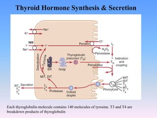
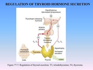
Thyroid Hormones
Physiology and Anatomy of The Thyroid Gland
The gland contains many follicles.
• A follicle is a fluid-filled lumen. The lumen contains material called colloid. The
major constituent of colloid is the large glycoprotein thyroglobulin, which
contains the thyroid hormones. Each follicle is lined by cuboidal epithelial cells,
also called the follicular cells.
• There are also extrafollicular hormone-secreting cells, called C cells.
• Thyroxine and triiodothyronine profoundly increase the metabolic
rate of the body.
• Complete lack of thyroid secretion usually causes the basal metabolic
rate to fall 40% to 50% below normal, and extreme excesses of
thyroid secretion can increase the basal metabolic rate to 60% to
100% above normal.
• The functions of these two hormones are qualitatively the same, but
they differ in rapidity and intensity of action.
Follicular cells require about 50 milligrams Iodine a year, or 1mg / week to
form normal quantities of thyroxine.
➢ Thyroxine (T4): made in follicle
➢ Triiodothyronine (T3): made in follicle
➢ Calcitonin: made by C cells
Hormones Produced in the thyroid
SYNTHESIS OF THYROID HORMONES
1. Thyroglobulin Synthesis
Endoplasmic reticulum and Golgi apparatus in the follicular cells of thyroid
gland synthesize and secrete thyroglobulin. Thyroglobulin molecule is a
large glycoprotein containing molecules of amino acid tyrosine. After
synthesis, thyroglobulin is stored in the follicle.
2. Iodide Trapping
Iodide is actively transported from blood into follicular cell, by sodium-
iodide symporter pump. The energy for transporting Iodide is provided by
the Na+-K+ ATPase pump.
This process of concentrating the iodide in the cell is called iodide trapping.
In a normal gland, the iodide pump concentrates the iodide to about 30
times its concentration in the blood.
It occurs in some stages.
3. Oxidation of Iodide
Iodide must be oxidized to elementary iodine. The oxidation of iodide into
iodine occurs inside the follicular cells in the presence of thyroid
peroxidase, which is located in the apical membrane of the cell thus
providing the oxidized iodine at exactly the point in the cell where the
thyroglobulin molecule enters the colloid.
4. Transport of Iodine into Follicular Cavity
From the follicular cells, iodine is transported into the follicular
cavity by an iodide-chloride pump called pendrin.
5. Iodination of Tyrosine and Coupling Reactions
Iodine is transported from follicular cells into the follicular lumen,
where it binds with thyroglobulin. This process is called organification of
thyroglobulin.
Combination of iodine with tyrosine is known as iodination. It takes place in
thyroglobulin.
Iodine (I) combines with tyrosine, which is already present in thyroglobulin.
Iodination of tyrosine occurs in several stages.
Tyrosine is iodized first into monoiodotyrosine (MIT) and later into
diiodotyrosine (DIT). MIT and DIT get coupled with one another. The
coupling occurs in different configurations, to give rise to different thyroid
hormones.
Tyrosine + I = Monoiodotyrosine (MIT)
MIT + I = Di-iodotyrosine (DIT)
DIT + MIT = Tri-iodothyronine (T3)
MIT + DIT = Reverse T3
DIT + DIT = Tetraiodothyronine or Thyroxine (T4)
RELEASE OF THYROID HORMONES FROM THE THYROID
GLAND
Thyroglobulin itself is not released into the bloodstream.
The hormones are first cleaved from thyroglobulin and released into the
blood.
1. Follicular cell sends foot-like extensions called pseudopods, which close
around the thyroglobulin-hormone complex.
2. Pseudopods convert thyroglobulin-hormone complex into small pinocytic
vesicles.
3. Then, lysosomes of the cell fuse with these vesicles.
4. Digestive enzymes such as proteinases present in lysosomes digest
(proteolysis) the thyroglobulin and release the hormones.
5. The hormones diffuse through base of the follicular cell and enter the
capillaries.
6. Only T3 and T4 are released into the blood.
7. MIT and DIT are not released into blood. These residues are deiodinated
by an enzyme called iodotyrosine deiodinase, resulting in the release of
iodine.
Thyroid Hormone Synthesis & Secretion
Each thyroglobulin molecule contains 140 molecules of tyrosine. T3 and T4 are
breakdown products of thyroglobulin
TRANSPORT OF THYROXINE AND TRIIODOTHYRONINE
TO TISSUES
• Upon entering the blood, the thyroxine and triiodothyronine combines
immediately with several of the plasma proteins.
• Then the thyroid hormones are released to the tissue cells slowly.
• Upon entering the tissue cells, the hormones again bind with
intracellular proteins.
• In the target tissue cells, they are again stored and are used slowly over a
period of days or weeks.
• The thyroid hormones therefore have a slow onset and long duration of
action probably due to their binding with proteins both in the plasma and
in the tissue cells, followed by their slow release.
Thyroid hormone
activation of
target cells
• Thyroxine (T4) and triiodothyronine (T3) readily diffuse through
the cell membrane.
• Much of the T4 is deiodinated to form T3 as thyroid hormone
receptors have a high affinity for T3.
• T3 interacts with the thyroid hormone receptor, bound as a
heterodimer with a retinoid X receptor, on the thyroid hormone
response element of the target genes.
• This causes either increases or decreases in transcription of target
genes that lead to formation of new proteins.
• Therefore, great numbers of protein enzymes, structural proteins,
transport proteins, and other substances are synthesized in the
almost all the cells.
• Most of the actions of thyroid hormone result from the
subsequent enzymatic and other functions of these new proteins
to cause a generalized increase in functional activity throughout
the body.
Thyroid hormone activation of target cells
REGULATION OF THYROID HORMONE SECRETION
• To maintain normal levels of metabolic activity in the body,
precisely the right amount of thyroid hormones must be secreted at
all times.
• To achieve this ideal level of secretion, specific feedback
mechanisms operate through the hypothalamus and anterior
pituitary gland to control the rate of thyroid secretion.
• Thyroid-Stimulating Hormone (TSH), also known as thyrotropin,
is an anterior pituitary hormone. It increases secretion of thyroxine
and triiodothyronine.
• Secretion of TSH from the Anterior pituitary is controlled by a
hypothalamic hormone, thyrotropin-releasing hormone (TRH),
which is synthesized by neurons of the hypothalamus and secreted
from their nerve endings in the pituitary stalk. From here, TRH is
then transported to the anterior pituitary by way of the
hypothalamic-hypophysial portal blood.
Figure 77-7: Regulation of thyroid secretion. T3, triiodothyronine; T4, thyroxine.
REGULATION OF THYROID HORMONE SECRETION

Lec-8 Thyroid Hormones and its usage1.pdf

  • 1.
  • 3.
    Physiology and Anatomyof The Thyroid Gland The gland contains many follicles. • A follicle is a fluid-filled lumen. The lumen contains material called colloid. The major constituent of colloid is the large glycoprotein thyroglobulin, which contains the thyroid hormones. Each follicle is lined by cuboidal epithelial cells, also called the follicular cells. • There are also extrafollicular hormone-secreting cells, called C cells.
  • 4.
    • Thyroxine andtriiodothyronine profoundly increase the metabolic rate of the body. • Complete lack of thyroid secretion usually causes the basal metabolic rate to fall 40% to 50% below normal, and extreme excesses of thyroid secretion can increase the basal metabolic rate to 60% to 100% above normal. • The functions of these two hormones are qualitatively the same, but they differ in rapidity and intensity of action. Follicular cells require about 50 milligrams Iodine a year, or 1mg / week to form normal quantities of thyroxine. ➢ Thyroxine (T4): made in follicle ➢ Triiodothyronine (T3): made in follicle ➢ Calcitonin: made by C cells Hormones Produced in the thyroid
  • 5.
    SYNTHESIS OF THYROIDHORMONES 1. Thyroglobulin Synthesis Endoplasmic reticulum and Golgi apparatus in the follicular cells of thyroid gland synthesize and secrete thyroglobulin. Thyroglobulin molecule is a large glycoprotein containing molecules of amino acid tyrosine. After synthesis, thyroglobulin is stored in the follicle. 2. Iodide Trapping Iodide is actively transported from blood into follicular cell, by sodium- iodide symporter pump. The energy for transporting Iodide is provided by the Na+-K+ ATPase pump. This process of concentrating the iodide in the cell is called iodide trapping. In a normal gland, the iodide pump concentrates the iodide to about 30 times its concentration in the blood. It occurs in some stages.
  • 6.
    3. Oxidation ofIodide Iodide must be oxidized to elementary iodine. The oxidation of iodide into iodine occurs inside the follicular cells in the presence of thyroid peroxidase, which is located in the apical membrane of the cell thus providing the oxidized iodine at exactly the point in the cell where the thyroglobulin molecule enters the colloid. 4. Transport of Iodine into Follicular Cavity From the follicular cells, iodine is transported into the follicular cavity by an iodide-chloride pump called pendrin.
  • 7.
    5. Iodination ofTyrosine and Coupling Reactions Iodine is transported from follicular cells into the follicular lumen, where it binds with thyroglobulin. This process is called organification of thyroglobulin. Combination of iodine with tyrosine is known as iodination. It takes place in thyroglobulin. Iodine (I) combines with tyrosine, which is already present in thyroglobulin. Iodination of tyrosine occurs in several stages. Tyrosine is iodized first into monoiodotyrosine (MIT) and later into diiodotyrosine (DIT). MIT and DIT get coupled with one another. The coupling occurs in different configurations, to give rise to different thyroid hormones. Tyrosine + I = Monoiodotyrosine (MIT) MIT + I = Di-iodotyrosine (DIT) DIT + MIT = Tri-iodothyronine (T3) MIT + DIT = Reverse T3 DIT + DIT = Tetraiodothyronine or Thyroxine (T4)
  • 8.
    RELEASE OF THYROIDHORMONES FROM THE THYROID GLAND Thyroglobulin itself is not released into the bloodstream. The hormones are first cleaved from thyroglobulin and released into the blood. 1. Follicular cell sends foot-like extensions called pseudopods, which close around the thyroglobulin-hormone complex. 2. Pseudopods convert thyroglobulin-hormone complex into small pinocytic vesicles. 3. Then, lysosomes of the cell fuse with these vesicles. 4. Digestive enzymes such as proteinases present in lysosomes digest (proteolysis) the thyroglobulin and release the hormones. 5. The hormones diffuse through base of the follicular cell and enter the capillaries. 6. Only T3 and T4 are released into the blood. 7. MIT and DIT are not released into blood. These residues are deiodinated by an enzyme called iodotyrosine deiodinase, resulting in the release of iodine.
  • 9.
    Thyroid Hormone Synthesis& Secretion Each thyroglobulin molecule contains 140 molecules of tyrosine. T3 and T4 are breakdown products of thyroglobulin
  • 10.
    TRANSPORT OF THYROXINEAND TRIIODOTHYRONINE TO TISSUES • Upon entering the blood, the thyroxine and triiodothyronine combines immediately with several of the plasma proteins. • Then the thyroid hormones are released to the tissue cells slowly. • Upon entering the tissue cells, the hormones again bind with intracellular proteins. • In the target tissue cells, they are again stored and are used slowly over a period of days or weeks. • The thyroid hormones therefore have a slow onset and long duration of action probably due to their binding with proteins both in the plasma and in the tissue cells, followed by their slow release.
  • 11.
  • 12.
    • Thyroxine (T4)and triiodothyronine (T3) readily diffuse through the cell membrane. • Much of the T4 is deiodinated to form T3 as thyroid hormone receptors have a high affinity for T3. • T3 interacts with the thyroid hormone receptor, bound as a heterodimer with a retinoid X receptor, on the thyroid hormone response element of the target genes. • This causes either increases or decreases in transcription of target genes that lead to formation of new proteins. • Therefore, great numbers of protein enzymes, structural proteins, transport proteins, and other substances are synthesized in the almost all the cells. • Most of the actions of thyroid hormone result from the subsequent enzymatic and other functions of these new proteins to cause a generalized increase in functional activity throughout the body. Thyroid hormone activation of target cells
  • 13.
    REGULATION OF THYROIDHORMONE SECRETION • To maintain normal levels of metabolic activity in the body, precisely the right amount of thyroid hormones must be secreted at all times. • To achieve this ideal level of secretion, specific feedback mechanisms operate through the hypothalamus and anterior pituitary gland to control the rate of thyroid secretion. • Thyroid-Stimulating Hormone (TSH), also known as thyrotropin, is an anterior pituitary hormone. It increases secretion of thyroxine and triiodothyronine. • Secretion of TSH from the Anterior pituitary is controlled by a hypothalamic hormone, thyrotropin-releasing hormone (TRH), which is synthesized by neurons of the hypothalamus and secreted from their nerve endings in the pituitary stalk. From here, TRH is then transported to the anterior pituitary by way of the hypothalamic-hypophysial portal blood.
  • 14.
    Figure 77-7: Regulationof thyroid secretion. T3, triiodothyronine; T4, thyroxine. REGULATION OF THYROID HORMONE SECRETION