2. Neuropraxia Of The Inferior Alveolar Nerve Secondary To Odontogenic Infection: A Case Report
Fig.2 Computed tomography scan of patient (Axial view)
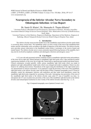
Fig.3 Computed tomography scan of patient (Sagittal view)
Patient was informed about the treatment plan and the possibility of injury to the inferior alveolar nerve
and the measures that would be taken during the surgery to prevent the same. With medication, patient’s general
condition improved a lot and the paraesthesia of the right mental nerve reduced considerably. Under local
anaesthesia, transalveolar extraction of the mandibular right third molar was carried out by sectioning of the
tooth (Fig.4). Postoperatively, the patient did not experience mental nerve paraesthesia and the surgical wound
healed uneventfully.
Fig.4 The impacted mandibular right third molar removed surgically by sectioning of tooth.
DOI: 10.9790/08531503071417 www.iosrjournals.org 15 | Page
3. Neuropraxia Of The Inferior Alveolar Nerve Secondary To Odontogenic Infection: A Case Report
III. Discussion
Third molar surgery is one of the most commonly performed minor surgeries by a maxillofacial
surgeon. Mandibular third molars are located in the angle region of mandible which marks the junction of the
ramus with the body of mandible. The third molars are the last teeth to erupt in the oral cavity, usually erupting
at the age of 16 to 25 years 5
. However, owing to the changing dietary habits and evolutionary changes, the jaw
size has been found to be showing signs of reduction, one of them being the mandibular third molars getting
impacted more frequently. At times, if the third molars erupt completely, they are placed in an unfavourable
position i.e. buccally or lingually or at an angle to the adjacent second molars. This unfavourable position of the
third molars makes it very difficult to keep them clean and so the third molars are found to be affected by dental
caries more frequently 5, 6, 7
. Infections from the third molars spread to involve the masticator spaces more
commonly8
. However, the periapical infection from the mandibular third molars is also in the close vicinity of
the mandibular canal and it may spread around the inferior alveolar nerve and cause paraesthesia. The irritation
from the periapical infection around the inferior alveolar nerve may cause neuritis thereby manifesting the
cardinal signs of inflammation (rubor, calor, dolor, tumor, functiolaesa). However, as the inferior alveolar nerve
is confined within a bony canal, there is not enough room for the inflamed nerve to swell and this may cause
compression of the nerve itself thereby causing ischaemic injury and further aggravating the neurological signs
(paraesthesia). The irritation from the pus also causes inflammation of the inferior alveolar nerve and the
pressure from the pus can cause ischaemic injury to the inferior alveolar nerve.
Surgical management of impacted mandibular third molars is a great challenge to the maxillofacial
surgeon. It is important to take into consideration the patients’ anatomical factors, patients’ viewpoint of the
surgery along with his/her past experiences, radiological assessment of the impacted mandibular third molars.
Establishing a correct diagnosis demands comprehensive clinicoradiological assessment and the surgeons’
experience in handling complicated cases. It is an important aspect of patient management to warn the patient of
the possibility of injury to the inferior alveolar nerve in the course of management of deep seated impacted
mandibular third molars9, 10
. Patients naturally have the “fear of the unknown” which raises their anxiety; but the
best and the easiest way to control the patients’ anxiety is by providing them with the detailed information about
the nature of the disease, the surgical management planned for treating the disease, likely complications
associated with the surgical management and the assurance to be competent enough to handle the complications,
if any, which may occur during the course of surgical management. Having a healthy and comprehensive
conversation with the patient pertaining to the disease they may be suffering from and the proposed surgical
management preoperatively is an important step towards prevention of litigation11
.
When surgically removing the impacted mandibular third molars, all aseptic precautions should be
followed. Copious irrigation should be done when preparing the buccal gutter to prevent thermal injury to the
bone. If radiological assessment 12, 13, 14, 15, 16
is suggestive of unfavourable root configuration, sectioning of the
tooth is indicated. Sectioning of tooth offers several advantages like less of bone is removed, postoperative pain,
swelling and discomfort are kept to a minimum, the axis of rotation of the tooth (to be removed surgically)
becomes favourable following sectioning of tooth17
, sectioning of the roots allows unfavourable roots to be
removed individually along their path of withdrawal 15
(Path of withdrawal is the path along which a tooth or a
tooth root can be removed easily by the least application of force), possibility of damage to the adjacent tooth is
also reduced due to the favourable axis of rotation of the third molar following sectioning of the tooth, chances
of injury to the inferior alveolar nerve are minimized following the sectioning of the tooth.After the tooth has
been delivered, overzealous exploration of the socket should be avoided to prevent likely injury to an exposed
nerve. Also, suctioning of the extraction wound to look for the exposed nerve should be avoided to prevent
injury to the nerve. The socket can be gently dried with gauze and inspected for any loose bone fragments which
may impinge on an exposed nerve, and hence should be removed before closure of the wound. Bleeding from
the surgical wound should be controlled prior to closure to prevent compression injury to an exposed inferior
alveolar nerve from an impending haematoma. When the surgeon anticipates a fair amount of postoperative
swelling due to the handling of the tissues, measures to prevent compression of an exposed nerve may be
adopted eg. use of a glove drain for 23 days, use of steroids in tapering dosages. Postoperative care of the
surgical wound with medication and proper oral hygiene care is equally important for uneventful healing.
Many factors determine the outcome of injury to nerve tissue such as the age of the patient, host response,
experience of the surgeon, handling of the tissues at the time of surgery.
DOI: 10.9790/08531503071417 www.iosrjournals.org 16 | Page
4. Neuropraxia Of The Inferior Alveolar Nerve Secondary To Odontogenic Infection: A Case Report
IV. Conclusion
Surgical skill is dependent on two important aspects: assessment of the case and management. “It is a
fundamental principle of medicine that in the clinical setting, assessment and management begin and proceed
simultaneously.” Complications can arise in a surgical procedure, but the best and the easiest way to manage a
complication is “to prevent it.” It is important to be well equipped with knowledge, surgical skill and the
necessary armamentarium to prevent a complication during a surgical procedure; and to handle one if it occurs
during the course of surgery.
References
[1]. Gintaras Juodzbalys, HomLay Wang, Gintautas Sabalys, Antanas Sidlauskas, Pablo GalindoMoreno, Inferior alveolar nerve
injury associated with implant surgery.Clin.Oral Impl. Res. 24,2013, 183–190.
[2]. Andrew B.G Tay, Inferior alveolar nerve injury in traumainduced mandible fractures, Journal of Oral and Maxillofacial
Surgery September 2007 Volume 65, Issue 9, Supplement, Page 40.
[3]. Tara Renton , Prevention of iatrogenic inferior alveolar nerve injuries in relation to dental procedures, Dent Update 2010; 37:
350–363.
[4]. Hanlie Engelbrecht ,Shabnum Meera, Jeff F. Kourie, Perineural infiltration of the inferior alveolar nerve in mandibular
ameloblastomas, Br. J. Oral Maxillofac Surg(2013),http://dx.doi.org/10.1016/j.bjoms.2013.02.002
[5]. GökselŞimşek Kaya, Muzaffer Aslan, Mehmet MelihÖmezli, ErtunçDayr, Some morphological features related to
mandibular third molar impaction ,J Clin Exp Dent. 2010;2(1):e127
[6]. Nazir A, Akhtar MU, Ali S. Assessment of different patterns of impacted mandibular third molars and their associated
pathologies. J Adv Med Dent Scie 2014;2(2):1422.
[7]. Dr Ajay Kumar Pillai, Dr Parimala Kulkarni, Dr Swapnil Moghe, Dr Vineesh Vishnu, Dr Saurabh Dhanraj Yadav, Dr Syed
Saquib Dastagir, Infra temporal & temporal abscess – Retrograde infection from mandibular molars, IOSR Journal of Dental
and Medical Sciences (IOSRJDMS)Volume 13, Issue 11 Ver. VI (Nov. 2014), PP 9699.
[8]. Dr G.V Thakur, ,Dr V.T Kandakure, , DrA.Thote, , Dr Ayesha .K,Masticator Space Abscess: A Case Report, IOSR Journal of
Dental and Medical Sciences (IOSRJDMS), Volume 7, Issue 2 (May. Jun. 2013), PP 6467.
[9]. JózsefSzalma, Inferior alveolar nerve injuries and impacted lower third molars: The importance of third dimension, Edorium J
Surg 2015;2:12–15.
[10]. Pitekova L, Satko I, Novotnakova D, Complications after third molar surgery, Bratisi lLekListy 2010, 111 (5), 296298.
[11]. Hesham F. Marei, Medical litigation in oral surgery practice:Lessons learned from 20 lawsuits, Journal of Forensic and Legal
Medicine, 20 (2013): 223225.
[12]. Hemamalini Balaji ,Dr.K.Laliytha, Evaluation of impacted mandibular third molar using panoramic radiographs, J. Pharm.
Sci. & Res. Vol. 7(11), 2015, 940945.
[13]. FábioWildsonGurgel Costa, Erick Helton Lima Fontenele, TácioPinheiroBezerra, Thyciana Rodrigues Ribeiro,
BárbaraGressy Duarte Souza Carneiro, Eduardo Costa Studart Soares, Correlation between radiographic signs of third molar
proximity with inferior alveolar nerve and postoperative occurrence of neurosensory disorders. A prospective, doubleblind
study, Acta CirúrgicaBrasileira Vol. 28 (3) 2013, 221227.
[14]. HangGul Kim, JaeHoon Lee, Analysis and evaluation of relative positions of mandibular third molar and mandibular canal
impacts, J Korean Assoc Oral Maxillofac Surg 2014;40:278284.
[15]. T. Renton, N. Smeeton, and M. McGurk, Factors predictive of difficulty of mandibular third molar surgery, British Dental
Journal volume 190 no. 11 June 9 2001; 607610.
[16]. Pallavi Sinha, Anuradha Pai, Assessment of proximity of impacted mandibular third molar roots to the mandibular canal
using intraoral periapical radiography and conebeam computerized tomography: A comparative study, International Dental
& Medical Journal of Advanced Research (2015), 1, 1–5.
[17]. Sam E. Farish, DMD,Gary F. Bouloux, General Technique of Third Molar Removal, Oral Maxillofacial Surg Clin N Am 19
(2007) 23–43.
DOI: 10.9790/08531503071417 www.iosrjournals.org 17 | Page