Radiology of theinternal hernia
Gastrointestinal
imaging
Presented by :
Dr.Ahmed Abdelkarim, MD
2.
Radiology of theinternal hernia
Definition
&
classificati
on
How to
diagnose
Small
bowel
related
hernia
Large
bowel
related
hernia
Omentum
related
hernia
Others
Differential
diagnosis
General principles Types DD
3.
Radiology of theinternal hernia
Definition &
classification
How to
diagnose
General principles
4.
Radiology of theinternal hernia
Definition
Protrusion of bowel loops through a peritoneal defect
Peritoneal defect = Hernia orifice
Most common signs/symptoms
Asymptomatic
Intermittent abdominal pain
Symptoms of acute small bowel obstruction
Nausea, vomiting, abdominal pain, and abdominal distention
5.
Radiology of theinternal hernia
Normal foramen
Foramen of Winslow hernia
Unusual retroperitoneal recess / fossa
Paraduodenal hernia
Pericecal hernia
Intersigmoid hernia
Most types of pelvic internal hernia
Abnormal opening in a mesentery or peritoneal ligament
Small bowel mesentery related hernia
Transverse mesocolon related hernia
Sigmoid mesocolon related hernia
Greater omentum related hernia
Most types of lesser sac hernia
Falciform ligament hernia
Broad ligament hernia
Mechanism & Classification
of the internal hernia
According to the
peritoneal
defect/hernia
orifice
6.
Radiology of theinternal hernia
Closed-loop obstruction
A segment of the bowel is occluded at two adjacent points.
Type of bowel obstruction seen in the internal hernia.
Can be seen in other conditions ”volvulus , adhesion ”.
Can easily develop into intestinal strangulation and ischemia
How to Diagnose
7.
Radiology of theinternal hernia
A direct sign of a closed loop at CT
U- or C-shaped, fluid-filled, distended intestinal loop,
Radial distribution of stretched and thickened mesenteric vessels toward hernia orifice
Best diagnostic
clue
8.
Radiology of theinternal hernia
Saclike appearance
Sacculation and crowding of small bowel loops owing to encapsulation within the hernia sac
Seen in unusual fossa in the retroperitoneum or intramesenteric-type internal hernia
Best diagnostic
clue
9.
Radiology of theinternal hernia
Displacement of surrounding structures and key vessels
Best
diagnostic clue
10.
Radiology of theinternal hernia
Small bowel
related hernia
Large bowel
related hernia
Omentum
related hernia
Others
Types
11.
Radiology of theinternal hernia
Small bowel
related hernia
Small bowel
mesentery
related hernia
Paraduodenal
hernia
Types
12.
Paraduodenal Hernia
Definition
Protrusion of the small intestine into a retroperitoneal fossa near duodenum
Types ; Right and left
Left paraduodenal hernias are
approximately three times more common
than right paraduodenal hernias
13.
Paraduodenal Hernia
Leftparaduodenal hernia
Saclike appearance is observed in the left anterior pararenal space
IMV is anteriorly displaced and seen along medial border of sac
Best diagnostic
clue
14.
Paraduodenal Hernia
Rightparaduodenal hernia
A saclike appearance is seen right anterior pararenal space
Inferior and to the right of 3rd par of the duodenum
SMV anteriorly displaced and seen along medial border of sac
Best
diagnostic clue
Small bowel mesenteryrelated hernia
Definition
Protrusion of the small intestine through defect within small bowel mesentery
Types ;
Transmesentreic = Most common type ,involve two layer of peritoneal folds
Intramesenteric = mesenteric pouch (rare)
17.
Small bowel mesenteryrelated hernia
Signs of closed loop obstruction
Can be seen at the upper or lower abdomen.
Congested crowded mesenteric vessels toward the mesentery.
Hernia orifice near the terminal ileum or near the ligament of Treiz
Best diagnostic
clue
Transmesenteric type
No sac like configuration
18.
Small bowel mesenteryrelated hernia
Displacement of the main mesenteric trunk to the right
Radiology of theinternal hernia
Large bowel
related hernia
Sigmoid
mesocolon
related hernia
Pericecal
related hernia
Types
21.
Pericecal internal hernia
Definition
Protrusion of bowel into an unusual recess near the cecum.
Types
Four kinds of pericecal recesses have been defined:
Superior and inferior ileocecal recess, retrocecal recess, and paracolic sulcus
22.
Pericecal internal hernia
Saclike appearance is seen
Lateral to the cecum & ascending colon ”Right Para-colic gutter”.
Displaces the cecum and ascending colon anteriorly or medially.
Best
diagnostic clue
23.
Sigmoid Mesocolon–related Hernia
Definition
Herniation of the bowel through the sigmoid mesentery
Types
Three subtypes transmesosigmoid, intramesosigmoid, and intersigmoid
24.
Sigmoid Mesocolon–related Hernia
Sac like appearance or closed loop obstruction can be present
At the left iliac fossa region along the left side of the descending colon ”LT Paracolic gutter”.
Hernia orifice is seen between the psoas muscle and sigmoid colon.
25.
Radiology of theinternal hernia
Omentum related
hernia
Lesser sac
related hernia
Greater
Omentum
related hernia
Types
26.
Greater omentum relatedinternal hernia
Definition
Protrusion of the bowel loops through defect within greater omentum
Type
The herniated intestine passes through both leaves (ie, four peritoneal layers)
27.
Greater omentum relatedinternal hernia
Signs of closed loop obstruction
Bowel loops close to anterior abdominal wall and inferior to the transverse colon
Omental vessels are seen displaced anteriorly
Best diagnostic
clue
28.
Lesser sac relatedinternal hernia
Definition
Protrusion of the bowel loops within the lesser sac.
Types : 4 (most common FOW hernia)
29.
Lesser sac relatedinternal hernia
Sign of closed loop obstruction
Seen in the lesser sac between stomach and pancreas
Hernia orifice is between portal vein and IVC
Best
diagnostic clue
30.
Radiology of theinternal hernia
Others
Broad
ligament
related hernia
Falciform
ligament
related hernia
Types
31.
Falciform ligament relatedinternal hernia
Definition
Protrusion of bowel loops through defect within the Falciform ligament
Vere rare type
32.
Falciform ligament relatedinternal hernia
Closed loop obstruction
Related to or just inferior to the anterior surface of the liver
Paraumbilical vein represent the inferior land mark of the ligament
33.
Broad ligament relatedinternal hernia
Definition
Protrusion of the bowel through defect within the broad ligament
Types
Pouch type = Intramesenteric type
Fenestra type = Transmesenteric type
34.
Broad ligament relatedinternal hernia
Closed loop or sac like appearance of bowel loops
Seen at the pelvis
Hernia orifice is seen directed toward the Parametrium
Enlargement of the distance between the uterus and ovary, deviating in opposite directions
key vessels
Tubal and ovarian branches of the ovarian artery (run inside the superior portion of the ligament).
The uterine artery and venous plexus,( run along the inferior border of the broad ligament)
Best diagnostic
clue
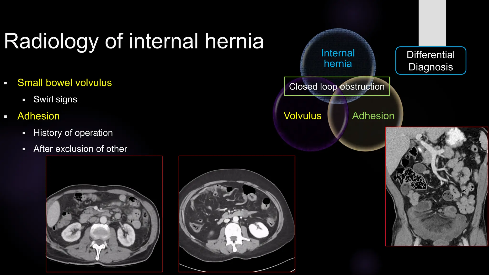
Radiology of internalhernia
Small bowel volvulus
Swirl signs
Adhesion
History of operation
After exclusion of other
Differential
Diagnosis
Internal
hernia
Adhesion
Volvulus
Closed loop obstruction