well as the healing status of the periapical region. The
primary goal of endodontic treatment is to eliminate
microorganisms from the root canal system to encourage
periapical healing. Success and failure of non-surgical
endodontic treatment is influenced by factors such as patient
factors, the effectiveness of the infection control, and
procedural complications, as well as overall response to
treatment. Root canal treatment usually fails when treatment
falls short of acceptable standards. Both poorly treated
and well-treated root canals might fail because of chronic
intraradicular or secondary infections. However, carefully
adhering to treatment protocols enhances the success rate as
well as the quality of endodontic treatment.
A flare-up is described as "an acute exacerbation of
periradicular pathosis after initiation or in continuation
of root canal treatment". 33
2. Endodontic flare-ups can significantly impact the
outcome of endodontic treatment, as highlighted in
recent research. These flare-ups, characterized by
post-operative pain, swelling, or discomfort, can
compromise the success of root canal therapy. 29
3. A study conducted by Smith et al. emphasized
that patients experiencing flare-ups were more likely
to exhibit increased treatment failure rates and
decreased healing compared to those without such
complications. 26
4. Mechanical injuries from overinstrumentation,
insufficient debridement, or insufficient removal of
pulp tissue are among the factors that cause flare-ups.
5. Debris extrusion from the periapical region, chemical
damage to the periapical tissues from irrigants,
intracanal medications, overextended root filling, or
microbiological injury are the most important factors
in the etiology of flare-ups.
6. It is also possible for iatrogenic and microbiological
factors to interact resulting in inter-appointment pain.
7. Management of flare-ups can be categorized as
preventive and definitive.
8. Preventative management includes: proper diagnosis,


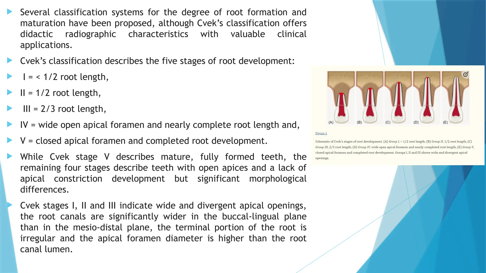
![ Clinical management of nonvital definitive immature anterior teeth presents both an
endodontic and restorative challenge.
Consequent interruption of continuous root development in immature teeth leads to
thin dentin walls, parallel or inverted taper of the root canals, open-apex, and an
unfavorable crown/root ratio
The absence of an apical closure in immature teeth precludes conventional
endodontic treatment, therefore requiring specific approaches with greater
predictability, safety, and effectiveness.
Apexification is a nonsurgical technique that allows the creation of an apical barrier
in open-apex teeth.
Traditionally performed apexification procedure included the sequential application
of calcium hydroxide (Ca[OH]2) in multiple sessions until obtaining an apical hard
tissue barrier5 .
Although clinical cases treated with Ca(OH)2 evidence high success rates, several
well-known disadvantages include treatment extension5,6 , higher likelihood of
coronal microleakage and recontamination. This is possibly attributed to its
hygroscopic and proteolytic properties, which induces desiccation of dentinal
proteins, thus predisposing the tooth to root fracture.](https://image.slidesharecdn.com/jc3-250923120011-2f011b4f/75/IMMATURE-APEX-TREATED-WITH-APEXIFICATION-PROCESS-AND-CORONO-RADICULAR-ADHRENCE-RESTORATION-12-2048.jpg)