1. THE PENNSYLVANIA STATE UNIVERSITY
SCHREYER HONORS COLLEGE
DEPARTMENT OF BIOCHEMISTRY AND MOLECULAR BIOLOGY
HOMOPOLYMERIC TRACTS: POTENTIAL NON-MUTATOR/MUTATOR SWITCHES
STEVE CHUNG
SPRING 2016
A thesis
submitted in partial fulfillment
of the requirements
for a baccalaureate degree
in Biochemistry and Molecular Biology
with honors in Biochemistry and Molecular Biology
Reviewed and approved* by the following:
Sarah Ades
Associate Professor of Biochemistry and Molecular Biology
Thesis Supervisor and Honors Adviser
James Howell
Lecturer of Biochemistry and Molecular Biology
Faculty Reader
Scott Selleck
Department Head for Biochemistry and Molecular Biology
* Signatures are on file in the Schreyer Honors College.
2. i
ABSTRACT
MRSA is defined as a strain of Staphylococcus aureus that has the gene mecA. While β-
lactam antibiotic resistance has been linked to mecA, it does not fully explain the rapid
acquisition of antibiotic resistance (1). A past clinical study identified a MRSA strain that
acquired resistance to the last line of defense drug (2). After treatment with vancomycin, the
infection still did not clear. MRSA isolates from the study were sequenced, and it was found that
the MRSA strains were acquiring mutations quickly. Among the genes found to be mutated, the
mutL gene was of interest because of a homopolymeric tract located midway in the gene.
Homopolymeric tracts are prone to slippage, making expansion and contraction highly likely (3).
This means that mutL could be frameshifted at a high probability, causing a nonfunctional
truncated mutL protein. The knockout of the mutL gene leads to a mutator phenotype, since the
gene is responsible for DNA mismatch repair.
In this study, we showed that a frameshift, contraction of the homopolymeric tract, in
mutL leads to an increase of mutation rate in MRSA, which might have indirectly supported the
bacteria in developing antibiotic resistance at a rate 10-times faster than the wild-type MRSA
strain. In addition, the study extends the concept of a homopolymeric tract as a potential gene
regulator in the ClpX gene. Preliminary data suggests that long homonucleotide tracts located in
other genes may play a role in β-lactam antibiotic resistance.
3. ii
TABLE OF CONTENTS
LIST OF FIGURES .....................................................................................................iii
LIST OF TABLES.......................................................................................................iv
ACKNOWLEDGEMENTS.........................................................................................v
Chapter 1 Introduction .................................................................................................1
1.1 The Patient’s MRSA Endocarditis and Accompanying Bacteremia..........................2
1.2 Patient X Was Treated With Imipenem, Rifampicin, and Vancomycin ....................3
1.3 Old Study ...................................................................................................................5
1.4 New Study..................................................................................................................7
1.5 MutL and the 9A Homopolymeric Tract....................................................................9
Chapter 2 Materials and Methods................................................................................14
2.1 Constructing the Vector With the MutL Insert...........................................................14
2.2 Transforming the Vector into E. coli .........................................................................15
2.3 Transforming the Vector into IM08...........................................................................16
2.4 Transforming the Vector into JE2..............................................................................17
2.5 Integrating the 8A MutL into the Genome of JE2 .....................................................17
2.6 Fluctuation Assay.......................................................................................................18
Chapter 3 Results.........................................................................................................19
3.1 Constructing the 8A mutL JE2...................................................................................19
3.2 Calculating the Mutation Rate ...................................................................................23
3.3 Investigating the Homopolymeric Tract in ClpX.......................................................31
Chapter 4 Discussion ...................................................................................................34
Appendix A Abbreviations .........................................................................................38
Appendix B Additional Data Sets...............................................................................39
BIBLIOGRAPHY........................................................................................................42
4. iii
LIST OF FIGURES
Figure 1. Infection and antibiotic duration timeline for patient X. ..........................................3
Figure 2. Timeline of samples collected from patient X before and after antibiotic treatment. 6
Figure 3. Possible scenarios during a replication slippage in a homopolymeric tract. ............10
Figure 4. Gel image of the colony PCR using primers that direct mutL amplification............20
Figure 5. Gel image of the PCR using primers that direct mutL amplification in well 2, and
pIMAY amplification in well 3........................................................................................21
Figure 6. Gel image of the colony PCR of JE2 integrant using primers that direct mutL
amplification. ...................................................................................................................22
Figure 7. The computed model vs. data graph taken from bz-rates of the integrant JE2.........27
Figure 8. Computed functions of the integrant JE2. ................................................................28
Figure 9. The computed model vs. data graph taken from bz-rates of wild-type JE2. ............29
Figure 10. Computed functions of the wild-type JE2. .............................................................30
Figure 11. Gel image of a colony PCR after transformation of the modified plasmid into C2987H.32
5. iv
LIST OF TABLES
Table 1. Order of mutations at individual locus.......................................................................8
Table 2. Order of mutations at individual locus with mutation characterization.....................12
Table 3. Average colony count results from the fluctuation test. ............................................24
Table 4. The first set of Colony counts of JE2 wild-type and JE2 8A integrant......................39
Table 5. The second set of Colony counts of JE2 wild-type and JE2 8A integrant.................40
Table 6. The third set of Colony counts of JE2 wild-type and JE2 8A integrant. ...................41
6. v
ACKNOWLEDGEMENTS
I would like to express my sincere gratitude to Dr. Michael Mwangi for always being a
supportive mentor, and friend. My gratitude extends to Dr. Sarah Ades and Dr. Kenneth Keiler
for the encouragement when my confidence gets misplaced. They were always willing to help. I
would like to thank Dr. James Howell for helping me push this thesis through. Without all of
their guidance, I would not have a well-rounded experience in the scientific field. I would also
like to thank Vikas Koundal, Juan Antonio Raygoza Garay, Caitlin Grube, and Trevor Kanasie in
the Mwangi lab for their guidance, contribution, and support in, and outside of the lab. Ray has
been patient, understanding, and supportive since I have joined the Mwangi lab.
I would like to thank my father, Dr. Rhayteh Chung, and my mother, Hsiang Chung, for
their unconditional support in my decisions. Although financial instability was always a worry
for my parents, I owe my education and life to them for their heavy sacrifice and trust in me. I
hope to repay them in the near future for the greatest form of joy they have provided me. I am
also grateful for my sister, Katherine Chung, for constantly pushing for my success. Katherine
has been my role model, and my best friend. Her relentless will to succeed inspired me to strive
for the best. I could only hope to continue to stay close to her.
Finally, I would like to thank my friends and acquaintances for their support and
suggestions along the way. This extends to the classmates of BMB488 for their constant push for
learning, and improving. Without Garrick Treaster and Spencer Lovrinic, I would not have the
capability to maintain a positive mentality for research, and view learning from a diverse set of
aspects.
7. 1
Chapter 1
Introduction
The introduction of antibiotics in the 20th
century has greatly reduced casualties in the
war against bacteria. However, microbes, driven by coevolution of predator and prey, evolved
quickly to combat the new “weaponry.” Methicillin-Resistant Staphylococcus aureus, or MRSA,
is a real-life example of antibiotic resistance. The misuse of antibiotics has been persisting even
to date, and microorganisms are continuing to withstand our weapons against bacteria. One
example of this resistance can be seen in the following case study: Patient X, infected with
MRSA developed endocarditis and bacteremia. Even with treatment by various forms of
antibiotics that target gram-positive bacteria, the infection persisted and Patient X passed away
(2). Throughout the course of the infection, bacterial isolates were taken and studied extensively
to attempt to characterize the infection.
Through this and many similar cases, we discovered our greatest weapons in the fight
against pathogenic microorganisms had been rendered useless. In this patient, pathogenic
microorganisms were once again untreatable due to their developed resistance. Without a
thorough understanding of antibiotic resistance, the post-antibiotic era may be reflective of the
pre-antibiotic era. In order to develop this understanding, a series of casefiles have been
assembled to study the development of antibiotic resistance in several strains of S. aureus. This
series of casefiles contains histories of patients, like patient X, infected with antibiotic resistant
MRSA strains. Among these casefiles is the case of a MRSA infection that contained a
population of mutators in Patient X- this is his story.
8. 2
1.1 The Patient’s MRSA Endocarditis and Accompanying Bacteremia
Patient X had a MRSA infection in the early 2000s (2). After diagnosis, the patient was
sent to Johns Hopkins Hospital. The MRSA infection caused the patient to develop endocarditis,
and eventually, bacteremia. Endocarditis is the bacterial infection of the heart, usually a valve.
Eventual inflammation leads to vegetation, where bacteria, platelets, fibrin, and inflammatory
cells amass. Vegetation can cause cardiac arrest, and can cause strokes by the breaking of cells.
The vegetation can cause bacteremia by allowing bacterial cells into the bloodstream (4). The
bacteria can produce toxins in the bloodstream that lead to sepsis, toxic shock syndrome, or both.
Sepsis and toxic shock syndrome triggers a whole-body inflammatory response that is harmful to
organs, and can lead to death (5). The patient died within 3 months due to the MRSA infection
that could not be treated. Figure 1 illustrates a crude timeline, taken from M. Mwangi, from the
start of the infection until the patient’s eventual mortality. During this time, patient X underwent
various chemotherapeutic treatments. The antimicrobials used in these treatments include β-
lactam class antibiotics.
9. 3
Figure 1. Infection and antibiotic duration timeline for patient X.
The infection lasted roughly three months. After starting rifampicin and imipenem,
vancomycin was administered when patient X was diagnosed with MRSA. The imipenem and
rifampicin treatment stopped shortly after the patient was on vancomycin.
1.2 Patient X Was Treated With Imipenem, Rifampicin, and Vancomycin
β-lactams work by inhibiting cell wall biosynthesis through a variety of pathways. These
antibiotics share a common characteristic of four-membered, nitrogen-containing β-lactam ring
at the base of their structure, which is indicative of its importance to the mode of action. β-lactam
antibiotics are bactericidal- they inhibit the synthesis of the peptidoglycan layer of the cell wall
which is present in bacterial cells (6). The peptidoglycan layer in gram-positive microbes is
responsible for their structural integrity, thus making the peptidoglycan layer a target for β-
lactam drugs. While β-lactam antibiotics share a common trait, they may differ in
pharmacokinetics, antimicrobial activity, and potential to induce allergic reactions (7).
10. 4
Imipenem belongs to the carbapenem class of β-lactams. Imipenem is the first line of
defense in treating a vicious bacterial infection, inhibiting cell wall synthesis. It is a broad-
spectrum antibiotic, usually administered first to clear infections since it is effective in killing
both gram-positive and gram-negative bacteria (8).
The non-β-lactam antibiotic, rifampicin, targets the RNA polymerase, thereby inhibiting
RNA synthesis. Rifampicin also acts as a broad-spectrum antibiotic. Rifampicin blocks the
formation of the phosphodiester bond in RNA, and prevents RNA extension. Rifampicin is
mainly used to treat tuberculosis, but a combination of other drugs with rifampicin can be used to
treat MRSA infections (9).
Vancomycin was the last line of defense against MRSA infections in the early 2000s
(10). Vancomycin is not a β-lactam antibiotic, but it does target peptidoglycan synthesis.
Vancomycin acts specifically to inhibit cell wall synthesis in gram-positive bacteria.
Vancomycin inhibits the formation of the peptidoglycan structure- bonds linking NAM and NAG
(11). Today, new antibiotics have been developed to treat MRSA infections, and vancomycin is
used more commonly. Often, treating MRSA with vancomycin clears the infection.
Doctors treated patient X with imipenem initially, since the cause of the infection was
unknown. Over time, the infection did not clear. Doctors combined the drug with rifampicin, for
a stronger therapy. The magnitude of the infection worsened, and physicians characterized the
infection as MRSA. Patient X had taken the two antibiotics, and started vancomycin. The
imipenem and rifampicin treatment was discontinued shortly after the patient started the
vancomycin regimen. The relative duration of the antibiotic treatment is shown in Figure 1,
taken from M. Mwangi, in relation to each other.
11. 5
Vancomycin treatment in MRSA usually controlled the infection in the early 2000s.
Resistance to vancomycin was unheard of as doctors often rely on vancomycin treatment as a
last line of defense against MRSA. Through steady administration of vancomycin, doctors
realized patient X was infected with MRSA that is resistant to vancomycin. The emergence of
vancomycin resistance was problematic: the last line of defense was ineffective.
1.3 Old Study
In the three months’ timeframe, three antibiotics were used in the drug therapy:
rifampicin, imipenem, and vancomycin. To characterize the infection progress, McAleese et. al
analyzed isolates that were taken from the patient through blood samples before and after
antibiotic treatment, and a heart biopsy after patient X had died (2). Figure 2, taken from M.
Mwangi, reflects the characterization of isolates taken from patient X.
12. 6
Figure 2. Timeline of samples collected from patient X before and after antibiotic
treatment.
Isolates were taken periodically throughout the infection. The genetic profiling, PCR or
whole genome sequencing, of the isolates was performed to compare the mutations present in the
infection.
Transcriptome profiling and whole genome sequencing were performed on cultures JH1
and JH9. Only 98% of the genome was resolved in each culture. The remaining 2% of the
genome was missing due to the technological limits of the time. In addition, JH2, JH5, JH6, and
JH14 isolates could only be PCR sequenced in a piecewise manner, which leaves large stretches
of genomic material unanalyzed. Whole genome sequencing was not done because of the relative
cost of sequencing. In addition, the study focused on the mutations found in JH9 that are absent
in JH1. These piecewise fragments were selected according to the mutations observed in the JH1
and JH9 isolates, which were fully (98%) sequenced. McAleese et. al suspected that the
13. 7
mutations could have arisen sometime during the treatment (2). Figure 2 also outlines the
profiling methods used for each of the individual isolates. Technological advancements over
time have allowed for more thorough genomic analysis and a more thorough investigation of the
piecewise analyzed genomes.
1.4 New Study
A more recent study resolved the limitations from the old study, and found an important
gene that caused the mutations seen in the isolates. Further sequencing of the JH1 and JH9
isolates updated the past genomic data from 98% coverage to 100% (12). In addition, the whole
genome of isolates JH2, JH5, JH6, and JH14 were sequenced to 100% coverage to identify all
mutations. The whole genome profiling of isolates JH2 and JH14 found many mutations in loci
different in each isolate. Since the mutation in the mutL locus was found in isolates JH2 to JH14,
isolates JH2 to JH14 are expected to be mutators. The mutL mutation was first found early in the
infection timeline, in isolate JH2. All of the following isolates had the same mutL mutation.
These mutators suggest that mutL may play a role in the aggressiveness of resistance in MRSA.
14. 8
Table 1. Order of mutations at individual locus.
After overcoming the limitations of the old study, the same mutL mutation was found in
JH2 and all the following isolates, but not in JH1. This mutL mutation is outlined in the table in
bold. The cells shaded in gray represent genes or loci mutated. This table was taken from M.
Mwangi.
Isolate JH1 JH2 JH5 JH6 JH9 JH14
Day 1 63 74 79 86 90
Locus Blood Heart
blaR1
mutL
rpoB
rpoC
SA1129
SA1702
SA1249
SA0019
SAS014
SA0582
SA0980
SA1659
SA1843
SA2094
SA2125
SA2320
SAP007
SA tRNA34
other loci +12 +8 +19 +13 +15
15. 9
1.5 MutL and the 9A Homopolymeric Tract
Although the name implies methicillin resistance, MRSA includes resistance to all β-
lactam drugs. The underlying gene responsible for β-lactam antibiotic resistance was found to be
mecA (1). The gene product of mecA is a penicillin-binding protein, PBP2a. PBP2a is a
penicillin-binding protein with a low affinity for β-lactams. With the low affinity for β-lactam
antibiotic binding, PBP2a allows transpeptidase activity for the continuation of cell wall
synthesis (13). Mediated by horizontal gene transfer between bacteria, S. aureus strains that
obtain the mecA gene exhibit β-lactam resistance (14). In this case, the MRSA strain, found in
the isolates, contained mutations that helped the infection persist with vancomycin treatment.
The JH2-JH14 isolates had a mutation in the mutL gene. The mutation was a result of a
truncated protein. A nine-adenine homopolymeric tract is found midway in the mutL gene in all
sequenced S. aureus strains (12). Homopolymeric tracts are DNA sequences that repeat
tandemly, and are thought to be a regulatory mechanism in prokaryotes (15). Homopolymeric
tracts are extremely unstable because they are prone to insertions or deletions due to
misalignment of the DNA strands during DNA replication. In general, the slippage in a given
homopolymeric tract does not occur with base pair (bp) lengths below seven, the critical
threshold (3). Figure 3, taken from Ellegren (16), illustrates the two possible slippages that could
occur in a homopolymeric tract.
16. 10
Figure 3. Possible scenarios during a replication slippage in a homopolymeric tract.
In (a), replication of the DNA strand forms a nick on the newly synthesized strand,
causing an addition of a nucleotide. In (b), replication of the DNA strand forms a nick on the
template strand, causing a deletion of a nucleotide.
The bacterial gene mutL codes for MutL, a subunit of the DNA mismatch repair system
MutHLS recognizes and repairs erroneous insertion, deletion, and misincorporation of bases that
arise during DNA replication. In S. aureus strains, the gene mutL has a length of 2010 bp and a
9A homopolymeric tract, AAAAAAAAA (9A), that starts midway into the gene at bp 1020. A
significant fraction of S. aureus cells in a population would be expected to carry a frameshift in
the 9A homopolymeric tract in mutL. The spontaneous mutation rate in bacteria has been
estimated to be roughly one per genome per 300 replications (17). Bacteria deficient in mutL
have been found to have a higher mutation rate than wildtype (18). Therefore, a significant
fraction of S. aureus cells in a population would be expected to be mutators. Generally, mutators
may have decreased fitness because they accumulate damaging mutations rapidly. However,
17. 11
when faced with an antibiotic selection pressure, mutators may have an advantage because they
can also acquire mutations that confer resistance more quickly. In order to balance these
competing interests, some S. aureus strains may have evolved to generate populations that are
mixtures of non-mutators and mutators.
Isolates with mutL deficiency were mutators that pass a core set of mutations from one
generation to the next. For example, in Table 2, taken from M. Mwangi, JH2 to JH14 contained
the same mutations in rpoB, rpoC, SA1129, and SA1702. This finding suggests that those
mutations were passed down from the initial isolate, JH2. The isolates also contained mutations
that are specific, and different from other isolates (12). This phenomenon suggests that the
homopolymeric tract in mutL can act as a switch, keeping certain mutations in certain genes.
18. 12
Table 2. Order of mutations at individual locus with mutation characterization.
Mutations outlined in blue indicate mutations passed down from generations. Mutations
outlined in orange indicate mutations specific to the isolate. The cells shaded in gray represent
genes or loci that were mutated.
Isolate JH1 JH2 JH5 JH6 JH9 JH14
Day 1 63 74 79 86 90
Locus Blood Heart
blaR1
mutL
rpoB
rpoC
SA1129
SA1702
SA1249
SA0019
SAS014
SA0582
SA0980
SA1659
SA1843
SA2094
SA2125
SA2320
SAP007
SA
tRNA34
other loci 12 8 9 13 15
The homopolymeric tract in mutL can expand or contract, creating mutators, and might
act as a genetic switch (15). As mentioned previously, mutators may have an advantage because
they acquire mutations that confer resistance quicker. Once an advantageous mutation that
allows antibiotic resistance occur, the homopolymeric tract can expand or contract back to the
19. 13
normal length, 9A. The advantageous mutation(s) can then propagate to the following
generations. Since mutator isolates with mutL deficiency pass a core set of mutations from one
generation to the next, studying these potential gene regulation phenomena could give us a new
set of insights to prevent an all-out pandemic of antibiotic resistance. My thesis experiment
served to demonstrate that a fraction of the MRSA population is expected to be mutators. I
hypothesized that a frameshift in the mutL gene will increase the mutation rate in MRSA.
Consequently, the goal of my thesis research was to show that a contraction in the mutL gene
could cause an increased mutation rate in MRSA. The increased mutation rate will assert that a
fraction of the MRSA population are mutators.
20. 14
Chapter 2
Materials and Methods
To test the phenomenon of an increased mutation rate due to the mutL frameshifts, a JE2
strain with 8A homopolymeric tract was constructed. This construct simulates the contraction of
the 9A homopolymeric tract, and the mutation frequency can be estimated by using a fluctuation
test.
Genetic manipulation in S. aureus has been highly difficult, and limited due to the type
IV restriction system, SauUSI, which recognizes cytosine methylated DNA. Monk et. al
constructed a DNA cytosine methyltransferase mutant in a high-efficiency Escherichia coli
cloning strain, DC10B, or IM08. The pIMAY plasmids were also engineered by Monk et. al so
that the sequences found in the plasmid can be integrated into the genome of S. aureus, making
genetic manipulation in MRSA USA300 possible (19).
2.1 Constructing the Vector With the MutL Insert
The Escherichia coli strain, DC10B, was obtained from Monk. The DC10B strain was
incubated at 37o
C in Luria broth (LB) for 12 hours. The pIMAY plasmid was extracted from
DC10B using the Zyppy™ Plasmid Miniprep Kit. The mutL fragment was amplified by PCR
from an overnight growth of a methicillin-resistant Staphylococcus aureus strain, JE2, at 37o
C in
tryptic soy broth (TSB). The primers used in the amplification of the mutL fragment were
modified such that the additional sequences that were added to both the 5’ and 3’ end were
21. 15
identical with a fragment in the pIMAY plasmid. Using Gibson Assembly, the mutL fragment
was inserted into the KpnI digested pIMAY plasmid in a 20µL reaction mixture: 1µL of insert,
1µL of vector, 8uL dH2O, and 10uL of 2X Gibson Assembly Master Mix (20).
2.2 Transforming the Vector into E. coli
The resulting vector from the Gibson Assembly mixture was transformed into E. coli
strain C2987H, obtained from New England Biolabs (NEB), using the high efficiency
transformation protocol (21). After 60 minutes incubation at 37o
C with vigorous shaking, cells
were diluted 10-fold. 100µL of both diluted and undiluted cells were plated on Luria agar (LA).
At 37o
C, the cells were incubated for 12 hours.
Six random colonies were placed into 100µL dH2O. A colony PCR was prepared for the
six colonies in a 20µL reaction mixture to check for the insert. PCR samples were analyzed via
1% agarose gel electrophoresis.
The 9A homopolymeric tract in mutL insert was mutated to 8A by site directed
mutagenesis. Using the Zyppy™ Plasmid Miniprep Kit, the pIMAY plasmid with insert was
extracted from C2987H. Primers were designed to carry the mutation, 8A instead of 9A, and
after replication, non-mutated parental DNA strands were digested with DpnI overnight (22).
Both PCR reactions were combined after digestion and denatured at 95o
C, then cooled to 55o
C.
The plasmid containing the mutated insert was transformed back into C2987H. The plasmid was
extracted again using the Zyppy™ Plasmid Miniprep Kit, and a PCR was prepared to check the
presence of the insert and plasmid in a 20µL reaction mixture. PCR samples were analyzed via
1% agarose gel electrophoresis.
22. 16
The DNA concentration was quantified by using a Qubit 2.0 Fluorometer and Nanodrop
2000 to confirm the purity (23, 24). From the values obtained from the Qubit 2.0 Fluorometer,
250ng DNA was sequenced at the Genomics Core Facility from the Huck Institutes of Life
Sciences. Sanger sequencing verified the correct sequences of the plasmid.
2.3 Transforming the Vector into IM08
Competent cell preparation of IM08, a descendant strain of DC10B, and JE2 was done
similar to the procedure outlined in Monk et. al (19). IM08 and JE2 cells were grown overnight
in 50mL TSB at 37o
C, and then diluted in a fresh prewarmed 6mL TSB. After the optical density
at 600nm (OD600) reached 0.5, the cultures were chilled on ice for 10 minutes, and all subsequent
steps were performed at 4o
C on ice. Cells were harvested by centrifugation at 2000 × 𝑔 for 30
min, and resuspended in equal volume of autoclaved water. The centrifugation and resuspension
steps were repeated twice. The cells were then thrice centrifuged and resuspended in 2mL 10%
glycerol. Aliquots of 75µL were frozen at -80o
C.
For transformation, the electroporation technique was used to insert the plasmid into
IM08. Competent cells were electroporated with 5µg plasmid extracted from C2987H at 2.5kV.
After electroporation, cells were incubated in SOC at 37o
C for 60min, then plated on tryptic soy
agar (TSA) and incubated overnight at 37o
C.
The plasmid was extracted from the cells using the Zyppy™ Plasmid Miniprep Kit. The
DNA concentration was quantified by using a Qubit 2.0 Fluorometer and Nanodrop 2000 (23,
24). From the values obtained from the Qubit 2.0 Fluorometer, 250ng DNA was sequenced at the
23. 17
Genomics Core Facility from the Huck Institutes of Life Sciences. Sanger sequencing verified
the correct sequences of the plasmid.
2.4 Transforming the Vector into JE2
The electroporation of JE2 competent cells was done similarly to Monk et. al (19).
Competent cells were electroporated with 5µg plasmid extracted from IM08 at 2.5kV. After
electroporation, cells were incubated in SOC at 28o
C for 60min, then plated on TSA and
incubated overnight at 28o
C.
The plasmid was extracted from the cells using the Zyppy™ Plasmid Miniprep Kit. The
DNA concentration was quantified by using a Qubit 2.0 Fluorometer and Nanodrop 2000 (23,
24). From the values obtained from the Qubit 2.0 Fluorometer, 250ng DNA was sequenced at the
Genomics Core Facility from the Huck Institutes of Life Sciences. Sanger sequencing verified
the correct sequences of the plasmid.
2.5 Integrating the 8A MutL into the Genome of JE2
To integrate pIMAY into the chromosome, a colony from the JE2 transformation plate
was diluted 10-fold to 10-3
with water. 100µL of each dilution was plated on brain heart infusion
agar (BHA) plus 10ug/mL chloramphenicol (CAM) and incubated overnight at 37o
C. A colony
PCR was performed to check the integration of the plasmid in JE2. Once integrated, cells were
plated on BHA plus 1µg/mL anhydrotetracycline (ATc). Cells that grew on ATc are indicative of
colonies that have expelled the empty pIMAY plasmid. The colonies were then inoculated at
24. 18
28o
C without CAM, and screened for the direction of recombination by PCR using primers
designed to bind to sequences of the desired direction.
2.6 Fluctuation Assay
Using JE2 and the 8A JE2 strains, the fluctuation test was used to determine the mutation
frequency of the given strains. In four 50mL TSB inoculated overnight at 37o
C with both strains,
one of JE2 and three of the 8A integrant JE2 strains, the cells were pelleted at 2000 × 𝑔 for 30
min, then resuspended with 6mL TSB. Strains were diluted 10-fold until 10-7
, and then 50µL of
each dilution was plated and incubated on TSA plus 100µg/mL rifampicin (RIF) at 37o
C. The
experiment was repeated nine times. Once the colony count was completed for each 24-hour and
48-hour time intervals, the online web tool, bz-rates (25), was used to compute the parameters
for the fluctuation test in determining the mutation frequency.
25. 19
Chapter 3
Results
To show that a contraction in the mutL gene leads to a mutator phenotype in MRSA, a
frameshift construct of the mutL gene must be built first. The JE2 strain is type of community-
associated (CA) MRSA strain that is most commonly used for molecular genetics studies (26).
After a series of transformation and integration steps, the completed 8A mutL JE2 construct
caused a non-functional truncated mutL protein. The construct was required to measure the
mutation rate by comparing the rates with the wild-type strain.
3.1 Constructing the 8A mutL JE2
To construct the strain, the 9A homopolymeric tract in mutL must be mutated to 8A in
JE2. To start, the entire mutL gene was copied and used as the insert from a template wild-type
JE2 strain in this study. The vector used in this study was the pIMAY plasmid. The temperature-
sensitive plasmid was made by Monk et. al with sequences that can direct homologous
recombination with S. aureus strains (19). Using this plasmid, it allowed for the mutation of the
9A homopolymeric tract in mutL in the MRSA genome. The mutL fragment was modified by
inserting additional sequences in the primers in the initial PCR of the gene such that they were
identical to a sequence fragment found on the pIMAY plasmid. The identical sequences added
on the mutL insert was required for the insertion and ligation into the pIMAY plasmid, since the
Gibson Assembly technique requires a fragment of overlapping sequences in the vector with its
insert. After amplification, the insert was ligated into the pIMAY plasmid by using Gibson
Assembly. With the modified plasmid of interest, modification of the sequences could be done
26. 20
after transformation. Since E. coli is easy to manipulate and transform, the resulting plasmid was
transformed into C2987H, a 5-alpha competent NEB E.coli strain. Figure 4 shows the colony
PCR result, which confirmed the presence of mutL from six of colonies that formed after
incubation of transformed C2987H.
Figure 4. Gel image of the colony PCR using primers that direct mutL amplification.
Bands seen in all six wells indicate a fragment of 1.5kb. The 1.5kb bands seen in the
wells indicated the presence of the mutL gene in C2987H.
The insert containing the pIMAY plasmid in C2987H was mutagenized such that the 9A
homopolymeric tract became eight adenines long by site-directed mutagenesis. Now, the pIMAY
plasmid contained the mutL gene with 8A. The mutagenesis of the 9A homopolymeric tract was
necessary because it causes the contraction of the homopolymeric tract, resulting in a frameshift
in the coding region. As a result, this frameshift causes a non-functional truncated mutL protein.
The presence of the insert and plasmid was checked by PCR, and the gel image in Figure 5
represents the result. The sequences then were verified by Sanger sequencing.
27. 21
Figure 5. Gel image of the PCR using primers that direct mutL amplification in well
2, and pIMAY amplification in well 3.
Band seen in well 2 indicate a fragment of 1.5kb, and 5.7kb in well 3. The 1.5kb band
seen in well 2 indicated the presence of the mutL gene. The 5.7kb band in well 3 indicated the
presence of the empty pIMAY plasmid.
The plasmid with the desired insert was ready to be transformed into MRSA. In order for
the MRSA strain to accept foreign DNA, the plasmid must contain methylated cytosines (19).
We used IM08 strains as an intermediate since the strains methylate cytosine in DNA. By
transforming the plasmid into IM08, it allowed the pIMAY plasmid for MRSA transformation to
be ready. The pIMAY plasmid was extracted from C2987H, and then transformed into IM08.
The sequences were checked by Sanger sequencing. Since the objective of this experiment was
to compare mutation rate in wild-type and 8A JE2, the 8A mutL fragment must replace the 9A
mutL in wild-type JE2 to construct the 8A integrant. First, the pIMAY plasmid was extracted,
28. 22
and transformed into JE2, with the sequences checked again by Sanger sequencing. At this point,
the plasmid with the modified mutL insert was ready to be integrated into the JE2 chromosome.
To reiterate, JE2 was used in this study because the strain is a CA-MRSA. The readiness and
virulence of this MRSA strain modeled the clinical relevance of MRSA infections (27).
Integration of the 8A mutL fragment was desirable since it creates the construct with the
frameshifted mutL gene. The frameshifted 8A mutL gene in the integrant JE2 strain generated a
non-functional truncated mutL protein, allowing the comparison of mutation rates from a mutL
contraction. After integrating the desired mutL gene into JE2, the sequences were verified by
colony PCR and Sanger sequencing.
Figure 6. Gel image of the colony PCR of JE2 integrant using primers that direct
mutL amplification.
Bands were seen in wells 4, 5, 6, and 7. These bands indicate a fragment of 1.5kb. The
1.5kb band seen in those wells indicated the presence of the mutL gene.
29. 23
3.2 Calculating the Mutation Rate
The contraction of the mutL gene, 8 adenines long, represents the loss function mutL;
therefore, according to our hypothesis, induces a mutator phenotype. If this is the case, then the
8A JE2 integrant on plates with RIF will have more colonies compared to the wild-type JE2
strain. This construct of the 8A integrant JE2 allowed for the investigation of the mutator
phenotype.
The rate at which mutations arise can be calculated by using the fluctuation analysis,
which is based on the properties of Luria-Delbrück distribution (28, 29). In this experiment, we
calculated the mutation rate at which cells develop RIF resistance. The RIF antibiotic was used
to select for a resistance response because of its clinical relevance to this study. The fluctuation
test of both the wild-type and 8A integrant JE2 could tell us the relative rate at which they
develop resistance, and the rates could be compared to determine the mutation effects of a
contraction in mutL. Without a thorough understanding of the statistical methods behind the
fluctuation test, the number of colonies on each plate could also determine whether the
frameshift generated a mutator phenotype.
To start, four cultures, one JE2 wild-type and three 8A integrant JE2, were diluted and
plated on TSA with and without RIF, and incubated for 24 hours. The dilutions allowed for the
calculation of the total number of cells, since the integrant JE2 and wild-type JE2 had confluent
growth on plates without RIF up until the 1 × 10⁻⁷
dilution. The number of colonies were
counted for each plate, and the plates were incubated for an additional 24 hours. The additional
incubation allowed for the fitness analysis of the strains. The cells were counted again, and the
colony counts were recorded in Tables 4-6 in Appendix B.
30. 24
Table 3. Average colony count results from the fluctuation test.
C indicates confluent growth; H indicates colony counts greater than 1000. The table
represents the average counts from Tables 4-6 in Appendix B.
24 Hours
Strain JE2
JE2
Integrants
RIF Concentration (µg/mL) 0 100 0 100
Number of colonies
with respective
dilution
1 C 21 C 119
1 × 10⁻¹ C 2 C 19
1 × 10⁻² C 0 C 3
1 × 10⁻³ C 0 C 0
1 × 10⁻⁴ C 0 C 0
1 × 10⁻⁵ C 0 C 0
1 × 10⁻⁶ H 0 H 0
1 × 10⁻⁷ 652 0 599 0
48 Hours
Strain JE2
JE2
Integrants
RIF Concentration (µg/mL) 0 100 0 100
Number of colonies
with respective
dilution
1 C 23 C 123
1 × 10⁻¹ C 2 C 19
1 × 10⁻² C 0 C 3
1 × 10⁻³ C 0 C 0
1 × 10⁻⁴ C 0 C 0
1 × 10⁻⁵ C 0 C 0
1 × 10⁻⁶ H 0 H 0
1 × 10⁻⁷ 686 0 615 0
Table 3 displays the average colony count results from the three experimental repeats,
with each experimental repeats containing three integrant JE2 repeats. After an additional 24
hours of incubation, new colonies did not form, since colony counts from 1 × 10⁻¹
did not
change. In 100µg/mL RIF, there was a noticeable difference between the numbers of colonies in
comparison with JE2 wild-type.
31. 25
The 8A integrant JE2 strains have more colony growths than the wild-type JE2 in
presence of RIF. Without a detailed understanding of the statistical methods behind the
fluctuation test, the contraction of the homopolymeric tract in mutL created a mutator phenotype
since there were noticeably more integrant JE2 colonies that grew on RIF plates.
The mutation rate could be calculated by performing the fluctuation analysis of JE2 and
integrant JE2 (28). As described before, the cultures were grown in parallel under identical
conditions. Each culture was started in an inoculum such that individual cells acquired mutations
that were independent of other cells in the culture. The cultures were plated on TSA plates
containing either 100µg/mL or no RIF. The colonies that grew on the selective plate were mutant
cells. The colonies that grew on plain TSA represented total number of cells from the initial
culture.
The first step for calculating mutation rates requires the estimation of mutations per
culture, m, under the assumption of the Luria-Delbrück distribution model (28, 29). The mutation
rate, µ, can be calculated by dividing m by the total number of plated cells per culture. An
obstacle that affects the estimation of m is the differential fitness, b, between mutant and wild-
type cells. In other words, b is the ratio of the mutant to wild-type growth rates. In addition to the
differential fitness, the plating efficiency, PE or z, accounts for the missed detection of all
mutants from the fraction of the culture that was plated. Incorporating the two factors that
impacts m, the differential fitness and plating efficiency, a web tool called bz-rates can estimate
the corrected m value, and consequently µ (28).
Using bz-rates (24, 28), the mutation rate per cell per division can be calculated by µcorr.
The tool required the input of the number of mutants on the plate, number of plated cells per
32. 26
culture, and plating efficiency, PE (0.008333). The number of plated cells per culture and the
plating efficiency (PE) can be determined by using the following equations:
𝑵 =
(𝒙 × 𝑫𝑳)
𝑽
Where:
𝑁 = Total number of plated cells per volume culture
𝑥 = Number of counted colonies in RIF or no RIF plates
𝐷𝐿 = Dilution factor of plated cells
𝑉 = Volume of media from original flask (0.05L)
𝑷𝑬 = (𝑫𝑭)(𝑽𝑷)(𝑽𝑻)
Where:
𝑃𝐸 = Proportion of cells from each culture that was plated
𝐷𝐹 = Dilution from the original flask (
1
1
)
𝑉𝑃 = Dilution of volume pipetted (50µL from 1mL, or
1
20
)
𝑉𝑇 = Dilution from test tube (1mL from 6mL, or
1
6
)
The web tool, bz-rates, makes a graph based on those entries. It plots cumulative
distribution versus mutants per culture. Cumulative distribution is the probability that a random
culture of cells will have the number of mutants per culture described in the x-axis. This
distribution is the summed probability of finding the highest to the lowest number of mutants in
all of the experimental repeats. This cumulative distribution function in the y-axis allows us to
judge the relative correctness from the Luria-Delbrück model distribution, which is plotted for
comparison (28).
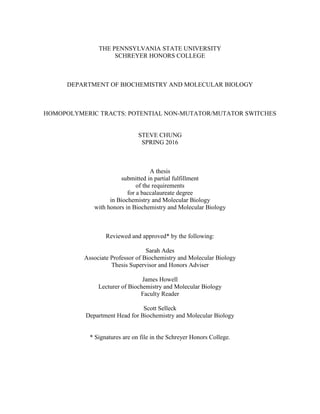
33. 27
Figure 7. The computed model vs. data graph taken from bz-rates of the integrant
JE2.
The data shown represents the graph of a cumulative distribution function of each repeat
fitted to the integrant JE2 data. The closer the fluctuation data is from the LD, or Luria-Delbrück,
model, the more reliable the functions that are calculated.
34. 28
Figure 8. Computed functions of the integrant JE2.
The computed functions were taken from Figure 7 using bz-rates of the integrant 8A
JE2. The µcorr value represents the mutation rate per cell per division corrected by the plating
efficiency.
35. 29
Figure 9. The computed model vs. data graph taken from bz-rates of wild-type JE2.
The data shown represents the graph of a cumulative distribution function of each repeat
fitted to the integrant JE2 data. The closer the fluctuation data is from the LD, or Luria-Delbrück,
model, the more reliable the functions that are calculated.
36. 30
Figure 10. Computed functions of the wild-type JE2.
The computed functions were taken from Figure 9 using bz-rates of the wild-type JE2.
The µcorr value represents the mutation rate per cell per division corrected by the plating
efficiency.
The function symbols are represented as follows.
m Mean number of mutations per culture not corrected by the PE
µ Mutation rate per cell per division not corrected by the PE
mcorr Number of mutations per culture corrected by the PE
µcorr Mutation rate per cell per division corrected by the PE
CLlower Lower 95% confidence limit for mcorr
CLupper Upper 95% confidence limit for mcorr
b Predicted mutant cells relative fitness
blower Lower 95% confidence limit for b
bupper Upper 95% confidence limit for b
meanNc Average number of plated cells per culture
sdNc Standard deviation of the number of plated cells
37. 31
chi2
Pearson's chi-square value
chi2
-p Pearson's chi-square p-value
The µcorr, or mutation frequency, of wild-type JE2 was 6.36 × 10⁻¹¹
mutation rate per
cell per division, whereas the mutant 8A JE2 was 8.934 × 10⁻¹⁰
mutation rate per cell per
division. The µcorr values suggested that 8A mutant JE2 had a mutation frequency 10-fold higher
than the wild-type counterpart. From this experiment, we have shown that the frameshift in MutL
does indeed increase the mutation rate in S. aureus, which might aid the bacteria in obtaining
antibiotic resistance at a rate 10-fold faster than the wild-type.
3.3 Investigating the Homopolymeric Tract in ClpX
In addition to the study of the mutator phenotype caused by mutL, the homopolymeric
tract found in ClpX was also investigated. In the ClpX gene, there is a 7A homopolymeric tract
located at the start of bp 69 of the 5’ end of the coding region. ClpX and ClpP form a dimer,
ClpXP, which functions as a protease essential for the virulence of S. aureus. The ClpX gene in
USA300 (a virulent CA-MRSA) serves to specifically recognize, unfold, and translocate
substrates into ClpP. It has been previously determined that a loss-function of ClpX in S. aureus
leads to β-lactam antibiotic resistance. This suggests that ClpX may be necessary for
antimicrobial inhibition of cell wall synthesis, and its role is not limited to protease activity (30).
Inactivated ClpX can give rise to β-lactam resistance, which could be damaging in clinical
settings. In the case of a homopolymeric tract, a deletion or addition of a base will lead to a
frameshift mutation, thereby rendering the gene inoperative. In other words, strains with a
38. 32
frameshift in ClpX may have an advantage by resisting the antimicrobial effects of β-lactam
antibiotics.
Figure 11. Gel image of a colony PCR after transformation of the modified plasmid
into C2987H.
The fragments were amplified from the ClpX gene containing the 7A homopolymeric
tract. The primers were designed to amplify the insert fragment. The sizes of all bands were
approximately 750bp, which indicates the presence of the desired insert.
Similar to using the pIMAY plasmid to transform S. aureus, the pCL plasmid was
introduced to modify a 7A homopolymeric tract. The pCL plasmid allowed for the controlled
expression of selected genes in target cells (31). Following the methods to integrate the insert
with the plasmid used in the mutL project, the 7A ClpX project stopped after the transformation
into an E. coli strain.
To test the frequency of frameshifts, the 7A homopolymeric tract would be paired with a
resistance gene. Once the homopolymeric tract frameshit, either contraction or expansion, the
resistance gene would be read in frame. The colonies that grows on the antibiotic plate can be
39. 33
compared to the total number of colonies present in the original culture. In this project, the
measurement of the frequency of expansion in the 7A homopolymeric tract was planned, but not
completed.
40. 34
Chapter 4
Discussion
Patient X, as well as many others, died due to an untreatable S. aureus infection. The
phenomenon of antibiotic resistance development in S. aureus strain is problematic. The
difficulties in treating MRSA highlights the need of find alternative methods of treatment. This
includes the primary research of mutL, a gene found responsible for inducing mutagenesis
through a bacteria’s genome once mutated. As a result, this question arises: would a frameshift in
the homopolymeric tract present in mutL gene cause a mutator phenotype?
Homopolymeric tracts are seen as a gene regulator that allows the gene to be mutated at a
given probability (15). In the case of mutL, once its inactivation occurs, the entire genome is
subject to mutations. Mutations, whether damaging or beneficial, will continue to occur
throughout the genome until mutL returns back to its non-mutated state. The homopolymeric
tract, due to its instability, in mutL governs the rate at which mutL becomes mutated by
frameshift. The impact of mutL on mutators developing antibiotic resistance can be a serious
clinically relevant phenomenon. To label it as such, it is necessary to measure and compare
mutation rates between wild-type and frameshifted mutL from USA300. Additionally, it is
necessary to compute the rate at which the 9A homopolymeric tract expands or contracts.
The construction of an 8A homopolymeric tract in USA300 demonstrated a contraction
of the 9A homopolymeric tract. The purpose of creating this construct was to compare the
mutation frequency with its wild-type counterpart. This construct differed from completely
knocking out the mutL gene since the 8A construct had the capability to frameshift back into
41. 35
frame. Usually a mutL knockout would have a decreased fitness. Although a mutL knockout may
develop an ability to survive antibiotic selection pressure, the genes that were favored in fitness,
such as metabolic genes, could be mutated as a result. Once the 8A construct had developed a
favorable gene for countering induced stress, it could frameshift back to 9A to prevent mutations
of important genes.
Using the fluctuation test, the 8A construct JE strain showed more resistance to
100µg/mL RIF than the wild-type JE2 strain after 24 hours of incubation. After 48 hours of
incubation, the 8A construct still showed more resistance to RIF. The 8A construct should have
exhibited a decreased fitness at a longer time point of incubation, however, the longevity of
acquiring damaging mutations varies among cells. By using the calculated µcorr values from bz-
rates (28), the 8A construct has a 10-fold antibiotic resistance rate higher than the wild-type JE2.
The procedure outlined by Monk et. al led to the successfully transformed 8A construct.
The detection of a decreased fitness described in the 48-hour incubation time point is difficult:
when a single cell in a colony acquires a deleterious mutation, the colony remains regardless of
the fate of that cell. The third integrant JE2 in Table 5 did not form any colonies. This could be
likely due to human error. Also, the chi2
values for the wild-type JE2 strain were not available. A
more in-depth statistical analysis should be performed in a multiple replication experiment to
describe the mutation rate phenomenon more accurately. This includes replicating the fluctuation
test of the wild-type and 8A integrant JE2 more than nine times. In addition, since the construct
and its wild-type were tested on RIF, future experiments should be aimed to compare the results
from RIF with other antibiotics. Aside from the mutation rate differences, the difficult nature of
transforming S. aureus strains limited the majority of the lab’s resources. This limitation includes
sequencing of each colonies seen on the plates in the fluctuation test. Each colony may have
42. 36
developed resistance through other mechanisms, besides the frameshift in mutL. By detecting the
contraction in mutL through sequencing, the colonies would have confirmed to develop antibiotic
resistance through the frameshift. The frameshift frequency of the 9A homopolymeric tract could
not be calculated. The experimental setup for calculating the frameshift frequency involved
pairing the 9A tract with a resistance marker out of frame. Once the desired direction of
frameshift occured, the cells could be plated on selection plates to calculate the frequency of
frameshift in the desired direction. The ClpX experiment terminated after the transformation of
C2987H cells. Early termination of the experiment suggested that future experiments should
attempt to calculate the frequency at which 7A homopolymeric tract in ClpX frameshifts. This
future experiment allows a deeper understanding of ClpX and the role of its 7A homopolymeric
tract in its inactivation.
The results of this experiment supported the idea that a fraction of the MRSA population
are mutators. The contraction of the 9A homopolymeric tract in mutL led to an increased
mutation rate. A similar experiment was done by Shaver and Sniegowski compared the effects of
changes in repeat length in the mutL gene of E. coli (18). They have found that mutators were
created because of the changes in repeat length in the mutL gene in E. coli. Although the study of
the mutL gene in E. coli did not investigate a homopolymeric tract, the experiment had similar
findings that a frameshift in the mutL gene could cause a mutator phenotype. With this new
knowledge, there may be a greater incentive to develop different treatment methods for MRSA
infections. In the case of patient X, isolate JH2 was found to have a mutation in mutL. The result
of this experiment suggested that the MRSA strain might have continually mutated to obtain
vancomycin resistance. From isolate JH2 to JH4, the mutated mutL gene might have developed
43. 37
beneficial mutations rapidly that helped the MRSA strain survive though the vancomycin
treatment.
The development of new antibiotics is very costly, and antibiotic resistance will continue
to render antibiotics ineffective. With a thorough understanding of the impact of a potential gene
regulator (i.e. homopolymeric tracts) on the development of mutator phenotype genes, a more
effective therapy could be employed to reduce antibiotic resistance. While this experiment shows
that there is an increased mutation rate in the 8A JE2 construct, it is necessary to perform
additional studies to completely confirm the results.
44. 38
Appendix A
Abbreviations
ATc: Anhydrotetracycline
BHA: Brain Heart Infusion Agar
BP: Base Pair
CA-MRSA: Community-Associated MRSA
CAM: Chloramphenicol
LA: Luria Agar
LB: Luria Broth
MRSA: Methicillin-Resistant Staphylococcus aureus
NAG: N-Acetylglucosamine
NAM: N-Acetylmuramic Acid
NEB: New England BioLabs
OD: Optical Density
PBP: Penicillin Binding Protein
PE: Plating Efficiency
RIF: Rifampicin, or Rifampin
TSA: Tryptic Soy Agar
TSB: Tryptic Soy Broth
45. 39
Appendix B
Additional Data Sets
The following tables (Tables 4-6) represents the colony counts from the fluctuation test
of a wild-type JE2, and three of the 8A integrant. All plates in the profile were inoculated from
the same overnight culture of USA300 wild-type or 8A integrant.
Table 4. The first set of Colony counts of JE2 wild-type and JE2 8A integrant.
This table was used in part to average all colony counts from Table 3. C indicates
confluent growth; H indicates colony counts greater than 1000.
24 Hours
Strain JE2
JE2
Integrant 1
JE2
Integrant 2
JE2
Integrant 3
RIF Concentration (µg/mL) 0 100 0 100 0 100 0 100
Number of colonies
with respective
dilution
1 C 5 C 166 C 158 C 144
1 × 10⁻¹ C 0 C 34 C 17 C 18
1 × 10⁻² C 0 C 3 C 1 C 1
1 × 10⁻³ C 0 C 0 C 0 C 0
1 × 10⁻⁴ C 0 C 1 C 0 C 0
1 × 10⁻⁵ C 0 C 0 C 0 C 0
1 × 10⁻⁶ H 0 H 0 H 0 H 0
1 × 10⁻⁷ 647 0 387 0 733 0 658 0
48 Hours
Strain JE2
JE2
Integrant 1
JE2
Integrant 2
JE2
Integrant 3
RIF Concentration (µg/mL) 0 100 0 100 0 100 0 100
Number of colonies
with respective
dilution
1 C 7 C 172 C 163 C 146
1 × 10⁻¹ C 0 C 35 C 19 C 10
1 × 10⁻² C 0 C 3 C 1 C 1
1 × 10⁻³ C 0 C 0 C 0 C 0
1 × 10⁻⁴ C 0 C 1 C 0 C 0
1 × 10⁻⁵ C 0 C 0 C 0 C 0
1 × 10⁻⁶ H 0 H 0 H 0 H 0
1 × 10⁻⁷ 695 0 404 0 764 0 684 0
46. 40
Table 5. The second set of Colony counts of JE2 wild-type and JE2 8A integrant.
This table was used in part to average all colony counts from Table 3. C indicates
confluent growth; H indicates colony counts greater than 1000.
24 Hours
Strain JE2
JE2
Integrant 4
JE2
Integrant 5
JE2
Integrant 6
RIF Concentration (µg/mL) 0 100 0 100 0 100 0 100
Number of
colonies with
respective dilution
1 C 18 C 174 C 129 C 0
1 × 10⁻¹ C 1 C 23 C 26 C 0
1 × 10⁻² C 0 C 0 C 8 C 0
1 × 10⁻³ C 0 C 0 C 1 C 0
1 × 10⁻⁴ C 0 C 0 C 0 C 0
1 × 10⁻⁵ C 0 C 0 C 0 C 0
1 × 10⁻⁶ H 0 H 0 H 0 C 0
1 × 10⁻⁷ 425 0 386 0 628 0 H 0
48 Hours
Strain JE2
JE2
Integrant 4
JE2
Integrant 5
JE2
Integrant 6
RIF Concentration (µg/mL) 0 100 0 100 0 100 0 100
Number of
colonies with
respective dilution
1 C 18 C 176 C 131 C 0
1 × 10⁻¹ C 1 C 24 C 26 C 0
1 × 10⁻² C 0 C 0 C 8 C 0
1 × 10⁻³ C 0 C 0 C 1 C 0
1 × 10⁻⁴ C 0 C 0 C 0 C 0
1 × 10⁻⁵ C 0 C 0 C 0 C 0
1 × 10⁻⁶ H 0 H 0 H 0 H 0
1 × 10⁻⁷ 441 0 395 0 648 0 H 0
47. 41
Table 6. The third set of Colony counts of JE2 wild-type and JE2 8A integrant.
This table was used in part to average all colony counts from Table 3. C indicates
confluent growth; H indicates colony counts greater than 1000.
24 Hours
Strain JE2
JE2
Integrant 7
JE2
Integrant 8
JE2
Integrant 9
RIF Concentration (µg/mL) 0 100 0 100 0 100 0 100
Number of
colonies with
respective dilution
1.00E+00 C 41 C 111 C 118 C 67
1.00E+01 C 6 C 26 C 17 C 7
1.00E+02 C 0 C 5 C 3 C 4
1.00E+03 C 0 C 0 C 0 C 0
1.00E+04 C 0 C 0 C 0 C 0
1.00E+05 C 0 C 0 C 0 C 0
1.00E+06 H 0 H 0 H 0 H 0
1.00E+07 884 0 253 0 833 0 792 0
48 Hours
Strain JE2
JE2
Integrant 7
JE2
Integrant 8
JE2
Integrant 9
RIF Concentration (µg/mL) 0 100 0 100 0 100 0 100
Number of
colonies with
respective dilution
1.00E+00 C 44 C 114 C 139 C 68
1.00E+01 C 6 C 28 C 18 C 8
1.00E+02 C 0 C 4 C 3 C 4
1.00E+03 C 0 C 1 C 0 C 0
1.00E+04 C 0 C 0 C 0 C 0
1.00E+05 C 0 C 0 C 0 C 0
1.00E+06 H 0 H 0 H 0 H 0
1.00E+07 921 0 252 0 836 0 807 0
48. 42
BIBLIOGRAPHY
1. Fuda, C., Suvorov, M., Vakulenko, S.B., Mobashery, S. 2004. The basis for resistance
to β-lactam antibiotics by penicillin-binding protein 2a of methicillin-
resistant Staphylococcus aureus. J. Biol. Chem. 279, 40802–40806.
2. McAleese, F., Wu, S.W., Sieradzki, K., Dunman, P., Murphy, E., Projan, S., Tomasz,
A. 2006. Overexpression of genes of the cell wall stimulon in clinical isolates of
Staphylococcus aureus exhibiting vancomycin-intermediate-S. aureus-type
resistance to vancomycin. J Bacteriol 188:1120–1133. 10.1128/JB.188.3.1120-
1133.2006.
3. Dechering, K.J., Cuelenaere, K., Konings, R.N., Leunissen, J.A. 1998. Distinct
frequency-distributions of homopolymeric DNA tracts in different genomes.
Nucleic Acids Res. 26, 4056-4062. 10.1093/nar/26.17.4056.
4. Cabell, C.H., Abrutyn, E., Karchmer, A.W. 2003. Cardiology patient page. Bacterial
endocarditis: the disease, treatment, and prevention. Circulation 107(20), e185-
e187. 10.1161/01.CIR.0000071082.36561.F1.
5. Ramachandran, G. 2014. Gram-positive and gram-negative bacterial toxins in sepsis:
A brief review. Virulence. 5(1), 213-218. 10.4161/viru.27024.
6. Bycroft, B.W., Shute, R.E. 1985. The Molecular Basis for the Mode of Action of Beta-
Lactam Antibiotics and Mechanisms of Resistance. Pharm Res. 2(1): 3-14.
7. Bergan, T. 1987. Pharmacokinetics of beta-lactam antibiotics. Scand J Infect Dis
Suppl. 42, 83–98.
49. 43
8. Barza, M. 1985. Imipenem: first of a new class of β-lactam antibiotics. Ann. Intern.
Med. 103:552–560. 10.7326/0003-4819-103-4-552.
9. Campbell, E.A., Korzheva, N., Mustaev, A., Murakami, K., Nair, S., Goldfarb, A.,
Darst S.A. 2001. Structural mechanism for rifampicin inhibition of bacterial RNA
polymerase. Cell 104:901–912.
10. Moellering, R.C. 2006. Vancomycin: a 50-year reassessment. Clin Infect Dis. 42, S3-
S4. 10.1086/491708.
11. Levine, D.P. 2006. Vancomycin: a history. Clin. Infect. Dis. 42:S5–S12.
10.1086/491709.
12. Mwangi, M.M., Wu, S.W., Zhou, Y., Sieradzki, K., de Lencastre, H., Richardson, P.,
Bruce, D., Rubin, E., Myers, E., Siggia, E.D., Tomasz, A. 2007. Tracking the in
vivo evolution of multidrug resistance in Staphylococcus aureus by whole-
genome sequencing. Proc Natl Acad Sci U S A 104:9451–9456.
10.1073/pnas.0609839104.
13. Leski, T.A., Tomasz, A. 2005. Role of penicillin-binding protein 2 (PBP2) in the
antibiotic susceptibility and cell wall cross-linking of Staphylococcus aureus:
evidence for the cooperative functioning of PBP2, PBP4, and PBP2A. J Bacteriol
187, 1815–1824. 10.1128/JB.187.5.1815-1824.
14. Wielders, C.L.C., Fluit, A.C., Brisse, S., Verhoef, J., Schmitz, F.J. 2002. mecA Gene
Is Widely Disseminated in Staphylococcus aureus Population. Journal of Clinical
Microbiology 40(11), 3970-3975. 10.1128/JCM.40.11.3970-3975.
50. 44
15. Orsi, R. H., Bowen, B. M., Wiedmann, M. 2010. Homopolymeric tracts represent a
general regulatory mechanism in prokaryotes. BMC Genomics 11, 102.
10.1186/1471-2164-11-102.
16. Ellegren, H. 2000. Microsatellite mutations in the germline: implications for
evolutionary inference. Trends in Genetics 16:551-558. 10.1016/S0168-
9525(00)02139-9.
17. Drake, J.W., Charlesworth, B., Charlesworth, D., Crow, J.F. 1998. Rates of
Spontaneous Mutation. GENETICS 148, 1667-1686.
18. Shaver, A.C., Sniegowski, P.D. 2003. Spontaneously arising mutL mutators in
evolving Escherichia coli populations are the result of changes in repeat length. J
Bacteriol 185: 6076–6082. 10.1128/JB.185.20.6076-6082.
19. Monk, I.R., Shah, I.M., Xu, M., Tan, M.W., Foster, T.J. 2012. Transforming the
untransformable: Application of direct transformation to manipulate genetically
Staphylococcus aureus and Staphylococcus epidermidis. MBio 3(2):e00277-11.
20. Gibson, D.G., Young, L., Chuang, R., Venter, J. C., Hutchingson III, C. A., Smith,
H.O. 2009. Enzymatic assembly of DNA molecules up to several hundred
kilobases. Nature Methods 6, 3433-3345. 10.1038/nmeth.1318.
21. New England BioLabs Incorporation. 2016. High Efficiency Transformation Protocol
(C2987H/C2987I). Ipswich, MA.
22. New England BioLabs Incorporation. 2016. Site Directed Mutagenesis. Ipswich, MA.
23. Life Technologies Corporation. 2010. Qubit 2.0 Fluorometer Catalog no. Q32866.
Carlsbad, CA.
51. 45
24. Thermo Fisher Scientific. 2009. NanoDrop 2000/2000c Spectrophotometer V1.0 User
Manual. Wilmington, DE.
25. Laboratory of Computational and Quantitative Biology. 2015. Bz-rates mutation rate
calculator. The University Pierre and Marie Curie, Paris, France.
26. BEI Resources. 2014. Product Information Sheet for NR-46643. ATCC.
27. Fey, P.D., Endres, J.L., Yajjala, V.K., Widhelm, T.J., Boissy, R.J., Bose, J.L., Bayles,
K.W. 2013. A genetic resource for rapid and comprehensive phenotype screening
of nonessential Staphylococcus aureus genes. mBio 4:e00537-12.
10.1128/mBio.00537-12.
28. Gillet-Markowska, A., Louvel, G., Fischer, G. 2015. bz-rates: A Web Tool to
Estimate Mutation Rates from Fluctuation Analysis. G3:
Genes|Genomes|Genetics 5(11), 2323-2327. 10.1534/g3.115.019836.
29. Lea D., Coulson C. A., 1949. The distribution of the numbers of mutants in bacterial
populations. J. Genet. 49, 264–285.
30. Baek, K. T. et al. beta-Lactam Resistance in Methicillin-Resistant Staphylococcus
aureus USA300 Is Increased by Inactivation of the ClpXP Protease. Antimicrob
Agents Chemother 58, 4593–4603.
31. Naviaux, R.K., Costanzi, E., Haas, M., Verma, I.M. 1996. The pCL vector system:
rapid production of helper-free, high-titer, recombinant retroviruses. Journal of
Virology 70(8), 5701-5705.
52. ACADEMIC VITA
STEVE CHUNG
Stevechung1993@gmail.com
EDUCATION
The Pennsylvania State University: University Park
Eberly College of Science & Schreyer Honors College
B.S. Biochemistry and Molecular Biology
AWARDS AND SCHOLARSHIPS
2014 Intel Scholarship for Employee’s Children
2013 President’s Freshman Award
2013, 2014 Eberly College of Science Travel Grant
2014 Education Abroad Whole World Scholarship
2014 Education Abroad Diversity Grant-in-Aid Scholarship
2015 Sperling Scholarship
RESEARCH AND OTHER EXPERIENCES
Research Assistant under Dr. Michael Mwangi on Mechanisms of Antibiotic Resistance 2013-2016
Department of Biochemistry and Molecular Biology, The Pennsylvania State University
Investigated and explored mechanisms that S. aureus (MRSA) obtain drug resistance
PCR, gel electrophoresis, plasmid digestion, DNA quantification, DNA purification, molecular genetics
Maintained media and equipment, and practiced safe techniques while working with biosafety level 1 and
2 strains.
Learning Assistant for BMB 251: Molecular Cell Biology I 2014-2015
Department of Biochemistry and Molecular Biology, The Pennsylvania State University
Held learning sessions to promote critical and analytical evaluation of materials taught in lecture
Discuss possible improvements in learning sessions and worksheets with professors
FDT Incubator – ForexMaster Brand Ambassador 2015
www.forexmaster.io
Represented ForexMaster, a product of Financial Data Technologies (FDT), throughout Penn State
LANGUAGE PROFICIENCY
FLUENT IN ENGLISH AND MANDARIN
5 YEARS OF CLASSROOM SPANISH LANGUAGE
ORGANIZATIONS
EARTH HOUSE ORGANIZATION – SECRETARY
PRE-PHARMACY SOCIETY– VICE-PRESIDENT, CO-FOUNDER
CHINESE CHESS CLUB – CO-FOUNDER
EARTH HOUSE ORGANIZATION – SECRETARY
PRE-PHARMACY SOCIETY– VICE-PRESIDENT, CO-FOUNDER
FUJIANESE FRIENDSHIP ASSOCIATION
CHINESE CHESS CLUB – CO-FOUNDER
NORTH HALLS STUDENT ASSOCIATION
PROJECT HAITI
TAIWANESE AMERICAN STUDENT ASSOCIATION
UNICEF PENN STATE