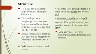
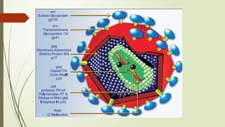
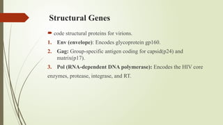
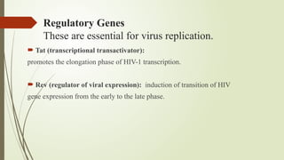
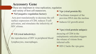
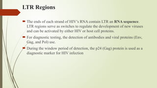
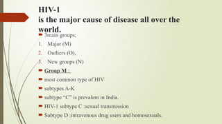
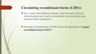
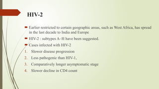
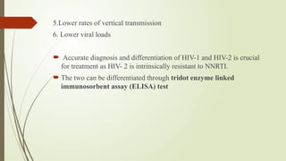
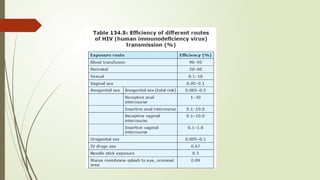
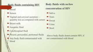
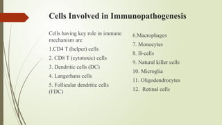
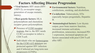
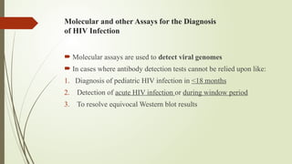
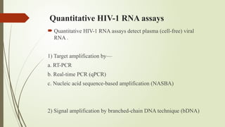
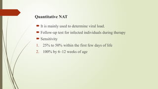
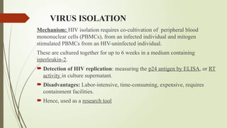
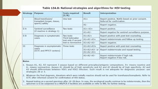
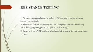
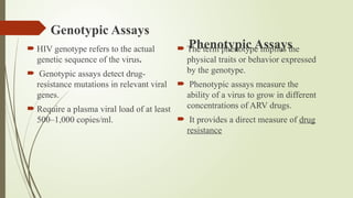
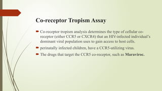

HUMAN IMMUNODEFICIENCY VIRUS (HIV)
Virology
Immunopathogenesis
Laboratory
Dr. Shruti Pawar
JR1 , MD Skin and VD
NDMVP Nashik
Origin
 HIV-1 virus: Simian immunodeficiency virus (SIV) of chimpanzees,
found in forests of Central Africa.
 HIV-2 : SIV of sooty Mangabey, an old world monkey inhabiting Western
Africa.
video
Structure
 It is a 120 nm icosahedral,
single-stranded, enveloped
RNA virus.
 The envelope : two
phospholipid layers derived
from the host cell membrane
contains glycoprotein (gp)160
(gp 120 + gp 41)
 Gp120: contains sites that bind
CD4 cells and co-receptors on
the surface of human CD4 T-
cells.
 Gp41: transmembrane bound
protein.
• Inside the viral envelope there is a
layer called the matrix: the protein
p17.
• Viral core (capsid): protein p24
contains HIV genetic material, two
positive strands of single-stranded
RNA.
 Viral enzymes—Reverse
transcriptase, (RT), Integrase, and
Protease.
Structural Genes
 code structural proteins for virions.
1. Env (envelope): Encodes glycoprotein gp160.
2. Gag: Group-specific antigen coding for capsid(p24) and
matrix(p17).
3. Pol (RNA-dependent DNA polymerase): Encodes the HIV core
enzymes, protease, integrase, and RT.
Regulatory Genes
These are essential for virus replication.
 Tat (transcriptional transactivator):
promotes the elongation phase of HIV-1 transcription.
 Rev (regulator of viral expression): induction of transition of HIV
gene expression from the early to the late phase.
Accessory Gene
These are important in virus replication, regulation
and variety of host cell maneuvers.
 Nef (negative regulation factor):
Acts post translationally to decrease the cell
surface expression of CD4, reduces T-cell
activation, and stimulates the infectivity of
HIV virions.
 Vif (viral infectivity):
the reproduction of HIV in peripheral blood
lymphocytes, macrophages.
 Vpr (viral protein R):
 Facilitates transport of the
provirus DNA into the nucleus
 Induces G2 growth arrest
 Vpu (viral protein unique):
decaying of CD4 in the
endoplasmic reticulum triggers
the release of virions from
infected cells
 HIV-2 lacks the vpu gene.
LTR Regions
 The ends of each strand of HIV’s RNA contain LTR an RNA sequence.
LTR regions serve as switches to regulate the development of new viruses
and can be activated by either HIV or host cell proteins.
 For diagnostic testing, the detection of antibodies and viral proteins (Env,
Gag, and Pol) use.
 During the window period of detection, the p24 (Gag) protein is used as a
diagnostic marker for HIV infection
HIV-1
is the major cause of disease all over the
world.
 3main groups;
1. Major (M)
2. Outliers (O),
3. New groups (N)
 Group M :
 most common type of HIV
 subtypes A-K
 subtype “C” is prevalent in India.
 HIV-1 subtype C :sexual transmission
 Subtype D :intravenous drug users and homosexuals.
Circulating recombinant forms (CRFs)
 Two viruses from different subtypes infect the same cell in an
infected person and result in recombinant virus strains that may
transmit within a population.
 Secondary recombination of CRFs lead to the appearance of unique
recombinant forms (URFs)
HIV-2
 Earlier restricted to certain geographic areas, such as West Africa, has spread
in the last decade to India and Europe
 HIV-2 : subtypes A–H have been suggested.
 Cases infected with HIV-2
1. Slower disease progression
2. Less pathogenic than HIV-1,
3. Comparatively longer asymptomatic stage
4. Slower decline in CD4 count
5.Lower rates of vertical transmission
6. Lower viral loads
 Accurate diagnosis and differentiation of HIV-1 and HIV-2 is crucial
for treatment as HIV- 2 is intrinsically resistant to NNRTI.
 The two can be differentiated through tridot enzyme linked
immunosorbent assay (ELISA) test
Replication Cycle of HIV
CD4 (cluster of differentiation 4) :
 58-kDa monomeric glycoprotein that can be detected on the cell
surface of many cells.
 CD4 is a primary receptor for HIV and can be detected
predominantly on cell surface of T-lymphocytes (CD4+ T-helper
cells).
 The virus may also infect other cell types with CD4 expressed on
the surface (e.g., resting CD4 T-cells, monocytes/macrophages,
dendritic/Langerhans cells). Besides these, CD4 independent cells
susceptible to HIV are astrocytes, follicular dendritic cells from
tonsils and renal epithelial cells.
Modes of transmission
Body fluids containing HIV
 Blood
 Semen
 Vaginal and cervical secretion—
quantity less as compared with semen
 Breast milk
 Amniotic fluid
 Cerebrospinal fluid
 Pleural, pericardial, peritoneal fluids
 Any body fluid contaminated with
blood
Body fluids with no/low
concentration of HIV
 Saliva
 Tears
 Sweat
 Urine
 Stool
Above body fluids donot contain HIV, if
not contaminated with blood
Cells Involved in Immunopathogenesis
Cells having key role in immune
mechanism are
1.CD4 T (helper) cells
2. CD8 T (cytotoxic) cells
3. Dendritic cells (DC)
4. Langerhans cells
5. Follicular dendritic cells
(FDC)
6.Macrophages
7. Monocytes
8. B-cells
9. Natural killer cells
10. Microglia
11. Oligodendrocytes
12. Retinal cells
Factors Affecting Disease Progression
• Viral factors: HIV strain (HIV-
1/HIV-2), co-receptor usage,
generation of escape mutants,
latency.
• Host genetic factors: HLA
polymorphism and chemokine
receptor genes polymorphism.
 Presence of CCR5 receptor
tropism, that is, the HIV needs
CCR5 co-receptor to infect a
CD4 T-cell.
 Individuals who are homozygous
for the CCR5-Δ32 deletions are
protected against HIV infection
and if infected are long-term non
progressors (LTNPs).
 Environmental factors: Nutrition,
coinfections, smoking, and alcoholism.
 Co-infection: Tuberculosis, STIs
especially herpes progenitalis, Hepatitis
B.
 Immunological factors: Low thymic
output, poor bone marrow function,
increased immune activation,
proliferation, senescence, increased PD-
1 expression, increased apoptosis are
factors associated with poor CD4 T-cell
after ART.
LABORATORY DIAGNOSIS
Objectives of Testing
• Transfusion and transplant safety
• Diagnosis of HIV in symptomatic and asymptomatic
• Prevention of parent-to-child transmission
• Postexposure prophylaxis
• Epidemiology
• Research
Approach to Testing
Unlinked anonymous testing
Used for HIV surveillance
purposes to monitor
epidemiological trends
 Voluntary confidential
counseling and testing
Used for the diagnosis of HIV
infection in symptomatic and
asymptomatic individuals
 Mandatory testing
1. Screening for HIV and other
bloodborne infections of all
blood destined for transfusion or
for the manufacture of blood
products.
2. Screening of donors prior to all
procedures:
 Artificial insemination
 Corneal grafts
 Organ transplant
 WHO Suggests Two New
Approaches to Enhance HIV-
Testing Services
• Self-testing
 HIV self-testing is easily accessible and can
reach high-risk population as well as
adolescent and young people who may not
otherwise opt for testing.
 Performed using kits authenticated by
concerned authorities.
 It has been observed that increase frequency
of self-testing is not associated with increase
in high-risk behavior, or any adverse effects.
 A reactive (positive) HIV self-testing result
always requires confirmatory HIV
testing.
 People who test negative for HIV on self-
testing need confirmation only if they have
had a potential HIV exposure in 3 months
prior to testing. Retesting after 6–12 weeks is
recommended in recent potential HIV
exposure.
 All people having high ongoing risk should
be advised to have an HIV test every 6
months. HIV self-testing should never be
used when people are on cART to prevent
false negative HIV test results.
HIV testing through voluntary-assisted
partner notification
 The goal of partner notification is to offer HIV testing for high-risk
partners of PLHIV. While maintaining confidentiality, partner
notification has to be provided on voluntary basis.
 Partner notification is to be promoted only through health care
workers and should not include other authorities.
DIRECT PREDICTORS OF HIV INFECTION
1. Specific antibody detection:
- ELISA/ EIA
- Rapid tests
- Western Blot (WB)
2. Specific antigen detection:
a. Detection of p24 antigen—
Dissociation EIA
b. Reverse transcriptase assay
3. Detection of viral nucleic acid:
a. In situ hybridization
b. PCR
 Genotyping of HIV
 Viral load assay
4. Isolation/culture of virus
a. Syncytium inducing
b. Non syncytium inducing
B. INDIRECT PREDICTORS OF HIV
INFECTION
1. Decreased CD4 cells
2. Increased β2 microglobulin
3. Increased serum neopterin
4. Increased IL-2 receptors
5. AIDS defining illnesses (Candidiasis, Cryptococcosis, CMV
retinitis, Kaposi sarcoma)
Laboratory Diagnosis During Window
Period
Virological tests can be used for the detection of
 Acute HIV infection during the “window period”, before HIV
antibodies become detectable.
 Although positive NAT results confirm the HIV diagnosis, the NAT
result may turn out negative if tested within 7–10 days of exposure.
Nucleic acid tests include tests for the qualitative detection of HIV-1
DNA or RNA, as well as the quantitative detection of HIV-1 RNA
(viral load determination).
 Need for laboratory diagnosis in window period:
1. Recent blood transfusion
2. Risky heterosexual/homosexual exposure
3. Needle stick injury (contaminated)
 1. ELISA
 2. Rapid tests
 3. Western blot test
1. ELISA
 ELISA is the most commonly performed screening test at blood
banks and tertiary care sites.
 Takes 2–3 hours.
ELISA types
 1. Indirect ELISA
 2. Competitive ELISA
 3. Sandwich ELISA—it can
detect all classes of HIV
antibodies and p24 antigen
 4. Capture ELISA:
• Antigen capture ELISA: It is
more specific than an indirect
assay.
• Antibody capture ELISA:
Developed to test specimens with
low concentrations of HIV
antibodies (e.g., urine and saliva)
or to detect a specific class of
antibodies (e.g., IgG, IgM, or
IgA)
False-positive ELISA
 Autoimmune diseases
 Alcoholic hepatitis
 Primary biliary cirrhosis
 Leprosy
 Multiple pregnancies
False-negative ELISA
 Technical errors
 The test may be negative during the
window period and during the end
stage of the disease
Rapid tests
 Detect antibodies to HIV Types 1 and 2 in human serum, plasma, whole blood,
saliva, and urine.
1) Immunoconcentration/dot blot assay (vertical flow):
HIV lysate, immobilized on nitrocellulose paper and microliter quantity of serum
added for Ab detection.
2) Agglutination assay (gelatin, red blood cell, latex, and microbeads): clumping
due to antigen and antibody interaction
3) Immunochromatographic assay (lateral flow) : The capture antibodies are
impregnated over paper strip and the liquid moves over the strip by capillary
action , target antigen detected by colorimetric change in paper strip
4) Dipstick assay based on enzyme immune assay (EIA)
Advantages of rapid tests:
 Visual point of care tests that do not require any special equipment.
 Technically simple to perform.
 Most of them have sensitivity and specificity comparable to an
ELISA
Western blot test
 confirmatory test with high specificity
 used in cases of indeterminate/discordant result of ELISA/rapid tests.
 identifies with specific antibodies, the proteins that have been separated from
one another according to their size by gel electrophoresis.
 The various HIV-specific recombinant or synthetic antigens are absorbed onto
blot which is a membrane, almost always of nitrocellulose or polyvinylidene
fluoride.
 detect the presence of antibodies against specific HIV proteins ( p24,
gp41, and gp120/160), which are seen as
bands on the test strip.
Limitations of Antibody Assays
 not detectable in the window period.
 Not useful in the diagnosis of infection in children below 18
months of age
Molecular and other Assays for the Diagnosis
of HIV Infection
 Molecular assays are used to detect viral genomes
 In cases where antibody detection tests cannot be relied upon like:
1. Diagnosis of pediatric HIV infection in <18 months
2. Detection of acute HIV infection or during window period
3. To resolve equivocal Western blot results
Nucleic Acid Tests
 Qualitative detection of HIV-1 DNA or RNA PCR Test.
 Quantitative detection of HIV-1 RNA (viral load determination).
 Qualitative PCR for HIV DNA.
PCR helps to find a specific gene sequence among an abundance of other
DNA.
 HIV RT produces DNA from the RNA genome of the virus.
 A complementary strand of DNA is then produced through viral DNA
polymerase to form double stranded viral DNA.
 This DNA, which may integrate into the host cell genome or remain
unintegrated in the cytoplasm, can be amplified by PCR.
 Qualitative transcription-mediated amplification assay for HIV
RNA:
 The APTIMA HIV-1 qualitative RNA assay (GenProbe)
Quantitative HIV-1 RNA assays
 Quantitative HIV-1 RNA assays detect plasma (cell-free) viral
RNA .
1) Target amplification by—
a. RT-PCR
b. Real-time PCR (qPCR)
c. Nucleic acid sequence-based amplification (NASBA)
2) Signal amplification by branched-chain DNA technique (bDNA)
Quantitative NAT
 It is mainly used to determine viral load.
 Follow-up test for infected individuals during therapy
 Sensitivity
1. 25% to 50% within the first few days of life
2. 100% by 6–12 weeks of age
VIRUS ISOLATION
Mechanism: HIV isolation requires co-cultivation of peripheral blood
mononuclear cells (PBMCs), from an infected individual and mitogen
stimulated PBMCs from an HIV-uninfected individual.
These are cultured together for up to 6 weeks in a medium containing
interleukin-2.
 Detection of HIV replication: measuring the p24 antigen by ELISA, or RT
activity in culture supernatant.
 Disadvantages: Labor-intensive, time-consuming, expensive, requires
containment facilities.
 Hence, used as a research tool
ANTIGEN DETECTION
 P24 antigen or immune complex of p24 with anti p24 is detected
using ELISA.
 Positive test: confirms diagnosis
 Negative test: does not rule out HIV infection
 Sensitivity is lower than NAT
HIV HOME-BASED TEST
Oral Mucosal Transudate Test
 Sample: noninvasively collected specimens of oral fluids, although
generally referred to as saliva; the fluid used for testing is actually oral
mucosal transudate.
 Testing: antibodies that are comparable to or exceed those from serum
samples.
 It qualitatively detects antibodies against HIV-1 and HIV-2 in human oral
fluid, whole blood obtained from a finger puncture or a venipuncture, and
plasma by immunoassay.
 99.8% specific and 99.3% sensitive.
 The only drawback is the “window period”
Monitoring Laboratory tests
Monitoring Parameters
Virological HIV1 viral load
Resistance to ART
Immunological and Hematological Total Lymphocyte count
CD4 + T cell count
Opportunistic infections Reactivation of TB
Occurrence of new OI
Recurrence of treated OI
Antimicrobial susceptibility of
bacterial pathogen
Adverse drug reactions Liver and kidney function tests
Hematological parameters
Technologies available for HIV 1 Viral Load
Estimation
 Real-time PCR
 Nucleic acid sequence-based amplification assays with real-time
detection
 Transcription-mediated amplification
 Signal amplification
 Branched DNA assay
Timings for HIV-1 Viral load
test
Advantages of viral load testing
 It is the earliest indicator of viral reproduction
 Useful sentinel for virological response during treatment because the decay
of viral load in tissues typically corresponds with virological responses in
plasma.
 Early and more accurate indicator of treatment failure.
 To determine the need to switch to second-line drugs. This helps to reduce
the accumulation of drug-resistance mutations and improve clinical outcomes
of the PLHIV on cART.
CD4 T-lymphocyte Count
 important marker of immune function in HIV infected individuals.
 predicts subsequent disease progression and survival
 A baseline CD4 count on ART initiation is necessary for determining
immunological failure in future and understanding presence of
opportunistic infections (OIs).
 Test and treat referring to HIV testing and the offer of antiretroviral
therapy, irrespective of WHO clinical stage or CD4 cell count
Patient group Timing for CD4 test
New Patient Every 6 months
Existing patient (on cART for atleast
12 months)
CD4 testing at baseline during ART
initiation
CD4 to be done every 6 months
thereafter
RESISTANCE TESTING
1. At baseline, regardless of whether ARV therapy is being initiated
(genotypic testing).
2. Treatment failure or incomplete viral suppression while receiving
ARV therapy (genotypic and/or phenotypic testing).
3. Cases still on cART or those who have left therapy for not more than
1 year.
Genotypic Assays
 HIV genotype refers to the actual
genetic sequence of the virus.
 Genotypic assays detect drug-
resistance mutations in relevant viral
genes.
 Require a plasma viral load of at least
500–1,000 copies/ml.
Phenotypic Assays
 The term phenotype implies the
physical traits or behavior expressed
by the genotype.
 Phenotypic assays measure the
ability of a virus to grow in different
concentrations of ARV drugs.
 It provides a direct measure of drug
resistance
Co-receptor Tropism Assay
 Co-receptor tropism analysis determines the type of cellular co-
receptor (either CCR5 or CXCR4) that an HIV-infected individual’s
dominant viral population uses to gain access to host cells.
 perinatally infected children, have a CCR5-utilizing virus.
 The drugs that target the CCR5 co-receptor, such as Maraviroc.
Human Leukocyte Antigen (HLA) Testing to
Predict Abacavir Hypersensitivity
 Clinicians should perform HLA-B5701, HLA-DR7 and HLA-DQ3
testing before initiating Abacavir based therapy as they have an
genetic predisposition for the development of Abacavir
hypersensitivity
Thank You

human immunodeficiency virus [HIV] PDF SQ

  • 1.
     HUMAN IMMUNODEFICIENCY VIRUS(HIV) Virology Immunopathogenesis Laboratory Dr. Shruti Pawar JR1 , MD Skin and VD NDMVP Nashik
  • 2.
    Origin  HIV-1 virus:Simian immunodeficiency virus (SIV) of chimpanzees, found in forests of Central Africa.  HIV-2 : SIV of sooty Mangabey, an old world monkey inhabiting Western Africa.
  • 3.
  • 4.
    Structure  It isa 120 nm icosahedral, single-stranded, enveloped RNA virus.  The envelope : two phospholipid layers derived from the host cell membrane contains glycoprotein (gp)160 (gp 120 + gp 41)  Gp120: contains sites that bind CD4 cells and co-receptors on the surface of human CD4 T- cells.  Gp41: transmembrane bound protein. • Inside the viral envelope there is a layer called the matrix: the protein p17. • Viral core (capsid): protein p24 contains HIV genetic material, two positive strands of single-stranded RNA.  Viral enzymes—Reverse transcriptase, (RT), Integrase, and Protease.
  • 6.
    Structural Genes  codestructural proteins for virions. 1. Env (envelope): Encodes glycoprotein gp160. 2. Gag: Group-specific antigen coding for capsid(p24) and matrix(p17). 3. Pol (RNA-dependent DNA polymerase): Encodes the HIV core enzymes, protease, integrase, and RT.
  • 7.
    Regulatory Genes These areessential for virus replication.  Tat (transcriptional transactivator): promotes the elongation phase of HIV-1 transcription.  Rev (regulator of viral expression): induction of transition of HIV gene expression from the early to the late phase.
  • 8.
    Accessory Gene These areimportant in virus replication, regulation and variety of host cell maneuvers.  Nef (negative regulation factor): Acts post translationally to decrease the cell surface expression of CD4, reduces T-cell activation, and stimulates the infectivity of HIV virions.  Vif (viral infectivity): the reproduction of HIV in peripheral blood lymphocytes, macrophages.  Vpr (viral protein R):  Facilitates transport of the provirus DNA into the nucleus  Induces G2 growth arrest  Vpu (viral protein unique): decaying of CD4 in the endoplasmic reticulum triggers the release of virions from infected cells  HIV-2 lacks the vpu gene.
  • 9.
    LTR Regions  Theends of each strand of HIV’s RNA contain LTR an RNA sequence. LTR regions serve as switches to regulate the development of new viruses and can be activated by either HIV or host cell proteins.  For diagnostic testing, the detection of antibodies and viral proteins (Env, Gag, and Pol) use.  During the window period of detection, the p24 (Gag) protein is used as a diagnostic marker for HIV infection
  • 10.
    HIV-1 is the majorcause of disease all over the world.  3main groups; 1. Major (M) 2. Outliers (O), 3. New groups (N)  Group M :  most common type of HIV  subtypes A-K  subtype “C” is prevalent in India.  HIV-1 subtype C :sexual transmission  Subtype D :intravenous drug users and homosexuals.
  • 11.
    Circulating recombinant forms(CRFs)  Two viruses from different subtypes infect the same cell in an infected person and result in recombinant virus strains that may transmit within a population.  Secondary recombination of CRFs lead to the appearance of unique recombinant forms (URFs)
  • 12.
    HIV-2  Earlier restrictedto certain geographic areas, such as West Africa, has spread in the last decade to India and Europe  HIV-2 : subtypes A–H have been suggested.  Cases infected with HIV-2 1. Slower disease progression 2. Less pathogenic than HIV-1, 3. Comparatively longer asymptomatic stage 4. Slower decline in CD4 count
  • 13.
    5.Lower rates ofvertical transmission 6. Lower viral loads  Accurate diagnosis and differentiation of HIV-1 and HIV-2 is crucial for treatment as HIV- 2 is intrinsically resistant to NNRTI.  The two can be differentiated through tridot enzyme linked immunosorbent assay (ELISA) test
  • 14.
    Replication Cycle ofHIV CD4 (cluster of differentiation 4) :  58-kDa monomeric glycoprotein that can be detected on the cell surface of many cells.  CD4 is a primary receptor for HIV and can be detected predominantly on cell surface of T-lymphocytes (CD4+ T-helper cells).  The virus may also infect other cell types with CD4 expressed on the surface (e.g., resting CD4 T-cells, monocytes/macrophages, dendritic/Langerhans cells). Besides these, CD4 independent cells susceptible to HIV are astrocytes, follicular dendritic cells from tonsils and renal epithelial cells.
  • 16.
  • 19.
    Body fluids containingHIV  Blood  Semen  Vaginal and cervical secretion— quantity less as compared with semen  Breast milk  Amniotic fluid  Cerebrospinal fluid  Pleural, pericardial, peritoneal fluids  Any body fluid contaminated with blood Body fluids with no/low concentration of HIV  Saliva  Tears  Sweat  Urine  Stool Above body fluids donot contain HIV, if not contaminated with blood
  • 20.
    Cells Involved inImmunopathogenesis Cells having key role in immune mechanism are 1.CD4 T (helper) cells 2. CD8 T (cytotoxic) cells 3. Dendritic cells (DC) 4. Langerhans cells 5. Follicular dendritic cells (FDC) 6.Macrophages 7. Monocytes 8. B-cells 9. Natural killer cells 10. Microglia 11. Oligodendrocytes 12. Retinal cells
  • 21.
    Factors Affecting DiseaseProgression • Viral factors: HIV strain (HIV- 1/HIV-2), co-receptor usage, generation of escape mutants, latency. • Host genetic factors: HLA polymorphism and chemokine receptor genes polymorphism.  Presence of CCR5 receptor tropism, that is, the HIV needs CCR5 co-receptor to infect a CD4 T-cell.  Individuals who are homozygous for the CCR5-Δ32 deletions are protected against HIV infection and if infected are long-term non progressors (LTNPs).  Environmental factors: Nutrition, coinfections, smoking, and alcoholism.  Co-infection: Tuberculosis, STIs especially herpes progenitalis, Hepatitis B.  Immunological factors: Low thymic output, poor bone marrow function, increased immune activation, proliferation, senescence, increased PD- 1 expression, increased apoptosis are factors associated with poor CD4 T-cell after ART.
  • 22.
    LABORATORY DIAGNOSIS Objectives ofTesting • Transfusion and transplant safety • Diagnosis of HIV in symptomatic and asymptomatic • Prevention of parent-to-child transmission • Postexposure prophylaxis • Epidemiology • Research
  • 23.
    Approach to Testing Unlinkedanonymous testing Used for HIV surveillance purposes to monitor epidemiological trends  Voluntary confidential counseling and testing Used for the diagnosis of HIV infection in symptomatic and asymptomatic individuals  Mandatory testing 1. Screening for HIV and other bloodborne infections of all blood destined for transfusion or for the manufacture of blood products. 2. Screening of donors prior to all procedures:  Artificial insemination  Corneal grafts  Organ transplant
  • 24.
     WHO SuggestsTwo New Approaches to Enhance HIV- Testing Services • Self-testing  HIV self-testing is easily accessible and can reach high-risk population as well as adolescent and young people who may not otherwise opt for testing.  Performed using kits authenticated by concerned authorities.  It has been observed that increase frequency of self-testing is not associated with increase in high-risk behavior, or any adverse effects.  A reactive (positive) HIV self-testing result always requires confirmatory HIV testing.  People who test negative for HIV on self- testing need confirmation only if they have had a potential HIV exposure in 3 months prior to testing. Retesting after 6–12 weeks is recommended in recent potential HIV exposure.  All people having high ongoing risk should be advised to have an HIV test every 6 months. HIV self-testing should never be used when people are on cART to prevent false negative HIV test results.
  • 25.
    HIV testing throughvoluntary-assisted partner notification  The goal of partner notification is to offer HIV testing for high-risk partners of PLHIV. While maintaining confidentiality, partner notification has to be provided on voluntary basis.  Partner notification is to be promoted only through health care workers and should not include other authorities.
  • 26.
    DIRECT PREDICTORS OFHIV INFECTION 1. Specific antibody detection: - ELISA/ EIA - Rapid tests - Western Blot (WB) 2. Specific antigen detection: a. Detection of p24 antigen— Dissociation EIA b. Reverse transcriptase assay 3. Detection of viral nucleic acid: a. In situ hybridization b. PCR  Genotyping of HIV  Viral load assay 4. Isolation/culture of virus a. Syncytium inducing b. Non syncytium inducing
  • 27.
    B. INDIRECT PREDICTORSOF HIV INFECTION 1. Decreased CD4 cells 2. Increased β2 microglobulin 3. Increased serum neopterin 4. Increased IL-2 receptors 5. AIDS defining illnesses (Candidiasis, Cryptococcosis, CMV retinitis, Kaposi sarcoma)
  • 28.
    Laboratory Diagnosis DuringWindow Period Virological tests can be used for the detection of  Acute HIV infection during the “window period”, before HIV antibodies become detectable.  Although positive NAT results confirm the HIV diagnosis, the NAT result may turn out negative if tested within 7–10 days of exposure. Nucleic acid tests include tests for the qualitative detection of HIV-1 DNA or RNA, as well as the quantitative detection of HIV-1 RNA (viral load determination).  Need for laboratory diagnosis in window period: 1. Recent blood transfusion 2. Risky heterosexual/homosexual exposure 3. Needle stick injury (contaminated)
  • 29.
     1. ELISA 2. Rapid tests  3. Western blot test
  • 30.
    1. ELISA  ELISAis the most commonly performed screening test at blood banks and tertiary care sites.  Takes 2–3 hours.
  • 31.
    ELISA types  1.Indirect ELISA  2. Competitive ELISA  3. Sandwich ELISA—it can detect all classes of HIV antibodies and p24 antigen  4. Capture ELISA: • Antigen capture ELISA: It is more specific than an indirect assay. • Antibody capture ELISA: Developed to test specimens with low concentrations of HIV antibodies (e.g., urine and saliva) or to detect a specific class of antibodies (e.g., IgG, IgM, or IgA)
  • 32.
    False-positive ELISA  Autoimmunediseases  Alcoholic hepatitis  Primary biliary cirrhosis  Leprosy  Multiple pregnancies False-negative ELISA  Technical errors  The test may be negative during the window period and during the end stage of the disease
  • 33.
    Rapid tests  Detectantibodies to HIV Types 1 and 2 in human serum, plasma, whole blood, saliva, and urine. 1) Immunoconcentration/dot blot assay (vertical flow): HIV lysate, immobilized on nitrocellulose paper and microliter quantity of serum added for Ab detection. 2) Agglutination assay (gelatin, red blood cell, latex, and microbeads): clumping due to antigen and antibody interaction 3) Immunochromatographic assay (lateral flow) : The capture antibodies are impregnated over paper strip and the liquid moves over the strip by capillary action , target antigen detected by colorimetric change in paper strip 4) Dipstick assay based on enzyme immune assay (EIA)
  • 34.
    Advantages of rapidtests:  Visual point of care tests that do not require any special equipment.  Technically simple to perform.  Most of them have sensitivity and specificity comparable to an ELISA
  • 35.
    Western blot test confirmatory test with high specificity  used in cases of indeterminate/discordant result of ELISA/rapid tests.  identifies with specific antibodies, the proteins that have been separated from one another according to their size by gel electrophoresis.  The various HIV-specific recombinant or synthetic antigens are absorbed onto blot which is a membrane, almost always of nitrocellulose or polyvinylidene fluoride.  detect the presence of antibodies against specific HIV proteins ( p24, gp41, and gp120/160), which are seen as bands on the test strip.
  • 37.
    Limitations of AntibodyAssays  not detectable in the window period.  Not useful in the diagnosis of infection in children below 18 months of age
  • 38.
    Molecular and otherAssays for the Diagnosis of HIV Infection  Molecular assays are used to detect viral genomes  In cases where antibody detection tests cannot be relied upon like: 1. Diagnosis of pediatric HIV infection in <18 months 2. Detection of acute HIV infection or during window period 3. To resolve equivocal Western blot results
  • 39.
    Nucleic Acid Tests Qualitative detection of HIV-1 DNA or RNA PCR Test.  Quantitative detection of HIV-1 RNA (viral load determination).  Qualitative PCR for HIV DNA. PCR helps to find a specific gene sequence among an abundance of other DNA.  HIV RT produces DNA from the RNA genome of the virus.  A complementary strand of DNA is then produced through viral DNA polymerase to form double stranded viral DNA.  This DNA, which may integrate into the host cell genome or remain unintegrated in the cytoplasm, can be amplified by PCR.
  • 40.
     Qualitative transcription-mediatedamplification assay for HIV RNA:  The APTIMA HIV-1 qualitative RNA assay (GenProbe)
  • 41.
    Quantitative HIV-1 RNAassays  Quantitative HIV-1 RNA assays detect plasma (cell-free) viral RNA . 1) Target amplification by— a. RT-PCR b. Real-time PCR (qPCR) c. Nucleic acid sequence-based amplification (NASBA) 2) Signal amplification by branched-chain DNA technique (bDNA)
  • 42.
    Quantitative NAT  Itis mainly used to determine viral load.  Follow-up test for infected individuals during therapy  Sensitivity 1. 25% to 50% within the first few days of life 2. 100% by 6–12 weeks of age
  • 43.
    VIRUS ISOLATION Mechanism: HIVisolation requires co-cultivation of peripheral blood mononuclear cells (PBMCs), from an infected individual and mitogen stimulated PBMCs from an HIV-uninfected individual. These are cultured together for up to 6 weeks in a medium containing interleukin-2.  Detection of HIV replication: measuring the p24 antigen by ELISA, or RT activity in culture supernatant.  Disadvantages: Labor-intensive, time-consuming, expensive, requires containment facilities.  Hence, used as a research tool
  • 44.
    ANTIGEN DETECTION  P24antigen or immune complex of p24 with anti p24 is detected using ELISA.  Positive test: confirms diagnosis  Negative test: does not rule out HIV infection  Sensitivity is lower than NAT
  • 45.
    HIV HOME-BASED TEST OralMucosal Transudate Test  Sample: noninvasively collected specimens of oral fluids, although generally referred to as saliva; the fluid used for testing is actually oral mucosal transudate.  Testing: antibodies that are comparable to or exceed those from serum samples.  It qualitatively detects antibodies against HIV-1 and HIV-2 in human oral fluid, whole blood obtained from a finger puncture or a venipuncture, and plasma by immunoassay.  99.8% specific and 99.3% sensitive.  The only drawback is the “window period”
  • 46.
    Monitoring Laboratory tests MonitoringParameters Virological HIV1 viral load Resistance to ART Immunological and Hematological Total Lymphocyte count CD4 + T cell count Opportunistic infections Reactivation of TB Occurrence of new OI Recurrence of treated OI Antimicrobial susceptibility of bacterial pathogen Adverse drug reactions Liver and kidney function tests Hematological parameters
  • 47.
    Technologies available forHIV 1 Viral Load Estimation  Real-time PCR  Nucleic acid sequence-based amplification assays with real-time detection  Transcription-mediated amplification  Signal amplification  Branched DNA assay
  • 48.
    Timings for HIV-1Viral load test
  • 49.
    Advantages of viralload testing  It is the earliest indicator of viral reproduction  Useful sentinel for virological response during treatment because the decay of viral load in tissues typically corresponds with virological responses in plasma.  Early and more accurate indicator of treatment failure.  To determine the need to switch to second-line drugs. This helps to reduce the accumulation of drug-resistance mutations and improve clinical outcomes of the PLHIV on cART.
  • 50.
    CD4 T-lymphocyte Count important marker of immune function in HIV infected individuals.  predicts subsequent disease progression and survival  A baseline CD4 count on ART initiation is necessary for determining immunological failure in future and understanding presence of opportunistic infections (OIs).  Test and treat referring to HIV testing and the offer of antiretroviral therapy, irrespective of WHO clinical stage or CD4 cell count
  • 51.
    Patient group Timingfor CD4 test New Patient Every 6 months Existing patient (on cART for atleast 12 months) CD4 testing at baseline during ART initiation CD4 to be done every 6 months thereafter
  • 53.
    RESISTANCE TESTING 1. Atbaseline, regardless of whether ARV therapy is being initiated (genotypic testing). 2. Treatment failure or incomplete viral suppression while receiving ARV therapy (genotypic and/or phenotypic testing). 3. Cases still on cART or those who have left therapy for not more than 1 year.
  • 54.
    Genotypic Assays  HIVgenotype refers to the actual genetic sequence of the virus.  Genotypic assays detect drug- resistance mutations in relevant viral genes.  Require a plasma viral load of at least 500–1,000 copies/ml. Phenotypic Assays  The term phenotype implies the physical traits or behavior expressed by the genotype.  Phenotypic assays measure the ability of a virus to grow in different concentrations of ARV drugs.  It provides a direct measure of drug resistance
  • 55.
    Co-receptor Tropism Assay Co-receptor tropism analysis determines the type of cellular co- receptor (either CCR5 or CXCR4) that an HIV-infected individual’s dominant viral population uses to gain access to host cells.  perinatally infected children, have a CCR5-utilizing virus.  The drugs that target the CCR5 co-receptor, such as Maraviroc.
  • 56.
    Human Leukocyte Antigen(HLA) Testing to Predict Abacavir Hypersensitivity  Clinicians should perform HLA-B5701, HLA-DR7 and HLA-DQ3 testing before initiating Abacavir based therapy as they have an genetic predisposition for the development of Abacavir hypersensitivity
  • 57.