Hemodialysis
•
0 likes•67 views

The document outlines a lesson plan for teaching 1st year MSc nursing students about hemodialysis. The plan covers defining hemodialysis, its purpose and indications, access sites, how it works based on diffusion and osmosis principles, pre-procedure preparation, demonstrating the procedure, post-procedure care, and potential complications. Teaching methods include lectures, demonstrations, discussions, and assignments. The goal is for students to understand hemodialysis and be able to assist with the procedure.

Recommended
More Related Content
What's hot
What's hot (20)
Similar to Hemodialysis
Similar to Hemodialysis (20)
Recently uploaded
Recently uploaded (20)
Hemodialysis
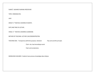
- 1. SUBJECT: ADVANCE NURSING PROCEDURE TOPIC: HEMODIALYSIS. UNIT: GROUP: 1st YEAR M.Sc NURSING STUDENTS. DATE AND TIME OF LECTURE : VENUE: 1st YEAR M.Sc NURSING CLASSROOM. METHOD OF TEACHING: LECTURE CUM DEMONSTRATION. TEACHING AIDS: Transparency-definition,purpose, indication Flip card-scientific principle. Chart- site, how hemodialysis work. Flash card-complication. KNOWLEDGE ASSUMED: Students have previous knowledge about dialysis.
- 2. AIM:AT THE END OF CLASS THE STUDENTS WILL BE ABLE TO ASSISST FOR HEMODYALYSIS.. OBJECTIVE:THE STUDENTS WILL BE ABLE TO DEFINE DIALYSIS DEFINE HEMODIALYSIS.. LIST PURPOSES OF HEMODIALYSIS. STATE INDICATION OF HEMODIALYSIS. LIST THE ACCESS SITE FOR HEMODIALYSIS. EXPLAIN THE SCIENTIFIC PRINCIPLE. ELABORATE ON HOW HEMODIALYSIS WORK DESCRIBE THE PREPREPARATION OF PROCEDURE. DEMONSTRATE THE PROCEDURE. ELABORATE ON POSTPREPARATION OF PROCEDURE. ENLIST COMPLICATIONS
- 3. CONTRIBUTORY OBJECTIVE TIME CONTENT Tg Lg ACTIVITY A V AIDS INTRODUCTION Students define term dialysis 1min Dialysis is the movement of fluids and molecules across a semi permeable membrane from one compartment to another. Peritoneal dialysis and hemodialysis are the two type of dialysis. Teacher ask, “what is the definition of dialysis”. Student responds and teacher appreciates. Transparency with definition. Students define the term hemodialysis. 1min Hemodialysis removes toxic wastes and other impurities from blood of a patient. In hemodialysis blood is removed from patient body through a surgically created access site pumped through a filtration exit to remove toxins and then returned to the body. Teacher asks,” what is the definition of hemodialysis”. Student responds and teacher appreciates. Transparency with definition. Students list the purposes of hemodialysis. 2min To extract toxic nitrogenous substance from the blood and to remove excess water. Teacher ask, what are the purpose of hemodialysis? Student’s responds and teacher appreciates. Transparency with purpose. Students state indication of hemodialysis. 2min ESRD Acutely ill patients who require short term dialysis. Teacher ask s,” what are the indication of hemodialysis?”. Student responds. Transparency with indication. Student enlist the hemodialysis acess site. 5min HEMODIALYSIS ACCESS SITE Hemodialysis requires vascular site. The site and type of access may vary, depending on the expected duration of dialysis, the surgeon’s preference, and the patient’s condition. Teacher elaborate on hemodialysis site. Chart with site
- 4. Subclavian vein catheterization-using the seldinger technique, the physician or surgeon inserts an introducer needle into the subclavian vein. He then insert a guide wire through the introducer needle and removes the needle. Using the guide wire, he then threads a 5” to 12” (12-30cm)plastic or Teflon catheter (with a Y hub) into the patient’s vein. Femoral vein catheterization-using seldinger technique, the physician or surgeon inserts an introducer needle into the left or right femoral vein. He then insert a guide wire through the introducer needle and removes the needle. Using the guide wire he then threads a 5” to 12” plastic or Teflon catheter with a Y hub or two catheters, one for inflow and other placed about ½”(1.3cm)distal to the first for outflow. Arteriovenous or AV Fistula- An AV fistula requires advance planning because a fistula takes a while after surgery to develop—usually from two to six months. A properly formed fistula is less likely than other kinds of vascular accesses to form clots or become infected. Also, properly formed fistulas tend to last years longer than any other kind of vascular access.A surgeon creates an AV fistula by connecting an artery directly to a vein, usually in the wrist or forearm. It is preferred that the fistula be placed in the arm you use the least. Connecting the artery to the vein causes more blood to flow into the vein. You will be able to feel the vibration or pulse of the blood flowing through your access. The vibration you feel is sometimes referred to as the "buzz." This feeling is called a thrill. Your healthcare professional will also listen for the blood flowing in your access with a stethoscope. The "whoosh" sound that is heard through the stethoscope is called a bruit (pronounced brew-ee). You will become familiar with how to check your access daily to make sure it is working properly. The high blood flow from the artery through the vein helps the access grow larger and stronger, making needle insertions for hemodialysis treatments easier. The fistula takes some time to develop. You will need to exercise the arm with the fistula to help it develop. For the surgery, you'll be given a local anesthetic. In most cases,
- 5. the procedure can be performed on an outpatient basis. Although it is the preferred access, small veins or other conditions could make it difficult for some people to have a successful fistula. Your surgeon may order a test (vessel mapping) to check the size and quality of your veins and blood flow to determine the best access for you. These and other problems with vascular access can be corrected when determined in the early stages. Your healthcare team will monitor your access regularly to help you keep your access healthy and working properly. Arteriovenous AV Graft- If you have small or weak veins that won't develop properly into a fistula, you can have a vascular access placed that connects an artery to a vein using a synthetic tube, or graft, implanted under the skin in your arm. The graft is usually a soft, man-made tube that connects to an artery on one end and to a vein on the other end, allowing blood to flow through it. The artificial graft material is used for needle placement for blood access during hemodialysis. A graft does not need as long to develop as a fistula does, so it can be used sooner after placement, often within two to four weeks. The thrill (vibration felt due to the high blood flow) can also be felt in a graft, and the bruit ("whooshing" noise) can be heard with a stethoscope if the graft is functioning properly. You will become familiar with how to check your access daily to make sure it is working properly. Compared with properly formed fistulas, grafts tend to have more problems with clotting and infection and need replacement sooner. However, a well-cared-for graft can last several years.
- 6. Students identify the types of dialyzer 3min TYPES OF DIALYZERS THE HALLOW FIBRE DIALYZER-the most common type, contains fine capillaries, with a semipermeable membrane enclosed in a plastic cylinder. Blood flows through these capillaries as the system pumps dialysate in the opposite direction on the outside of the capillaries. THE FLAT PLATE OR PARALLEL PLATE DIALYZER- has two or more layers of semipermeable membrane,bound by a semirigid structure. Blood ports are located at both ends, between the membranes. Blood flow between the membranes, and dialysate flow in the opposite direction along the outside of the membrane. THE COIL DIALYZER(no longer widely used) consist of one or more semipermeable membrane tubes supported by mesh and wrapped concentrically around a central core. Blood passes through a coils as dialysate circulate at high speed around the coils and meshwork. Teacher explain about types of dialyzer. Transparency with types. Students states the principles on which hemodialysis work. 3min PRINCIPLES Diffusion, osmosis, and ultra filtration are the principles on which hemodialysis is based. The toxins and wastes in the blood are removed by diffusion- that is they move from an area of higher concentration in the blood to an area of lower concentration in the dialysate. The dialysate is a solution made up of all the important electrolytes in their ideal extra cellular concentration. Excess water is removed from the blood by osmosis, in which water moves from an area of higher solute concentration (the blood) to an area of lower solute concentration (the dialysate bath). In ultra filtration, water moves under high pressure to an area of low pressure. This pressure is much more efficient than osmosis at water removal and is accomplished by applying Teacher explains about principles on which hemodialysis work.then ask the student to answer, student responds and teacher appretiates ask the student to sit down. Flip chart with principles.
- 7. negative pressure or a suctioning force to the dialysis membrane. Student describe how hemodialysis work 4min HOW HEMODIALYSIS WORK. In hemodialysis, blood flow from the patient to an external dialyzer(or the artificial kidney) through an arterial access site. Inside the dialyzer, blood and dialysate flow countercurrently, divided by a semipermeable membrane. The composition of the dialysate resembles normal extracellular fluid. The composition of the dialysate resembles normal extracellular fluid. The blood contains an excess of specific solute and the dialyate contains electrolyte that may be at abnormal levels in the patient’s blood stream. The dialysate’s electrolyte composition can be modified to raise or lower electrolyte levels, depending on need. Excretory function and electrolyte homeostasis are achieved by diffusion. Water crosses the membrane from the blood into the dialysate by ultrafiltration. This process removes excess water, waste product and other metabolites through osmotic pressure and hydrostatic pressure. Cleaned of impurities and excess water, the blood returns to the body through a venous site. Teacher explain how dialysis work. Ans then ask student. Student respond and teacher appretiates. chart Students explain pre procedure preparation 2min PRE PROCEDURE PREPARATION Explain the procedure. Weigh the patient. Check vital signs, take blood pressure while siting and standing. Provide comfortable position- supine or sitting in recliner chair with feet elevated. Maintain sterility. Check whether the machine is working. Wash hands. Teacher describes the pre procedure preparation. demonstration
- 8. Students list the articles. ARTICLES RATIONALE. Dialysis machine For dialysis Dialysate To filter blood Two winged fistula needle For cannulation Betadine To clean site Spirit To clean site 10 ml syringe To flush heparin To prevent clotting Needle To aspirate Gauze To clean Tourniquet To apply pressure Clean gloves To prevent infection Adhesive tape To tape puncture site Teacher explain articles and the rationale. models Students demonstrate the procedure. 20min PROCEDURE Put mackintosh and towel under the av fistula site. Wash hands and dry it. Put clean gloves. Then flush the fistula needle using attached syringe containing heparin flush solution,and set them aside. Clean the area with betadine and spirit 3 longotuidinal stroke. Apply a tourniquet above the fistula to distend the vein and facilitate venipuncture. Make sure you avoid occluding the fistula. Perform venipuncture with a fistula needle.remove the needle guard and squeeze the wing tips firmly together. Insert the arterial needle at least 1”(2.5cm)above the anastomosis, being careful not to puncture the fistula. Remove tourniquet and Flush the needle with heparin flush solution to prevent clotting.clamp the arterial needle tubing with a hemostat and secure the wing tip of the needle to the skin with adhesive tape. Teacher demonstrates the procedure. demonstration
- 9. Perform another venipuncture with the venous needle a few inches above the arterial needle. Flush the needle with heparin. Clamp the venous needle tubing and secure the wing tip of the venous needle. Remove the syringe from the end of arterial tubing, uncap the arterial line from the hemodialysis machine and connect the two line.tap the connection securely. Repeat this with venous line. Release the hemostat and start hemodialysis. Blood flow rate 200-500ml/min and dialysate flow rate 300-900 ml/min. It takes 4 hours. After 4 hours wash hands. Turn the blood pump on the hemodialysis machine to 50-100ml/min. Put the gloves and remove the tape from the connection site of the arterial lines. Clamp the needle tubing with the hemostat and disconnect the line. The blood in the machine’s arterial line will continue to flow towards the dialyzer, followed by a column of air. Just before the blood reaches the point where the saline solution enters the line, clamp the blood line with another hemostat. Unclamp the saline solution. After blood is retransfused, clamp the venous needle tubing and the machine’s venous line with hemostats. Turn off the blood pump. Remove the tape from the connection site of the venous lines and disconnect the lines. Remove venipuncture needle, and apply pressure to the site. Apply adhesive tape. Repeat the procedure on the arterial line. Students elaborate on post preparation. 1min POST PROCEDURE Remove the mackintosh. Assess the patient’s weight and vitals sign. Assess for bleeding. DOCUMENTATION-time, day, hours of dialysis, any Teacher explains on post procedure preparation. demonstration
- 10. complication. Students list complication 1min COMPLICATION Infection. Catheter clotting. Excessive removal of fluid during ultrafiltration can cause hypovolemia and hypotension. Cardiac arrhythmias can occur during hemodialysis as a result of electrolyte and ph changes in the blood. Thrombosis. Teacher ask,” what are the complication?”.students respond and teacher appretiates. Flash card with complication. SUMMARY Today we have learnt about the definition, purpose, indication, access site ,how hemodialysis work,scientific principle, preprocedure preparation, procedure, post procedure preparation of hemodialysis. RECAPITALATION List indication of hemodialysis What are the scientific principle. CONCLUSION I hope you have understood todays topic. ASSIGNMENT Read about Duocart biofiltration.
- 11. BIBLIOGRAPHY Langley, C. Nursing Procedure. Philadelphia: Lippincott Williams & Wilkins Smeltzer, S. C. (2009). Brunner & Suddarth’s Textbook Of Medical Surgical Nursing, New Delhi: Wolter Kluwer (India) Pvt. Ltd. Crisp, J., & Taylor, C. (2008). Potter And Perry’s Fundamentals Of Nursing. Australia: Elsevier Publishers.
- 12. LESSON PLAN ON HEMODIALYSIS. SUBMITTED TO: Ms.DEEPA. SUBMITTED BY: Ms.NESSY