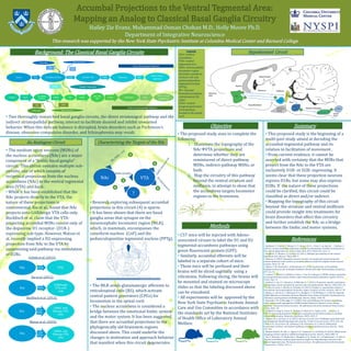
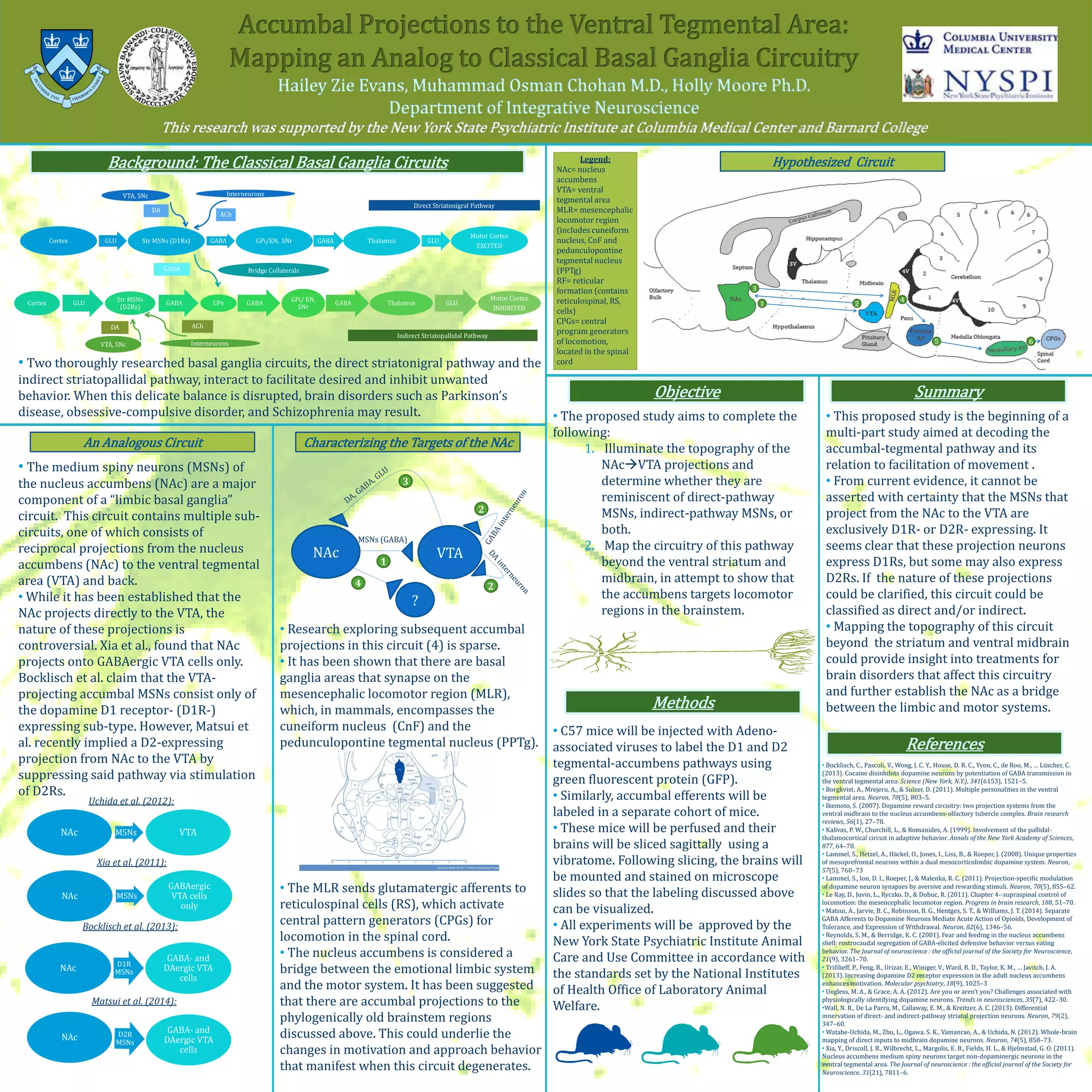
Methods
Background: The Classical Basal Ganglia Circuits
An Analogous Circuit
Objective
• The proposed study aims to complete the
following:
1. Illuminate the topography of the
NAcVTA projections and
determine whether they are
reminiscent of direct-pathway
MSNs, indirect-pathway MSNs, or
both.
2. Map the circuitry of this pathway
beyond the ventral striatum and
midbrain, in attempt to show that
the accumbens targets locomotor
regions in the brainstem.
• C57 mice will be injected with Adeno-
associated viruses to label the D1 and D2
tegmental-accumbens pathways using
green fluorescent protein (GFP).
• Similarly, accumbal efferents will be
labeled in a separate cohort of mice.
• These mice will be perfused and their
brains will be sliced sagittally using a
vibratome. Following slicing, the brains will
be mounted and stained on microscope
slides so that the labeling discussed above
can be visualized.
• All experiments will be approved by the
New York State Psychiatric Institute Animal
Care and Use Committee in accordance with
the standards set by the National Institutes
of Health Office of Laboratory Animal
Welfare.
Cortex GLU
Str MSNs
(D2Rs)
GABA GPe GABA
GPi/ EN,
SNr
GABA Thalamus GLU
Motor Cortex
INHIBITED
Cortex GLU Str MSNs (D1Rs) GABA GPi/EN, SNr GABA Thalamus GLU
Motor Cortex
EXCITED
GABA
DA
VTA, SNc Interneurons
ACh
VTA, SNc
DA
Interneurons
ACh
Bridge Collaterals
Direct Striatonigral Pathway
Indirect Striatopallidal Pathway
• Bocklisch, C., Pascoli, V., Wong, J. C. Y., House, D. R. C., Yvon, C., de Roo, M., … Lüscher, C.
(2013). Cocaine disinhibits dopamine neurons by potentiation of GABA transmission in
the ventral tegmental area. Science (New York, N.Y.), 341(6153), 1521–5.
• Borgkvist, A., Mrejeru, A., & Sulzer, D. (2011). Multiple personalities in the ventral
tegmental area. Neuron, 70(5), 803–5.
• Ikemoto, S. (2007). Dopamine reward circuitry: two projection systems from the
ventral midbrain to the nucleus accumbens-olfactory tubercle complex. Brain research
reviews, 56(1), 27–78.
• Kalivas, P. W., Churchill, L., & Romanides, A. (1999). Involvement of the pallidal-
thalamocortical circuit in adaptive behavior. Annals of the New York Academy of Sciences,
877, 64–70.
• Lammel, S., Hetzel, A., Häckel, O., Jones, I., Liss, B., & Roeper, J. (2008). Unique properties
of mesoprefrontal neurons within a dual mesocorticolimbic dopamine system. Neuron,
57(5), 760–73
• Lammel, S., Ion, D. I., Roeper, J., & Malenka, R. C. (2011). Projection-specific modulation
of dopamine neuron synapses by aversive and rewarding stimuli. Neuron, 70(5), 855–62.
• Le Ray, D., Juvin, L., Ryczko, D., & Dubuc, R. (2011). Chapter 4--supraspinal control of
locomotion: the mesencephalic locomotor region. Progress in brain research, 188, 51–70.
• Matsui, A., Jarvie, B. C., Robinson, B. G., Hentges, S. T., & Williams, J. T. (2014). Separate
GABA Afferents to Dopamine Neurons Mediate Acute Action of Opioids, Development of
Tolerance, and Expression of Withdrawal. Neuron, 82(6), 1346–56.
• Reynolds, S. M., & Berridge, K. C. (2001). Fear and feeding in the nucleus accumbens
shell: rostrocaudal segregation of GABA-elicited defensive behavior versus eating
behavior. The Journal of neuroscience : the official journal of the Society for Neuroscience,
21(9), 3261–70.
• Trifilieff, P., Feng, B., Urizar, E., Winiger, V., Ward, R. D., Taylor, K. M., … Javitch, J. A.
(2013). Increasing dopamine D2 receptor expression in the adult nucleus accumbens
enhances motivation. Molecular psychiatry, 18(9), 1025–3
• Ungless, M. A., & Grace, A. A. (2012). Are you or aren’t you? Challenges associated with
physiologically identifying dopamine neurons. Trends in neurosciences, 35(7), 422–30.
•Wall, N. R., De La Parra, M., Callaway, E. M., & Kreitzer, A. C. (2013). Differential
innervation of direct- and indirect-pathway striatal projection neurons. Neuron, 79(2),
347–60.
• Watabe-Uchida, M., Zhu, L., Ogawa, S. K., Vamanrao, A., & Uchida, N. (2012). Whole-brain
mapping of direct inputs to midbrain dopamine neurons. Neuron, 74(5), 858–73.
• Xia, Y., Driscoll, J. R., Wilbrecht, L., Margolis, E. B., Fields, H. L., & Hjelmstad, G. O. (2011).
Nucleus accumbens medium spiny neurons target non-dopaminergic neurons in the
ventral tegmental area. The Journal of neuroscience : the official journal of the Society for
Neuroscience, 31(21), 7811–6.
• The medium spiny neurons (MSNs) of
the nucleus accumbens (NAc) are a major
component of a “limbic basal ganglia”
circuit. This circuit contains multiple sub-
circuits, one of which consists of
reciprocal projections from the nucleus
accumbens (NAc) to the ventral tegmental
area (VTA) and back.
• While it has been established that the
NAc projects directly to the VTA, the
nature of these projections is
controversial. Xia et al., found that NAc
projects onto GABAergic VTA cells only.
Bocklisch et al. claim that the VTA-
projecting accumbal MSNs consist only of
the dopamine D1 receptor- (D1R-)
expressing sub-type. However, Matsui et
al. recently implied a D2-expressing
projection from NAc to the VTA by
suppressing said pathway via stimulation
of D2Rs.
NAc MSNs VTA
Uchida et al. (2012):
Xia et al. (2011):
NAc MSNs
GABAergic
VTA cells
only
NAc D1R
MSNs
GABA- and
DAergic VTA
cells
Bocklisch et al. (2013):
Matsui et al. (2014):
NAc
GABA- and
DAergic VTA
cells
• This proposed study is the beginning of a
multi-part study aimed at decoding the
accumbal-tegmental pathway and its
relation to facilitation of movement .
• From current evidence, it cannot be
asserted with certainty that the MSNs that
project from the NAc to the VTA are
exclusively D1R- or D2R- expressing. It
seems clear that these projection neurons
express D1Rs, but some may also express
D2Rs. If the nature of these projections
could be clarified, this circuit could be
classified as direct and/or indirect.
• Mapping the topography of this circuit
beyond the striatum and ventral midbrain
could provide insight into treatments for
brain disorders that affect this circuitry
and further establish the NAc as a bridge
between the limbic and motor systems.
• Two thoroughly researched basal ganglia circuits, the direct striatonigral pathway and the
indirect striatopallidal pathway, interact to facilitate desired and inhibit unwanted
behavior. When this delicate balance is disrupted, brain disorders such as Parkinson’s
disease, obsessive-compulsive disorder, and Schizophrenia may result.
Characterizing the Targets of the NAc
• Research exploring subsequent accumbal
projections in this circuit (4) is sparse.
• It has been shown that there are basal
ganglia areas that synapse on the
mesencephalic locomotor region (MLR),
which, in mammals, encompasses the
cuneiform nucleus (CnF) and the
pedunculopontine tegmental nucleus (PPTg).
• The MLR sends glutamatergic afferents to
reticulospinal cells (RS), which activate
central pattern generators (CPGs) for
locomotion in the spinal cord.
• The nucleus accumbens is considered a
bridge between the emotional limbic system
and the motor system. It has been suggested
that there are accumbal projections to the
phylogenically old brainstem regions
discussed above. This could underlie the
changes in motivation and approach behavior
that manifest when this circuit degenerates.
NAc VTA
MSNs (GABA)
?
1
2
2
3
4
Hypothesized Circuit
References
Summary
1 2
3
4
5 6
Legend:
NAc= nucleus
accumbens
VTA= ventral
tegmental area
MLR= mesencephalic
locomotor region
(includes cuneiform
nucleus, CnF and
pedunculopontine
tegmental nucleus
(PPTg)
RF= reticular
formation (contains
reticulospinal, RS,
cells)
CPGs= central
program generators
of locomotion,
located in the spinal
cord

Hailey_Evans NAc VTA Poster 2014

  • 1.
    Methods Background: The ClassicalBasal Ganglia Circuits An Analogous Circuit Objective • The proposed study aims to complete the following: 1. Illuminate the topography of the NAcVTA projections and determine whether they are reminiscent of direct-pathway MSNs, indirect-pathway MSNs, or both. 2. Map the circuitry of this pathway beyond the ventral striatum and midbrain, in attempt to show that the accumbens targets locomotor regions in the brainstem. • C57 mice will be injected with Adeno- associated viruses to label the D1 and D2 tegmental-accumbens pathways using green fluorescent protein (GFP). • Similarly, accumbal efferents will be labeled in a separate cohort of mice. • These mice will be perfused and their brains will be sliced sagittally using a vibratome. Following slicing, the brains will be mounted and stained on microscope slides so that the labeling discussed above can be visualized. • All experiments will be approved by the New York State Psychiatric Institute Animal Care and Use Committee in accordance with the standards set by the National Institutes of Health Office of Laboratory Animal Welfare. Cortex GLU Str MSNs (D2Rs) GABA GPe GABA GPi/ EN, SNr GABA Thalamus GLU Motor Cortex INHIBITED Cortex GLU Str MSNs (D1Rs) GABA GPi/EN, SNr GABA Thalamus GLU Motor Cortex EXCITED GABA DA VTA, SNc Interneurons ACh VTA, SNc DA Interneurons ACh Bridge Collaterals Direct Striatonigral Pathway Indirect Striatopallidal Pathway • Bocklisch, C., Pascoli, V., Wong, J. C. Y., House, D. R. C., Yvon, C., de Roo, M., … Lüscher, C. (2013). Cocaine disinhibits dopamine neurons by potentiation of GABA transmission in the ventral tegmental area. Science (New York, N.Y.), 341(6153), 1521–5. • Borgkvist, A., Mrejeru, A., & Sulzer, D. (2011). Multiple personalities in the ventral tegmental area. Neuron, 70(5), 803–5. • Ikemoto, S. (2007). Dopamine reward circuitry: two projection systems from the ventral midbrain to the nucleus accumbens-olfactory tubercle complex. Brain research reviews, 56(1), 27–78. • Kalivas, P. W., Churchill, L., & Romanides, A. (1999). Involvement of the pallidal- thalamocortical circuit in adaptive behavior. Annals of the New York Academy of Sciences, 877, 64–70. • Lammel, S., Hetzel, A., Häckel, O., Jones, I., Liss, B., & Roeper, J. (2008). Unique properties of mesoprefrontal neurons within a dual mesocorticolimbic dopamine system. Neuron, 57(5), 760–73 • Lammel, S., Ion, D. I., Roeper, J., & Malenka, R. C. (2011). Projection-specific modulation of dopamine neuron synapses by aversive and rewarding stimuli. Neuron, 70(5), 855–62. • Le Ray, D., Juvin, L., Ryczko, D., & Dubuc, R. (2011). Chapter 4--supraspinal control of locomotion: the mesencephalic locomotor region. Progress in brain research, 188, 51–70. • Matsui, A., Jarvie, B. C., Robinson, B. G., Hentges, S. T., & Williams, J. T. (2014). Separate GABA Afferents to Dopamine Neurons Mediate Acute Action of Opioids, Development of Tolerance, and Expression of Withdrawal. Neuron, 82(6), 1346–56. • Reynolds, S. M., & Berridge, K. C. (2001). Fear and feeding in the nucleus accumbens shell: rostrocaudal segregation of GABA-elicited defensive behavior versus eating behavior. The Journal of neuroscience : the official journal of the Society for Neuroscience, 21(9), 3261–70. • Trifilieff, P., Feng, B., Urizar, E., Winiger, V., Ward, R. D., Taylor, K. M., … Javitch, J. A. (2013). Increasing dopamine D2 receptor expression in the adult nucleus accumbens enhances motivation. Molecular psychiatry, 18(9), 1025–3 • Ungless, M. A., & Grace, A. A. (2012). Are you or aren’t you? Challenges associated with physiologically identifying dopamine neurons. Trends in neurosciences, 35(7), 422–30. •Wall, N. R., De La Parra, M., Callaway, E. M., & Kreitzer, A. C. (2013). Differential innervation of direct- and indirect-pathway striatal projection neurons. Neuron, 79(2), 347–60. • Watabe-Uchida, M., Zhu, L., Ogawa, S. K., Vamanrao, A., & Uchida, N. (2012). Whole-brain mapping of direct inputs to midbrain dopamine neurons. Neuron, 74(5), 858–73. • Xia, Y., Driscoll, J. R., Wilbrecht, L., Margolis, E. B., Fields, H. L., & Hjelmstad, G. O. (2011). Nucleus accumbens medium spiny neurons target non-dopaminergic neurons in the ventral tegmental area. The Journal of neuroscience : the official journal of the Society for Neuroscience, 31(21), 7811–6. • The medium spiny neurons (MSNs) of the nucleus accumbens (NAc) are a major component of a “limbic basal ganglia” circuit. This circuit contains multiple sub- circuits, one of which consists of reciprocal projections from the nucleus accumbens (NAc) to the ventral tegmental area (VTA) and back. • While it has been established that the NAc projects directly to the VTA, the nature of these projections is controversial. Xia et al., found that NAc projects onto GABAergic VTA cells only. Bocklisch et al. claim that the VTA- projecting accumbal MSNs consist only of the dopamine D1 receptor- (D1R-) expressing sub-type. However, Matsui et al. recently implied a D2-expressing projection from NAc to the VTA by suppressing said pathway via stimulation of D2Rs. NAc MSNs VTA Uchida et al. (2012): Xia et al. (2011): NAc MSNs GABAergic VTA cells only NAc D1R MSNs GABA- and DAergic VTA cells Bocklisch et al. (2013): Matsui et al. (2014): NAc GABA- and DAergic VTA cells • This proposed study is the beginning of a multi-part study aimed at decoding the accumbal-tegmental pathway and its relation to facilitation of movement . • From current evidence, it cannot be asserted with certainty that the MSNs that project from the NAc to the VTA are exclusively D1R- or D2R- expressing. It seems clear that these projection neurons express D1Rs, but some may also express D2Rs. If the nature of these projections could be clarified, this circuit could be classified as direct and/or indirect. • Mapping the topography of this circuit beyond the striatum and ventral midbrain could provide insight into treatments for brain disorders that affect this circuitry and further establish the NAc as a bridge between the limbic and motor systems. • Two thoroughly researched basal ganglia circuits, the direct striatonigral pathway and the indirect striatopallidal pathway, interact to facilitate desired and inhibit unwanted behavior. When this delicate balance is disrupted, brain disorders such as Parkinson’s disease, obsessive-compulsive disorder, and Schizophrenia may result. Characterizing the Targets of the NAc • Research exploring subsequent accumbal projections in this circuit (4) is sparse. • It has been shown that there are basal ganglia areas that synapse on the mesencephalic locomotor region (MLR), which, in mammals, encompasses the cuneiform nucleus (CnF) and the pedunculopontine tegmental nucleus (PPTg). • The MLR sends glutamatergic afferents to reticulospinal cells (RS), which activate central pattern generators (CPGs) for locomotion in the spinal cord. • The nucleus accumbens is considered a bridge between the emotional limbic system and the motor system. It has been suggested that there are accumbal projections to the phylogenically old brainstem regions discussed above. This could underlie the changes in motivation and approach behavior that manifest when this circuit degenerates. NAc VTA MSNs (GABA) ? 1 2 2 3 4 Hypothesized Circuit References Summary 1 2 3 4 5 6 Legend: NAc= nucleus accumbens VTA= ventral tegmental area MLR= mesencephalic locomotor region (includes cuneiform nucleus, CnF and pedunculopontine tegmental nucleus (PPTg) RF= reticular formation (contains reticulospinal, RS, cells) CPGs= central program generators of locomotion, located in the spinal cord