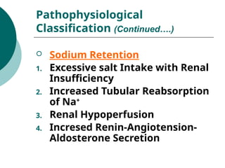
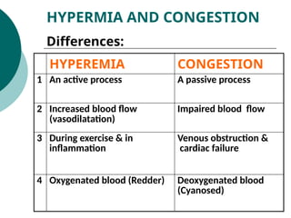
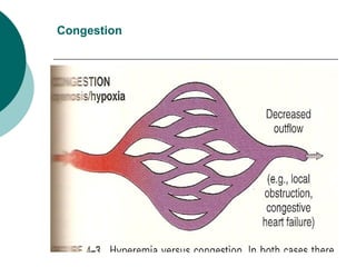
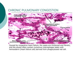
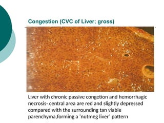
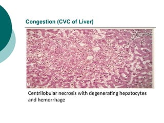
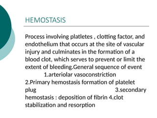
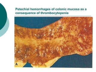
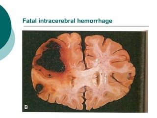
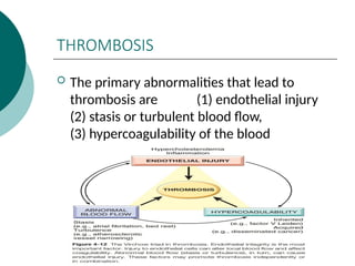
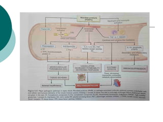
HAEMODYNAMICS
AND SHOCK
Presented by-Dr.Mana ram
coordinator-Dr.samay singh meena
INTRODUCTION
Health of Cells & Organs:
 Uninterrupted Circulation (O2 &
Nutrients, Wastes)
 Normal Fluid & Electrolyte
Balance
Normal Fluid Homeostasis
 Vessel Wall Integrity
 Intravascular Pressure
 Intravascular Osmolarity
(CONTIN…….)
INTRODUCTION (contin……)
Normal Fluid Homeostasis
 Maintenance of blood as
fluid
 Formation of clot in case of
injury
INTRODUCTION (contin……)
Abnormalities of
Haemodynamic System
 Primary (e.g. Pathology in
discrete blood vessels like
Hemorrhage, Thrombosis or
Infarction)
 Secondary (e.g. Pulmonary
Edema, Shock etc.)
INTRODUCTION (contin……)
EDEMA
Definition:
“Increased Fluid in the Interstitial
Tissue Spaces”
Also Includes:
Hydrothorax, Hydropericardium
Hydroperitonium or Ascites and
Anasarca.
Pathophysiological
Classification
 Inflammatory Edema
 Non-Inflammatory Edema
1. Increased Hydrostatic
Pressure
2. Reduced Plasma Osmotic
Pressure
3. Lymphatic Obstruction
4. Sodium Retention
 Increased Hydrostatic Pressure:
1. Impaired Venous Return: (e.g. CCF,
Constrictive Pericarditis, Liver
Cirrhosis, Venous Obstruction)
2. Arteriolar dilatation: (e.g. Exposure
to Heat, Neurohormonal
dysregulation)
Pathophysiological
Classification (Continued….)
 Reduced Plasma Osmotic Pressure
1. Protein-Loosing Glomerulopathies
(Nephrotic Syndrome)
2. Liver Cirrhosis (Ascites)
3. Malnutrition
4. Protein-Loosing
gastroenteropathies
Pathophysiological
Classification (Continued….)
 Lymphatic Obstruction
1. Inflammatory
2. Neoplastic
3. Postsurgical
4. Postirradiation
Pathophysiological
Classification (Continued….)
Factors Affecting Fluid Balance Across
Capillary Wall
 Sodium Retention
1. Excessive salt Intake with Renal
Insufficiency
2. Increased Tubular Reabsorption
of Na+
3. Renal Hypoperfusion
4. Incresed Renin-Angiotension-
Aldosterone Secretion
Pathophysiological
Classification (Continued….)
HYPERMIA AND
CONGESTION
Definitions:
“Both indicates a local increased
volume of blood in a particular
tissue. ”
HYPERMIA AND CONGESTION
Differences:
HYPEREMIA CONGESTION
1 An active process A passive process
2 Increased blood flow
(vasodilatation)
Impaired blood flow
3 During exercise & in
inflammation
Venous obstruction &
cardiac failure
4 Oxygenated blood (Redder) Deoxygenated blood
(Cyanosed)
Normal Vasculature Regarding Blood Volume
Hyperemia
Congestion
CHRONIC PULMONARY CONGESTION
Caused by congestive heart failure, the septa are thickened and fibrotic,
and the alveoli often contain numerous macrophages laden with
hemosiderin (heart failure cell) derived from phagocytosed red cell.
Congestion (CVC of Liver; gross)
Liver with chronic passive congetion and hemorrhagic
necrosis- central area are red and slightly depressed
compared with the surrounding tan viable
parenchyma,forming a ‘nutmeg liver’ pattern
Congestion (CVC of Liver)
Centrilobular necrosis with degenerating hepatocytes
and hemorrhage
HEMOSTASIS
Process involving platletes , clotting factor, and
endothelium that occurs at the site of vascular
injury and culminates in the formation of a
blood clot, which serves to prevent or limit the
extent of bleeding.General sequence of event
1.arteriolar vasoconstriction
2.Primary hemostasis formation of platelet
plug 3.secondary
hemostasis : deposition of fibrin 4.clot
stabilization and resorption
Hemorrhage
Definition:
“Extravasation of blood due to vessel
rupture”
Types: (depending on the site, extent
and location)
External
Internal
Hematoma: ‘Blood within the tissue’
(small; like a Bruise, or sufficiently
large as to be fatal)
Petechial hemorrhages of colonic mucosa as a
consequence of thrombocytopenia
Fatal intracerebral hemorrhage
THROMBOSIS
 The primary abnormalities that lead to
thrombosis are (1) endothelial injury
(2) stasis or turbulent blood flow,
(3) hypercoagulability of the blood
SHOCK
Defination
Shock is a state of circulatory failure that
impaire tissue perfusion and lead to cellular
hypoxia
Three categories
1.Cardiogenic shock
2.Hypovolemic shock
3.Septic shock
STAGES OF SHOCK
 1.An initial nonprogressive stage
During which reflex
compensatory mechanism are activated and
vital organ perfusion is maintained .
2.Progressive stage
characterized by tissue hypoperfusion and
onset of worsening circulatory and metabolic
derangement, including acidosis
3.Irreversible stage in which cellular and
tissue injury is so sever that even if the
hemodynamic defects are corrected, survival
Cardiogenic shock
 Result from low cardiac output due to
myocardial pump failure.This can be due to
myocardial damage,ventricular
arrythmias,cardiac tamponade,pulmonary
embolism
Hypovolumic shock
 Results from low cardiac output due to low
blood volume ,such as can occur with
massive hemorrage or fluid loss from severe
burn
Septic shock
 Septic is defined as a subset of sepsis in
which particularly profound circulatory ,
cellular, and metabolic abnormalities are
associated with a greater risk of mortality
than with sepsis alone.
Thank You !

Haemodynamics and shock powerpoint presentation

  • 1.
    HAEMODYNAMICS AND SHOCK Presented by-Dr.Manaram coordinator-Dr.samay singh meena
  • 2.
    INTRODUCTION Health of Cells& Organs:  Uninterrupted Circulation (O2 & Nutrients, Wastes)  Normal Fluid & Electrolyte Balance
  • 3.
    Normal Fluid Homeostasis Vessel Wall Integrity  Intravascular Pressure  Intravascular Osmolarity (CONTIN…….) INTRODUCTION (contin……)
  • 4.
    Normal Fluid Homeostasis Maintenance of blood as fluid  Formation of clot in case of injury INTRODUCTION (contin……)
  • 5.
    Abnormalities of Haemodynamic System Primary (e.g. Pathology in discrete blood vessels like Hemorrhage, Thrombosis or Infarction)  Secondary (e.g. Pulmonary Edema, Shock etc.) INTRODUCTION (contin……)
  • 6.
    EDEMA Definition: “Increased Fluid inthe Interstitial Tissue Spaces” Also Includes: Hydrothorax, Hydropericardium Hydroperitonium or Ascites and Anasarca.
  • 7.
    Pathophysiological Classification  Inflammatory Edema Non-Inflammatory Edema 1. Increased Hydrostatic Pressure 2. Reduced Plasma Osmotic Pressure 3. Lymphatic Obstruction 4. Sodium Retention
  • 8.
     Increased HydrostaticPressure: 1. Impaired Venous Return: (e.g. CCF, Constrictive Pericarditis, Liver Cirrhosis, Venous Obstruction) 2. Arteriolar dilatation: (e.g. Exposure to Heat, Neurohormonal dysregulation) Pathophysiological Classification (Continued….)
  • 9.
     Reduced PlasmaOsmotic Pressure 1. Protein-Loosing Glomerulopathies (Nephrotic Syndrome) 2. Liver Cirrhosis (Ascites) 3. Malnutrition 4. Protein-Loosing gastroenteropathies Pathophysiological Classification (Continued….)
  • 10.
     Lymphatic Obstruction 1.Inflammatory 2. Neoplastic 3. Postsurgical 4. Postirradiation Pathophysiological Classification (Continued….)
  • 11.
    Factors Affecting FluidBalance Across Capillary Wall
  • 12.
     Sodium Retention 1.Excessive salt Intake with Renal Insufficiency 2. Increased Tubular Reabsorption of Na+ 3. Renal Hypoperfusion 4. Incresed Renin-Angiotension- Aldosterone Secretion Pathophysiological Classification (Continued….)
  • 13.
    HYPERMIA AND CONGESTION Definitions: “Both indicatesa local increased volume of blood in a particular tissue. ”
  • 14.
    HYPERMIA AND CONGESTION Differences: HYPEREMIACONGESTION 1 An active process A passive process 2 Increased blood flow (vasodilatation) Impaired blood flow 3 During exercise & in inflammation Venous obstruction & cardiac failure 4 Oxygenated blood (Redder) Deoxygenated blood (Cyanosed)
  • 15.
  • 16.
  • 17.
  • 18.
    CHRONIC PULMONARY CONGESTION Causedby congestive heart failure, the septa are thickened and fibrotic, and the alveoli often contain numerous macrophages laden with hemosiderin (heart failure cell) derived from phagocytosed red cell.
  • 19.
    Congestion (CVC ofLiver; gross) Liver with chronic passive congetion and hemorrhagic necrosis- central area are red and slightly depressed compared with the surrounding tan viable parenchyma,forming a ‘nutmeg liver’ pattern
  • 20.
    Congestion (CVC ofLiver) Centrilobular necrosis with degenerating hepatocytes and hemorrhage
  • 21.
    HEMOSTASIS Process involving platletes, clotting factor, and endothelium that occurs at the site of vascular injury and culminates in the formation of a blood clot, which serves to prevent or limit the extent of bleeding.General sequence of event 1.arteriolar vasoconstriction 2.Primary hemostasis formation of platelet plug 3.secondary hemostasis : deposition of fibrin 4.clot stabilization and resorption
  • 23.
    Hemorrhage Definition: “Extravasation of blooddue to vessel rupture” Types: (depending on the site, extent and location) External Internal Hematoma: ‘Blood within the tissue’ (small; like a Bruise, or sufficiently large as to be fatal)
  • 24.
    Petechial hemorrhages ofcolonic mucosa as a consequence of thrombocytopenia
  • 25.
  • 26.
    THROMBOSIS  The primaryabnormalities that lead to thrombosis are (1) endothelial injury (2) stasis or turbulent blood flow, (3) hypercoagulability of the blood
  • 27.
    SHOCK Defination Shock is astate of circulatory failure that impaire tissue perfusion and lead to cellular hypoxia Three categories 1.Cardiogenic shock 2.Hypovolemic shock 3.Septic shock
  • 28.
    STAGES OF SHOCK 1.An initial nonprogressive stage During which reflex compensatory mechanism are activated and vital organ perfusion is maintained . 2.Progressive stage characterized by tissue hypoperfusion and onset of worsening circulatory and metabolic derangement, including acidosis 3.Irreversible stage in which cellular and tissue injury is so sever that even if the hemodynamic defects are corrected, survival
  • 29.
    Cardiogenic shock  Resultfrom low cardiac output due to myocardial pump failure.This can be due to myocardial damage,ventricular arrythmias,cardiac tamponade,pulmonary embolism
  • 30.
    Hypovolumic shock  Resultsfrom low cardiac output due to low blood volume ,such as can occur with massive hemorrage or fluid loss from severe burn
  • 31.
    Septic shock  Septicis defined as a subset of sepsis in which particularly profound circulatory , cellular, and metabolic abnormalities are associated with a greater risk of mortality than with sepsis alone.
  • 33.