![Physicochemical And Biological Studies...
www.ijpsi.org 56 | Page
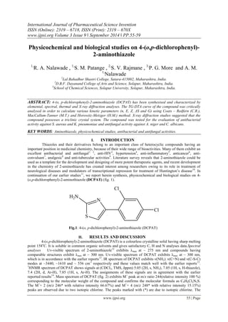
molecular ion undergoes rupture of thiazole ring15 to give fragments at m/z 202/204*/206* (relative intensity
58/39.39/6.06 respectively). These fragment further loose two chlorine atoms one by one to give fragments
having m/z 167 (23.8%) and 132 (11.3%). Fragmentation pattern is depicted in Scheme I. The fragment m/z 132
(11.3%) then undergoes decomposition to give smaller fragments, m/z (relative intensity %):123 (18.18), 104
(17.00), 87 (11.36), 73 (12.27), 45 (12.27).
Fig. 2: Mass spectrum of DCPAT
S
Cl
Cl
S
N
H2N
Cl
Cl
+.
-2Cl S
+.
m/z 244 m/z 202 m/z 132
Scheme I: Fragmentation pattern of DCPAT
Thermal analysis : Thermogravimetric analysis is one of the important techniques for the study of thermal
properties of substances. From the TG-DTA curve, various kinetic parameters can be estimated using different
methods which are broadly classified as differential methods and integral methods The TG-DTA curve of
DCPAT was critically analysed in view of evaluating various kinetic parameters such as n (order of reaction), E
(energy of activation), Z (pre-exponential factor), ΔS (entropy change) and G (free energy change) using
integral methods viz. Coats – Redfern (C.R.), MacCallum-Tanner (M.T.) and Horowitz-Metzger (H.M.) method
as follows:
Coats-Redfern method16
1
2
1 1 1
log log
(1 ) 2.303
n
ZR E
n T Eq R T
.....1
MacCallum- Tanner Method17
α
log
1-(1- )1-n
(1- n)
= log
ZE
Rq
0.485E 0.435 ......2
0.449 + 0.217E
T
. 103
Horowitz – Metzger Method18
α
log
1-(1- )1-n
(1- n)
=
2
θ
2
log
ZRTS
Eq
E
2.303 RTs
+
E
2.303 RTs
......3
In all three equations: α is the fraction of weight loss at the particular temperature, Ts is the temperature
at half weight loss, q is the rate of heating, θ is difference of particular temperature and temperature at half
weight loss (T-Ts). From the calculated values of E and Z, the values of ΔS and G were determined by using the
equations as follows:
ΔS = 2.303 x Log [(Z x h) / (Ts x k)]
G = E-(ΔS x Ts)](data:image/gif;base64,R0lGODlhAQABAIAAAAAAAP///yH5BAEAAAAALAAAAAABAAEAAAIBRAA7)
Recommended
Recommended
More Related Content
What's hot
What's hot (18)
Viewers also liked
Viewers also liked (14)
Similar to H039055059
Similar to H039055059 (20)
Recently uploaded
Recently uploaded (20)
H039055059
- 1. International Journal of Pharmaceutical Science Invention ISSN (Online): 2319 – 6718, ISSN (Print): 2319 – 670X www.ijpsi.org Volume 3 Issue 9 ‖ September 2014 ‖ PP.55-59 www.ijpsi.org 55 | Page Physicochemical and biological studies on 4-(o,p-dichlorophenyl)- 2-aminothiazole 1,R. A. Nalawade , 1,S. M. Patange , 2,S. V. Rajmane , 3,P. G. More and A. M. 1,Nalawade 1Lal Bahadhur Shastri College, Satara-415002, Maharashtra, India. 2D.B.F. Dayanand College of Arts and Science, Solapur, Maharashtra, India. 3School of Chemical Sciences, Solapur University, Solapur, Maharashtra, India. ABSTRACT: 4-(o, p-dichlorophenyl)-2-aminothiazole (DCPAT) has been synthesised and characterized by elemental, spectral, thermal and X-ray diffraction analyses. The TG-DTA curve of the compound was critically analysed in order to calculate various kinetic parameters (n, E, Z, ΔS and G) using Coats – Redfern (C.R.), MacCallum-Tanner (M.T.) and Horowitz-Metzger (H.M.) method. X-ray diffraction studies suggested that the compound possesses a triclinic crystal system. The compound was tested for the evaluation of antibacterial activity against S. aureus and K. pneumoniae and antifungal activity against A. niger and C. albicans. KEY WORDS: Aminothiazole, physicochemical studies, antibacterial and antifungal activities. I. INTRODUCTION Thiazoles and their derivatives belong to an important class of heterocyclic compounds having an important position in medicinal chemistry, because of their wide range of bioactivities. Many of them exhibit an excellent antibacterial and antifungal1, 2, anti-HIV3, hypertension4, anti-inflammatory5, anticancer6, anti-convulsant 7, analgesic8 and anti-tubercular activities9. Literature survey reveals that 2-aminothiazole could be used as a template for the development and designing of more potent therapeutic agents, and recent development in the chemistry of 2-aminothiazole has created interest among researchers owing to its role in treatment of neurological diseases and modulators of transcriptional repression for treatment of Huntington’s disease10. In continuation of our earlier studies11, we report herein synthesis, physicochemical and biological studies on 4- (o,p-dichlorophenyl)-2-aminothiazole (DCPAT) (fig. 1). S N H2N Cl Cl Fig.1: 4-(o, p-dichlorophenyl)-2-aminothiazole (DCPAT) II. RESULTS AND DISCUSSION 4-(o,p-dichlorophenyl)-2-aminothiazole (DCPAT) is a colourless crystalline solid having sharp melting point 158oC. It is soluble in common organic solvents and gives satisfactory C, H and N analyses data.Spectral analyses Uv-visible spectrum of 2-aminothiazole12 exhibits λmax at ~ 275 nm and compounds having comparable structures exhibit λmax at ~ 300 nm. Uv-visible spectrum of DCPAT exhibits λmax at ~ 300 nm, which is in accordance with the earlier reports12. IR spectrum of DCPAT exhibits ν(NH2), ν(C=N) and ν(C-S-C) modes at ~3440, ~1610 and ~ 556 cm-1 respectively and these values match well with the earlier reports13. 1HNMR spectrum of DCPAT shows signals at (CDCl3, TMS, δppm) 5.05 (2H, s, NH2), 7.05 (1H, s, H-thiazole), 7.4 (2H, d, Ar-H), 7.85 (1H, s, Ar-H). The assignments of these signals are in agreement with the earlier reported results14. Mass spectrum of DCPAT (fig. 2) exhibits M+ peak at m/z ratio 244(relative intensity 100 %) corresponding to the molecular weight of the compound and confirms the molecular formula as C9H6Cl2N2S. The M++ 2 (m/z 246* with relative intensity 66.67%) and M++ 4 (m/z 248* with relative intensity 15.15%) peaks are observed due to two isotopic chlorine. The peaks marked with (*) are due to isotopic chlorine. The
- 2. Physicochemical And Biological Studies... www.ijpsi.org 56 | Page molecular ion undergoes rupture of thiazole ring15 to give fragments at m/z 202/204*/206* (relative intensity 58/39.39/6.06 respectively). These fragment further loose two chlorine atoms one by one to give fragments having m/z 167 (23.8%) and 132 (11.3%). Fragmentation pattern is depicted in Scheme I. The fragment m/z 132 (11.3%) then undergoes decomposition to give smaller fragments, m/z (relative intensity %):123 (18.18), 104 (17.00), 87 (11.36), 73 (12.27), 45 (12.27). Fig. 2: Mass spectrum of DCPAT S Cl Cl S N H2N Cl Cl +. -2Cl S +. m/z 244 m/z 202 m/z 132 Scheme I: Fragmentation pattern of DCPAT Thermal analysis : Thermogravimetric analysis is one of the important techniques for the study of thermal properties of substances. From the TG-DTA curve, various kinetic parameters can be estimated using different methods which are broadly classified as differential methods and integral methods The TG-DTA curve of DCPAT was critically analysed in view of evaluating various kinetic parameters such as n (order of reaction), E (energy of activation), Z (pre-exponential factor), ΔS (entropy change) and G (free energy change) using integral methods viz. Coats – Redfern (C.R.), MacCallum-Tanner (M.T.) and Horowitz-Metzger (H.M.) method as follows: Coats-Redfern method16 1 2 1 1 1 log log (1 ) 2.303 n ZR E n T Eq R T .....1 MacCallum- Tanner Method17 α log 1-(1- )1-n (1- n) = log ZE Rq 0.485E 0.435 ......2 0.449 + 0.217E T . 103 Horowitz – Metzger Method18 α log 1-(1- )1-n (1- n) = 2 θ 2 log ZRTS Eq E 2.303 RTs + E 2.303 RTs ......3 In all three equations: α is the fraction of weight loss at the particular temperature, Ts is the temperature at half weight loss, q is the rate of heating, θ is difference of particular temperature and temperature at half weight loss (T-Ts). From the calculated values of E and Z, the values of ΔS and G were determined by using the equations as follows: ΔS = 2.303 x Log [(Z x h) / (Ts x k)] G = E-(ΔS x Ts)
- 3. Physicochemical And Biological Studies... www.ijpsi.org 57 | Page DCPAT undergoes decomposition in two stages, Stage-I: 1580C to 2620C (83.59% weight loss) and Stage-II: 262oC to 600oC (8.25% weight loss). Two DTA peaks (endothermic) are located at 158.740C and 262.580C. The residue (5.414%) remaining at the end may be due to formation of thermally stable compound at high temperature. Major weight loss occurs in Stage I only and hence the kinetic parameters (n, E, Z, ΔS and G) have been calculated for this stage. The values of kinetic parameters calculated by Coats – Redfern (C.R.), MacCallum-Tanner (M.T.) and Horowitz-Metzger (H.M.) method are given in Table1. The values of E (occurring in the range 21-27 Kcal mol-1) and G (22 – 28 Kcal mol-1) are sufficiently high and suggest that DCPAT is a thermally stable compound. The TG-DTA curve is depicted in figure 3. Fig. 3: TG-DTA curve of DCPAT Table 1: Kinetic parameters estimated by Coats – Redfern (C.R.), MacCallum-Tanner (M.T.) and Horowitz- Metzger (H.M.) method. Kinetic parameters Stage - I C.R. M.T. H.M. n 0.47 0.45 0.75 E 21.507 21.329 26.826 Z 3.83x10⁶ 6.20x10¹² 9.36x10¹¹ ΔS -15.15 -0.8514 -2.44 G 24.032 22.4715 27.128 Units: E (kcal mol-1), Z (S-1), ΔS (JK-1mol-1), G (kcal mol-1) X-ray diffraction study DCPAT has been characterized by powder x-ray diffraction studies to predict the crystal system. The diffractogram is depicted in fig. 4 which shows 47reflection (2θ) between 10.670 to 93.030 with maximum at 2θ = 34.830 and d = 2.57Ao. The cell parameters calculated are mentioned in parenthesis (a = 8.1851Ao, b = 8.7875 Ao, c = 7.0573 Ao and α = 90.3050o, β = 97.7880o, γ = 109.4140o) and these values are found to be in agreement with those required for a triclinic crystal system19 where a ≠ b ≠ c and α ≠ β ≠ γ. Therefore it may be concluded that the crystal system of the DCPAT is triclinic. The volume of unit cell is 473.90 Ao 3.
- 4. Physicochemical And Biological Studies... www.ijpsi.org 58 | Page Fig. 4: X-ray diffractogram of DCPAT Biological activity : We have tested DCPAT for the evaluation of antibacterial activity against S. aureus and K. pneumoniae and antifungal activity against A. niger and C. albicans in DMF as solvent using serial dilution technique20. Eight test tubes containing 5 ml of sterile nutrient / sabouraud broth were inoculated with 0.02ml of 24 h old culture of bacteria S. aureus and K. pneumoniae and fungi A. niger and C. albicans respectively. Different amounts of DCPAT were added with the help of sterile pipette from the stock solution 200 μg/ml to 5 ml quantities of respective media so as to reach the concentration from 1μg/ml to 20μg/ml. All test tubes were inoculated at 370C and at room temperature for bacteria and fungi respectively. Test tubes inoculated with organisms were observed for presence of turbidity after 24h and 48h respectively. The lowest concentration of DCPAT inhibiting the growth of test organism was determined as MIC value. The minimum inhibition concentration (MIC) values for DCPAT lie in the range 8-12 μg/ml for antibacterial activity and 4-8 μg/ml antifungal activity (Table 2 and 3). DCPAT was found to exhibit pronounced antifungal activity compared to antibacterial activity Table 2: Antibacterial activity of DCPAT Table 3: Antifungal activity of DCPAT Conc. of thiazole in μg/ml Grouth (+) /inhibition (-) of ( S. aureus ) Grouth (+) /inhibition (-) of (K. pneumoniae ) 4 + + 6 + + 8 - + 10 - + 12 - - 14 - - Conc. of thiazole in μg/ml Grouth (+) /inhibition (-) of ( A. Niger) Grouth (+) /inhibition (-) of (C. albicans) 2 + + 4 - + 6 - + 8 - - 10 - - 12 - -
- 5. Physicochemical And Biological Studies... www.ijpsi.org 59 | Page III. CONCLUSION DCPAT is a thermally stable compound having sharp melting point and possesses a triclinic crys tal system. It is biologically active and exhibits pronounced antifungal activity compared to antibacterial activity. Experimental : All the chemicals used were of A. R. Grade. The solvents were dried according to standard procedures and distilled before use. Elemental analyses (C, H and N) were performed using micro analytical technique. Physical measurements were performed as reported in our earlier communication11. Synthesis of 4-(o, p-dichlorophenyl)-2-aminothiazole (DCPAT) 21 A mixture of 2,4-dichloroacetophenone (0.05mol), thiourea (0.1mol) and iodine (0.1mol) was refluxed on water bath for eight hours and again 12 to 16 hours after removal of the condenser. The crude reaction product was kept in contact with diethyl ether with occasional shaking for 48 hours. The ether layer was then removed and reaction product was treated with sodium thiosulphate solution to remove traces of iodine. The product was boiled with water and filtered in hot condition. The filtrate was treated with concentrated ammonia to obtain DCPAT, which was recrystallized from 50% ethanol and dried under reduced pressure and its purity was tested by TLC. Scheme II represents synthesis of DCPAT. O Cl Cl + NH2 S 2H2N + I2 S N -I+H3N Cl Cl NH2 +I-SH + H2N Scheme II: Synthesis of DCPAT IV. ACKNOWLEDGEMENT Authors are thankful to the authorities of Lal Bahadur Shastri College, Satara for providing research facilities. REFERENCES [1] N. Ulusoy, M. Kiraz and O. Kucukbasmaci, Monatsh. Chem. 2002, 133, 1305. [2] Z. A. Kaplancikli, G. T. Zitouni, G. Revial and K. Guven, Arch. Pharm. Res.,2004, 27, 1081. [3] P. Bhattacharya, J. T. Leonard and K. Roy, Bioorg. Med. Chem., 2005, 13, 1159; M. S. Al-Saddi, H. M. Faidallah and S. A. F. Rostom, Archived der pharmazie, 2008, 341, 424. [4] K. D. Tripathi, “Essential of Medical Pharmacology”, 5th edition, Jaypee Publishers, New Delhi, 2003, p-627. [5] K. A. Karpov, A. V. Nazarenko, B. V. Pekarevskii and V. M. Potekhin, Russ. J. Appl. Chem., 2001, 74, 998; R. N. Sharma, F. P. Xavier, K. K. Vasu, S. C. Chaturvedi and S. S. Pancholi, J. Enzyme Inhib. Med. Chem., 2009, 24, 890. [6] T. Baselt and K. Rehse, Archived Der Pharmazie, 2008, 341, 645. [7] H. N. Karade, B. N. Acharya, S. Manisha and M. P. Kaushik, Med. Chem. Res.,2008, 17, 19. [8] I. Argyropoulou, A. Geronikaki, P. Vicini and F. Zanib, Arkivoc, 2009, 6, 89. [9] K. Karimain, Indian J. Chem., 2009, 19B, 369. [10] L. Samantha, M. Cesare, K. Aleksey, S. Mattia, M. Stefano, C. Elena, R. Dorotea and C. Alessandro, Bioorg. Med. Chem., 2008, 16, 5695. [11] P. G. More, A. S. Lawand, N. V. Dalave and A. M. Nalawade, J. Indian Chem. Soc., 2008, 85, 862. [12] R. A. Mathews and J. T. Gregory, J. Am. Chem. Soc., 1952, 74, 3867; L. H. Convover and D. S. Tarbell, J. Am. Chem. Soc., 1950, 72, 5221. [13] J. E. Kovacic, Spectrochim. Acta, 1967, 23A, 183; S. Saydam, Synth. React. Inorg. Met.-Org. Chem., 2002, 32(3), 434. [14] R. M. Silverstein, G. C. Bassler and T. C. Morill, “Spectroscopic Identification of Organic Compounds”, 4th edition, John Wiley and Sons, New York, 1991. [15] S. M. Mohamed, M. Unis and H. Abd El-Hady, Indian J. Chem., 2006, 45B, 1453. [16] A.W. Coats and J. P. Redfern, Nature, 1964, 201, 68. [17] J. R. MacCallum and J. Tanner, Euro. Poly. J., 1970, 6, 907. [18] H. H. Horowitz and G. Metzger, Anal. Chem., 1963, 35, 1464. [19] N.F.M. Henry, H. Lipson and W. A. Wooster, “Interpretation of X-ray Diffraction [20] Photography”, MacMillan, London, 1959, p-179; M. J. Burger, “X-ray Crystallography”, Wiley, New York, 1953, p-100; M. M. Woolfson, “An Introduction to X-ray Crystallography”, Cambridge University press, Cambridge, 1980, p-125; B. D. Cullity, “Elements of X-ray Diffraction”, Addition Wesley, Massachusetts, 1956. [21] D. I. Spooner and G. Sykes, “Methods in Microbiology”, Academic, London, 1972. [22] B. Das and M. K. Rout, J. Indian Chem. Soc., 1955, 32, 663.