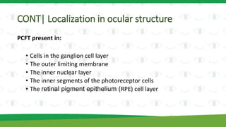
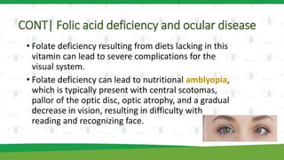
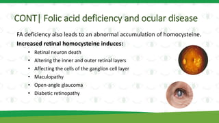
Folic Acid
Deficiency
and Vision
Abrar Alturki – Nouf Almousa
Outline
1. Definition of folate
2. Human eye development
3. Folate transport proteins and their localization in ocular structure
4. Folic acid deficiency and ocular disease
• FA deficiency in patients
• FA deficiency in animal models
Folate B9 and Folic acid
Folate (B9) is a water-soluble vitamin. It cannot be synthesized by the
human body (essential), it must be obtained either through the diet or
from supplementation.
Folic acid is the synthetic form of B9, not naturally present in foods but
is produced synthetically for use in food fortification and dietary
supplementation.
• Folate can be found in many natural foods, such as
During pregnancy
The requirement for FA increases and it becomes very
important for both the mother’s health and fetal development.
A daily FA supplement (400 - 800 mcg/ day).
Low levels of folate are associated with several birth defects, such as
neural tube defects, low infant birth weight, and cleft palate.
Periconceptional administration of FA helps prevent the occurrence and
recurrence of many congenital abnormalities, especially those related to
the neural tube, which forms the embryonic nervous system.
Human eye development
• Day 22nd
• The eye begins to develop at this stage.
• The diencephalon stick out on either side as two lateral
outgrowths forming the optic vesicle.
• Day 27th
• The optic vesicles come into contact with the adjoining
surface ectoderm, which thickens to form the lens
placode.
• The distal part of the optic vesicle is invaginated into its
more proximal part to form a double-layered optic cup:
the inner and the outer layers.
CONT|Human eye development
• Day 36th
The lens vesicle is formed and detaches from the
surface ectoderm. The ectodermal layer in this region
forms the future corneal epithelium.
If a failure occurs to the lens vesicle separation from
the surface ectoderm it will result in lenticulocorneal
fusion.
CONT|Human eye development
• 4th and 6th weeks
The neural retina is composed of two zones: the inner and outer
neuroblastic layers.
• The first layer → ganglion, Müller, and amacrine cells.
• The outer neuroblastic cell layer → photoreceptor, bipolar, and
horizontal cells.
A failure of organization or delayed differentiation of the neuroblastic
cells during development leads to retinal dysplasia.
The inner and outer
neuroblastic layers
CONT|Human eye development
• By the end of the 3rd month
The pupillary membrane becomes fully formed. The lip of
the optic cup begins to elongate.
Outer layer of the optic cup forms:
• The anterior iris epithelium
• Outer pigmented epithelium of the ciliary body
Inner layer of the optic cup forms:
• The posterior iris epithelium
• Inner non-pigmented epithelium of the ciliary body
The secondary vitreous composed of fine fibrillar material
becomes evident.
Summary for the
previous stages
EmbryonAnimation
CONT|Human eye development
• 4th and 5th week
The choriocapillaris starts to differentiate.
• 6th week
The innermost layer of the choroid membrane begins to develop and
regulates the mutual exchange of biomolecules, nutrients, and oxygen
between the retina and the general circulation.
• 15th week
The first choroidal arterioles and veins can be seen; become
distinguishable at the 22nd week.
Folate transport proteins
Folate and its one-carbon derivatives cannot penetrate biological
membranes by simple diffusion.
Folate transporter :
1. Folate receptor (FR)
2. Reduced folate transporter (RFT-1)
3. Proton-coupled folate transporter (PCFT)
CONT| Folate transport proteins
Folate receptor (FR)
There are four FR isoforms (α, β, γ, and δ), all of them are tissue-specific.
• FRα, also known as folate receptor 1 (Folr1)
• Enhance the cellular uptake of folate from the blood
• Bind with folate at a high affinity to mediate transport into the cell cytoplasm
• FRβ or Folr2, is the most common folate receptor
• Has high expression in the nervous system.
• A biomarker for activated macrophages in the eye, macrophages are present in
the ocular tissues.
• No much information has been published about the protein receptor
FRγ and its role in folate transport.
• FRδ or Folr4
• Expressed in regulatory T cells and may play a role in controlling immune
responses.
• The ocular pigment epithelial cells of the iris, the ciliary body, and the retina
contribute to the immune property of the eye.
CONT| Folate transport proteins
• Like FRα, the reduced folate transporter-1 protein (RFT-1)
• regulates the cellular uptake of molecules inside the cell.
• The proton-coupled folate transporter (PCFT)
• The transport of folates into the central nervous system.
Localization in ocular structure
FA transport proteins are localized in many eye structures.
Numerous studies have shown that the expression of FRα is
ubiquitous in:
• all layers of the retina
• ganglion cell layer
• Müller cells
• Outer limiting membrane
The principal role of this receptor is to mediate the uptake of folate into
the different cells of the retina
CONT| Localization in ocular structure
• FR-β is present in the activated macrophage in the retina.
• FRδ is present in retinal pigment epithelium (RPE) cells.
CONT| Localization in ocular structure
PCFT present in:
• Cells in the ganglion cell layer
• The outer limiting membrane
• The inner nuclear layer
• The inner segments of the photoreceptor cells
• The retinal pigment epithelium (RPE) cell layer
Folic acid deficiency and ocular disease
It is known that folate is essential during the early stages of human
development especially during pregnancy.
This vitamin plays an essential role in embryonic development, but its
importance is not confined to the fetal period, it is also important for
adults.
FA can enhance growth even in adulthood and repair mechanisms.
CONT| Folic acid deficiency and ocular disease
• Folate deficiency resulting from diets lacking in this
vitamin can lead to severe complications for the
visual system.
• Folate deficiency can lead to nutritional amblyopia,
which is typically present with central scotomas,
pallor of the optic disc, optic atrophy, and a gradual
decrease in vision, resulting in difficulty with
reading and recognizing face.
CONT| Folic acid deficiency and ocular disease
FA deficiency also leads to an abnormal accumulation of homocysteine.
Increased retinal homocysteine induces:
• Retinal neuron death
• Altering the inner and outer retinal layers
• Affecting the cells of the ganglion cell layer
• Maculopathy
• Open-angle glaucoma
• Diabetic retinopathy
CONT| Folic acid deficiency and ocular disease
Folate deficiency is also a possible risk factor for
cataracts.
• A cross-sectional study has shown that elderly
patients with cataracts had a lower plasma folate
level than those without cataracts, and that folate
insufficiency was significantly associated with
cataracts.
• Also, another study found that long-term use of
vitamin B2, B12, and folate supplements was
associated with a reduced prevalence of cataracts
Folic acid deficiency in animal models
• Nutritional animal models are essential to understand
human diseases.
• Animal studies can play an important role in public health
nutrition including the prevention of several physiological,
biochemical, metabolic, and molecular diseases.
CONT| Folic acid deficiency in animal models
To determine the role folate intake has in lens development, a study
was recently carried out. On embryonic and adult lenses, using mice.
• This study reveals that :
• embryos with aberrant lens fiber organization presented smaller
lenses.
• Adult eyes accompanied by cataracts were also has small lenses.
These results observed that maternal FA deficiency alters ocular
biometry of mouse embryos.
CONT| Folic acid deficiency in animal models
• Another research team also found that mice exposed to a FA
deficient diet exhibited anophthalmia and microphthalmia
CONT|Folic acid deficiency in animal models
• The impact of folate deficiency in zebrafish was studied. It
has been observed that
• folate deficiency induced oxidative stress and
• impaired neural tube closure in embryos.
As a result, many abnormalities of eye development occurred.
Questions
section
How much folic acid should a pregnant woman take?
1. 500 mcg/ day
2. 400 - 800 mcg/ day
3. 400 mcg/ day
In which week the retina and the general circulation
exchanges of biomolecules, nutrients, and oxygen begin?
1. Sixth week
2. 22nd week
3. 22 day
Folate deficiency can lead to:
1. Amblyopia.
2. Accumulation of homocysteine.
3. Cataracts.
4. All of above
Nutritional animal models are essential to understand
human diseases.
1. True
2. False
Thank you for listening

Folic acid deficiency and vision

  • 1.
  • 2.
    Outline 1. Definition offolate 2. Human eye development 3. Folate transport proteins and their localization in ocular structure 4. Folic acid deficiency and ocular disease • FA deficiency in patients • FA deficiency in animal models
  • 3.
    Folate B9 andFolic acid Folate (B9) is a water-soluble vitamin. It cannot be synthesized by the human body (essential), it must be obtained either through the diet or from supplementation. Folic acid is the synthetic form of B9, not naturally present in foods but is produced synthetically for use in food fortification and dietary supplementation. • Folate can be found in many natural foods, such as
  • 4.
    During pregnancy The requirementfor FA increases and it becomes very important for both the mother’s health and fetal development. A daily FA supplement (400 - 800 mcg/ day). Low levels of folate are associated with several birth defects, such as neural tube defects, low infant birth weight, and cleft palate. Periconceptional administration of FA helps prevent the occurrence and recurrence of many congenital abnormalities, especially those related to the neural tube, which forms the embryonic nervous system.
  • 5.
    Human eye development •Day 22nd • The eye begins to develop at this stage. • The diencephalon stick out on either side as two lateral outgrowths forming the optic vesicle. • Day 27th • The optic vesicles come into contact with the adjoining surface ectoderm, which thickens to form the lens placode. • The distal part of the optic vesicle is invaginated into its more proximal part to form a double-layered optic cup: the inner and the outer layers.
  • 6.
    CONT|Human eye development •Day 36th The lens vesicle is formed and detaches from the surface ectoderm. The ectodermal layer in this region forms the future corneal epithelium. If a failure occurs to the lens vesicle separation from the surface ectoderm it will result in lenticulocorneal fusion.
  • 7.
    CONT|Human eye development •4th and 6th weeks The neural retina is composed of two zones: the inner and outer neuroblastic layers. • The first layer → ganglion, Müller, and amacrine cells. • The outer neuroblastic cell layer → photoreceptor, bipolar, and horizontal cells. A failure of organization or delayed differentiation of the neuroblastic cells during development leads to retinal dysplasia.
  • 8.
    The inner andouter neuroblastic layers
  • 9.
    CONT|Human eye development •By the end of the 3rd month The pupillary membrane becomes fully formed. The lip of the optic cup begins to elongate. Outer layer of the optic cup forms: • The anterior iris epithelium • Outer pigmented epithelium of the ciliary body Inner layer of the optic cup forms: • The posterior iris epithelium • Inner non-pigmented epithelium of the ciliary body The secondary vitreous composed of fine fibrillar material becomes evident.
  • 10.
    Summary for the previousstages EmbryonAnimation
  • 11.
    CONT|Human eye development •4th and 5th week The choriocapillaris starts to differentiate. • 6th week The innermost layer of the choroid membrane begins to develop and regulates the mutual exchange of biomolecules, nutrients, and oxygen between the retina and the general circulation. • 15th week The first choroidal arterioles and veins can be seen; become distinguishable at the 22nd week.
  • 12.
    Folate transport proteins Folateand its one-carbon derivatives cannot penetrate biological membranes by simple diffusion. Folate transporter : 1. Folate receptor (FR) 2. Reduced folate transporter (RFT-1) 3. Proton-coupled folate transporter (PCFT)
  • 13.
    CONT| Folate transportproteins Folate receptor (FR) There are four FR isoforms (α, β, γ, and δ), all of them are tissue-specific. • FRα, also known as folate receptor 1 (Folr1) • Enhance the cellular uptake of folate from the blood • Bind with folate at a high affinity to mediate transport into the cell cytoplasm • FRβ or Folr2, is the most common folate receptor • Has high expression in the nervous system. • A biomarker for activated macrophages in the eye, macrophages are present in the ocular tissues.
  • 14.
    • No muchinformation has been published about the protein receptor FRγ and its role in folate transport. • FRδ or Folr4 • Expressed in regulatory T cells and may play a role in controlling immune responses. • The ocular pigment epithelial cells of the iris, the ciliary body, and the retina contribute to the immune property of the eye. CONT| Folate transport proteins
  • 15.
    CONT| Folate transportproteins • Like FRα, the reduced folate transporter-1 protein (RFT-1) • regulates the cellular uptake of molecules inside the cell. • The proton-coupled folate transporter (PCFT) • The transport of folates into the central nervous system.
  • 16.
    Localization in ocularstructure FA transport proteins are localized in many eye structures. Numerous studies have shown that the expression of FRα is ubiquitous in: • all layers of the retina • ganglion cell layer • Müller cells • Outer limiting membrane The principal role of this receptor is to mediate the uptake of folate into the different cells of the retina
  • 17.
    CONT| Localization inocular structure • FR-β is present in the activated macrophage in the retina. • FRδ is present in retinal pigment epithelium (RPE) cells.
  • 18.
    CONT| Localization inocular structure PCFT present in: • Cells in the ganglion cell layer • The outer limiting membrane • The inner nuclear layer • The inner segments of the photoreceptor cells • The retinal pigment epithelium (RPE) cell layer
  • 19.
    Folic acid deficiencyand ocular disease It is known that folate is essential during the early stages of human development especially during pregnancy. This vitamin plays an essential role in embryonic development, but its importance is not confined to the fetal period, it is also important for adults. FA can enhance growth even in adulthood and repair mechanisms.
  • 20.
    CONT| Folic aciddeficiency and ocular disease • Folate deficiency resulting from diets lacking in this vitamin can lead to severe complications for the visual system. • Folate deficiency can lead to nutritional amblyopia, which is typically present with central scotomas, pallor of the optic disc, optic atrophy, and a gradual decrease in vision, resulting in difficulty with reading and recognizing face.
  • 21.
    CONT| Folic aciddeficiency and ocular disease FA deficiency also leads to an abnormal accumulation of homocysteine. Increased retinal homocysteine induces: • Retinal neuron death • Altering the inner and outer retinal layers • Affecting the cells of the ganglion cell layer • Maculopathy • Open-angle glaucoma • Diabetic retinopathy
  • 22.
    CONT| Folic aciddeficiency and ocular disease Folate deficiency is also a possible risk factor for cataracts. • A cross-sectional study has shown that elderly patients with cataracts had a lower plasma folate level than those without cataracts, and that folate insufficiency was significantly associated with cataracts. • Also, another study found that long-term use of vitamin B2, B12, and folate supplements was associated with a reduced prevalence of cataracts
  • 23.
    Folic acid deficiencyin animal models • Nutritional animal models are essential to understand human diseases. • Animal studies can play an important role in public health nutrition including the prevention of several physiological, biochemical, metabolic, and molecular diseases.
  • 24.
    CONT| Folic aciddeficiency in animal models To determine the role folate intake has in lens development, a study was recently carried out. On embryonic and adult lenses, using mice. • This study reveals that : • embryos with aberrant lens fiber organization presented smaller lenses. • Adult eyes accompanied by cataracts were also has small lenses. These results observed that maternal FA deficiency alters ocular biometry of mouse embryos.
  • 25.
    CONT| Folic aciddeficiency in animal models • Another research team also found that mice exposed to a FA deficient diet exhibited anophthalmia and microphthalmia
  • 26.
    CONT|Folic acid deficiencyin animal models • The impact of folate deficiency in zebrafish was studied. It has been observed that • folate deficiency induced oxidative stress and • impaired neural tube closure in embryos. As a result, many abnormalities of eye development occurred.
  • 27.
  • 28.
    How much folicacid should a pregnant woman take? 1. 500 mcg/ day 2. 400 - 800 mcg/ day 3. 400 mcg/ day
  • 29.
    In which weekthe retina and the general circulation exchanges of biomolecules, nutrients, and oxygen begin? 1. Sixth week 2. 22nd week 3. 22 day
  • 30.
    Folate deficiency canlead to: 1. Amblyopia. 2. Accumulation of homocysteine. 3. Cataracts. 4. All of above
  • 31.
    Nutritional animal modelsare essential to understand human diseases. 1. True 2. False
  • 32.
    Thank you forlistening

Editor's Notes

  • #26 ​Microphthalmia: Microphthalmia literally means 'small eye'. Children may be born with one, or both eyes, small and underdeveloped. Some children may be blind, but others may have some residual sight or light perception. Anophthalmia: Anophthalmia means an absence of the eye. As with Microphthalmia, a child may be born with one, or both eyes, missing from the eye socket.