hyderabad call girl.pdfRussian Call Girls in Hyderabad Amrita 9907093804 Inde...
Fisiopatologia[21630]
1. PATHOPHYSIOLOGY
OF ACUTE ISCHEMIC
STROKE
Tudor G. Jovin, Andrew M. Demchuk, Rishi Gupta
ABSTRACT
In acute ischemic stroke, abrupt vessel occlusion results in a drop in regional CBF,
leading to time-dependent compartmentalization of the ischemic brain into tissue
that is irreversibly damaged (ischemic core), tissue that is functionally impaired but
structurally intact and thus potentially salvageable (penumbra), and tissue that is
hypoperfused but not threatened under normal circumstances (oligemic brain).
At a cellular level, neuronal damage occurs through a complex interaction of
mechanisms (necrosis, apoptosis, excitotoxicity, inflammation, peri-infarct depo-
larization, acidosis, and free radical formation) that are characteristic for each
compartment. All these mechanisms are potential targets for neuroprotective
therapy, which, combined with flow restoration strategies, is likely to improve
outcome significantly in human stroke.
Continuum Lifelong Learning Neurol 2008;14(6):28–45.
INTRODUCTION
Acute ischemic stroke is characterized
by abrupt neurologic dysfunction due
to focal brain ischemia resulting in
persistent neurologic deficit or accom-
panied by characteristic abnormalities
on brain imaging (Albers et al, 2002).
Until recently, stroke was defined
using clinical criteria alone, based on
duration of symptoms lasting 24 hours
or longer (Ad Hoc Committee, 1975). If
the symptoms persisted for less than 24
hours, the condition was termed tran-
sient ischemic attack (TIA) However,
modern neuroimaging techniques, es-
pecially diffusion MRI, have shown that
defining stroke or TIA based only on
duration of symptoms may not be ac-
curate, since permanent brain damage
can occur even when symptoms last
only minutes (Ay et al, 1999; Engelter
et al, 1999; Kidwell et al, 1999). There-
fore, recently proposed definitions of
TIA take into account both the dura-
tion of symptoms (typically less than
an hour) and lack of acute infarction
on brain imaging (Albers et al, 2002).
Ovbiagele and colleagues (2003) esti-
mated that adopting a definition of
TIA based on the above criteria would
reduce estimates of the annual inci-
dence of TIA by 33% (currently esti-
mated to be 180,000 annually) and
increase annual number of strokes
by 7% (currently estimated at 820,000
per year).
28
Relationship Disclosure: Dr Jovin has received personal compensation for activities with CoAxia and
Concentric Medical, Inc. Dr Jovin has received personal compensation as associate editor of Journal of
Neuroimaging. Dr Demchuk has received personal compensation for activities with AstraZeneca, Boehringer
Ingelheim Pharmaceuticals, Inc., and sanofi-aventis. Dr Demchuk has received grant support from Novo
Nordisk, Inc. Dr Gupta has received personal compensation for activities with Concentric Medical, Inc.
Unlabeled Use of Products/Investigational Use Disclosure: Drs Jovin and Gupta have nothing to disclose.
Dr Demchuk discusses the unlabeled use of emerging therapies with numerous references to potential therapies.
Copyright # 2008, American Academy of Neurology. All rights reserved.
Copyright @ American Academy of Neurology. Unauthorized reproduction of this article is prohibited.
2. ETIOLOGIC AND
PATHOLOGIC ASPECTS
It is important to recognize that ische-
mic stroke results from a heteroge-
neous group of disorders whose final
common pathway leading to clinical
manifestations is interruption of blood
flow through vascular occlusion. This
results in an infarct of which the size is
dependent on extent, duration, and
severity of ischemia. Brain infarcts
resulting from arterial occlusion are
divided based on their macroscopic
appearance into white (bland) and red
(hemorrhagic) infarcts (Garcia et al,
1998). By gross anatomy, the former
are composed of few or no petechiae
while the latter are characterized by
grossly visible blood. This latter term
is equivalent to hemorrhagic transfor-
mation, which refers to leaking of red
blood cells into a dying and ischemic
brain tissue, and should not be con-
fused with parenchymal hematoma,
which represents a homogenous col-
lection of blood usually resulting from
a ruptured blood vessel. Serial brain
imaging studies in patients with acute
stroke have demonstrated that hem-
orrhagic transformation of an initially
bland infarct can occur in up to 80% of
patients (Hart and Easton, 1986; Lyden
and Zivin, 1993; Mayer et al, 2000). The
risk of early hemorrhagic transformation
and parenchymal hematoma is greatly
increased by administration of thrombo-
lytics or anticoagulants in acute ische-
mic stroke (Larrue et al, 1997).
Gross anatomy studies reveal that
arterial infarcts evolve over several
stages (Garcia et al, 1998). In the first
12 to 24 hours after the ictus, the le-
sion is barely visible to the naked eye.
Swelling reaches its zenith at days 3 to
5; in large strokes this can become life
threatening due to displacement and
compression of neighboring structures.
Between days 5 and 10, the infarcted
brain becomes sharply demarcated
from the unaffected brain tissue. The
chronic stage, occurring weeks or
months after the ictus, features a fluid-
filled cavity that results from reabsorp-
tion of necrotic debris, hence the name
liquefaction necrosis.
STROKE MECHANISMS
Two major mechanisms are responsible
for ischemia in acute stroke: thrombo-
embolism and hemodynamic failure.
The former usually occurs as a result
of embolism or in situ thrombosis and
leads to an abrupt fall in regional
cerebral blood flow (CBF). The latter
usually occurs with arterial occlusion or
stenosis, when collateral blood supply
maintains CBF at levels that are suffi-
cient for preservation of brain function
under normal circumstances. In these
cases, cerebral ischemia may be trig-
gered by conditions that decrease per-
fusion proximally to the arterial lesion
(systemic hypotension or low cardiac
output) and increase metabolic de-
mands (fever, acidosis) or conditions
that lead to ‘‘steal’’ of blood from af-
fected to unaffected areas in the brain
(carbon dioxide retention) (Alexandrov
et al, 2007). Strokes occurring through
these mechanisms are located predom-
inantly in the so-called borderzones
or watershed regions, which are areas
in the brain bordering major vascular
territories such as the middle cerebral
artery (MCA)/internal carotid artery or
MCA/posterior cerebral artery interface
(Klijn et al, 1997). Caplan and Hennerici
(1998) postulated that embolism and
hypoperfusion oftentimes coexist and
potentiate each other. They proposed
impaired clearance of emboli due to
low flow states as a link between these
two factors in the pathophysiology of
brain infarction.
Embolism
Embolic material formed within the
heart or vascular system travels through
the arterial system, lodging in a vessel
and partially or completely occluding it.
29
A Two major
mechanisms are
responsible for
ischemia in
acute stroke:
thromboembolism
and
hemodynamic
failure.
A Embolism and
hypoperfusion
can coexist and
potentiate each
other.
Continuum: Lifelong Learning Neurol 2008;14(6)
KEY POINTS
A Ischemic stroke
results from a
heterogeneous
group of
disorders whose
final common
pathway is
interruption of
blood flow
through vascular
occlusion.
Copyright @ American Academy of Neurology. Unauthorized reproduction of this article is prohibited.
3. The most common sources of emboli
are the heart and large arteries. Other
rare sources of emboli are air, fat, cho-
lesterol, bacteria, tumor cells, and par-
ticulate matter from injected drugs
(Caplan, 2000).
Cardioembolism. Cardioembolism
accounts for 20% to 30% of all ischemic
stroke (Grau et al, 2001; Kolominsky-
Rabas et al, 2001; Petty et al, 2000;
Sacco et al, 1995). Table 2-1 outlines
the high risk versus low or uncertain
risk conditions for cardioembolic stroke
(Ferro, 2003). Conditions considered at
high risk for embolization to the brain
are atrial fibrillation, sustained atrial
flutter, sick sinus syndrome, left atrial
thrombus, left atrial appendage throm-
bus, left atrial myxoma, mitral stenosis,
prosthetic valve, infective endocarditis,
noninfective endocarditis, left ventric-
ular thrombus, left ventricular myxoma,
recent anterior myocardial infarct, and
dilated cardiomyopathy.Conditionscon-
sidered at low or uncertain risk for brain
embolization include patent foramen
ovale, atrial septal aneurysm, sponta-
neous atrial contrast, mitral annulus
calcification, mitral valve prolapse, cal-
cified aortic stenosis, fibroelastoma,
giant Lambl excrescences, akinetic or
dyskinetic ventricular wall segment,
subaortic hypertrophic cardiomyop-
athy, and congestive heart failure
(Marchal et al, 1999).
Artery-to-artery embolism. Em-
boli occluding brain arteries can also
originate from large vessels situated
more proximally, such as the aorta,
extracranial carotid, or vertebral ar-
teries or intracranial arteries. In these
circumstances, the embolic material is
composed of clot, platelet aggregates
or plaque debris that usually breaks off
from atherosclerotic plaques (Furlan
et al, 1996). This is a major mechanism
responsible for stroke due to large
vessel atherosclerosis, which accounts
for 15% to 20% of all ischemic strokes
(Grau et al, 2001; Kolominsky-Rabas
30
Continuum: Lifelong Learning Neurol 2008;14(6)
TABLE 2-1 High Risk
Versus Low or
Uncertain Risk
Conditions for
Cardioembolic
Stroke
" High Risk Conditions
Atrial fibrillation
Sustained atrial flutter
Sick sinus syndrome
Left atrial thrombus
Left atrial appendage
thrombus
Left atrial myxoma
Mitral stenosis
Mechanical valve
Infective endocarditis
Noninfective endocarditis
Left ventricular myxoma
Recent anterior myocardial
infarct
Dilated cardiomyopathy
" Low or Uncertain
Risk Condition
Patent foramen ovale
Atrial septal aneurysm
Spontaneous atrial contrast
Mitral valve prolapse
Calcified aortic stenosis
Fibroelastoma
Giant Lambel excrescence
Akinetic or dyskinetic
ventricular wall segment
Subaortic hypertrophic
cardiomyopathy
Congestive heart failure
Reprinted from Ferro JM. Cardioembolic
stroke: an update. Lancet Neurol 2003;2(3):
177–188. Copyright # 2003, with permission
from Elsevier.
KEY POINT
A The most
common sources
of emboli are the
heart and large
arteries.
"PATHOPHYSIOLOGY
Copyright @ American Academy of Neurology. Unauthorized reproduction of this article is prohibited.
4. et al, 2001; Petty et al, 2000; Sacco et al,
1995).
Thrombosis
Thrombosis represents an obstruction
of flow with thrombus formation result-
ing from an occlusive process initiated
within the vessel wall. In the vast
majority of cases this is caused by
atherosclerotic disease, hence the name
atherothrombosis. Less common vascu-
lar pathologies leading to vessel stenosis
or occlusion include arterial dissection
(intracranial or extracranial), fibromus-
cular dysplasia, vasospasm (drug in-
duced, inflammatory, or infectious),
radiation-induced vasculopathy, extrin-
sic compression such as tumor or other
mass lesion, or moyamoya disease.
Small vessel disease. Thrombotic
occlusion of the small penetrating ar-
teries in the brain is another important
cause of strokes, accounting for an-
other approximately 20% to 30% of all
ischemic strokes. This type of vascu-
lar lesion is strongly associated with
hypertension and is characterized path-
ologically by lipohyalinosis, micro-
atheroma, fibrinoid necrosis, and
Charcot-Bouchard aneurysms (Bamford
and Warlow, 1988; Fisher, 1982; Fisher,
1998; Mohr, 1982). Lipohyalinosis is
characterized by replacement of the
normal vessel wall with fibrin and col-
lagen and is specifically associated with
hypertension. Microatheroma repre-
sents an atheromatous plaque of the
small vessel that may involve the origin
of a penetrating artery (Caplan, 1989).
This latter mechanism is believed to be
responsible for larger subcortical in-
farcts. Fibrinoid necrosis is usually as-
sociated with extremely high blood
pressure, leading to necrosis of smooth
muscle cells and extravasation of plas-
ma proteins, which appear micros-
copically as fine granular eosinophilic
deposits in the connective tissue of
the vessel wall. Charcot-Bouchard an-
eurysms are areas of focal dilata-
tion in the small vessel wall, which
may thrombose, leading to vessel
occlusion.
CEREBRAL BLOOD
FLOW CHANGES
Introduction
Following vessel occlusion, the main
factors ultimately determining tissue
outcome are regional CBF and duration
of vessel occlusion. A decrease in re-
gional CBF leads to diminished tissue
perfusion. In persistent large vessel oc-
clusion, local perfusion pressure, which
is the main factor influencing the even-
tual outcome of tissue (Baron, 2001),
depends on several factors such as the
presence and extent of collaterals and
systemic arterial pressure (due to loss
of the ischemic brain’s autoregulatory
capacity). It is inversely correlated to
the local tissue pressure (which is
increased by ischemic edema).
Cerebral Blood Flow Thresholds
in Acute Cerebral Ischemia
The difference in tissue outcome follow-
ing arterial occlusion is based on the
concept that CBF thresholds exist, below
which neuronal integrity and function
are differentially affected (Figure 2-1).
Early human studies performed in the
1950s during carotid artery clamping for
carotid endarterectomy using intracaro-
tid xenon 133 injections (Boysen, 1971;
Jennett et al, 1966) reported that hemi-
paresis occurred when regional CBF fell
below 50% to 30% of normal, and per-
manent neurologic deficit occurred if
mean CBF fell below 30% of normal.
Evidence also indicated that develop-
ment of permanent neurologic sequelae
is a time-dependent process; for any
given blood flow level, low CBF values
are tolerated only for a short period
of time, while higher CBF values re-
quire longer time for infarction to
occur. Several investigators (Sundt et al,
1974; Trojaborg and Boysen, 1973) have
31
Continuum: Lifelong Learning Neurol 2008;14(6)
KEY POINTS
A Thrombosis is
associated with
thrombus
formation as
a consequence
of an occlusive
process
initiated within
the vessel wall
and is usually
caused by
atherosclerotic
disease.
A Occlusive
disease of small
penetrating
arteries in the
brain (small
vessel disease)
accounts for
20% to 30% of
ischemic strokes.
A Following vessel
occlusion, the
main factors
ultimately
determining tissue
outcome are
regional cerebral
blood flow and
duration of vessel
occlusion.
A The difference in
tissue outcome
following arterial
occlusion is based
on the concept
that cerebral
blood flow
thresholds exist,
below which
neuronal integrity
and function are
differentially
affected.
Copyright @ American Academy of Neurology. Unauthorized reproduction of this article is prohibited.
5. studied the relationship between EEG
changes and regional CBF during carotid
clamping. EEG would slow down when
mean CBF fell below 23 mL/100 g/min,
while at values below 15 mL/100 g/min
the EEG would become flat.
The concept of CBF threshold in
focal cerebral ischemia proposed by
these early human studies was then
reinforced by landmark studies per-
formed by Symon and colleagues
(1977), who investigated the relation-
ship between severity of local CBF im-
pairment and degree of neurologic
dysfunction at various durations of is-
chemia in a baboon model of MCA oc-
clusion. Symon and colleagues (1977)
demonstrated that brain tissue perfu-
ses between certain CBF values (22 mL/
100 mg/min to 8 mL/100 mg/min) even
when prolonged hypoperfusion stops
functioning, but maintains its structural
integrity and, most importantly, can be
salvaged with reperfusion. Figure 2-2
depicts a summary of metabolic and
electrophysiologic disturbances accord-
ing to reductions of cortical blood flow
at different thresholds.
The time dependence of ischemic
thresholds in producing permanent or
transient neurologic damage has been
demonstrated by Jones and colleagues
(1981) in primate studies using a
temporary or permanent MCA occlu-
sion model. These experiments dem-
onstrated that the CBF values below
which brain tissue becomes infarcted
are dependent on the duration of
vessel occlusion. Two hours of contin-
uous MCA occlusion in awake ma-
caque monkeys required CBF values of
5 mL/100 g/min to produce infarction,
while 3 hours of continuous occlusion
resulted in infarction if CBF values
were 12 mL/100 mg/min or less. Per-
manent occlusion resulted in infarc-
tion if flows were 18 mL/100 mg/min
or less. It should be noted that
30 minutes of occlusion did not result
in infarction even at CBF values below
5 mL/100 mg/min.
It is important to understand that
the blood flow thresholds studied in
most animal and human experiments
refer to ischemic tolerance of the brain
cortex. Thresholds for deep white
32
Continuum: Lifelong Learning Neurol 2008;14(6)
FIGURE 2-1 Schematic drawing of the different cerebral blood flow thresholds in man.
a
Furlan M, Marchal G, Viader F, et al. Spontaneous neurological recovery after stroke and the fate
of the ischemic penumbra. Ann Neurol 1996;40(2):216–226.
Marchal G, Beaudouin V, Rioux P, et al. Prolonged persistence of substantial volumes of potentially
viable brain tissue after stroke. Stroke 1996;27(4):599–606.
Marchal G, Benali K, Igle´sias S, et al. Voxel-based mapping of irreversible ischaemic damage
with PET in acute stroke. Brain 1999;122(pt 12):2387–2400.
b
Heiss WD, Grond M, Thiel A, et al. Tissue at risk of infarction rescued by early reperfusion: A positron
emission tomography study in systemic recombinant tissue plasminogen activator thrombolysis
of acute stroke. J Cereb Blood Flow Metab 1998;18(12):1298–1307.
Heiss WD, Thiel A, Grond M, Graf R. Which targets are relevant for therapy of acute ischemic
stroke? Stroke 1999;30(7):1486–1489.
Baron JC. Perfusion thresholds in human cerebral ischemia: historical perspective and therapeutic
implications. Cerebrovasc Dis 2001;11(suppl 1):2–8. Reprinted with permission from S. Karger, AG, Basel.
"PATHOPHYSIOLOGY
Copyright @ American Academy of Neurology. Unauthorized reproduction of this article is prohibited.
6. matter or basal ganglia have not been
studied rigorously and are simply un-
known. It is believed, however, that
gray matter is more susceptible to
infarction than white matter, and that
within the gray matter the basal ganglia
have a lower ischemic tolerance than
the cortex (Marcouz et al, 1982).
Concept of Ischemic Core and
Ischemic Penumbra
The notion that in acute stroke, de-
pending on the extent and duration
of hypoperfusion, the tissue supplied
by the occluded artery is compartmen-
talized into areas of irreversibly dam-
aged brain tissue and areas of brain
tissue that are hypoperfused but viable
led to the concept of ischemic core
and ischemic penumbra proposed
by Astrup and colleagues (1981). The
ischemic core represents tissue that is
irreversibly damaged. PET studies in
humans suggest that beyond a certain
time limit (probably no longer than an
hour) the ischemic core corresponds
to CBF values of less than 7 mL/100
mg/min (Furlan et al, 1996; Marchal
et al, 1996; Marchal et al, 1999) to
12 mL/100 mg/min (Heiss, 2000; Heiss
et al, 2001b). The ischemic penum-
bra represents tissue that is function-
ally impaired but structurally intact
and, as such, potentially salvageable.
It corresponds to a high CBF limit of
17 mL/100 mg/min to 22 mL/100 mg/
min and a low CBF limit of 7 mL/100
mg/min to 12 mL/100 mg/min. Salvag-
ing this tissue by restoring its flow to
nonischemic levels is the aim of acute
stroke therapy. Another compartment,
termed by Symon and colleagues (1977)
oligemia, represents mildly hypoper-
fused tissue from the normal range
down to around 22 mL/100 mg/min. It
is believed that under normal circum-
stances this tissue is not at risk of
infarction (Baron, 2001). It is con-
ceivable, however, that under certain
circumstances, such as hypotension,
fever, or acidosis, oligemic tissue can
be incorporated into penumbra and
subsequently undergo infarction.
Evidence in the literature suggests
that there is temporal evolution of the
core, which grows at the expense of
33
KEY POINTS
A The ischemic
core represents
tissue that is
irreversibly
damaged.
A The ischemic
penumbra
represents
tissue that is
functionally
impaired but
structurally intact
and, as such,
potentially
salvageable.
Salvaging
this tissue by
restoring its flow
to nonischemic
levels is the aim
of reperfusion
therapy in acute
stroke.
Continuum: Lifelong Learning Neurol 2008;14(6)
FIGURE 2-2 Thresholds of metabolic (left) and electrophysiologic (right) disturbances
during gradual reduction of cortical blood flow.
SEP = somatosensory evoked potentials; EEG = electroencephalogram; ATP = adenosine triphosphate.
Hossmann KA. Pathophysiology and therapy of experimental stroke. Cell Mol Neurobiol 2006;26(7–8):1057–1083.
Reprinted with permission from Springer Science and Business Media.
Copyright @ American Academy of Neurology. Unauthorized reproduction of this article is prohibited.
7. penumbra (Ginsberg, 2003; Heiss et al,
2001a; Raichle, 1982) (Figure 2-3). This
process occurs because of the interac-
tion of a multitude of complex factors
acting concomitantly or sequentially
(Hossmann, 2006). It is known that
the ischemic penumbra represents a
dynamic phenomenon that evolves
in space and time. If vessel occlusion
persists, the penumbra may shrink
because of progressive recruitment
into the core. Alternatively, it may re-
turn to a normal state following vessel
recanalization or possibly neuropro-
tective interventions. It thus appears
that the ischemic penumbra repre-
sents a transitional state between evo-
lution into permanent ischemia as one
possibility and transformation into
normal tissue as the other possibil-
ity. On the basis of a rat model of MCA
occlusion studied in a multimodal
fashion assessing CBF, metabolism,
and gene expression, Ginsberg (2003)
concluded that the penumbra lies
within a narrow range of perfusion
and thus is precariously dependent
on small perfusion pressure changes;
that the penumbra is electrophysi-
ologically dynamic and undergoes
recurrent depolarizations; and that
it is metabolically unstable, being
34
FIGURE 2-3 Experimental model of middle cerebral artery occlusion in rats. Serial MRIs (coronal sections) at three
levels in the brain depicting the apparent diffusion coefficient of water (ADC) (blue) demonstrating the
time-dependent growth of the ischemic core.
Hossmann KA. Pathophysiology and therapy of experimental stroke. Cell Mol Neurobiol 2006;26(7–8):1057–1083. Reprinted with permission from
Springer Science and Business Media.
Continuum: Lifelong Learning Neurol 2008;14(6)
"PATHOPHYSIOLOGY
Copyright @ American Academy of Neurology. Unauthorized reproduction of this article is prohibited.
8. the site of severe metabolism/flow
dissociation.
Restriction of acute stroke therapy
aimed at vessel recanalization to 3
hours from onset of symptoms for IV
thrombolysis and 6 hours for intraar-
terial thrombolysis is based on the
concept that the ischemic penumbra
has a short lifespan, being rapidly in-
corporated into the core within hours
of the ictus (Heiss et al, 2001b; Kaufman
et al, 1999). Recent evidence suggests,
however, that penumbral brain tissue
of significant extent is present even
beyond 6 hours of stroke onset. PET
studies using quantitative CBF assess-
ment (Furlan et al, 1996; Marchal et al,
1999) or markers of tissue hypo-
xia such as 18F fluoromisonidazole
(Read et al, 2000) to assess penumbra,
included patients studied within 6
hours to as late as 51 hours after stroke
onset and reported the existence of
penumbra comprising 30% to 45% of
the total ischemic tissue at risk. Several
investigators have estimated the pen-
umbra based on diffusion/perfusion
MRI (diffusion-weighted imaging [DWI]/
perfusion-weighted imaging [PWI]) mis-
match in acute stroke (Hjort et al, 2005;
Schlaug et al, 1999). Since the diffusion
abnormalities are presumed to repre-
sent an approximation of the irrevers-
ible ischemic lesion and the perfusion
abnormalities are presumed to repre-
sent the brain territory at risk, the area
of mismatch between DWI and PWI
is considered a territory still viable but
at risk of undergoing infarction and
corresponds theoretically to the con-
cept of ischemic penumbra. The major
shortcoming of this concept derives
from the lack of quantitative data
provided by MRI imaging. It has been
shown that the DWI lesion is not
precise in distinguishing between ir-
reversible and reversible ischemia
(Guadagno et al, 2005). It incorporates
both types of ischemia and therefore
cannot be considered equivalent to the
ischemic core (Guadagno et al, 2004;
Guadagno et al, 2005; Sobesky et al,
2005). Additionally, the PWI lesion has
been shown to incorporate both immi-
nently threatened brain and brain that
will not undergo infarction as a conse-
quence of persistent vessel occlusion
(Heiss et al, 2004). Since, by definition,
penumbra represents tissue that will
undergo infarction with continuous
vessel occlusion, assessment of penum-
bral extent based on perfusion MRI is
also not precise.
Using MRI technology, Schlaug and
colleagues (1999) demonstrated that
penumbra comprises about 40% of
the total ischemic territory in a
cohort of patients that was studied
within 24 hours of symptom onset.
Similar extent of penumbral volumes
has been reported by numerous other
investigators (Barber et al, 1999;
Rordorf et al, 1998; Schellinger et al,
2001; Staroselskaya et al, 2001). These
reports have also described that the
presence of diffusion/perfusion mis-
match is highly correlated with the
presence of large vessel (internal
carotid artery, MCA, or major division)
occlusion. SPECT studies performed
acutely in patients with large vessel
occlusion have confirmed these find-
ings (Ogasawara et al, 2000; Ueda et al,
1999). Insights into the pathophysiol-
ogy of acute stroke as it relates to
reversible versus irreversible brain tis-
sue are provided by a study in which a
homogenous group of patients with
stroke due to angiographically proven
M1 MCA occlusion were studied within
6 hours of symptom onset with xenon-
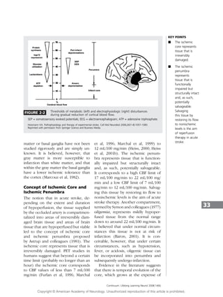
CT-CBF technology (Jovin et al, 2003).
This study, in which core and penum-
bra were determined based on estab-
lished perfusion thresholds, indicated
that within this time frame, irrespec-
tive of the point in time at which the
patients were studied, the ischemic
penumbra was consistently present
and relatively constant, comprising
35
Continuum: Lifelong Learning Neurol 2008;14(6)
Copyright @ American Academy of Neurology. Unauthorized reproduction of this article is prohibited.
9. approximately one-third of the MCA
territory. In contrast to the penumbra,
the ischemic core was highly variable,
ranging from 20% to 70% of cortical
MCA territory. The authors found that
both in patients who recanalized and
in those who did not recanalize, the
extent of core and not that of penum-
bra was correlated with clinical outcome.
CELLULAR MECHANISMS OF
ISCHEMIC NEURONAL INJURY
IN ACUTE STROKE
Introduction
At a cellular level, the biochemical and
electrophysiologic mechanisms in-
volved in the ischemic brain injury
vary according to the extent of cere-
bral ischemia. Neuronal cell death
occurs as a result of two main mech-
anisms: necrosis and apoptosis. Ne-
crosis is a process that is not regulated
or programmed and is the predomi-
nant mechanism that follows acute
permanent focal vascular occlusion.
Necrosis occurs mainly as a conse-
quence of disruption of cellular ho-
meostasis due to energy failure and is
accompanied by cellular swelling,
membrane lysis, inflammation, vascu-
lar damage, and edema formation
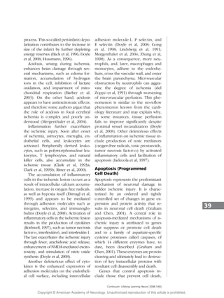
(Bhardwaj et al, 2003) (Figure 2-4).
Apoptosis, or programmed cell death,
is characterized by cell shrinkage,
chromatin clumping, and cytoplasmic
blebbing and is not associated with
inflammation or secondary injury to
surrounding brain (Graham and Chen,
2001; Thompson, 1995; Vaux et al,
1994) (Figure 2-5). These two distinct
types of neuronal death appear to rep-
resent opposite poles of a spectrum
that coexist within the ischemic brain,
with necrosis being the main mecha-
nism of neuronal injury in the ischemic
core and apoptosis being the main
mechanism of neuronal injury in the
penumbra where, because of the milder
degree of ischemia, sufficient energy is
produced to allow for expression of
new proteins that mediate apoptosis
(Bhardwaj et al, 2003; Dirnagl et al,
1999; Graham and Chen, 2001).
Acute vascular occlusion triggers a
complex sequence of pathophysiologic
events that evolve over time and space.
Major pathogenic mechanisms of the
ischemic cascade leading to neuronal
injury constitute active targets for
various neuroprotective strategies and
include cytotoxicity, peri-infarct depo-
larization, inflammation, tissue acidosis,
nitric oxide, and free radical produc-
tion, as well as, at a later stage, apop-
tosis (Barber et al, 2003; Doyle et al,
2008; Dirnagl et al, 1999).
Excitotoxicity, Peri-infarct
Depolarizations, Acidosis,
Inflammation
The reduction in regional CBF through
insufficient delivery of the neuron’s
main energy substrates, oxygen and
glucose, results in inadequate produc-
tion of energy required to maintain
ionic gradients (Martin et al, 1994).
Since the transport of calcium from
the cell into the extracellular space is an
energy-dependent process, this leads to
intracellular accumulation of calcium.
Calcium influx is further enhanced by
impairment in the energy-dependent
reuptake of excitatory amino acids,
especially glutamate, and by release
of excitatory amino acids into the
extracellular space. An increase in
extracellular glutamate leads to in-
creased calcium influx, through in-
creased stimulation of the NMDA or
non-NMDA (mainly -amino-3-hydroxy-
5-methylisoxazole-4-propionicacid[AMPA])
receptor (Budd, 1998). At the same
time, sodium and chloride enter the
neuron via channels for monovalent
ions (Tyson et al, 1996). Water follows
osmotic gradients, leading to edema,
which is predominantly cytotoxic and
can further diminish perfusion in re-
gions surrounding the core, leading to
36
Continuum: Lifelong Learning Neurol 2008;14(6)
KEY POINTS
A At a cellular
level, the
biochemical and
electrophysiologic
mechanisms
involved in the
ischemic brain
injury vary
according to the
extent of cerebral
ischemia.
A Neuronal cell
death occurs
as a result of
two main
mechanisms:
necrosis and
apoptosis.
A Necrosis occurs
predominantly in
the hyperacute
stage within the
ischemic core. It
occurs mainly as
a consequence
of disruption
of cellular
homeostasis
due to energy
failure and is
accompanied by
cellular swelling,
membrane lysis,
inflammation,
vascular damage,
and edema
formation.
PATHOPHYSIOLOGY
Copyright @ American Academy of Neurology. Unauthorized reproduction of this article is prohibited.
10. recruitment of penumbral areas into
the core (Hossmann, 2006; Raichle,
1982). Effects of delayed edema for-
mation (at this stage predominantly
vasogenic) include increased intracra-
nial pressure, shift and displacement
of brain structures, vascular compres-
sion, and herniation (Hossmann, 2006).
The accumulation of intracellular cal-
cium leads to a series of events at both
the cytoplasmic and nuclear levels that
result in cell death through several
37
Continuum: Lifelong Learning Neurol 2008;14(6)
FIGURE 2-4 Cellular mechanisms of ischemic neuronal injury in acute stroke.
IL-1b = interleukin 1b; TNF = tumor necrosis factor-; ICAM = intercellular adhesion molecule;
ECAM = endothelial cell adhesion molecule.
Reprinted with permission from Barber PA, Demchuk AM, Hirt L, Buchan AM. Biochemistry of ischemic stroke. In: Barnett HJM, Bogousslavsky
J, Meldrum H, eds. Advances in neurology: ischemic stroke. Philadelphia: Lippincott Williams Willkins, 2003:151.
Copyright @ American Academy of Neurology. Unauthorized reproduction of this article is prohibited.
11. mechanisms: activation of enzymes that
degrade cytoskeletal proteins (Baudry
et al, 1981; Chen and Strickland, 1997),
activation of lipoxygenase and cyclo-
oxygenase, xanthine oxydase and nitric
oxide synthase with resultant accumu-
lation of highly cytotoxic oxygen free
radicals oxygen (O2), hydrogen perox-
ide (H2O2), hydroxyl (OH), and nitric
oxide (NO). These reactions occur
both in the cytoplasm and in the mito-
chondria. Mitochondria are an impor-
tant source of reactive oxygen species.
As a consequence of free radical–
mediated disruption of the inner mito-
chondrial membrane and the oxidation
of the proteins that mediate electron
transport (Dugan and Choi, 1994),
the mitochondrial membrane becomes
leaky through the formation of a so-
called mitochondrial permeability tran-
sition pore in the mitochondrial mem-
brane (Mergenthaler et al, 2004). This
results in mitochondrial swelling, intra-
mitochondrial calcium accumulation,
impaired energy production, and reac-
tive oxygen species production (Kristian
and Siesjo, 1998). Another consequence
of disrupted mitochondrial permeability
is the release of proapoptotic molecules,
such as cytochrome c and caspase-9
(Dirnagl et al, 1999; Doyle et al, 2008).
Following energy loss, membrane
potentials cannot be maintained, leading
to depolarization of neurons and glia
(Dirnagl et al, 1999). While in the core
region depolarization may be perma-
nent, in the penumbral area cells can
depolarize and then undergo repetitive
depolarization, an active energy-requiring
38
Continuum: Lifelong Learning Neurol 2008;14(6)
FIGURE 2-5 Overview of molecular events involved in apoptosis.
PKC = protein kinase C; CAD = capase activated DNAase.
Reprinted with permission from Barber PA, Demchuk AM, Hirt L, Buchan AM. Biochemistry
of ischemic stroke. In: Barnett HJM, Bogousslavsky J, Meldrum H, eds. Advances in neurology:
ischemic stroke. Philadelphia: Lippincott Williams Willkins, 2003:151.
PATHOPHYSIOLOGY
Copyright @ American Academy of Neurology. Unauthorized reproduction of this article is prohibited.
12. process. This so-called peri-infarct depo-
larization contributes to the increase in
size of the infarct by further depleting
energy reserves (Back et al, 1996; Doyle
et al, 2008; Hossmann, 1996).
Acidosis, arising during ischemia,
enhances brain damage through sev-
eral mechanisms, such as edema for-
mation, accumulation of hydrogen
ions in the cell, inhibition of lactate
oxidation, and impairment of mito-
chondrial respiration (Barber et al,
2003). On the other hand, acidosis
appears to have antiexcitotoxic effects,
and therefore some authors argue that
the role of acidosis in focal cerebral
ischemia is complex and poorly un-
derstood (Mergenthaler et al, 2004).
Inflammation further exacerbates
the ischemic injury. Soon after onset
of ischemia, astrocytes, microglia, en-
dothelial cells, and leukocytes are
activated. Peripherally derived leuko-
cytes, such as polymorphonuclear leu-
kocytes, T lymphocytes, and natural
killer cells, also accumulate in the
ischemic tissue (Clark et al, 1993a;
Clark et al, 1993b; Ritter et al, 2000).
The accumulation of inflammatory
cells in the ischemic lesion occurs as a
result of intracellular calcium accumu-
lation, increase in oxygen free radicals,
as well as hypoxia itself (Dirnagl et al,
1999) and appears to be mediated
through adhesion molecules such as
integrins, selectins, and immunoglo-
bulins (Doyle et al, 2008). Activation of
inflammatory cells in the ischemic lesion
results in the production of cytokines
(Rothwell, 1997), such as tumor necrosis
factor-, interleukin-6, and interleukin-1.
The last exacerbates the ischemic injury
through fever, arachidonic acid release,
enhancement of NMDA-mediated excito-
toxicity, and stimulation of nitric oxide
synthesis (Doyle et al, 2008).
Another deleterious effect of cyto-
kines is the enhanced expression of
adhesion molecules on the endotheli-
al cell surface, including intercellular
adhesion molecule-1, P selectin, and
E selectin (Doyle et al, 2008; Gong
et al, 1998; Lindsberg et al, 1991;
Mergenthaler et al, 2004; Zhang et al,
1998). As a consequence, more neu-
trophils, and, later, macrophages and
monocytes, adhere to the endothe-
lium, cross the vascular wall, and enter
the brain parenchyma. Microvascular
obstruction by neutrophils can aggra-
vate the degree of ischemia (del
Zoppo et al, 1991) through worsening
of microvascular perfusion. This phe-
nomenon is similar to the no-reflow
phenomenon known from the cardi-
ology literature and may explain why,
in some instances, tissue perfusion
fails to improve significantly despite
proximal vessel recanalization (Doyle
et al, 2008). Other deleterious effects
of inflammation on ischemic tissue in-
clude production of toxic mediators
(oxygen free radicals, toxic prostanoids,
tumor necrosis factor-) by activated
inflammatory cells and facilitation of
apoptosis (Iadecola et al, 1997).
Apoptosis (Programmed
Cell Death)
Apoptosis represents the predominant
mechanism of neuronal damage in
milder ischemic injury. It is charac-
terized by an ordered and tightly
controlled set of changes in gene ex-
pression and protein activity that re-
sults in neuronal cell death (Graham
and Chen, 2001). A central role in
apoptosis-mediated mechanisms of is-
chemic injury is attributed to genes
that suppress or promote cell death
and to a family of aspartate-specific
cysteine proteases called caspases, of
which 14 different enzymes have, to
date, been described (Graham and
Chen, 2001). These enzymes are protein
cleaving and ultimately lead to destruc-
tion of key intracellular proteins with
resultant cell disassembly and death.
Genes that control apoptosis in-
clude those that prevent cell death,
39
Continuum: Lifelong Learning Neurol 2008;14(6)
Copyright @ American Academy of Neurology. Unauthorized reproduction of this article is prohibited.
13. such as BCL2, and genes that promote
cell death, such as BAX or p53. The
main sites at which apoptosis can be
initiated are the mitochondria, cell
membrane receptors, and chromo-
somal DNA. Mitochondrial injury may
result in release of cytochrome c, lead-
ing to activation of apoptosis through
caspase-dependent mechanisms. How-
ever, a caspase-independent mecha-
nism may also initiate apoptosis at
the mitochondrial level (Green and
Read, 1998). DNA damage can trigger
apoptosis by inducing expression of
the transcription factor p53 (Miyashita
et al, 1994). This leads to alteration
of transcription of several genes (in-
cluding BAX) and in the initiation of
apoptosis.
Blood-Brain Barrier and the
Neurovascular Unit
The integrity of the blood-brain barrier
plays an important role in the patho-
physiology of acute stroke. Cellular
elements that form the blood-brain
barrier matrix include endothelial cells
and astrocytes. During cerebral ische-
mia, the normal structure of this matrix
and its intercellular signal exchange are
affected by the ischemic process. A
prominent role in the changes underly-
ing blood-brain barrier dysfunction is
attributed to a family of proteases called
matrix metalloproteases (Lo, 2008;
Mergenthaler et al, 2004). Increased
presence of these enzymes, especially
metalloproteinase-9, has been corre-
lated with damage to the blood-brain
barrier, with an increased risk of hemor-
rhagic transformation after tissue plas-
minogen activator (t-PA) administration
and with the extent of neuronal dam-
age (Mergenthaler et al, 2004).
An emerging concept in the patho-
physiology of acute stroke is that of
the neurovascular unit comprising
endothelium and astrocytes in addi-
tion to the neuron (Lo, 2008; Lo et al,
2005). While, traditionally, stroke has
been seen as primarily a neuronal
disorder, the interaction between neu-
rons, endothelium, and astrocytes
through cell-cell signaling and cell-
matrix interactions is regarded as
increasingly important in the under-
standing of stroke and its response to
stroke treatments (Lo, 2008).
REFERENCES
Ad Hoc Committee. Advisory Council for the National Institute of Neurological and
Communicative Disorders and Stroke. A classification and outline of cerebrovascular
diseases. II. Stroke 1975;6(5):564–616.
Albers GW, Caplan LR, Easton JD, et al. Transient ischemic attack—proposal for new
definition. N Engl J Med 2002;347(21):1713–1716.
Alexandrov AV, Sharma VK, Lao AY, et al. Reversed Robin Hood syndrome in acute
ischemic stroke patients. Stroke 2007;38(11):3045–3048.
Astrup J, Siesjo BK, Symon L. Thresholds in cerebral ischemia—the ischemic penumbra.
Stroke 1981;12(6):723–725.
Ay H, Buonanno FS, Rordorf G, et al. Normal diffusion-weighted MRI during stroke-like
deficits. Neurology 1999;52(9):1784–1792.
Back T, Ginsberg MD, Dietrich WD, Watson BD. Induction of spreading depression in
the ischemic hemisphere following experimental middle cerebral artery occlusion:
effect on infarct morphology. J Cereb Blood Flow Metab 1996;16(2):202–213.
40
Continuum: Lifelong Learning Neurol 2008;14(6)
KEY POINT
A Apoptosis is the
main mechanism
of neuronal injury
in the penumbra
where, because
of the milder
degree of
ischemia,
sufficient energy
is produced to
allow for
expression of
new proteins
that mediate cell
death through
an ordered and
tightly controlled
set of changes in
gene expression
and protein
activity.
PATHOPHYSIOLOGY
Copyright @ American Academy of Neurology. Unauthorized reproduction of this article is prohibited.
14. Bamford JM, Warlow CP. Evolution and testing of the lacunar hypothesis. Stroke
1988;19(9):1074–1082.
Barber PA, Davis SM, Darby DG, et al. Absent middle cerebral artery flow predicts
the presence and evolution of the ischemic penumbra. Neurology 1999;52(6):
1125–1132.
Barber PA, Demchuk AM, Hirt L, Buchan AM. Biochemistry of ischemic stroke. In:
Barnett HJM, Bogousslavsky J, Meldrum H, eds. Advances in neurology: ischemic stroke.
Philadelphia: Lippincott Williams Willkins, 2003:151.
Baron JC. Perfusion thresholds in human cerebral ischemia: historical perspective and
therapeutic implications. Cerebrovasc Dis 2001;11(suppl 1):2–8.
Baudry M, Bundman MC, Smith EK, Lynch GS. Micromolar calcium stimulates
proteolysis and glutamate binding in rat brain synaptic membranes. Science 1981;
212(4497):937–938.
Bhardwaj A, Alkayed NJ, Kirsch JR, Hurn PD. Mechanisms of ischemic brain damage.
Curr Cardiol Rep 2003;5(2):160–167.
Boysen G. Cerebral blood flow measurement as a safeguard during carotid
endarterectomy. Stroke 1971;2(1):1–10.
Budd SL. Mechanisms of neuronal damage in brain hypoxia/ischemia: focus on
the role of mitochondrial calcium accumulation. Pharmacol Ther 1998;80(2):
203–229.
Caplan LR. Intracranial branch atheromatous disease: a neglected, understudied, and
underused concept [published errata appears in Neurology 1990;40(4):725]. Neurology
1989;39(9):1246–1250.
Caplan LR. Basic pathology, anatomy, and pathophysiology of stroke. In: Caplan’s
stroke: a clinical approach. Boston: Butterworth-Heinemann, 2000:19.
Caplan LR, Hennerici M. Impaired clearance of emboli (washout) is an important link
between hypoperfusion, embolism, and ischemic stroke. Arch Neurol 1998;55(11):1475–1482.
Chen ZL, Strickland S. Neuronal death in the hippocampus is promoted by
plasmin-catalyzed degradation of laminin. Cell 1997;91(7):917–925.
Clark RK, Lee EV, Fish CJ, et al. Development of tissue damage, inflammation and
resolution following stroke: an immunohistochemical and quantitative planimetric study.
Brain Res Bull 1993a;31(5):565–572.
Clark WM, Coull BM, Briley DP, et al. Circulating intercellular adhesion molecule-1
levels and neutrophil adhesion in stroke. J Neuroimmunol 1993b;44(1):123–125.
del Zoppo GJ, Schmid-Schonbein GW, Mori E, et al. Polymorphonuclear leukocytes
occlude capillaries following middle cerebral artery occlusion and reperfusion in
baboons. Stroke 1991;22(10):1276–1283.
Dirnagl U, Iadecola C, Moskowitz MA. Pathobiology of ischaemic stroke: an integrated
view. Trends Neurosci 1999;22(9):391–397.
Doyle KP, Simon RP, Stenzel-Poore MP. Mechanisms of ischemic brain damage
[published online ahead of print January 25, 2008]. Neuropharmacology PMID: 18308346.
Dugan LL, Choi DW. Excitotoxicity, free radicals, and cell membrane changes. Ann
Neurol 1994;35(suppl):S17–S21.
41
Continuum: Lifelong Learning Neurol 2008;14(6)
Copyright @ American Academy of Neurology. Unauthorized reproduction of this article is prohibited.
15. Engelter ST, Provenzale JM, Petrella JR, Alberts MJ. Diffusion MR imaging and
transient ischemic attacks. Stroke 1999;30(12):2762–2763.
Ferro JM. Cardioembolic stroke: an update. Lancet Neurol 2003;2(3):177–188.
Fisher CM. Lacunar strokes and infarcts: a review. Neurology 1982;32(8):871–876.
Fisher CM. Lacunes: small, deep cerebral infarcts. 1965. Neurology 1998;50(4):841–852.
Furlan M, Marchal G, Viader F, et al. Spontaneous neurological recovery after stroke
and the fate of the ischemic penumbra. Ann Neurol 1996;40(2):216–226.
Garcia JH, Ho K, Pantoni L. Pathology. In: Barnett HJM, Mohr JP, Stein BM, Yatsu FM,
editors. Stroke pathophysiology, diagnosis and management. 3rd ed. Philadelphia:
Churchill Livingstone, 1998:147–148.
Ginsberg MD. Adventures in the pathophysiology of brain ischemia: penumbra, gene
expression, neuroprotection: the 2002 Thomas Willis Lecture. Stroke 2003;34(1):214–223.
Gong C, Qin Z, Betz AL, et al. Cellular localization of tumor necrosis factor alpha
following focal cerebral ischemia in mice [published errata appears in Brain Res
1999;818(1):184]. Brain Res 1998;801(1–2):1–8.
Graham SH, Chen J. Programmed cell death in cerebral ischemia. J Cereb Blood Flow
Metab 2001;21(2):99–109.
Grau AJ, Weimar C, Buggle F, et al. Risk factors, outcome, and treatment in subtypes of
ischemic stroke: the German stroke data bank. Stroke 2001;32(11):2559–2566.
Green DR, Read JC. Mitochondria and apoptosis. Science 1998;281(5381):1309–1312.
Guadagno JV, Warburton EA, Aigbirhio FI, et al. Does the acute diffusion-weighted
imaging lesion represent penumbra as well as core? A combined quantitative PET/MRI
voxel-based study. J Cereb Blood Flow Metab 2004;24(11):1249–1254.
Guadagno JV, Warburton EA, Jones PS, et al. The diffusion-weighted lesion in acute
stroke: heterogeneous patterns of flow/metabolism uncoupling as assessed by
quantitative positron emission tomography. Cerebrovasc Dis 2005;19(4):239–246.
Hart RG, Easton JD. Hemorrhagic infarcts. Stroke 1986;17(4):586–589.
Heiss WD. Ischemic penumbra: evidence from functional imaging in man. J Cereb
Blood Flow Metab 2000;20(9):1276–1293.
Heiss WD, Forsting M, Diener HC. Imaging in cerebrovascular disease. Curr Opin
Neurol 2001a;14(1):67–75.
Heiss WD, Kracht LW, Thiel A, et al. Penumbral probability thresholds of cortical
flumazenil binding and blood flow predicting tissue outcome in patients with cerebral
ischaemia. Brain 2001b;124(pt 1):20–29.
Heiss WD, Sobesky J, Hesselmann V. Identifying thresholds for penumbra and
irreversible tissue damage. Stroke 2004;35(11 suppl 1):2671–2674.
Hjort N, Butcher K, Davis SM, et al. Magnetic resonance imaging criteria for
thrombolysis in acute cerebral infarct. Stroke 2005;36(2):388–397.
Hossmann KA. Periinfarct depolarizations. Cerebrovasc Brain Metab Rev 1996;8(3):195–208.
Hossmann KA. Pathophysiology and therapy of experimental stroke. Cell Mol
Neurobiol 2006;26(7-8):1057–1083.
42
Continuum: Lifelong Learning Neurol 2008;14(6)
PATHOPHYSIOLOGY
Copyright @ American Academy of Neurology. Unauthorized reproduction of this article is prohibited.
16. Iadecola C, Zhang F, Casey R, et al. Delayed reduction of ischemic brain injury and
neurological deficits in mice lacking the inducible nitric oxide synthase gene.
J Neurosci 1997;17(23):9157–9164.
Jennett WB, Harper AM, Gillespie FC. Measurement of regional cerebral blood-flow
during carotid ligation. Lancet 1966;2(7474):1162–1163.
Jones TH, Morawetz RB, Crowell RM, et al. Thresholds of focal cerebral ischemia in
awake monkeys. J Neurosurg 1981;54(6):773–782.
Jovin TG, Yonas H, Gebel JM, et al. The cortical ischemic core and not the consistently
present penumbra is a determinant of clinical outcome in acute middle cerebral
artery occlusion. Stroke 2003;34(10):2426–2433.
Kaufmann AM, Firlik AD, Fukui MB, et al. Ischemic core and penumbra in human
stroke. Stroke 1999;30(1):93–99.
Kidwell CS, Alger JR, Di Salle F, et al. Diffusion MRI in patients with transient ischemic
attacks. Stroke 1999;30(6):1174–1180.
Klijn CJ, Kappelle LJ, Tulleken CA, van Gijn J. Symptomatic carotid artery occlusion: a
reappraisal of hemodynamic factors. Stroke 1997;28(10):2084–2093.
Kolominsky-Rabas PL, Weber M, Gefeller O, et al. Epidemiology of ischemic stroke
subtypes according to TOAST criteria: incidence, recurrence, and long-term survival in
ischemic stroke subtypes: a population-based study. Stroke 2001;32(12):2735–2740.
Kristian T, Siesjo BK. Calcium in ischemic cell death. Stroke 1998;29(3):705–718.
Larrue V, von Kummer R, del Zoppo G, Bluhmki E. Hemorrhagic transformation in
acute ischemic stroke. Potential contributing factors in the European Cooperative
Acute Stroke Study. Stroke 1997;28(5):957–960.
Lindsberg PJ, Hallenbeck JM, Feuerstein G. Platelet-activating factor in stroke and
brain injury. Ann Neurol 1991;30(2):117–129.
Lo EH. Experimental models, neurovascular mechanisms and translational issues in
stroke research. Br J Pharmacol 2008;153(suppl 1):396–405.
Lo EH, Moskowitz MA, Jacobs TP. Exciting, radical, suicidal: how brain cells die after
stroke. Stroke 2005;36(2):189–192.
Lyden PD, Zivin JA. Hemorrhagic transformation after cerebral ischemia: mechanisms and
incidence. Cerebrovasc Brain Metab Rev 1993;5(1):1–16.
Marchal G, Beaudouin V, Rioux P, et al. Prolonged persistence of substantial volumes of
potentially viable brain tissue after stroke: a correlative PET-CT study with voxel-based
data analysis. Stroke 1996;27(4):599–606.
Marchal G, Benali K, Iglesias S, et al. Voxel-based mapping of irreversible ischaemic
damage with PET in acute stroke. Brain 1999;122(pt 12):2387–2400.
Marcoux FW, Morawetz RB, Crowell RM, et al. Differential regional vulnerability in
transient focal cerebral ischemia. Stroke 1982;13(3):339–346.
Martin RL, Lloyd HG, Cowan AI. The early events of oxygen and glucose deprivation:
setting the scene for neuronal death? Trends Neurosci 1994;17(6):251–257.
Mayer TE, Schulte-Altedorneburg G, Droste DW, Bru¨ ckmann H. Serial CT and MRI of
ischaemic cerebral infarcts: frequency and clinical impact of haemorrhagic transformation.
Neuroradiology 2000;42(4):233–239.
43
Continuum: Lifelong Learning Neurol 2008;14(6)
Copyright @ American Academy of Neurology. Unauthorized reproduction of this article is prohibited.
17. Mergenthaler P, Dirnagl U, Meisel A. Pathophysiology of stroke: lessons from animal
models. Metab Brain Dis 2004;19(3–4):151–167.
Miyashita T, Krajewski S, Krajewska M, et al. Tumor suppressor p53 is a regulator of
bcl-2 and bax gene expression in vitro and in vivo. Oncogene 1994;9(6):1799–1805.
Mohr JP. Lacunes. Stroke 1982;13(1):3–11.
Ogasawara K, Ogawa A, Doi M, et al. Prediction of acute embolic stroke outcome after
local intraarterial thrombolysis: value of pretreatment and posttreatment 99mTc-ethyl
cysteinate dimer single photon emission computed tomography. J Cereb Blood Flow
Metab 2000;20(11):1579–1586.
Ovbiagele B, Kidwell CS, Saver JL, et al. Epidemiological impact in the United States of a
tissue-based definition of transient ischemic attack. Stroke 2003;34(4):919–924.
Petty GW, Brown RD Jr, Whisnant JP, et al. Ischemic stroke subtypes: a population-based
study of functional outcome, survival, and recurrence. Stroke 2000;31(5):1062–1068.
Raichle ME. The pathophysiology of brain ischemia and infarction. Clin Neurosurg
1982;29:379–389.
Read SJ, Hirano T, Abbott DF, et al. The fate of hypoxic tissue on 18F-fluoromisonidazole
positron emission tomography after ischemic stroke. Ann Neurol 2000;48(2):228–235.
Ritter LS, Orozco JA, Coull BM, et al. Leukocyte accumulation and hemodynamic
changes in the cerebral microcirculation during early reperfusion after stroke. Stroke
2000;31(5):1153–1161.
Rordorf G, Koroshetz WJ, Copen WA, et al. Regional ischemia and ischemic injury in
patients with acute middle cerebral artery stroke as defined by early diffusion-weighted
and perfusion-weighted MRI. Stroke 1998;29(5):939–943.
Rothwell NJ. Cytokines and acute neurodegeneration. Mol Psychiatry 1997;2(2):120–121.
Sacco RL, Kargman DE, Gu Q, Zamanillo MC. Race-ethnicity and determinants of
intracranial atherosclerotic cerebral infarction. The Northern Manhattan Stroke Study.
Stroke 1995;26(1):14–20.
Schellinger PD, Fiebach JB, Jansen O, et al. Stroke magnetic resonance imaging within
6 hours after onset of hyperacute cerebral ischemia. Ann Neurol 2001;49(4):460–469.
Schlaug G, Benfield A, Baird AE, et al. The ischemic penumbra: operationally defined
by diffusion and perfusion MRI. Neurology 1999;53(7):1528–1537.
Sobesky J, Zaro Weber O, Lehnhardt FG, et al. Does the mismatch match the
penumbra? Magnetic resonance imaging and positron emission tomography in early
ischemic stroke. Stroke 2005;36(5):980–985.
Staroselskaya IA, Chaves C, Silver B, et al. Relationship between magnetic resonance
arterial patency and perfusion-diffusion mismatch in acute ischemic stroke and its
potential clinical use. Arch Neurol 2001;58(7):1069–1074.
Sundt TM Jr, Sharbrough FW, Anderson RE, Michenfelder JD, et al. Cerebral blood
flow measurements and electroencephalograms during carotid endarterectomy.
J Neurosurg 1974;41(3):310–320.
Symon L, Branston NM, Strong AJ, Hope TD. The concepts of thresholds of ischaemia
in relation to brain structure and function. J Clin Pathol Suppl (R Coll Pathol)
1977;11:149–154.
44
Continuum: Lifelong Learning Neurol 2008;14(6)
PATHOPHYSIOLOGY
Copyright @ American Academy of Neurology. Unauthorized reproduction of this article is prohibited.
18. Thompson CB. Apoptosis in the pathogenesis and treatment of disease. Science 1995;
267(5203):1456–1462.
Trojaborg W, Boysen G. Relation between EEG, regional cerebral blood flow and
internal carotid artery pressure during carotid endarterectomy. Electroencephalogr
Clin Neurophysiol 1973;34(1):61–69.
Tyson RL, Sutherland GR, Peeling J. 23Na nuclear magnetic resonance spectral changes
during and after forebrain ischemia in hypoglycemic, normoglycemic, and hyperglycemic
rats. Stroke 1996;27(5):957–964.
Ueda T, Sakaki S, Yuh WT, et al. Outcome in acute stroke with successful intra-arterial
thrombolysis and predictive value of initial single-photon emission-computed
tomography. J Cereb Blood Flow Metab 1999;19(1):99–108.
Vaux DL, Haecker G, Strasser A. An evolutionary perspective on apoptosis. Cell
1994;76(5):777–779.
Zhang Z, Chopp M, Goussev A, Powers C. Cerebral vessels express interleukin 1beta
after focal cerebral ischemia. Brain Res 1998;784(1–2):210–217.
45
Copyright @ American Academy of Neurology. Unauthorized reproduction of this article is prohibited.