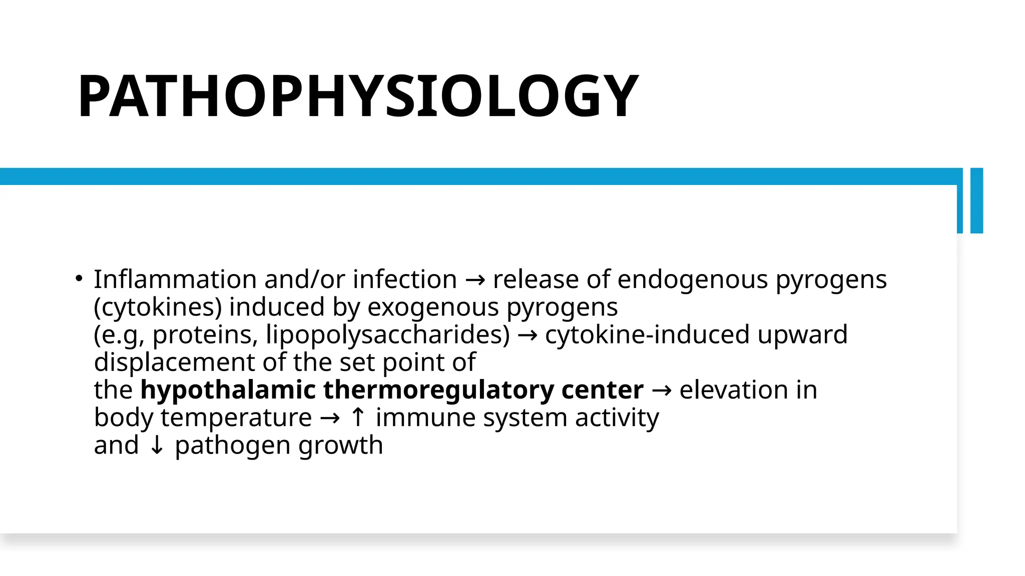
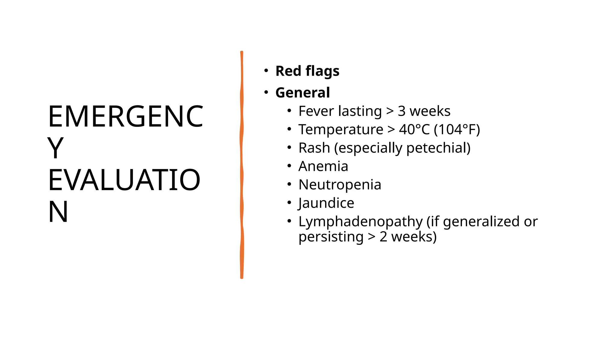
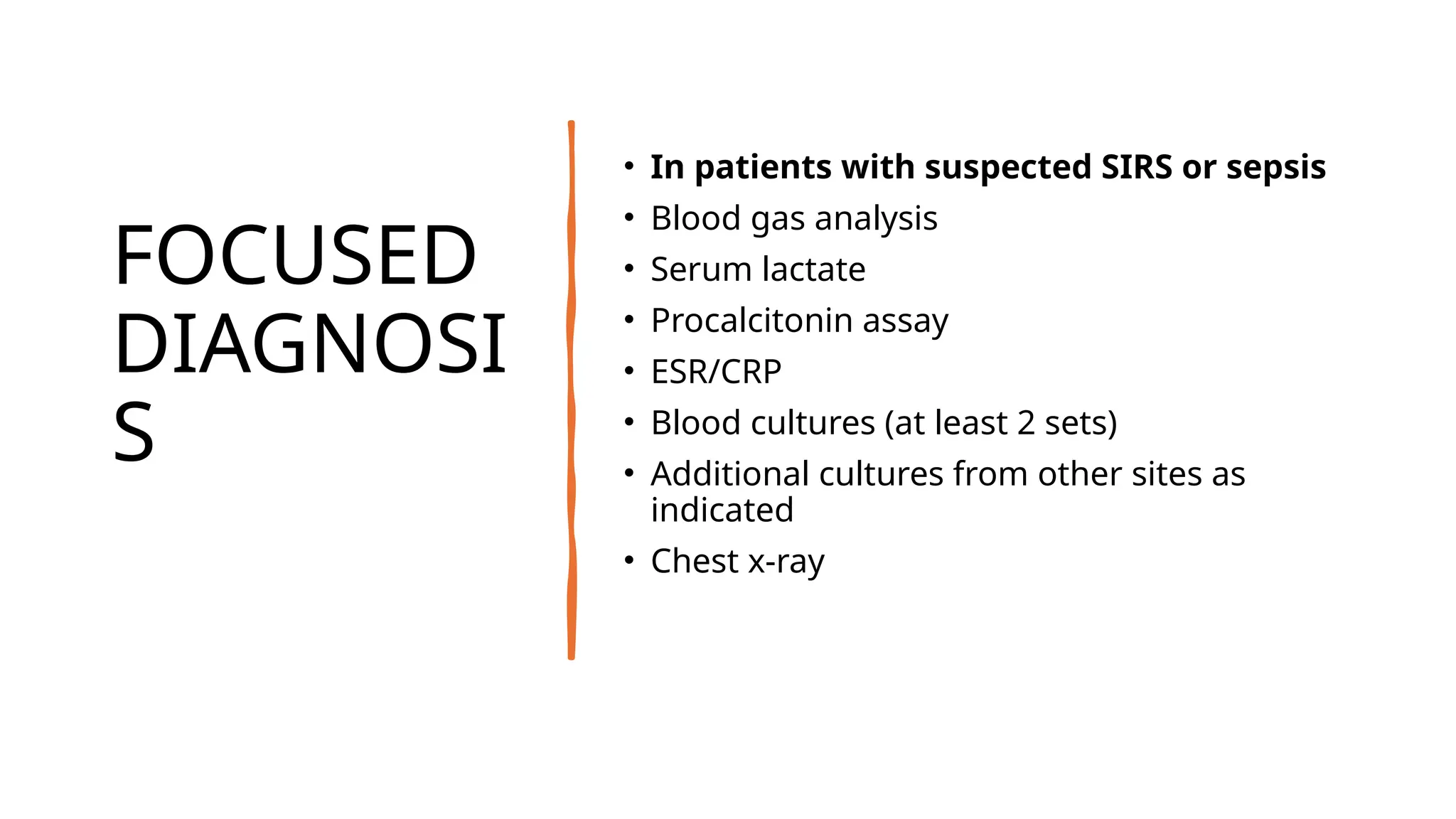
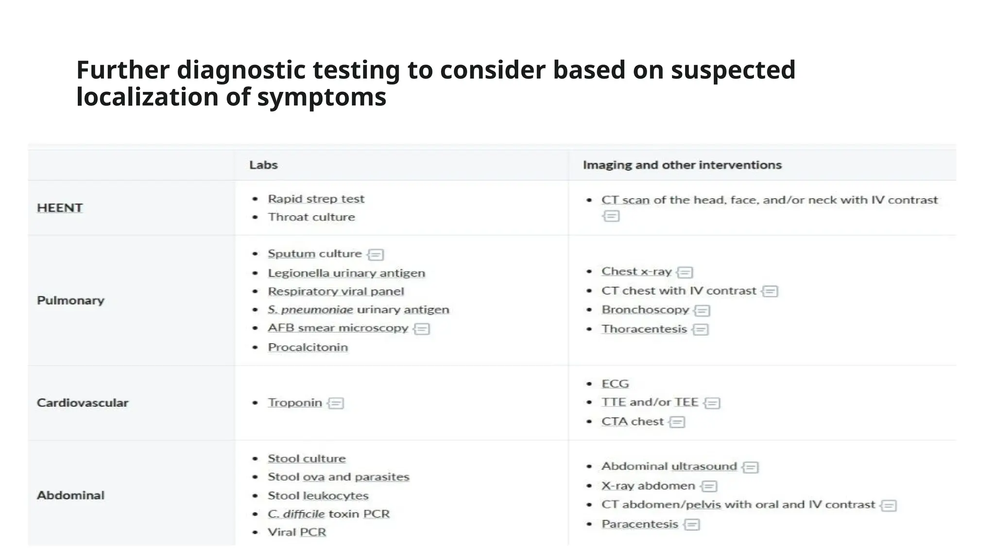
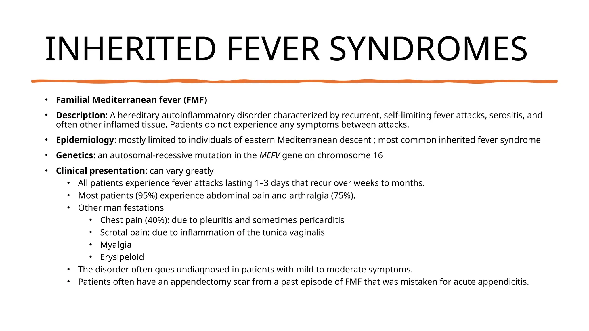
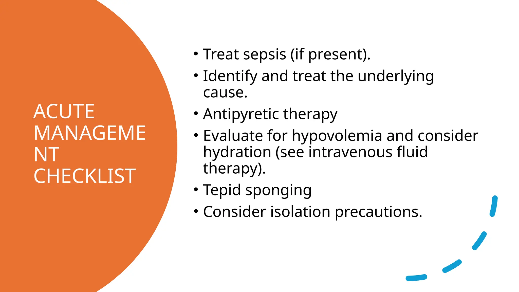
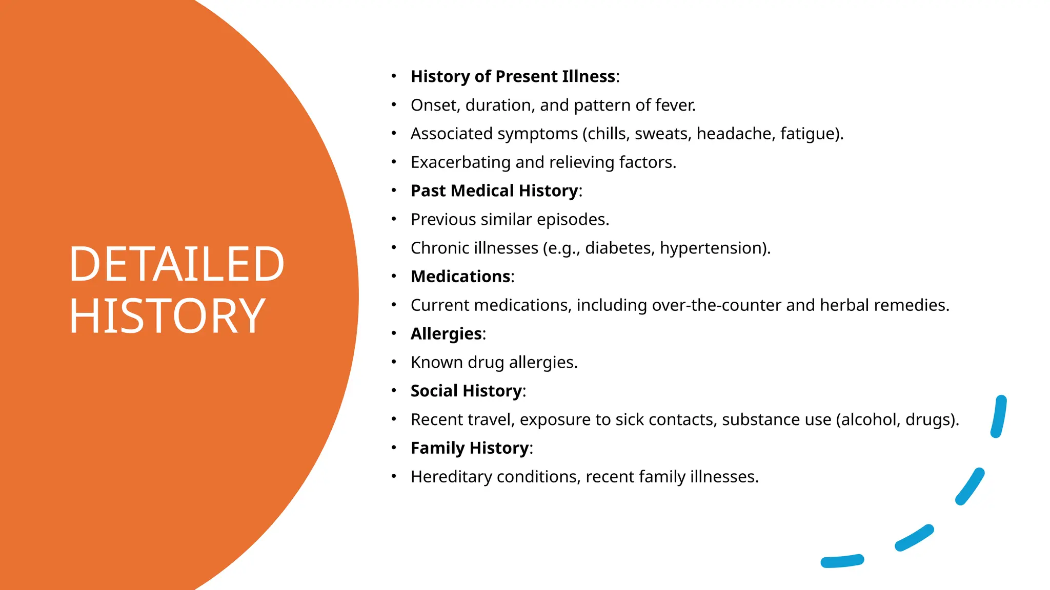
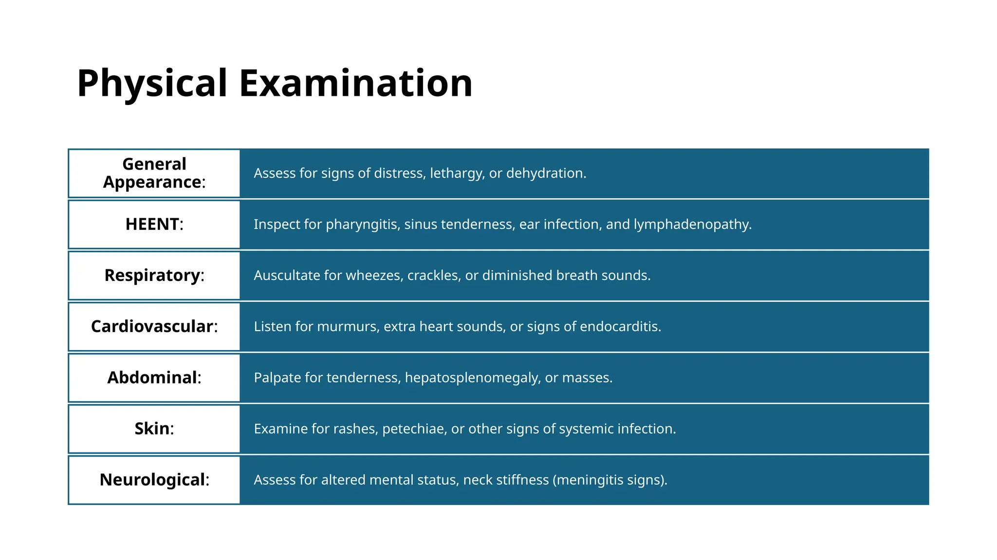
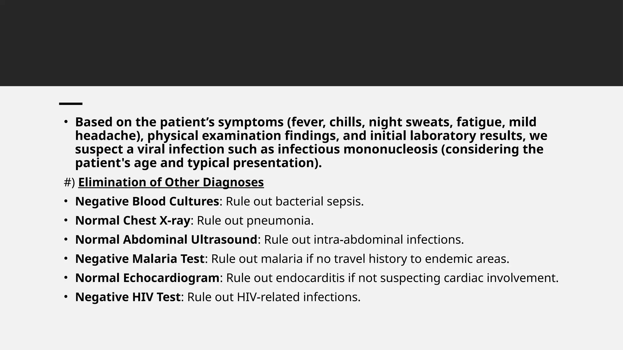
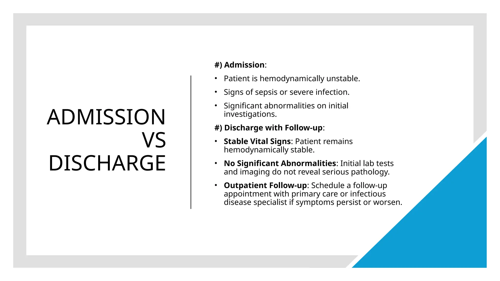
FEVER
MOHAMMED KAIF SHAIKH
MEDF21-2C
PATHOPHYSIOLOGY
• Inflammation and/or infection release of endogenous pyrogens
→
(cytokines) induced by exogenous pyrogens
(e.g, proteins, lipopolysaccharides) cytokine-induced upward
→
displacement of the set point of
the hypothalamic thermoregulatory center elevation in
→
body temperature immune system activity
→ ↑
and pathogen growth
↓
EMERGENCY EVALUATION
Basic approach
Identify and
treat sepsis, if
present.
Identify and treat the
underlying cause.
Provide supportive
therapy
(antipyretics, IV fluids,
tepid sponging).
EMERGENC
Y
EVALUATIO
N
• Red flags
• General
• Fever lasting > 3 weeks
• Temperature > 40°C (104°F)
• Rash (especially petechial)
• Anemia
• Neutropenia
• Jaundice
• Lymphadenopathy (if generalized or
persisting > 2 weeks)
EMERGENCY EVALUATION
• Localized
• CNS: neck stiffness, seizures, headache, altered level of
consciousness, altered mental status
• ENT: purulent ear discharge, mastoid pain
• Chest: hemoptysis, hypoxemia
• Abdomen: guarding, organomegaly, ascites, melena, hematochezia,
persistent vomiting, voluminous diarrhea
• Musculoskeletal: joint swelling, reduced range of motion
FOCUSED
HISTORY
History of present illness
Onset
Duration
Characteristics (e.g., continuous vs.
intermittent or nocturnal, high grade vs. low
grade)
FOCUSED
HISTORY
Recent exposures
Travel
Sick contacts
New medication or substance
Animals, including farm and wild animals
Associated symptoms
General
Chills, rigors
Night sweats
Unintentional weight loss
FOCUSED
DIAGNOSIS
• Laboratory studies
• Routine
• CBC with differential
• Blood glucose
• BMP
• LFTs
• Coagulation
studies (e.g., INR, PTT)
• Urinalysis with microscopy
FOCUSED
DIAGNOSI
S
• In patients with suspected SIRS or sepsis
• Blood gas analysis
• Serum lactate
• Procalcitonin assay
• ESR/CRP
• Blood cultures (at least 2 sets)
• Additional cultures from other sites as
indicated
• Chest x-ray
Further diagnostic testing to consider based on suspected
localization of symptoms
DIFFERENTIAL
DIAGNOSIS BY
FEVER
CHARACTERISTICS
TREATMENT
• Antipyretics: indicated in case of intolerable fever; do not prevent febrile
seizures
• Acetaminophen
• NSAIDs
• Ibuprofen
• Aspirin
• Antibiotics in case of suspected bacterial infection (e.g., pneumonia)
• Acetaminophen is the preferred antipyretic during pregnancy but should be
avoided in patients with severe hepatic dysfunction.
• NSAIDs are contraindicated in pregnancy and hemorrhagic fevers. They
should be used with caution in breastfeeding patients and those with CAD.
INHERITED FEVER SYNDROMES
• Familial Mediterranean fever (FMF)
• Description: A hereditary autoinflammatory disorder characterized by recurrent, self-limiting fever attacks, serositis, and
often other inflamed tissue. Patients do not experience any symptoms between attacks.
• Epidemiology: mostly limited to individuals of eastern Mediterranean descent ; most common inherited fever syndrome
• Genetics: an autosomal-recessive mutation in the MEFV gene on chromosome 16
• Clinical presentation: can vary greatly
• All patients experience fever attacks lasting 1–3 days that recur over weeks to months.
• Most patients (95%) experience abdominal pain and arthralgia (75%).
• Other manifestations
• Chest pain (40%): due to pleuritis and sometimes pericarditis
• Scrotal pain: due to inflammation of the tunica vaginalis
• Myalgia
• Erysipeloid
• The disorder often goes undiagnosed in patients with mild to moderate symptoms.
• Patients often have an appendectomy scar from a past episode of FMF that was mistaken for acute appendicitis.
INHERITED
FEVER
SYNDROMES
• Complication: AA amyloidosis
• Therapy: Prevention of acute
episodes and progression to AA
amyloidosis through inhibition
of granulocyte function
by colchicine.
• Other hereditary fever
syndromes
• Hyper-IgD syndrome
• TNFα reception-associated periodic
syndrome
ACUTE
MANAGEME
NT
CHECKLIST
• Treat sepsis (if present).
• Identify and treat the underlying
cause.
• Antipyretic therapy
• Evaluate for hypovolemia and consider
hydration (see intravenous fluid
therapy).
• Tepid sponging
• Consider isolation precautions.
PATIENT
PRESENTATIO
N
• A 22-year-old male presents to the
Emergency Department (ED) with a
complaint of continuous fever for one
week. He reports associated symptoms
including intermittent chills, night
sweats, fatigue, and a mild headache.
He denies any cough, shortness of
breath, abdominal pain, urinary
symptoms, or recent travel.
IMMEDIATE
ACTIONS
#) Triage and Initial Assessment
1.Vital Signs:
• Temperature: Confirm presence of fever.
• Heart Rate: Assess for tachycardia.
• Respiratory Rate: Check for tachypnea.
• Blood Pressure: Monitor for hypotension.
• Oxygen Saturation: Ensure adequate oxygenation.
2.Patient Stability: Address Airway, Breathing,
Circulation (ABCs).
#) Initial Management:
3. IV Access: Establish intravenous access for fluids and
medications.
4.IV Fluids: Administer IV fluids if there are signs of
dehydration or hypotension.
5.Antipyretics: Administer acetaminophen or ibuprofen
to reduce fever and alleviate discomfort.
DETAILED
HISTORY
• History of Present Illness:
• Onset, duration, and pattern of fever.
• Associated symptoms (chills, sweats, headache, fatigue).
• Exacerbating and relieving factors.
• Past Medical History:
• Previous similar episodes.
• Chronic illnesses (e.g., diabetes, hypertension).
• Medications:
• Current medications, including over-the-counter and herbal remedies.
• Allergies:
• Known drug allergies.
• Social History:
• Recent travel, exposure to sick contacts, substance use (alcohol, drugs).
• Family History:
• Hereditary conditions, recent family illnesses.
Physical Examination
Assess for signs of distress, lethargy, or dehydration.
General
Appearance:
Inspect for pharyngitis, sinus tenderness, ear infection, and lymphadenopathy.
HEENT:
Auscultate for wheezes, crackles, or diminished breath sounds.
Respiratory:
Listen for murmurs, extra heart sounds, or signs of endocarditis.
Cardiovascular:
Palpate for tenderness, hepatosplenomegaly, or masses.
Abdominal:
Examine for rashes, petechiae, or other signs of systemic infection.
Skin:
Assess for altered mental status, neck stiffness (meningitis signs).
Neurological:
Diagnostic
Workup
• Laboratory Tests:
• CBC with Differential: Evaluate for leukocytosis or leukopenia,
indicative of infection or inflammation.
• Basic Metabolic Panel (BMP): Assess electrolytes, renal
function.
• Liver Function Tests (LFTs): Check for hepatic involvement.
• Coagulation Studies (INR, PTT): Evaluate coagulation status.
• Blood Cultures: Obtain at least 2 sets to identify bacteremia.
• Urinalysis with Microscopy: Detect urinary tract infection.
• Procalcitonin Assay: Marker for bacterial infection.
• ESR/CRP: Inflammatory markers.
• HIV Test: To rule out immunosuppression and potential
opportunistic infections.
• Imaging Studies:
• Chest X-ray: Evaluate for pneumonia, lung abscess, or other
thoracic abnormalities.
• Abdominal Ultrasound/CT: If abdominal pain or
hepatosplenomegaly is present.
• Head CT/MRI: If neurological symptoms suggest central nervous
system involvement.
• Echocardiogram: If endocarditis is suspected.
Differential Diagnosis and
Approach
Infectious Causes:
• Bacterial Infection: Positive
blood cultures, leukocytosis,
elevated procalcitonin.
• Viral Infection: Normal/slightly
elevated WBC, specific viral
serology or PCR.
• Tuberculosis: Consider in
presence of night sweats,
weight loss, and contact with TB
patients.
• Malaria: Consider if travel
history to endemic areas.
Non-Infectious Causes:
• Autoimmune/Inflammatory
Diseases: Elevated ESR/CRP,
specific autoantibodies (e.g.,
ANA for lupus).
• Malignancy (e.g., Lymphoma,
Leukemia): Unexplained weight
loss, night sweats, persistent
lymphadenopathy, abnormal
CBC.
• Based on the patient’s symptoms (fever, chills, night sweats, fatigue, mild
headache), physical examination findings, and initial laboratory results, we
suspect a viral infection such as infectious mononucleosis (considering the
patient's age and typical presentation).
#) Elimination of Other Diagnoses
• Negative Blood Cultures: Rule out bacterial sepsis.
• Normal Chest X-ray: Rule out pneumonia.
• Normal Abdominal Ultrasound: Rule out intra-abdominal infections.
• Negative Malaria Test: Rule out malaria if no travel history to endemic areas.
• Normal Echocardiogram: Rule out endocarditis if not suspecting cardiac involvement.
• Negative HIV Test: Rule out HIV-related infections.
MANAGEMENT AND STABILIZATION
Symptomatic
Treatment:
Antipyretics:
Continue
acetaminophen or
ibuprofen for fever
management.
Hydration: Encourage
oral fluids or continue
IV fluids if necessary.
Antiviral Therapy:
Not typically required
for suspected viral
infection like
infectious
mononucleosis.
Patient Education:
Advise rest, adequate
hydration, and
symptomatic care.
Educate on the self-
limiting nature of viral
infections and when
to seek further
medical attention.
ADMISSION
VS
DISCHARGE
#) Admission:
• Patient is hemodynamically unstable.
• Signs of sepsis or severe infection.
• Significant abnormalities on initial
investigations.
#) Discharge with Follow-up:
• Stable Vital Signs: Patient remains
hemodynamically stable.
• No Significant Abnormalities: Initial lab tests
and imaging do not reveal serious pathology.
• Outpatient Follow-up: Schedule a follow-up
appointment with primary care or infectious
disease specialist if symptoms persist or worsen.
FEVER MANAGMENT , OVERVIEW , AND CAUSES.

FEVER MANAGMENT , OVERVIEW , AND CAUSES.

  • 1.
  • 2.
    PATHOPHYSIOLOGY • Inflammation and/orinfection release of endogenous pyrogens → (cytokines) induced by exogenous pyrogens (e.g, proteins, lipopolysaccharides) cytokine-induced upward → displacement of the set point of the hypothalamic thermoregulatory center elevation in → body temperature immune system activity → ↑ and pathogen growth ↓
  • 3.
    EMERGENCY EVALUATION Basic approach Identifyand treat sepsis, if present. Identify and treat the underlying cause. Provide supportive therapy (antipyretics, IV fluids, tepid sponging).
  • 4.
    EMERGENC Y EVALUATIO N • Red flags •General • Fever lasting > 3 weeks • Temperature > 40°C (104°F) • Rash (especially petechial) • Anemia • Neutropenia • Jaundice • Lymphadenopathy (if generalized or persisting > 2 weeks)
  • 5.
    EMERGENCY EVALUATION • Localized •CNS: neck stiffness, seizures, headache, altered level of consciousness, altered mental status • ENT: purulent ear discharge, mastoid pain • Chest: hemoptysis, hypoxemia • Abdomen: guarding, organomegaly, ascites, melena, hematochezia, persistent vomiting, voluminous diarrhea • Musculoskeletal: joint swelling, reduced range of motion
  • 6.
    FOCUSED HISTORY History of presentillness Onset Duration Characteristics (e.g., continuous vs. intermittent or nocturnal, high grade vs. low grade)
  • 7.
    FOCUSED HISTORY Recent exposures Travel Sick contacts Newmedication or substance Animals, including farm and wild animals Associated symptoms General Chills, rigors Night sweats Unintentional weight loss
  • 8.
    FOCUSED DIAGNOSIS • Laboratory studies •Routine • CBC with differential • Blood glucose • BMP • LFTs • Coagulation studies (e.g., INR, PTT) • Urinalysis with microscopy
  • 9.
    FOCUSED DIAGNOSI S • In patientswith suspected SIRS or sepsis • Blood gas analysis • Serum lactate • Procalcitonin assay • ESR/CRP • Blood cultures (at least 2 sets) • Additional cultures from other sites as indicated • Chest x-ray
  • 10.
    Further diagnostic testingto consider based on suspected localization of symptoms
  • 13.
  • 15.
    TREATMENT • Antipyretics: indicatedin case of intolerable fever; do not prevent febrile seizures • Acetaminophen • NSAIDs • Ibuprofen • Aspirin • Antibiotics in case of suspected bacterial infection (e.g., pneumonia) • Acetaminophen is the preferred antipyretic during pregnancy but should be avoided in patients with severe hepatic dysfunction. • NSAIDs are contraindicated in pregnancy and hemorrhagic fevers. They should be used with caution in breastfeeding patients and those with CAD.
  • 16.
    INHERITED FEVER SYNDROMES •Familial Mediterranean fever (FMF) • Description: A hereditary autoinflammatory disorder characterized by recurrent, self-limiting fever attacks, serositis, and often other inflamed tissue. Patients do not experience any symptoms between attacks. • Epidemiology: mostly limited to individuals of eastern Mediterranean descent ; most common inherited fever syndrome • Genetics: an autosomal-recessive mutation in the MEFV gene on chromosome 16 • Clinical presentation: can vary greatly • All patients experience fever attacks lasting 1–3 days that recur over weeks to months. • Most patients (95%) experience abdominal pain and arthralgia (75%). • Other manifestations • Chest pain (40%): due to pleuritis and sometimes pericarditis • Scrotal pain: due to inflammation of the tunica vaginalis • Myalgia • Erysipeloid • The disorder often goes undiagnosed in patients with mild to moderate symptoms. • Patients often have an appendectomy scar from a past episode of FMF that was mistaken for acute appendicitis.
  • 17.
    INHERITED FEVER SYNDROMES • Complication: AAamyloidosis • Therapy: Prevention of acute episodes and progression to AA amyloidosis through inhibition of granulocyte function by colchicine. • Other hereditary fever syndromes • Hyper-IgD syndrome • TNFα reception-associated periodic syndrome
  • 18.
    ACUTE MANAGEME NT CHECKLIST • Treat sepsis(if present). • Identify and treat the underlying cause. • Antipyretic therapy • Evaluate for hypovolemia and consider hydration (see intravenous fluid therapy). • Tepid sponging • Consider isolation precautions.
  • 19.
    PATIENT PRESENTATIO N • A 22-year-oldmale presents to the Emergency Department (ED) with a complaint of continuous fever for one week. He reports associated symptoms including intermittent chills, night sweats, fatigue, and a mild headache. He denies any cough, shortness of breath, abdominal pain, urinary symptoms, or recent travel.
  • 20.
    IMMEDIATE ACTIONS #) Triage andInitial Assessment 1.Vital Signs: • Temperature: Confirm presence of fever. • Heart Rate: Assess for tachycardia. • Respiratory Rate: Check for tachypnea. • Blood Pressure: Monitor for hypotension. • Oxygen Saturation: Ensure adequate oxygenation. 2.Patient Stability: Address Airway, Breathing, Circulation (ABCs). #) Initial Management: 3. IV Access: Establish intravenous access for fluids and medications. 4.IV Fluids: Administer IV fluids if there are signs of dehydration or hypotension. 5.Antipyretics: Administer acetaminophen or ibuprofen to reduce fever and alleviate discomfort.
  • 21.
    DETAILED HISTORY • History ofPresent Illness: • Onset, duration, and pattern of fever. • Associated symptoms (chills, sweats, headache, fatigue). • Exacerbating and relieving factors. • Past Medical History: • Previous similar episodes. • Chronic illnesses (e.g., diabetes, hypertension). • Medications: • Current medications, including over-the-counter and herbal remedies. • Allergies: • Known drug allergies. • Social History: • Recent travel, exposure to sick contacts, substance use (alcohol, drugs). • Family History: • Hereditary conditions, recent family illnesses.
  • 22.
    Physical Examination Assess forsigns of distress, lethargy, or dehydration. General Appearance: Inspect for pharyngitis, sinus tenderness, ear infection, and lymphadenopathy. HEENT: Auscultate for wheezes, crackles, or diminished breath sounds. Respiratory: Listen for murmurs, extra heart sounds, or signs of endocarditis. Cardiovascular: Palpate for tenderness, hepatosplenomegaly, or masses. Abdominal: Examine for rashes, petechiae, or other signs of systemic infection. Skin: Assess for altered mental status, neck stiffness (meningitis signs). Neurological:
  • 23.
    Diagnostic Workup • Laboratory Tests: •CBC with Differential: Evaluate for leukocytosis or leukopenia, indicative of infection or inflammation. • Basic Metabolic Panel (BMP): Assess electrolytes, renal function. • Liver Function Tests (LFTs): Check for hepatic involvement. • Coagulation Studies (INR, PTT): Evaluate coagulation status. • Blood Cultures: Obtain at least 2 sets to identify bacteremia. • Urinalysis with Microscopy: Detect urinary tract infection. • Procalcitonin Assay: Marker for bacterial infection. • ESR/CRP: Inflammatory markers. • HIV Test: To rule out immunosuppression and potential opportunistic infections. • Imaging Studies: • Chest X-ray: Evaluate for pneumonia, lung abscess, or other thoracic abnormalities. • Abdominal Ultrasound/CT: If abdominal pain or hepatosplenomegaly is present. • Head CT/MRI: If neurological symptoms suggest central nervous system involvement. • Echocardiogram: If endocarditis is suspected.
  • 24.
    Differential Diagnosis and Approach InfectiousCauses: • Bacterial Infection: Positive blood cultures, leukocytosis, elevated procalcitonin. • Viral Infection: Normal/slightly elevated WBC, specific viral serology or PCR. • Tuberculosis: Consider in presence of night sweats, weight loss, and contact with TB patients. • Malaria: Consider if travel history to endemic areas. Non-Infectious Causes: • Autoimmune/Inflammatory Diseases: Elevated ESR/CRP, specific autoantibodies (e.g., ANA for lupus). • Malignancy (e.g., Lymphoma, Leukemia): Unexplained weight loss, night sweats, persistent lymphadenopathy, abnormal CBC.
  • 25.
    • Based onthe patient’s symptoms (fever, chills, night sweats, fatigue, mild headache), physical examination findings, and initial laboratory results, we suspect a viral infection such as infectious mononucleosis (considering the patient's age and typical presentation). #) Elimination of Other Diagnoses • Negative Blood Cultures: Rule out bacterial sepsis. • Normal Chest X-ray: Rule out pneumonia. • Normal Abdominal Ultrasound: Rule out intra-abdominal infections. • Negative Malaria Test: Rule out malaria if no travel history to endemic areas. • Normal Echocardiogram: Rule out endocarditis if not suspecting cardiac involvement. • Negative HIV Test: Rule out HIV-related infections.
  • 26.
    MANAGEMENT AND STABILIZATION Symptomatic Treatment: Antipyretics: Continue acetaminophenor ibuprofen for fever management. Hydration: Encourage oral fluids or continue IV fluids if necessary. Antiviral Therapy: Not typically required for suspected viral infection like infectious mononucleosis. Patient Education: Advise rest, adequate hydration, and symptomatic care. Educate on the self- limiting nature of viral infections and when to seek further medical attention.
  • 27.
    ADMISSION VS DISCHARGE #) Admission: • Patientis hemodynamically unstable. • Signs of sepsis or severe infection. • Significant abnormalities on initial investigations. #) Discharge with Follow-up: • Stable Vital Signs: Patient remains hemodynamically stable. • No Significant Abnormalities: Initial lab tests and imaging do not reveal serious pathology. • Outpatient Follow-up: Schedule a follow-up appointment with primary care or infectious disease specialist if symptoms persist or worsen.

Editor's Notes

  • #8 Leukocytosis with neutrophilia suggests bacterial infection. Lymphocytic leukocytosis is more strongly associated with viral infections and certain bacterial infections, e.g., TB. A low white blood cell count or thrombocytopenia may be due to an infectious cause in patients with a history of recent travel. An increase in eosinophil count suggests drug-induced fever or parasitic infection………. Hyperglycemia or hypoglycemia in a nondiabetic patient should raise concern for sepsis or septic shock………… To evaluate renal function and electrolytes; also required to document baseline levels before initiating potentially nephrotoxic antibiotics, e.g., aminoglycosides………. To evaluate liver function and to document baseline levels to monitor possible drug toxicity…………… New-onset coagulopathy should raise concern for sepsis, DIC, and/or viral hemorrhagic fevers………. Pyuria with bacteriuria suggests a UTI……….
  • #9 Blood gas analysis is indicated in severely ill patients or those with recurrent vomiting, acute abdominal/chest pain, or dyspnea to evaluate for acid-base and electrolyte abnormalities…………. Elevated lactate indicates organ hypoperfusion, which, in the setting of fever, may be due to sepsis or septic shock…………. Procalcitonin is a biomarker that can be helpful in identifying bacterial causes of LRTI and sepsis………… Nonspecific inflammatory markers that, if elevated, suggest an infectious or inflammatory process