Federated learning powered lung tumor detection using hybrid deep learning model and explainable ai on ct scan images
1.
BGDFGSEDGRHG
Federated Learning-Powered LungTumor Detection
using Hybrid Deep Learning model and
Explainable AI on CT Scan Images
6 Sem, Minor Project
th
Under the guidance of : Presented by:
Ms. Jyotirmayee Rautaray Priyansu Upadhyay (2211100144)
Asst. Professor, CSE, OUTR Satyajit Panda (2211100147)
Introduction
This project focuseson detecting lung cancer from CT scan images using deep learning
techniques, specifically Convolutional Neural Networks (CNNs) for feature extraction and
classification into normal, benign, and malignant cases.
To ensure data privacy and collaboration across multiple sources, Federated Learning is
implemented along with Explainable AI (XAI) to highlight the specific lung regions affected,
enhancing both accuracy and interpretability.
Lung cancer diagnosis and treatment face complex challenges that require the integration
of medicine, data science, and technology. Machine learning models, especially deep
learning, have shown great promise in early detection, tumor classification, and treatment
prediction using CT scans and other medical data. Interpretability, validation, and
reproducibility remain essential to ensure clinical trust and effectiveness.
4.
Motivation
Lung cancer isthe leading cause of cancer-related deaths worldwide, accounting for ~1.8 million deaths
annually (WHO 2022). Early detection could drastically improve survival rates.
Lack of radiologists and unequal healthcare access, especially in rural or under-resourced regions,
delays diagnosis. An AI-based detection system can act as a virtual assistant to radiologists.
Manual interpretation of CT scans varies between experts and institutions, leading to misdiagnoses.
An AI model ensures consistent, reproducible, and objective analysis.
Hospitals are often unwilling to share sensitive patient data due to privacy laws like HIPAA (USA) or
GDPR (EU). Federated Learning allows them to collaborate without data leakage.
Time is critical in lung cancer treatment—the earlier the diagnosis, the higher the survival rate. AI-powered
tools offer faster diagnosis and decision support in emergencies.
5.
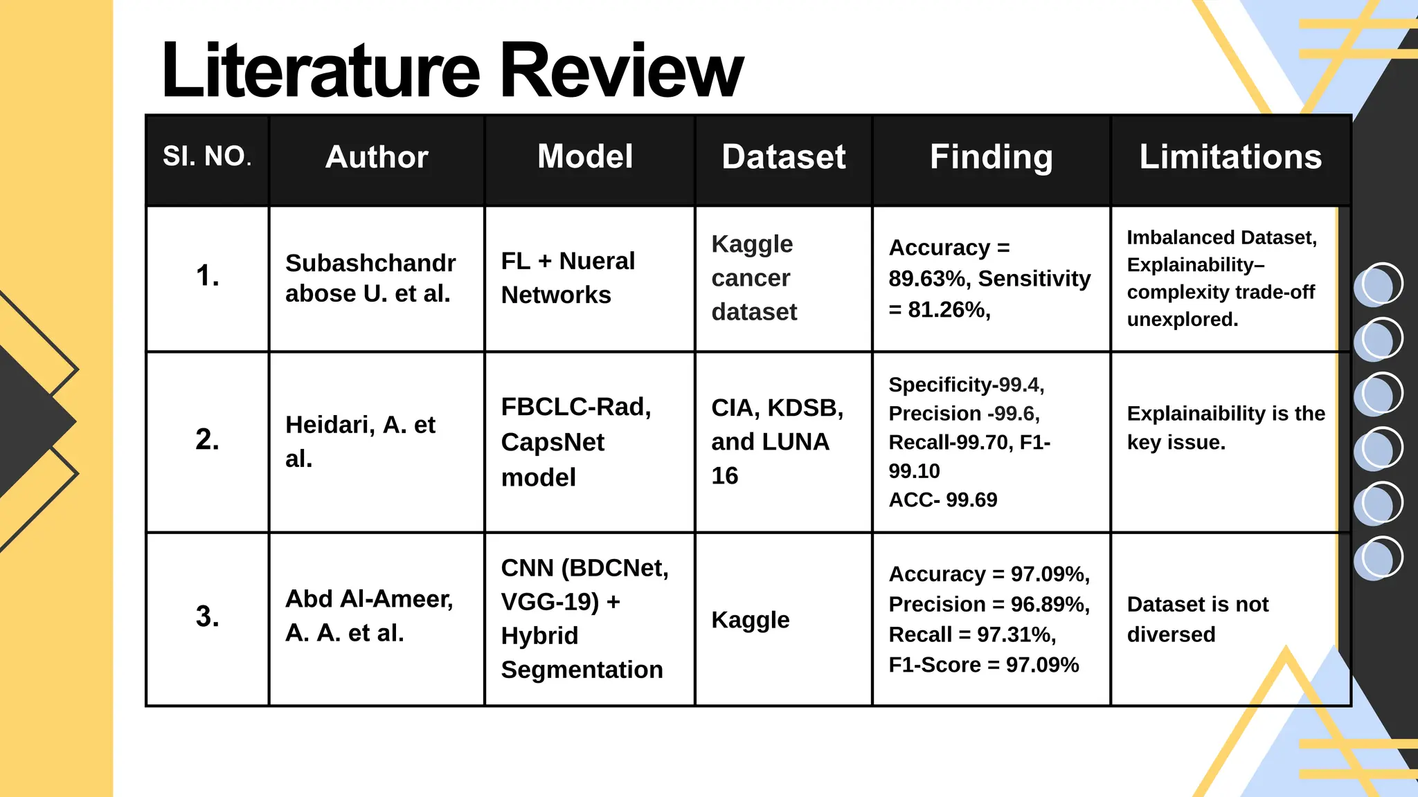
SI. NO. AuthorModel Dataset Finding Limitations
1. Subashchandr
abose U. et al.
FL + Nueral
Networks
Kaggle
cancer
dataset
Accuracy =
89.63%, Sensitivity
= 81.26%,
Imbalanced Dataset,
Explainability–
complexity trade-off
unexplored.
2.
Heidari, A. et
al.
FBCLC-Rad,
CapsNet
model
CIA, KDSB,
and LUNA
16
Specificity-99.4,
Precision -99.6,
Recall-99.70, F1-
99.10
ACC- 99.69
Explainaibility is the
key issue.
3.
Abd Al-Ameer,
A. A. et al.
CNN (BDCNet,
VGG-19) +
Hybrid
Segmentation
Kaggle
Accuracy = 97.09%,
Precision = 96.89%,
Recall = 97.31%,
F1-Score = 97.09%
Dataset is not
diversed
Literature Review
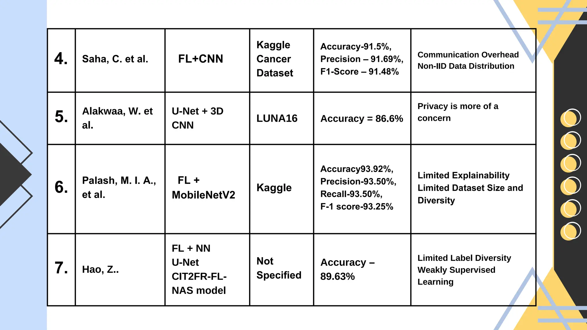
6.
4. Saha, C.et al. FL+CNN
Kaggle
Cancer
Dataset
Accuracy-91.5%,
Precision – 91.69%,
F1-Score – 91.48%
Communication Overhead
Non-IID Data Distribution
5. Alakwaa, W. et
al.
U-Net + 3D
CNN
LUNA16 Accuracy = 86.6%
Privacy is more of a
concern
6.
Palash, M. I. A.,
et al.
FL +
MobileNetV2
Kaggle
Accuracy93.92%,
Precision-93.50%,
Recall-93.50%,
F-1 score-93.25%
Limited Explainability
Limited Dataset Size and
Diversity
7. Hao, Z..
FL + NN
U-Net
CIT2FR-FL-
NAS model
Not
Specified
Accuracy –
89.63%
Limited Label Diversity
Weakly Supervised
Learning
7.
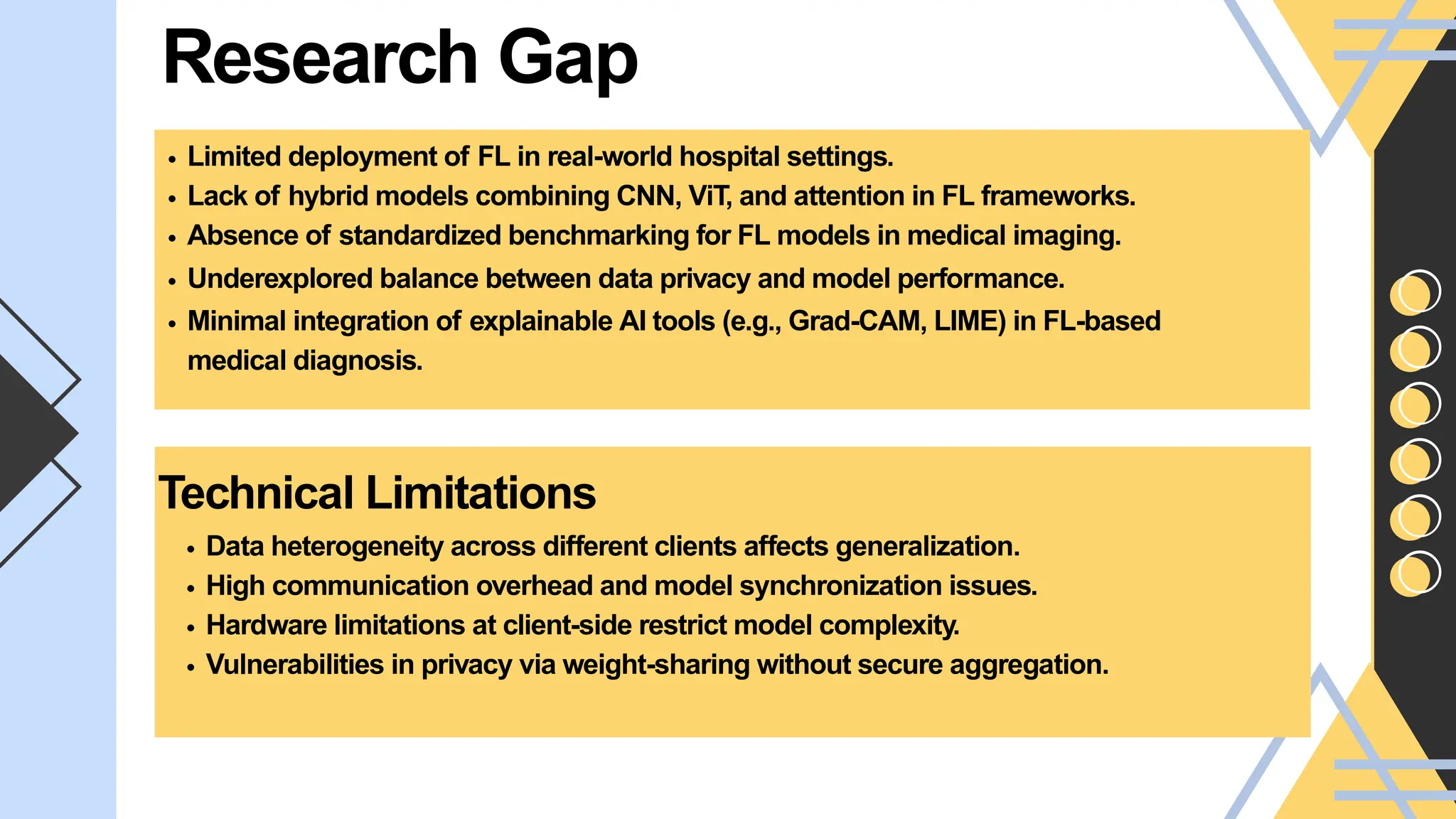
Research Gap
Limited deploymentof FL in real-world hospital settings.
Lack of hybrid models combining CNN, ViT, and attention in FL frameworks.
Absence of standardized benchmarking for FL models in medical imaging.
Underexplored balance between data privacy and model performance.
Minimal integration of explainable AI tools (e.g., Grad-CAM, LIME) in FL-based
medical diagnosis.
Technical Limitations
Data heterogeneity across different clients affects generalization.
High communication overhead and model synchronization issues.
Hardware limitations at client-side restrict model complexity.
Vulnerabilities in privacy via weight-sharing without secure aggregation.
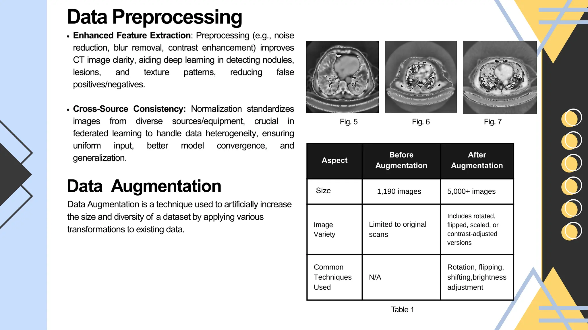
Aspect
Before
Augmentation
After
Augmentation
Size 1,190 images5,000+ images
Image
Variety
Limited to original
scans
Includes rotated,
flipped, scaled, or
contrast-adjusted
versions
Common
Techniques
Used
N/A
Rotation, flipping,
shifting,brightness
adjustment
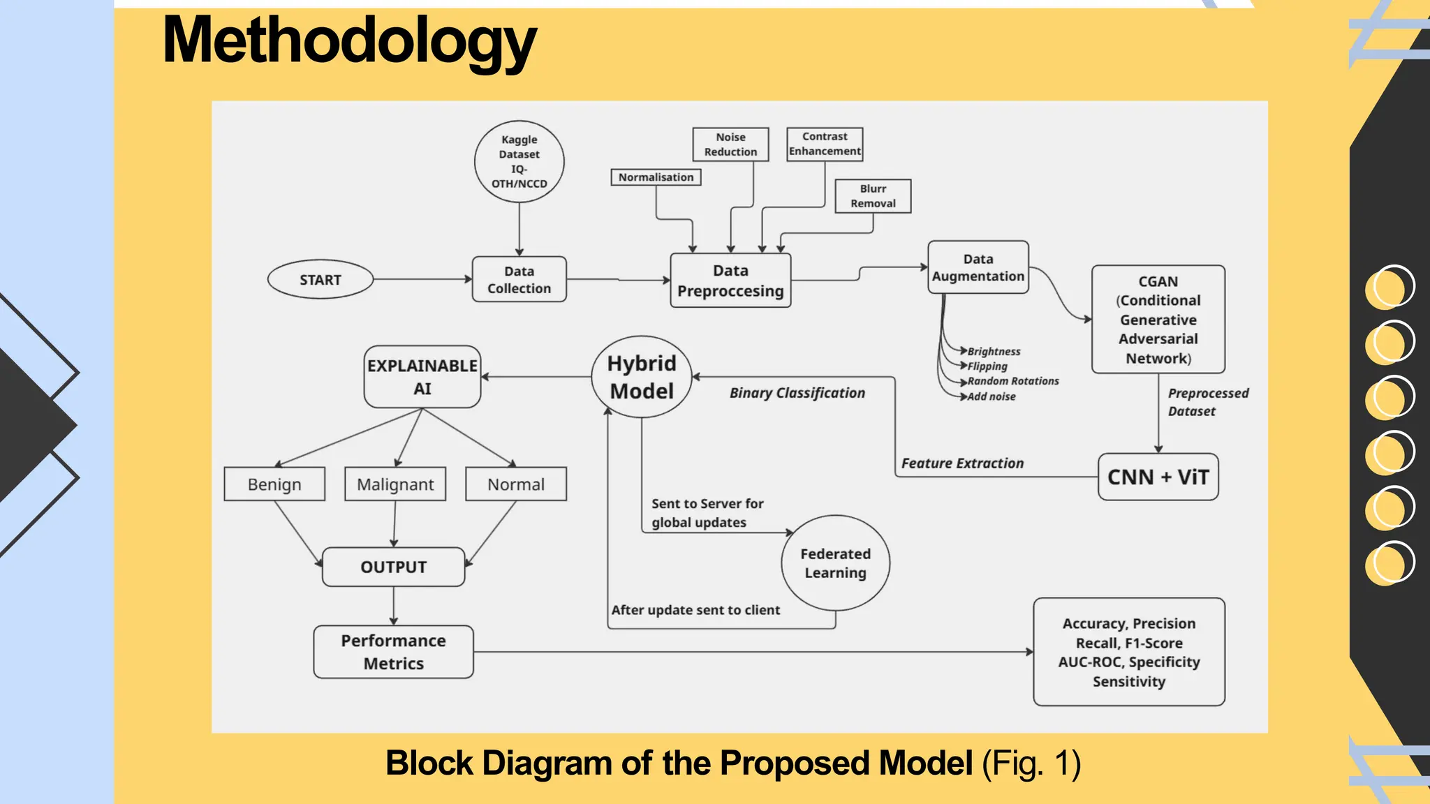
Data Preprocessing
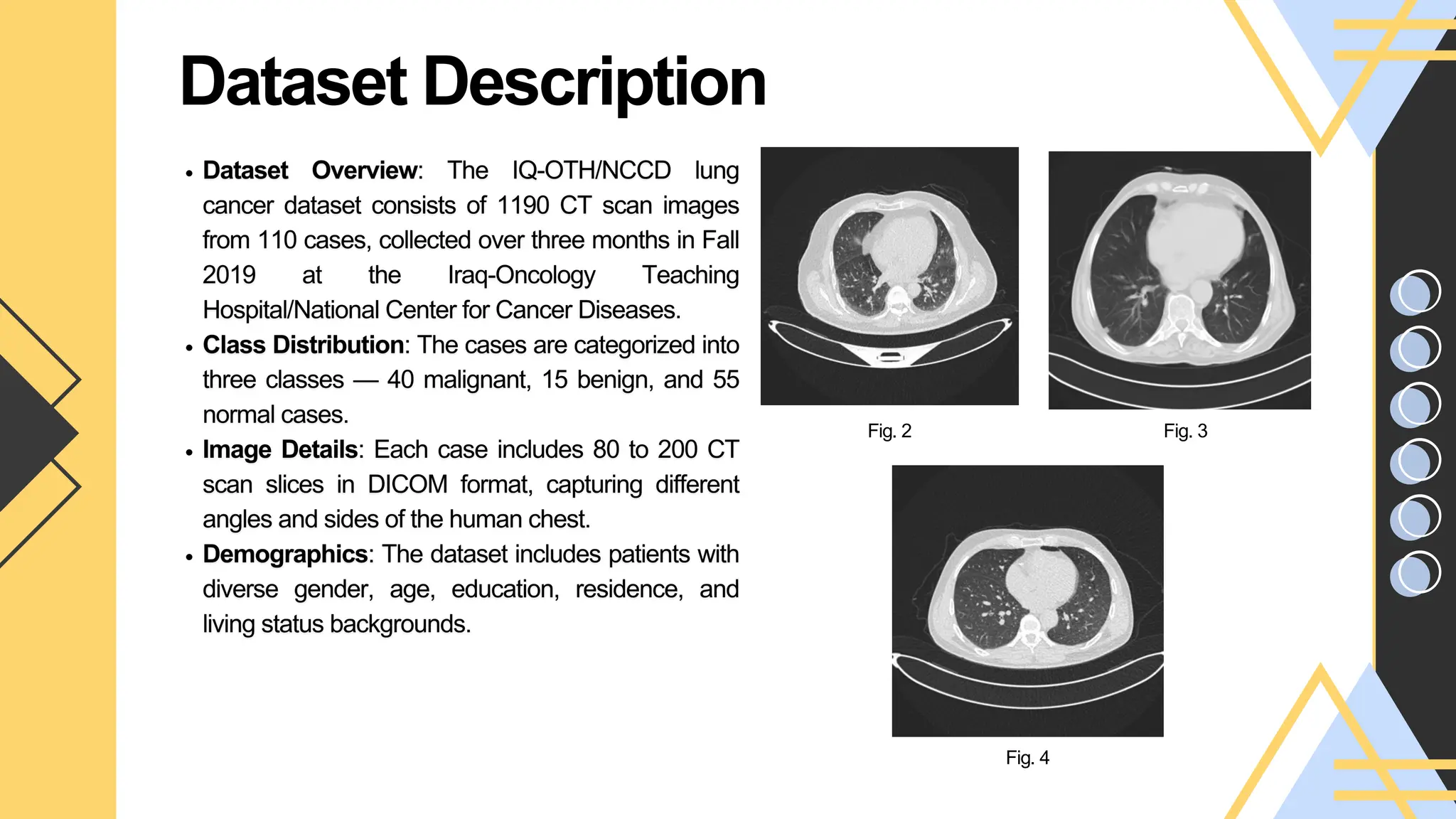
Fig. 7
Fig. 5 Fig. 6
Data Augmentation
Data Augmentation is a technique used to artificially increase
the size and diversity of a dataset by applying various
transformations to existing data.
Table 1
12.
Future Work
Multi-Modal Integration- This can enhance model accuracy and personalization by providing a more
holistic view of the patient.
Integrate causal inference into XAI to identify true contributing features.
Use domain adaptation techniques to generalize across hospitals, scanners, and populations.
Optimize CNN+ViT hybrid using quantization, pruning, or distillation.
Deploy on edge devices for point-of-care diagnostics.
Your paragraph text
Conclusion
Your paragraph text
In this project, we have successfully completed the initial and essential phase of lung cancer detection—
comprehensive data preprocessing. This includes cleaning, normalization, augmentation, and preparation of CT
scan images to ensure consistency, quality, and suitability for deep learning model training. Such preprocessing is a
critical foundation for developing robust, accurate, and generalizable AI models, particularly when combining
convolutional neural networks (CNNs) with Vision Transformers (ViTs).
13.
References
Link Name:
Brief Description:
1.Subashchandrabose U, John R, Anbazhagu UV
, Venkatesan VK, Thyluru Ramakrishna M. Ensemble Federated
Learning Approach for Diagnostics of Multi-Order Lung Cancer. Diagnostics (Basel). 2023 Sep 25;13(19):3053. doi:
10.3390/diagnostics13193053. PMID: 37835796; PMCID: PMC10572651.
2. Heidari, A., Javaheri, D., Toumaj, S., Navimipour, N. J., Rezaei, M., & Unal, M. (2023). A new lung cancer detection
method based on the chest CT images using Federated Learning and blockchain systems. Artificial intelligence in
medicine, 141, 102572.
3. Abd Al-Ameer, A. A., Hussien, G. A., & Al Ameri, H. A. (2022). Lung cancer detection using image processing and
deep learning. Indones. J. Electr. Eng. Comput. Sci, 28(2), 987-993.
4. Saha, C., Saha, S., Rahman, M. A., Milu, M. H., Higa, H., Rashid, M. A., & Ahmed, N. (2025). Lung-AttNet: An
Attention Mechanism based CNN Architecture for Lung Cancer Detection with Federated Learning. IEEE Access.
5. Alakwaa, W., Nassef, M., & Badr, A. (2017). Lung cancer detection and classification with 3D convolutional neural
network (3D-CNN). International Journal of Advanced Computer Science and Applications, 8(8).
6. Palash, M. I. A., & Yousuf, M. A. (2024, May). A Federated Learning-based Model for the Detection of Lung Cancer
from CT Scan Images. In 2024 6th International Conference on Electrical Engineering and Information & Communication
Technology (ICEEICT) (pp. 741-745). IEEE.
7. Hao, Z. Advancing Lung Cancer Diagnosis: Federated Learning-Based Privacy Innovations.


![Coded Agents – with UiPath SDK + LangGraph [Virtual Hands-on Workshop]](https://cdn.slidesharecdn.com/ss_thumbnails/codedagentsdeck-251215155422-5497c599-thumbnail.jpg?width=640&height=640&fit=bounds)
