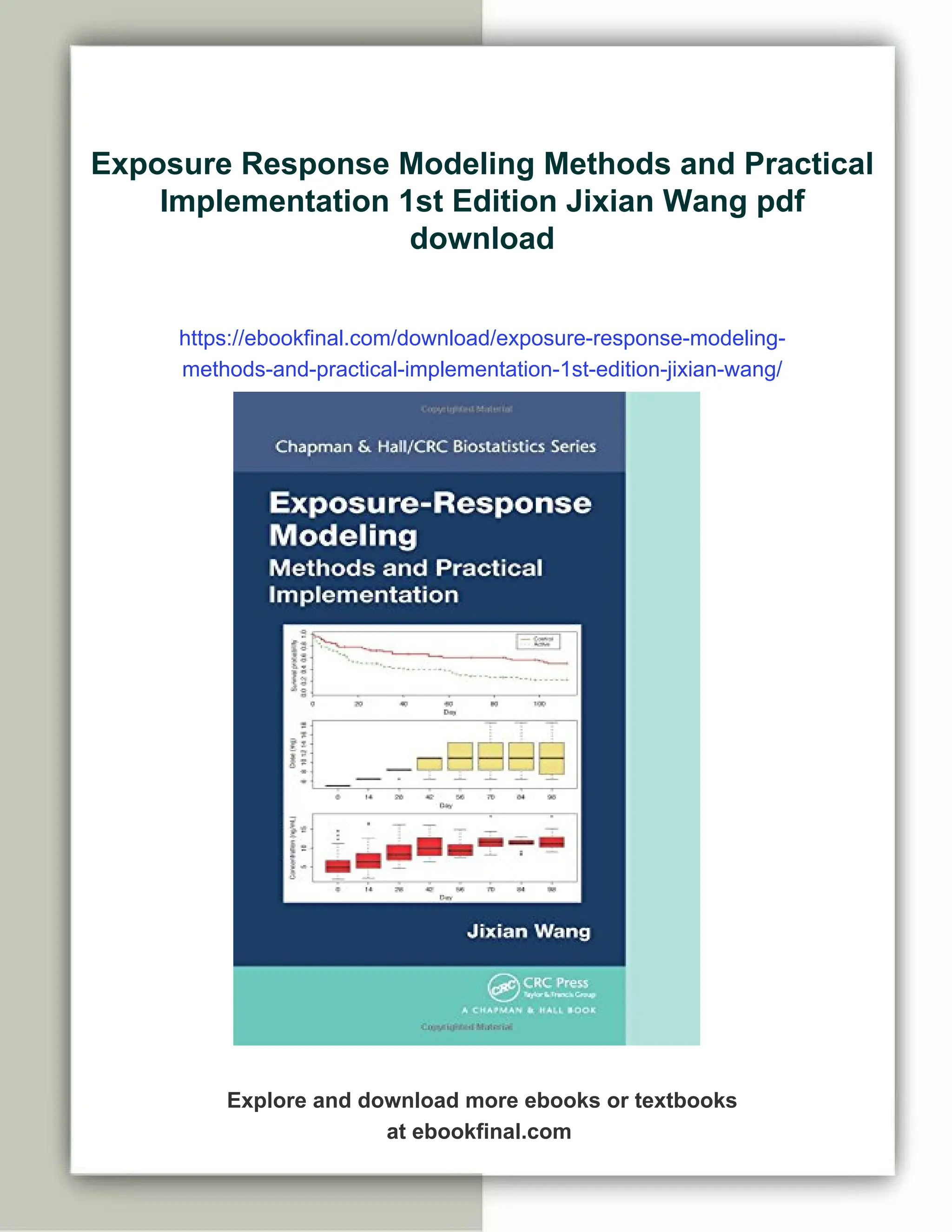
Exposure Response Modeling Methods and Practical Implementation 1st Edition Jixian Wang
Exposure Response Modeling Methods and Practical Implementation 1st Edition Jixian Wang
Exposure Response Modeling Methods and Practical Implementation 1st Edition Jixian Wang