SlideShare a Scribd company logo
ORTHOPEDIC DEPARTEMENT
PR BENHABILES PRESENTED BY
DR AGGOUN
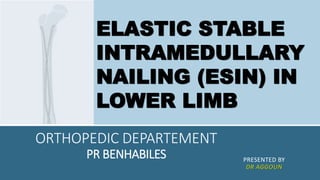
ELASTIC STABLE
INTRAMEDULLARY
NAILING (ESIN) IN
LOWER LIMB
PLAN
1. Introduction
2. Principales of ESIN
3. Indication
4. ESIN technique
5. Post-op
6. Complications
7. Benifits of ESIN
8. Limitations of ESIN
9. conclusion
INTRODUCTION
Elastic stable intramedullary nailing (ESIN) is a
minimally invasive surgical technique used to treat
fractures in the long bones, particularly in children. It's
become a popular choice for treating fractures in the
lower limbs.
PRINCIPLE OF ESIN AND BIOMECHANIC
▪ is based on the symmetrical
bracing action of two elastic
nails inserted into the
metaphysis, each of which
bears against the inner bone at
three points:
▪ Entry point
▪ Fracture zone
▪ Far end in dense metaphyseal
area
PRINCIPLE OF ESIN
▪ This produces the following
four biomechanical properties:
▪ flexural stability, axial stability,
translational stability and
rotational stability.
INDICATIONS
▪ Elastic stable intramedullary nailing is used
primarily for the management of diaphyseal and
metaphyseal fractures in children. Whether the
ESIN is indicated or not depends upon the age of
the patient and the type and site of the fracture.
▪ All three factors must be considered together.
INDICATIONS
1.Age
▪ The age limit depends on the biological development of the child.
▪ Experience has shown that the lower limit is 3–4 years and the upper limit
13–15 years.
2.Type of fracture
▪ transverse fractures
▪ short oblique or transverse fractures with broken-off wedges
▪ long oblique fractures with the possibility of cortical support
▪ spiral fractures
▪ multi-fragment and bifocal fractures
▪ pathological fractures with juvenile bone cysts
INDICATIONS
3.Fracture site
▪ femur: diaphyseal
▪ distal femur: metaphyseal
▪ femur: subtrochanteric
▪ lower leg: diaphyseal
▪ lower leg: distal metaphyseal
INDICATIONS
Other possible special indications:
▪ polytrauma in combination with craniocerebral trauma, even
outside the age range specified above
▪ prophylactic stabilization with juvenile bone cysts
▪ osteogenesis imperfecta
Contraindications
▪ intraarticular fractures
▪ complex femoral fractures, particularly in connection with
overweight (50–60 kg) and/or age (15–16 years)
ESIN LOWER LIMB TECHNIQUE
This surgical technique is explained using
the example of a
▪ femoral shaft fracture and the
ascending technique.
FEMORAL SHAFT-ASCENDING TECHNIQUE
Position child
▪ Place the child in a supine position on a
radiolucent operating Table , The extension
table can be used for larger children.
▪ Secure small children to the operating table.
▪ The assistant extends the injured extremity.
▪ Free positioning allows better control of the
nail position and rotation.
▪ Position the image intensifier so that AP and
lateral X-rays can be recorded over the full
length of the femur.
FEMORAL SHAFT-ASCENDING TECHNIQUE
Reduce fracture
▪ If the extension table is used, reduce the fracture preoperatively,
while closed, under image intensifier control.
▪ If the child is freely positioned, the fracture is reduced during the
operation.
▪ For complex fractures, cover both legs with sterile sheets so that a
rotation comparison can be performed during operation.
▪ Fracture reduction can be facilitated by the use of the small F-tool.
▪ Position the F-tool at the level of the fracture
▪ so that the two identically aligned arms of the lever bring the
fragments into the desired position.
FEMORAL SHAFT-ASCENDING TECHNIQUE
Determine nail diameter
▪ Measure the isthmus of the medullary
cavity on the X-ray image.
▪ The diameter of the individual nail (A)
should be
▪ 30–40% of the diameter of the
medullary cavity (B).
▪ Choose nails with identical diameter to
avoid varus or valgus malpositioning.
FEMORAL SHAFT-ASCENDING TECHNIQUE
Determine nail insertion points
▪ For the ascending technique, the insertion
points on the femur are 1–2 cm proximal to
the distal epiphyseal plate.
▪ In children, this is about one fingerbreadth
proximal to the upper pole of the patella.
▪ If necessary, check the intended insertion
points under the image intensifier.
FEMORAL SHAFT-ASCENDING TECHNIQUE
Perform incisions
▪ Make the opposing medial and lateral skin
incisions at the planned insertion points
and cut distally for 3–4 cm, depending on
the size of the child.
▪ On the lateral side especially, the incision
of the fascia should be of the same
length.
FEMORAL SHAFT-ASCENDING TECHNIQUE
Open medullary cavity
▪ Precisely matched opening of the medullary cavity on both
sides is essential for optimal symmetrical bracing.
▪ Divide the fascia lata over a sufficient length.
▪ Vertically insert the Awl down to the bone and firmly make a
centre mark.
▪ With rotating movements, lower the awl down to an angle of
45° in relation to the shaft axis and continue perforating the
cortical bone at an upward angle.
▪ The opening should be slightly larger than the selected nail
diameter.
▪ Check the position and insertion depth of the awl with the
image intensifier.
▪ Repeat this procedure for access on the opposite side.
FEMORAL SHAFT-ASCENDING TECHNIQUE
Pre-bend nails
▪ We have to pre-bend the implanted
part of the nails to three times the
diameter of the medullary canal.
▪ The vertex of the arch should be
located at the level of the fracture
zone.
▪ The nail tip should form the
continuation of the arch.
▪ Pre-bend both nails in exactly the
same way.
FEMORAL SHAFT-ASCENDING TECHNIQUE
Load first nail in the inserter
▪ Load the first nail in the inserter.
▪ Align the laser marking on the end of the nail with one
of the guide markings on the inserter (laser markings at
the tip, asymmetrical transverse bolts at the end).
▪ This permits direct visual control of the alignment and
rotation of the nail tip in the bone without an image
intensifier, thus preventing excessive crossover of the
nails (corkscrew effect).
▪ Tighten the nail in the inserter in the desired position
using the Pin Wrench or the Spanner Wrench.
FEMORAL SHAFT-ASCENDING TECHNIQUE
Insert first nail
▪ Insert the nail into the medullary cavity with
the nail tip at right angles to the bone shaft
(1).
▪ Turn the inserter through 180° (2) ,and align
the nail tip with the axis of the medullary
cavity (3).
FEMORAL SHAFT-ASCENDING TECHNIQUE
Advance first nail to the fracture zone
▪ Advance the nail manually up to the fracture
site, using rotating movements or gentle
taps of the Combined Hammer against the
striking surface of the inserter.
FEMORAL SHAFT-ASCENDING TECHNIQUE
Insert second nail
▪ Repeat the three last steps for the
second nail at the opposing insertion
point, thereby producing the first
crossover of the nails.
FEMORAL SHAFT-ASCENDING TECHNIQUE
▪ Advance nails
▪ If necessary, perform indirect fracture reduction
either by turning the nails, pulling the leg or using
the F-tool.
▪ Then advance the nails alternately across the
fracture zone.
▪ Survey the passage of the nails with the image
intensifier in both
▪ planes also on the other side of the fracture zone.
FEMORAL SHAFT-ASCENDING TECHNIQUE
Check position of nail tips
▪ Correctly align the nail tips in the proximal fragment in
relation to the medullary cavity in the frontal plane.
▪ If the tips are correctly located, advance the nails in a
proximal direction until the fracture is secured.
▪ The tips of the nails should only just reach the metaphysis
(A).
▪ Ensure that the nails cross over for the second time only
after they have passed the fracture zone.
FEMORAL SHAFT-ASCENDING TECHNIQUE
Check rotation
▪ When the fracture is provisionally but firmly fixed, check
rotation before final anchoring and, if necessary, align the
nail tips correctly.
▪ If an extension table is used, aseptically release the leg
from the extension so that the image intensifier can be used
to check the axial alignment in the proximal femur.
FEMORAL SHAFT-ASCENDING TECHNIQUE
Trim nails
▪ The nails must be trimmed to the desired length during the operation.
▪ The ideal cutting point is measured from the bone to the distal end of
the nail.
▪ Starting at the proximal end, estimate the distance (X) between the
current position of the nail tips (A) and the definitive anchoring position
(B) on the image intensifier projection.
▪ This distance plus an extraction length of approx. 1 cm (Y) produces the
distance from the bone to the cutting point.
▪ Note: Excessively long nail ends result in pseudobursa formation and
prevent free flexion of the knee.
▪ They can also perforate the skin and cause infections.
FEMORAL SHAFT-ASCENDING TECHNIQUE
Final positioning and anchoring of nails
▪ Advance the nails to the planned final position by
applying gentle hammer taps to the bevelled
Impactor
▪ The bevelled part of the impactor must reach the
cortical bone.
▪ This will ensure a projection of approx. 1 cm (Y) for
subsequent removal.
▪ Bend the nail ends upwards slightly with the bevelled
impactor to facilitate subsequent implant removal.
FEMUR – DESCENDING TECHNIQUE
▪ The descending monolateral technique is
preferable for fractures of the distal third of the
femur or the distal metaphysis.
▪ The fixation of metaphyseal fractures with the
nailing technique does not correspond to the
same biomechanical principles as the fixation of
shaft fractures. However, a correct inner
support for the stabilisation of the nail tips and
therefore of the metaphyseal fragment must be
guaranteed.
FEMUR – DESCENDING TECHNIQUE
Determine nail insertion points
▪ For the descending technique, the
monolateral insertion points are
located antero-laterally in the
subtrochanteric area.
▪ They are separated from each other
vertically by approx. 1–2 cm and
horizontally by 0.5–1 cm.
FEMUR – DESCENDING TECHNIQUE
Perform incisions
▪ The incisions should be 4–5 cm long so as to
expose the femur through a short L-shaped
cut in the M. vastus lateralis.
Pre-bend nails
▪ To ensure correct internal bracing, i.e. with 3-
point support , bend one of the nails into an
S-shape so that the bracing occurs at the
level of the fracture zone ( ).
FEMUR – DESCENDING TECHNIQUE
Insert first nail
▪ Introduce the simply pre-bent nail, reduce
the fracture with the nail and achieve
primary stabilisation.
Insert second nail
▪ Insert the S-shaped pre-bent nail (1). After
the first contact with the cortical bone on
the opposite side, turn the nail through
180° (2).
FEMUR – DESCENDING TECHNIQUE
Final positioning and anchoring of nails
▪ Advance the nails to the epiphyseal plate
and align the nail tips
▪ so that they diverge from each other.
ESIN LOWER LEG(TIBIA)
Indications
▪ Lower leg and isolated tibial fractures should preferably be treated conservatively.
▪ Lower leg fractures constitute a special indication for internal fixation by TEN.
Nailing is indicated in:
▪ closed, unstable lower leg fractures from the age of 9
▪ irreducible and non-retainable fractures
▪ polytrauma and severe craniocerebral trauma
▪ Since the tibia is positioned off-centre in relation to the surrounding muscles and
since it possesses a triangular crosssection, particular care is indicated when
placing the nails.
▪ Always nail the tibia using the descending technique. Do not use the ascending
technique for the tibia.
ESIN LOWER LEG(TIBIA)
Determine nail insertion points
▪ The nail insertion points are located on
the medial and lateral
▪ sides of the tibial tuberosity.
ESIN LOWER LEG(TIBIA)
Perform incisions
▪ Make a 2–3 cm skin incision from each planned insertion point
▪ in the cranial direction.
Check position of nail tips
▪ Because of the triangular shape of the tibial medullary canal,
both nails tend to lie dorsally, which would result in recurvation.
▪ Before hammering the nails home, turn the tips of both nailsslightly
posteriorly so as to achieve the physiological antecurvation of the tibia.
Trim nails
▪ In view of the minimal soft tissue cover, keep the nail ends short
▪ and do not bend upward.
POST-OP
▪ radiographies at 15, 30, 45 days and 3 months
▪ No complementary immobilization is needed mostly
▪ Partial weight bearing with crutches allowed on the
15th day
▪ removal of nails at the 4th month
▪ Sports activities should be stopped for 6 months.
COMPLICATIONS
▪ Pain at insertion site(most common)
▪ Nail tip irritation
▪ Skin infection
▪ Implant failure
▪ Unacceptable angulation
▪ Malrotation
BENEFITS OF ESIN
▪ Minimally invasive
▪ Reduced pain
▪ Earlier weight-bearing and mobilization
▪ Shorter hospital stay
▪ Lower complication rates
LIMITATIONS OF ESIN
▪ Not suitable for all types of fractures.
▪ Severe comminuted (fragmented)
fractures or fractures with significant
angulation might not be ideal candidates.
CONCLUSION
The fixation of paediatric diaphyseal fractures with elastic
nails is a rapid, well-codified and effective method for
treating long-bone closed fractures in children.
Advantages over other fixation techniques include a lower
infection rate, lower refracture rate, lack of secondary
displacement and a greater ease of management for the
patients and their parents owing to early mobility and
reduced school absenteeism. All of these could
compensate for this technique’s greater direct cost.
REFERENCES
▪ J Child Orthop. 2011 Aug; 5(4): 297–304. Published online 2011 Jun 2.
doi: 10.1007/s11832-011-0343-5
▪ Nielsen, E.; Bonsu, N.; Andras, L.M.; Goldstein, R.Y. The effect of canal fill on
paediatric femur fractures treated with titanium elastic nails. J. Child. Orthop. 2018,
12, 15–19. [Google Scholar] [CrossRef]
▪ https://www.ncbi.nlm.nih.gov/pmc/articles/PMC6456152/
▪ Frei, Benjamin MMeda; Mayr, Johannes MD, PhDa,∗; de Bernardis, Gaston MDa;
Camathias, Carlo MD, PhDb; Holland-Cunz, Stefan MD, PhDa; Rutz, Erich MD, PhDc.
Elastic stabile intramedullary nailing (ESIN) of diaphyseal femur fractures in children
and adolescents: A strobe-compliant study. Medicine 98(14):p e15085, April 2019. |
DOI: 10.1097/MD.0000000000015085
THANK YOU

More Related Content

Similar to ESIN LOWER LIMB FEMUR AND TIBIA FRACTURES .pptx

The inman aligner
The inman alignerThe inman aligner
The inman aligner
MaherFouda1
 
Surgical technique for gama nail
Surgical technique for gama nailSurgical technique for gama nail
Surgical technique for gama nail
orthopedic Implant
 
Occlusion and Mmf vv-1.pptx
Occlusion and Mmf  vv-1.pptxOcclusion and Mmf  vv-1.pptx
Occlusion and Mmf vv-1.pptx
Shubhanshu Gaurav
 
Occlusion and Mmf vv-1.pptx
Occlusion and Mmf  vv-1.pptxOcclusion and Mmf  vv-1.pptx
Occlusion and Mmf vv-1.pptx
Shubhanshu Gaurav
 
Dental Implant
Dental ImplantDental Implant
Dental Implant
Saleh Bakry
 
Extraction of primary teeth along with permanent teeth
Extraction of primary teeth along with permanent teeth Extraction of primary teeth along with permanent teeth
Extraction of primary teeth along with permanent teeth
Dr. Atasi Chakraborty
 
Extrusion by reverse curves archwires by Dr Maher Fouda
Extrusion by reverse curves archwires by Dr Maher FoudaExtrusion by reverse curves archwires by Dr Maher Fouda
Extrusion by reverse curves archwires by Dr Maher Fouda
Maher Fouda
 
Mandibular osteotomies.pptx
Mandibular osteotomies.pptxMandibular osteotomies.pptx
Mandibular osteotomies.pptx
KathirvelGopalakrish
 
Direct bracketing technique for dental professionals
Direct bracketing technique  for dental professionalsDirect bracketing technique  for dental professionals
Direct bracketing technique for dental professionals
Jean-Marc Retrouvey
 
200330 Controversies in the intramedullary nailing of tibia
200330 Controversies in the intramedullary nailing of tibia200330 Controversies in the intramedullary nailing of tibia
200330 Controversies in the intramedullary nailing of tibia
Dr MADAN MOHAN
 
JAW RELATIONS IN CAST PARTIAL DENTURE
JAW RELATIONS IN CAST PARTIAL DENTUREJAW RELATIONS IN CAST PARTIAL DENTURE
JAW RELATIONS IN CAST PARTIAL DENTURE
DiyaSharma39
 
Surgical Approaches to distal humerus fractures - DR.S.SENTHIL SAILESH, M.S...
 Surgical Approaches to distal  humerus fractures - DR.S.SENTHIL SAILESH, M.S... Surgical Approaches to distal  humerus fractures - DR.S.SENTHIL SAILESH, M.S...
Surgical Approaches to distal humerus fractures - DR.S.SENTHIL SAILESH, M.S...
Senthil sailesh
 
Conventional begg's stage 3 in orthodontics /certified fixed orthodontic cour...
Conventional begg's stage 3 in orthodontics /certified fixed orthodontic cour...Conventional begg's stage 3 in orthodontics /certified fixed orthodontic cour...
Conventional begg's stage 3 in orthodontics /certified fixed orthodontic cour...
Indian dental academy
 
Armamentarium of exodontia
Armamentarium of exodontiaArmamentarium of exodontia
Armamentarium of exodontia
MonikaIrmal
 
The level anchorage system-Dr.Pooja Kale
The level anchorage system-Dr.Pooja KaleThe level anchorage system-Dr.Pooja Kale
The level anchorage system-Dr.Pooja Kale
Pooja Kale
 
Leveling and aligning
Leveling and aligningLeveling and aligning
Leveling and aligning
Dr.Rahul Tiwari
 
Twin block /certified fixed orthodontic courses by Indian dental academy
Twin block /certified fixed orthodontic courses by Indian dental academy  Twin block /certified fixed orthodontic courses by Indian dental academy
Twin block /certified fixed orthodontic courses by Indian dental academy
Indian dental academy
 
Anchorage preparation in PAE
Anchorage preparation in PAEAnchorage preparation in PAE
Anchorage preparation in PAE
Indian dental academy
 

Similar to ESIN LOWER LIMB FEMUR AND TIBIA FRACTURES .pptx (20)

The inman aligner
The inman alignerThe inman aligner
The inman aligner
 
Surgical technique for gama nail
Surgical technique for gama nailSurgical technique for gama nail
Surgical technique for gama nail
 
Occlusion and Mmf vv-1.pptx
Occlusion and Mmf  vv-1.pptxOcclusion and Mmf  vv-1.pptx
Occlusion and Mmf vv-1.pptx
 
Occlusion and Mmf vv-1.pptx
Occlusion and Mmf  vv-1.pptxOcclusion and Mmf  vv-1.pptx
Occlusion and Mmf vv-1.pptx
 
Dental Implant
Dental ImplantDental Implant
Dental Implant
 
Extraction of primary teeth along with permanent teeth
Extraction of primary teeth along with permanent teeth Extraction of primary teeth along with permanent teeth
Extraction of primary teeth along with permanent teeth
 
Extrusion by reverse curves archwires by Dr Maher Fouda
Extrusion by reverse curves archwires by Dr Maher FoudaExtrusion by reverse curves archwires by Dr Maher Fouda
Extrusion by reverse curves archwires by Dr Maher Fouda
 
Mandibular osteotomies.pptx
Mandibular osteotomies.pptxMandibular osteotomies.pptx
Mandibular osteotomies.pptx
 
Direct bracketing technique for dental professionals
Direct bracketing technique  for dental professionalsDirect bracketing technique  for dental professionals
Direct bracketing technique for dental professionals
 
200330 Controversies in the intramedullary nailing of tibia
200330 Controversies in the intramedullary nailing of tibia200330 Controversies in the intramedullary nailing of tibia
200330 Controversies in the intramedullary nailing of tibia
 
24.refine denture setup
24.refine denture setup24.refine denture setup
24.refine denture setup
 
Alignment and leveling
Alignment and levelingAlignment and leveling
Alignment and leveling
 
JAW RELATIONS IN CAST PARTIAL DENTURE
JAW RELATIONS IN CAST PARTIAL DENTUREJAW RELATIONS IN CAST PARTIAL DENTURE
JAW RELATIONS IN CAST PARTIAL DENTURE
 
Surgical Approaches to distal humerus fractures - DR.S.SENTHIL SAILESH, M.S...
 Surgical Approaches to distal  humerus fractures - DR.S.SENTHIL SAILESH, M.S... Surgical Approaches to distal  humerus fractures - DR.S.SENTHIL SAILESH, M.S...
Surgical Approaches to distal humerus fractures - DR.S.SENTHIL SAILESH, M.S...
 
Conventional begg's stage 3 in orthodontics /certified fixed orthodontic cour...
Conventional begg's stage 3 in orthodontics /certified fixed orthodontic cour...Conventional begg's stage 3 in orthodontics /certified fixed orthodontic cour...
Conventional begg's stage 3 in orthodontics /certified fixed orthodontic cour...
 
Armamentarium of exodontia
Armamentarium of exodontiaArmamentarium of exodontia
Armamentarium of exodontia
 
The level anchorage system-Dr.Pooja Kale
The level anchorage system-Dr.Pooja KaleThe level anchorage system-Dr.Pooja Kale
The level anchorage system-Dr.Pooja Kale
 
Leveling and aligning
Leveling and aligningLeveling and aligning
Leveling and aligning
 
Twin block /certified fixed orthodontic courses by Indian dental academy
Twin block /certified fixed orthodontic courses by Indian dental academy  Twin block /certified fixed orthodontic courses by Indian dental academy
Twin block /certified fixed orthodontic courses by Indian dental academy
 
Anchorage preparation in PAE
Anchorage preparation in PAEAnchorage preparation in PAE
Anchorage preparation in PAE
 

Recently uploaded

CMHPSM Regional Compliance Training 2024
CMHPSM Regional Compliance Training 2024CMHPSM Regional Compliance Training 2024
CMHPSM Regional Compliance Training 2024
JColaianne
 
Letter to MREC - application to conduct study
Letter to MREC - application to conduct studyLetter to MREC - application to conduct study
Letter to MREC - application to conduct study
Azreen Aj
 
Professional Secrecy: Forensic Medicine Lecture
Professional Secrecy: Forensic Medicine LectureProfessional Secrecy: Forensic Medicine Lecture
Professional Secrecy: Forensic Medicine Lecture
DIVYANSHU740006
 
NEEDLE STICK INJURY - JOURNAL CLUB PRESENTATION - DR SHAMIN EABENSON
NEEDLE STICK INJURY - JOURNAL CLUB PRESENTATION - DR SHAMIN EABENSONNEEDLE STICK INJURY - JOURNAL CLUB PRESENTATION - DR SHAMIN EABENSON
NEEDLE STICK INJURY - JOURNAL CLUB PRESENTATION - DR SHAMIN EABENSON
SHAMIN EABENSON
 
Child Welfare Clinic and Well baby clinicin Sri Lanka.ppsx
Child Welfare Clinic and Well baby clinicin Sri Lanka.ppsxChild Welfare Clinic and Well baby clinicin Sri Lanka.ppsx
Child Welfare Clinic and Well baby clinicin Sri Lanka.ppsx
Sankalpa Gunathilaka
 
NKTI Annual Report - Annual Report FY 2022
NKTI Annual Report - Annual Report FY 2022NKTI Annual Report - Annual Report FY 2022
NKTI Annual Report - Annual Report FY 2022
nktiacc3
 
定制(wsu毕业证书)美国华盛顿州立大学毕业证学位证书实拍图原版一模一样
定制(wsu毕业证书)美国华盛顿州立大学毕业证学位证书实拍图原版一模一样定制(wsu毕业证书)美国华盛顿州立大学毕业证学位证书实拍图原版一模一样
定制(wsu毕业证书)美国华盛顿州立大学毕业证学位证书实拍图原版一模一样
khvdq584
 
PET CT beginners Guide covers some of the underrepresented topics in PET CT
PET CT  beginners Guide  covers some of the underrepresented topics  in PET CTPET CT  beginners Guide  covers some of the underrepresented topics  in PET CT
PET CT beginners Guide covers some of the underrepresented topics in PET CT
MiadAlsulami
 
IMCI LECTURE PRESENTATION 1.pptx by Ronald
IMCI LECTURE PRESENTATION 1.pptx by RonaldIMCI LECTURE PRESENTATION 1.pptx by Ronald
IMCI LECTURE PRESENTATION 1.pptx by Ronald
NatungaRonald1
 
Champions of Health Spotlight On Leaders Shaping Germany's Healthcare.pdf
Champions of Health Spotlight On Leaders Shaping Germany's Healthcare.pdfChampions of Health Spotlight On Leaders Shaping Germany's Healthcare.pdf
Champions of Health Spotlight On Leaders Shaping Germany's Healthcare.pdf
eurohealthleaders
 
Gemma Wean- Nutritional solution for Artemia
Gemma Wean- Nutritional solution for ArtemiaGemma Wean- Nutritional solution for Artemia
Gemma Wean- Nutritional solution for Artemia
smuskaan0008
 
INFECTION OF THE BRAIN -ENCEPHALITIS ( PPT)
INFECTION OF THE BRAIN -ENCEPHALITIS ( PPT)INFECTION OF THE BRAIN -ENCEPHALITIS ( PPT)
INFECTION OF THE BRAIN -ENCEPHALITIS ( PPT)
blessyjannu21
 
Tips for Pet Care in winters How to take care of pets.
Tips for Pet Care in winters How to take care of pets.Tips for Pet Care in winters How to take care of pets.
Tips for Pet Care in winters How to take care of pets.
Dinesh Chauhan
 
Deep Leg Vein Thrombosis (DVT): Meaning, Causes, Symptoms, Treatment, and Mor...
Deep Leg Vein Thrombosis (DVT): Meaning, Causes, Symptoms, Treatment, and Mor...Deep Leg Vein Thrombosis (DVT): Meaning, Causes, Symptoms, Treatment, and Mor...
Deep Leg Vein Thrombosis (DVT): Meaning, Causes, Symptoms, Treatment, and Mor...
The Lifesciences Magazine
 
DECODING THE RISKS - ALCOHOL, TOBACCO & DRUGS.pdf
DECODING THE RISKS - ALCOHOL, TOBACCO & DRUGS.pdfDECODING THE RISKS - ALCOHOL, TOBACCO & DRUGS.pdf
DECODING THE RISKS - ALCOHOL, TOBACCO & DRUGS.pdf
Dr Rachana Gujar
 
Top Rated Massage Center In Ajman Chandrima Spa
Top Rated Massage Center In Ajman Chandrima SpaTop Rated Massage Center In Ajman Chandrima Spa
Top Rated Massage Center In Ajman Chandrima Spa
Chandrima Spa Ajman
 
ABDOMINAL COMPARTMENT SYSNDROME
ABDOMINAL COMPARTMENT SYSNDROMEABDOMINAL COMPARTMENT SYSNDROME
ABDOMINAL COMPARTMENT SYSNDROME
Rommel Luis III Israel
 
Feeding plate for a newborn with Cleft Palate.pptx
Feeding plate for a newborn with Cleft Palate.pptxFeeding plate for a newborn with Cleft Palate.pptx
Feeding plate for a newborn with Cleft Palate.pptx
SatvikaPrasad
 
Bringing AI into a Mid-Sized Company: A structured Approach
Bringing AI into a Mid-Sized Company: A structured ApproachBringing AI into a Mid-Sized Company: A structured Approach
Bringing AI into a Mid-Sized Company: A structured Approach
Brian Frerichs
 
TEST BANK For Accounting Information Systems, 3rd Edition by Vernon Richardso...
TEST BANK For Accounting Information Systems, 3rd Edition by Vernon Richardso...TEST BANK For Accounting Information Systems, 3rd Edition by Vernon Richardso...
TEST BANK For Accounting Information Systems, 3rd Edition by Vernon Richardso...
rightmanforbloodline
 

Recently uploaded (20)

CMHPSM Regional Compliance Training 2024
CMHPSM Regional Compliance Training 2024CMHPSM Regional Compliance Training 2024
CMHPSM Regional Compliance Training 2024
 
Letter to MREC - application to conduct study
Letter to MREC - application to conduct studyLetter to MREC - application to conduct study
Letter to MREC - application to conduct study
 
Professional Secrecy: Forensic Medicine Lecture
Professional Secrecy: Forensic Medicine LectureProfessional Secrecy: Forensic Medicine Lecture
Professional Secrecy: Forensic Medicine Lecture
 
NEEDLE STICK INJURY - JOURNAL CLUB PRESENTATION - DR SHAMIN EABENSON
NEEDLE STICK INJURY - JOURNAL CLUB PRESENTATION - DR SHAMIN EABENSONNEEDLE STICK INJURY - JOURNAL CLUB PRESENTATION - DR SHAMIN EABENSON
NEEDLE STICK INJURY - JOURNAL CLUB PRESENTATION - DR SHAMIN EABENSON
 
Child Welfare Clinic and Well baby clinicin Sri Lanka.ppsx
Child Welfare Clinic and Well baby clinicin Sri Lanka.ppsxChild Welfare Clinic and Well baby clinicin Sri Lanka.ppsx
Child Welfare Clinic and Well baby clinicin Sri Lanka.ppsx
 
NKTI Annual Report - Annual Report FY 2022
NKTI Annual Report - Annual Report FY 2022NKTI Annual Report - Annual Report FY 2022
NKTI Annual Report - Annual Report FY 2022
 
定制(wsu毕业证书)美国华盛顿州立大学毕业证学位证书实拍图原版一模一样
定制(wsu毕业证书)美国华盛顿州立大学毕业证学位证书实拍图原版一模一样定制(wsu毕业证书)美国华盛顿州立大学毕业证学位证书实拍图原版一模一样
定制(wsu毕业证书)美国华盛顿州立大学毕业证学位证书实拍图原版一模一样
 
PET CT beginners Guide covers some of the underrepresented topics in PET CT
PET CT  beginners Guide  covers some of the underrepresented topics  in PET CTPET CT  beginners Guide  covers some of the underrepresented topics  in PET CT
PET CT beginners Guide covers some of the underrepresented topics in PET CT
 
IMCI LECTURE PRESENTATION 1.pptx by Ronald
IMCI LECTURE PRESENTATION 1.pptx by RonaldIMCI LECTURE PRESENTATION 1.pptx by Ronald
IMCI LECTURE PRESENTATION 1.pptx by Ronald
 
Champions of Health Spotlight On Leaders Shaping Germany's Healthcare.pdf
Champions of Health Spotlight On Leaders Shaping Germany's Healthcare.pdfChampions of Health Spotlight On Leaders Shaping Germany's Healthcare.pdf
Champions of Health Spotlight On Leaders Shaping Germany's Healthcare.pdf
 
Gemma Wean- Nutritional solution for Artemia
Gemma Wean- Nutritional solution for ArtemiaGemma Wean- Nutritional solution for Artemia
Gemma Wean- Nutritional solution for Artemia
 
INFECTION OF THE BRAIN -ENCEPHALITIS ( PPT)
INFECTION OF THE BRAIN -ENCEPHALITIS ( PPT)INFECTION OF THE BRAIN -ENCEPHALITIS ( PPT)
INFECTION OF THE BRAIN -ENCEPHALITIS ( PPT)
 
Tips for Pet Care in winters How to take care of pets.
Tips for Pet Care in winters How to take care of pets.Tips for Pet Care in winters How to take care of pets.
Tips for Pet Care in winters How to take care of pets.
 
Deep Leg Vein Thrombosis (DVT): Meaning, Causes, Symptoms, Treatment, and Mor...
Deep Leg Vein Thrombosis (DVT): Meaning, Causes, Symptoms, Treatment, and Mor...Deep Leg Vein Thrombosis (DVT): Meaning, Causes, Symptoms, Treatment, and Mor...
Deep Leg Vein Thrombosis (DVT): Meaning, Causes, Symptoms, Treatment, and Mor...
 
DECODING THE RISKS - ALCOHOL, TOBACCO & DRUGS.pdf
DECODING THE RISKS - ALCOHOL, TOBACCO & DRUGS.pdfDECODING THE RISKS - ALCOHOL, TOBACCO & DRUGS.pdf
DECODING THE RISKS - ALCOHOL, TOBACCO & DRUGS.pdf
 
Top Rated Massage Center In Ajman Chandrima Spa
Top Rated Massage Center In Ajman Chandrima SpaTop Rated Massage Center In Ajman Chandrima Spa
Top Rated Massage Center In Ajman Chandrima Spa
 
ABDOMINAL COMPARTMENT SYSNDROME
ABDOMINAL COMPARTMENT SYSNDROMEABDOMINAL COMPARTMENT SYSNDROME
ABDOMINAL COMPARTMENT SYSNDROME
 
Feeding plate for a newborn with Cleft Palate.pptx
Feeding plate for a newborn with Cleft Palate.pptxFeeding plate for a newborn with Cleft Palate.pptx
Feeding plate for a newborn with Cleft Palate.pptx
 
Bringing AI into a Mid-Sized Company: A structured Approach
Bringing AI into a Mid-Sized Company: A structured ApproachBringing AI into a Mid-Sized Company: A structured Approach
Bringing AI into a Mid-Sized Company: A structured Approach
 
TEST BANK For Accounting Information Systems, 3rd Edition by Vernon Richardso...
TEST BANK For Accounting Information Systems, 3rd Edition by Vernon Richardso...TEST BANK For Accounting Information Systems, 3rd Edition by Vernon Richardso...
TEST BANK For Accounting Information Systems, 3rd Edition by Vernon Richardso...
 

ESIN LOWER LIMB FEMUR AND TIBIA FRACTURES .pptx

  • 1. ORTHOPEDIC DEPARTEMENT PR BENHABILES PRESENTED BY DR AGGOUN ELASTIC STABLE INTRAMEDULLARY NAILING (ESIN) IN LOWER LIMB
  • 2. PLAN 1. Introduction 2. Principales of ESIN 3. Indication 4. ESIN technique 5. Post-op 6. Complications 7. Benifits of ESIN 8. Limitations of ESIN 9. conclusion
  • 3. INTRODUCTION Elastic stable intramedullary nailing (ESIN) is a minimally invasive surgical technique used to treat fractures in the long bones, particularly in children. It's become a popular choice for treating fractures in the lower limbs.
  • 4. PRINCIPLE OF ESIN AND BIOMECHANIC ▪ is based on the symmetrical bracing action of two elastic nails inserted into the metaphysis, each of which bears against the inner bone at three points: ▪ Entry point ▪ Fracture zone ▪ Far end in dense metaphyseal area
  • 5. PRINCIPLE OF ESIN ▪ This produces the following four biomechanical properties: ▪ flexural stability, axial stability, translational stability and rotational stability.
  • 6. INDICATIONS ▪ Elastic stable intramedullary nailing is used primarily for the management of diaphyseal and metaphyseal fractures in children. Whether the ESIN is indicated or not depends upon the age of the patient and the type and site of the fracture. ▪ All three factors must be considered together.
  • 7. INDICATIONS 1.Age ▪ The age limit depends on the biological development of the child. ▪ Experience has shown that the lower limit is 3–4 years and the upper limit 13–15 years. 2.Type of fracture ▪ transverse fractures ▪ short oblique or transverse fractures with broken-off wedges ▪ long oblique fractures with the possibility of cortical support ▪ spiral fractures ▪ multi-fragment and bifocal fractures ▪ pathological fractures with juvenile bone cysts
  • 8. INDICATIONS 3.Fracture site ▪ femur: diaphyseal ▪ distal femur: metaphyseal ▪ femur: subtrochanteric ▪ lower leg: diaphyseal ▪ lower leg: distal metaphyseal
  • 9. INDICATIONS Other possible special indications: ▪ polytrauma in combination with craniocerebral trauma, even outside the age range specified above ▪ prophylactic stabilization with juvenile bone cysts ▪ osteogenesis imperfecta Contraindications ▪ intraarticular fractures ▪ complex femoral fractures, particularly in connection with overweight (50–60 kg) and/or age (15–16 years)
  • 10. ESIN LOWER LIMB TECHNIQUE This surgical technique is explained using the example of a ▪ femoral shaft fracture and the ascending technique.
  • 11. FEMORAL SHAFT-ASCENDING TECHNIQUE Position child ▪ Place the child in a supine position on a radiolucent operating Table , The extension table can be used for larger children. ▪ Secure small children to the operating table. ▪ The assistant extends the injured extremity. ▪ Free positioning allows better control of the nail position and rotation. ▪ Position the image intensifier so that AP and lateral X-rays can be recorded over the full length of the femur.
  • 12. FEMORAL SHAFT-ASCENDING TECHNIQUE Reduce fracture ▪ If the extension table is used, reduce the fracture preoperatively, while closed, under image intensifier control. ▪ If the child is freely positioned, the fracture is reduced during the operation. ▪ For complex fractures, cover both legs with sterile sheets so that a rotation comparison can be performed during operation. ▪ Fracture reduction can be facilitated by the use of the small F-tool. ▪ Position the F-tool at the level of the fracture ▪ so that the two identically aligned arms of the lever bring the fragments into the desired position.
  • 13. FEMORAL SHAFT-ASCENDING TECHNIQUE Determine nail diameter ▪ Measure the isthmus of the medullary cavity on the X-ray image. ▪ The diameter of the individual nail (A) should be ▪ 30–40% of the diameter of the medullary cavity (B). ▪ Choose nails with identical diameter to avoid varus or valgus malpositioning.
  • 14. FEMORAL SHAFT-ASCENDING TECHNIQUE Determine nail insertion points ▪ For the ascending technique, the insertion points on the femur are 1–2 cm proximal to the distal epiphyseal plate. ▪ In children, this is about one fingerbreadth proximal to the upper pole of the patella. ▪ If necessary, check the intended insertion points under the image intensifier.
  • 15. FEMORAL SHAFT-ASCENDING TECHNIQUE Perform incisions ▪ Make the opposing medial and lateral skin incisions at the planned insertion points and cut distally for 3–4 cm, depending on the size of the child. ▪ On the lateral side especially, the incision of the fascia should be of the same length.
  • 16. FEMORAL SHAFT-ASCENDING TECHNIQUE Open medullary cavity ▪ Precisely matched opening of the medullary cavity on both sides is essential for optimal symmetrical bracing. ▪ Divide the fascia lata over a sufficient length. ▪ Vertically insert the Awl down to the bone and firmly make a centre mark. ▪ With rotating movements, lower the awl down to an angle of 45° in relation to the shaft axis and continue perforating the cortical bone at an upward angle. ▪ The opening should be slightly larger than the selected nail diameter. ▪ Check the position and insertion depth of the awl with the image intensifier. ▪ Repeat this procedure for access on the opposite side.
  • 17. FEMORAL SHAFT-ASCENDING TECHNIQUE Pre-bend nails ▪ We have to pre-bend the implanted part of the nails to three times the diameter of the medullary canal. ▪ The vertex of the arch should be located at the level of the fracture zone. ▪ The nail tip should form the continuation of the arch. ▪ Pre-bend both nails in exactly the same way.
  • 18. FEMORAL SHAFT-ASCENDING TECHNIQUE Load first nail in the inserter ▪ Load the first nail in the inserter. ▪ Align the laser marking on the end of the nail with one of the guide markings on the inserter (laser markings at the tip, asymmetrical transverse bolts at the end). ▪ This permits direct visual control of the alignment and rotation of the nail tip in the bone without an image intensifier, thus preventing excessive crossover of the nails (corkscrew effect). ▪ Tighten the nail in the inserter in the desired position using the Pin Wrench or the Spanner Wrench.
  • 19. FEMORAL SHAFT-ASCENDING TECHNIQUE Insert first nail ▪ Insert the nail into the medullary cavity with the nail tip at right angles to the bone shaft (1). ▪ Turn the inserter through 180° (2) ,and align the nail tip with the axis of the medullary cavity (3).
  • 20. FEMORAL SHAFT-ASCENDING TECHNIQUE Advance first nail to the fracture zone ▪ Advance the nail manually up to the fracture site, using rotating movements or gentle taps of the Combined Hammer against the striking surface of the inserter.
  • 21. FEMORAL SHAFT-ASCENDING TECHNIQUE Insert second nail ▪ Repeat the three last steps for the second nail at the opposing insertion point, thereby producing the first crossover of the nails.
  • 22. FEMORAL SHAFT-ASCENDING TECHNIQUE ▪ Advance nails ▪ If necessary, perform indirect fracture reduction either by turning the nails, pulling the leg or using the F-tool. ▪ Then advance the nails alternately across the fracture zone. ▪ Survey the passage of the nails with the image intensifier in both ▪ planes also on the other side of the fracture zone.
  • 23. FEMORAL SHAFT-ASCENDING TECHNIQUE Check position of nail tips ▪ Correctly align the nail tips in the proximal fragment in relation to the medullary cavity in the frontal plane. ▪ If the tips are correctly located, advance the nails in a proximal direction until the fracture is secured. ▪ The tips of the nails should only just reach the metaphysis (A). ▪ Ensure that the nails cross over for the second time only after they have passed the fracture zone.
  • 24. FEMORAL SHAFT-ASCENDING TECHNIQUE Check rotation ▪ When the fracture is provisionally but firmly fixed, check rotation before final anchoring and, if necessary, align the nail tips correctly. ▪ If an extension table is used, aseptically release the leg from the extension so that the image intensifier can be used to check the axial alignment in the proximal femur.
  • 25. FEMORAL SHAFT-ASCENDING TECHNIQUE Trim nails ▪ The nails must be trimmed to the desired length during the operation. ▪ The ideal cutting point is measured from the bone to the distal end of the nail. ▪ Starting at the proximal end, estimate the distance (X) between the current position of the nail tips (A) and the definitive anchoring position (B) on the image intensifier projection. ▪ This distance plus an extraction length of approx. 1 cm (Y) produces the distance from the bone to the cutting point. ▪ Note: Excessively long nail ends result in pseudobursa formation and prevent free flexion of the knee. ▪ They can also perforate the skin and cause infections.
  • 26. FEMORAL SHAFT-ASCENDING TECHNIQUE Final positioning and anchoring of nails ▪ Advance the nails to the planned final position by applying gentle hammer taps to the bevelled Impactor ▪ The bevelled part of the impactor must reach the cortical bone. ▪ This will ensure a projection of approx. 1 cm (Y) for subsequent removal. ▪ Bend the nail ends upwards slightly with the bevelled impactor to facilitate subsequent implant removal.
  • 27. FEMUR – DESCENDING TECHNIQUE ▪ The descending monolateral technique is preferable for fractures of the distal third of the femur or the distal metaphysis. ▪ The fixation of metaphyseal fractures with the nailing technique does not correspond to the same biomechanical principles as the fixation of shaft fractures. However, a correct inner support for the stabilisation of the nail tips and therefore of the metaphyseal fragment must be guaranteed.
  • 28. FEMUR – DESCENDING TECHNIQUE Determine nail insertion points ▪ For the descending technique, the monolateral insertion points are located antero-laterally in the subtrochanteric area. ▪ They are separated from each other vertically by approx. 1–2 cm and horizontally by 0.5–1 cm.
  • 29. FEMUR – DESCENDING TECHNIQUE Perform incisions ▪ The incisions should be 4–5 cm long so as to expose the femur through a short L-shaped cut in the M. vastus lateralis. Pre-bend nails ▪ To ensure correct internal bracing, i.e. with 3- point support , bend one of the nails into an S-shape so that the bracing occurs at the level of the fracture zone ( ).
  • 30. FEMUR – DESCENDING TECHNIQUE Insert first nail ▪ Introduce the simply pre-bent nail, reduce the fracture with the nail and achieve primary stabilisation. Insert second nail ▪ Insert the S-shaped pre-bent nail (1). After the first contact with the cortical bone on the opposite side, turn the nail through 180° (2).
  • 31. FEMUR – DESCENDING TECHNIQUE Final positioning and anchoring of nails ▪ Advance the nails to the epiphyseal plate and align the nail tips ▪ so that they diverge from each other.
  • 32. ESIN LOWER LEG(TIBIA) Indications ▪ Lower leg and isolated tibial fractures should preferably be treated conservatively. ▪ Lower leg fractures constitute a special indication for internal fixation by TEN. Nailing is indicated in: ▪ closed, unstable lower leg fractures from the age of 9 ▪ irreducible and non-retainable fractures ▪ polytrauma and severe craniocerebral trauma ▪ Since the tibia is positioned off-centre in relation to the surrounding muscles and since it possesses a triangular crosssection, particular care is indicated when placing the nails. ▪ Always nail the tibia using the descending technique. Do not use the ascending technique for the tibia.
  • 33. ESIN LOWER LEG(TIBIA) Determine nail insertion points ▪ The nail insertion points are located on the medial and lateral ▪ sides of the tibial tuberosity.
  • 34. ESIN LOWER LEG(TIBIA) Perform incisions ▪ Make a 2–3 cm skin incision from each planned insertion point ▪ in the cranial direction. Check position of nail tips ▪ Because of the triangular shape of the tibial medullary canal, both nails tend to lie dorsally, which would result in recurvation. ▪ Before hammering the nails home, turn the tips of both nailsslightly posteriorly so as to achieve the physiological antecurvation of the tibia. Trim nails ▪ In view of the minimal soft tissue cover, keep the nail ends short ▪ and do not bend upward.
  • 35. POST-OP ▪ radiographies at 15, 30, 45 days and 3 months ▪ No complementary immobilization is needed mostly ▪ Partial weight bearing with crutches allowed on the 15th day ▪ removal of nails at the 4th month ▪ Sports activities should be stopped for 6 months.
  • 36.
  • 37. COMPLICATIONS ▪ Pain at insertion site(most common) ▪ Nail tip irritation ▪ Skin infection ▪ Implant failure ▪ Unacceptable angulation ▪ Malrotation
  • 38. BENEFITS OF ESIN ▪ Minimally invasive ▪ Reduced pain ▪ Earlier weight-bearing and mobilization ▪ Shorter hospital stay ▪ Lower complication rates
  • 39. LIMITATIONS OF ESIN ▪ Not suitable for all types of fractures. ▪ Severe comminuted (fragmented) fractures or fractures with significant angulation might not be ideal candidates.
  • 40. CONCLUSION The fixation of paediatric diaphyseal fractures with elastic nails is a rapid, well-codified and effective method for treating long-bone closed fractures in children. Advantages over other fixation techniques include a lower infection rate, lower refracture rate, lack of secondary displacement and a greater ease of management for the patients and their parents owing to early mobility and reduced school absenteeism. All of these could compensate for this technique’s greater direct cost.
  • 41. REFERENCES ▪ J Child Orthop. 2011 Aug; 5(4): 297–304. Published online 2011 Jun 2. doi: 10.1007/s11832-011-0343-5 ▪ Nielsen, E.; Bonsu, N.; Andras, L.M.; Goldstein, R.Y. The effect of canal fill on paediatric femur fractures treated with titanium elastic nails. J. Child. Orthop. 2018, 12, 15–19. [Google Scholar] [CrossRef] ▪ https://www.ncbi.nlm.nih.gov/pmc/articles/PMC6456152/ ▪ Frei, Benjamin MMeda; Mayr, Johannes MD, PhDa,∗; de Bernardis, Gaston MDa; Camathias, Carlo MD, PhDb; Holland-Cunz, Stefan MD, PhDa; Rutz, Erich MD, PhDc. Elastic stabile intramedullary nailing (ESIN) of diaphyseal femur fractures in children and adolescents: A strobe-compliant study. Medicine 98(14):p e15085, April 2019. | DOI: 10.1097/MD.0000000000015085

Editor's Notes

  1. All four are essential for achieving optimal results
  2. humerus: diaphyseal and subcapital humerus: supracondylar radius and ulna: shaft radius: neck
  3. humerus and forearm in adults
  4. This surgical technique is explained using the example of a femoral shaft fracture and the ascending technique. Variants of this standard technique are described in “Additional applications” on page 12. Careful preoperative planning, the correct choice of implant and a precise rotation check on the basis of the non-operated extremity are all vital for a good surgical result.
  5. Important: Ensure that the insertion points are outside the joint capsule and be careful to avoid the epiphyseal plates.
  6. Alternative If the cortical bone is very hard, open up the medullary cavity with the corresponding Drill Bit (315.280/290/480) and the Double Drill Guide 4.5/3.2 (312.460). Check the position and insertion depth of the drill bit with the image intensifier. Note: Lower the drill by 45° only when the drill is running,otherwise the tip may break.
  7. Note: The pressure applied internally can be increased by prebending the nails to a smaller diameter, thus shifting the nail crossover points more towards the metaphyses. This can increase the stability in complex fractures.
  8. If necessary, check the position of the nail tip with the image intensifier.
  9. Do not strike the T-pieces. Option If more forceful hammer blows prove necessary, or if the nail needs to be moved back and forth in a targeted manner to achieve fracture reduction, screw the Hammer Guide (359.218) firmly into the inserter, if necessary with the aid of the pin wrench (321.170). Use the combined hammer or Slotted Hammer (357.026).
  10. Note: Any nails that buckle as a result of the reduction manipulations must be replaced and discarded.
  11. Note: Do not, under any circumstances, turn the nail through more than 180° about its own axis or produce a “corkscrew effect” (more than two nail crossover points).
  12. Note: The bone may split during nail insertion if the insertion points are placed too close to each other.
  13. Note: Do not damage the proximal tibial epiphyseal plate and tibial apophysis during perforation of the cortical bone. Note: Compress the fracture to prevent fixation in distraction.
  14. Minimally invasive: Smaller incisions compared to traditional methods. Reduced pain: Less soft tissue disruption translates to less post-operative pain. Earlier weight-bearing and mobilization: Patients can often start putting weight on the affected limb sooner, aiding recovery. Shorter hospital stay: Faster recovery allows for earlier discharge. Lower complication rates: Minimally invasive approach reduces the risk of infection and other complications.