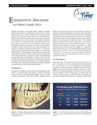
Endo diagnosis oct2002
•
1 like•24 views

This document discusses the importance of conducting a complete endodontic examination for all patients. It states that without such an examination, the pulpal status of teeth is unknown. A complete endodontic examination involves a clinical examination, radiographic examination, and vital pulp testing to diagnose the pulpal status and identify any endodontically involved teeth. Conducting these examinations can lead to the identification and treatment of many previously undiagnosed endodontic problems, improving oral health outcomes and generating additional income for the dental practice.

Recommended
More Related Content
What's hot
What's hot (20)
Similar to Endo diagnosis oct2002
Similar to Endo diagnosis oct2002 (20)
Recently uploaded
Recently uploaded (20)
Endo diagnosis oct2002
- 1. Dentists are trained to thoroughly review medical and dental histories and perform comprehensive extraoral and intraoral examinations. Yet, in spite of these efforts to optimally serve patients, the dominant clinical reality is the vast majority of dentists do not know the status of the pulps within the teeth of the patients who visit them daily. If a complete endodontic examination is not conducted, then the pulpal status of any given tooth is unknown. The rationale for conducting a complete endodontic examination is similar to conducting a complete intraoral screening for soft tissue pathology; con- ducting a complete periodontal examination; or a physician conducting a complete physical examination on a seemingly healthy patient. Clinical judgment should be used to determine which patients, and which teeth, should receive an endodontic examination. Regretfully, a significant number of endodontically involved teeth are not diagnosed or treated. In fact, the vast majority of all endodontic procedures are performed secondarily to patients presenting with symptoms. It is critically important for dentists to accurately diagnose endodontic disease associated with both asymptomatic and symptomatic teeth. THE REALITY It should be completely understood and fully appreciated pulpal health is not guaranteed just because a tooth is clinically asymptomatic or a well-angulated radiograph does not reveal a lesion of endodontic origin (LEO).1 Many pulpally involved teeth do not exhibit symptoms or demonstrate a LEO even though considerable breakdown and destruction may have already occurred in the less dense trabecular bone. Research has demonstrated diagnosticians only see an “incipient” radiolucency when the more dense buccal or lingual cortical plates of bone have been invaded by a lesion.2 If a complete endodontic examination was conducted on each patient before commencing with any dental procedure, then a staggering and sobering number of quiescent, pulpally involved teeth would be identified (Figure 1).3 Frequently, patients report they were comfortable before treatment, then following a so-called “routine” procedure developed a “toothache”. In summary, clinicians regularly treat the “toothache”, yet only sporadically find other “obvious” endodontic problems. THE POSSIBILITY It has been said, “At the end of the rainbow there is a pot of gold.” However, the “real” endodontic pot of gold is typically not discovered. If found, undiagnosed endodontics represents a significant source of additional practice income (Figure 2). As a conservative example, assume a mature practice has 1,000 active patients. Assume that each patient has an average of only 20 teeth. Then it could be said this practice is the custodian of 20,000 teeth. Appreciate the enormous endodontic implications if pulpally involved teeth ENDODONTIC DIAGNOSIS by Clifford J. Ruddle,D.D.S. DENTISTRY TODAY October 2002 Figure 1. This human skull demonstrates several important anatomical relationships and serves to illustrate how a LEO can hide between intact cortical plates of bone. Figure 2. This table serves to reveal the enormous health and financial consequences when endodontically involved teeth are not identified and treated. © ADVANCED ENDODONTICS
- 2. ENDODONTIC DIAGNOSIS ▲ 2 are not diagnosed between 1% and 5% of the time. In this hypothetical scenario, if the endodontic diagnosis is missed just 5%, then this would represent 1,000 teeth. Assume all endodontic procedures cost $500 per tooth, then potentially this could result in not performing $500,000 of endodontic work. Additionally, the vast majority of all endodontically treated teeth require a core build-up and restoration. If we assume these restorative efforts cost $500, then another $500,000 worth of dentistry could have been produced. This assumption demonstrates there is a conservative yet massive potential to diagnose, treat and produce an additional $1,000,000 of dentistry.4 Although it is impossible to quantify the magnitude of undiagnosed endodontically involved teeth, it is an unmistakable fact that a significant number of endodontic problems are not identified or treated.5 When a complete endodontic exam is performed, the results clearly communicated, and mutual trust has been established, then patients will generally schedule, as convenient, the appropriate treatment. Dentists have a professional responsibility to clearly communicate the risks versus benefits associated with the recommended treatment, alternatives to treatment, and the clinical ramifications of no treatment. Each clinician should carefully evaluate all the diag- nostic information, consider the strategic nature of the tooth, and anticipate the treatment challenges. Additionally, each practitioner needs to evaluate their training and experience and, if they have the appropriate technology, decide if a referral would be in the patient’s best interest.6 Importantly, when a comprehensive endodontic examination is conducted, then virtually all of the “non-obvious” endodontically involved teeth can be identified. The endodontic examination serves to improve treatment planning, performance and prognosis. Endodontic diagnosis and treatment represent important aspects of ideal oral health. In fact, oral infection has been identified as a risk factor for certain systemic diseases.7 The possibility is present to improve oral health by accurately diagnosing previously undiagnosed endodontically involved teeth. Regarding the expression, “At the end of the rainbow there is a pot of gold,” my assertion is, “There is a pot of gold and it can be found within the teeth that visit you daily.” ETIOLOGY OF PULPAL BREAKDOWN The dental pulp is a dynamic tissue whose status at any given time can be assigned a position on a continuum that ranges from optimal pulpal health to pulpal necrosis. It is wise to appreciate that not all asymptomatic teeth that exhibit a normal response to pulp tests have the same degree of health or capacity to heal.8 The dental pulp has a restricted capacity to heal because it has a limited blood supply, is encased in unyielding dentinal walls and represents terminal circulation. As such, the ultimate fate of the dental pulp is dependent on the magnitude and duration of an injury.9 The major threats to the pulp are caries, traumatic episodes, specific developmental anomalies, certain periodontal conditions and related treatment efforts, and extensive dental proce- dures.10 As an example, following many operative procedures, patients may report transient pain to a cold stimulus indicating a pulpal inflammatory response that is potentially reversible. In other instances, patients may report intense and lingering pain to a cold stimulus which generally infers an irreversible pulpitis.11 Obviously, recurrent caries, a leaking restorative and repeated episodes of dentistry on the same tooth sharply escalate the potential for pulpal break- down and disease flow. Pulpal injuries frequently progress from reversible to irreversible inflammatory conditions and rapidly advance from ischemia, infarction and partial necrosis to complete pulpal death (Figure 3).12 The progressive cascade of pathohistological events that occur within a degenerating pulp as it symbolically journeys along this continuum are well understood.8-12 Regretfully, clinicians who perform dental procedures without diagnostic pulp tests are unable to forecast the ultimate fate of the pulp. THE ENDODONTIC EXAMINATION The comprehensive endodontic examination is a “three-step” diagnostic process comprised of the clinical examination, the radiographic examination and vital pulp testing. This examina- tion should be appropriately scheduled and performed on all new and existing patients regardless of whether they are asymptomatic or symptomatic. In the instance where there is a chief complaint, it is important to inquire as to the region, magnitude and duration of the pain. Additionally, the dentist should ask if the sensitivity is diffuse or localized, intermittent or continuous, and if there is a specific stimulus that provokes the pain. It is important to listen, clarify and then accurately record this information.13 The purpose of a full mouth endodontic examination is to differentially diagnose between odontogenic versus nonodontogenic problems. Specifically, the endodontic examination serves to identify endodontically involved teeth and additionally enables the clinician to classify any given tooth into one of four categories: 1) Teeth that are asymptomatic and do not have a LEO 2) Teeth that are asymptomatic and have a LEO 3) Teeth that are symptomatic and do not have a LEO 4) Teeth that are symptomatic and have a LEO Figure 3. A radiograph of a maxillary first bicuspid showing a gutta percha point tracing a sinus tract and pointing to a lesion of endodontic origin. © ADVANCED ENDODONTICS - www.endoruddle.com
- 3. CLINICAL EXAMINATION The purpose of the clinical examination is to thoroughly evaluate all aspects of the extraoral and intraoral tissues. The extraoral examination allows the dentist to observe a patient’s face and look for symmetry, color and the overall complexion. Further, the examination reveals the presence of various diseases, traumatic injuries, and facial scars. Examining dentists should bilaterally palpate the sub- mandibular nodes for lymphadenopathy as this is the site for regional drainage from the head and neck. The intraoral portion of the examination is directed towards inspecting all aspects of the soft and hard tissues. The soft tissue portion of the examination includes, but is not limited to, carefully evaluating the oral mucosa, oral pharynx, tongue, and floor of the mouth. Additionally, a thorough intraoral soft tissue exam reveals the color, texture, consistency and contour of the soft tissues including the presence of a swelling, sinus tract or tattoo (Figure 4). The periodontal examination provides the opportunity to evaluate the mucogingival soft tissues, attach- ment apparatus, and probable pocket depths. Importantly, broad crater-shaped defects are more often associated with periodontal disease, whereas narrow vertical defects suggest either endodontic etiology or a radicular fracture (Figure 5). The intraoral hard tissue exam reveals missing teeth, fractured teeth, dark teeth and developmental anomalies (Figure 6). Further, all existing restoratives are evaluated for marginal adaptation, contour and esthetics. The diagnostician looks for caries, recurrent caries, and inspects the cervical area of teeth for erosions, abrasions and abfractions. The presence of inflammation or infection can contribute to the loss of attachment and excessive mobility of a tooth. Roots should be palpated laterally and apically, both on the facial and lingual aspects as a lesion of endodontic origin can invade through the cortical plate. The percussion test is performed gently and conducted laterally, then vertically on the incisal edges of anterior teeth or on the buccal and lingual cusps of posterior teeth. A positive percussion test indicates an injury to the attachment apparatus, may cast suspicion regarding the status of the pulp, but, in and of itself, does not disclose information regarding the health of the pulp. The bite test is useful to identify teeth with incomplete or complete dentinal fractures and is best performed with a cotton roll, q-stick or the Tooth Slooth (Sullivan Schein Dental; Melville, New York). These devices are placed interocclusally and patients are instructed to initially bite gently and, if possible, to then bite firmly. Additionally, patients should demonstrate they can ENDODONTIC DIAGNOSIS ▲ 3 Figure 5. A surgical photograph of an endodontically failing molar reveals a vertical root fracture possibly caused by the use of excessive force during obturation. Figure 4a. A photograph reveals significant swelling secondary to a LEO that has broken through the buccal cortical plate of bone. Figure 4b. A photograph demonstrates a gutta percha point tracing a sinus tract and a tattoo associated with an endodontically failing maxil- lary left central incisor. © ADVANCED ENDODONTICS - www.endoruddle.com
- 4. move their mandibles into working and balancing excursions. A fiberoptic wand can be used to transilluminate the clinical crowns of teeth without extensive, full restorative coverage. When a natural crown is transilluminated facial to lingual, light will uniformly pass through tooth structure if there is no fracture. On the contrary, when a tooth exhibits a coronal fracture, then light will not uniformly pass through the clinical crown, and the fracture breaks a beam of light. The clinical examination focuses on the masticatory system including the jaws, temporomandibular joint and muscles of mastication. The occlusion is carefully checked by having the patient move into various lateral and protrusive excursions. Marking paper can be used to identify and address prematurities that can contribute to harmful wear facets, increased mobility and thermal sensitivity. Habitual grinding is a behavior that pro- motes malocclusion and is frequently associated with fractured teeth. In summary, the clinical examination reveals valuable information regarding a patient’s dental history, and can serve as an indicator of their motivation to pursue oral health. RADIOGRAPHIC EXAMINATION The radiographic examination is generally performed following the clinical examination. In fact, the clinical portion of the diag- nostic work-up often serves to identify the specific location(s) where the radiographic exam should be focused. The endodontic radiographic examination is optimized when three different, well-angulated, and high quality images are obtained.14 A straight-on diagnostic film should be taken such that the xray cone is aimed perpendicular to both the facial aspect and long axis of the tooth. A second, mesially angulated film is attained by horizontally aiming the xray cone up to 30° mesial to the straight-on angle and perpendicular to the long axis of the tooth. A third, distally angulated film is attained by horizontally aiming the xray cone up to 30° distal to the straight-on angle and perpendicular to the long axis of the tooth. Frequently, dentists inquire as to the need for three pre-operative radiographs when, indeed, a single film, in conjunction with the results from a vital pulp test and the clinical examination, will generally confirm a definitive diagnosis. The answer is simple: The best film is still a two-dimensional image of a three-dimensional object. A single film, along with the other diagnostic information, may endodontically condemn a tooth; however, a single radiographic image will not adequately prepare the clinician for optimal treatment planning and patient communication. The diagnostic quality of a radiographic image is definitely enhanced using film holding and aiming devices and adhering to well-recognized and successful darkroom protocols. Digital radiography is improving the field of dental radiology as it provides several advantages over film-based radiography.15 Digital radiography reduces radiation, eliminates chemicals and film processing, and provides nearly instant, high quality images that patients can clearly see. Centralized storage and retrieval allows clinicians to send, receive, and print images. Software tools afford several features that can be utilized to enhance images, such as zoom, measurements, adjustable contrast, image colorization, black/white reversal, and density measurement and comparison. Perhaps the greatest advantages of digital radiography is the potential to more effectively communicate with patients by allowing them to participate in co-discovery, co-diagnosis and co-treatment planning. When performing the radiographic examination, the clinician will observe that different angulated images enhance detecting the location and extent of caries or recurrent caries. A restoration should be evaluated radiographically regarding marginal adaptation, contour, relative depth and relationship to the pulp chamber. At times, a bitewing film is useful as it can provide additional information as to splinted teeth, and the presence of pins and build-up materials. Radiographic images frequently allow the clinician to determine the size of the pulp chamber as compared to adjacent teeth, the presence of stones, and if calcific material projects into the coronal aspect of a canal (Figure 3). Clinicians can visualize a radiograph to appreciate the crown/root ratio and orientation, and the angle of the coronal aspect of a canal relative to the long axis of the root (Figure 7). Different, horizontally angled films disclose ENDODONTIC DIAGNOSIS ▲ 4 Figure 6a. A photograph shows discoloration of the clinical crown of the mandibular left central incisor as a result of a traumatic accident. Figure 6b. A clin- ical photograph of the lingual surface of this maxillary incisor reveals a dens evaginatus and a sinus tract located high in the palatal vault. © ADVANCED ENDODONTICS - www.endoruddle.com
- 5. information regarding the length and curvature of a root and, when present, the depth of an external concavity. High quality radiographic images can be studied to better appreciate the root canal system and, at times, disclose canals that merge, curve, recurve, dilacerate or divide. The astute clinician will identify atypical tooth morphology such as a C-shaped molar, taurodontism or dens invaginatus (Figure 8).16 Different, well-angulated films allow clinicians to observe the result of a traumatic episode such as a coronal fracture, horizontal root fracture, and at times, a vertical root fracture (Figure 9). The clinician needs to carefully observe films for the possible sequelae to traumatic events, such as internal and external resorptions (Figure 10). High quality images clarify root end proximity to normal anatomical structures such as the maxillary sinus, mental foramen or mandibular canal. In fact, at times clinicians should expose a contralateral film to rule out a normal radiolucent anatomical landmark versus an abnormal radiolucent lesion. At times, additional films may be prescribed to augment an examination; including a panograph, lateral jaw, or occlusal radiograph. Diagnosticians should ENDODONTIC DIAGNOSIS ▲ 5 Figure 7. A radiograph suggests the anterior bridge abutment is endodontically involved. Note the crown/root orientation and inclina- tion of the canal coronally. Figure 8. A radi- ograph suggests this maxillary right central incisor has a dens in dente, internal resorption, a large asymmetri- cal lesion and multiple canals. Figure 9. A radiograph of a maxillary central incisor reveals a horizontal root fracture with displacement and a previously accessed lateral incisor. Figure 10a. A radiograph of a mandibular left lateral incisor shows evidence of internal resorp- tion and an apical lesion of endodontic origin. Figure 10b. This radiograph reveals massive root resorption associated with the maxillary incisors possibly caused by the erupting and mesially inclined canine. © ADVANCED ENDODONTICS - www.endoruddle.com
- 6. recognize that regardless of the various radiographic options, in the final analysis, interpreting a radiographic image is subjective and is a learned skill.17 Various horizontally angulated radiographs also provide critical information as to the etiology of endodontically failing teeth.18 Many endodontic failures can be attributable to coronal leakage resulting from failed restorations. Radiographs can clarify if the obturation material was gutta percha, a silver point, carrier-based obturator or paste filler. Additionally, radiographs reveal a particular canal was well-shaped, and the vertical extent of obturation. Dentists who expend a considerable amount of their clinical time performing retreat- ment appreciate that a short fill could suggest a blocked canal. Off-angled films enhance the diagnostic assessment of root canals that exhibit a ledge, transportation or perforation. A radiograph will generally reveal the presence of a post and additionally provide information as to its length, diameter and orientation relative to the long axis of the root. Off-angled images can demonstrate the presence and position of a broken instrument or a missed canal. At times, a patient will be asymptomatic and demonstrate a radiographic radiolucency associated with a root apex. If there was a history of endodontic surgery, then the differential diagnosis should include the possibility of a surgical scar. However, discounting radicular fractures and hopelessly involved periodontal teeth, virtually all other endodontic surgical failures should be attributable to microleakage and bacterial invasion. The radiographic examination also provides information regarding the periodontal supporting structures. Certain probable defects masquerade as a periodontal lesion when, in fact, the etiology may be attributable to significant lateral canals disseminating pulpal irritants.19 Clearly, pulp testing schemes must be conducted to corroborate this suspicion allowing the clinician to differentially diagnose the presence or absence of a LEO. Over time it is becoming well understood that LEO’s occur in the furcations of multi-rooted teeth (Figure 11a). Additionally, a LEO may be positioned crestally, laterally along a root surface, or symmetrically or asymmetrically around the apex of a root (Figures 11b, 11c). It must be under- stood that radiographic radiolucencies or radiopacities could represent a normal anatomical landmark, a nonodontogenic lesion, or a LEO, and the differential diagnosis is made by performing vital pulp tests.20 ENDODONTIC DIAGNOSIS ▲ 6 Figure 11a. A radiograph of an endodontically involved mandibular first molar showing a gutta percha point passing through the buccal sulcus to a furcal lesion. Figure 11b. A radiograph of an endodontically involved maxillary first bicuspid reveals a distocrestal lesion that is threatening the sulcus. Figure 11c. A radiograph shows a mandibular first molar with a poor fitting crown, incomplete endodontics and a LEO associated with the mesial root. © ADVANCED ENDODONTICS - www.endoruddle.com
- 7. VITAL PULP TESTING The clinical and radiographic steps of the examination often- times cast suspicion of endodontic involvement of a specific tooth. Vital pulp tests (VPT) are essential components of the endodontic examination and serve to disclose the status of the dental pulp.13 Frequently, patients present reporting pain to a thermal stimulus in a specific quadrant. In these instances, vital pulp testing schemes should be performed first on presumably “pain-free” teeth, away from the area of the chief complaint. Specifically, the preferred sequence is to test contralateral teeth first, opposing teeth second, then presumably healthy teeth within the thermally painful quadrant, and finally, the most suspicious tooth last. This strategy of sequencing the vital pulp tests allows both the doctor and the patient to appreciate the range of “normal” pulpal responses exhibited by asymptomatic teeth. Importantly, performing repetitive pulp tests, as described, will tend to relax the patient, build confidence and reduce the probability of a false positive or false negative report. VPT procedures are initially performed to establish a normal “baseline” for any given tooth on any single patient. Once a baseline has been established then, and only then, should the appropriate VPT be performed in the quadrant where the patient is experiencing symptoms. Performing VPT on asymptomatic teeth establishes the baseline for testing and comparing an “abnormal” response in a symptomatic tooth. In fact, when VPT schemes are conducted in this manner, patients will frequently question why another tooth is either overreactive or nonreactive to the specific test. In these instances, additional diagnostic evaluation may be required to clarify the endodontic status of any given tooth. When pulpal inflammation is confined to the root canal space, diagnosticians should be skeptical when patients attempt to identify a specific tooth they perceive as the source of their pain. This doubt is justified since the dental pulp does not have proprioceptive nerve fibers.21 On the contrary, the attachment apparatus has proprioceptive nerve fibers that allow a patient to identify a tooth that is sensitive to biting pressure. As such, inflammatory conditions involving the dental pulp are diagnosed by reproducing the patient’s chief complaint as this is diagnostic. Thermal pain is pulpal in origin whereas biting or chewing pain is related to injuries involving the periodontal attachment apparatus. The origins of attachment apparatus injuries are multifactorial and, as examples, could be periodontal or endodontic in etiology or attributable to a recently placed restoration in hyperocclusion. In summary, it is wise to appreciate that a symptomatic patient can present with two separate, distinct and unrelated problems and, as an example, the tooth that is symptomatic to biting pressure may not be the tooth that is symptomatic to a thermal stimulus (Figure 12). There are four methods that may be employed to determine the vitality of the dental pulp: the cold, hot, electric, and cavity tests. Selection of the cold test or the hot test is based on the patient’s chief complaint. If a patient does not report any history of thermal pain then, for ease, the cold test is selected. However, it should be recognized that once the pulp is stimu- lated with cold, there is a refractory period of several minutes before a second cold or hot test can be accurately conducted. The electric pulp test is more technique sensitive, requires a dry field and is oftentimes impractical to utilize in teeth with full restorative coverage. The cavity test is rarely used, and only considered when the clinical and radiographic informa- tion and pulp test results prove inconclusive. In these instances and, when the patient situation supports intervention, then the cavity test could be considered as a last resort. If employed, the cavity test is initiated on a suspicious tooth, without anesthetic, and involves drilling a small window through either enamel or a restoration to dentin. The cavity test will stimulate a vital pulp and provoke a painful response when dentin is invaded. In the event of a vital response, a simple restoration is placed. On the contrary, the cavity test will not stimulate a partially necrotic pulp to the same extent as a vital pulp. In this situation, the dentist initiates the access cavity, invades progressively deeper into dentin and often reaches the pulp chamber uneventfully. Thermal tests should be conducted on the cervical aspect of a tooth, and as close as possible to the free gingival margin. This location represents the thinnest aspect of enamel or a restoration and, importantly, the closest distance to the pulp chamber. When performing a thermal test, the clinician is evaluating the “immediacy”, the “intensity”, and the “duration” of the response. The immediacy and intensity of a response to thermal testing can vary significantly depending on, as examples, the depth of a carious lesion, the placement of a new restoration, or recent periodontal surgery. It is useful to have the patient subjectively rate the intensity of a response utilizing a zero to ten (0-10) scale where zero (0) is a no response and ten (10) represents maximum pain. Regardless of the immediacy and intensity, if the response rapidly dissipates upon removing the thermal stimulus, then ENDODONTIC DIAGNOSIS ▲ 7 Figure 12. This radiograph suggests the mandibular first bicuspid has a carious pulp exposure and reveals a LEO associated with the mesial root of the molar. © ADVANCED ENDODONTICS - www.endoruddle.com
- 8. although the pulp may have tested inflamed, this may be a reversible condition. Importantly, it is the “duration” of the response, compared to the baseline that was established by testing other teeth, that is most diagnostic. In certain instances, a tooth tested with a thermal stimulus may elicit a “no response” which could infer the pulp is necrotic. It should be recognized a patient may not respond to a thermal test if the pulp chamber has significantly calcified or receded apical to the crest of bone. Further, a no response to a thermal test could imply a tooth has been involved in a recent episode of trauma, has an immature apex, or the patient may have premedicated. Additionally, a patient will not generally respond to a thermal stimulus on a tooth that has had root canal treatment. However, an endodontically failing tooth with a missed canal will, at times, illicit a painful response when tested with a hot stimulus. COLD TEST When a patient presents and reports a history of pain to a cold stimulus, then the clinician should logically conduct the “cold test”. Although there are a few different methods that may be selected to apply a cold stimulus, one reliable source is to utilize an ice pencil.22 A pencil of ice is easily formed by first purging all the anesthetic from an unused carpule. Dental floss may then be cut about one (1) inch longer than the length of the carpule and inserted into this glass tube. Several of these carpules are filled with water, held upright in a cup and then placed in a freezer. When a pencil of ice is needed, a frozen carpule is briskly rolled between the gloved palms of the hands. This action serves to warm and contract the ice pencil which may then be liberated by gently pulling the floss. The pencil of ice is placed in a 2x2 gauze to prevent warmth from the fingers from prematurely melting the ice. Before initiating any thermal pulp test the diagnostician needs to establish reliable hand signals. The patient is instructed to raise their hand when they first feel the sensa- tion from the thermal stimulus in the tooth, to keep their hand up as long as this sensation lingers, and to lower their hand when the sensation dissipates. It is wise to repeat and clarify these instructions as both asymptomatic and, especially, symptomatic patients are frequently nervous and may inadvertently not follow directions. This is precisely why thermal pulp tests should not be initially performed on suspicious or symptomatic teeth. As such, before instituting any pulp test, advise the patient how this test works, ask permission to test, and then initiate the test on pain-free teeth. The specific technique for pulp testing is straightforward.23 The ice stick is placed towards the cervical aspect of a tooth on either the buccal or lingual aspect of a crown and quickly moved back and forth (Figure 13). This action creates a slurry of cold water which will effectively bathe, conduct and penetrate into a tooth. To prevent a false positive result, a cotton pellet should be placed just distal to the tooth being tested to prevent ice water from potentially contacting a more posterior tooth. When the patient signals they feel cold in the tooth being tested, the ice is immediately removed. However, the patient is reminded to keep their hand up as long as the cold sensation lingers in the tooth. When testing teeth with healthy pulps, once the patient has signaled they feel cold in a tooth, their hand will generally remain raised approximately 2-5 seconds after the stimulus is removed. As mentioned, the diagnostician is observing the immediacy, intensity, and importantly, the duration of the response. At times, the ice pencil and resultant slurry of cold water will not elicit a response and reproduce the patient’s chief complaint. In these instances, a tooth or group of teeth may be individually isolated with a rubber dam, and ice water syringed onto each tooth. Although more time consuming, this method of testing is very effective at simultaneously bathing the entire clinical crown of a tooth and stimulating an inflamed pulp. Astute clinicians appreciate that a test that elicits a lingering response is diagnostic and separates the reversible pulpal conditions from the irreversible conditions. HOT TEST When a patient reports a history of pain to a hot stimulus, then the clinician should logically conduct the “hot test”. A “toothache” precipitated by hot liquids or foods usually suggests an acutely inflamed or partially necrotic pulp. Necrotic tissue frequently harbors bacteria which can pro- duce gasses that potentially expand against tissue encased inside unyielding dentinal walls. This phenomenon causes sensory fibers of the pulp to transmit pain.21 There are a few different devices that may be selected to apply a hot stimulus, including the Touch ‘n’ Heat or System B (SybronEndo; Orange, California). Either device has a handpiece which is designed to receive various inserts such as the Hot Pulp Test Tip (SybronEndo; Orange, California). Regardless of the device chosen, the continuous mode is selected and the ENDODONTIC DIAGNOSIS ▲ 8 Figure 13. A clinical photograph demonstrates a reliable method and technique for performing the cold test. © ADVANCED ENDODONTICS - www.endoruddle.com
- 9. intensity is set at the manufacturer’s recommendation for performing the hot pulp test. Within a few seconds, the insert tip’s metallic end becomes sufficiently hot. The clinician may use the heat from the insert tip to thermosoften a gutta percha cone into a round ball, which is then attached to the Hot Pulp Test Tip. A thermosoftened ball of gutta percha will readily adapt to the morphological contour of a tooth, which results in achieving better conductivity into the pulp chamber. As with the cold test, the diagnostician must first establish a baseline by testing asymptomatic teeth. The hot test and related hand signals are performed as described for the cold test. Thermosoftened gutta percha is placed towards the cervical aspect of a moist or lubricated tooth, on either the buccal or lingual aspects of the crown (Figure 14). When the patient raises their hand, the diagnostician should immediately remove the hot stimulus. In the event the patient does not perceive any heat sensation in their tooth after 5-6 seconds, then the stimulus should be removed. However, certain pulpally involved teeth may not initially be stimulated by the hot test, then after several seconds, elicit significant pain. For this reason, it is advisable to wait several seconds before placing a hot stimulus on the next tooth. Some teeth with irreversible pulpitis require a repeated hot stimulus over time to reach a threshold that provokes pain. Therefore, when a patient reports pain upon drinking hot coffee, it is informative to inquire if the pain is experienced on the first sip or after repeated sips. This information may be useful when performing and sequencing the hot pulp test. At times, after carefully conducting the hot test as described, the diagnostician may not be able to reproduce the patient’s chief complaint. An alternate method of heat testing involves isolating the crown of a tooth with a rubber dam, and then applying hot water with a syringe.23 The advantage of this method is that hot water instantaneously bathes the entire clinical crown, improves conductivity, and more closely replicates the way heat naturally contacts the tooth during the ingestion of hot foods and liquids (Figure 15). The disadvantage of this test is the cooperation required to comfortably place a clamp on a tooth, then the time required to perform this test on several teeth. Regardless of which hot pulp test method was utilized, the clinician is assessing the immediacy, intensity and duration of each response. As with the cold test, the response that lingers disproportionately, as compared to contralateral, opposing and adjacent teeth, is diagnostic. On occasion, certain patients present with a glass of cold water tightly grasped in their hand. These patients typically have a necrotic pulp and the attendant pain can be “turned off” when they drink cold water or when the diagnostician places an ice pencil on the offending tooth. Provoking a “toothache” with a hot stimulus then turning off the pain with a cold stimulus is profoundly diagnostic. On occasion, after conducting a thorough clinical and radiographic examination and performing VPT, there may be a diagnostic dilemma, such as when a patient reports acute, radiating or diffuse pain that cannot be localized. In these situations, it may be helpful to remove a specific crown or administer block anesthesia in either the maxillary or the mandibular arches to help localize the source of the chief complaint. Clinicians should recognize that in spite of performing a thorough and comprehensive endodontic exam- ination, there will be times when a definitive diagnosis cannot be made. It is wise to remember the Hippocratic oath which states, “Do no harm while doing good.” In these instances, it is appropriate to dismiss the patient and reschedule when their symptoms can be localized. CONCLUSION A comprehensive endodontic examination serves to system- atically identify pulpally involved teeth and is critical for providing optimal dental care. The endodontic examination should be performed and the results recorded before initiating any dental procedure. Specifically, full mouth vital pulp testing procedures establish a baseline and provide information which can be utilized to make more predictable decisions. In many states, trained members of the dental team can legally perform and record the observations and results from the endodontic examination. Reliable information serves to improve diagnostics, treatment planning and patient communication. The comprehensive endodontic examination increases the possibility for patients to receive more timely care and for dentists to find the proverbial pot of gold! ▲ ENDODONTIC DIAGNOSIS ▲ 9 Figure 14. A clinical photograph shows the Hot Pulp Test Tip with thermosoftened gutta percha contacting the cervical aspect of a maxillary lateral incisor. Figure 15. A clinical photograph demonstrates syringing 60°C hot water onto a tooth that has been carefully isolated with a rubber dam. © ADVANCED ENDODONTICS - www.endoruddle.com
- 10. ENDODONTIC DIAGNOSIS ▲ 10 REFERENCES 1. Bender IB: Factors influencing radiographic appearance of bony lesions, J Endod 8:4, pp. 161-170, 1982. 2. Bender IB, Seltzer S: Roentgenographic and direct observation of experimental lesions in bone (part 1), JADA 62, pp. 152-160, 1961. 3. Ruddle C J: Nonsurgical endodontic retreatment: issues influencing treatment, Dentistry Today 17:2, pp. 64-71, 1998. 4. Ruddle C J: How to profit from endo: finding the fair fee for endodontics, Dental Economics 88:11, pp. 30-42, 1998. 5. Endodontic trends reflect change in care provided, Dental Products Report 30:12, pp. 94-98, 1996. 6. Dorn SO, Gartner AH: Ch. 4, Case selection and treatment planning. In Cohen S, Burns RC, editors: Pathways of the Pulp, pp. 60-76, 6th ed., Mosby, St. Louis, 1994. 7. Mattila KJ, Valtonen VV, Nieminen M, Hattunen JK: Dental infection and the risk of new coronary events: Prospective study of patients with documented coronary artery disease, Clin Infect Dis 20:588-592, 1995. 8. Van Hassel HJ: Physiology of the human dental pulp, Oral Surg Oral Med Oral Pathol 32, pp. 126-134, 1971. 9. Kim S: Microcirculation of the dental pulp in health and disease, J Endod 11:11, pp. 465-471, 1985. 10. Takahashi K: Changes in the pulp vasculature during inflammation, J Endod 16:2, pp. 92-97, 1990. 11. Stanley HR, Swerdlow H: Reaction of the human pulp to cavity preparation, results produced by eight different operative grinding techniques, JADA 58, pp. 49-59, 1959. 12. Kim S, Trowbridge H, Suda H: Ch. 15, Pulpal reaction to caries and dental procedures. In Cohen S, Burns RC, editors: Pathways of the Pulp, pp. 573-600, 8th ed., Mosby, St. Louis, 2002. 13. Cohen S: Ch. 1, Diagnostic procedures. In Cohen S, Burns RC, editors: Pathways of the Pulp, pp. 2-24, 6th ed., Mosby, St. Louis, 1994. 14. Kaffe I, Gratt BM: Variations in the radiographic interpretation of the periapical dental region, J Endod 14:7, pp. 330-335, 1988. 15. Antenucci EL: Digital radiography, clinical applications of a maturing technology, AGD Impact 30:7, pp. 18-19, 2002. 16. Mangani F, Ruddle CJ: Endodontic treatment of a “very paticular” maxillary central incisor, J Endod 20:11, pp. 560-561, 1994. 17. Goldman M, Pearson A, Darzenta N: Reliability of radiographic interpretation, Oral Surg 38, pp. 282-293, 1974. 18. Ruddle CJ: Ch. 25, Nonsurgical endodontic retreatment. In Cohen S, Burns RC, editors: Pathways of the Pulp, pp. 875-929, 8th ed., Mosby, St. Louis, 2002. 19. Schilder H: Cleaning and shaping the root canal system, Dent Clin North Am 18:2, pp.269-296, 1974. 20. Bhaskar SN: Part III pathology of the teeth and jaws. In Bhaskar SN, editor: Synopsis of Oral Pathology, pp. 119-337, 4th ed., Mosby, St. Louis, 1973. 21. Gluskin AH, Goon WWY: Ch. 2, Orofacial dental pain emergencies: endodontic diagnosis and management. In Cohen S, Burns RC, editors: Pathways of the Pulp, pp. 25-50, 6th ed., Mosby, St. Louis, 1994. 22. Augsburger RA, Peters DD: In vitro effects of ice, skin refrigerant, and CO2 snow on intrapulpal temperature, J Endod 7:3, pp. 110-116, 1981. 23. Ruddle CJ: “Ruddle on Clean•Shape•Pack”, 2-tape video series. Studio 2050, producer. Santa Barbara, California: Advanced Endodontics, 2002. © ADVANCED ENDODONTICS - www.endoruddle.com