This document provides information about the editors and goals of a book on emergency laparoscopy. It contains biographies of the four editors - Ferdinando Agresta, Fabio Cesare Campanile, Gabriele Anania, and Carlo Bergamini. The editors aimed to provide an updated review of the current evidence on the use of laparoscopy in different abdominal emergencies, including an evaluation of its efficacy and cost-effectiveness. They sought to offer guidance to both experienced laparoscopic surgeons and those less experienced, helping identify appropriate treatment for several emergencies.


![Note to the Reader
The authors of this book adopted the following methodology for literature search
and appraisal: the primary objective of the search was to identify all clinical relevant
randomized controlled trials (RCT) and meta-analysis. Afterward, other reports,
population-based outcome studies, case series, and case reports have been also
included. A systematic review based on comprehensive literature search has been
made on PubMed according to the following criteria: Limits Activated: Humans,
Clinical Trial, Meta-Analysis, Practice Guideline, Randomized Controlled Trial,
Review, English, All Adult: 19C years, published in the last 20 years. Search details:
[((“laparoscopy” [MeSH Terms] OR “laparoscopic” [All Fields]) AND (“condition-
specific key word” [MeSH Terms] OR “condition-specific key word” [All Fields]))
AND (“humans” [MeSH Terms] AND (Clinical Trial [ptyp] OR Meta-Analysis [ptyp]
OR Practice Guideline [ptyp] OR Randomized Controlled Trial [ptyp] OR Review
[ptyp]) AND English [lang] AND “adult” [MeSH Terms] AND “2010/25/11”[PDat]:
“2015/10/31” [PDat])]. Then, limits regarding language, age, and publication date
and study type have been removed, to search for additional papers. Cross-link
control was performed with Google Scholar and Cochrane library databases. The
full text paper was obtained for all relevant articles. The papers have been selected
and classified on the basis of the highest level of evidence, design of the study,
and most recent publication. The 2011 Oxford hierarchy for grading clinical
studies according to levels of evidence (LoE) has been used (http://www.cebm.
net/index.aspx?o=5653). Studies containing severe methodological flaws have been
downgraded as necessary. For each intervention, the validity and homogeneity of
study results, effect sizes, safety, and economic consequences have been considered.
xv](https://image.slidesharecdn.com/emergencylaparoscopy-191115144149/85/Emergency-laparoscopy-16-320.jpg)

![2 C. Bergamini et al.
of emergency laparoscopic surgery: it was in 2006, in fact, when the European
Association for Endoscopic Surgery (EAES) published its consensus statement on
laparoscopy for abdominal emergencies concluding that “ : : : available evidence
clearly demonstrates the superiority of a laparoscopic approach in various emer-
gency situations” [1]. Furthermore, it is interesting to note that these guidelines
outline the role of emergency abdominal laparoscopy as a diagnostic tool as well,
allowing the resolutions of preoperative diagnostic doubts besides the treatment of
the underlying disease.
Different pathologic conditions are responsible for various acute abdominal
situations; in particular, the most common cause of acute abdominal pain is the
nonspecific abdominal pain (NSAP), followed by acute appendicitis, acute biliary
disease, and bowel obstruction or diverticulitis. Despite noninvasive diagnostic
procedures (ultrasonography and CT scan) help surgeon to establish a preoperative
diagnosis, in most of the cases these are not completely accurate and conclusive
about many authors [2–6]. On the contrary, the rate of diagnostic accuracy of
laparoscopy is variable from 89 to 100 % in international literature [1, 6–9].
Concerning the trauma setting, prior to ultrasonography and CT scan, laparotomy
for abdominal trauma was negative and nontherapeutic in approximately one-third
of cases [10]. Nowadays, laparoscopy has a dominant role in well-selected traumatic
patients as a diagnostic tool or therapeutic tool in repairing injuries to the diaphragm,
liver, or spleen and limited gastrointestinal injuries.
1.3 Notes on Lap Cholecystectomy
Laparoscopic approach actually is generally considered to be a gold standard for
cholecystectomies, and the safety of this procedure has also been proved in case of
acute cholecystitis. In the last decades, the number of cholecystectomies increased
worldwide. However, even if several national guidelines suggest laparoscopic
cholecystectomy as a standard of care, it is surprising to find in some national reports
that acute cholecystitis is treated in 50 % of cases. In a population-based study
conducted in the USA, from January 2000 through December 2005, there were an
estimated 2.5 million patients with acute cholecystitis. Among these patients, fewer
than half underwent cholecystectomy and, among them, the 85 % was treated with
a laparoscopic procedure while the 15 % with an open procedure [11]; however,
this study did not consider the patients who underwent delayed cholecystectomy
after the conservative resolution of the cholecystitis. On the other hand, in a
study conducted in the USA on 4011 patients, early laparoscopic cholecystectomy
was performed in 38.0 % of patients and delayed laparoscopic cholecystectomy
in 62.0 %. In this study, authors state that laparoscopic cholecystectomy for
acute cholecystitis in patients older than 65 years with significant comorbidities
is associated with shorter postoperative stay and no increase in postoperative
complications or conversion to open cholecystectomy [12].
On the contrary, in Denmark, among 28,379 patients who underwent chole-
cystectomy between 2006 and 2009, a laparoscopic procedure was performed in](https://image.slidesharecdn.com/emergencylaparoscopy-191115144149/85/Emergency-laparoscopy-21-320.jpg)
![1 A Worldwide Overview of Emergency Laparoscopic Procedures 3
97.7 % [13]. When surgery is performed laparoscopic cholecystectomy represents
the treatment of choice for acute setting with more than 80 % of the operations done
with a laparoscopic approach.
1.4 Notes on Lap Appendectomy
Acute appendicitis represents the greatest cause of emergency surgery; in fact,
250,000 operations are performed every year in the USA [14]. Open appendectomy
has been the standard procedure for acute appendicitis for more than 100 years.
It was in 1983 when Semm [15] reported the first video-assisted laparoscopic
appendectomy in an adult patient, and since then the acceptance of laparo-
scopic appendectomy by surgeons was quite slow if compared to cholecistectomy.
Laparoscopy for acute appendicitis, fortunately, has spread widely in the last 10
years. In a national audit conducted in Italy, for example, results confirm the trend
of the dissemination of LA to peripheral district hospital: in fact, it is practiced
in 93 % of Italian hospitals, and the percentage is increasing [16]. In a national
survey conducted in Germany, results show that the technical infrastructure for
laparoscopic appendectomy was provided in all units, but only in 79 % of units
laparoscopic technique was the standard approach for appendectomy [17]. In a
national survey conducted in Massachusetts, among 2565 patients with appendicitis,
90.1 % of appendicitis patients underwent some intervention. Of these patients,
15.3 % underwent open appendectomy and 72.9 % underwent laparoscopic appen-
dectomy; meanwhile, 1.9 % underwent radiologically guided drain placement [18].
While the laparoscopic approach for appendectomy is widely diffused in the
majority of countries and represents a standard of care, in other countries this
technique is still in the process of development and diffusion. In a nationwide
population-based study conducted in Taiwan, the proportion of patients undergoing
laparoscopic appendectomy was lower (13.3 %) than that of patients receiving open
appendectomy for acute appendicitis. Nevertheless, the frequency of laparoscopic
appendectomy increased over time, passing from less than 1 % in 2001 to 37.2 % in
2008 [19]. In a survey of Polish pediatric surgeon, the low rate of laparoscopic
appendectomy in Poland (33 % of cases) is confirmed: these data suggest that
laparoscopic appendectomy still remains far from being a standard in that country
[20].
1.5 Other Lap in the Urgency Setting
The role of laparoscopy in the diagnosis and treatment of many other acute
gynecological disorders is fundamental, due to the nonspecific clinical presentation
of both diseases in the majority of cases. Similarly, there are many causes of acute
pancreatitis and a large spectrum of clinical presentation, and even if laparoscopy
is unnecessary for diagnosis, it plays a key role in the management of necrotizing
pancreatitis.](https://image.slidesharecdn.com/emergencylaparoscopy-191115144149/85/Emergency-laparoscopy-22-320.jpg)
![4 C. Bergamini et al.
It was in 1990 when Mouret et al. [21] reported the first laparoscopic repair
of a perforated peptic ulcer and actually this technique is universally accepted. A
laparoscopic approach is indicated also in case of complicated diverticulitis as well
as in case of small-bowel obstruction due to adhesions.
Small-bowel obstruction (SBO) requiring adhesiolysis is a frequent and
costly problem in the USA: in 2005, 119 per 100,000 hospitalized patients had
adhesiolysis-related disease [22].
Open adhesiolysis is accepted as the standard surgical intervention for adhesive
SBO, but since the first successful laparoscopic adhesiolysis performed in the
early 1990s, many small-scale studies have found this procedure to be feasible
and safe. In a systematic review of 29 studies with a total of 2005 patients
undergoing laparoscopic treatment of acute SBO, O’Connor et al. found that 64 %
of the operations were completed without conversion to an open procedure with
a postoperative morbidity of 14.8 % and mortality of 1.5 % [23]. A recent study
in the USA performed on a total of 9619 patients who underwent adhesiolysis
between 2005 and 2010 demonstrated that only 14.9 % of operations for SBO
were performed laparoscopically with a significant reduction in 30-day mortality,
major complications, and incisional complications associated with laparoscopic
adhesiolysis compared with open adhesiolysis for the treatment of acute SBO [24].
Laparoscopy, furthermore, plays an important role in the diagnosis and in the
management of acute mesenteric ischemia because it is relatively quick, is well
tolerated, and if necessary can be performed at the bedside in the intensive care unit
or emergency room [25].
1.6 Lap Emergency Surgery in Developing Countries
While the spread of laparoscopy in emergency surgery is a standard practice in
most of the countries of the world, open surgery unfortunately remains the standard
of care in developing nations. The introduction of laparoscopy in resource-poor
areas has been a topic of debate; in fact, within poor countries surgical services
are concentrated almost wholly in cities and priority is given to surgical abdomens,
severe trauma, and other potentially fatal pathologies that do not have laparoscopic
indications [26]. There are few studies in the literature about this topic, but they
demonstrate the use of laparoscopy in emergency surgery in most countries of
the world. A study conducted in Pakistan [27], for example, demonstrates that
also in a developing country early diagnostic laparoscopy is a safe procedure with
high efficacy. In this study, authors excluded the use of diagnostic laparoscopy in
trauma patients because of the nonavailability of technical expertise at all times. In
another study conducted in Nigeria [28], in which authors explain their experience in
laparoscopic appendectomy, emerges the fact that the development of laparoscopic
surgery in this country is slow if compared to other developing nations like India,
but, at this moment, in some institutions surgeons with a basic training in minimal
access surgery who perform most of the operations with a good outcome are present.
Another Nigerian study [29] conducted on pediatric patients demonstrated that](https://image.slidesharecdn.com/emergencylaparoscopy-191115144149/85/Emergency-laparoscopy-23-320.jpg)
![1 A Worldwide Overview of Emergency Laparoscopic Procedures 5
laparoscopy has been used to perform cholecystectomies, appendectomies, and also
adhesiolysis for intestinal obstruction due to adhesions. Authors conclude stating
that laparoscopic surgery technique is feasible in developing countries despite scarce
resources.
While the use of laparoscopy in emergency surgery is still under development in
few countries and widely used for appendectomies or diagnostic laparoscopy, in a
recent systematic review and meta-analysis conducted by Li et al. [30], a comparison
between laparoscopy and laparotomy in the management of abdominal trauma was
performed. Authors analyzed 64 studies (countries included China, Finland, Turkey,
Brazil, Russia, the USA) including 9058 patients with abdominal trauma. The type
of abdominal trauma reported mainly involved gastrointestinal injury, spleen injury,
hepatic injury, pancreatic injury, mesenteric injury, and omentum majus injury (most
of the studies enrolled patients with multiple injuries). Moreover, laparoscopy was
used as a screening, diagnostic, or therapeutic tool. Authors conclude by stating
that laparoscopy is effective and superior in the treatment of abdominal trauma than
conventional open laparotomy if patients have appropriate indications.
In conclusion, laparoscopic surgery represents a valuable standard of surgical
care for most of the country, for both diagnostic and therapeutic intent, in most
abdominal emergencies and it surely has improved our management of surgical
emergencies being an essential part of our clinical and therapeutic approach.
References
1. Sauerland S, Agresta F, Bergamaschi R, Borzellino G, Budzynski A, Champault G et al
(2006) Laparoscopy for abdominal emergencies: evidence-based guidelines of the European
Association for Endoscopic Surgery. Surg Endosc 20(1):14–29
2. Salem TA, Molloy RG, O’Dwyer PJ (2005) Prospective study on the role of the CT scan in
patients with an acute abdomen. Colorectal Dis 7(5):460–466
3. Andersson RE (2011) Routine ultrasound and limited computed tomography for the diagnosis
of acute appendicitis: a surgeon’s perspective. World J Surg 35(2):295–296
4. Golash V, Willson PD (2005) Early laparoscopy as a routine procedure in the management of
acute abdominal pain: a review of 1,320 patients. Surg Endosc 19(7):882–885
5. Kirshtein B, Roy-Shapira A, Lantsberg L, Mandel S, Avinoach E, Mizrahi S (2003) The use of
laparoscopy in abdominal emergencies. Surg Endosc 17(7):1118–1124
6. Domínguez LC, Sanabria A, Vega V, Osorio C (2011) Early laparoscopy for the evaluation of
nonspecific abdominal pain: a critical appraisal of the evidence. Surg Endosc 25(1):10–18
7. Agresta F, Mazzarolo G, Ciardo LF, Bedin N (2008) The laparoscopic approach in abdominal
emergencies: has the attitude changed? A single-center review of a 15-year experience. Surg
Endosc 22(5):1255–1262
8. Champault G, Descottes B, Dulucq JL, Fabre JM, Fourtanier G, Gayet B et al (2006)
Laparoscopic surgery. The recommendations of specialty societies in 2006 (SFCL-SFCE).
J Chir (Paris) 143(3):160–4
9. Hori Y (2008) Diagnostic laparoscopy guidelines: this guideline was prepared by the SAGES
Guidelines Committee and reviewed and approved by the Board of Governors of the Society of
American Gastrointestinal and Endoscopic Surgeons (SAGES), November 2007. Surg Endosc
22(5):1353–1383
10. Renz BM, Feliciano DV (1995) Unnecessary laparotomies for trauma: a prospective study of
morbidity. J Trauma 38(3):350–356](https://image.slidesharecdn.com/emergencylaparoscopy-191115144149/85/Emergency-laparoscopy-24-320.jpg)
![2Acute Calculous Cholecystitis
Fabio Cesare Campanile, Domenico Giannotti, Ferdinando Agresta,
Nereo Vettoretto, and Rao Ivatury
2.1 Introduction
Acute calculous cholecystitis is one of the most common conditions faced by a
general surgeon. Cholecystectomy is the most common digestive operation and
about 30 % of them are performed after an acute cholecystitis [1]. Still, many aspects
about its treatment are controversial and common practice often differs from the
indications provided by the literature.
The disease itself is very heterogeneous: the degree of inflammation may vary
from a self-limiting to a life-threatening form; furthermore, associated conditions
often impose a deviation from the most common course of action, and the general
status of the patient has a relevant impact on both the severity of the illness and the
treatment outcomes.
Such heterogeneity should correspond to a tailored approach, able to grant the
more appropriate treatment to every single patient; however, the literature is devoid
of clear indications about how to adapt the clinical pathway to the diverse scenarios,
the parameters to stratify the risk of surgery in this population are not clear, the most
commonly adopted risk scores are not validated for the acute cholecystitis, and no
instrument is available to select the best course of action in different situations [2,3].
F.C. Campanile ( ) • D. Giannotti
AUSL Viterbo, Ospedale San Giovanni Decollato Andosilla, via Ferretti 169,
01033 Civita Castellana, VT, Italy
e-mail: campanile@surgical.net
F. Agresta
Department of General Surgery, Azienda ULSS19 del Veneto, Adria (RO), Italy
N. Vettoretto
Montichiari Surgery Department, ASST Spedali Civili di Brescia, via G. Ciotti 154, 25018
Montichiari, BS, Italy
R.R. Ivatury
Virginia Commonwealth University, Richmond, VA, USA
© Springer International Publishing Switzerland 2016
F. Agresta et al. (eds.), Emergency Laparoscopy,
DOI 10.1007/978-3-319-29620-3_2
7](https://image.slidesharecdn.com/emergencylaparoscopy-191115144149/85/Emergency-laparoscopy-26-320.jpg)
![8 F.C. Campanile et al.
The available guidelines provide a very valuable support for the daily surgical
practice; however, their recommendations are affected by the lack of literature
evidence about several crucial aspects.
We will try to examine the controversial clinical issues about acute calculous
cholecystitis, analyzing the evidence supporting the guidelines and the most recent
scientific literature data.
2.2 Diagnostic Issues in the Acute Calculous Cholecystitis
The diagnosis of acute calculous cholecystitis relies on a combination of local,
systemic, and imaging signs. A systematic review by Trowbridge [4] suggested that
no single clinical finding or laboratory test carries sufficient weight to establish
or exclude acute cholecystitis. In 2006, based on these results, the European
Association for Endoscopic Surgery (EAES) suggested that the presence of acute
right upper quadrant pain for more than 6 h along with ultrasound evidence of acute
cholecystitis (the presence of gallstones with a thickened and edematous gallbladder
wall, positive ultrasound Murphy’s sign, and pericholecystic fluid collections) could
achieve a high diagnostic specificity. The demonstration of gallstones without
clear imaging cholecystitis should be supported by one or more of the following:
temperature above 38ı
, leukocytosis greater than 10,000/dL, and C-reactive protein
higher than 10 mg/L [5]. The 2012 update of those guidelines maintained the validity
of those criteria [6].
In 2007, the Tokyo Guidelines (TG07) proposed a similar set of criteria that
quickly became widely adopted worldwide [7, 8]. The Tokyo Guidelines Revision
Committee reported that 92.1 % sensitivity and 93.3 % specificity was achieved
when those criteria were adopted in clinical practice. Their last revision (TG13)
is reported in Table 2.1; the imaging findings necessary for the definite diagnosis
are detailed in Table 2.2.
Table 2.1 TG13 diagnostic
criteria for acute
cholecystitisa
A. Local signs of inflammation, etc.
(1) Murphy’s sign and (2) RUQ mass/pain/tenderness
B. Systemic signs of inflammation, etc.
(1) Fever, (2) elevated CRP, and (3) elevated WBC count
C. Imaging findings
Imaging findings characteristic of acute cholecystitis
Suspected diagnosis: One item in A + one item in B
Definite diagnosis: One item in A + one item in B + C
a
RUQ, right upper abdominal quadrant, CRP, C-reactive
protein, and WBC, white blood cell
With kind permission from Springer Science+Business
Media: Yokoe M et al. (2013) TG13 diagnostic criteria
and severity grading of acute cholecystitis (with videos).
J Hepatobiliary Pancreat Sci 20:35–46. ©Japanese Society
of Hepato-Biliary-Pancreatic Surgery and Springer 2012,
Table 1 [8] (modified)](https://image.slidesharecdn.com/emergencylaparoscopy-191115144149/85/Emergency-laparoscopy-27-320.jpg)
![2 Acute Calculous Cholecystitis 9
Table 2.2 TG07 Imaging findings of acute cholecystitisa
Ultrasonography findings
Sonographic Murphy sign (tenderness elicited by pressing the gallbladder with the ultrasound
probe)
Thickened gallbladder wall (>4 mm; if the patient does not have chronic liver disease and/or
ascites or right heart failure)
Enlarged gallbladder (long axis diameter >8 cm, short axis diameter >4 cm)
Incarcerated gallstone, debris echo, and pericholecystic fluid collection
Sonolucent layer in the gallbladder wall, striated intramural lucencies, and Doppler signals
Magnetic resonance imaging (MRI) findings
Pericholecystic high signal
Enlarged gallbladder
Thickened gallbladder wall
Computed tomography (CT) findings
Thickened gallbladder wall
Pericholecystic fluid collection
Enlarged gallbladder linear high-density areas in the pericholecystic fat tissue
Tc-HIDA scans
Non-visualized gallbladder with normal uptake and excretion of radioactivity
Rim sign (augmentation of radioactivity around the gallbladder fossa)
a
With kind permission from Springer Science+Business Media: Hirota M et al. (2007) Diagnostic
criteria and severity assessment of acute cholecystitis: Tokyo Guidelines. J Hepatobiliary Pancreat
Sci 14:78–82. ©Springer-Verlag Tokyo 2007 [7] (modified)
2.3 Laparoscopic or Laparotomic Surgery for Acute
Cholecystitis?
Early after the introduction of laparoscopic surgery, acute cholecystitis was con-
sidered a contraindication for mini-invasive surgery: laparoscopic cholecystectomy
in the acute inflammatory phase of cholecystitis required uncommon dexterity and
experience for those early times, and a higher complication rate was reported [9,10].
In later years, the increased experience and progressive acquisition of the
required skills allowed us to safely complete a laparoscopic cholecystectomy even
in the most severe acute conditions. All the recent guidelines consider laparoscopic
cholecystectomy as the gold standard for the treatment of acute calculous cholecys-
titis [6, 11–13]. Nevertheless, practice pattern varies. Most patients, at least in the
western world, receive laparoscopic cholecystectomy [14, 15]; however, many do
not undergo surgery [16,17] and almost 50 % of all complicated acute cholecystitis
worldwide are still operated by laparotomy, as shown by the CIAO and CIAOW
studies [18,19].
Therefore, it is quite important to examine the clinical advantages reported in the
literature in favor of the laparoscopic approach.](https://image.slidesharecdn.com/emergencylaparoscopy-191115144149/85/Emergency-laparoscopy-28-320.jpg)
![10 F.C. Campanile et al.
2.3.1 Laparoscopic Cholecystectomy for Acute Cholecystitis: The
Evidence
The superiority of laparoscopic cholecystectomy in the treatment of acute calculous
cholecystitis has been mainly demonstrated in four randomized controlled trials
(RCTs) [20–23]. They show that laparoscopic cholecystectomy is associated with
faster recovery and shorter hospital stay. They also indicated that the morbidity of
the laparoscopic approach was not higher, as initially supposed, but actually lower.
However, a full demonstration of a statistically significant reduction in the com-
plication rate could only be obtained by the meta-analysis just recently published
by Coccolini et al. [24]. This study took into consideration both randomized and
non-randomized researches; however, some of the outcomes could be separately
analyzed on the basis of RCTs data only. In this way, it was possible to demonstrate,
with the highest level of evidence, that laparoscopic cholecystectomy in acute
cholecystitis carries a lower risk of complications. Mortality also appeared lower
in the laparoscopic group but the data were available only for non-randomized
comparative studies. Among the complications, wound infection, pneumonia, and
bile leakage deserved a separate inquiry: the wound infection and pneumonia rate
favored laparoscopic surgery, while the bile leakage did not differ significantly
between the two groups. These data represent a strong support for the role of
laparoscopic cholecystectomy as the gold standard in the treatment of the acute
calculous cholecystitis.
The interpretation of the results about faster recovery and postoperative length of
stay deserves a word of caution. As a matter of fact, the evaluation of these param-
eters is partially subjective, and it cannot be excluded that the improved outcome
of laparoscopic surgery be related to an expectation bias of the medical staff rather
than to real clinical and physiopathologic changes. The trial by Johannson [21] was
designed to avoid this bias and included a blind assessment of the outcomes: both
patients and postoperative care staff were unaware of the surgical access received
by the patient because the wounds were kept concealed at all times. Still a reduced
hospital stay could be shown.
One of the RCTs [22] demonstrated a reduction in the surgical trauma and
immunosuppression by measuring serum C-reactive protein and tumor necrosis
factor-˛ (TNF-˛) secretion of peripheral blood mononuclear cells.
If the data of the RCTs with their meta-analysis constitute the strongest evidence
in support of the laparoscopic approach also in the acute setting, the findings
reported by the large observational studies provide an insight into the reality of
the everyday practice. In 2008, Csikesz conducted a population-based analysis
on the USA National Hospital Discharge Surveys about more than one million
cholecystectomies performed for acute cholecystitis from 2000 through 2005.
The study confirmed that laparoscopic cholecystectomy is associated with lower
morbidity, mortality, and shorter length stay. Open surgery was associated with a
1.3-fold increase in perioperative morbidity (95 % CI 1.1–1.4) after adjusting for
patient and hospital factors [14].](https://image.slidesharecdn.com/emergencylaparoscopy-191115144149/85/Emergency-laparoscopy-29-320.jpg)
![2 Acute Calculous Cholecystitis 11
2.4 When to Operate an Acute Cholecystitis? A Question of
Timing
The optimal timing for surgical interventions in an acute cholecystitis is difficult to
determine and controversial; most of the recent literature is devoted to this aspect.
In the pre-laparoscopic era, several randomized controlled trials had shown
the superiority of early versus delayed open cholecystectomy: early surgery was
associated with lower morbidity and shorter hospital stay [25–28].
As we underlined in section “Laparoscopic or Laparotomic Surgery for Acute
Cholecystitis?,” right after the introduction of laparoscopic surgery the skills to
manage an acute cholecystitis by laparoscopy were uncommon, and the condition
was considered to be a contraindication for mini-invasive cholecystectomy. The
safety of laparoscopic surgery in the acute phase of the disease was questioned [9],
and, again, practice evolved in favor of delayed laparoscopic cholecystectomy as an
alternative to immediate open surgery: initial antibiotic treatment and resuscitation
followed by elective laparoscopic surgery about 6 weeks after the acute attack.
Later, after having developed the necessary experience, the performance of
mini-invasive cholecystectomies even in the acute phase of the disease became
more and more common, and trials comparing early versus delayed laparoscopic
cholecystectomy were possible.
2.4.1 The Evidence Supporting Early Laparoscopic
Cholecystectomy
A systematic review of the literature found 15 randomized controlled trials compar-
ing early versus delayed laparoscopic cholecystectomy [29–43], analyzed in several
meta-analyses [1,44–49].
The definitions of “early” and “delayed” differ among the trials; however, most
of them take into consideration patients operated on less than 72 h or less than 7
days from the onset of symptoms as “early”; their treatment is planned at least 6
weeks after the initial diagnosis in the “delayed” groups. Here, we do not mean to
analyze in detail this large amount of literature; however, all the studies agree that
early laparoscopic cholecystectomy is superior because it reduces the total hospital
stay (due to the two episodes of admission) without a significant difference in the
complication or conversion rate. One of the meta-analyses shows that the rate of bile
duct injury, the most feared complication, seems to be even higher in the delayed
treated patients but the difference was not statistically significant [1]. The same
review also revealed that 18.3 % of the patients included in the delayed groups,
in five RCTs, had to undergo emergency surgery in the interval period for either
non-resolution or recurrence of symptoms before their planned operation [1]. The
conversion rate was 45 % in this subgroup.
A very large population-based study confirmed those findings on 10,304 acute
cholecystitis patients who did not undergo cholecystectomy on the first admission.
The probability of a gallstone-related event was 14 % at 6 weeks and 29 % at](https://image.slidesharecdn.com/emergencylaparoscopy-191115144149/85/Emergency-laparoscopy-30-320.jpg)
![12 F.C. Campanile et al.
12 weeks. At 1 year after discharge the likelihood of an acute gallstone-related
episode raised to 29 %. Of these events, 30 % were for biliary tract obstruction or
pancreatitis [50]. A Swedish study found that 6.1 % of patients discharged without
cholecystectomy had emergency surgery in the 2-year study period [16].
Other recent and large population-based studies sustain early surgery and show
that morbidity (included common bile duct injuries) is lower in the early treated
patients [51–53].
These data further support the practice of early laparoscopic cholecystectomy.
Despite the growing evidence in favor of it, the optimal time to perform
early laparoscopic cholecystectomy is still not clear. Is there any difference if the
operation is performed at 24, 48, and 72 h after the onset of symptoms or even later?
Can we establish a threshold of delay after which cholecystectomy can no longer
considered “early” and surgery had better be deferred to a later date (delayed)?
The already mentioned Cochrane review [1] performed a subgroup analysis
comparing the data obtained from the trials that included only patients treated less
than 4 days from the onset of symptoms to those including also patients with a
longer delay, and did not find any statistically significant difference between them.
Once again, population-based study helps to clarify the issue. In 2011, Banz
et al. examined the outcome of 4113 patients divided into six different groups
according to their preoperative length of stay (adopted as a surrogate measure of
the onset of symptoms): operated on the day of admission, on the following day, 2,
3, 4 or 5, 6 days later, or afterwards. There was no significant difference between
preoperative length of stay and postoperative morbidity or mortality. However, the
longer was the preoperative stay and the higher were the operative times and the
conversion rate [51]. Their work did not include any risk adjustment, but similar
results were reported by Brooks et al. on the American College of Surgeons National
Surgical Quality Improvement Program database [54]. Their risk-adjusted analysis
showed an increasing morbidity for cholecystectomies performed from the day of
the admission to 4 days later (from 6 to 19 %) even if they could not demonstrate
a statistical significance. In their 2015 paper, Zafar et al. used propensity score
matching techniques to account for severity differences in a very large (95,523
patients) population-based series from the Nationwide Inpatient Sample (NIS).
They demonstrated that surgery performed within the first 48 h of presentation was
associated with the lowest complication, length of stay, mortality, and hospital cost.
In fact, patients who had surgery during days 2 through 5 and days 6 through
10 had increasingly worse outcomes than those undergoing surgery on days 0
through 1. They could show statistically significant differences for mortality (OR =
1.26; 95 % CI 1.00–1.58 and 1.93; 95 % CI 1.38–2.68) and postoperative infections
(OR = 0.88; 95 % CI 0.69–1.12 and 1.53; 95 % CI, 1.05–2.23) for days 2 through
5 and days 6 through 10, respectively. Also significant were the differences for
pneumonia, UTI, and postoperative length of stay. Their extensive analysis took also
into consideration the adjusted mean hospital cost, and showed it to increase from
$8974 (days 0–1) to $17,745 (days 6–10). The analysis by each incremental day
demonstrated the optimal time of surgery to be within the first 48 h of presentation.
As already mentioned, the relevant size of the series allowed to properly assess](https://image.slidesharecdn.com/emergencylaparoscopy-191115144149/85/Emergency-laparoscopy-31-320.jpg)
![2 Acute Calculous Cholecystitis 13
the differences in the rare event of perioperative death: the lowest risk-adjusted
mortality was found for surgery performed within 1 day or 2 days of presentation
(0.36 % and 0.37 %, respectively), and sudden increase in mortality appeared from
day 3 and later (0.45 %, P D 0:01). It is interesting to point out that mortality for
surgery performed on day 0 was higher (0.42 %) than later. The authors explain
the finding with the higher probability of under-resuscitated patients among those
operated on the day of the admission [55].
Then, laparoscopic cholecystectomy should be performed as early as possible
after the onset of symptoms, and preceded by an adequate resuscitation.
A recent large randomized controlled trial compared laparoscopic cholecystec-
tomy performed within 24 h of admission versus surgery between 7 and 45 days
after initial diagnosis [40]. This trial demonstrated that the morbidity rates were
lower in the delayed than early laparoscopic cholecystectomy group and the length
of hospital stay was 5 days shorter.
In conclusion, several RCTs and their meta-analyses show that laparoscopic
cholecystectomy in the first 7 days after the diagnosis is preferable for its reduced
length of stay, while morbidity is similar if the operation is performed after 6
weeks; on the other side, one trial reports that cholecystectomy between 7 and 45
days carries a higher morbidity rate. Therefore, we can affirm that laparoscopic
cholecystectomy is safe within 7 days from the diagnosis but, outside that window,
is probably better to wait until the 6th week. Large and sophisticated retrospective
studies demonstrate that, within the 7 days of diagnosis, the earlier the operation the
better the outcome and, ideally, laparoscopic cholecystectomy should be performed
within the first 48 h of presentation.
Of course, it is just the case to underline that some of the above examined data
use the time of diagnosis or day of admission as surrogate indicators of the onset of
symptoms because the knowledge of the real beginning of the latter was not easy
to determine; it can be assumed that the symptoms occur just before the hospital
admission but this is not always the case. The everyday clinical practice has to keep
into consideration this variability.
2.5 Laparoscopic Cholecystectomy in Particular Conditions
If the gold standard role of laparoscopic cholecystectomy for acute cholecystitis in
the general population is well clarified by the above reported evidence, still a few
particular conditions deserve a separate review. Are the above described principles
applicable in every case irrespective of the severity of the inflammation and the local
conditions? Are there groups in which a different conduct may be more appropriate?
2.5.1 Severe Cholecystitis
Is laparoscopic surgery indicated for empyema, perforated or gangrenous cholecys-
titis (severe cholecystitis)? Local inflammatory changes can be a real challenge for](https://image.slidesharecdn.com/emergencylaparoscopy-191115144149/85/Emergency-laparoscopy-32-320.jpg)
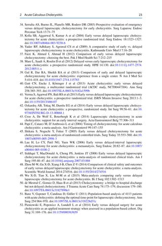
![14 F.C. Campanile et al.
Fig. 2.1 A case of severe
cholecystitis: gallbladder
empyema
any surgeon, and the question of safety of laparoscopy in these extreme conditions
needs to be addressed.
The EAES guidelines published in 2012 stated that empyema, perforated or
gangrenous cholecystitis do not preclude the indication for laparoscopic chole-
cystectomy [6]. However, the Tokyo guidelines incorporate severe cholecystitis
in Grade II of their classification (moderate cholecystitis), likely to be associated
with increased operative difficulty (Fig. 2.1). Therefore, they advocate more cau-
tious indications with a major propensity for gallbladder drainage and delayed
surgery [8].
As a matter of fact, some of the RCTs mentioned in section “Laparoscopic Chole-
cystectomy for Acute Cholecystitis: The Evidence” specifically included patients
with severe cholecystitis [20,23], and a systematic review of observational series of
severe cholecystitis did not show an increase in local postoperative complications
despite a threefold conversion rate [56]. In their case-series, Nikfarjam et al. did
not find any difference in the complication rate between the gangrenous and non-
gangrenous cholecystitis [57].
In addition, some observational reports examined the treatment of severe chole-
cystitis with the aim to compare open versus laparoscopic surgery. A recent very
large retrospective population-based series about gangrenous cholecystitis studied a
total of 141,970 cholecystectomies from the 2005–2011 National Surgical Quality
Improvement Project Participant User File. The authors extracted 7017 gangrenous
cholecystitis. Although they were associated with increased morbidity and mortality
compared with the general series of acute cholecystitis, the multivariate logistic
regression model demonstrated a significant decrease in overall complication rate
(odds ratio = 0.46; P < 0:001) for laparoscopic versus laparotomic cholecystec-
tomy, with a lower, although not significant, perioperative mortality (OR = 0.59;
P D 0:12) [58].
The retrospective investigation by Lo et al. [59] included 74 patients with
cholecystitis and gallbladder perforation divided into 3 groups: early open chole-
cystectomy, early laparoscopic cholecystectomy, and percutaneous gallbladder](https://image.slidesharecdn.com/emergencylaparoscopy-191115144149/85/Emergency-laparoscopy-33-320.jpg)
![2 Acute Calculous Cholecystitis 15
drainage followed by delayed elective surgery. There were no differences in oper-
ative time, blood loss, conversion rate, and morbidity between the groups, but the
length of hospital stay was significantly shorter for laparoscopic cholecystectomy.
The authors concluded that early LC should be considered the optimal treatment for
gallbladder perforation.
If in Lo’s study early laparoscopic cholecystectomy compared favorably with
both early open and delayed laparoscopic surgery for perforated cholecystitis,
other observational reports examined the treatment of different forms of severe
cholecystitis, with the aim to compare early laparoscopic surgery versus some kind
of delayed treatment. Recently Choi et al., in a retrospective study about gangrenous
cholecystitis, compared patients who had early versus delayed laparoscopic surgery,
the latter often preceded by percutaneous cholecystostomy. They failed to find a
significant difference in the morbidity rate, and the total hospital stay was longer
in the delayed group [60]. Similar results were reported by Kwon et al. about
gallbladder empyema [61].
Therefore, laparoscopic surgery is not precluded by severe cholecystitis and
actually seems to be beneficial in terms of morbidity and hospital stay. Early
surgery appears to be advantageous and there is little support for the assumption
that deferring the definitive treatment of these conditions may improve the outcome
and reduce complications.
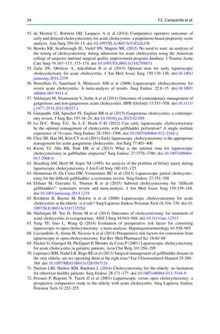
Nonetheless, the operation may be extremely demanding in those cases in
which the intense inflammation around the Calot’s triangle increases the risk of
serious complications. When a positive identification of the vascular and biliary
structures cannot be achieved with the “critical view of safety” (Fig. 2.2) [62]
a laparotomy is advocated. However, conversion not always grants an improved
Fig. 2.2 Identification of the
“critical view of safety”
window in a gangrenous
cholecystitis](https://image.slidesharecdn.com/emergencylaparoscopy-191115144149/85/Emergency-laparoscopy-34-320.jpg)
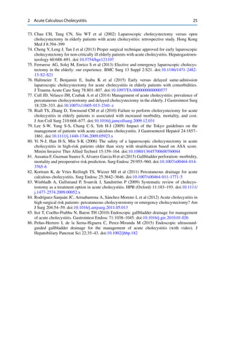
![16 F.C. Campanile et al.
Fig. 2.3 Subtotal
laparoscopic cholecystectomy
in a patient with “frozen”
Calot’s triangle: suture of the
infundibulum
understanding of the biliary anatomy, and experienced laparoscopic surgeons may
proceed laparoscopically with a subtotal (or “partial”) cholecystectomy that appears
to be a reasonable alternative (Fig. 2.3).
The two most recent systematic reviews about partial cholecystectomy report
relatively low morbidity rates, comparable to those of total cholecystectomy in
simple cases. Nowadays, most of them are carried out laparoscopically with reduced
risk of subhepatic collection (odds ratio OR = 0.4; 95 % CI = 0.2–0.9), retained
stones (OR = 0.5; 95 % CI = 0.3–0.9), wound infection (OR = 0.07; 95 % CI =
0.04–0.2), reoperation (OR = 0.5; 95 % CI = 0.3–0.9), and mortality (OR = 0.2;
95 % CI = 0.05–0.9) but more bile leaks (OR = 5.3; 95 % CI = 3.9–7.2) compared
with the open approach [63,64].
2.5.2 Acute Cholecystitis in the Elderly
In a later chapter, we will treat the issue of laparoscopic emergency surgery in the
elderly. Here, we will examine some specific aspects concerning acute cholecystitis.
The prevalence of gallstones increases with age and the life expectancy rises
worldwide. Therefore, the analysis of the more appropriate treatment modalities in
this age group is becoming more and more relevant. In addition, the co-morbidities
in the elderly are obviously more frequent and the acute biliary disease itself appears
to be more severe in this age group. It does not surprise, then, that age is often
considered a risk factor for laparoscopic biliary surgery, particularly in the acute
setting, both for morbidity [65,66] and conversion, [67,68], but it is very difficult to
reach solid conclusions comparing younger versus older patients, because the two
age groups are generally not comparable in terms of co-morbidities and functional
status.](https://image.slidesharecdn.com/emergencylaparoscopy-191115144149/85/Emergency-laparoscopy-35-320.jpg)
![2 Acute Calculous Cholecystitis 17
In the case-series published in 2001 by Decker, cholecystectomy lethality
in acute calculous cholecystitis was much higher in the laparoscopic group in
patients older than 80 [69]. Lupinacci et al., in their retrospective study, pointed
out that the increase in mortality was relevant for cholecystectomy performed in
emergency but not for the elective cases [70]. They suggested that, in the aged,
laparoscopic cholecystectomy should be performed early after they are found to
have symptomatic gallstones, preventing the occurrence of the inflammation, its
increased morbidity and mortality risk. Of course, their series had more high risk
patients (ASA III and IV) and less laparoscopic cholecystectomies in the urgent
(acute cholecystitis) than in the elective groups; these factors can be responsible for
part of the worse outcome of the emergency treatment.
Nielsen et al., on the other hand, could demonstrate a significantly worse
mortality in the patients older than 80 even if they had a low anesthetic risk (ASA I
and II). Their retrospective study showed that the odds for mortality are much higher
in the aged (>80) than in the younger groups (65–79 vs. 50–64), with Odds Ratio
of 30.86 vs. 5.51 vs. 1 [71].
However, the few observational studies able to compare directly open versus
laparoscopic cholecystectomy for acute cholecystitis in the elderly, demonstrated
a reduction in hospital stay [72, 73], and morbidity either unchanged [72] or even
improved [73] in the patients who had laparoscopic surgery.
Having clarified that mini-invasive cholecystectomy is beneficial even in the
aged, one question arises from the above reported data: if it is true that mortality is
higher for laparoscopic surgery performed in the acute but not in the elective setting
in the elderly group, could delayed cholecystectomy be beneficial at this age?
There is no definitive evidence on this particular aspect. Only a few retrospective
cohort studies compare the outcome of early versus delayed cholecystectomy in
elderly acute calculous cholecystitis patients. Mortality and morbidity do not appear
to be significantly different [74–76]. Furthermore, a recent cohort study showed
that recurrent episodes of pancreatitis, cholecystitis, and cholangitis were four
times more likely after delayed cholecystectomy, and the conversion rate did not
change [77]. These findings are confirmed by a recent population-based study on
a sample of the US Medicare Claims Data System. In this analysis, a lack of a
definitive surgical treatment at the index admission in the elderly is associated
with 38 % gallstone-related readmission rate in 2 years versus 4.4 % in similar
patients who had early cholecystectomy and a worse 2-year survival (hazard ratio
1.56, 95 % CI 1.47–1.65) even taking into account patient demographics and co-
morbidities [78]. The very large series reported in the study (29,818 Medicare
beneficiaries urgently or emergently admitted for acute cholecystitis from 1996 to
2005) showed that most US surgeons are confident in operating elderly cholecystitis
patients in emergency: 75 % of the patients aged 66 years and older had an early
cholecystectomy (laparoscopic in 71 % of them). The authors conclude in favor
of early cholecystectomy for elderly patients to prevent recurrent episodes of
cholecystitis, multiple readmissions, and increased costs.](https://image.slidesharecdn.com/emergencylaparoscopy-191115144149/85/Emergency-laparoscopy-36-320.jpg)
![18 F.C. Campanile et al.
2.6 The High Risk Patient
Acute calculous cholecystitis is a very heterogeneous condition. The severity
and complexity of the clinical picture is determined not only by the degree of
inflammation and the local conditions but also by the general status of the patient.
In the previous chapters we examined the data about laparoscopic cholecystectomy
as the definitive therapy of the disease. As a matter of fact, surgery is the gold
standard treatment. Nevertheless, quite often, local or general circumstances suggest
or even impose a different course of action. The optimal surgical treatment should
be examined according to the severity of the disease. In fact, it could be argued that
alternative treatment could better fit the needs of patients with reduced functional
reserve [2, 3]. Should surgery be avoided in some very high risk patients? Could
a delayed treatment be beneficial in some of them? Are there any alternatives to
surgery?
The identification of the parameters and instruments to stratify the risk of surgery
in this population would be of paramount importance to evaluate the role for
alternative therapeutic pathways.
2.6.1 How to Stratify the Risk?
Let’s examine the literature to verify if there is any available instrument to select the
best course of action in particular high risk groups.
Recently, the Tokyo guidelines attempted to address the acute cholecystitis
heterogeneity with a therapeutic algorithm that includes some elements of risk
evaluation. Their staging system is based upon severity assessment criteria such
as degree of local inflammation and associated co-morbidities, without including
any of the most commonly adopted risk stratification scores [11]. However, a
retrospective series failed to find any significant benefit following the introduction
of their guidelines [79].
Advanced age is often identified as a risk factor for the surgical treatment of acute
cholecystitis. We have examined in section “Acute Cholecystitis in the Elderly”
the literature about laparoscopic cholecystectomy in the elderly and in particular
about the timing for surgery; however, the possibility to compare directly surgical
treatment versus an alternative strategy is not available.
In 2006 Yi stratifies the risk in relation to the ASA score, showing a significant
difference in morbidity (20 % vs 9.1 %) in patients in ASA III vs ASA I, with
no significant differences on the conversion rate, recovery time, and hospital
postoperative stay [80].
The only other available comparison of risk assessment scores (ASA, APACHE
II, and POSSUM) is limited to the perforated acute cholecystitis. It highlights
a significant association of the three scores with morbidity and mortality. Both
POSSUM and APACHE II were superior to ASA in risk prediction [81]. However
APACHE II is built as an evaluation score in patient admitted to intensive care units,](https://image.slidesharecdn.com/emergencylaparoscopy-191115144149/85/Emergency-laparoscopy-37-320.jpg)
![2 Acute Calculous Cholecystitis 19
and its use as preoperative risk prediction instrument may be suboptimal. As a matter
of fact, a validated score to choose the best treatment in relation to the patient’s
surgical risk is currently not available.
2.6.2 The Percutaneous Cholecystostomy
A large amount of literature addresses the role of gallbladder drainage (tube
cholecystostomy), generally percutaneous, as an alternative to early surgery in
septic high risk patients. More than 100 papers have been published in the last
few years about this topic. They are generally small case-series of poor quality.
Their inclusion criteria, results, and conclusions are largely not homogeneous. The
only randomized controlled trial of cholecystostomy versus surgery is under way
and not even preliminary results are available [82]. The purpose of gallbladder
drainage is decompression of the infected bile, removal of the purulent collection,
and solution of the sepsis with an improvement of the clinical conditions. Surgery
may be planned at a later date. In particular, the panel of the Tokyo guidelines states
that it is known to be a safe option in critically ill patients, and their guidelines
consider the percutaneous (or surgical) drainage as mandatory in the severe grade
of acute cholecystitis. Its use is also suggested in the moderate grade. However,
cholecystostomy has never been proven to be an effective alternative to early
surgery, and the evidence on its role is still poor. With all the methodological
limits mentioned above, a survey of the literature shows that in-hospital mortality
for cholecystostomy varies between 4 and 50 % and its morbidity between 8.2 and
62 %. A recent systematic review performed a particularly detailed examination of
53 papers about gallbladder drainage as an option in acute cholecystitis. It found
no evidence to support the recommendation of tube cholecystostomy rather than
straight early emergency cholecystectomy even in critically ill patients. Actually,
it suggested that cholecystectomy seems to be a better option for treating acute
cholecystitis in the elderly and/or critically ill population. The authors include 53
studies with 1918 patients; once again, they warn that the results obtained from the
studies reviewed are very heterogeneous. They outline a high success rate of the
procedure (85.6 %) with a low mortality directly related to the procedure (0.36 %)
but a 30-day mortality of 15.4 %, significantly higher (P < 0:001) than after early
cholecystectomy (4.5 %), as reported in published series of similar patients [83]. A
recent prospective study in high risk patients examined the outcomes of 29 patients
treated by percutaneous cholecystostomy and 32 by emergency cholecystectomy.
The groups were homogeneous by age and surgical risk, estimated by physiological
POSSUM, Charlson, APACHE II, and ASA scores. Eight patients (29.6 %) in the
cholecystostomy group required emergency cholecystectomy anyway. The mortality
rate was significantly higher in the cholecystostomy group (17.2 % vs nil). The
authors concluded that percutaneous drainage appears of little benefit and should be
reserved for the patients with surgical contraindication [84]. A large retrospective
cohort study, based on administrative databases capturing all emergency department
(ED) visits and hospital admissions in a populous area, examined 27,718 acute](https://image.slidesharecdn.com/emergencylaparoscopy-191115144149/85/Emergency-laparoscopy-38-320.jpg)
![20 F.C. Campanile et al.
cholecystitis patients. Of them, 890 (3.3 %) underwent tube cholecystostomy. In-
hospital mortality was 5 %, but an additional 18 % had died without surgery. Only
40 % had cholecystectomy within 1 year (due to intercurrent mortality or ongoing
contraindications) and 49 % was evaluated in the emergency department or admitted
to the hospital for a gallstone-related complication [50].
Therefore, at the moment, tube cholecystostomy, as a bridge to surgery or
definitive management of acute cholecystitis, cannot be considered as an established
and safe option. More studies are needed.
2.6.3 Endoscopic Gallbladder Drainage
In the attempt to find less invasive solutions in the old-frail patients and those
with increased surgical risk, endoscopic gallbladder drainage methods have been
proposed. Two techniques have been described: the endoscopic transpapillary
(ETGD) and endoscopic ultrasound-guided transmural gallbladder drainage (EUS-
GBD).
In the former, endoscopic naso-gallbladder drainage and gallbladder stenting via
a transpapillary endoscopic approach are included.
In retrospective studies, endoscopic naso-gallbladder drainage and gallbladder
stenting have a technical success rate of 81 % and 96 %, but a clinical success
rate of 75 % and 88 %, respectively [85]. However, this technique is not feasible if
obstruction or tortuosity of the cystic duct does not allow a guidewire to be advanced
into the gallbladder.
As an alternative, a drain or a stent can be placed in the gallbladder by the
transgastric or transduodenal route under the guide of endoscopic ultrasound (EUS-
GBD). A recent systematic review of the English language literature about this
technique collected 155 reported patients with acute cholecystitis treated with EUS-
GBD in eight studies and 12 case reports. Their technical and clinical success rates
were very high and adverse events were reported only in 8 % of the cases [86]. Of
course non-comparative observational studies and case reports carry a very high risk
of selection bias and further studies are needed to ascertain the potential role of the
endoscopic techniques. Besides, the scarce availability of the technical expertise to
complete these procedures is an additional limitation factor.
2.7 Conclusions
Laparoscopic cholecystectomy is the gold standard for the definitive management
of acute calculous cholecystitis. It should be performed as soon as possible after
the symptoms occurrence. The disease, however, is very heterogeneous as far as
degree of inflammation and local conditions are concerned; besides, the general
characteristics of the patient (age, associated co-morbidities, and functional status)
have a very relevant impact on the disease itself, and surgical intervention may
result in increased morbidity and mortality in the elderly, patients with multiple co-](https://image.slidesharecdn.com/emergencylaparoscopy-191115144149/85/Emergency-laparoscopy-39-320.jpg)
![3Acute Pancreatitis
Mario Campli, Alessandra Cerioli, Ari Leppäniemi, Alberto Arezzo,
and Carlo Bergamini
3.1 Introduction
“Acute pancreatitis is the most terrible of all the calamities that occur in connection
to the abdominal viscera. The suddenness of its onset, the illimitable agony which
accompanies it, and the mortality attendant upon it, render it the most formidable
of catastrophes.”—B. Moynihan, 1925
Throughout much of history, acute pancreatitis (AP) was a mysterious and
dramatic disease: delayed recognition, when necrosis had been established, the
remote location of the gland in the lesser sac, difficulties in differential diagnosis,
and incomplete understanding of the metabolic needs of the seriously ill patients
made AP an impossible challenge for surgeons [1]. We actually achieve great
advancements of our understanding, but the disease remains problematic.
AP is an inflammatory process of the pancreas with a highly variable clinical
course. Most patients experience a mild, self-limiting disease that resolves sponta-
M. Campli, MD
Division of Surgery, “Nuova Itor” Private Health Facility, Rome, Italy
e-mail: mario.campli@gmail.com
A. Cerioli, MD, PhD Candidate
Division of Surgery, “Nuova Itor” Private Health Facility, Rome, Italy
Department of General Surgery, Tor Vergata University, Rome, Italy
A. Leppäniemi, MD, PhD
Second Department of Surgery, Meilahti Hospital, Helsinki, Finland
A. Arezzo, MD
Department of Surgical Sciences, University of Torino, Torino, Italy
C. Bergamini, MD
SOD Chirurgia Generale, d’Urgenza e Mini-invasiva, AOU Careggi, Firenze, Italy
© Springer International Publishing Switzerland 2016
F. Agresta et al. (eds.), Emergency Laparoscopy,
DOI 10.1007/978-3-319-29620-3_3
27](https://image.slidesharecdn.com/emergencylaparoscopy-191115144149/85/Emergency-laparoscopy-45-320.jpg)
![28 M. Campli et al.
neously or successfully treated with supportive therapy. But no medications exist
to treat specifically acute pancreatitis; no actual drugs are available to improve the
course of the disease, and this is a big deal for patients going through a severe attack.
Acute pancreatitis and its serious complications are still a tough challenge for
surgeons. After an “heroic” age of surgery, nowadays a more cautious attitude is
adopted than in the past, with extensive use of laparoscopic and minimally invasive
approach, according to the principle that “less is more” in pancreatic surgery.
Due to the high morbidity and mortality of surgical procedures in the course
of severe AP, the indications for surgery are strictly regulated: early assessment of
the severity and risk stratification are important for well-timed intensive therapy
and timely intervention and have been shown to improve prognosis and survival.
Currently, AP is not an absolute indication for surgery: only the complications of
the disease lead the patient to the operating room to resolve a bowel obstruction,
bleeding, ischemia, or to perform drainage of septic/necrotic collections. In the
gallstone-related disease, the aim of the surgeon is the clearance of the common
bile duct and laparoscopic cholecystectomy with no curative intent but to prevent
the risk of recurrence.
Indications, limits, and advantages of surgical treatment will be clarified and
specified with the current progress in the knowledge of the natural development
and pathophysiology of the disease.
3.2 Classification: Diagnostic and Therapeutic Hints and Tips
During the years many classifications were proposed like Scuro (1984), Marseilles-
Rome (1988), and the Atlanta classification, established in 1992 and probably
the most frequently used by pancreatologists until its revision. The classification
with its empirical conclusions, limits, and gaps was rated more suitable for a
retrospective evaluation and for scientific purposes, rather than for a perspective
classification of patients. The current Atlanta classification, revised in 2012 [2], is
designed to describe AP in a practical way, in order to standardize terminology
across specialties, to help in treatment planning, and to compare management and
results in different departments and institutions or recruitment in clinical trials.
It inserts an additional category to the old classification, providing three classes
of acute pancreatitis concerning severity: “mild,” “moderate,” and “severe.” Mild
AP is the most common form without organ failure (OF) and local or systemic
complications; it does not need an imaging definition and usually resolves in
the first week with no risk of mortality. Moderate AP is characterized by the
presence of transient OF (less than 48 h) and/or the presence of local complications
or exacerbation of comorbid disease and it can eventually require a prolonged
specialist care. Its mortality is lower than in the severe form. Finally, severe AP
consists in persistent OF (more than 48 h) and represents 15–20 % of cases. If the
OF is persistent from the first days, the mortality grows up to 36–50 %. Moreover,
morphologically a classification exists, dividing AP into acute oedematous or
interstitial pancreatitis and necrotizing that can involve both the pancreas and the](https://image.slidesharecdn.com/emergencylaparoscopy-191115144149/85/Emergency-laparoscopy-46-320.jpg)
![3 Acute Pancreatitis 29
peripancreatic tissues, only the peripancreatic tissues (less commonly), and only the
pancreatic parenchyma (rarely). Temporally, the AP can be divided into two phases:
early, in the first week when only clinical parameters are important for treatment
planning, and late, after the first week.
Nevertheless, a consensus about the classification has not been reached yet
[3]. In fact, even in 2012 another international multidisciplinary classification has
been done as a result of a consultative process among specialists in pancreatic
diseases from the entire world [4]. The determinant-based classification provides
a set of concise up-to-date definitions of all the main entities pertinent to classify
the severity of AP in clinical practice and research and adds a further category in
severity classification: the critical one. Complications are classified local or systemic
and sterile or infected. Local complications are acute peripancreatic fluid collections
(APFC), acute necrotic collections (ANC), pseudocyst, walled-off necrosis (WON),
systemic inflammatory response syndrome (SIRS), splenic-portal thrombosis, colic
necrosis, gastric outlet dysfunction, and organ failure (OF). OF is defined as a score
of two or more for one of these three organ systems (cardiovascular, renal, and
respiratory) using the modified Marshall scoring system on the basis of the worst
measurement over a 24-h period. APFC contain only fluid and are not or partially
encapsulated, develop in 4 weeks in interstitial pancreatitis, and frequently regress
spontaneously. ANC contain a mixture of fluid and necrotic material, are not or
partially encapsulated, and develop in 4 weeks, usually in necrotizing pancreatitis.
Pseudocyst is a fluid encapsulated collection, develops after 4 weeks in interstitial
pancreatitis, and also contains necrotic material. WON are necrotic collections, are
fully encapsulated, and develop after 4 weeks.
No significant differences were found comparing the two grading systems [5], but
surgeons may be slightly more interested in the determinant-based classification,
since surgical interventions are indicated only on a few occasions: never in the
early stages, unless general complications such as bleeding, bowel ischemia, or
perforation; and in the late stages only in case of infected necrosis.
It would be very important to define and stratify the predicted severity of the
disease on admission in order to identify potential patients with stones or developing
severe AP to set up an aggressive treatment, or a possible transfer to a specialist
care [6, 7], but till now it is a challenge to determine the right prediction of AP
progression. Furthermore, it is almost impossible to know why a patient remains
in mild state of AP or if he could develop complications [4, 8, 9]. Moreover, the
improvements in diagnostic image, the better understanding of pathophysiology of
the disease, and the development of radiologic, endoscopic, and minimally invasive
operative techniques for the management of complications led to a continuous
revision of the classifications [8, 10, 11].
Score systems are used to predict hospital mortality and they are a good landmark
to classify AP. They are all based on clinical, radiologic, and laboratory variabilities
to calculate the severity of the disease. The scores need to be periodically recali-
brated to reflect changes in practice and patient demographics. In recent years, many
grading systems have been used: Ranson score, Glasgow score, BISAP (Bed Side
Index of Severity in Acute Pancreatitis), Balthazar for the valuation of CT images,](https://image.slidesharecdn.com/emergencylaparoscopy-191115144149/85/Emergency-laparoscopy-47-320.jpg)
![30 M. Campli et al.
and even the APACHE II (Acute Physiology and Chronic Health Evaluation) score,
which is not specific for AP but rather generically for critically ill patients. In recent
comparisons of scoring systems to predict the severity of AP [8, 11], APACHE
II score appeared to have highest accuracy for prediction of severe AP, although
its predictive accuracy was not significantly different when compared to other
scoring systems. Grading systems show a similar predictive accuracy; no simple
assessment score capable of reaching maximal utility for prediction of severe AP
is still available. They are devised to identify groups of high-risk patients rather
than individuals. They work best at the opposite ends of the spectrum (i.e., high
negative or positive predictive value in patients with very low or high scores) [12].
An ideal prognostic evaluation system should be simple, noninvasive, accurate, and
quantitative, and the assessment methods should be easily applicable at the time
of diagnosis [11, 13], but it does not exist. Conversely, application of a severity
assessment score is time-consuming and typically requires 48 h to become accurate.
Frequently, when a score demonstrates severe disease, the patient’s condition is
obvious regardless of the score [8, 14]. While they are cumbersome, are not
mandatory, and do not help with patient management, they are considered useful
tools for risk stratification and to compare the care received by patients with similar
risk characteristics in different units, but in the Western World scores for severity
assessment of pancreatitis are performed only by one-third of hospitals, and the
opinion that the judgment of an experienced surgeon provides evaluation of the
disease outcome as good as any severity scoring system is widely spread [8, 14].
The main problem is still the definition of severity, proposed contemporarily
by many authors; despite the availability of several clinical and radiological
scoring systems, accurate prediction of the AP grade remains uncertain, and a
unique reliable classification is still an open matter [9, 15]. Coexistence of several
classification systems is a questionable peculiarity that is still waiting for a solution
[12, 16].
However, it seems appropriate to deepen understanding of some aspects of the
pathophysiology of AP and some diagnostic issues that have considerable implica-
tions in refining definitions of the various grading systems and in determining timing
and treatment choices.
The proposal takes hold to distinguish between limited and expansive necrosis,
considering the spreading of necrosis in various degrees throughout the retroperi-
toneum and into the small and large mesentery, as there are no anatomical barriers
[12]. If expansive, the necrosis cannot be said to be located into a “collection.”
In CT images it can sometimes be difficult, if not impossible, to distinguish fluid
from necrosis. MRI can clearly distinguish fluid from solid components, but in
daily practice MRI is not as easily performed as CT. The timing of encapsulation,
however, differs markedly among patients and can only be judged on contrast-
enhanced CT, rather than according to time from symptom onset. The presence and
necrosis extension is not valuable in the first days and it is not directly involved
with organ failure and severity. Finally, in the first week a local complication does
not require a surgical treatment. The predictive accuracy of CT scoring systems
for severity of AP is similar, with no statistically significant differences, to clinical](https://image.slidesharecdn.com/emergencylaparoscopy-191115144149/85/Emergency-laparoscopy-48-320.jpg)
![3 Acute Pancreatitis 31
scoring systems. Hence, a CT on admission solely for severity assessment in AP
is not recommended because early CT does not reveal any other diagnosis nor
any local pancreatic complication and underestimates the presence of parenchymal
necrosis in a substantial number of patients [8]. Therefore, CT studies should be
reserved only for patients with predicted severe AP by clinical assessment, for those
who fail to improve clinically with conservative management, for patients whose
diagnosis is unclear, or in suspected severe complication (bleeding, bowel ischemia,
or perforation, etc.) [4, 8]. Predicted mild AP on early CT (i.e., low CT scores) does
not imply that the patient will not develop clinically severe AP, especially when
significant baseline comorbidity is present. The presence of systemic inflammatory
response syndrome (SIRS) increases the risk of developing organ failure, essential
determinant of severity. SIRS is a clinical condition, but also a diagnostic criterion
that can be used to monitor the AP progression [12, 17–19] and, with repetitive
monitoring during the first days of admission, to guide treatment decisions.
Infection of necrosis, both pancreatic and peripancreatic, is the primary indi-
cation for a specific operative treatment in AP. Extensive research has addressed
the possibility of prediction of infected necrosis: several multifactorial scoring
systems (e.g., Simplified Acute Physiology Score II—SAPS 2, Ranson and Imrie
scores, APACHE II, Balthazar CT severity index—CTSI, and multiple organ
dysfunction scores—MODS) and a broad spectrum of biochemical markers (pan-
creatic proteases and anti-proteases, C-reactive protein, procalcitonin, interleukins,
tumor necrosis factor-’—TNF-’, adhesion molecules, serum amyloid A) have been
evaluated and systematically reviewed not only for early stratification of severity but
for prediction of infected necrosis. Although all these scoring systems have been
shown to correlate with morbidity and mortality, it remains difficult to accurately
identify on admission or early in the course of their hospitalization individual
patients who develop infected necrosis.
Apart from clinical symptoms and blood test findings, a clear diagnostic sign
of infected necrosis is represented by gas bubbles within (peri)pancreatic necrosis
on CT, with an almost 100 % specificity in the diagnosis of pancreatic infection.
However, it is worth mentioning that on rare occasions the presence of gas can
indicate the presence of a communication with the gastrointestinal tract. A positive
culture of (peri)pancreatic necrosis by image-guided fine needle aspiration (FNA) is
not recommended as routine today: FNA should be used selectively when there is no
clinical response to adequate therapy or when the clinical and/or imaging features
of infection are uncertain [17, 20, 21].
If the pathophysiologic mechanism of necrosis plays a key role in severe AP
development, infusion therapy can influence the course of the disease. Massive
fluid supplementation to correct or preferably prevent intravascular hypovolemia
is commonly accepted as the first step in supportive care, but it may worsen accu-
mulation of fluid in the third space. Development of intra-abdominal hypertension is
partly related to the effects of the inflammatory process itself causing retroperitoneal
edema, fluid collections, ascites, and ileus, and partly iatrogenic resulting from
aggressive fluid resuscitation. Moreover, it is also difficult to differentiate the SIRS
of the pancreatitis process itself and the effects of intra-abdominal hypertension. It](https://image.slidesharecdn.com/emergencylaparoscopy-191115144149/85/Emergency-laparoscopy-49-320.jpg)
![32 M. Campli et al.
is likely that the processes exacerbate each other in a multifactorial manner. Raised
intra-abdominalpressure causes splinting of the diaphragms, leading to compression
atelectasis and hypercapnia. These events, in combination with bilateral pleural
effusion, which are common in severe pancreatitis, result in severe hypoxia.
The high intra-abdominal pressure with the compression of vessels and viscera
further reduces organ perfusion and causes worsening of ischemia, causing multiple
system organ failure. At this point, an abdominal compartment syndrome is thus
established.
Nowadays, nutritional support is considered a therapeutic measure rather than
merely a way to provide calories in a patient with severe pancreatitis. Enteral
nutrition through a nasoenteric feeding tube has been shown to reduce organ
failure, infected necrosis, and mortality compared with parenteral nutrition. These
complications are thought to be mediated by bacterial translocation from the
gut provoked by disturbed intestinal motility, bacterial overgrowth, and increased
mucosal permeability. Enteral nutrition is believed to stimulate intestinal motility—
thus reducing bacterial overgrowth—and is believed to increase splanchnic blood
flow, which helps to preserve the integrity of the gut mucosa. The improved
outcomes found in the trials following the use of enteral nutrition might also be the
result of complications specifically related to parenteral nutrition, such as catheter-
related infections [22].
Prophylactic administration of intravenous antibiotics has been used with the aim
to prevent infected necrosis and other infectious complications. Several randomized
trials have shown that routine use of antibiotics does not prevent infected necrosis.
Therefore, antibiotics in AP are indicated in case of proven infection or in case of a
very strong clinical suspicion of cholangitis or infected necrosis [14, 22].
Outcome of male patients is worse than those of females [23].
3.3 Mild Pancreatitis
3.3.1 Biliary Pancreatitis
Acute gallstone-associated pancreatitis is initiated and sustained by the passage of
stones in the bile ducts. Biliary etiology is the leading cause of AP, 15 cases of
gallstone AP per 100,000 persons [24], from one to two-thirds of all pancreatitis
depending on the series; when an AP is suspected, an ultrasound scan should always
be performed, as well as cholestasis indices. In mild AP, if no common bile duct
obstruction but gallstones are detected, laparoscopic cholecystectomy should be
performed during the index hospitalization, as soon as pancreatitis symptoms cool
off. Although this assumption is widely adopted, a recent Cochrane review failed
to demonstrate that early laparoscopic cholecystectomy has a better result, that is,
a decreased risk of complications, in comparison with interval cholecystectomy
[25] (LE3). For various reasons, the quality of this evidence is poor; anyway the
outcome of cholecystectomy is not the only intent to perform an early intervention:
between 32 and 61 % of patients with AP are subject to repeated acute episodes.](https://image.slidesharecdn.com/emergencylaparoscopy-191115144149/85/Emergency-laparoscopy-50-320.jpg)
![3 Acute Pancreatitis 33
Recurrent biliary events are common in the first 30 days from index episode,
and subsequent attacks could be more severe than the first one [26, 27] (LE3).
Delayed cholecystectomy increases risk of readmission for recurrent biliary events,
especially recurrent biliary pancreatitis [17, 26, 28–30] (LE2). Therefore, the
current recommendation for patients with mild gallstone pancreatitis is to undergo
a laparoscopic cholecystectomy during initial hospitalization as soon as pancreatitis
has been resolved, before discharge [31]. There is strong evidence that a same-
admission cholecystectomy can be performed safely, without increased difficulty
or surgery-related complications [32] although a very early cholecystectomy (within
48 h after admission) is contraindicated because patients can still develop pancreatic
necrosis or organ failure during this phase of the disease, both representing a surgical
contraindication [33, 34]. Routine intraoperative cholangiography at the time of
cholecystectomy does not improve outcomes [35]: clinical suspicion of common
bile duct stones requests preoperative adequate assessment and possible clearance
of bile duct stones with endoscopic retrograde cholangiopancreatography. At the
present moment same-admission cholecystectomy in mild biliary pancreatitis is
no longer a matter of debate in current guidelines [17, 18, 21], but adherence
to the above recommendation is low: in real world early cholecystectomy is not
performed for a fairly large proportion of patients [36, 37] often because concerns
still exist about an increased risk of cholecystectomy-related complications with this
approach. Cholecystectomy during initial hospitalization may also not be feasible
because of clinical factors, patient preference, or operating room resources that may
be fully. We should also consider the eventuality that some physicians do not follow
guidelines. This is not an evidence-based medicine approach, but there are studies
showing that patients suffering from pancreatitis and admitted in medical services
rather than in surgical wards are treated more conservatively and undergo same-stay
cholecystectomy less frequently [38–40] (LE3). This attitude is even more prevalent
toward the elderly population: more than half of the patients who do not receive
cholecystectomy are never evaluated by a surgeon [41] (LE3).
3.3.2 ERCP and Endoscopic Sphincterotomy
About 20 % of biliary pancreatitis have persistent bile duct stones. Many treatments
are available for cholecysto-choledocholithiasis, including laparoscopic, endo-
scopic, percutaneous, and open traditional techniques, either through a combination
of these interventions or by conducting them in a stepwise sequence. But a great
number of studies failed to strongly demonstrate the superiority of any technique,
and it is unlikely that in the future some rigorous clinical trials will be conducted
to compare so many different types of therapeutic options. Therefore, the choice of
the best management is often driven by the local presence of professional expertise
and resources, rather than by a real superiority of one strategy over another [42].
Nowadays, endoscopic procedures are among the preferred strategies for treatment
of bile duct stones. Considered as the least invasive effective technique, they have
replaced open choledochotomy as the first intervention for biliary clearance [43].](https://image.slidesharecdn.com/emergencylaparoscopy-191115144149/85/Emergency-laparoscopy-51-320.jpg)
![34 M. Campli et al.
A real “paradigm shift” has developed, and today associated gallbladder and bile
duct stones undergo more often an ERCP and a separate operation than a single open
or laparoscopic exploration associated with cholecystectomy [44] (LE3). A recent
Cochrane review demonstrates superiority of open bile duct surgery in achieving
common bile duct stone clearance, but this statement is based on the evidence
available from the early endoscopy age [45]. A recent survey demonstrated that the
preferred method of US surgeons for managing common bile duct stones discovered
preoperatively is the “sequential” one, ERCP and sphincterotomy followed by
laparoscopic cholecystectomy (79–88 % of responders versus 5–8 % preferring
laparoscopic common bile duct exploration, and 4–16 % preferring intraoperative
or postoperative ERCP) [46].
At present ERCP and endoscopic sphincterotomy are effective in common bile
duct clearance and should be performed “as soon as possible” in case of acute biliary
pancreatitis to reestablish normal biliary and pancreatic outflow by removal of bile
duct stones and/or stenting of the common bile duct. Indeed, there was a debate
between those who argued the early use of conservative medical treatment and
ERCP only if signs and symptoms suggest an infection (cholangitis) or blockage
of the bile duct by a stone (even within the first 24 h of symptom onset according
to some guidelines) [22], and those who advise early routine use of ERCP. There
is no evidence that early routine ERCP significantly affects mortality and local or
systemic complications of pancreatitis, regardless of predicted severity [47] (LE1).
Not surprisingly, however, this argument is used to support current recommendation
that early ERCP should be considered only in patients with coexisting cholangitis
or biliary obstruction: ERCP and endoscopic sphincterotomy have small but not
negligible morbidity and mortality, and endoscopists do not perform ERCP without
a conclusive evidence of persistent biliary obstruction. Probably a waiting period
of 48 h may give more opportunities to a small stone wedged in the papilla of
Oddi to pass spontaneously into the duodenum, saving an unnecessary ERCP. A
similar waiting period could be useful for a more accurate diagnosis of infection,
since cholangitis is difficult to diagnose in patients with severe pancreatitis because
of the systemic inflammatory response syndrome [22]. Some authors have recently
stressed the uncertainty on the role of routine early ERCP and sphincterotomy in
patients with predicted severe acute biliary pancreatitis [12, 48]. The “APEC” trial
(ISRCTN97372133), specifically designed to give an answer to this question, will
end only in 2017. While the indication for ERCP in case of severe AP is arguable,
there is no doubt that it should not be performed in case of mild AP without
cholangitis.
Endoscopic sphincterotomy alone may still be considered in poor operative can-
didates (elderly and frail patients unfit for surgery) [39, 40, 49, 50], but the optimal
treatment following sphincterotomy remains early laparoscopic cholecystectomy
[30, 51] (LE3).](https://image.slidesharecdn.com/emergencylaparoscopy-191115144149/85/Emergency-laparoscopy-52-320.jpg)
![3 Acute Pancreatitis 35
3.3.3 Non-biliary Mild Pancreatitis
In case of alcohol-related or idiopathic pancreatitis the clinic of the patient becomes
paramount: previous acute episodes, serum triglycerides level, serum calcium level,
use of drugs or alcohol, abdominal trauma, infective agents, and the possible tumoral
differential diagnosis must be considered. In case of idiopathic AP there is no
consensus about expecting another AP episode or transferring the patient in a
second-or third-level center. IAP/APA guidelines recommend, after negative routine
work-up for biliary etiology, echoendoscopy to assess for occult microlithiasis (less
than 3 mm stones) or a neoplasia [17]. Eventually, an MRI for pancreas divisum,
anatomic variant of Santorini duct, dysfunctional Oddi sphincter, or finally a genetic
study are advised. Only aggressive fluid resuscitation and supportive therapy are
suggested in non-biliary mild pancreatitis.
3.4 Severe Acute Pancreatitis
Necrosis of the pancreatic parenchyma and/or peripancreatic fat occurs in about
20 % of AP. In the first phase, about 1 or 2 weeks after the onset of symptoms,
a systemic inflammatory response syndrome occurs, which is often associated
with multiple organ failure. Approximately, half the deaths from necrotizing
pancreatitis are caused by multiple organ failure in the early phase. At this stage
open or laparoscopic surgery is harmful and contraindicated unless complications.
The only permitted surgical indications requiring an emergency laparotomy are
acute complications such as abdominal compartment syndrome, bowel ischemia,
ongoing acute bleeding, or gastric outlet, intestinal, or biliary obstruction [22, 52]
(LE2). During these surgical interventions, drainage of pancreatic collections and/or
necrosectomy are not indicated. At this stage pancreatic necrosis is still sterile, but
any operative action may increase the risk of developing infected necrosis.
Another problem exists: about 15 % of all patients with predicted mild pan-
creatitis will progress erratically to severe pancreatitis. Despite the numerous
assessment scores, a patient with a mild clinical picture at admission could undergo
cholecystectomy and then could develop a severe necrotizing pancreatitis, with high
risk of mortality. Prophylactic cholecystectomy in case of severe biliary pancreatitis
must be postponed until the clinical recovery: early surgery and severe pancreatitis
do not get along.
In the second phase, approximately 30 % of necrotizing pancreatitis develop
infection that eventually leads to death if no intervention is performed or if surgery
is too long postponed. Infected necrosis is the main surgical indication, while the
sterile one can be treated conservatively in most of the cases. In these patients,
risk of infection rises because of the downregulation of the immune system that
occurs at the end of the first week, when systemic inflammation regresses and a
compensatory suppressed inflammatory response takes hold: pathogens can migrate
unopposed from the hollow viscera lumen and colonize necrotic tissue in and around](https://image.slidesharecdn.com/emergencylaparoscopy-191115144149/85/Emergency-laparoscopy-53-320.jpg)
![36 M. Campli et al.
the damaged pancreas [53, 54]. No specific drug therapy is available to improve
the course of disease, particularly in patients with AP who develop a systemic
inflammatory response syndrome early after the onset of symptoms and hence are
prone to develop organ failure.
Timing of intervention in patients with severe AP (usually open necrosectomy)
has been a debated matter for years. Many series and reviews [12, 22, 55, 56]
show that best results are achieved when intervention is performed after about
4 weeks after the onset of symptoms. Delaying surgery affords acute necrotic
collections to become walled off while the necrotic tissue liquefies partially during
the third and the fourth week, resulting in collections that contain both liquid and
solid necrotic material. The encapsulation of peripancreatic collections improves
conditions for intervention, facilitates necrosectomy, and thereby decreases the risk
of complications such as bleeding and perforation.
Faced with the high morbidity and mortality of an early intervention, postponing
surgery when walled-off necrosis occurs improves outcome and decreases morbidity
and mortality.
Late open necrosectomy is still an option (the last one) in the treatment of
infected pancreatic necrosis. Surgical technique involves a midline longitudinal or
a bilateral subcostal incision. The retroperitoneum is entered through the lesser sac
to expose the pancreas. In cases that the above approach is not feasible, infracolic
approach has been described as an alternative. The blunt dissection is preferred to
the formal resection to minimize the incidence of bleeding, fistulae, and removal of
vital residual pancreatic tissue. Cholecystectomy can be added to the procedure in
cases of biliary pancreatitis. The completion of surgical procedure involves various
techniques, from open or closed packing, to continuous irrigation of the lesser sac
and retroperitoneum, to planned staged relaparotomies [20]. The latest advancement
in the managing of open abdomen is the vacuum-assisted closure, with a packing
combined with negative pressure that easily allows repeated laparotomies, prevents
abdominal compartment syndrome, simplifies the nursing of patient, and should
reduce time to definitive abdominal closure [57].
The need to postpone resorting to surgery led to the proposal of therapeutic
maneuvers such as radiology-assisted percutaneous drainage and minimally invasive
surgical techniques. These therapies were initially proposed as a bridge therapy to
delayed open necrosectomy, but they proved valid alternative measures, resulting in
improved patient outcomes.
In 2006, the “PANTER” trial [58] validated the “step-up approach” to severe
AP, which utilizes a gradual approach adopting minimally invasive techniques and
progressively increases the aggressiveness of the surgical approach that is regulated
to the actual need.
Medical management is effective in about two-thirds of patients with sterile
necrosis. If patients do not improve on conservative treatment or develop infection,
then the first intervention is percutaneous catheter drainage, in about half the
cases without any need for subsequent treatments. The minimally invasive nature
of the technique allows intervention even in the early phase of severe necrosis,
when an open approach could be harmful. Usually, via a retroperitoneal approach](https://image.slidesharecdn.com/emergencylaparoscopy-191115144149/85/Emergency-laparoscopy-54-320.jpg)
![3 Acute Pancreatitis 37
through the left flank, 12–30 Fr. catheters are placed with the guidance of CT
or ultrasound [59]. If catheter drainage is unsuccessful, pancreatic necrosectomy
has to be performed using a minimally invasive approach [22, 52, 60–64] (LE2).
Laparoscopy for necrosectomy is not so widely supported. Most commonly a
retroperitoneoscopic route is used [65, 66], taking advantage of the previous
percutaneous tract, usually created under CT-guided drainage, or making a first
brand new tract, then adding one or two further trocars or, in case of “hybrid”
technique, a small—5 to 7 cm—subcostal incision to perform necrosectomy and
irrigation. All variants of retroperitoneoscopy are collectively known either as sinus
tract endoscopy or video-assisted retroperitoneal debridement (VARD).
There is an increasing strong evidence that endoscopic drainage and necrosec-
tomy using a transgastric or transduodenal approach ensures good results, but these
data still require validation [61, 67–70] (LE3). Endoscopic necrosectomy is a form
of natural orifice trans-luminal endoscopic surgery (NOTES) and is theoretically the
less invasive intervention: it can be performed under conscious sedation without the
need for general anesthesia and it can be repeated, but it can only be used when
stomach or duodenum is apposed to the area of necrosis, while retroperitoneoscopic
approach is effective to access fluid collections close to the lateral abdominal wall,
usually in the pancreatic tail or the left paracolic gutter and less frequently for fluid
collections of the right paracolic gutter [62] (LE3).
The rationale behind the step-up approach is the aim to control the sepsis, rather
than complete removal of the infected necrosis, in a disease which evolves progres-
sively. This reduces the rate of complications and death by minimizing the surgical
trauma and the inflammatory response to a surgical intervention in already critically
ill patients [20, 63]. Minimally invasive techniques such as percutaneous drainage
and video-assisted retroperitoneoscopicdebridement reduce complications, whereas
open necrosectomy does not maintain abdominal compartmentalization and may
therefore cause further intra-abdominal spread of the infection [71].
3.5 Abdominal Compartment Syndrome
Abdominal compartment syndrome (ACS), a life-threatening sustained elevation
of the intraabdominal pressure associated with new onset organ failure or acute
worsening of existing organ failure [72], is the most lethal complication in the
course of severe AP. This syndrome has been described in many severe clinical
situations such as abdominal trauma, burns, hemoperitoneum, and in the course
of emergency abdominal surgery. Its development in AP is thought to be directly
related to the inflammation of the pancreas. Proinflammatory cytokines start a
cascade of pancreatic and visceral edema, acute peripancreatic fluid collections,
capillary leakage causing ascites, paralytic ileus, and gastric dilatation by upper
gastrointestinal tract obstruction leading to an elevated intra-abdominal pressure.
It seems that the incidence of ACS has increased, probably as a result of current
therapeutic strategy: conservative or a minimally invasive approach and efforts to
delay open surgery with massive aggressive fluid resuscitation and prolonged stay](https://image.slidesharecdn.com/emergencylaparoscopy-191115144149/85/Emergency-laparoscopy-55-320.jpg)
![38 M. Campli et al.
in the intensive care unit worsen the evolving visceral edema further increasing
the intra-abdominal pressure [73]. This complication leads to reduced perfusion
and subsequent ischemia of splanchnic organs followed by further progression
of the existing organ failure leading to a potentially lethal downward spiral; also
diaphragm motility is compromised, with evolving respiratory failure, and cardiac
output is diminished, with lower organ perfusion pressure subject to further decrease
due to the intra-abdominal pressure [74, 75].
Nonoperative strategies to prevent and counteract intra-abdominal hypertension
are sedation and mechanical ventilation, enteral decompression with nasogastric and
rectal tube, prokinetic and diuretic medication, and hemodialysis, but conservative
measures can provide only a short period of relief from the increase in abdominal
pressure, after that early surgical decompression should be performed. Currently,
there is no uniform consensus on the indication and the optimal timing of invasive
intervention and which method is most effective. The clinical outcome of patients
suffering from ACS is very poor, whatever the adopted technique [76].
Traditional full-thickness midline laparostomy or transverse bilaterally extended
incision below the costal margins ensures decompression, but facilitates infection,
fistula formation, and incisional hernia [77]. To avoid open abdomen, subcutaneous
linea alba fasciotomy was proposed through three short horizontal skin incisions to
perform an anterior abdominal midline fasciotomy with the peritoneum left intact
[78].
In the recent literature, evidence has grown on vacuum-assisted closure in the
management of laparostomy, but very few articles specifically on ACS were pub-
lished [79]. Regarding minimally invasive procedures, three works about minimally
invasive drainage in case of abdominal compartment syndrome were written in
the last 5 years, but on low impact factor journals and/or with a low level of
evidence [80–82]. A trial named the “DECOMPRESS Study” was designed in
2010 to compare decompressive laparotomy and percutaneous drainage [83], but
to date no definitive result has yet been published. The optimal timing and method
of minimally invasive interventions, as well as their efficacy, should be further
evaluated. Pancreatic necrosectomy is not advised during surgery for decompression
and the standard indications for necrosectomy apply [84].
3.6 Acute Pancreatitis and Pregnancy
The most common causes of AP during pregnancy are biliary disease and congenital
or acquired hypertriglyceridemia, which can occur during any trimester but over
a half comes about in the third one. In case of gallstone-related mild disease,
many small series have confirmed the safety of both ERCP and laparoscopic
cholecystectomy in any trimester of pregnancy [85, 86], but, despite the lack of
significant adverse fetal outcomes in published literature, the uncertainty of long-
term effects of radiation on the fetus still poses a significant concern to clinicians
[87], suggesting the use of ERCP and endoscopic sphincterotomy without radiation.](https://image.slidesharecdn.com/emergencylaparoscopy-191115144149/85/Emergency-laparoscopy-56-320.jpg)
![3 Acute Pancreatitis 39
The most adopted strategy during the first trimester consists in conservative
treatment; antepartum cholecystectomy with correct patient positioning and deep
vein thrombosis prophylaxis should be strongly considered in case of gallstone
pancreatitis of the second trimester, and not only after failure of a nonoperative
approach. Patients treated with nonoperative management are at a significant risk
for recurrent symptoms; during the third trimester, if surgery is not performed,
antepartum ERCP and endoscopic sphincterotomy appear to be protective with a
decrease of symptom recurrence, but early postpartum cholecystectomy is strongly
recommended [88–90]. In case of severe AP, most often due to hypertriglyc-
eridemia, and life-threatening for the mother and fetus, a pregnancy interruption
is mandatory, as soon as possible, either by abortion or by delivery if possible [91].
References
1. Bradley EL, Dexter ND (2010) Management of severe acute pancreatitis: a surgical odyssey.
Ann Surg 251:6–17. doi:10.1097/SLA.0b013e3181c72b79
2. Banks PA, Bollen TL, Dervenis C et al (2013) Classification of acute pancreatitis—2012:
revision of the Atlanta classification and definitions by international consensus. Gut 62:102–
111
3. Talukdar R, Bhattacharrya A, Rao B et al (2014) Clinical utility of the revised Atlanta
classification of acute pancreatitis in a prospective cohort: have all loose ends been tied?
Pancreatol Off J Int Assoc Pancreatol IAP Al 14:257–262. doi:10.1016/j.pan.2014.06.003
4. Dellinger EP, Forsmark CE, Layer P et al (2012) Determinant-based classification of acute
pancreatitis severity: an international multidisciplinary consultation. Ann Surg 256:875–880
5. Acevedo-Piedra NG, Moya-Hoyo N, Rey-Riveiro M et al (2014) Validation of the determinant-
based classification and revision of the Atlanta classification systems for acute pancreatitis.
Clin Gastroenterol Hepatol Off Clin Pract J Am Gastroenterol Assoc 12:311–316. doi:10.1016/
j.cgh.2013.07.042
6. van Santvoort H, Bakker O, Besselink M et al (2011) Prediction of common bile duct stones
in the earliest stages of acute biliary pancreatitis. Endoscopy 43:8–13. doi:10.1055/s-0030-
1255866
7. Schepers NJ, Besselink MGH, van Santvoort HC et al (2013) Early management of acute
pancreatitis. Best Pract Res Clin Gastroenterol 27:727–743. doi:10.1016/j.bpg.2013.08.007
8. Bollen TL, Singh VK, Maurer R et al (2012) A comparative evaluation of radiologic and
clinical scoring systems in the early prediction of severity in acute pancreatitis. Am J
Gastroenterol 107:612–619. doi:10.1038/ajg.2011.438
9. Brisinda G, Crocco A, Giustacchini P (2014) Classification of the severity of acute pan-
creatitis: how much is really needed for a new classification? Ann Surg. doi:10.1097/SLA.
0000000000000625
10. Bernardoni L, Frulloni L, Benini L, Gabbrielli A (2014) Aggiornamento delle linee guida della
pancreatite acuta. Giorn Ital Endosc Dig 37:25–30
11. Cho JH, Kim TN, Chung HH, Kim KH (2015) Comparison of scoring systems in predicting
the severity of acute pancreatitis. World J Gastroenterol 21:2387–2394. doi:10.3748/wjg.v21.
i8.2387
12. Bakker OJ, Issa Y, van Santvoort HC et al (2014) Treatment options for acute pancreatitis. Nat
Rev Gastroenterol Hepatol 11:462–469. doi:10.1038/nrgastro.2014.39](https://image.slidesharecdn.com/emergencylaparoscopy-191115144149/85/Emergency-laparoscopy-57-320.jpg)
![42 M. Campli et al.
54. Phillip V, Steiner JM, Algül H (2014) Early phase of acute pancreatitis: assessment and
management. World J Gastrointest Pathophysiol 5:158–168. doi:10.4291/wjgp.v5.i3.158
55. Rodriguez JR, Razo AO, Targarona J et al (2008) Debridement and closed packing for sterile or
infected necrotizing pancreatitis: insights into indications and outcomes in 167 patients. Ann
Surg 247:294–299. doi:10.1097/SLA.0b013e31815b6976
56. Raraty MGT, Halloran CM, Dodd S et al (2010) Minimal access retroperitoneal pancreatic
necrosectomy: improvement in morbidity and mortality with a less invasive approach. Ann
Surg 251:787–793. doi:10.1097/SLA.0b013e3181d96c53
57. Sermoneta D, Di Mugno M, Spada PL et al (2010) Intra-abdominal vacuum-assisted closure
(VAC) after necrosectomy for acute necrotising pancreatitis: preliminary experience. Int
Wound J 7:525–530. doi:10.1111/j.1742-481X.2010.00727.x
58. Besselink MGH, van Santvoort HC, Nieuwenhuijs VB et al (2006) Minimally invasive
“step-up approach” versus maximal necrosectomy in patients with acute necrotising pan-
creatitis (PANTER trial): design and rationale of a randomised controlled multicenter trial
[ISRCTN13975868]. BMC Surg 6:6. doi:10.1186/1471-2482-6-6
59. Mamlouk MD, van Sonnenberg E (2011) Interventional radiology for necrotizing pancreatitis.
J Gastrointest Surg Off J Soc Surg Aliment Tract 15:1101–1103. doi:10.1007/s11605-011-
1507-9
60. Fink D, Soares R, Matthews JB, Alverdy JC (2011) History, goals, and technique of laparo-
scopic pancreatic necrosectomy. J Gastrointest Surg 15:1092–1097. doi:10.1007/s11605-011-
1506-x
61. Bakker OJ, van Santvoort HC, van Brunschot S et al (2012) Endoscopic transgastric vs surgical
necrosectomy for infected necrotizing pancreatitis: a randomized trial. JAMA 307:1053.
doi:10.1001/jama.2012.276
62. Bausch D, Wellner U, Kahl S et al (2012) Minimally invasive operations for acute necrotizing
pancreatitis: comparison of minimally invasive retroperitoneal necrosectomy with endoscopic
transgastric necrosectomy. Surgery 152:S128–S134. doi:10.1016/j.surg.2012.05.021
63. Babu RY, Gupta R, Kang M et al (2013) Predictors of surgery in patients with severe acute
pancreatitis managed by the step-up approach. Ann Surg 257:737–750. doi:10.1097/SLA.
0b013e318269d25d
64. Dupuis CS, Baptista V, Whalen G et al (2013) Diagnosis and management of acute pancreatitis
and its complications. Gastrointest Interv 2:36–46. doi:10.1016/j.gii.2013.03.001
65. Tang C, Wang B, Xie B et al (2011) Treatment of severe acute pancreatitis through retroperi-
toneal laparoscopic drainage. Front Med 5:302–305. doi:10.1007/s11684-011-0145-7
66. Wronski M, Cebulski W, Slodkowski M et al (2012) Retroperitoneal minimally invasive
pancreatic necrosectomy using single-port access. Surg Laparosc Endosc Percutan Tech 22:e8–
e11. doi:10.1097/SLE.0b013e31823fbec5
67. Fogel EL (2011) Endoscopic pancreatic necrosectomy. J Gastrointest Surg Off J Soc Surg
Aliment Tract 15:1098–1100. doi:10.1007/s11605-011-1505-y
68. Worhunsky DJ, Qadan M, Dua MM et al (2014) Laparoscopic transgastric necrosectomy
for the management of pancreatic necrosis. J Am Coll Surg 219:735–743. doi:10.1016/j.
jamcollsurg.2014.04.012
69. Bang JY, Holt BA, Hawes RH et al (2014) Outcomes after implementing a tailored endoscopic
step-up approach to walled-off necrosis in acute pancreatitis. Br J Surg 101:1729–1738. doi:10.
1002/bjs.9664
70. Tan V, Charachon A, Lescot T et al (2014) Endoscopic transgastric versus surgical necrosec-
tomy in infected pancreatic necrosis. Clin Res Hepatol Gastroenterol 38:770–776. doi:10.1016/
j.clinre.2014.06.016
71. Bausch D, Keck T (2013) Laparoscopic pancreatic resections. Langenbecks Arch Surg
398:939–945. doi:10.1007/s00423-013-1108-z
72. Kirkpatrick AW, Roberts DJ, De Waele J et al (2013) Intra-abdominal hypertension and
the abdominal compartment syndrome: updated consensus definitions and clinical practice
guidelines from the World Society of the Abdominal Compartment Syndrome. Intensive Care
Med 39:1190–1206. doi:10.1007/s00134-013-2906-z](https://image.slidesharecdn.com/emergencylaparoscopy-191115144149/85/Emergency-laparoscopy-60-320.jpg)
![4Laparoscopic Appendectomy
Nereo Vettoretto, Sarah Molfino, Giulia Montori,
Fabio Cesare Campanile, and Mauro Zago
4.1 Introduction
Five years after our previous guidelines on laparoscopic surgery in appendicitis [1]
technical aspects and outcomes are still debated, despite recent guidelines [2,3].
The publication, in recent years, of many longitudinal or retrospective database
studies [4] on large national populations has added more hints to the problem
[5] concerning new complications which deserve further confirmation by higher
evidence comparative studies to come: wound rupture, negative appendectomy ratio
and incidence of small bowel obstruction, intestinal injury, hospital readmission,
and urinary infections. The former seem to carry a higher incidence in open
appendectomy (OA) series, while the latter have become typical complications of
laparoscopic appendectomy (LA). A Swedish national database study [5], including
a large cohort of 169,896 patients comparing LA with OA, evidenced a shorter
length of hospital stay, a lower frequency of negative appendicectomy (adjusted
odds ratio 0.59; P < 0.001), lower rates of wound infection (adjusted OR 0.54;
N. Vettoretto ( )
Montichiari Surgery Department, ASST Spedali Civili di Brescia, V.le Ciotti 154, 25018
Montichiari, BS, Italy
e-mail: nereovet@gmail.com
S. Molfino
Surgical Clinic, University of Brescia, P.zza Mercato 15, 25121 Brescia, Italy
G. Montori
Surgical Clinic, University of Parma, Via Gramsci 14, 43100 Parma, Italy
F.C. Campanile
General Surgery Department, Ospedale Andosilla, Via Ferretti 169, 01033 Civita Castellana,
VT, Italy
M. Zago
General Surgery Department, Minimally Invasive Surgery Unit, Policlinico San Pietro, Via
Forlanini 15, 24036 Ponte San Pietro, BG, Italy
© Springer International Publishing Switzerland 2016
F. Agresta et al. (eds.), Emergency Laparoscopy,
DOI 10.1007/978-3-319-29620-3_4
45](https://image.slidesharecdn.com/emergencylaparoscopy-191115144149/85/Emergency-laparoscopy-62-320.jpg)
![46 N. Vettoretto et al.
P D 0.004), and wound rupture (adjusted OR 0.44; P D 0.010) associated with
LA; on the other hand, laparoscopy carries higher rates of previously infrequent
complications, like intestinal injuries (adjusted OR 1.32; P D 0.042), readmission
(adjusted OR 1.10; P < 0.001), postoperative abdominal abscess (adjusted OR 1.58;
P < 0.001), and urinary infection (adjusted OR 1.39; P D 0.020). Moreover, this
paper analyzes small bowel obstructions during the first 2 years after surgery,
proving a lower hospitalization in Las, during the first 2 years following operation.
4.2 Preoperative Study
Concerning preoperative study, where possible, ultrasound (US) should be added
to clinical examination and computed tomography proves useful, especially in
equivocal cases, in order to lower negative appendectomy rate (NAR) and missed
perforations. A systematic review of the literature, including a total of 802 patients,
showed that an additional radiological imaging was routinely performed in 96.3 %
of patients with suspected appendicitis (n D 164). The use of CT scan was kept
to a minimum (17.9 %), due to radiation risk and costs, with a US:CT ratio of
approximately 6:1. Different positive and negative predictive values have been
referred to clinical examination alone (63 % and 98 %), US (94 % and 97 %), and
CT (100 % and 100 %). The negative appendicitis rate was 3.3 %, the perforation
rate was 23.5 %, and the missed perforated appendicitis rate was 3.4 %. This
meta-analysis supports the hypothesis that the use of preoperative abdominal CT
is associated with lower negative appendectomy rates [6].
This is particularly true for obese patients: in these the difference of negative
appendectomy rates for overweight patients, obese patients, and morbidly obese
patients who underwent preoperative CT, compared with patients in the same
BMI category who did not undergo preoperative CT, was statistically significant
(P Ä 0.001). An analogous significance was found for negative appendectomy rates
in favor of preoperative CT scan [7].
Many diagnostic scoring systems have been studied in recent years, in order
to stratify patients affected by acute appendicitis. These scores consider clini-
cal history, physical examination, and laboratory values. Popular and validated
examples include Alvarado score [8], Pediatric Appendicitis Score (PAS) [9], and
RIPASA score [10]. Other scores have been published, including variations on
the abovementioned scores. The primary data are derived from retrospective and
prospective cross-sectional studies and represent either level 2 or 3 evidence.
The Alvarado score is the most extensively studied score, and its validity has been
summarized in a recent meta-analysis [11], including 5960 patients in 29 studies.
The score’s performance is dependent on the cutoff value: a clinical cutoff score of
less than 5 results in a sensitivity of 99 % (95 % CI 97–99 %) and a specificity of
43 % (36–51 %), while a cutoff score of less than 7 results in a sensitivity of 82 %
(76–86 %) and a specificity of 81 % (76–85 %).
Alvarado score (with a cut-off of 4) is also useful for the selection of candidates
to CT scan [12] (Tables 4.1 and 4.2).](https://image.slidesharecdn.com/emergencylaparoscopy-191115144149/85/Emergency-laparoscopy-63-320.jpg)
![4 Laparoscopic Appendectomy 47
Table 4.1 Characteristics of
Alvarado scorea
Characteristics
Score when
present
Migration of pain 1
Anorexia 1
Nausea 1
Tenderness in right lower qua 2
Rebound pain 1
Elevated temperature 1
Leukocytosis 2
Neutrophilia 1
Total (maximum) 10
a
Modified from Jones RP et al., The Alvarado
score as a method for reducing the number of
CT studies when appendiceal ultrasound fails
to visualize the appendix in adults
Table 4.2 CT scan using Alvarado scorea
Alvarado
score
Pathologically proved
appendicitis, evident on
CT (%)
Pathologically proved
appendicitis (%)
Patients with CT showing neither
significant alternative findings nor
appendicitis (%)
Ä3 0 0 85.7
4 15.7 17.1 58.6
P 0.003 0.001 0.002
a
Modified from Jones RP et al., The Alvarado score as a method for reducing the number of CT
studies when appendiceal ultrasound fails to visualize the appendix in adults
In 2014, Shogiley and colleagues published an evidence-based review on diag-
nosis of acute appendicitis, comprehending studies regarding imaging, Alvarado
score, common laboratory markers, and novel markers. They stated that CT is
the most accurate mode of imaging for suspected cases of appendicitis but the
associated increase in radiation exposure remains an unsolved issue. Alvarado score
can help by potentially decreasing the use of CT imaging. Regarding laboratory
tests, white blood cell (WBC), C-reactive protein (CRP), granulocyte count, and
proportion of polymorphonuclear (PMN) cells are frequently elevated in patients
with appendicitis, but are insufficient as isolated tests, for their low specificity.
Whenever used in combination, their diagnostic sensitivity is greatly increased
[13]. Still, at present, no specific biologic marker has changed the diagnostic or
therapeutic management of acute appendicitis.
4.3 Patient Selection
LA is to be considered the gold standard in premenopausal women. A meta-analysis
carried out in the Cochrane Library, MEDLINE, EMBASE, LILACS, CNKI,
SciSearch, study registries, and the congress proceedings of endoscopic surgical](https://image.slidesharecdn.com/emergencylaparoscopy-191115144149/85/Emergency-laparoscopy-64-320.jpg)
![48 N. Vettoretto et al.
societies and including randomized clinical trials comparing laparoscopic (LA)
versus open appendectomy (OA) in adults or children demonstrated as diagnostic
laparoscopy reduced the risk of a negative appendectomy, but this effect was
stronger in fertile women (RR 0.20; CI 0.11–0.34) as compared to unselected adults
(RR 0.37; CI 0.13–1.01). In those clinical settings where surgical expertise and
equipment are available and affordable, diagnostic laparoscopy and LA (either in
combination or separately) seem to have various advantages over OA. Some of the
clinical effects of LA, however, are small and of limited clinical relevance [14].
Regarding the elderly, a recent database study on more than 250,000 patients
aged >65 years entails improved clinical outcomes for laparoscopic appendectomy
compared with OA in terms of length of stay (LOS, 4.44 days vs. 7.86 days,
P < 0.001), fewer total patient safety indicator events (1.8 % vs. 3.5 %, P < 0.001),
and a decreased mortality rate (0.9 % vs. 2.8 %, P < 0.001). On multivariate analysis,
a decreased probability of patient safety events occurring in LA vs. OA was
observed (odds ratio, 0.68) as measured by patient safety indicators [15].
As for obese patients, a meta-analysis of prospective and retrospective com-
parative series evidences superiority of LA vs. OA also in BMI > 30, with a
significantly lower wound infection rate (P < 0.001) and postoperative compli-
cation rate (P < 0.001). Also, operation time was considered as a hallmark of
technical challenge and was diminished in the laparoscopic group (P D 0.018).
Intra-abdominal abscesses formation rate was higher in the open appendectomy
group (P D 0.058), although slightly above the statistical significance threshold [16].
LA is also feasible in men, even if advantages over OA in the latter group are
not demonstrated. In fact in a Greek comparative study, 147 men with suspected
acute appendicitis were randomized to either OA (n D 75) or LA (n D 72). LA took
longer operating time (60 min; range, 20–120 min vs. 45 min; range, 20–90 min;
P D 0.0027), and LA did not result in any significant difference for the safety issues,
and the length of hospital stay (LOS) did not differ significantly [17].
4.4 Complicated Appendicitis
Complicated appendicitis (defined as abscess, perforation, gangrene, or general-
ized peritonitis) can be approached laparoscopically in experienced context, with
significant advantages, comprehending lower overall complications, readmission
rate, SBO rate, infections of the surgical site (minor advantage following Clavien’s
criteria), and faster recovery [18]. Several database series involving more than
20,000 patients demonstrated that age, comorbidity, and severity of appendicitis
were determinants of costs and LOS for both LA and OA. Although the costs and
LOS for appendectomy increased with age and number of comorbidities, a sharper
increase was noted for OA patients. Laparoscopic appendectomy mildly decreased
LOS at the expense of significantly higher costs for young patients, those without
comorbidities, and patients with uncomplicated appendicitis. In contrast, compared
with OA, LA was associated with comparable costs and reduced LOS for the elderly,
patients with comorbidities, and those with complicated appendicitis. In addition,](https://image.slidesharecdn.com/emergencylaparoscopy-191115144149/85/Emergency-laparoscopy-65-320.jpg)
![4 Laparoscopic Appendectomy 49
Table 4.3 Risk of conversion for LAa
Author Risk factors Definition threshold Conversion rate (%) P value
X Abnormal CT findings Abscess, phlegmon, rupture 17.0 0.001
X Age >45 years 9.1 0.006
X Symptoms duration >48 h 10.4 0.012
Y Retrocecal appendix Yes 15.2 0.016
Y Gangrenous Yes 19.4 0.0001
Y Perforated Yes 26.3 0.0001
Y Abscess Yes 27.7 0.0001
Y Peritonitis Diffuse 27.8 0.0001
a
Modified from Gupta N et al., Identification of preoperative risk factors associated with the
conversion of laparoscopic to open appendectomies [X] and Antonacci N et al., Laparoscopic
appendectomy: which factors are predictors of conversion? A high-volume prospective cohort
study [Y]
hospital mortality and readmission rates for postoperative complications did not
differ significantly between LA and OA [19–21].
Conversion to laparotomy is another interesting issue. Two prospective obser-
vational cohorts have been analyzed for the detection of factors associated with
conversion: higher rates of conversion affect patients with more than 5 days since
the onset of symptoms, a WBC count higher than 20,000, males aged more than 45
years, or ruptured appendicitis detected on CT scan; anatomical and pathological
intraoperative findings which bring to a higher conversion rate are perforation,
retrocecal position, abscess and generalized peritonitis [22, 23] (Table 4.3).
Peritoneal lavage and aspiration have been suggested by a low-powered study
to be detrimental [24], but these conclusions are based on low-volume lavage and
scarce numbers; a definitive conclusion cannot be drawn even though a study
in children [25] has not demonstrated advantages in terms of intra-abdominal
abscesses (IAA) of >500 ml, although >6–8 l are needed to lower bacterial charge
significantly [26]. Further studies are auspicated as the issue is still controversial
and guidelines on behavior are of utmost importance.
Routine drainage has not proven its utility, as established in a case match study
in which 130 patients operated for complicated appendicitis with prophylactic
intraperitoneal drainage were matched one by one to 130 patients operated without
drainage. Drainage is not useful except in generalized peritonitis and seems to cause
more complications, LOS, and transit recovery time [27], despite the widespread
opinion that aspiration of the residual fluid after peritoneal lavage in the first 24 h
postoperatively might lower the incidence of IAA in case of insufficient lavage [28].
The increase in postoperative IAA has been a major issue in favor of OA since the
Cochrane meta-analysis by Sauerland [14] which demonstrated a threefold increase
in spite of a threefold decrease in superficial wound infections. More recent random-
ized studies have reappraised the problem, ascribing it to the initial experiences, and
not confirming those data [29]. On the contrary, database longitudinal studies still
demonstrate a higher incidence in LA although lowered to a twofold increase [30].](https://image.slidesharecdn.com/emergencylaparoscopy-191115144149/85/Emergency-laparoscopy-66-320.jpg)
![50 N. Vettoretto et al.
Interval appendectomy in adults has been advised in case of appendiceal masses
(3.8 % of appendicites) [31], as an initial nonsurgical treatment carries a lower
morbidity when compared to immediate surgery; moreover, nonsurgical treatment
followed by interval appendectomy has higher morbidity than no appendectomy
[32], but controversies over the treatment options are present, since there is a 7.2 %
rate of failure of conservative antibiotic treatment, a 7.4 % rate of recurrence of
symptoms and disease, and a 1.5 % rate of malignancies in the adult population,
which grows in the patients aged over 40 years [32–34].
When indicated, interval appendectomy carries better outcomes if performed by
means of laparoscopy [31].
Despite evidence and American guidelines [2], which considers LA safe in
pregnancy [35], the choice must be put in each surgeons’ hands, as advantages are
minor (less pain, less infections, less early deliveries) if compared to the risk of fetal
loss; in fact recent data from reviews of comparative studies (599 LA vs. 2816 OA)
show an increased fetal loss for LA, without significant advantages [36], while a
database study on 859 pregnant women with appendicitis confirms a better outcome
for those treated surgically while seeking no difference in maternal complications
between LA and OA [37].
Removal of a normal appendix in the presence of other diseases at exploration
is not opportune. If no other pathology is encountered while a normal appearing
appendix is found, the surgeon is entailed with a therapeutical dilemma. In order
to clarify therapeutical options, an English database data on 20,000 patients who
underwent either simple exploratory laparoscopy or normal appendectomy suggest
a removal to prevent a 32.9 % of readmission for abdominal pain [38]. In support
of these conclusions, the data on morbidity of appendectomy do not significantly
exceed those of explorative laparoscopy. Thus, if the practice’s rate of abscesses is
minimal and the surgeon carries a thorough experience in laparoscopic surgery, then
appendectomy might be advised in order to prevent recurrent pain and readmission
and to gain the “endoappendicites,” which accounts for 11–26 % of normal
appendices at pathologic examination [39]. Another important hint comes from
preoperative findings regarding the presence of fecaliths in the appendix base. If
a normal appendix is associated with preoperatory discovery of fecaliths or fecal
impaction, then appendectomy is advised. A case series of more than 1000 patients
demonstrates an association with recurrence of symptoms and fecaliths’ presence
also in healthy appendix: the fecalith rate was 13–16 % and the main groups
with fecaliths were those with acute/nonperforated appendicitis (n D 121, 65.1 %,
P D 0.041) and those with a pathologically normal appendix (n D 65, 34.9 %,
P D 0.003) [40].
4.5 Technical Notes
Appendiceal stump closure can be performed either with the use of endoloops or
with a mechanical suture. Stapler reduces operative time and superficial wound
infections [41], but since no significant differences are evidenced in the rate of](https://image.slidesharecdn.com/emergencylaparoscopy-191115144149/85/Emergency-laparoscopy-67-320.jpg)
![4 Laparoscopic Appendectomy 51
IAA [42], higher costs (6- to 12-fold) influence the choice toward loop-closure;
nevertheless, attention must be paid to training issues since LA is mostly performed
by younger surgeons; hence, an easier and technically standardized closure device
might prove advantageous [28].
Trocar size and position are variable in different practices: according to the more
often used technique, once pneumoperitoneum is established, another two trocars
are required, in order to obtain the proper triangulation: lower left quadrant (LLQ)
and suprapubic, LLQ and lower right quadrant (LRQ), suprapubic and LRQ, or
both trocars in the suprapubic position (the latter one is particularly appreciated for
cosmetic reasons since scars are hidden in pubic hair) [43].
One trocar technique is particularly used in children: the so-called transumbil-
ical laparoscopic-assisted appendectomy (TULAA). There are many advantages in
the use of TULAA: excellent diagnostic and therapeutic approach to the acute
abdomen, observation of the entire abdominal cavity, high therapeutic reliability,
high versatility, optimal cosmetic result, excellent postoperative recovery, and
high feasibility, even in obese patients. The current literature does not report real
contraindications to the use of this technique apart from those generally indicated for
pneumoperitoneum.As for laparoscopy, it is important to remember that insufflation
pressure and flow rate must be kept as low as possible, especially in the pediatric
age, in order to reduce postoperative pain. Specifically speaking for TULAA, it is
necessary to deflate the abdomen before extracting the appendix since this prevents
excessive traction on the mesoappendix and facilitates extraction of the appendix
through the umbilicus even if a 4.4 % rate of umbilical infections is reported [44].
Needlescopy might be applied only in selected and not complicated cases due to
its higher rate of conversions and prolonged OT time [45] and failure to prove any
benefit in a recent comparison with standard LA [46].
Single-port appendectomy has proven equivalent in experienced hands, as
demonstrated in a recent Italian meta-analysis [47] even with conventional and
low-cost devices [48] with benefits in the patient’s perceived cosmesis. This
technique could be the next step in minimally invasive surgery after conventional
laparoscopy, but an economic evaluation of the various techniques for single access
must be performed before its widespread clinical introduction. Better-designed
randomized controlled trials are necessary to define a population in which single-
port appendectomy could have major benefits [49].
Natural orifice translumenal endoscopic surgery (NOTES) appendectomy, prin-
cipally performed via a transvaginal route (both with flexible operative endoscopes
and with hybrid rigid and flexible devices) in women with uncomplicated appen-
dicitis, has seen a growing interest, with more than 100 cases published in a recent
systematic review [50], and seems to carry advantages on pain and recovery over
LA [51].
Fast-track procedures in the post-op care of LA are studied prevalently in
children [52] and 1-day or outpatient surgery is beginning to be studied also for
adults. A recent prospective protocol involving more than 300 patients demonstrates
that outpatient laparoscopic appendectomy can be performed with a high rate of](https://image.slidesharecdn.com/emergencylaparoscopy-191115144149/85/Emergency-laparoscopy-68-320.jpg)
![52 N. Vettoretto et al.
success, a low morbidity, and a low readmission rate for uncomplicated appendicitis
[53].
Costs should not be determining in favoring open or LA, unless there is a routine
application of costly technology, as analyzed in an European meta-analysis in which
from a health-economic view, laparoscopic and open appendectomies are generally
similar with respect to the direct in-patient and indirect costs [54].
References
1. Agresta F, Ansaloni L, Baiocchi GL, Bergamini C, Campanile FC, Carlucci M, Cocorullo G,
Corradi A, Franzato B, Lupo M, Mandalà V, Mirabella A, Pernazza G, Piccoli M, Staudacher
C, Vettoretto N, Zago M, Lettieri E, Levati A, Pietrini D, Scaglione M, De Masi S, De Placido
G, Francucci M, Rasi M, Fingerhut A, Uranüs S, Garattini S (2012) Laparoscopic approach
to acute abdomen from the Consensus Development Conference of the Società Italiana di
Chirurgia Endoscopica e nuove tecnologie (SICE), Associazione Chirurghi Ospedalieri Italiani
(ACOI), Società Italiana di Chirurgia (SIC), Società Italiana di Chirurgia d’Urgenza e del
Trauma (SICUT), Società Italiana di Chirurgia nell’Ospedalità Privata (SICOP), and the
European Association for Endoscopic Surgery (EAES). Surg Endosc 26(8):2134–2164
2. Korndorffer JR Jr, Fellinger E, Reed W (2010) SAGES guideline for laparoscopic appendec-
tomy. Surg Endosc 24(4):757–761
3. Vettoretto N, Gobbi S, Corradi A, Ricciardelli L, Belli F, Piccolo D, Mannino L (2011)
Consensus conference on laparoscopic appendectomy: development of guidelines. Colorectal
Dis 13(7):748–754
4. Vettoretto N, Agresta F, Presenti L, Morino M (2013) Evidence-based laparoscopic appendec-
tomy practice requires national database studies. Surg Endosc 27(7):2652–2653
5. Andersson RE (2014) Short-term complications and long-term morbidity of laparoscopic and
open appendicectomy in a national cohort. Br J Surg 101(9):1135–1142
6. Toorenvliet BR, Wiersma F, Bakker RFR, Merkus JWS, Breslau PJ, Hamming JF (2010)
Routine ultrasound and limited computed tomography for the diagnosis of acute appendicitis.
World J Surg 34:2278–2285
7. Coursey CA, Nelson RC, Moreno RD, Patel MB, Beam CA, Vaslef S (2011) Appendicitis,
body mass index, and CT: is CT more valuable for obese patients than thin patients? Am Surg
77(4):471–475
8. Alvarado A (1986) A practical score for the early diagnosis of acute appendicitis. Ann Emerg
Med 15:557
9. Samuel M (2002) Pediatric appendicitis score. J Ped Surg 37(6):877
10. Chong CF, Adi MIW, Thien A et al (2010) Development of the RIPASA score: a new
appendicitis scoring system for the diagnosis of acute appendicitis. Singapore Med J 51:220
11. Ohle R, O’Reilly F, O’Brien KK et al (2011) The Alvarado score for predicting acute
appendicitis: a systematic review. BMC Med 9:139
12. Jones RP, Jeffrey RB, Shah BR, Desser TS, Rosenberg J, Olcott EW (2015) Journal Club:
The Alvarado score as a method for reducing the number of CT studies when appendiceal
ultrasound fails to visualize the appendix in adults. Am J Roentgenol 204(3):519–526
13. Shogilev DJ, Duus N, Odom SR, Shapiro NI (2014) Diagnosing appendicitis: evidence-based
review of the diagnostic approach in 2014. West J Emerg Med 15(7):859–871
14. Sauerland S, Jaschinski T, Neugebauer EA (2010) Laparoscopic versus open surgery for
suspected appendicitis. Cochrane Database Syst Rev 10, CD001546
15. Ward NT, Ramamoorthy SL, Chang DC, Parsons JK. Laparoscopic appendectomy is safer than
open appendectomy in an elderly population. JSLS. 2014 Jul-Sep;18(3). pii: e2014.00322
16. Ciarrocchi A, Amicucci G (2014) Laparoscopic versus open appendectomy in obese patients:
a meta-analysis of prospective and retrospective studies. J Minim Access Surg 10(1):4–9](https://image.slidesharecdn.com/emergencylaparoscopy-191115144149/85/Emergency-laparoscopy-69-320.jpg)
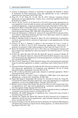
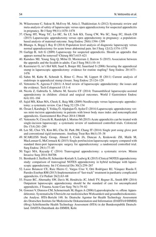
![5Laparoscopy in Gynecologic Emergencies
Federico Coccolini, Giuseppe D’Amico, Giulia Montori,
Fausto Catena, Gustavo Fraga, and Luca Ansaloni
5.1 Introduction
An acute abdomen results usually from peritoneal irritation due to inflammation or
rupture of an abdominal organ or obstruction of a hollow organ. In hospital practice,
patients with acute abdominal pain either go spontaneously to the emergency
room or are sent there by their family doctor with a provisional diagnosis, which
reportedly has no more than a 50 % chance of being correct [1].
Determining the cause of lower acute abdominal pain is one of the greatest
challenges faced by physicians in emergency services. It is a common reason for
hospitalization since 67 % of all patients show nonspecific lower acute abdominal
pain. Among women, for anatomic, physiological, and pregnancy reasons, it is the
most frequent cause of hospitalization [2].
In women, acute pain can be essentially caused by pelvic inflammatory disease
(PID), appendicitis, ectopic pregnancy, abortion, torsion of adnexa, endometriosis,
and hemorrhagic ovarian cysts.
“Wait and see” has been the most widely used clinical, or paraclinical, method
of observation of patients with nontypical clinical signs. The predictive value of
clinical diagnosis reached with this method, which varies with the underlying cause,
has been estimated between 68 % and 92 %. On the one hand, this method entails
F. Coccolini • G. D’Amico • G. Montori • L. Ansaloni ( )
Department of General Surgery, Papa Giovanni XXIII Hospital, 24127 Bergamo, Italy
e-mail: lansaloni@hpg23.it
F. Catena
Department of Emergency and Trauma Surgery, University Hospital of Parma, Parma, Italy
G. Fraga
Division of Trauma Surgery, Department of Surgery, School of Medical Sciences, University
of Campinas (Unicamp), Campinas, SP, Brazil
© Springer International Publishing Switzerland 2016
F. Agresta et al. (eds.), Emergency Laparoscopy,
DOI 10.1007/978-3-319-29620-3_5
55](https://image.slidesharecdn.com/emergencylaparoscopy-191115144149/85/Emergency-laparoscopy-72-320.jpg)
![56 F. Coccolini et al.
risk to patients because of possible complications such as peritonitis, hemorrhage,
or infertility; on the other hand, laparotomy might be unnecessarily performed.
Laparoscopy is an alternative way of making an early diagnosis of the cause of
pain and treating the underlying disease. The accuracy of laparoscopy in acute pelvic
pain varies within a range of 65–97 %. It depends on the complete visualization
of the whole abdominal pelvic cavity and the diagnostic standard to which it is
compared. However, laparoscopy should not be considered as a routine diagnostic
tool for every case of acute abdomen, as it carries its own morbidity and requires
a general anesthesia. Although it is incorrect to hypothesize that all patients with
acute abdominal pain would benefit from laparoscopic surgery, it is evident that
over the years an increasing number of patients have been managed successfully in
emergency thanks to the laparoscopic approach [3].
5.2 Clinical Evaluation
In cases of pelvic pain, taking a thorough patient history is mandatory prior
to physical examination. The obstetric history is important, particularly if the
woman has suffered a late miscarriage, ectopic pregnancy, or undergone a Cesarean
section. The number and outcome of all pregnancies should be noted together with
eventual complications suffered such as abortion, prenatal and postnatal infections
(endometritis), and thromboembolism.
The gynecologic history should focus on conditions such as pelvic inflammatory
and infective processes which are relatively common in women of reproductive
age. Previous surgical interventions such as laparoscopy, hysteroscopy, and surgical
and medical termination of pregnancy, particularly if recent, have to be taken
into careful consideration. The patient’s menstrual history should be checked
completely, gathering information relating to her menarche (first menstruation),
menstrual cycle pattern, and menopausal symptoms. The date of her last men-
struation should be recorded as well. The surgeon should enquire as to whether
the woman has used any contraceptive methods and their nature. A delay in the
menstruation should lead the surgeon to suspect an ectopic pregnancy, even when
the urine pregnancy test result is negative. In such circumstances, a serum beta-
hCG evaluation would confirm or exclude the diagnosis of a pregnancy. An ectopic
pregnancy should also be suspected in the presence of a positive pregnancy test
where an intrauterine contraceptive device (IUCD) or oral contraception has been
used. Other irregularities in the menstrual cycle should be recorded.
Clinical examination can confirm or refute a diagnosis suggested by the patient
history. Generally, a painful abdomen with localized or widespread tenderness,
rebound tenderness, and reduced bowel movements should be considered a surgical
problem. Urgent hematologic and abnormal ultrasound will help to establish the
correct diagnosis. The clinical assessment must always be completed by checking
for hernias in the groin or in the site of previous surgery. Rectal and vaginal
examination should not be unduly painful and the reason for performing it must be
explained to the patients. The vaginal examination should be avoided in very young](https://image.slidesharecdn.com/emergencylaparoscopy-191115144149/85/Emergency-laparoscopy-73-320.jpg)
![5 Laparoscopy in Gynecologic Emergencies 57
patients and in those with virgo intacta. The physical examination will include
palpating the cervix, the uterus, and the adnexa. Local tumor, fluid collection, and
signs of pelvic and peritoneal tenderness will be searched for. Signs of tenderness
may indicate the presence of inflammations and infections and suggest peritonitis.
Laboratory investigations should include a full blood count. The red blood cell
count and hematocrit will give information on possible anemia. Leukocytosis is
always present in cases of infections and inflammations such as salpingitis, PID,
and pelvic abscesses. It is also often present in the initial stages of an ovarian cyst
rupture and in ovarian torsion. Blood cross-matching is advisable before surgery.
Likewise, the clotting screen is also necessary in the presence of heavy blood loss or
if acute anemia is suspected. The serum human chorionic gonadotropin (beta-hCG)
should be urgently tested, particularly in women with irregular menstrual cycles or
those using contraception. The serum beta-hCG test should be carried out even when
a negative urine pregnancy test result has been obtained, due to a false-negative rate
of 5 %. Testing for CA-125 is not required in an emergency setting. However, it
is worth knowing that a raised CA-125 is a specific marker of peritoneal reactions.
Levels can be elevated up to hundred times in endometriosis and pregnancy and a
thousand times in ovarian cancer.
5.3 Most Common Gynecologic Emergencies
The most common gynecologic emergencies causing acute pelvic pain are reported
below:
1. Adnexal–ovarian torsion
2. Pelvic inflammatory disease (PID)
3. Ectopic pregnancy
4. Endometriosis
5. Hemorrhagic ovarian cysts
5.3.1 Adnexal Torsion
5.3.1.1 Etiology
The adnexa of the uterus, formed by the ovaries and fallopian tube, are prone
to torsion. Ovary and/or fallopian tube torsion happens when the adnexa suffer a
complete or partial rotation on its ligamentous support, resulting in compromise
of their blood supply and in sudden onset of pelvic pain. Adnexal torsion can be
unilateral or bilateral, with the last one being extremely rare. It is the fifth most
common gynecological emergency [4], with a reported incidence ranging from
2.7 % to 3.0 % [4, 5]. Adnexal torsion can affect women of all ages 28–31, but it is
more frequent among women of reproductive age 26. Most of these women desire
future fertility; thus, an accurate diagnosis and prompt treatment are important in an
attempt to preserve ovarian function.](https://image.slidesharecdn.com/emergencylaparoscopy-191115144149/85/Emergency-laparoscopy-74-320.jpg)
![58 F. Coccolini et al.
The major risk factors for adnexal torsion are an ovarian mass (physiologic cyst
or neoplasm) [6], pregnancy and ovarian stimulation [6], previous pelvic surgery,
particularly tubal ligation [6], past history of ovarian torsion [7], hypermobile
adnexa [8], and an excessive length of the utero-ovarian ligament, particularly in
the pediatric population [9].
5.3.1.2 Signs and Symptoms
Patients presenting with adnexal torsion complain of an acute, intermittent, unilat-
eral pelvic pain. This pain may be exacerbated by positional shifts. Approximately,
70 % of patients have associated gastrointestinal complaints including nausea and
vomiting, which can lead some providers to include in their differential appendicitis,
bowel obstruction, or mesenteric ischemia. The episodes may have intermittently
persisted for the past few days to weeks. On physical examination, a tender,
unilateral adnexal mass is appreciated in roughly 70 % of patients [10]. This
tender mass becomes enlarged because in a partial torsion, there is enough arterial
pressure for blood to flow into the adnexa. The venous blood is unable to return,
however, because of the pressure dampening of the torsion. If the torsion persists
or progresses, then the arterial blood flow is unable to oxygenate the ovary and the
tissue begins to undergo necrosis. Many women only develop a fever once necrosis
ensues.
5.3.1.3 Diagnosis
Adnexal torsion remains as one as the most challenging conditions to diagnose
preoperatively. Traditionally, diagnosis has stood on the clinical and sonography
findings. Numerous retrospective analyses were undertaken to evaluate the clinical
profile of adnexal torsion. Abdominal pain was the most common symptom (77.8–
98 %), followed by nausea and vomiting (46–80.5 %), mass in clinical examination
(22.2–72 %), elevated white blood count (22.1–75 %), lower urinary tract symptoms
(14.5 %), fever (5.6–7.8 %), and abnormal genital tract bleeding (4 %) [4, 5]. A
gynecological ultrasound is usually the first imaging tool used in the approach of
pelvic pain. The sonography findings that may be found in adnexal torsion are
ovarian enlargement, visualization of a cyst, solid or complex mass at the location
of the adnexa, abnormal ovarian position in the pouch of Douglas, thickening of
the adnexal wall, peripherally enlarged follicles, cystic hemorrhage, and free pelvic
fluid [5]. Lo et al. reported that an adnexal or pelvic mass could be detected using
gynecological ultrasound in almost all of the patients (98.3 %) [4]. Oltmann et al.
stated that the finding most consistent with torsion on transvaginal ultrasound (TVU)
was simply the presence of an enlarged ovary or adnexal mass and confirmed that
the sole presence of a mass larger than 5 cm had the best sensitivity for torsion
(83 %) [11].
Color doppler ultrasound detects the ovarian blood flow, while spectral Doppler
can enable the differentiation between venous and arterial flow in the ovary. These
techniques have been suggested as an additional tool to improve the accuracy of
diagnosing ovarian torsion. The absence of venous, or arterial and venous flow in an
enlarged or cystic ovary on color and spectral doppler studies, is highly predictive](https://image.slidesharecdn.com/emergencylaparoscopy-191115144149/85/Emergency-laparoscopy-75-320.jpg)
![5 Laparoscopy in Gynecologic Emergencies 59
of ovarian torsion [12]. Studies reported that, although highly specific, doppler
sonography was not sufficiently sensitive. A normal flow by doppler sonography
does not rule out ovarian torsion and should not delay surgical exploration if the
patient presents with suggestive signs and symptoms.
Computerized tomography scan (CT scan) and magnetic resonance image (MRI)
can detect ovarian torsion, but they are not necessary in most cases and should not
delay treatment. Moreover, CT scan is contraindicated in pregnant women. Tumor
markers, such as beta-human chorionic gonadotropin (“-HCG), alpha-fetoprotein
(’-FP), cancer antigen 125 (CA-125), and lactate dehydrogenase (LDH), are not
routinely screened in an emergency setting.
At the present time, there is no reliable method enabling the diagnosis of
adnexal torsion to be confirmed preoperatively. Studies still report a poor correlation
between preoperative diagnosis and surgery findings: Bar-On et al. found that
women who underwent laparoscopy for suspected ovarian torsion were correctly
diagnosed in only 46.1 % of cases [13]; Cohen et al. stated that among 100
emergency laparoscopies performed due to acute abdomen, ovarian torsion was the
most difficult to diagnose preoperatively, with only 44 % of cases being confirmed
by laparoscopy [14]; Houry et al. reported that, among women with ovarian torsion,
the diagnosis was initially considered in 47 % of patients [6], while White et
al. found that it was considered in only 19 % of patients [7]. Thus, diagnostic
laparoscopy can reduce the uncertainty of diagnosis in ovarian torsion. Also, it offers
a lot of other advantages: enables a rapid and accurate assessment [15], reduces
the incidence of negative laparotomies, reduces morbidity and mortality from the
underlying disease [16], and, most of all, enables treatment.
5.3.1.4 Treatment
Surgical approach by laparoscopy is the standard of care for adnexal torsion; it
allows the physician to diagnose adnexal torsion in a first step and to treat it
in a second step. Multiple studies have confirmed the safety and efficacy of the
laparoscopic approach for adnexal torsion in children [9, 17] and pregnant patients
[18].
Results of Lo et al. show that patients undergoing laparoscopy had a smaller
ovarian mass, a shorter hospital stay, and fewer of them suffered from postoperative
fever, comparing to patients undergoing laparotomy [4]. Oltmann et al. also reported
that a preoperative imaging mass size of less than 5 cm was associated with the use
of a laparoscopic approach. They also found that laparoscopy rates increased from
18 % in the 1993–2000 to 42 % in the 2001–2008 period, reflecting the change
over time in surgical practice [11]. Laparoscopy approach has demonstrated to
be superior to laparotomy as it results in lower rate of febrile morbidity, reduces
consumption of analgesic drugs, and shortens hospital stay [19, 20].
Intraoperative management of adnexal torsion has been largely debated. In the
past, treatment consisted of radical surgery. An ipsilateral oopherectomy by laparo-
tomy was the standard of care, due to both concerns regarding thromboembolic
phenomena and “black-bluish” appearance of twisted adnexa [19]. Nowadays,
treatment is rather conservative. It is initiated by untwisting the adnexa, even when](https://image.slidesharecdn.com/emergencylaparoscopy-191115144149/85/Emergency-laparoscopy-76-320.jpg)
![60 F. Coccolini et al.
it appears necrotic, and completed, as required, by treatment of any cyst present
and/or oophoropexy. All these steps are easily accomplished by laparoscopy [21,
22].
In a pregnant patient, there have been successful cases of laparoscopic adnexal
torsion reduction. The incidence of adnexal torsion in pregnancy is 1 in 5000
pregnancies. Many authors recommend entering in an open manner as opposed to
Verres needle or in a direct approach; however, there has been documentation of
complications in all three approaches. In pregnant patients, precautions should be
taken as the uterus may be in the abdominal cavity and surgical pneumoperitoneum
poses the risk for decreased uterine blood flow secondary to increased intra-
abdominal pressure [18, 23]. Fetal cardiac activity should be documented before
and after the procedure and the patient should be positioned in the dorsal supine
position with left lateral tilt during the operation.
5.3.2 Pelvic Inflammatory Disease
5.3.2.1 Etiology
Pelvic inflammatory disease refers to the acute infection of the upper genital
tract in women. It includes any combination of cervicitis, endometritis, salpingitis,
tubo-ovarian abscess, and pelvic peritonitis [24]. Originally, PID was associated
with Neisseria gonorrhea and Chlamydia trachomatis infection. Nowadays, it is
recognized that these microorganisms cause only two-thirds of cases. Gardnerella
vaginalis, Mycoplasma genitalium/hominis, Ureaplasma spp., Diphtheroids, Pre-
votella, Bacteroides, other anaerobes, and streptococci may also be involved [24]. It
is the most common infectious disease affecting young women, accounting for 94 %
of sexually transmitted disease-associated morbidity in well-resourced countries.
This condition can cause significant reproductive health sequelae, including tubo-
ovarian abscesses, pelvic adhesions, recurrent PID, chronic pelvic pain, infertility
due to obstruction of fallopian tubes, and ectopic pregnancy in an increased factor
of 7–10 [25]. Such complications are related to disease severity and timing of
diagnosis. Prompt investigation is essential in order to attenuate disease impact.
5.3.2.2 Signs and Symptoms
Approximately, 90 % of patients present with the chief complaint of abdominal
pain and approximately 75 % of those patients have an elevated white count. Other
signs and symptoms can be vaginal discharge, abnormal uterine bleeding, nausea,
vomiting, and fever.
5.3.2.3 Diagnosis
The accuracy of different signs and symptoms in predicting the presence of PID
has been extensively evaluated using laparoscopy as the gold standard [26, 27].
Currently, the clinical diagnosis of PID is based on recommendations from the
Center for Disease Control and Prevention (CDC). Minimum diagnostic criteria
(presence of cervical motion, uterine and adnexal tenderness) have been set with](https://image.slidesharecdn.com/emergencylaparoscopy-191115144149/85/Emergency-laparoscopy-77-320.jpg)
![5 Laparoscopy in Gynecologic Emergencies 61
a high sensitivity and low specificity, in order to detect as many cases of clinical
disease as possible. If all these criteria are present in sexually active young women,
empiric treatment should be instituted. The CDC also provides a list of additional
criteria that may help to improve diagnostic specificity: elevated oral temperature
(>38.3 ı
C), abnormal cervical or vaginal discharge, elevated erythrocyte sedimen-
tation rate, elevated C-reactive protein concentration, and laboratory documentation
of cervical infection with Neisseria gonorrhoeae or Chlamydia trachomatis [24].
Peipert et al. showed that the clinical finding of adnexal tenderness had a
sensitivity of 95.5 %, which was superior to that of the CDC’s minimal clinical
criteria (83 %) [28].
There is not a pathognomonic laboratory test for upper genital tract infection.
Ultrasound examination is able to detect thickening of fallopian tubal walls and
tubo-ovarian abscesses and rule out other possible diagnoses like ovarian cysts and
ovarian torsion [29]. However, the sensitivity and specificity of ultrasound features
is highly dependent on the severity of the disease [29].
Laparoscopic exploration is warranted whenever the diagnosis is unclear and the
symptomatology is unremitting, especially in young women for whom the preser-
vation of fertility is important. Laparoscopic visualization of the triad of edema,
hyperemia, and a purulent exudate from the ends or surface of the fallopian tube
has been considered the gold standard for PID diagnosis [30, 31]. Laparoscopy can
be used to directly visualize pelvic anatomy and diagnose PID without aggravating
the inflammatory process and exclude other pathologies (which may be present in
approximately 20 % of patients) [32]. However, its sensitivity varies depending on
the stage of illness, being less sensitive in milder forms where diagnostic criteria
are less objective. In cases where there is no visual evidence of salpingitis by
laparoscopy, endometrial biopsy is warranted because some women with PID can
have endometritis alone.
Gaitán et al. reported that endometrial biopsy showed 75 % of global accuracy,
comparing with a global accuracy of 91 % with laparoscopy. Laparoscopy showed
an optimum specificity (100 %), proving to be very useful in excluding other causes
of abdominal pain in patients presenting with an atypical course [31].
Among these lines, in cases of abdominal pain of unknown cause where there is a
suspicion of PID, laparoscopic surgery has become a diagnostic tool that permits not
only confirmation of the process but also to take a microbiologic culture from the
fallopian tubes to apply specific antibiotic therapy, a classification of the severity of
the disease, the evaluation of the future fertility status of the patient, and therapeutic
options, such as lysis of adhesions, aspiration of tubo-ovarian abscess, and irrigation
of the peritoneal cavity [15, 25].
In patients without PID, laparoscopy permits rapid recognition and treatment
of differential pathologies like appendicitis, ovarian cyst, ovarian torsion, and
endometriosis, avoiding unnecessary antibiotic therapy. Costs, limited access, sur-
gical risks, and the fact that not all institutions have the facilities or scheduling to
submit all patients with acute abdominal pain to diagnostic laparoscopy prevent the
universal use of laparoscopy for the diagnosis of PID [16]. However, the costs can](https://image.slidesharecdn.com/emergencylaparoscopy-191115144149/85/Emergency-laparoscopy-78-320.jpg)
![62 F. Coccolini et al.
be balanced by a shorter hospital stay and a reduction in unnecessary abdominal
imaging.
5.3.2.4 Treatment
Tubo-ovarian abscess is not a disease entity, but rather a finding in the spectrum
of PID. Treatment includes analgesia, fluids, and intravenous broad-spectrum
antibiotics.
Although 60–80 % will resolve without surgical intervention, it is prudent to
identify those individuals who require immediate surgical intervention because
delay can be fatal [12].
Currently, there are three situations where surgical intervention is recommended:
a concern for an alternative surgical emergency, failure of clinical response after
48–72 h of medical therapy, or ruptured tubo-ovarian abscess with generalized
peritonitis and septic shock. A review on the management options for tubo-ovarian
abscess reported that laparoscopy should be considered in all patients with tubo-
ovarian abscess who desire future conception. They reported pregnancy rates
ranging from 32 % to 63 % in patients treated with medical therapy followed
by immediate laparoscopy drainage within 24 h, comparing to pregnancy rates
ranging from 4 % to 15 % in patients treated with medical management alone,
both without suspected rupture. They also report the major advantages of an
immediate laparoscopy: it allows an accurate diagnosis and effective treatment
under magnification with minimal complications and shows faster response rates
with shorter hospitalization times and decreased infertility [33]. Surgical procedures
can include laparoscopy with drainage of purulent fluid, adhesion lysis, tubal lavage,
dissection and excision of necrotic tissue, and thorough irrigation of the peritoneal
cavity before completion of the procedure [32, 34]. A particularly simple, safe,
and efficacious alternative approach is the use of ultrasound-guided transvaginal
drainage of the abscess in combination with antibiotics.
5.3.3 Ectopic Pregnancy
5.3.3.1 Etiology
Ectopic pregnancy occurs when the embryo fails to implant within the uterine cavity.
The majority of ectopic pregnancies (95 %) are located in the fallopian tube. The
rising incidence of ectopic pregnancy in the past 30 years has been attributed to
a number of factors including a greater prevalence of sexually transmitted disease
(PID), tubal sterilization and reversal, delayed childbearing, assisted reproductive
technologies, and more successful clinical detection [35]. Ectopic pregnancy is the
leading cause of maternal mortality during the first trimester of pregnancy [36].
Although spontaneous resolution of ectopic pregnancy can occur, patients are at risk
of tubal rupture and catastrophic hemorrhage. Most fatal cases result from delayed
diagnosis and inappropriate management. However, recent improvements made in
diagnostic techniques have led to a marked reduction in mortality rates.](https://image.slidesharecdn.com/emergencylaparoscopy-191115144149/85/Emergency-laparoscopy-79-320.jpg)
![5 Laparoscopy in Gynecologic Emergencies 63
5.3.3.2 Signs and Symptoms
Patients who have ectopic pregnancies usually show abdominal pain, amenorrhea,
and irregular vaginal bleeding. On physical examination, the most common findings
include ipsilateral adnexal tenderness, abdominal tenderness, and an adnexal mass
[37].
Not all patients who have an ectopic pregnancy, even those who have a hemoperi-
toneum, have rebound tenderness or peritoneal signs. Also of note, vigorous
pressure during bimanual examination can cause a previously unruptured ectopic
pregnancy to rupture; therefore, gentle examination is required.
5.3.3.3 Diagnosis
In women of early pregnancy/history of amenorrhea presenting with acute pelvic
pain and/or vaginal bleeding, a diagnosis of ectopic pregnancy should always
be considered. On physical examination, an adnexal mass may be palpated. The
diagnosis of ectopic pregnancy has evolved since the 1980s. Advanced ultrasonog-
raphy techniques as well as improved “-HCG measurements have largely replaced
laparoscopy as primary diagnostic tools. These tests allow us to detect an early
ectopic pregnancy before clinical symptoms have the chance to set in [34]. These
days, the role of laparoscopy is limited to cases in which the diagnosis is still in
doubt. In such cases, a choice must be made between expectant observation and
laparoscopic confirmation.
5.3.3.4 Treatment
To date, the management options of tubal ectopic pregnancy are threefold: expec-
tant management, medical treatment, or surgery. These three options have been
compared in terms of efficacy, financial costs, and future fertility. The criteria for
choosing between therapeutic modalities in ectopic pregnancy remain unsettled.
Expectant management has been advocated based on the knowledge that an early
ectopic pregnancy can be self-limiting, resulting in tubal abortion or re-absorption.
It has been advised in a selective group of patients asymptomatic with serum “-HCG
cutoff level relatively low and declining. Medical treatment with methotrexate has
been widely used in patients with minimal symptoms, who are hemodynamically
stable, have no more than a moderate amount of intra-abdominal free-fluid on TVU
scan, and have “-HCG concentrations <3000 IU/L [38]. The major contraindications
for medical management are “-HCG level greater than 15,000 IU/L, fetal cardiac
activity, and free fluid in the cul-de-sac on TVU. Surgical management is indicated
if the patient is not eligible for medical therapy, if medical therapy has failed, or if
there are symptoms and signs of tubal rupture [38].
Ectopic pregnancy represents a hallmark in the history of laparoscopic surgery:
it was one of the first surgical pathology being treated through a laparoscope [39].
Nowadays, laparoscopy is the accepted approach to perform both a salpingostomy
and salpingectomy. Mol et al. reported that although laparoscopic approach was
significantly less successful than the open surgical approach in eliminating tro-
phoblastic tissue, it was significantly less costly. The cost savings after laparoscopic
surgery resulted from a significantly shorter operation time, less perioperative blood](https://image.slidesharecdn.com/emergencylaparoscopy-191115144149/85/Emergency-laparoscopy-80-320.jpg)
![64 F. Coccolini et al.
loss, shorter duration of hospital stay, and shorter convalescence time. Laparoscopy
was the most cost-effective treatment in women with tubal ectopic pregnancy. The
fertility outcome was comparable to laparotomy [40]. Laparoscopy also results in
less postoperative adhesion formation and impairment of the pelvic status than
laparotomy [35].
Laparoscopic surgery is the gold standard for the treatment of ectopic pregnancy
in hemodynamically stable women. However, it is estimated that approximately
15 % of women with ectopic pregnancy experience hypovolemic shock. In these
cases, both the volume of hemoperitoneum and the hemodynamic status of the
patient have been critical factors in deciding whether to perform a laparoscopy
or a laparotomy. Although historically contraindicated, numerous case reports
and studies have proven that laparoscopy can be successful in cases of ruptured
ectopic pregnancy with hemodynamic instability [41, 42]. Laparoscopy has also
been contraindicated in certain situations such as interstitial and cornual pregnancy
and severe adhesions. The improvement of surgeons’ skills and instrumentation
has made these contraindications obsolete. Laparoscopy efficacy has been reported
in interstitial and cornual pregnancies in various case reports [43, 44]. Thus,
laparoscopy leaves little space for laparotomy in ectopic pregnancy management.
There is a controversy on whether to perform a conservative treatment (salpin-
gostomy) or a radical treatment (salpingectomy) in ectopic pregnancy. The possible
advantage of radical approach would be to eliminate the possibility of recurrence,
but with lower rates of fertility (66 %). In conservative approach, rates of 8 % of
persistent ectopic pregnancy have been reported, but with higher rates of fertility
(89 %). So the conservative approach should be preferred whenever future fertility
is required. Most authors agree that salpingectomy should only be performed in
women with uncontrolled bleeding, recurrent ectopic pregnancy in the same tube, a
severely damaged tube, or a tubal gestational sac greater than 5 cm in diameter [36].
5.3.4 Endometriosis
5.3.4.1 Etiology
Endometriosis, the presence of endometrial glands and stroma outside of the
endometrial cavity, represents one of the most challenging gynecologic conditions
to manage given its insidious onset, surgical diagnosis, association with pelvic pain
and infertility, and often progressive nature.
The cause of endometriosis is unclear. There are several theories postulated to
explain its pathogenesis. These include retrograde menstruation and implantation,
the metaplasia theory, lymphatic and vascular spreading, and genetic predisposition
[45, 46].
Endometriosis is a chronic disease affecting at least 10 % of reproductive-aged
women, but is found in approximately 40 % of infertile women and up to 90 %
of women with pelvic pain [47]. Risk factors include family history, low body
mass index, alcohol use, smoking, particularly in the setting of infertility, Caucasian](https://image.slidesharecdn.com/emergencylaparoscopy-191115144149/85/Emergency-laparoscopy-81-320.jpg)
![5 Laparoscopy in Gynecologic Emergencies 65
race, prolonged estrogen exposure as with early menarche or late menopause, and
nutritional/environmental factors.
5.3.4.2 Signs and Symptoms
During menstruation these ectopic tissue areas undergo changes such as edema and
bleeding, causing symptoms like acute pelvic pain, dysmenorrhea, dyspareunia,
signs of peritoneal irritation, dyschezia, rectal pain and bleeding, each being
dependent upon the location of endometriosis. Chronic symptoms with acute bouts
of pain are characteristics of endometriosis affecting the ovaries with bleeding,
torsion, or rupture of an endometrioma.
5.3.4.3 Diagnosis
Apart from the clinical evaluation, diagnosis is helped by transvaginal or trans-
abdominal US and by MRI. Laparoscopy is the gold standard to evaluate pelvic
endometriosis as it allows the collection of histologic specimens needed to confirm
diagnosis. Deep infiltrating or subperitoneal endometriosis is not always visible
at laparoscopy and MRI can therefore be helpful in such conditions. Laboratory
investigations are restricted to serum CA-125 which usually increases to values of
up to 100 200 IU/mL, but is not useful for the diagnosis. The role of CA-125 is
limited to the follow-up of recurrences.
5.3.4.4 Treatment
Medical treatment with NSAIDs reduces the production of local prostaglandin,
therefore reducing inflammation but more importantly reducing the pain. The
medical treatment of endometriosis consists of ceasing ovarian estrogen production.
Estroprogestinic preparations are the most useful drugs as they can be used for
months if common contraindications such as obesity, breast cancer, smoking, and
high blood pressure do not exist.
Medical treatment by danazol or a GnRH analog are also very effective as a
means of stopping ovarian function and estrogen production, but are associated,
due to hypo-estrogenization, with undesired side effects including osteoporosis,
vaginal dryness, hot flushes, and other vasomotor symptoms. The use of danazol
or a GnRH analog if protracted for more than 6 months should be associated with
an estroprogestinic preparation to reduce the menopausal symptoms, osteoporosis,
and fracture risk. The medical treatment is only effective in minimal endometriosis,
while moderate and severe forms need to be treated by surgery [48].
Surgical treatment consists of trying to remove as much endometriotic tissue as
possible. It is rare to perform emergency surgery for pelvic pain exclusively due to
endometriosis, unless it is secondary to an operation performed for hemoperitoneum
due to an ovarian cyst rupture (endometriomas) or for suspected appendicitis. At
surgery the removal of endometriosis requires careful manipulation of the organs
involved. A careful approach needs to be applied during surgery for endometriosis
infiltrating the serosa of the bowel, above the urethra and bladder and above major
blood vessels [49].](https://image.slidesharecdn.com/emergencylaparoscopy-191115144149/85/Emergency-laparoscopy-82-320.jpg)
![66 F. Coccolini et al.
Laparoscopy allows a mapping of the disease and the collection of material for
histology that will eventually confirm the diagnosis. Surgical staging of endometrio-
sis consists of a scoring to be attributed to each site, localization, and extension of
the lesion. The most commonly used scoring system suggested by the American
Fertility Society helps in the evaluation of patient fertility prognosis [50].
In young women, surgical treatment of ovarian endometriosis must not compro-
mise the reproductive function of the ovary and electrosurgery should be minimally
used. The laparoscopic approach is favored compared to open surgery as it allows
clearer visibility and access to the pelvic cavity and reduces the postoperative
morbidity and hospital stay. Moreover, laparoscopy may increase chances of future
pregnancy and live birth [51, 52].
It is important to remember that the endometriotic tissue has the capacity to
implant and therefore careful peritoneal cavity washing and adequate removal of
the surgical specimens should be performed to reduce the risk of distal implant on
scars and trocar access points.
5.3.5 Hemorrhagic Ovarian Cysts
5.3.5.1 Etiology
Approximately 2–4 days after ovulation, the ovary becomes increasingly vascular.
With neovascularization, blood from the vascular theca zone often fills the cavity of
the cyst. Often, the cyst reabsorbs the blood; however, if the amount of bleeding is
large or a cyst ruptures the bleeding may continue, producing a hemoperitoneum.
Increased caution should be taken in patients who are anticoagulated.
5.3.5.2 Signs and Symptoms
Pertinent history should include when the patient’s last menstrual period took place.
Pain can occur with ovulation of a follicular cyst, otherwise known as Mittelschmerz
syndrome. Enlarging ovarian follicles often produce a colicky or dull unilateral
tenderness in the lower abdomen or pelvic region. Rarely, patients may present with
hypovolemia and hemodynamic instability. Nausea and/or vomiting are associated
as nonspecific symptoms. A bimanual examination should be performed to evaluate
the size of the cyst and tenderness. On physical examination, a corpus luteal cyst can
mimic an ectopic pregnancy. Originally described by Halban, a triad of symptoms
includes a delay in menses, followed by spotting, unilateral pelvic pain, and a small,
tender, adnexal mass [53].
5.3.5.3 Diagnosis
Diagnosis of a hemorrhagic or ruptured cyst is often made by narrowing the
differential. Transvaginal ultrasound often demonstrates an adnexal mass; however,
the ovary may be normal in size if rupture has decompressed any cysts. In these
cases, a large amount of pelvic fluid is usually noted [54]. A quantitative pregnancy
test can rule out an ectopic pregnancy. A CBC should be taken to assess the current
blood count. An elevated white count is rarely seen in a ruptured luteal cyst. Once](https://image.slidesharecdn.com/emergencylaparoscopy-191115144149/85/Emergency-laparoscopy-83-320.jpg)
![5 Laparoscopy in Gynecologic Emergencies 67
Table 5.1 Indications for different surgical approaches
Laparotomy
High suspicion for malignancy
Large cysts
History of or risk factors for abdominopelvic adhesions
Hemodynamically unstable patient—hypovolemia
Significant cardiopulmonary disease
Obesity
Laparoscopy
Minimizing the risks of subsequent infertility resulting from pelvic adhesions
Every effort should be made to conserve ovarian tissue
pregnancy is ruled out, an abdominal/pelvic CT with contrast can further assist in
evaluating other potential causes, including acute appendicitis.
5.3.5.4 Treatment
Initial management of a suspected follicular or hemorrhagic cyst is supportive man-
agement and continued observation with a repeat pelvic ultrasound in approximately
4–6 weeks to document resolution. Oral contraceptive medication can be started in
patients to halt further ovulation.
Indications for immediate operative intervention include a large amount of
peritoneal fluid found on a transvaginal ultrasound, hemodynamic instability, and
severe pain. Delayed operative management is indicated for patients in whom pain
is not improving with conservative management or for persistent tumors to rule
out a neoplastic process. A cystectomy is recommended as opposed to a unilateral
salpingo-oophorectomy in reproductive-aged women. Laparoscopic evaluation is
usually feasible; however, if cancer is suspected, laparotomy may be necessary to
ensure intact removal and for staging purposes [53]. The indications for laparotomy
or laparoscopy are summarized in Table 5.1. Intraoperative spillage of a malignant
tumor does not necessarily worsen prognosis, but it does necessitate adjuvant
chemotherapy. If the cyst is benign, a ruptured cyst may still cause postoperative
abdominal pain secondary to chemical peritonitis.
5.4 Discussion and Conclusion
Anteby et al. highlighted the value of laparoscopy in the evaluation of acute pelvic
pain in 1974. Since then, great advances in anesthesia care and cardiovascular
monitoring, together with the development of a great number of devices and
different techniques, have made possible the use of diagnostic and therapeutic
laparoscopy procedures for gynecologic emergencies.
Nowadays, early diagnostic laparoscopy in the emergency setting improves
diagnostic rates and reduces hospital stays and costs, when compared to other](https://image.slidesharecdn.com/emergencylaparoscopy-191115144149/85/Emergency-laparoscopy-84-320.jpg)
![68 F. Coccolini et al.
diagnostic modalities. Still, laparoscopy is an invasive technique and it should
only be used after a first-line investigation combining clinical and sonography
examination.
Therapeutic laparoscopy procedures in gynecology emergencies are now univer-
sal practice. The efficacy and safety are comparable to laparotomy procedures but
with several additional advantages: shorter operation times, impairment of pelvic
status with less postoperative adhesions; few postoperative pain, fever, and blood
loss; reduction on consumption of analgesic drugs; fast recovery rates; shorter
hospital stays; better cosmetics outcomes; and lower costs. However, the safest
approach should always be the one that the surgeon feels more comfortable with,
and this should always be a factor regarding the type of intervention.
In a pregnant woman laparoscopy and laparotomy may increase the possibility
of spontaneous abortion in the first trimester and premature delivery in the last
trimester of pregnancy, respectively. It is, however, a common opinion that the
use of laparoscopy is preferable to laparotomy. In this regard, the advantages of
laparoscopy over open surgery are represented by a reduced administration of
anesthetic drugs, a reduced need for postoperative analgesia, a low incidence of
infective complications, and a faster recovery. Major risks of surgery in pregnant
women include uterine injuries due to the increased uterine size, with or without
rupture of the amniotic membrane, bleeding, and gaseous embolism, as well as
technical difficulties related to the limited working space [55]. Therefore, when
operating on a pregnant woman, all the necessary precautions must be taken prior
to the use of laparoscopy including a low intra-abdominal pressure and deep vein
thrombosis prophylactic measures even for minor procedures.
In conclusion, it is important to remember that the multidisciplinary approach
to the patient is essential for establishing the correct diagnosis and treatment in the
shortest time. The use of modern laboratory and radiologic resources associated
with the progress in endoscopic surgical techniques provides both general surgeons
and gynecologists with all the means necessary to ensure the best surgical result.
References
1. Jones PF (1987) Emergency abdominal surgery. Blackwell Science, Oxford
2. Sheridan WG, White AT, Havard T, Crosby DL (1992) Non-specific abdominal pain: the
resource implications. Ann R Coll Surg Engl 74:181–185
3. Sauerland S, Agresta F, Bergamaschi R, Borzellino G, Budzynski A, Champault G et al
(2006) Laparoscopy for abdominal emergencies: evidence-based guidelines of the European
Association for Endoscopic Surgery. Surg Endosc 20:14–29
4. Lo LM, Chang SD, Horng SG, Yang TY, Lee CL, Liang CC (2008) Laparoscopy versus
laparotomy for surgical intervention of ovarian torsion. J Obstet Gynaecol Res 34:1020–1025
5. Vijayalakshmi K, Reddy GM, Subbiah VN, Sathiya S, Arjun B (2014) Clinico-pathological
profile of adnexal torsion cases: a retrospective analysis from a tertiary care teaching hospital.
J Clin Diagn Res 8:OC04–OC07
6. Houry D, Abbott JT (2001) Ovarian torsion: a fifteen-year review. Ann Emerg Med 38:156–
159](https://image.slidesharecdn.com/emergencylaparoscopy-191115144149/85/Emergency-laparoscopy-85-320.jpg)
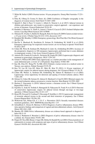
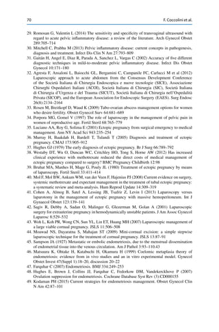
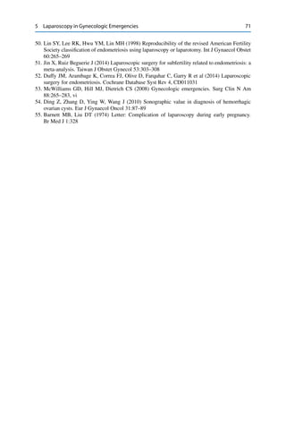
![6Nonspecific Abdominal Pain
Michele Carlucci, Aldo Alberto Beneduce, Guido Fiorentini,
and Giovanni Burtulo
6.1 Introduction
Five to ten percent of all admissions to the emergency department (ED) are due to
acute abdominal pain. Within this patient population, up to a third of all patients
will leave the hospital without a clear and unambiguous diagnosis explaining the
cause of their abdominal pain, despite a mean hospital stay of up to 6 days. Acute
nonspecific abdominal pain (NSAP) is defined as acute abdominal pain less than
1 week in duration, for which there is no diagnosis despite investigations and
comprises a spectrum of undiagnosed conditions, both somatic and functional, and
remains a “diagnosis of exclusion.” Depending on the severity of the symptoms,
some patients are admitted to the hospital for repeated active clinical observation,
classically known as the “wait and see” approach, resulting in a substantial use of
financial and human resources [1].
Although attempts have been made toward developing consensus guidelines and
diagnostic algorithms, no prospective evidence-based clinical guidelines for the
exclusion of NSAP have been developed or validated to date [2, 3]. The necessity
to achieve a correct diagnosis and a systematic approach to NSAP should be
useful in order to reduce the admission rate for NSAP because of the costs and
morbidity associated with this condition in terms of excessive hospital stay, multiple
investigations, and unnecessary surgical explorations [3–5].
Systematic baseline character examination, abdominal ultrasound (US) [6],
abdominal computed tomography (CT) [7], and early laparoscopy (EL) [8–13] have
all been described as potential methods for improving the diagnostic accuracy in the
emergency setting.
M. Carlucci ( ) • A.A. Beneduce • G. Fiorentini • G. Burtulo
IRCCS Ospedale San Raffaele, Milan, Italy
e-mail: carlucci.michele@hsr.it
© Springer International Publishing Switzerland 2016
F. Agresta et al. (eds.), Emergency Laparoscopy,
DOI 10.1007/978-3-319-29620-3_6
73](https://image.slidesharecdn.com/emergencylaparoscopy-191115144149/85/Emergency-laparoscopy-89-320.jpg)
![74 M. Carlucci et al.
Nevertheless, the most important part of the evaluation remains a thorough
history and a careful physical examination given the wide variety of disorders
which may cause abdominal pain. The physician is called to establish a differential
diagnosis, plan appropriate imaging studies, and determine whether surgery is
necessary.
In conclusion, the management of acute NSAP can be divided into three stages.
The first stage is the initial history, physical examination, baseline investigations,
and formulation of a working and differential diagnosis. The second stage involves
the judicious use of radiologic techniques.
The third stage should be represented by diagnostic laparoscopy [12].
6.2 Baseline Investigation
Although standing as the bedrock of medical practice, the physical examination
often is both insensitive and nonspecific in the diagnosis of NSAP. Likewise,
diagnostic adjuncts such as laboratory studies and plain abdominal radiographs
have played fundamental yet controversial roles in the assessment of NSAP. When
combined with a plain chest radiograph, the upright and supine plain abdominal
radiographs compose the acute abdominal series (AAS) in many institutions. This
series is ordered frequently for the evaluation of abdominal pain; however, its low
sensitivity and specificity limit utility. Some authors suggest a systematic collection
of baseline data (Table 6.1) with the end-point of discriminate, among patients with
NSAP, patients who need urgent procedure or further diagnostic procedure [3].
However, history and clinical examination with the addition of laboratory tests
and abdominal radiographs do not reach a sufficient level of sensitivity and
specificity to lead to a proper management of NSAP (56 % and 81 %, respectively)
[3–5]. An early use of CT in the diagnostic workup of acute abdominal pain has led
to a correct diagnosis in 96.8 % of cases [14].
6.3 Imaging Studies
Several studies designed to identify the most effective diagnostic strategy for
patients with acute abdominal pain have demonstrated that the highest sensitivity
for detecting urgent diagnoses is achieved when ultrasonography is performed in all
patients and a CT only in the event of inconclusive or negative ultrasonography [3,
15, 16]. Using this strategy, CT is only needed when conventional radiology or US
does not achieve diagnosis.
Even though CT exposes patients to a higher radiation dose, it still remains the
preferred standard diagnostic modality if ultrasound fails to diagnose the cause](https://image.slidesharecdn.com/emergencylaparoscopy-191115144149/85/Emergency-laparoscopy-90-320.jpg)
![6 Nonspecific Abdominal Pain 75
Table 6.1 Baseline Data Collection
Age Does examination change during ED visit?
Sex Does pain resolve during ED visit?
Pulse rate initial Does patient vomit during ED visit?
Systolic blood pressure initial Urinalysis, leukocyte esterase positive?
Respiratory rate initial Urinalysis nitrite positive?
Temperature initial Urinalysis ketones positive?
Pain diffuselocalized Urinalysis leukocytes present?
Pain localization (if localized) Urinalysis erythrocytes present?
Pain onset (sudden/gradual) Venous pH
Does pain radiate? Venous base excess
Nausea in past 24 hours? Complete blood count, leukocyte count
Vomiting in past 24 hours? Amylase, serum
Anorexia in past 24 hours? Lipase, serum
Diarrhea in past 24 hours? AST, serum
Flatus in past 24 hours? ALT, serum
Melena in past 24 hours? ALP, serum
Hematochezia in past 24 hours? Total bilirubin, serum
Hematemesis in past 24 hours? Direct bilirubin, serum
Bowel sounds present? Glucose, serum
Rebound tenderness? Serum urea nitrogen
McBurney point tenderness? Creatinine, serum
Murphy sign present? AAS predicts UI required?
Rovsing sign present?
Costovertebral angle tenderness present?
Stool grossy bloody?
Stool heme positive?
of acute abdominal pain correctly. Plain radiography demonstrates low sensitivity
and accuracy and is, therefore, generally unhelpful; because of its low sensitivity
and negative predictive value, it can also be misleading in the workup of acute
abdominal pain. Although the radiation dose is lower than that of CT, the mediocre
test characteristics of plain radiography may delay appropriate treatment and are
therefore detrimental.
Moreover, CT is increasingly being used as a first-line investigation in abdominal
pain because it is not only good at determining the correct diagnosis but also
the severity of the disease and its potential complications. It can also accurately
demonstrate clinically relevant unexpected and important second abnormalities,
which may be critical to patient management [16].
Except certain patients, such as children or pregnant women, in which US or
MRI should be considered the method of choice because of concerns about radiation](https://image.slidesharecdn.com/emergencylaparoscopy-191115144149/85/Emergency-laparoscopy-91-320.jpg)
![76 M. Carlucci et al.
dose, a CT should be considered to guide treatment and avoid harmful delays [17,
18]. Therefore, if the diagnosis is still uncertain or CT is not accessible, the next
stage of the management of NSAP should be diagnostic laparoscopy.
6.4 The Role of Diagnostic Laparoscopy: When and Always?
Several studies have documented the feasibility and safety of diagnostic laparoscopy
(DL) using general anesthesia for patients with acute abdominal pain [19, 20]. The
diagnostic accuracy of the procedure is high, ranging between 90 and 100 %, and
prevents unnecessary laparotomies in 36–95 % of patients in the published series
[21]. Open access or the Veress technique has been used for initial access without
reported untoward events. Overall morbidity, also in ICU patients, has been reported
between 0 % and 8 % in expert hands and no mortality directly associated with the
procedure has been described [22, 23]. Contraindications for DL do not differ from
contraindications to exploratory laparotomy, except for patients unable to tolerate
pneumoperitoneum and those with a tense and distended abdomen (i.e., clinically
suspected abdominal compartment syndrome) [21]. The role of early laparoscopy
compared with the traditional “wait and see” in the management of NSAP in patients
with unclear diagnosis after baseline examinations and tests has been evaluated by
randomized controlled trials [9, 10, 12] with controversial results due to poor sample
size or no long-term follow-up. DL seems to drive to an improved diagnosis (81–
97 % vs. 28–36 % in observational group) and subsequent treatment of patients
with NSAP leading to save costs for hospitalization and reduced hospital stay but
seems unuseful in prevention of recurrence of symptoms [12, 14–24]. In conclusion,
DL is technically feasible and can be applied safely by surgeons with appropriate
training and expertise for selected patients with acute nonspecific abdominal pain
after baseline examination. The results of our systematic review suggest that there is
insufficient evidence at present to justify routine use of early laparoscopy in patients
with undifferentiated acute abdominal pain. Conversely, there is no evidence of
harm. Cost-effectiveness data are lacking and there have been no comparisons with
alternative strategies, particularly early cross-sectional imaging. There is a need for
robust large-scale randomized controlled trials to clarify the role of this technique.
The timing and the real necessity of laparoscopy should be evaluated on the basis
of patients’ course and observation.
6.5 Conclusions
Despite their initial presentation of NSAP, approximately 13 % of our subject
sample required UI within 24 hours of ED presentation, and an additional 34 %
of subjects underwent elective interventions that likely mitigated morbidity and
mortality, approximating proportions reported in larger retrospective studies [25,
26].](https://image.slidesharecdn.com/emergencylaparoscopy-191115144149/85/Emergency-laparoscopy-92-320.jpg)
![80 A. Mirabella et al.
have led to reduced need for acid-reductive surgery (vagotomy, gastric resection)
and a reduction of ulcerative recurrence (and so the need for re-intervention surgery)
[1, 2].
By contrast, the number of patients requiring surgical intervention for compli-
cations, such as perforations, remains relatively unchanged and the peptic ulcer
remains the most common cause of gastroduodenal perforation with incidence
ranging from 2 % to 10 % of the patients with a peptic ulcer [3]. This is attributed to
the increasingly widespread use of anti-inflammatory drugs (NSAIDs), especially
in the elderly with considerable comorbidity, where a high mortality rate (up to
25 %) and a morbidity rate of up to 50 % have been reported, even in recent studies.
Almost 70 % of deaths from peptic ulcer disease are the result of perforation [4].
Consequently, the treatment of perforated peptic ulcer (PPU) remains today the most
frequent indication for gastric emergency surgery [5].
Considering these conditions, the mini-invasive technique, as in other fields, has
been adopted to treat a PPU, because it allows the advantages of laparoscopic
surgery (better tolerability for the patients) and the definitive treatment of the
disease, with a simple surgical procedure. After the first report published in 1990
by Mouret [6] that described the closure of the PPU through an omental patch
maintained in situ by fibrin glue, in the same year Nathanson [7] reported the first
laparoscopy for PPU by direct suturing.
However, even today, some concerns on the use of laparoscopy in the PPU,
especially with regard to the real advantages that the technique would have
compared to traditional treatment, have partially limited the spread, at least in
terms predictable after the initial experiences. However, in most centers the rate
of laparoscopy is gradually increasing [8].
In January 2010, the SICE (Italian Society of Endoscopic Surgery), under the
auspices of the EAES, decided to revisit the clinical recommendations for the role
of laparoscopy in abdominal emergencies in adults, with the primary intent being to
update the 2006 EAES indications [9, 10].
The papers were selected and classified based on highest evidence design of the
study (following Oxford’s hierarchy of March 2009). The results of Consensus,
published in 2012, showed, surprisingly for PPU, that “previous strong recommen-
dations have to be challenged after review of recent research,” so as to advise some
caution in the use of laparoscopy in the PPU, waiting for new and more reliable
publications (Figs. 7.1 and 7.2; Table 7.1).
Fig. 7.1 From evidence based guidelines of the European Association for Endoscopic Surgery.
Surg Endosc 2006 [9]](https://image.slidesharecdn.com/emergencylaparoscopy-191115144149/85/Emergency-laparoscopy-96-320.jpg)
![7 Perforated Gastroduodenal Ulcer 81
Fig. 7.2 From laparoscopic
approach to acute
abdomen—Consensus
Development Conference,
Surg Endosc 2012 [10]
Table 7.1 EAES 2006 and 2012 Guidelines “evidence” of the effectiveness of laparoscopy in
acute abdomen
2006 consensus 2011 consensus
Perforated gastroduodenal ulcer CCC CC
Acute cholecystitis CCC CC
Acute pancreatitis C CC
Acute appendicitis CCC CCC
Acute diverticulitis –? C
Small bowel obstruction C? C
Incarcerated hernia C? C
Ventral hernias C
Mesenteric ischemia –? –
Gynecologic disorders CCC CCC
Nonspecific abdominal pain CCC CCC
Abdominal trauma C?/–? C
C, effectiveness from strongest (CCC) to weakest (C); –, no effectiveness; ?, doubtful
effectiveness
In this study, the results of the Consensus of 2012, which we consider a sort of
milestones about emergency laparoscopy, will be reconsidered, first of all in light of
the new classification Oxford of 2011. These results will also reevaluated based on
the experiences published in the last 4 years (after Consensus), for any confirmations
or criticisms. We also would like to refer to our monograph on the use of laparoscopy
in emergency, published by Springer in 2011 [11].
7.2 Radiological Imaging and Diagnostic Laparoscopy
The diagnosis of perforated peptic ulcer (PPU) is based on patient history (history
of ulcer disease, taking NSAIDs), clinical examination (absence or reduction of
hepatic obtuse area on physical examination), and imaging studies.
Sometimes the clinical examination may not be decisive, especially when the
leakage from the gastric cavity and the spread of free air in the peritoneum
are contained. Generally, laboratory tests are of little use in the acute phase
and nonspecific, even if the association of leukocytosis, metabolic acidosis, and
hyperamylasemia may be present in case of PPU [12].](https://image.slidesharecdn.com/emergencylaparoscopy-191115144149/85/Emergency-laparoscopy-97-320.jpg)
![82 A. Mirabella et al.
The finding of sickle air at the base of the right hemi-diaphragm on the
radiograph (plain abdomen and chest bases Rx), associated with a history of peptic
ulcer and a history and peritonitis on examination, is an indication of surgery [13].
Although the first instrumental investigation used, radiological examination has,
however, a limited sensitivity (50–70 %) for the detection of pneumoperitoneum
[14–16]. Current practice is to perform a CT scan in the stable patient and avoid
upright chest X-ray or plain radiographs due to the lower sensitivity and potential
added delay in diagnosis.
The CT is the most reliable survey not only in the diagnosis of perforation.
However, few studies have evaluated the reliability of CT to specify the location
of perforation.
The Consensus of 2012 has considered two prospective studies. Hainaux et al.
prospectively studied 85 consecutive patients with extraluminal air on MDCT
who had surgically proven gastrointestinal tract perforations. Two experienced
radiologists, blinded to the surgical diagnosis, reached a consensus on prediction
of the site of the perforation. Analysis of MDCT images was predictive of the site
of gastrointestinal tract perforation in 73 (86 %) of 85 patients [17].
Earls et al. examined 27 CT scans and 27 abdominal radiographs in 17 patients
after uncomplicated abdominal surgery. Fifteen patients were examined 3 days after
surgery and 12 were examined 6 days after surgery. The studies were evaluated in
a blinded fashion for the presence, location, and volume of free air. The results of
this study showed that CT is significantly more sensitive than plain radiography
for detecting small amounts of free intraperitoneal air in postoperative patients
[18].
Chen et al. in a retrospective study of 14 patients with PPU reported a 100 %
reliability of a CT scan to determine a pneumoperitoneum, but only 36 % in
determining the perforation site [19].
The Consensus concluded, based on two prospective studies, that “A CT scan of
the abdomen is the most reliable exam, not only for the diagnosis of perforation
(sensibility nearly 100 % for the detection of pneumo-peritoneum), but also to
identify the perforation site (specificity approximately 86 %).”
In the review of Bertleff [20] of 54 articles published on the subject in the
last two decades, the diagnosis of perforation with the presence of free air in the
abdomen was possible in 85 % of cases. Moreover, as can be seen from RCT of
the same authors, in 8 of 109 patients examined (7 %) intraoperative diagnosis was
different from that of a PPU undertaken prior to the operation [21]. This underlines
the importance of the role of laparoscopy to confirm the diagnosis and to define
the location, size, and nature of the perforation (differential diagnosis with other
diseases which can cause perforation of the visceral cavity), when the preoperative
imaging studies are inconclusive.
The Consensus 2012 that refers to two randomized controlled trials [21, 22], a
systematic review [23] and a meta-analysis [24], concludes “A DL is possible when
preoperative exams are not sufficiently clear for definitive diagnosis” and “However,
failing to identify a PPU represents one of the most frequent causes of conversion
to laparotomy.”](https://image.slidesharecdn.com/emergencylaparoscopy-191115144149/85/Emergency-laparoscopy-98-320.jpg)
![7 Perforated Gastroduodenal Ulcer 83
7.3 Selection of Patients/Prognostic Factors
From the data obtained from the literature, it is evident that laparoscopic technique
cannot be used in all patients with PPU. In an attempt to select preoperatively
the patients with objective criteria undergoing minimally invasive surgery, several
clinical parameters have been defined.
A number of risk scores are utilized and reported in PPU, as reviewed extensively
by Thorsen et al. [8].
Boey already in 1987 had established a scale of surgical risk, from 0 to 3, in the
treatment of perforated peptic ulcer, according to the presence of three parameters:
state of shock on admission (SBP < 90 mmHg), ASA III–V (presence of severe
comorbidities), and duration of symptoms (>24 h) [25]. The laparoscopic treatment
is preferred as a safe option for the classes 0 and 1 and should be avoided in those
capable of higher degree. For patients in extremis, a “damage control” approach
should be considered to minimize the insult on physiology and get early control on
sepsis.
The conclusions of Lau’s meta-analysis [24] are favorable to the use of
laparoscopy in the PPU in patients without risk factors (Boey score D 0). In higher
risk classes, the conversion rate and mortality are significantly higher. This fact is
evident especially in some limited experiences where there was a high proportion
of patients at risk: Katkhouda [26] presents 10 patients in shock (22 % of the total);
Robertson [27] contains 17 subjects (47 % of cases) with ASA III–IV, of which 11
underwent laparoscopy.
Studies by Bertleff et al. [20, 21] have shown that the risk classification according
to the Boey score is highly reliable to decide the choice of best technique. However,
the limited number of patients with advanced Boey score (2–3) in the study,
especially of those who underwent laparoscopic treatment, makes it difficult to find
a definitive confirmation of this statement.
Overall, the use of clinical risk scores is difficult and not reliable; valid score
exists with a high predictive value and wide generalizability. The investigations of
scores have proved difficult, likely due to the complexity and heterogeneity of the
disease.
The systematic review by Lunevicius and Morkevicius reached the same con-
clusion that the laparoscopic approach, which involves an extended operating time
compared to the open technique, is not recommended in patients with higher risk
factors [23]. The authors include among the risk factors of laparoscopic surgery the
advanced Boey score, patients over 70 years, the APACHE II D 5, and lastly the
surgeon’s skill in laparoscopic surgery (this parameter is not simple evaluation).
Even these authors underline that a conclusive opinion on the matter cannot be
given yet, because of the lack of sufficient quantitative data, concerning the high-risk
patients who undergo laparoscopic surgery.
Notashi et al. [28] have demonstrated the reliability of MPI (Mannheim Peritoni-
tis Index) score as a prognostic factor in patients with peritonitis: when the score](https://image.slidesharecdn.com/emergencylaparoscopy-191115144149/85/Emergency-laparoscopy-99-320.jpg)
![84 A. Mirabella et al.
is lower than 21 the mortality rate varies from 0 to 2.3 %; for higher values the
mortality rate is close to 100 %.
In other experiences [29], the APACHE II score has been used to identify patients
at risk. The latter is not widely used because it is considered not easily applicable.
The problem of the duration of symptoms and then the diagnostic delay is set in
correlation to the possibility that the PPU may become an unrecognized peritonitis,
which is responsible for a generalized sepsis. In this eventuality, laparoscopy could
play an important role in favoring, thanks to the action of CO2, the bacterial
translocation, as demonstrated in animal models [27]. These experimental results,
however, have not been confirmed by clinical experience. The RCT by Lau et al.
of 20 patients, which examined possible effects on the acute phase response and
the presence of endo-toxemia, found no difference between laparoscopy and open
repair [30]. A study by Waidya [31] suggests that the mini-invasive treatment is
safe even in patients with peritonitis and a delayed diagnosis: no mortality was
recorded on 31 patients treated by laparoscopy for peritonitis caused by PPU and
with symptoms for more than 24 h. These findings are confirmed by recent study
of Schietroma et al. [32] that showed, in case of peritonitis after PPU, increased
incidence of bacteremia, endo-toxemia, and systemic inflammation after open ulcer
repair compared with laparoscopy.
The Danish Clinical Register of Emergency Surgery [33], including 2668 patients
treated for PPU, however, reports the limits of surgical delay, in a cohort study. A
total of 708 patients (26.5 %) died within 30 days of surgery and it was showed that
every hour of delay from admission to surgery is associated with an adjusted 2–4 %
decreased probability of survival, compared with the previous hour. The authors
conclude that prompt surgery for the control of sepsis is of paramount importance
in treating patients with PPU.
In 2013, Thorsen et al. [8] compared 11 different scores used to predict outcome
in PPU patients, of which the Boey score, the Hacettepe score, the Jabalpur score,
the peptic ulcer perforation (PULP) score, the ASA score, the Charlson comorbidity
index, the Mannheim Peritonitis Index (MPI), the Acute Physiology and Chronic
Health Evaluation II (APACHE II), the simplified acute physiology score II
(SAPS II), the Mortality probability models II (MPM II), and the Physiological
and Operative Severity Score for the enumeration of Mortality and Morbidity
physical subscore (POSSUM-phys score). Only four of the scores were specifically
constructed for PPU patients. Considerable variation in performance both between
different scores and between different studies was found. The authors concluded
that the Boey and ASA scores are most commonly used to predict outcome for PPU
patients, while other scoring systems are hampered by a lack of validation or by their
complexity, which precludes routine clinical use. The PULP score (Presentation
or <24 h; presence of preoperative shock; ASA score, presence of aids, active
malignancy, liver failure; serum creatinine >130 mmol/L) seems promising, but it
needs external validation before widespread use.
The Consensus 2012 concludes: “Several studies suggest that Boey’s shock score
on admission [blood pressure (BP)90 mmHg)], ASA III–V (severe comorbidities),
and duration of symptomatology (>24 h) are the most reliable parameters for](https://image.slidesharecdn.com/emergencylaparoscopy-191115144149/85/Emergency-laparoscopy-100-320.jpg)
![7 Perforated Gastroduodenal Ulcer 85
selecting patients.” “The laparoscopic approach is safe in patients with no risk
factors (Boey score D 0).” “Other principles of selection have been considered:
Mannheim Peritonitis Index (MPI), age >70 years, APACHE II and surgeon’s skill
in mini-invasive surgery.”
However, to date, there is no unanimous agreement about which group of patients
might benefit from a laparoscopic approach of PPU and sufficient information
regarding the laparoscopic repair in high-risk surgical patients.
Teoh et al. [34], in a retrospective study published in 2015, concluded the
adoption of laparoscopy-first approach in patients suffering from PPU was asso-
ciated with acceptable rates of mortality and morbidity. The approach could also
be selectively adopted in patients hemodynamically stable with Boey score 2 and
low ASA grading.
7.4 Surgical Technique/Closure of the Perforation
Placing the patient in the Trendelenburg position at 15–20ı
is usually recommended.
The operator can position between the legs of the patient or at the patient’s side.
From the review of Bertleff [20], it has been shown that 44 % of the surgeons
prefer positioned between the legs of the patient (even in our experience this is
a privileged position), 33 % carry out the intervention from the left side, 16 %
either between the legs or the side left, and 6 % from the patient’s right side. The
assistants are positioned each at one side of the patient. The number, location, and
the section of the trocars may differ according to the habits and experience of the
surgeon, and in relationship to the patient’s build. Generally, the first optical trocar,
12 mm, is placed between or over the umbilical area using open technique. Once
the pneumoperitoneum has been insufflated (alternatively the Verres needle can be
used), the whole abdominal cavity is explored using a 30ı
optic. Then the other
trocars (usually in number of 3) are positioned under visual control. One 5 mm
trocar is placed in the epigastric for the lift of the liver and if necessary of the
gallbladder. The other two trocars are usually positioned, respectively, in the left
abdominal quadrant, on the mild-clavicular line, above the umbilical transverse line
(12 mm), and on the right side (5 mm). The position is diametrically opposite on the
projection plane of the abdominal wall of the trans-pyloric region. There are other
variations in the positioning of trocars, but the one described seems to respond better
to the needs of ergonomics and “triangulation” between optics and trocars for the
uneventful outcome of a laparoscopy (Fig. 7.3).
Usually, it is easy to identify PPU, but if the perforation is located in a “difficult”
site (lateral wall of the descending duodenum, posterior gastric wall) some addi-
tional maneuvers need to be performed, i.e., the mobilization of duodenum or the
section of the gastrocolic ligament to access to the retro-cavity of great omentum.
In case of missed site perforation, despite these maneuvers, the conversion may be
necessary. Conversion should not be considered a failure, as the detection and safe
closure of the perforation takes precedence over choice of surgical access. The gain](https://image.slidesharecdn.com/emergencylaparoscopy-191115144149/85/Emergency-laparoscopy-101-320.jpg)
![86 A. Mirabella et al.
TROCARS
camera
12 mm
5 mm
Fig. 7.3 The position of the optical and operating ports on the abdominal wall used to repair
perforated peptic ulcer
Fig. 7.4 Graham omentoplasty: a free graft of epiploon is used to repair the perforation
of laparoscopy may be lost if the complexity exceeds the level of technical ability
or diminish the performance of a safe procedure.
The closure of perforation, probably, represents the most demanding moment of
surgery and together with the subsequent extensive washing of the abdominal cavity
is responsible for the increase of the operative time of laparoscopy compared to the
traditional technique.
The choice of the method of closure depends fundamentally on the characteristics
of the lesion: if the margins are infiltrated, friable, and less mobile, the repair must
be performed only by applying an omental patch; for easily joined margins without
any tension, just a simple suture with an omental flap is sufficient. In the latter 2–3
stitches are suggested to close the lesion to which in the second moment an omental
patch is secured. Figs. 7.4, 7.5, and 7.6 show the techniques of laparoscopic closure
of PPU [35].](https://image.slidesharecdn.com/emergencylaparoscopy-191115144149/85/Emergency-laparoscopy-102-320.jpg)
![7 Perforated Gastroduodenal Ulcer 87
Fig. 7.5 Cellan-Jones repair:
a strand of omentum is drawn
over the perforation and held
in place by full thickness
sutures placed on either side
of the perforation
Fig. 7.6 Primary suture of
the perforation overlapped by
a pedicle omentum flap
As an alternative to classical suturing, several changes have been proposed to
simplify the method: “single-stitch laparoscopic repair” [22]; use of a clip to tighten
the suture (avoid a knot in the suture) [2]; use of clips full thickness self-locking
[36]; use of staplers (“stapled omental patch repair”) [37]; and use of combined
laparoscopic–endoscopic technique (“gastroscopy-aided repair”) [38].
In order to make the repair of the PPU less complex and, consequently, reduce
operating time, techniques have been proposed so-called suture-less, which avoids
the suture of the perforation (particularly laborious in the case of the edge of the
friable and edematous perforation) [39]. The lesion can be repaired by means of
an omental flap secured in situ by the affixing of fibrin glue (as in the first case
described by laparoscopic Mouret in 1990) [6], or by using a plug to close the
perforation, dimensioned on the size of the lesion [39, 40]. Nevertheless, there is
still an open question whether the reduction of operating time made possible by
simpler procedures does not reduce the safety of the laparoscopic technique at the
expense of the patient, favoring a higher incidence of postoperative sequelae (in
particular leakage) [5, 24].
The Consensus 2012 states: “The choice of perforation closure technique
depends on lesion characteristics: if margins are edematous, friable, and/or difficult
to mobilize, repair can be limited to an omental patch, eventually associated with](https://image.slidesharecdn.com/emergencylaparoscopy-191115144149/85/Emergency-laparoscopy-103-320.jpg)
![88 A. Mirabella et al.
one or more sealant devices.” “When the margins can be easily brought together,
without tension, direct suturing can be sufficient with or without omentoplasty.”
The reference is to the randomized study of Lau et al. [39], which compared
laparoscopic versus open repair and suture versus suture-less repair of perforated
duodenal and juxta-pyloric ulcers. One hundred three patients were randomly allo-
cated to laparoscopic suture repair (26 patients), laparoscopic suture-less repair (26
patients), open suture repair (25 patients), and open suture-less repair (26 patients).
The authors showed that the morbidity and mortality were the same between
patients who underwent laparoscopic repair and open repair. Similarly, there was
no significant difference between patients who had suture repair and suture-less
repair. The laparoscopic suture-less repair took significantly less operation time
than laparoscopic suture repair, but it took significantly longer than open suture-less
repair. They concluded, “Laparoscopic repair of perforated peptic ulcer is a viable
option. Suture-less repair is as safe as suture repair and it takes less time to perform.”
The RCT of Lau was scored as “moderate” [41], due to some bias present such as
the absence of blinded evaluation of outcome (all the patients were assessed by the
treating team) and learning curve of surgeons (surgeon’s experience was similar).
To consider also that larger studies with better statistical power are needed.
Another study considered by the Consensus of EAES is the systematic review
conducted by Sanabria et al. [42], updated to 2013 [43]. It involves only three
RCTs [21, 22, 39] (evaluated of acceptable quality), for a small diversified of just
315 patients. The authors examined laparoscopic versus open surgery correction of
the PPU with any mechanical method (primary suture, omentum patch, synthetic
material patch, or resection), with or without insufflation. The suture dehiscence,
one of the primary outcomes measured, in order to define the most reliable
technique, was reported in four patients in the laparoscopy group (2.4 %) versus two
patients in the open group (1.3 %), but any conclusion was not possible, because of
few events examined.
7.5 Drainage/Lavage
The withdrawal of intra-abdominal collections, in particular septic fluids, is more
frequent when the perforation is not recent, and peritoneal irrigation should reduce
the incidence of dangerous postoperative intra-abdominal infections, which are
often responsible for re-intervention. It is reasonable to perform irrigation only if
necessary and, however, proportional to the quantity and quality (presence of food)
of the fluids found in the peritoneal cavity. Saline solution (2–6 L) is generally
recommended, but others recommend using greater quantities, up to 30 L. [20, 24].
In this way, laparoscopic technique compared to the traditional technique offers
the advantage of easier access to various intra-peritoneal recesses (spaces above and
below liver, pericolic areas, left diaphragmatic region, pelvic cavity) without the
need to perform extensive and destructive laparotomies.
A single drainage is generally used for prophylactic purposes by the majority of
the authors [44, 45], positioned in the hepatic region or closer to the lesion. Only](https://image.slidesharecdn.com/emergencylaparoscopy-191115144149/85/Emergency-laparoscopy-104-320.jpg)
![7 Perforated Gastroduodenal Ulcer 89
a minority of authors consider it unnecessary [22, 46], considering it as a possible
source of contamination of the peritoneal cavity [47].
Consensus 2012 states: “Decontamination of the peritoneal cavity by washing
after treatment of PPU is a fundamental step in the surgical procedure.”
7.6 Conversion
It has an extreme variable percentage in the different case studies (from 0 to 29 %)
[20, 24], probably in relation to the different degrees of experience reached by the
various surgical teams involved in the studies. The LAMA randomized multicenter
trial, conducted by Bertleff et al. in 2009 [21], shows a conversion rate of only
8 %. This result is due, according to the authors, to good laparoscopic experience
gained by the nine centers involved (at least 50 previous laparoscopic procedures)
and confirms that this type of surgery should only be undertaken by surgeons with
sufficient experience in the field of mini-invasive surgery.
The most common causes of intraoperative conversion are the size of the
perforation (for some authors >6 mm, for others >10 mm) [22, 26], the missed
diagnosis, the friable margins of the lesion, the gastric non-juxta-pyloric site, and
the severity of peritonitis. Moreover, in 2010, Bertleff et al. [20] performed a review
including 2788 patients. They stress that, even if a perforation >10 mm should
be considered a contraindication to laparoscopy, the size of perforation might not
necessarily have to be a reason anymore to convert, using an omental patch.
The presence of those factors considered predictive of conversion can be useful
to prevent this eventuality, preferring to the traditional open treatment. Those
conditions include the state of shock at admission, risk of conversion up to 50 %
vs. 8 % in patients without shock, as well as the time between the beginning of
perforation and diagnosis >24 h, rate conversion up to 33 % vs. 0 % of patients
without diagnostic delay [6, 26].
The study of Katkhouda [26] was taken into account by Consensus of 2012.
The study, despite the limitation, nonrandomized controlled trial, as well as small
sample size, only 30 patients underwent laparoscopy using the classic Graham patch
technique for PPU), shows that the laparoscopy in PPU is safe and feasible. Two
clinical parameters, however, may preclude the results: shock at the admission,
defined as a systolic blood pressure less than 80 mmHg, and symptom duration
more than 24 h. Patients who presented with evidence of shock and were treated
laparoscopically had a high conversion rate and a significantly worse postoperative
course than patients without shock on admission. In this clinical situation, a
laparoscopic approach that has a high likelihood of failure should not be attempted,
and the patient should have an expeditious open repair.
Consensus 2012 states: “Predictive factors of conversion are shock on admittance
and the free interval between the beginning of perforation and the diagnosis >24 h).”
In 2015, in a nationwide real-life data on laparoscopic vs. open surgery from
Denmark [48], the authors showed the results of 726 patients of whom 238 (32.8 %)
were treated laparoscopically and 178 (24.5 %) had a laparoscopic procedure](https://image.slidesharecdn.com/emergencylaparoscopy-191115144149/85/Emergency-laparoscopy-105-320.jpg)
![90 A. Mirabella et al.
converted to laparotomy. The most frequent causes were a persistent leak and wound
dehiscence occurred in 5.9 % and 4.7 % of cases, respectively.
A special condition of conversion can be the suspicion of malignant perforated
ulcer. The perforation of a gastric cancer is a rare event (1 % of gastric cancers
and about 10–16 % of gastric perforations [49] should be suspected in patients over
65) which is diagnosed intraoperatively in only about 30 % of cases and often is
indistinguishable from a benign ulcer, unless a biopsy of the lesion is performed.
This is recommended as routine by some authors for all gastric perforations during
the same laparoscopic repair [50, 51].
The few studies available are of limited number of cases and limited to open
treatment and significant results on the treatment of this disease in emergency
surgery cannot be obtained.
According to some authors, a resective surgery (with or without conversion
to open?) in case of a perforated gastric cancer can be justified by clinical and
oncological criteria. Several studies concluded that the 5-year survival in patients
with a perforated gastric cancer is the same as a non-complicated gastric cancer [51,
52].
A multivariate analysis has shown that the degree of lymph node invasion is the
only factor related to long-term survival of patients with perforated gastric cancer
[53].
In 2000, Lehnert et al. [52] proposed a two-stage treatment: the first, a simple
repair of the perforation and the second a radical gastrectomy. This approach would
fully meet two fundamental requirements: avoid major surgical procedures in a frail
patient because of peritonitis and cancer; improve the patient’s clinical condition
and after appropriate cancer staging perform the correct oncologic procedure.
7.7 Re-intervention
In a systematic review of seven studies examined, Lunevicius and Mokevicius [23]
report no statistically significant difference between the incidence of reoperation
between laparoscopic technique and the open approach, although the incidence
is almost double after laparoscopy (5.3 % vs. 2.1 %). Even in the meta-analysis
of Lau [24], the incidence of reoperation is significantly higher after laparoscopy
(3.7 %) compared to traditional surgery (1.6 %). The complication that causes, most
frequently, reoperation is represented by the leakage of the suture (seven cases).
Lee has identified APACHE II (>5 points) and the diameter of the ulcer (>10 mm)
as independent factors of the risk of postoperative leakage in patients undergoing
suture-less laparoscopic repair (plug with fibrin glue) of PPU [29]. The importance
of the laparoscopic skill was underlined in this experience. This factor is also
confirmed in a study of Lunevicius and Morkevicius: all cases of leakage are
reported when the intervention is performed by surgeons with limited learning curve
(less than 10 laparoscopic sutures) [54].
Consensus 2012 states: “In Lau’s meta-analysis [24], the reoperation rate was
higher after the laparoscopic approach (3.7 %) than after conventional surgery](https://image.slidesharecdn.com/emergencylaparoscopy-191115144149/85/Emergency-laparoscopy-106-320.jpg)
![7 Perforated Gastroduodenal Ulcer 91
(1.6 %). Suture site leakage represents the most important cause of reoperation”
and “APACHE II (5 points) and ulcer size (>10 mm) are independent risk factors
for postoperative leak after laparoscopic suture-less fibrin glue repair.”
Wilhemsen et al. [48], already mentioned above, showed that 17.1 % of patients
needed a re-intervention. The crude risk of re-operative surgery was higher in
patients who underwent laparotomy and those with procedures converted to open
surgery than in patients who had laparoscopic repair: OR 1.98 (95 % C.I. 1.19–
3.27) and 2.36 (1.37–4.08), respectively. The mortality was higher in this group,
with crude and adjusted OR 1.53 (1.00–2.34) and 1.06 (0.65–1.72), respectively.
They concluded that reoperation was necessary in almost one in every five patients
operated on for PPU. Laparoscopy was associated with lower risk of reoperation
than laparotomy or a converted procedure. However, there was a risk of bias,
including confounding by indication.
7.8 Results
The first RCT that compared the surgical techniques for PPU, open vs. laparoscopic
and sutures vs. sutureless repair, was published by Lau et al. in Hong Kong [39]
and included 103 procedures. The study showed that the total operative time for
laparoscopic repair was significantly increased but resulted in a reduced requirement
for postoperative analgesia.
In the same study there was no significant difference found in NG tube drainage,
intravenous fluid usage, hospital stay, and return to normal diet. There was also
no statistically significant difference between the different techniques, in terms of
mortality and morbidity.
Similarly, a second small randomized trial of 20 patients, which examined
possible effects on the acute phase response and the presence of endo-toxemia,
found no difference between laparoscopy and open repair [30].
A further RCT, from Hong Kong, that included 130 patients reported in 2002
[22], and the only European study (the Dutch LAMA trial [21]) reported in 2009,
confirmed these findings. All three RCTs favored laparoscopic repair, as being at
least not inferior to open surgery for PPU.
However, a subsequent meta-analysis of Sanabria [43] has provided inconclusive
results. From the information provided by the clinical trials included in this review,
outcomes from laparoscopic surgery for perforated peptic ulcers were not different
from those of open surgery. The authors conclude that laparoscopic surgery could be
the first therapeutic after considering other variables such as experience, costs, and
availability. However, it is necessary to have randomized controlled trials with large
sample sizes, better outcomes assessments, and in different populations. Besides,
variables that measure the experience of surgeons with laparoscopic repair must be
introduced and assessed. Data for cost and resources consumption variables are also
needed.
We will compare the main results obtained by the two approaches, open and
laparoscopic.](https://image.slidesharecdn.com/emergencylaparoscopy-191115144149/85/Emergency-laparoscopy-107-320.jpg)
![92 A. Mirabella et al.
7.9 Postoperative Pain
In the meta-analysis conducted by Lau in 2004 [24], the first on this topic, which
included 13 trials (with 2 RCTs [22, 39]), in 8 was demonstrated a significant
reduction in the dose of analgesics in the patients who underwent laparoscopy
and in three studies [22, 39, 52] VAS pain score was significantly decreased after
laparoscopy.
In contrast, in the RCT of Lau et al. [39] in the earlier measurements (first 24 h
postoperative), the VAS pain score was comparable in the two techniques.
According to the authors, there are three possibilities for these apparently
inconsistent results. First, laparoscopic surgery was as painful as open surgery,
and the less demand in the postoperative analgesic was due to the influence of the
psychological impact of minimally invasive surgery and the medical and nursing
staffs’ positive attitudes toward the procedure. This explanation is difficult to prove
or disprove because it is difficult to perform a double-blind study to eliminate these
biases. Second, laparoscopic repair actually was less painful than open repair, but
it was difficult for the patients to differentiate between pain due to the peritonitis
and postoperative pain within 24 h of surgery. Third, there was an actual difference
in the postoperative VAS pain score between laparoscopic and open repair, but the
difference was too small to be detected by the small number of patients studied (type
II statistical error).
Differences regarding postoperative pain in favor of the laparoscopic procedure
were confirmed later in the only European study (the Dutch LAMA trial [21])
reported in 2009, which showed a VAS score on postoperative days 1, 3, and 7
significantly lower in the laparoscopic group.
Consensus 2012 concludes: “One of the advantages of laparoscopic surgery is
less postoperative pain, but earlier data about pain (within 24 h postoperatively) do
not show any difference, probably because of peritoneal inflammation.”
7.10 Operating Time
In the meta-analysis of 2004, Lau [24] showed that operative time was significantly
higher for laparoscopy in five trials. In further five studies there was no difference
between laparoscopy and open treatment. Only in the RCT of Siu the operating time
was lower, 42 min for laparoscopy vs. 52.3 min for open treatment [22]. This result
in favor of laparoscopy is attributed to the particular technique used: a “single-stitch
laparoscopic repair” for the closure of the perforation and the subsequent omental
patch.
Contrasting results have been reported in the other two RCTs of Lau [39] and
LAMA trial of Bertleff [21], where the results are more favorable for the open
surgery. The variability of these results is attributable mainly to the different levels
of experience and organization achieved by the surgical teams involved in the study,
as well as the technological improvement, in particular the laparoscopic irriga-](https://image.slidesharecdn.com/emergencylaparoscopy-191115144149/85/Emergency-laparoscopy-108-320.jpg)
![7 Perforated Gastroduodenal Ulcer 93
tion/drainage system, as also emphasized in the systematic review of Lunevicius
and Morkevicius [23].
Consensus 2012 states: “The operative times are longer for laparoscopy (except
one study); however, a progressive and constant reduction of operative times over
the past 10 years has been seen, probably due to an improvement in the surgeon’s
skill, better technology, and better organization of the surgical teams.”
7.11 Complications
In the evaluation of postoperative morbidity, the parameters most frequently
considered are represented by septic sequelae (sepsis, infection of the wall, intra-
abdominal abscesses, lung infections), complications of perforated lesion per-
foration (suture dehiscence, hemorrhage, recurrence of ulcer), systemic compli-
cations (e.g., cardiovascular), or other conditions, such as prolonged dynamic
ileus.
The first RCT about the use of laparoscopy in PPU performed by Lau, in
1996, showed that there were no statistically significant differences between the
two techniques, mini-invasive and traditional, in terms of mortality and morbidity
[39].
These results were confirmed later by the other two RCTs of Bertleff [21] and Siu
[23], which reaffirmed the excellence of the laparoscopic option in terms of safety
and feasibility.
Recent studies, such as the review of Bertleff published in 2010, confirm previous
data equivalence in terms of rate of complications between the two techniques and
lower incidence of mortality after laparoscopy: 14.3 % and 3.6 % vs. 26.9 % and
6.4 %, respectively, after laparoscopy and open surgery [20]. Even the review of
Sanabria [43] (updated to 2013) is indicative of a trend toward a decrease in some
complications, such as wall abdominal infections, postoperative ileus, pulmonary
infections, as well as the mortality rate, after laparoscopic surgery, although none of
these results are statistically significant. In contrast, although not statistically signif-
icant, the incidence of intra-abdominal abscesses and re-intervention in comparison
with traditional surgery has increased. Lastly, no conclusion has been reached with
regard to other complications, such as the incidence of suture dehiscence or the risk
of incisional hernia [43].
Antoniou et al. [55], in meta-analysis published in 2013, conclude that laparo-
scopic surgery could be the first therapeutic after considering other variables such as
surgeon’s experience, costs, and availability, however, it is necessary to have RCT
with large sample sizes, better outcome assessments, and in different populations.
They stress the same conclusions of previous work [43, 55].](https://image.slidesharecdn.com/emergencylaparoscopy-191115144149/85/Emergency-laparoscopy-109-320.jpg)
![94 A. Mirabella et al.
7.12 Hospitalization
The three most important systemic reviews [23, 24, 43] report favorable results to
laparoscopy in terms of time of postoperative hospital stay or at least not worse than
those obtained from conventional surgery.
This is highlighted in the RCT by Siu et al. [22], which showed a reduction in
hospitalization (6 vs. 7 days) and a significantly earlier shorter return to normal
activities (10.4 vs. 26.1 days; p D 0.001), in patients treated laparoscopically than
those treated traditionally.
In contrast, in the RCTs of Lau [39] and Bertleff [21] the hospital stay was
comparable between laparoscopy and open surgery: median, respectively, of 5 vs. 6
days and 6.5 vs. 8.0 days, in the two studies.
Lunevicius and Morkevicius [23] stress that these aspects are also linked to
factors outside the medical fields, like the cultural habits of the patients and the
different health policies followed by the various surgical centers involved.
Consensus 2012 concludes: “The hospital stay has been shown to be more
favorable for the laparoscopic approach compared to traditional surgery in Siu et al.,
but not in Lau and Bertleff et al.”
7.13 Non-operative Management (NOM)
Studies by Taylor, in the first half of the twentieth century, have shown that the
positive effects of the adjacent organs buffer at least half of the patients with
PPU, in particular by the greater omentum [56]. This has tended to suggest that
nonoperative management (NOM) is a possible valid alternative to surgery for
PPU in selected cases. Conservative treatment known as the “Taylor method”
consists of positioning of nasogastric tube aspiration, administration of parenteral
therapy, antibiotics, and proton pump inhibitor (PPI) therapy. Croft et al. conducted
a prospective randomized trial comparing the outcome of nonoperative treatment
with that of emergency surgery in patients with a clinical diagnosis of PPU
[57]. Of the 83 patients entered in the study over a 13-month period, 40 were
randomly assigned to conservative treatment. The other 43 patients were assigned
to immediate laparotomy and repair of the perforation. Eleven of 40 patients (28 %)
who underwent NOM had no clinical improvement after 12 h and required an
operation. The overall mortality rates in the two groups were similar (two deaths
in each, 5 %) and did not differ significantly in the morbidity rates (40 % in surgical
group and 50 % in nonsurgical group). The authors concluded that in patients
with perforated peptic ulcer, an initial period of conservative treatment with careful
observation may be safely allowed except in patients over 70 years old (where a
high failure rate was noted), because such an observation period can avoid the need
for emergency surgery in more than 70 % of patients. This is the only RCT on
conservative treatment performed thus far. Notably, this was performed before the
PPI era, and ranitidine was used as acid suppressant.](https://image.slidesharecdn.com/emergencylaparoscopy-191115144149/85/Emergency-laparoscopy-110-320.jpg)
![7 Perforated Gastroduodenal Ulcer 95
Bucher et al. performed NOM on inoperable patients because of their poor
clinical condition [49]. The authors pointed out that the eradication of HP and
especially the introduction of PPI drugs can positively influence the results of
conservative treatment, mortality of 64 % in the group of patients with H2-blockers,
compared to 11 % of patients in therapy with PPI. On the other hand, shock is the
greatest predictive criterion for the failure of NOM, while no influence would be
given to the advanced age of the patient. Thus, hemodynamically unstable patients
should be candidates for surgery immediately.
Songne B et al. in 2004 conducted a prospective trial of 82 consecutive patients,
with diagnosis of PPU, initially treated with NOM [58]. Clinical improvement was
obtained in 44 patients (54 %) with a mortality rate of 1 %. Significant predictive
factors of failure of nonoperative treatment were size of pneumoperitoneum, heart
beat > 94 bpm, pain at digital rectal exam, age > 59 years, and abdominal meteorism.
These results suggested that more than 50 % of patients with perforated peptic ulcer
respond to conservative treatment without surgery and that the association of few
clinical criteria required emergency surgery.
As confirmed by the recent review of the WSES [59], in the 2013, the most
important factor regarding the likely success of NOM of PPU is whether the ulcer
has sealed. This can be shown by gastrografin contrast study: if there is free leak
of contrast from the ulcer, then surgery is needed; if the ulcer has sealed itself by
adherent omentum, etc., then NOM is indicated, provided the patient does not have
peritonitis or severe sepsis.
NOM management should be carefully considered in selected patients, knowing
that evidence is weak for its efficacy and risk may be high if disease symptoms do
not resolve.
7.14 Future Developments
7.14.1 Natural Orifice Transluminal Endoscopic Surgery
A Natural Orifice Transluminal Endoscopic Surgery (NOTES) approach may reduce
the physiologic impact of therapeutic intervention after peptic ulcer perforation and
provide a technically less challenging procedure. This is especially important in
elderly and/or immunocompromised patients.
A retrospective review of Bonin et al. suggested that up to 50 % of patients
presenting with perforated ulcer might be candidates for a NOTES repair [60].
Bingener et al. present a pilot clinical study evaluating the feasibility of
endoscopic transluminal omental-patch closure for perforated peptic ulcers [61].
Of seven patients (mean age 79, range 64–89) who consented to the study,
three underwent the study procedure. All patients had multiple comorbidities.
Two patients presented with 4–6 mm perforated peptic ulcers and underwent](https://image.slidesharecdn.com/emergencylaparoscopy-191115144149/85/Emergency-laparoscopy-111-320.jpg)
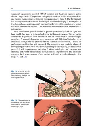
![7 Perforated Gastroduodenal Ulcer 97
The falciform ligament was used if a suitable omental patch was not available.
Following repair, the stomach or duodenum was insufflated to test for air leak. If the
NOTES procedure was unsuccessful, either a laparoscopic or open omental patch
repair was considered by the acute care surgical team.
Initial results from a laparoscopic-assisted NOTES approach for closure of perfo-
rated peptic ulcers appear promising and enable swift recovery in selected patients.
This is especially important in elderly and/or immunocompromised patients.
7.14.2 Stem Cells
In future, development takes place in the Stem Cell Technology. In the literature,
among several preclinic studies on stem cells, there is a Chinese experimental
study [62] published in 2015 on the effect of local injection of mesenchymal
stem cells (MSC) on healing of sutured gastric perforation (GP). Its results were
very encouraging. The local injection of adipose tissue-derived MSC significantly
promoted the healing of GP, with the highest pneumatic bursting pressure, minimal
wound adhesions, and lowest incidence of wound dehiscence and abdominal
abscess. The injected MSC favored a reduction of inflammation and an increase of
granulation and re-epithelization. The authors concluded that local injection of MSC
enhanced the healing of sutured GP by anti-inflammatory process and improved
cellular proliferation and earlier onset of granulation. The use of this technology
represents the new frontier, for instance, for secure gastrotomy closure after notes,
as advocated by the authors.
Thus, in the future, surgeons may take advantage of stem cell technology for
enhanced healing of wounds after closure, based on experimental studies. This has
yet to be confirmed in a clinical trial.
7.14.3 Other Techniques
Aside from the novel techniques previously described, there are some applications
very interesting. Søreide [63] enunciates these techniques superbly in an article
published in 2015, such as Over-the-scope-clip (OTSC), the OverStitch Endoscopic
Suturing System, Self-expanding metal stents (SEMS), Nitinol U-clips, Plug with
acellular matrix, a biodegradable patch, a patch coated with fibrinogen and
thrombin. Some of these are in an experimental phase.
7.15 Conclusions
After the laparoscopic repair of PPU was first reported in 1990 by Mouret, an
increasing numbers of combined meta-analyses of RCTs, prospective observations,
and retrospective cohorts have yielded more favorable advantages for laparoscopy,
as being at least not inferior to open surgery for PPU. Collective reviews also favor](https://image.slidesharecdn.com/emergencylaparoscopy-191115144149/85/Emergency-laparoscopy-113-320.jpg)
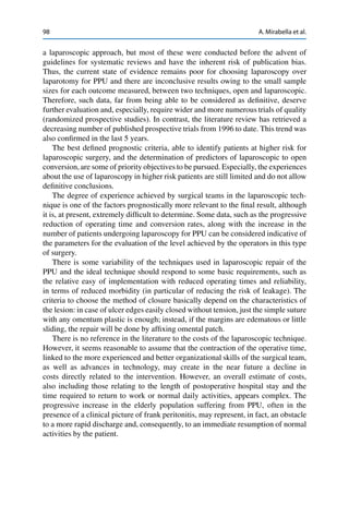
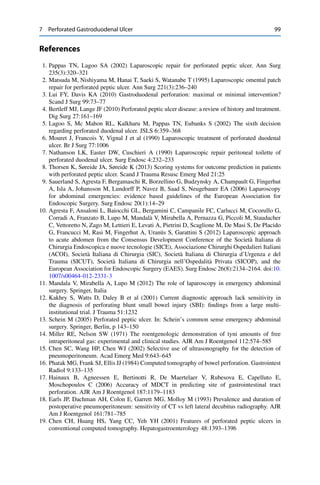
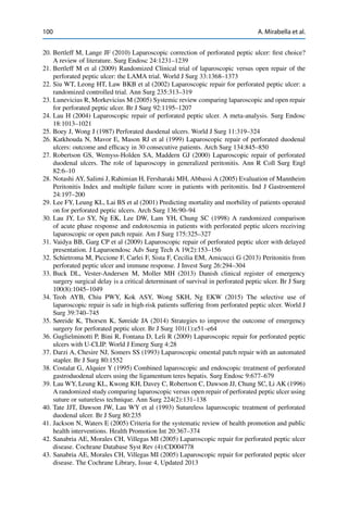
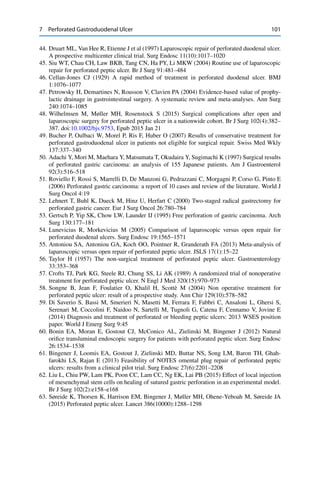
![8Emergency Lapararoscopy for Colon
Obstruction and Acute Diverticulitis
Alberto Arezzo, Marco E. Allaix, Mario Campli,
Carlo Bergamini, and Ferdinando Agresta
8.1 Acute Colon Obstruction
8.1.1 Introduction
About 30 % of patients with colorectal cancer present with acute colon obstruction
requiring an emergent treatment [1]. There are several surgical options and the
choice among these options depends on several factors: tumor location, clinical
condition, surgeon expertise, and intraoperative surgical findings.
The different surgical options include:
1. Colon resection and end colostomy (so-called Hartmann procedure). This
approach is most frequently performed for obstructive left-sided colon cancers,
although colostomy reversal is performed in much less than 50 % of patients [2];
2. Subtotal colectomy and primary ileorectal anastomosis. It is the alternative to
the Hartmann procedure avoiding the need for a loop oostomy and treating in
one stage possible synchronous cancers [3];
3. Loop oostomy followed by colorectal resection. Currently, this two- or three-
staged strategy is reserved to obstructive extraperitoneal rectal cancers to allow
neoadjuvant chemoradiation therapy before resection [4];
A. Arezzo, MD, PhD ( ) • M.E. Allaix, MD
Department of Surgical Sciences, University of Torino, Torino, Italy
e-mail: alberto.arezzo@gmail.com; alberto.arezzo@unito.it
M. Campli, MD
Division of Surgery, “Nuova Itor” Private Health Facility, Roma, Italy
C. Bergamini, MD
General, Emergency and Mini-invasive Surgery, Careggi Hospital, Firenze, Italy
F. Agresta, MD
Department of General Surgery, Azienda ULSS19 del Veneto, Adria (RO), Italy
© Springer International Publishing Switzerland 2016
F. Agresta et al. (eds.), Emergency Laparoscopy,
DOI 10.1007/978-3-319-29620-3_8
103](https://image.slidesharecdn.com/emergencylaparoscopy-191115144149/85/Emergency-laparoscopy-118-320.jpg)
![104 A. Arezzo et al.
4. Colon resection with intraoperative washout and primary anastomosis. This
strategy is still quite common in the USA despite it is still missing evidence that
intraoperative lavage significantly reduces the risk of postoperative anastomotic
leakage [5].
Acute colon obstruction is considered by many an absolute contraindication to
the laparoscopic approach. As a consequence, this surgery is mainly performed
by an open approach and is associated with significant perioperative mortality,
postoperative morbidity, and impaired quality of life [6].
However, the progressive diffusion of the laparoscopic approach to both benign
and malignant colorectal diseases and the development of endoluminal stenting is
challenging this practice.
8.1.2 Laparoscopic Right Hemicolectomy
Surgery is the standard of care for the treatment of right-sided obstructive colon
cancer as this is achieved in a single-stage procedure with no need of protective
ileostomy in the vast majority of cases. Elective laparoscopic right hemicolectomy
is a well-established procedure for the treatment of right colon cancer, with
significantly reduced early postoperative morbidity and mortality. Arezzo [7] has
recently published the results of a systematic review and meta-analysis of studies
comparing elective laparoscopic and open right hemicolectomy having as primary
endpoints 30-day mortality and overall morbidity. Twenty-seven studies were
included, representing 3049 patients although only 2 were randomized controlled
trials (RCTs) for a total of 211 patients. Mortality was significantly lower after
laparoscopic surgery (1.2 % vs. 3.4 %, p D 0.031) as well as the raw incidence of
overall complications (16.8 % vs. 24.2 %; p D 0.007).
Data about emergent laparoscopic right hemicolectomy for obstructing right-
sided colon cancer are lacking. The only study published in the literature is the
one by Ng [8]. This is a retrospective study comparing 14 patients who underwent
emergency laparoscopic-assisted right hemicolectomy and 29 treated by open
right hemicolectomy for obstructing right-sided colon cancer. Both groups were
similar in terms of age, gender, comorbidities, duration of symptoms, tumor size,
and pathological staging. Laparoscopic resection was associated with prolonged
operative time (187.5 min vs. 145 min; p D 0.034) and reduced intraoperative blood
losses (20 vs. 100 ml; p D 0.020) when compared to open surgery. Full patients’
mobilization occurred 2 days earlier after laparoscopic right hemicolectomy (4 vs. 6
days, p D 0.016). No differences were observed in time to resumption of normal
diet, time to return to gastrointestinal functions, and length of hospital stay. A
higher morbidity rate was observed after open surgery, but the difference despite
consistent did not reach statistical significance (55.2 % vs. 28.6 %, p D 0.119). The
interpretation of the results of this study is limited by the retrospective nature, the
small sample size, and the lack of long-term oncologic outcomes. Larger and RCTs](https://image.slidesharecdn.com/emergencylaparoscopy-191115144149/85/Emergency-laparoscopy-119-320.jpg)
![8 Emergency Lapararoscopy for Colon Obstruction and Acute Diverticulitis 105
are needed to investigate the role of laparoscopic surgery in the emergent treatment
of obstructing right-sided colon cancers.
8.1.3 Laparoscopic Left Hemicolectomy
Emergent open Hartmann procedure, subtotal colectomy, and colon resection with
intraoperative washout and primary anastomosis are all procedures proposed for the
treatment of left-sided obstructive colon cancer, but burdened by high postoperative
morbidity and mortality rates and concerns for quality of life and often need of loop
oostomy [6].
The insertion of a self-expandable metallic stent (SEMS) has been proposed for
the relief of colon obstruction in 1990 by Dohmoto [9], initially with palliative
intent in patients with unresectable locally advanced tumor, metastatic disease, or at
high operative risk, aiming to avoid an emergent operation and the surgery-related
morbidity.
Consequently, this approach has been proposed as a “bridge to surgery” strategy
in patients with resectable but obstructing colon cancer [10], the insertion of SEMS
allowing for postponed elective surgery and its benefits [11, 12]. There is actually
no more controversial topic than this with two RCTs [13, 14] stopped for evidence
of superiority of emergency surgery and one [15] stopped vice versa for evidence
of superiority of the SEMS bridge to surgery strategy. Still, a temptative meta-
analysis by Huang [16] showed despite high heterogeneity that SEMS insertion
followed by elective surgery improves the rates of one-stage surgery, increases the
rates of primary anastomosis, and reduces the rates of stoma creation and wound
infections, while overall short-term mortality and morbidity, anastomotic leakage
rates, and intra-abdominal complication rates do not significantly vary between
groups. These data, although limited by the heterogeneity of the RCTs published
in the literature, show that SEMS placement is a safe and effective procedure in
the “bridge to surgery” strategy. In addition, it seems to be associated with earlier
adjuvant chemotherapy administration [17, 18]. To explain why so different results
were achieved by the various different studies it has to be noticed that the expertise
of endoscopists and their case load in those studies that were stopped for advantage
of emergency surgery were critical and possibly criticized. At the same time in those
studies in which Hartmann’s procedure was usually performed in the emergency
surgery group, not always Hartmann’s reversal procedure was considered when
considering complications of surgery. On the other side, the considerably low rate of
reversal procedures allows us to say that Hartmann’s procedure and SEMS insertion
as bridge to surgery have different objectives.
After the first report of laparoscopic-assisted colectomy in 1991 [19], many
studies have shown the short-term benefits of laparoscopic resection when compared
with open resection for uncomplicated colon cancer, including reduced intraopera-
tive blood loss and postoperative pain, lower complication rates, shorter length of
hospital stay, and reduced costs [20–23]. Based on this high-quality evidence, the
use of laparoscopy has been proposed in the early 2000s as the second step of the](https://image.slidesharecdn.com/emergencylaparoscopy-191115144149/85/Emergency-laparoscopy-120-320.jpg)
![106 A. Arezzo et al.
“bridge to surgery” strategy. The SEMS placement relieves bowel distension, thus
allowing to create the working space for the laparoscopic procedure and minimizing
the risk of intestinal injury that is increased in the presence of dilated and fragile
bowel. For instance, Morino [24] reported in 2002 the short-term outcomes in four
patients with colon obstruction secondary to left-sided colon cancer. Laparoscopic
colon resection was performed 4–5 days after the SEMS placement. No patient
experienced postoperative complications and all patients were discharged 5–7 days
after laparoscopic resection.
More recently, few studies (Table 8.1) have shown the advantages of combining
these two minimally invasive procedures. Balagué [25] described the outcomes in
six patients undergoing SEMS insertion followed by laparoscopic colon resection.
Conversion to open surgery was necessary in one patient due to adhesions, while
conversion to hand-assisted surgery was necessary in one case for challenging
dissection of the tumor and the mobilization of the splenic flexure. Median interval
between SEMS placement and surgery was 8 days (range 5–14). Postoperative
morbidity rate was 16.7 %, including a perianastomotic collection that required a
percutaneous drainage. Similarly, Dulucq [26] reported, in 2006, the results in 10
patients diagnosed with acute colon obstruction and treated with SEMS followed
by laparoscopic surgery: they observed one case (10 %) of anastomotic leakage that
was treated by laparoscopic drainage and loop ileostomy. Chung [27] analyzed 17
patients. All procedures were completed laparoscopically: an Hartmann procedure
was performed in two cases (11.8 %), while a diverting ileostomy was created in
a patient undergoing anterior resection. Postoperative morbidity rate was 11.8 %.
Similar results were obtained by others [18, 28].
A word of caution comes from a recent comparative study on long-term
oncologic outcomes. Sabbagh [29] has retrospectively analyzed survival in 48
patients undergoing SEMS and 39 patients treated by emergency surgery. Five-year
overall survival and 5-year cancer-specific mortality were significantly worse in the
SEMS group, even though a higher number of lymph nodes were harvested in the
SEMS group and these patients had a better access to adjuvant chemotherapy. No
significant differences between the two groups were observed in 5-year disease-free
survival, recurrence rate, and mean time to recurrence. In the multivariable analysis,
SEMS insertion was independently correlated with overall survival. The analysis
of patients with no metastasis and no colon perforation showed similar worse
oncologic outcomes in the SEMS patients compared to the patients undergoing
emergency surgery. To justify this, a possible mechanism of cancer cell spreading
was advocated [30]. This confirms in a way the hypothesis of Kim [31] who showed
in a case-matched study that SEMS insertion had an adverse effect on the 5-year
overall survival rate (38.4 % vs. 65.6 %; p D 0.025) and the 5-year disease-free
survival rate (48.3 % vs. 75.5 %; p D 0.024). Nevertheless, it is unclear what
magnitude of this effect is related to the underlying obstruction rather than to the
SEMS. No other report of long-term results is present in the literature, although
necessary to clarify the crucial aspect of oncologic adequacy.](https://image.slidesharecdn.com/emergencylaparoscopy-191115144149/85/Emergency-laparoscopy-121-320.jpg)
![8 Emergency Lapararoscopy for Colon Obstruction and Acute Diverticulitis 107
Table8.1SEMSandlaparoscopiccolonresection:datafromtheliterature
AuthorTypeofstudyn
Intervalbetween
SEMSandsurgery
(days)Stoma(%)
Conversiontoopen
surgery(%)
Earlypostoperative
morbidity(%)Hospitalstay(days)
Morino[24]R45(4–5)0007(5–7)
Balagué[25]R68(5–14)016.7open16.76.5(5–12)
16.7hand-assisted
Dulucq[26]P146.5˙2001016.4˙5a
Chung[27]R177(2–11)17.6011.89(7–49)
Stipa[28]R616.5(6–18)0007
Lujan[18]R1219(4–60)8.30NANA
Rretrospective,Pprospective,NAnotavailable
a
Totalhospitalstaybetweenadmissionanddischarge](https://image.slidesharecdn.com/emergencylaparoscopy-191115144149/85/Emergency-laparoscopy-122-320.jpg)
![108 A. Arezzo et al.
8.1.4 Conclusions
The current evidence shows that elective laparoscopic colon resection could be
safely performed in patients who had previous successful SEMS insertion, with neg-
ligible intraoperative complications, very low stoma rates, and limited postoperative
morbidity. However, further larger RCTs including long-term oncologic results are
needed to confirm these data and to verify oncologic adequacy.
Based on the little evidence currently available, the European Society for
Gastrointestinal Endoscopy has recently published new guidelines for the use
of SEMS for obstructing colonic and extracolonic cancer [32, 33] that can be
summarized in the following four recommendations:
• Prophylactic colonic metal stent placement is not recommended and should be
reserved for symptomatic patients with imaging evidence of obstruction
• Colonic metal stent placement as a bridge to elective surgery is not recom-
mended.
• Colonic metal stent placement is an alternative to emergency surgery only in case
of increased risk of postoperative mortality.
• Colonic metal stent placement is recommended as the preferred treatment for
palliation.
This seems supported by very weak data and should be applied with caution.
The results of two ongoing RCTs comparing SEMS as “bridge to surgery”
and emergency surgery for left-sided colonic malignant obstruction are expected
to better clarify the oncologic impact of this minimally invasive strategy: the
Colorectal Stent Trial (CreST) [34] and the ESCO trial [35]. Both studies have
closed recruitment recently and publication of results is now awaited.
8.2 Acute Diverticulitis
8.2.1 Introduction
The Hinchey classification is the most common grading system used for the
classification and treatment of complicated diverticulitis [36]. While Hinchey grade
I and II patients are successfully managed in most cases by conservative treatment,
surgery is needed in the presence of peritonitis secondary to perforated diverticulitis
(grades III and IV according to Hinchey classification).
The traditional surgical treatment of Hinchey III and IV patients includes open
sigmoid colon resection with end colostomy and left hemicolectomy with primary
anastomosis with or without diverting stoma. Early postoperative mortality rate is
about 10–20 % after both sigmoid resection and end colostomy and resection and
primary anastomosis. In addition, up to 70 % of patients undergoing nonrestorative](https://image.slidesharecdn.com/emergencylaparoscopy-191115144149/85/Emergency-laparoscopy-123-320.jpg)
![8 Emergency Lapararoscopy for Colon Obstruction and Acute Diverticulitis 109
resection do not have their colostomy reversed and the anastomotic leakage rate
reported in the literature after primary anastomosis is as high as 14 % [37, 38].
In 1996, a minimally invasive approach to patients with Hinchey III diverticulitis
based on intravenous antibiotics and laparoscopic peritoneal lavage was proposed
to avoid urgent resective surgery, and stoma creation including their high morbidity
and mortality [39]. Since then, several studies have challenged both indications and
outcomes of the laparoscopic peritoneal lavage in Hinchey III patients [40–57].
8.2.2 Laparoscopic Peritoneal Lavage
This minimally invasive approach to perforated diverticulitis was first proposed by
O’Sullivan [39] in 1996. The rationale was that “in the vast majority of patients
with generalized peritonitis secondary to perforated diverticular disease, there is
no evidence of fecal contamination, and the perforation is already sealed or cannot
be found at surgery” [58]. They retrospectively reviewed the outcomes in eight
patients with generalized peritonitis secondary to perforated diverticular disease
of the left colon that was diagnosed laparoscopically and treated by laparoscopic
peritoneal lavage and administration of parenteral fluids and antibiotics. All patients
had diffuse purulent peritonitis with no fecal contamination. No patients developed
postoperative surgical complications. Full normal diet was resumed in all patients
within 5–8 days. Mean postoperative hospital stay was 10 days (range, 7–17). Two
patients were readmitted to the hospital at 4 and 8 months after the laparoscopic
peritoneal lavage with lower quadrant abdominal pain; both patients were treated
conservatively. No surgical intervention was required during the follow-up, ranging
from 12 to 48 months. Similar results were reported by other small retrospective
studies [40, 42].
The first large multicenter prospective study was published by Myers in 2008
[47]. They included 100 patients preoperatively diagnosed with generalized peri-
tonitis undergoing attempted laparoscopic peritoneal lavage. Primary endpoints
were operative success and resolution of symptoms. Eight patients with Hinchey
IV diverticulitis had conversion to open Hartmann’s procedure. The remaining
92 patients underwent laparoscopic peritoneal lavage. The postoperative medical
complication rate was 4 % (lower respiratory tract infection, acute myocardial
infarction, and pulmonary embolism). Pelvic abscesses occurred in two patients:
one patient underwent radiological percutaneous drainage, while the other required
an open Hartmann’s procedure. The postoperative mortality rate was 3 %, including
multiorgan failure in two patients and pulmonary embolism in one. During a median
follow-up of 36 (range 12–84) months, two patients were readmitted with acute
diverticulitis treated by intravenous antibiotics. No patient required reintervention.
The authors concluded that laparoscopic management of perforated diverticulitis
with generalized peritonitis is an alternative to open surgery, with low postoperative
morbidity and low risk of recurrence in the short term.
Since 2008, several papers have been published on the feasibility and efficacy
of laparoscopic peritoneal lavage for the treatment of diffuse peritonitis secondary](https://image.slidesharecdn.com/emergencylaparoscopy-191115144149/85/Emergency-laparoscopy-124-320.jpg)
![110 A. Arezzo et al.
to perforated diverticulitis. Cirocchi [59] recently published a systematic review
of those studies aiming to evaluate the outcomes of this minimally invasive
strategy, having as primary outcome for this systematic review the success rate
of laparoscopic peritoneal lavage. This was defined as the rate of patients alive
without surgical treatment for a recurrent episode of diverticulitis or complication
from diverticular disease. Of the 19 articles published between 1996 and 2013,
10 were cohort studies, 8 case series, and 1 controlled clinical trial for a total
of 871 patients analyzed with up to 96 months of follow-up. The success rate
of laparoscopic peritoneal lavage was 24.3 %. The overall conversion rate for
Hinchey III and Hinchey IV patients was 1 % and 45 %, respectively. The 30-
day postoperative mortality rate was 2.9 % as a result of multiple organ failure for
sepsis and pulmonary embolism. The 30-day postoperative reintervention (surgery
or percutaneous drainage) rate was 3.8 %: most common causes were sepsis
from generalized peritonitis or intra-abdominal abscess and colonic fistula The
hospital readmission rate after the laparoscopic peritoneal lavage was 3.4 %: main
reasons were recurrent diverticular phlegmon, generalized peritonitis, colo-vesical
fistula, missing colon cancer, inflammatory colonic stenosis, and pelvic abscess.
A reintervention (colon resection or percutaneous drainage) was necessary in 19
(63.3 %) of the 30 patients readmitted to the hospital. Elective laparoscopic sigmoid
resection was performed in 35.8 % of patients. The timing of the laparoscopic
procedure varied greatly, ranging from 2 weeks to 21 months from the laparoscopic
peritoneal lavage. In this group of patients, the conversion rate to open surgery was
4.2 %. It has to be pointed out that the technique of laparoscopic lavage is not yet
standardized so that there was a vast heterogeneity among the included studies, even
in considering the criteria for a further routine sigmoidectomy after resolution of the
acute phase.
Four RCTs have been initiated to better define the role of laparoscopic peritoneal
lavage in patients with purulent peritonitis secondary to perforated diverticulitis.
A few months ago, short-term results of the DILALA trial were published show-
ing feasibility and safety of laparoscopic lavage as treatment for patients with
Hinchey III diverticulitis [60]. A total of 83 patients with purulent peritonitis
were randomized. Some 39 patients undergoing laparoscopic peritoneal lavage and
36 treated by an Hartmann procedure were available for analysis. Similar early
postoperative morbidity and mortality rates were found in the two groups. Patients
undergoing laparoscopic lavage had a shorter operative time and shorter hospital
stay. On the other side, concerns have been risen about the use of laparoscopic
lavage even in selected patients after results of the LADIES trial and SCANDIV
trial were presented. The LADIES trial was the largest study on peritoneal lavage
designed. Patients with purulent peritonitis were randomized to laparoscopic lavage
and drainage, sigmoidectomy with primary anastomosis, and sigmoidectomy with
end colostomy in a 2:1:1 ratio. Recruitment was stopped early for the laparoscopic
lavage and drainage arm due to safety issues, while the two other arms are still
continuing the recruitment [61, 62].
The SCANDIV trial [63] is a multicenter, randomized clinical superiority trial
comparing the outcomes in patients undergoing laparoscopic lavage (101) or](https://image.slidesharecdn.com/emergencylaparoscopy-191115144149/85/Emergency-laparoscopy-125-320.jpg)
![8 Emergency Lapararoscopy for Colon Obstruction and Acute Diverticulitis 111
colon resection (98) for perforated diverticulitis. Ninety-day severe postoperative
complication rates (Clavien Dindo > IIIa) were similar in the laparoscopic lavage
and colon resection groups (30.7 % vs. 26 %, p D 0.53). Ninety-day mortality rates
did not significantly differ between the two groups (12.9 % vs. 11.5 %, p D 0.67).
The reoperation rate was significantly higher after the laparoscopic lavage (20.3 %)
than after colon resection (5.7 %) (p D 0.01). Operative time was significantly
shorter in the laparoscopic lavage group; length of postoperative hospital stay and
quality of life did not differ significantly between groups. Four sigmoid cancers
were missed with laparoscopic lavage. The results of this study do not support the
use of laparoscopic lavage for the treatment of perforated diverticulitis since it is not
associated with better postoperative outcomes than colon resection.
The long-term results of the DILALA trials and the results of the other ongoing
randomized controlled trial (LAPLAND [64] trial) are needed to more clearly
establish the role of laparoscopic peritoneal lavage in the treatment of patients with
purulent peritonitis secondary to acute perforated diverticulitis.
8.2.3 Conclusions
According to the evidence currently available in the literature, laparoscopic peri-
toneal lavage should be considered an option for the treatment of purulent peritonitis
secondary to perforated diverticulitis. In addition, the laparoscopic exploration of
the peritoneal cavity helps to categorize the patients and better plan the surgical
approach. However, the quality of evidence is still quite low; therefore, despite
promising results, it seems too early to recommend the routine use of laparoscopic
peritoneal lavage in clinical practice. As stated by the American Society of Colon
and Rectal Surgeons (ASCRS) [65], “the poor quality of the existing literature on
peritoneal lavage in aggregate and the inherent selection bias in the literature are
major obstacles in advocating the widespread adoption of the laparoscopic lavage;
the safety of lavage for purulent or fecal peritonitis has not been proven or disproven
by the published studies to date.”
Conflict of Interest The authors have no conflicts of interest to declare.
References
1. Landis SH, Murray T, Bolden S, Wingo PA (1998) Cancer statistics, 1998. CA Cancer J Clin
48(1):6–29
2. van de Wall BJ, Draaisma WA, Schouten ES, Broeders IA, Consten EC (2010) Conventional
and laparoscopic reversal of the Hartmann procedure: a review of literature. J Gastrointest Surg
14(4):743–752
3. The SCOTIA Study Group (1995) Single-stage treatment for malignant left-sided colonic
obstruction: a prospective randomized clinical trial comparing subtotal colectomy with seg-
mental resection following intraoperative irrigation. Br J Surg 82:1622–1627](https://image.slidesharecdn.com/emergencylaparoscopy-191115144149/85/Emergency-laparoscopy-126-320.jpg)
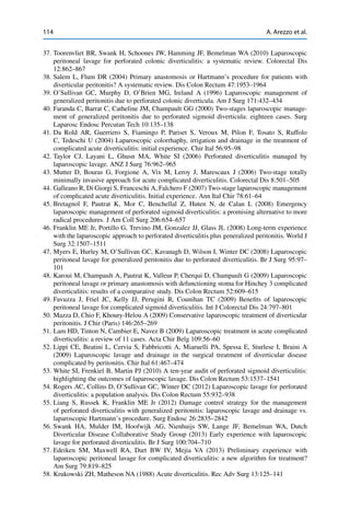
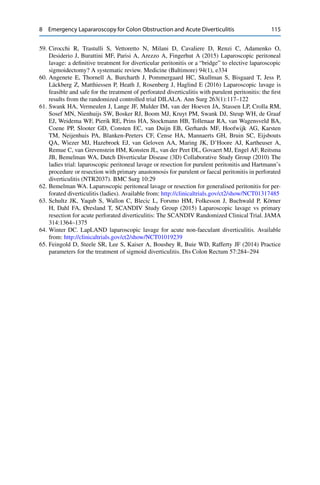
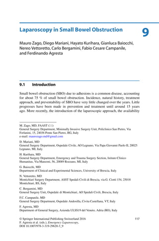
![118 M. Zago et al.
of more established criteria for conservative management, and improvement in
imaging techniques have all contributed to advancements in this field. This chapter
focuses on current evidences about laparoscopy for SBO, including its place in a
comprehensive algorithm of management, and some technical tips and tricks to be
respected when this surgical approach is adopted.
Topics such as pathophysiology, clinical presentation, and medical treatment of
SBO are out of the objectives of this chapter.
9.2 General Issues: Epidemiology, Costs, Etiology,
Classification
9.2.1 Epidemiology, Costs
In some papers of the first half of the last century, adhesions accounted for 7–27 %
of intestinal obstructions, respectively, in the UK and USA [1–3]. Epidemiology
of SBO has not significantly evolved over the years. Admissions for SBO range
between 0.9 and 2 % of all hospital admissions [4, 5]. The incidence rate of SBO
is not decreased [6–8], even if there is a growing evidence of a reduced rate of
clinically relevant postoperative SBO due to adhesions after laparoscopic abdominal
surgery. The burden of admissions for SBO entails an impressive impact on health
budgets. Some studies revealed that the medical costs for SBO are so high to be
comparable to those for some gastrointestinal malignancies [9]. Ray et al. reported
about a yearly 948,000 hospital days of care required for the treatment of SBO [10].
They estimated that Medicare alone was paying $3.2 billion per year for treatment
of SBO.
9.2.2 Etiology of SBO
Even though adhesions rarely lead to obstruction of the large bowel, they account
for more than 70 % of all SBOs. Hernia, cancer, inflammatory bowel disease,
intussusception, radiation, endometriosis, infection, and foreign body are other
relatively common causes of SBO. In patients without previous operations, the most
common cause of bowel obstruction is incarceration of small bowel in a hernia, but
adhesions can occur even in patients without previous abdominal surgery.
9.2.3 Classification of SBO
Small bowel obstruction is generally classified as complete vs. partial, high grade
vs. low grade, simple vs. closed loop, high vs. low, mechanical vs. ileus, and chronic
vs. acute.
Even though the elements of definitions are not strict and they are often expressed
in a subjective way (like “significant” or “less dramatic”), classification is useful for
understanding the probability for surgery and the urgency for appropriate treatment.](https://image.slidesharecdn.com/emergencylaparoscopy-191115144149/85/Emergency-laparoscopy-132-320.jpg)
![9 Laparoscopy in Small Bowel Obstruction 119
Laparoscopic approach could be used in all types when surgery is indicated,
but surgeons must consider the different risks of bowel injuries and or resection
according to the preoperative classification.
Complete SBO is defined by significantly distended bowel, failure to pass stool
or flatus, and lack of air in colon and rectum. It virtually always leads to surgical
operation. Partial SBO is associated with less dramatic small bowel distention,
evidence of flatus and/or stool passage, and evidence of gas throughout the entire
bowel on plain film. Partial SBO can often be conservatively treated.
A high-grade SBO is essentially a complete SBO associated with abdominal pain
and often an impressively distended abdomen. Successful conservative management
is quite rare. A low-grade SBO has the features of a partial SBO and mild to no pain
and discomfort. It frequently succeeds with watchful waiting.
A simple SBO is defined when the bowel is occluded at a single point along its
length, whereas a closed loop obstruction occurs when both the afferent and efferent
portions of a loop of bowel are occluded by a constrictive band, with higher risk of
strangulation and bowel necrosis.
A high SBO is defined when obstruction occurs very proximally in the jejunum,
whereas in a low SBO the occlusion is found in the distal ileum.
9.3 Pathophysiology of Adhesion Formation, SBO,
and Laparoscopy
Attard and Maclean [7] have well depicted the main mechanisms by which adhesion
formation can be avoided and prevented:
– Decreasing peritoneal damage
– Decreasing the early inflammatory response
– Prevention of fibrin formation
– Increasing fibrinolysis
– Preventing collagen deposition
– Providing barriers to adhesion formation
These issues pointed out that the growing use of laparoscopy in abdominal
operations was expected to reduce the rate of postoperative adhesions and at the
same time the incidence of SBO due to adhesions, due to lesser damage to the
parietal and visceral peritoneum, lesser handling of tissues, and the well-known
effect of laparoscopy in decreasing the inflammatory response.
9.3.1 Laparoscopic vs. Open Surgery and Adhesions
Laparoscopic surgery has potential theoretical advantages over open surgery in
reducing the rate of adhesion formation, but very few comparative studies are
available to prove this.](https://image.slidesharecdn.com/emergencylaparoscopy-191115144149/85/Emergency-laparoscopy-133-320.jpg)
![120 M. Zago et al.
An animal experimental study performed by Tittel showed that laparoscopy
decreased the incidence, extension, and strength of intraperitoneal adhesions com-
pared with laparotomy [11]. A retrospective cohort of 716 consecutive patients
undergoing either laparoscopic (211 pt) or open bowel resection (505 pt) suggested
that postoperative SBO requiring hospitalization with conservative management
was reduced in those patients who had laparoscopic surgery (n D 4) compared to
the open surgery cases (n D 31; p < 0.016) [12] (LoE 3).
Gutt and coworkers [13] reviewed 15 papers published from 1987 to 2001,
3 clinical and 12 experimental. In the three clinical studies [14–16], adhesions
following laparoscopy (mainly at the operative site) were less than after open
surgery in comparable groups of patients. The problem of distant adhesions was
poorly represented in the literature. Because of the important differences between
studies with regard to the design, endpoints, and statistical calculations, a meta-
analysis could not be achieved. The conclusions were based on the prevalence of
evidence.
The authors anyway concluded that laparoscopic surgery was associated with a
decrease in adhesion formation in all clinical and most of the experimental studies.
More recently, Schnüriger, in a collective literature review, found that the inci-
dence of adhesion-related readmissions was 7.1 % in open vs. 0.2 % in laparoscopic
cholecystectomy, 9.5 % vs. 4.3 % in respectively open and laparoscopic colectomy,
15.6 % vs. 0 % in hysterectomy, and 23.9 % vs. 0 % in adnexal surgery [17] (LoE 3).
Angenete and coworkers published a population-based retrospective register study,
including 108,141 patients. This large retrospective analysis showed a four times
increased risk of SBO for open surgery compared with laparoscopy [18] (LoE 3).
We should desume that with the wider diffusion of laparoscopic surgery, the
expected rate of this kind of SBO should decrease in the next years.
This seems not to be true for colorectal surgery. Two retrospective analysis
of prospectively collected databases, including homogenous groups of patients
submitted to open vs. laparoscopic colonic resections, showed a similar rate of SBO
[19, 20] (LoE 3). In the latter study, with a long-term FU ranging between 36 and
46 months, the need for operative intervention was significantly lower (8 % vs. 2 %,
p D 0.006) in the laparoscopic group.
9.4 Imaging of SBO, Decision Making, and Laparoscopy
Plain abdominal film remains the most employed imaging when an SBO is
suspected (Fig. 9.1).
Even when the plain film confirms the diagnosis of SBO, the nature and the
precise site of obstruction remain often only suspected and not confirmed. In case
a laparoscopic approach is suggested, plain film is not enough for surgical planning
in the vast majority of cases.
There are evidences that ultrasound (US) sensitivity in diagnosing SBO is
comparable to plain film even when performed by non-radiologists [21–23] (LoE 3).
Notwithstanding, US is unfortunately not widely used. The main US finding](https://image.slidesharecdn.com/emergencylaparoscopy-191115144149/85/Emergency-laparoscopy-134-320.jpg)
![9 Laparoscopy in Small Bowel Obstruction 121
Fig. 9.1 Plain upright
abdominal film, showing
dilated small bowel loops
with fluid levels. Neither the
exact point of obstruction nor
the etiology can be desumed
Fig. 9.2 Ultrasound view of
a SBO. Detection of dilated
(white markers), together
with empty small bowel loops
(yellow arrow), is a sensitive
marker of SBO
confirming the presence of SBO is the visualization of both dilated and empty
small bowel loops, which is highly sensitive for the diagnosis of SBO (Fig. 9.2).
Additional findings are detection of free fluid, characters of fluid (by US-guided
Diagnostic Peritoneal Aspiration, DPA), evaluation of peristalsis, mesurement of
dilated loops, aspect of the bowel wall, aspect of plicae, and assessment of bowel
viability with color flow mapping and power doppler. The main limit of US is to not
be as sensitive as CT in detecting the site and the nature of the obstruction.
Contrast-enhanced CT (CECT) scan has become the imaging of choice for
most patients with suspicion of SBO. A literature review performed by Mallo and
coworkers analyzed that the accuracy of CT in identifying SBO associated with
bowel ischemia or complete obstruction was very high [24]. Concerning bowel
ischemia, CT aggregated positive predictive value (PPV) was 79 % (range 69–
100 %), negative predictive value (NPV) 93 % (range 33–100 %), sensitivity
(SN) 83 % (range 63–100 %), and specificity (SP) 92 % (range 61–100 %). The](https://image.slidesharecdn.com/emergencylaparoscopy-191115144149/85/Emergency-laparoscopy-135-320.jpg)
![122 M. Zago et al.
Fig. 9.3 CT has a high
sensitivity for the
identification of the
transitional point due to a
band adhesion (arrow).
Empty small bowel loops are
clearly evident on the left
corner
aggregated PPV of CT for complete obstruction was 92 % (range 84–100 %), NPV
93 % (range 76–100 %), SN 92 % (range 81–100 %), and SP 94 % (range 68–
100 %).
CECT scan (with or without oral contrast) shows proximal dilated small bowel,
distal collapsed small bowel, and an empty colon. The presence of a transition point,
usually due to an adhesion band (Fig. 9.3), does not predict neither the need for
surgery nor the failure of nonoperative management [25]. CT scan can identify
the vast majority of causes of the SBO (neoplasms, internal hernias, IBD, estrinsic
stricture, etc.) and a careful identification of some CT signs can help in predicting
the right and successful indication for the laparoscopic approach.
The main CT signs to be searched and interpreted are listed here:
• free fluid
• bowel loop caliper
• reduced bowel wall enhancement
• bowel loop thickness
• mesenteric swirl
• small bowel feces sign
Looking at CT findings, Schwenter showed that the need for bowel resection in
SBO was significantly predicted in multivariate analysis by the presence of more
than 500 mL of free fluid, reduction bowel wall enhancement, and mesenteric swirl.
Adding clinical and biochemical criteria like WBC > 10,000/mmc, CRP > 75 mg/L,
abdominal guarding, and pain lasting for more than 4 days, patients with four or
more of these variables required resection. The presence of fever and the detection
of a transition point on CT were not independent predictors of ischemic bowel [26].
Zielinski and coworkers confirmed that fever and transition point on CT were
not independent predictors of operation. In a further prospective study, Zielinski
highlighted that the lack of small bowel feces sign on CT scan was predictive for](https://image.slidesharecdn.com/emergencylaparoscopy-191115144149/85/Emergency-laparoscopy-136-320.jpg)
![9 Laparoscopy in Small Bowel Obstruction 123
operation [27, 28]. The “small bowel feces sign” is the presence of particulate
material in dilated small bowel loops on CT. Around 50 % of patients with SBO
show it. The so-called whirl sign is defined as “swirl of mesenteric soft tissue and
fat attenuation with adjacent loops of bowel surrounding rotated intestinal vessels”
(Fig. 9.4).
In a retrospective study, Duda was able to identify the “whirl sign” in 20 % of
CT of a large series of patients with SBO. When detected, the whirl sign should
be carefully considered because it could be an ominous sign. Eighty percent of the
Duda’s series with the “whirl sign” required operation (PPV 80 %), whereas only
14 % without the sign underwent surgery (NPV 86 %) [29].
A comparative list of findings useful for both diagnosis and surgical planning is
depicted in Table 9.1.
As discussed below in this chapter, safety and effectivenes of the laparoscopic
approach are strictly related to careful patient selection. The use of laparoscopy as a
Fig. 9.4 The whirl sign
(circle). This finding is better
detected by an “up&down”
scrolling of CT axial images
on the screen
Table 9.1 Usefulness of imaging techniques for planning the laparoscopic approach for SBO
Plain abdominal X ray US CECT
Diagnosis of SBO C C CC
Bowel peristalsis – C –
Presence of free fluid – C C
Characters of free fluid – CC a
˙
Small bowel loops caliper C C C
Bowel loops viability – C CC
Site of obstruction – ˙ CC
Etiology – ˙ C
Preop. planning of trocars placement – C CC
US ultrasound, CECT contrast enhanced computed tomography
a
By DPA](https://image.slidesharecdn.com/emergencylaparoscopy-191115144149/85/Emergency-laparoscopy-137-320.jpg)
![124 M. Zago et al.
diagnostic tool without a full preoperative planning in this setting might be the door
toward a disaster.
The need for a surgeon-performed interpretation of CT findings is evident. Items
listed in Table 9.1, coupled with medical history, physiology, and clinical exam, are
all needed for properly deciding the urgency of operation (presence and nature of
free fluid, bowel viability), the probabilty of failure or intraoperative complications
(loops caliper, etiology), and the proper trocar placement.
9.5 Evidences for the Laparoscopic Approach to SBO
The first reports of laparoscopic treatment date from the early 1990s, initially in the
field of gynecologic surgery. In 1990, Clotteau first reported on laparoscopic adhe-
siolysis for SBO secondary to adhesions [30]. The main advantages of laparoscopy
vs. open surgery for SBO concern the postoperative recovery, as well as the reduced
rate of postoperative laparotomy-related adhesions and ventral hernia.
Neither RCTs nor prospective controlled studies are available in the scientific
literature comparing open vs. laparoscopic approach for SBO [31]. A Finnish
multicenter, prospective, randomized clinical trial was registered in 2013, compar-
ing laparoscopic vs. open adhesiolysis for SBO, with the primary endpoint being
the length of postoperative stay. Secondary endpoints include passage of stools,
commencement of enteral nutrition, morbidity, and mortality. Results are expected
in 2018 [32].
9.5.1 Guidelines
Some guidelines concerning laparoscopy and SBO are available in the literature and
show a progressive trend toward a selected and more precise use of this appoach.
The EAST Guidelines, published in 2008 [33], suggested that in a highly selected
group of patients the laparoscopic treatment of SBO should be considered, leading
to a shorter hospital length of stay. Notwithstanding, laparoscopy was not included
in the proposed flowchart. The 2008 SAGES guidelines on diagnostic laparoscopy
are very generic on this topic. They considered laparoscopy contraindicated in
patients with a clear indication for surgical intervention such as massive bowel
obstruction, perforated viscus (free air), and those with hemodynamic instability
[34].
A group of experts published in 2013 an update of the Bologna Guidelines [35].
Laparoscopy is suggested as an option in highly selected patients. The open access
technique is advised. The panel of an Italian consensus conference on laparoscopic
adhesiolysis agreed to consider hemodynamic instability and/or cardiopulmonary
impairment the only absolute contraindications to a minimally invasive approach
[36].
Agresta and coworkers, on behalf of five Italian Scientific Societies and EAES,
published in 2012 an update of the previous EAES guidelines on laparoscopy in](https://image.slidesharecdn.com/emergencylaparoscopy-191115144149/85/Emergency-laparoscopy-138-320.jpg)
![9 Laparoscopy in Small Bowel Obstruction 125
abdominal emergencies [37, 38]. A mild advice supporting the selective use of
laparoscopy in SBO was affirmed.
9.5.2 Conservative Management and Laparoscopy
The availability of the laparoscopic approach does not influence the current man-
agement of SBO suitable for conservative treatment. As specified above, the role
and timing for nasogastric tube placement, Gastrografin®
administration, antibiotic
use, and time for oral intake resumption are not discussed here [35, 39, 40]. It is
well known that surgery does not influence both the risk of recurrence and the need
for a future operation [8] (LoE 3). Duron et al. found that the rates of primary or
secondary recurrence (12 % and 18 %, respectively) were not different after open
compared to laparoscopic surgery [41] (LoE 3).
In other words, laparoscopy is a minimally invasive surgical treatment with
inherent morbidity and mortality, and according to present knowledge, it does not
decrease the rate of recurrence of SBO after adhesiolysis.
9.5.3 Indications for Surgical Treatment and Selection
for the Laparoscopic Approach
According to the above paragraph considerations, indications to surgical treatment
do not change even if the patient is considered suitable for a laparoscopic approach.
An accurate and complete evaluation of the patient history, physiology,abdomen,
and of the performed imaging must be performed.This is especially true for a patient
with the classic tetrad frequently associated with bowel strangulation, namely,
leukocytosis, tachycardia, fever, and severe unrelenting abdominal pain.
The management of patients with SBO is based on some critical issues: etiology,
the degree of obstruction (partial or complete), and small bowel viability (ischemia
or perforation). Therefore, CT plays a key role for a successful laparoscopic
approach, as the majority of the morphological criteria associated with conversion
to open surgery, intraoperative complications, and failure of laparoscopy are
detectable on CT (axial, sagittal, and coronal views). Even in case of patients with
bowel obstruction and a virgin abdomen, who virtually always undergo surgical
exploration, a preoperative CT is advisable whenever the laparoscopic approach is
evisageable.
Theoretically, all etiologies of SBO could be an indication to the laparoscopic
approach, but the best and more successful situation is a SBO due to a single
adhesion. It is an additional reason for a careful preoperative workup. A special
situation is SBO occurring in patients with known intra-abdominal malignancy,
for whom recurrent malignancy is possible. Although there is a comprehensible
reluctancy to operate, 30 % of patients will have a simple adhesion amenable to
simple lysis, 30 % have a resectable obstructing malignancy, and only 30 % have
carcinomatosis not suitable for any surgical solution. It is probably the only setting](https://image.slidesharecdn.com/emergencylaparoscopy-191115144149/85/Emergency-laparoscopy-139-320.jpg)
![126 M. Zago et al.
of SBO for which laparoscopy, when feasible, should be considered as a diagnostic
tool, in order to minimize the surgical trauma in very frail patients.
The only absolute contraindication to the laparoscopic approach is a physiologic
status (hemodynamics, respiratory) which makes impossible the pneumoperitoneum
[36]. Even the impossibility to get a sufficient peritoneal space (often related to
bowel loops dilation) is a technical contraindication [42].
One of the main concern about the laparoscopic approach for SBO is the high
conversion rate: complete laparoscopic treatment has been reported possible in the
past in only 50–60 % of patients. Papers published in the last decade, in an era of a
more standardized laparoscopic techniques and criteria, have shown a trend toward
a reduction in conversion rate, in the laparoscopically treated patients, constantly
lower than 50 % [42–50] (LoE 3).
A systematic review including all papers published up to 2007 (1236 patients)
found a successful therapeutic laparoscopy rate in the range of 40–88 % and a
conversion rate ranging from 0 to 52 %. Positive predictive factors for success are
less than two previous laparotomies and the absence of peritonitis [51] (LoE 3).
More recently, O’Connor et al. published a more comprehensive systematic review,
reporting a conversion rate of 29 % [52] (LoE 3).
Conversion can be due to many reasons [52–65]:
– Inability to visualize and control the operative field (massive loops dilation)
– Neoplasms
– Dense adhesions
– Intestinal ischemia with bowel infarction
– Iatrogenic bowel injury
– Inability to find the point of obstruction
– Inability to achieve pneumoperitoneum
Overall morbidity after laparoscopic adhesiolysis ranges between 15 and 39 %
[51, 53, 55, 56, 62, 63].
Inadvertent enterotomy rate is around 6.5 % and did not statiscally differ from
open surgery [48, 53, 55, 56, 65]. It could be repaired laparoscopically in half of
cases. The analysis of outcomes is discussed below. A bowel injury may be missed
at the time of operation. Although a missed enterotomy can occur during laparotomy
too, the incidence is reported to be higher with the laparoscopic approach. A careful
surgical technique (see below) is the only method for preventing the risk.
Laparoscopy should not be used as a first step tool for the diagnosis of SBO
without intention to treat.
Many studies have identified the factors that proved to be statistically signifi-
cantly associated with failure of the laparoscopic approach.
These criteria should be kept in mind and accurately considered in order to
optimize the selection of patient to be submitted to laparoscopy and are listed below:
– Small bowel loop diameter >4–5 cm
– More than two previous abdominal operations](https://image.slidesharecdn.com/emergencylaparoscopy-191115144149/85/Emergency-laparoscopy-140-320.jpg)
![9 Laparoscopy in Small Bowel Obstruction 127
Fig. 9.5 Intraoperative view.
Single band adhesion cut with
cold scissors
– Operation after more than 24 h from diagnosis
– Duration of surgery
– Dense and extensive adhesions
Previous appendectomy was statistically associated with a higher rate of success-
ful laparoscopic management, with the single band adhesion as the ideal condition
for the laparoscopic approach [54–61, 66] (LoE 3) (Fig. 9.5).
The authors, in a retrospective study over 100 consecutive patients [59], reported
a conversion rate of 34.2 % and found as statistically significant factor for
conversion a small bowel caliper >4 cm. Notwithstanding, the analysis of the
series revealed that 17.5 % of successful laparoscopic operations were performed
in patients with bowel loops larger than 4 cm.
Reduced port and single-port laparoscopic adhesiolysis has been reported, but
experiences are limited and only feasibility could be assessed. It is of interest to
note that preoperative imaging identifying a single band adhesion is deemed a
prerequisite for some authors [67] (LoE 4).
In pediatric patients, experience on the laparoscopic treatment of SBO is very
limited. Recent literature shows a trend in considering laparoscopy the first option
for adhesive small bowel obstruction in children [68, 69] (LoE 4).
9.5.4 Laparoscopy in the SBO Algorithm
In the last few years, many articles were published suggesting comprehensive
algorithms for SBO management, including paths for nonoperative management,
water-soluble contrast administration, timing for expectant management, and crite-
ria for immediate or delayed surgery [35, 36, 62, 63, 70]. This chapter is specifically
devoted to the role of laparoscopy in SBO, and a careful analysis and comparison
among them is mandatorily skipped.](https://image.slidesharecdn.com/emergencylaparoscopy-191115144149/85/Emergency-laparoscopy-141-320.jpg)
![128 M. Zago et al.
Fig. 9.6 Proposal of a simplified algorithm for the management of SBO, focused on the criteria for
successful laparoscopic approach. Preoperative and intraoperative findings (midline boxes) guide
the choice for laparoscopy or open approach
A simplified original algorithm focused on laparoscopic criteria for conversion
to open surgery is proposed in Fig. 9.6 and could be used to integrate a more
comprehensive protocol. Preoperative and intraoperative criteria obtained by the
updated literature review and useful for the critical choice of the laparoscopic
approach are highligted.
9.5.5 Laparoscopy and Clinical Outcomes
Laparoscopy adhesiolysis reveals advantages in most of the surgical outcomes.
A systematic review and meta-analysis, pooling the effects of outcomes of
a total of 334 patients in four retrospective comparative studies using meta-
analytic methods, showed that laparoscopic adhesiolysis was associated with a
reduction in overall complication rate (p < 0.01), prolonged ileus rate (p < 0.01),
and pulmonary complication rate (p < 0.04), compared with open approach. No
differences were observed for intraoperative bowel injuries, wound infection, and
mortality [71] (LoE 2). Saleh and coworkers [64] analyzed data of the ACS NSQIP
between 2005 and 2010, collecting 4616 patients submitted to surgery for SBO
(3697 open, 919 laparoscopic). They excluded the sicker patients, the converted
operations, and those with formal bowel resection in order to reduce potential biases.](https://image.slidesharecdn.com/emergencylaparoscopy-191115144149/85/Emergency-laparoscopy-142-320.jpg)
![9 Laparoscopy in Small Bowel Obstruction 129
Laparoscopy, in considering both unadjusted and adjusted odd ratios, revealed a
statistically significant advantage over open surgery in terms of morbidity and 30-
day postoperative morbidity (p < 0.01) and overall complications (p < 0.01). The
mean postoperative length of stay (LOS) was 8.4 compared to 3.8 days in the
open and laparoscopic groups, respectively (p < 0.01). The same group performed
a retrospective review about the outcome of their own series of 269 patients, 83 of
whom (30.9 %) were managed laparoscopically, with a conversion rate of 38.6 %.
Laparoscopy allowed a quicker recovery (time of passage of flatus, p < 0.005),
LOS (5 vs. 7 days, p < 0.031), and overall complication rate (27.7 vs. 43.6 %,
p < 0.014). The same results were obtained following exclusion of bowel resections
[65] (LoE 4). A significant reduction of 30-day mortality and of major complication
rate with the laparoscopic approach was confirmed in a further analysis of the ACS
NSQIP database done by Kelly and coworkers [72] (LoE 3).
The use of a tailored laparotomy (i.e., a reduced incision placed according to
laparoscopic findings) would be a potential benefit of the laparoscopic approach,
but this item has not been demonstrated yet. The use of a tailored mini-laparotomy
(less than 10 cm) is reported ranging from 20 to 35 % of cases [59, 73].
Even though successful laparoscopic treatments of patients with negative pre-
dictive criteria are described without complication in some papers [59, 73], there
is a current agreement among experts that a low threshold for open conversion
remains recommended, especially facing with dense adhesions (LoE 5). Provided a
full workup and a critical decision making have been done, conversion should not be
considered a failure, but simply the further and wise step in the optimal management
of a patient with SBO. From the other side, any surgeon with developed laparoscopic
skills who has performed a standard midline incision to release a single adhesive
band normally regrets that the laparoscopic approach was not chosen.
9.6 Technical Tips and Tricks
As stated by Azagury et al. [70], the ideal successful surgical management of
SBO should be defined as “a laparoscopic lysis of adhesions without inadvertent
enterotomy or laparoscopic with conversion to laparotomy before any intraoperative
complication.”
9.6.1 Equipment
No special laparoscopic instrumentation is needed for SBO adhesiolysis. Well-
functioning atraumatic graspers (Johanne like) and cold scissors are the most
important instruments. Recording any procedure is advisable, especially for review-
ing the procedure in case of complications and when inadvertent enterotomy occurs,
in order to improve the awareness and the skills of the entire surgical team.
Advanced sealing instruments (radiofrequency dissector, ultrasonic scalpel) could](https://image.slidesharecdn.com/emergencylaparoscopy-191115144149/85/Emergency-laparoscopy-143-320.jpg)
![130 M. Zago et al.
be helpful but not indispensable and should be used in selected situations. The use
of a 30ı
telescope is the rule.
9.6.2 Positioning of the Patient
Patient positioning strongly affects the feasibility of laparoscopic adhesiolysis.
A bad placing of the patient on the table might be the main cause of conversion.
You would need to tilt the table in every position during the operation for pulling
away the dilated loops and sometimes even to change the side where you started to
work.
Check that all the following issues were done:
– Both arms should be put along the patient’s sides (assistant is almost always
beside you).
– Always install shoulder keepers (and feet keepers if the patient is obese) to be
prepared for steep positions if needed.
– Control the table remote control is working.
9.6.3 Trocar Placement and Access
Nasogastric tube and Foley catheter should already be in place.
First access to the peritoneal cavity should be placed far from the scars of
previous surgery.
A preoperative individual planning, namely, a surgeon performed examination of
preoperative CT, is essential for a safe trocar placement and effective surgery.
No evidences support the use of open laparoscopy vs. Veress needle vs. optic
trocar for the induction of pneumoperitoneum. Some guidelines suggest the use of
open laparoscopy as the preferred method [35, 36, 70]. A muscle splitting incision
for the Hasson trocar insertion can be performed everywhere in the lateral abdominal
quadrants, except in morbid obese patients.
Gentle blunt dissection with a 30ı
telescope directly against parietal adhesions
is a smart trick for getting space enough for the positioning of the second trocar.
The goal is to place at least three ports with a good triangulation. Additional ports
should be inserted as needed.
A smart choice is to use a 5-mm angled scope that could be inserted in any port.
9.6.4 Surgical Technique
Adhesiolysis should start where adhesions are thinner, the bowel is easily identified,
and loops are empty. Pulling away dilated loops by gravity and gaining the
workspace is the first step, with the aim to focus on the transition point. For
dissection, prefer cold scissors and blunt dissection whenever possible. Be aware](https://image.slidesharecdn.com/emergencylaparoscopy-191115144149/85/Emergency-laparoscopy-144-320.jpg)
![9 Laparoscopy in Small Bowel Obstruction 133
24. Mallo RD, Salem L, Lalani T et al (2005) Computed tomography diagnosis of ischemia and
complete obstruction in small bowel obstruction: a systematic review. J Gastrointest Surg
9(5):690–694
25. Colon MJ, Telem DA, Wong D et al (2010) The relevance of transition zones on computed
tomography in the management of small bowel obstruction. Surgery 147:373–377
26. Schwenter F, Polleti PA, Platon A et al (2010) Clinicoradiological score for predicting the risk
of strangulated small bowel obstruction. Br J Surg 97:1119–1125
27. Zielinski MD, Eiken PW, Bannon MP et al (2010) Small bowel obstruction—who needs an
operation? A multivariate prediction model. World J Surg 34(5):910–919
28. Zielinski MD, Eiken PW, Lohse HSF et al (2011) Prospective, observational validation of
a multivariate small-bowel obstruction model to predict the need for operative intervention.
J Am Coll Surg 212(6):1068–1076
29. Duda JB, Bhatt S, Dogra VS (2008) Utility of CT whirl sign in guiding management of small-
bowel obstruction. Am J Roentgenol 191(3):743–747
30. Clotteau JE, Premont M (1990) Occlusion by adhesions treated by celioscopic section [in
French]. Presse Med 19(25):1196
31. Cirocchi R, Abraha I, Farinella E, Montedori A, Sciannameo F (2010) Laparoscopic versus
open surgery in small bowel obstruction (Review). The Cochrane Collaboration. Cochrane
Database Syst Rev 2, CD007511
32. Sallinen V, Wikstrom H, Victorzon M, Salaminen P, Koivukangas V, Haukijarvi E, Enholm
B, Leppaniemi A, Mentula P (2014) Laparoscopic versus open adhesiolysis for small bowel
obstruction – a multicentre, prospective, randomized, controlled trial. BMC Surg 14:77
33. Diaz JJ Jr, Bokhari F, Mowery NT, Acosta JA, Block EFJ, Bromberg WJ, Collier BR, Cullinane
DC, Dwyer KM, Griffen MM, Mayberry JC, Jerome R (2008) Guidelines for management of
small bowel obstruction. J Trauma 64:1651–1664
34. SAGES (2008) Diagnostic laparoscopy guidelines. Surg Endosc 22:1353–1383
35. Di Saverio S, Coccolini F, Galati M, Smerieri N, Biffl WL, Ansaloni L, Tugnoli G, Velmahos
GC, Sartelli M, Bendinelli C, Fraga G, Kelly M, Moore FA, Mandalà V, Mandalà S, Masetti
M, Jovine E, Pinna AD, Peitzman AB, Leppaniemi A, Sugarbaker PH, Van Goor H, Moore
EE, Jeekel J, Catena F (2013) Bologna Guidelines for diagnosis and management of adhesive
small bowel obstruction (ASBO): 2013 update of the evidence-based guidelines from the world
society of emergency surgery ASBO working group. WJES 8:42
36. Vettoretto N, Carrara A, Corradi A, De Vivo G, Lazzaro L, Ricciardelli L, Agresta F, Amodio
C, Bergamini C, Borzellino G, Catani M, Cavaliere D, Cirocchi R, Gemini S, Mirabella A,
Palasciano N, Piazza D, Piccoli M, Rigamonti M, Scatizzi M, Tamborrino E, Zago M (2012)
Laparoscopic adhesiolysis: consensus conference guidelines. Colorectal Dis 14:e208–e215
37. Sauerland S, Agresta F, Bergamaschi R, Borzellino G, Budzynsky A, Champault G, Fingerhut
A, Isla A, Johansson M, Lundorff P, Navez B, Saad S, Neugebauer EA (2006) Laparoscopyfor
abdominal emergencies: evidence based guidelines of the European Association for Endo-
scopic Surgery. Surg Endosc 20(1):14–29
38. Agresta F, Ansaloni L, Baiocchi L, Bergamini C, Campanile FC, Carlucci M, Cocorullo G,
Corradi A, Franzato B, Lupo M, Mandalà V, Mirabella A, Pernazza G, Piccoli M, Staudacher
C, Vettoretto N, Zago M, Fingerhut A, Uranues S (2012) Laparoscopic approach to acute
abdomen. Consensus Dev Conf Surg Endosc 26:2134–2164
39. Branco BC, Barmparas G, Schnüriger B et al (2010) Systematic review and meta-analysis of
the diagnostic and therapeutic role of water-soluble contrast agent in adhesive small bowel
obstruction. Br J Surg 97:470–478
40. Abbas S, Bissett IP, Parry BR (2007) Oral water soluble contrast for the management of
adhesive small bowel obstruction. Cochrane Database Syst Rev 3, CD004651
41. Duron JJ, Jourdan-Da Silva N, Tezenas du Montcel S, Berger A, Muscari F, Hennet H,
Veyrieres M, Hay JM (2006) Adhesive postoperative small bowel obstruction: incidence and
risk factors of recurrence after surgical treatment. A multicenter prospective study. Ann Surg
244:750–757](https://image.slidesharecdn.com/emergencylaparoscopy-191115144149/85/Emergency-laparoscopy-147-320.jpg)
![10Incarcerated Hernias
Giovanni Alemanno, Riccardo Somigli, Paolo Prosperi,
Mario Campli, Alberto Arezzo, Andrea Valeri, Carlo Bergamini,
and Mauro Zago
10.1 Introduction
The lifetime risk of having a groin hernia repair has been estimated to be 27 %
for men and 3 % for women. In the natural history of the inguinal or femoral
hernia, 0.29–2.9 % of cases become incarcerated. Incarceration is the state of an
external hernia which cannot be reduced into the abdomen. It implies an increased
risk of small bowel obstruction, being the second most common cause of it
[1]. Furthermore, among incarcerated hernias, 10–15 % become strangulated and
gangrenous, which is a severe complication with a mortality rate of up to 5 % in
the elderly. Incarceration has, therefore, great importance and should be considered
a surgical emergency [2]. Actually, roughly 5 % of all patients having an operation
for external hernia are explored as an emergency.
In 1993, Watson and colleagues demonstrated the feasibility of laparoscopic
groin hernia repair (GHR). A modified pre-peritoneal femoral hernia repair with
mesh and resection of incarcerated small bowel was been successfully completed by
these authors under laparoscopic guidance in a 64-year-old patient with incarcerated
femoral hernia [3].
G. Alemanno ( ) • R. Somigli • P. Prosperi • A. Valeri • C. Bergamini
Careggi University Hospital, Florence, Italy
e-mail: g.alemannomd@gmail.com
M. Campli
Division of Surgery, “Nuova Itor” Private Health Facility, Rome, Italy
A. Arezzo
Department of Surgical Science, University of Torino, Italy
M. Zago
General Surgery Department, Minimally Invasive Surgery Unit, Policlinico San Pietro,
Via Forlanini 15, 24036 Ponte San Pietro, BG, Italy
© Springer International Publishing Switzerland 2016
F. Agresta et al. (eds.), Emergency Laparoscopy,
DOI 10.1007/978-3-319-29620-3_10
137](https://image.slidesharecdn.com/emergencylaparoscopy-191115144149/85/Emergency-laparoscopy-150-320.jpg)
![138 G. Alemanno et al.
10.2 Literature Supporting the Role of Laparoscopy
in Emergency GHR
In 2003, a Cochrane library study showed that the outcome of elective laparoscopic
GHR is at least equivalent to that of the open approach. Indeed, the use of mesh
during laparoscopic hernia repair seemed to be associated with a relative reduction
in the risk of hernia recurrence of around 30–50 %. Furthermore, a less persisting
pain and numbness following laparoscopic repair was suggested and the return to
usual activities was faster. However, operation took a longer time and there appeared
to have a higher risk of serious complication rate in respect of visceral (especially
bladder) and vascular injuries [4].
This study was confirmed in 2010 by an extensive meta-analysis of randomized
controlled studies (RCT) and comparative studies concerning patients treated for
recurrent inguinal hernia with laparoscopic procedures versus open mesh repair.
Significantly, fewer cases of hematoma/seroma formation were observed in the
laparoscopic group in comparison with the Lichtenstein group, whereas the relative
risk of overall recurrence of the laparoscopic versus the Lichtenstein group was
comparable [5].
Concerning the incarcerated hernia, many non-comparative low-evidence level
studies have been published so far. A review of cohort studies on laparoscopic
repair of incarcerated groin hernias was published in 2010 by Deeba et al., updating
the information given in the previous guidelines on laparoscopic emergency [6]. It
reviews seven articles on this topic dating from 1989 to 2008, reporting on 328
cases treated with total extra-peritoneal (TEP) or trans-abdominal pre-peritoneal
(TAPP) repair (Table 10.1). Of these seven articles, two are LE 3 prospective cohort
studies and five are LE 4 small case series. The overall results of the Deeba et al.’s
study were that average operative time was 61.3 min, average hospital stay was
3.8 days, mortality rate was 0.28 %, complication rate was 10.3 %, conversion rate
was 1.8 %, intestinal laparoscopic or mini-laparotomy resection rate was 5.1 %, and
reoperation rate was 0.9 %. The most serious complications consisted of two colonic
lesions and one divided vas deferens. The others were infected mesh (0.6 %), wound
infection (0.3 %), deep venous thrombosis (0.3 %), and other minor complications.
The highest recurrence rate at 7 years was 5.8 %. The authors concluded that the
laparoscopic approach, either TEP or TAPP, is possible for repairing incarcerated
hernia taking into account the knowledge of anatomy and expertise needed to dissect
and reduce the sac. Laparoscopy can also be used to resect bowel, if necessary, or
to repair an occult contralateral hernia, present in 11.2–50 % of cases. The overall
complication rate, recurrence rate, and length of hospital stay seem to be very similar
to those documented for open repair of strangulated/incarcerated hernias.
The only comparative study so far available has been recently published by
Yang et al. These authors have described the low middle-term outcome of open
compared to laparoscopic strangulated groin hernia repair in adults [7]. In this series
of 188 cases, retrospectively studied, Yang et al. reported that the laparoscopic group
had a much lower wound infection rate compared to the open group. The mean](https://image.slidesharecdn.com/emergencylaparoscopy-191115144149/85/Emergency-laparoscopy-151-320.jpg)
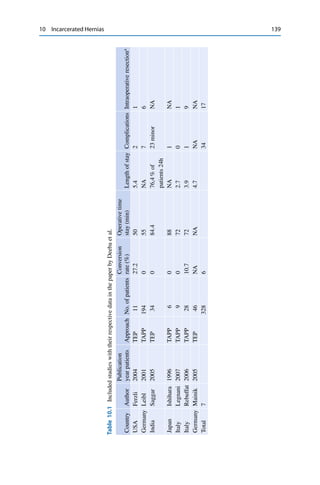
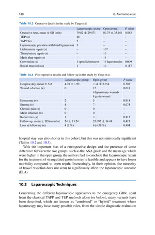
![142 G. Alemanno et al.
Hernial
Sec
Peritoneum
Mesh unrolled laterally
in preperitoneal space
toward Bogros space
Mesh unrolled
in prepritoneal
space
Peritoneum
Fig. 10.2 Completing placement of the mesh
viability of bowels at the end of the procedure, thereby avoiding unnecessary
bowel resection [8] (Fig. 10.2).](https://image.slidesharecdn.com/emergencylaparoscopy-191115144149/85/Emergency-laparoscopy-155-320.jpg)
![10 Incarcerated Hernias 143
Fig. 10.3 (a) Giant inguino-scrotal hernia. (b) Placement of ports
2. Hybrid laparoscopic/laparotomic using the TEP. Other authors prefer the TEP
because they feel that laparoscopic extra-peritoneal preparation of the pre-
peritoneal floor avoids the usual struggle of an open pre-peritoneal repair and
results in a larger and better positioned mesh than in an open anterior approach.
In addition, dissection of the cord structures does not rely on invagination of
the sac which makes the dissection more difficult when dealing with the scrotal
hernia [9] (Fig. 10.3a, b)](https://image.slidesharecdn.com/emergencylaparoscopy-191115144149/85/Emergency-laparoscopy-156-320.jpg)
![144 G. Alemanno et al.
Table 10.4 Outcome and follow-up in the study by Sgourakis et al.
Laparoscopic group Open group P value
Operstive time
(min)
67.78 ˙ 12.39 82.84 ˙ 21.08 <0.05
Presence of
hemorrhagic fluid
8 7 0.885
Extension of the
operation (no. of
patients)
2 (2 resections) 4 (4 standard laparotomies, 2 resections) 0.413
Median hospital
stay (h)
28 34 0.044
Hospital stay over
36 h
3 9 0.108
Major
complications
0 4 (20 %) <0.05
Minor
complications
3 (14 %) 3 (15 %) 0.955
Deaths 0 2 0.157
Return to ordinary
and professional
activities (days)
17.28 ˙ 5.34 24.23 ˙ 9.14 0.017
Recurrent hernia 0 0 -
3. The new “hernia sac laparoscopy (hernioscopy)” that is a new mixed laparo-
scopic/open technique for incarcerated hernias, which spontaneously reduce
during the surgical manipulations. This technique consists of inserting a 10-
mm trocar through the internal inguinal ring and place a tight purse-string suture
on the hernia sac. Patient is placed in the Trendelenburg position and pneumo-
peritoneum is established. A 10-mm laparoscope is inserted for the inspection of
the incarcerated content and for the assessment of its viability (color, swelling,
peristalsis, and ischemia). A step-by-step exploration of the whole abdominal
cavity is to be subsequently performed. When detecting gangrenous hernia
content an additional 10-mm port in the ipsilateral lower abdomen quadrant helps
retrieve the hernia content through the internal inguinal ring by using a clamp for
fixing the specimen. By pushing the hernia content through the internal inguinal
ring the laparoscope is simultaneously withdrawn. Alternatively, an open mini-
laparotomy may be performed at the site of the second trocar for retrieval and
resection. A randomized controlled study suggested that this was effective in
avoiding unnecessary laparotomy (Table 10.4) [10].
4. Combined “one stage” sequential TAPP/TEP. In this technique an intra-
peritoneal laparoscopic exploration and hernia reduction (followed by
mini-laparotomic bowel resection if necessary) is followed by a subsequent TEP
repair (Fig. 10.4). The results of the preliminary report suggest that this approach
is safe, feasible, and effective, offering an easy reduction of the hernia content
with a superb view for assessing its viability, followed by a standard TEP repair,](https://image.slidesharecdn.com/emergencylaparoscopy-191115144149/85/Emergency-laparoscopy-157-320.jpg)
![10 Incarcerated Hernias 145
Fig. 10.4 Placement of
trocars. After completion of
intraperitoneal laparoscopic
reduction, the umbilical
5-mm trocar is removed, a
Hasson trocar is introduced
into the pre-peritoneal space,
and a standard total
extra-peritoneal (TEP) repair
is performed
with minimal risk of major complications such as wound and mesh infection.
It also offers the opportunity to repair a concurrent bilateral hernia if found.
The authors answer to the predictable opposition that abdominal exploration
should naturally be followed by TAPP procedure and that this approach may
allow to perform a safer technique in urgent cases for such surgeons who are
used to perform TEP in their practice in elective cases. Furthermore, in their
series, despite incarceration of bowel in the inguinal canal, not a single case of
postoperative mesh infection was recorded. They attribute this positive result to
the particularities of TEP technique in which the post-reduction empty hernia sac
usually can be easily reduced unopened,and theoretically may serve as a barrier
between the intra-peritoneal portion of the sac containing the incarcerated bowel
and the pre-peritoneal space, in which the mesh is placed [9].
5. Combined “two-stage” laparoscopic hernioplasty. In this kind of approach,
which has been introduced for the first time by Ginestà and Sasaki [11, 12], the
second TEP procedure might be recommended within a week after first operation
and possible resection of the strangulated viscous, if inflammatory response of
the first surgery is settled and patient’s condition is fit for general anesthesia.
In such a way the inflammatory response of the first surgery is settled and
patient’s condition is fit for a second general anesthesia. Therefore, this treatment
is particularly suggested for elderly patients, in poor general health, and in case
of with femoral or obturator hernias requiring intestinal resection. Sometimes,
when the patients are particularly high aged and have relatively hyponutrition,
duration between first and second surgery may be longer.](https://image.slidesharecdn.com/emergencylaparoscopy-191115144149/85/Emergency-laparoscopy-158-320.jpg)
![146 G. Alemanno et al.
10.4 Less Frequently Found Abdominal Hernias
A number of small case-series or single-case reports have been reported on other
rare types of incarcerated hernia or other types of laparoscopic techniques that are
reported below.
1. Most case reports concern emergency obturator hernioplasties [13]. The obtura-
tor hernia is a rare type of abdominal wall hernia in which abdominal content
protrudes through the obturator foramen (Fig. 10.5). The obturator foramen,
which is usually 10 mm in diameter and oval in shape, is almost completely
closed by the obturator membrane, obturator internus, and obturator externus
muscles, except at the upper part. Emaciation has been known to be the most
important risk factor of obturator hernia because massive weight loss leads to loss
of the protective effect of pre-peritoneal fat over the obturator canal and increases
the likelihood of herniation. Thus, it commonly occurs in debilitated elderly
people in their seventh or eighth decades and is sometimes called the “skinny
old lady hernia.” Up to 50–60 % of cases present with partial bowel obstruction
in Richter’s type incarceration have been reported. Bowel resection is required in
25–75 % of cases, as most of them were diagnosed too late. The mortality rate has
Fig. 10.5 Incarcerated left obturator hernia on CT coronal reconstruction. The arrow indicates
the location of the left obturator hernia](https://image.slidesharecdn.com/emergencylaparoscopy-191115144149/85/Emergency-laparoscopy-159-320.jpg)
![10 Incarcerated Hernias 147
been reported to be between 12 and 70 %. Unfavorable outcomes are commonly
associated with delay in diagnosis and surgical intervention. The laparoscopic
approach may be advantageous for both early diagnosis and treatment with good
results in terms of resolution of symptoms and hospital stay. It enables clear
bilateral visualization of all of the potential hernia orifices of the region. It has
a lower rate of postoperative ileus due to gentler manipulation of the intestine,
decreased pulmonary complications, less postsurgical pain resulting in shorter
postoperative hospital stays, and better esthetic results. These advantages are
especially important in these old-aged, fragile, and malnourished patients.
2. The spigelian hernia, which is a rare condition that requires a low degree
of suspicion to diagnose because of the overlying intact external oblique and
vague presenting symptoms [14] (Fig. 10.6). It accounts for 2 % of all the
emergency surgeries with a high incarceration and strangulation rate (17–33 %
according to the literature). The laparoscopic approach may accurately and
quickly localize the obstruction site, which sometimes is a challenge in an open
repair of this type of hernia, especially in an obese individual. Laparoscopic
repair has lower recurrence rates, is associated with a quicker recovery, and
has a shorter hospital stay compared with open techniques also according to the
literature (5.2 vs. 1.4 days). This is true even in the elderly patients undergoing
laparoscopic repair of spigelian hernias. TEP has been the method of choice in
elective cases, whereas a combined laparotomy after a laparoscopic diagnosis or
an IPOM-like totally laparoscopic treatment has been the preferred method in
emergency settings, mainly because of small bowel incarceration and distension
in emergency presentation.
3. The De Garangeot hernia, consisting of a concomitant inflamed appendix incar-
cerated within the hernia sac [15]. Acute appendicitis within a femoral hernia
Fig. 10.6 The arrow indicates the location of the spigelian hernia](https://image.slidesharecdn.com/emergencylaparoscopy-191115144149/85/Emergency-laparoscopy-160-320.jpg)
![148 G. Alemanno et al.
accounts for 0.13 % of all cases of acute appendicitis. Usually, inflammation
of the appendix is attributed to intraluminal obstruction or obstruction at the
opening with the cecum. However, it is thought that external compression of
the appendix at the neck of the hernia explains the cause of acute appendicitis
in femoral hernia. The anatomy of the femoral canal usually prevents intraperi-
toneal spread of infection, and hence, the patient will not present with symptoms
of peritonitis but rather local signs such as erythema and groin tenderness. The
use of imaging in these cases is controversial. CT scanning has been shown to be
of benefit in some studies; however, it is quite unlikely and difficult to diagnose
appendicitis within the groin mass itself. The laparoscopy is therefore the best
modality of investigation when the clinical suspect is present and, at the same
time, it allows the contemporary resection of the appendix and repair of the hernia
defect. It is not recommended to place synthetic mesh plugs within the defect
because of the infective and inflammatory processes involved in appendicitis.
4. The “reduction en masse” incarcerated hernia, a rare complication of an
incarcerated inguinal hernia, occurring in approximately 1 of 13,000 hernias
[16]. It arises when the incarcerated sac is reduced into the pro-peritoneal space
with forcible manipulation or during the operation. The intestinal loop remains
trapped in the sac, however, and obstruction persists. If left unrecognized and
untreated, strangulation may develop and result in significant morbidity and
mortality. Reduction en masse occurs because repeated difficult reductions cause
trauma to the neck of the sac. Subsequent scarring makes it unyielding to the
incarcerated bowel. A relatively lax hernial ring allowing the passage of the
sac through it conjointly facilitates the development of reduction en masse. A
delay in diagnosis is not infrequent because of the ambiguous presentation. If
suspected, computed tomography of the abdomen helps to confirm the diagnosis.
A TAPP hernioplasty can be used for managing incarcerated inguinal hernias
with satisfactory outcome. The TAPP approach allows expeditious exploration of
the abdominal cavity for concomitant defects. The incarceration can be promptly
relieved and the viability of the bowel observed while performing hernioplasty.
Unnecessary bowel resections can therefore be avoided. In cases of irreversible
ischemia of the bowel, a resection can be performed after the conclusion of the
hernioplasty.
Diaphragmatic and internal incarcerated hernias are beyond the scope of this
chapter.
References
1. Kulah B, Kulacoglu IH, Oruc MT, Duzgun AP, Moran M, Ozmen MM, Coskun F (2001)
Presentation and outcome of incarcerated external hernias in adults. Am J Surg 181(2):101–
104
2. Gallegos NC, Dawson J, Jarvis M, Hobsley M (1991) Risk of strangulation in groin hernias.
Br J Surg 78(10):1171–1173](https://image.slidesharecdn.com/emergencylaparoscopy-191115144149/85/Emergency-laparoscopy-161-320.jpg)
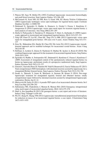
![11Incarcerated Incisional and Ventral Hernias
Micaela Piccoli, Grazia Maria Attina’, Vincenzo Trapani,
Domenico Marchi, Barbara Mullineris, and Bruno Scotto
11.1 Introduction
Laparoscopic repair of ventral and incisional hernia has gained wider acceptance
among surgeons than the open technique, due to the favorable results in terms of
incidence of recurrences, complications, and patient satisfaction. After postoper-
ative peritoneal adhesions, these diseases are the second most common cause of
occlusion of the small intestine and therefore deserve some considerations aside [1].
Complications such as incarcerations or strangulation may occur in 5 % of cases
[2, 3]. Urgent surgical procedures may be required in 5–13 % of incarcer-
ated abdominal wall hernia cases and intestinal resections may be required in
10–15 % [4].
The emergency laparoscopic treatment of incarcerated incisional and ventral
hernias seems to be one of the most interesting and debated topics [2]. Apart from
case reports and few case series, the role of the laparoscopic treatment of ven-
tral/incisional hernias in emergent situations for strangulation and/or incarceration
has not been established to date, due to the reluctance to use this approach in such
situations [2–5].
In these instances, a traditional open approach is preferred without placing a
mesh in most of the cases, aiming at the resolution of the life-threatening condition
and delaying the definitive repair. On the other hand, there are available evidences
that demonstrate the superiority of laparoscopic approach in various abdominal
emergencies [5].
M. Piccoli ( ) • G.M. Attina’ • V. Trapani • D. Marchi • B. Mullineris • B. Scotto
Dipartimento di Chirurgia. UOC di Chirurgia Generale, d’Urgenza e Nuove Tecnologie,
Nuovo Ospedale Civile Sant’Agostino Estense (NOCSAE), 41126 Modena, Italy
e-mail: m.piccoli@ausl.mo.it; graziamaria.attina@gmail.com; v.trapani@ausl.mo.it;
d.marchi@ausl.mo.it; b.mullineris@ausl.mo.it; dr.brunoscotto@gmail.com
© Springer International Publishing Switzerland 2016
F. Agresta et al. (eds.), Emergency Laparoscopy,
DOI 10.1007/978-3-319-29620-3_11
151](https://image.slidesharecdn.com/emergencylaparoscopy-191115144149/85/Emergency-laparoscopy-163-320.jpg)
![152 M. Piccoli et al.
11.2 Incarcerated Ventral and Incisional Hernia
In 2005 [6], the previous International Consensus Conference of the European
Association of Endoscopic Surgery (EAES) stated that the open approach remains
the standard treatment for incarcerated hernia, although laparoscopic surgery may be
considered in selected patients and restricted to surgeons with maximum expertise
in this field.
In 2010 [7], an Italian Consensus Conference of the main national scientific soci-
eties (SIC, ACOI, SICE, and the Italian Chapter of Hernia Society) on laparoscopic
treatment of ventral and incisional hernia underlined that the incidence of intra- and
postoperative complications and recurrences in emergency cases was the same as in
elective cases. Good experience in emergency surgery and in laparoscopic repair of
the abdominal wall in elective patients is always strictly required.
In 2012 [8], the Consensus Development Conference on Laparoscopic approach
to acute abdomen (SICE, ACOI, SIC, SICUT, SICOP, EAES) confirmed that
the laparoscopic approach to incarcerated ventral and incisional hernias may be
performed in selected patients.
In these three Consensus, the grade of recommendation was GoR C.
The objective of this study is to evaluate recent and relevant news about laparo-
scopic approach to incarcerated ventral and incisional hernia repair with respect to
previous Consensus Development Conference on “Laparoscopic approach to acute
abdomen” of 2012.
The main points were:
– Selection criteria for patients undergoing laparoscopic repair in emergency
– Surgical details
– Use of biological prosthesis in contaminated field
11.2.1 Selection Criteria for Patients Undergoing Laparoscopic
Repair in Emergency
Incarcerated or strangulated ventral/incisional hernias are due to small bowel
obstruction (SBO). In these cases, safety and feasibility of the laparoscopic
approach has been reported in several studies with various outcomes. According
to the previous recommendations (EAES 2005, Italian Consensus Conference
of laparoscopy in acute abdomen 2012, SICE 2013), laparoscopic treatment
of incarcerated or strangulated ventral/incisional hernia can be successfully
accomplished in selected patients [6–8] .](https://image.slidesharecdn.com/emergencylaparoscopy-191115144149/85/Emergency-laparoscopy-164-320.jpg)
![11 Incarcerated Incisional and Ventral Hernias 153
Either randomized control trials or prospective controlled studies are available in
the literature [9], and laparoscopy remains contraindicated in these cases [9] :
– High operative risk
– Hemodynamic instability
– Massive abdominal distension
– Perforation and/or bowel necrosis
– Limited laparoscopic experience
Best cases for laparoscopy are patients with incarcerated ventral incisional hernia
in these situations [9–11]:
• Absence of marked abdominal distension with reduced working space that
precludes entry into the peritoneal cavity. Moderate abdominal distension and
bowel distension with a diameter <5 cm are favorable situations with low risk
of conversion (Fig. 11.1). Laparoscopic management of acute SBO caused by
an incarcerated incisional hernia is challenging because of the reduced working
space due to ileus, the fragility of dilated intestinal loops, and the difficulty to
identify the cause of obstruction in some cases. Manipulation of bowel should
be done very carefully as intestinal injuries can have disastrous consequences
(conversion rates ranging from 0 % to 52 % according to surgeon’s skill and
patient selection) [9–14].
Fig. 11.1 Abdomen CT:
incarcerated ventral hernia
with moderate abdominal
distension and bowel
distension with a diameter of
4.2 cm](https://image.slidesharecdn.com/emergencylaparoscopy-191115144149/85/Emergency-laparoscopy-165-320.jpg)
![154 M. Piccoli et al.
• Some authors introduce the number (>4) of previous laparotomies as a predictive
factor of conversion [5]. However, not only the number but also the type of
previous procedure and the location of the surgical scars are very important.
As far as adhesiolysis is concerned, one surgical xipho-pubic scar following an
abdominal trauma or massive peritonitis will often give rise to more difficulties
than three scars (for example, in the right subcostal area, in the hypogastrium,and
in the right iliac region) resulting from elective and uncomplicated surgery [15];
• Absence of peritonitis with the need for bowel resection and bowel handling in a
highly inflamed environment and absence of clinical signs of intestinal ischemia
[2, 3, 5];
• Absence of high septic-risk situations, for example, concomitant execution of
contaminated abdominal procedures and the presence of contaminated skin
lesions or enterocutaneous fistulas [2, 3, 5] ;
• Absence of major defects with loss of domain or hernias that do not allow the
laparoscopic approach with adequate overlap of the mesh [2];
• Morbid obesity, old age, and debilitation are not considered contraindications to
laparoscopy [16, 17].
11.2.2 Details of Surgical Technique
The search showed no studies directly comparing different methods of adhesiolysis
and their risks, although the 2012 Italian Consensus Conference recommended
cold and sharp adhesiolysis. In an animal model, ultrasonic coagulating shears,
electrothermal bipolar vessel sealer, titanium laparoscopic clips, and plastic laparo-
scopic clips show safe hemostasis. In order to reduce the thermal damage on the
intestine, we reiterate the concept that it is safer to use cold and sharp adhesiolysis
or ultrasonic dissection [18, 19].
When an incisional hernia is found to be the cause of obstruction, all incarcerated
or strangulated intestinal contents are reduced back into the abdominal cavity and
inspected for viability [14]. The hernial contents are reduced by a combination of
manual external pressure, internal traction, and adhesiolysis.
As in open surgery, adequate traction and countertraction are the key elements
for a successful procedure, facilitating the identification of the avascular plane of
dissection [3, 13, 14, 20–22].
The best way to release the bowel is the enlargement of the hernia’s port using
careful incision of the fascia at the edge of the hernia’s ring. It is essential to use
atraumatic grasper and cold scissors close to bowels (Fig. 11.2A–D). The next step
is similar to the elective treatment: positioning of intraperitoneal mesh fixed with
tacks.
The dangerous part of the laparoscopic approach consists in the adhesiolysis
because it may lead to bowel perforation [2]. Most series reveal no differences](https://image.slidesharecdn.com/emergencylaparoscopy-191115144149/85/Emergency-laparoscopy-166-320.jpg)
![11 Incarcerated Incisional and Ventral Hernias 155
Fig. 11.2 (a–d) Laparoscopic view of the incarcerated incisional hernia of the middle line:
reduction of the intestinal contents with enlargement of the hernia’s port using careful incision
of the fascia at the edge of the hernia’s ring
between open and laparoscopic groups with respect to iatrogenic injuries to bowel.
In general, iatrogenic injuries were more likely in the presence of dense and
extensive adhesions [13]. Two bowel injury mechanisms have been described: direct
trauma by scissor with no intraoperative recognition and an indirect lesion due to
energy source with subsequent necrosis causing the perforation [15].
The need to perform proper adhesiolysis, in order to adequately identify the
hernia defect allowing the placing of the mesh, represents a crucial point [2]; this is
particularly important in the emergency setting due to the bowel distension of the
incarcerated bowel loops, associated with the vascular compromise with possible
development of necrosis and contaminated effusion and the impossibility to perform
an adequate preoperative bowel cleaning [22].
The incidence of recognized enterotomies varies from 6 to 14.3 %; the man-
agement of this condition is somewhat controversial: in many centers the hernia
procedure is terminated and the injury is repaired both laparoscopically and by
conversion to open technique and the hernia repair deferred [23]. Most recently,
some authors have reported the feasibility to repair the enterotomy laparoscopically](https://image.slidesharecdn.com/emergencylaparoscopy-191115144149/85/Emergency-laparoscopy-167-320.jpg)
![156 M. Piccoli et al.
and to complete the procedure with the application of mesh in cases where the
injury is not associated with a large amount of spillage of intestinal content [12,
20, 24]. We suggest to repair the lesions laparoscopically if the iatrogenic lesion
involves the small bowel, without fecal contamination of the abdomen, and to
proceed in positioning the mesh. If the lesion involves the colon or if there is a
fecal contamination, we prefer to suture the perforation, clean the abdomen, and
place the mesh during a second operation [3, 14, 22].
Moreover, surgeons must be very careful and check the bowel at the end of the
procedure to avoid any undiscovered lesion that, in the postoperative period, can
cause peritonitis. The peritonitis is the major complication that occurs in up to 6 %
of patient and can lead to fatal consequences involving a 0.3 % of mortality [25].
This implies a strict early postoperative follow-up to identify suspicious symptoms
and signs like fever, leukocytosis, increasing abdominal pain to establish a correct
diagnosis, and an early reoperation. Some authors identified three categories of
patients with high risk of visceral injury [5]:
1. Hernias with multiple bowel loops
2. Previous recent repair with intraperitoneal polypropylene mesh
3. Previous episodes of severe peritonitis
In the presence of incarceration under these circumstances, laparoscopy still can
be attempted. However, if release of the incarcerated bowel or release of the bowel
adherent to the peritoneum around the hernia’s defect cannot be performed safely,
conversion to open repair must be considered [13, 20].
Reasons for conversion are: 1. Reduced working space because of intestinal
distension; 2. Dense adhesions; 3. Iatrogenic perforation; 4. Bowel necrosis [3, 12,
14, 20, 26].
Iatrogenic perforation and bowel necrosis are relative reasons because they could
be managed laparoscopically [14].
The conversion is not a failure, but simply the necessary sequence of events in
the optimal management of these patients [13].
11.2.3 Surgical Experience
In several studies on laparoscopic repair in emergent situations, the authors talk
about “learning curves” and “experienced surgeon” without defining them evenly.
Studies, concerning elective surgery, suggest that the incidence of major compli-
cations such as a missed enterotomy and mortality tends to decrease as surgeon’s
experience increases after at least 50 cases [22, 25, 27]. Institutional and individ-
ual learning curves had no significant influence on clinical outcomes, although
improved experience was reflected in improved operation time, conversion rate, and
hospital stay [28].
However, this type of surgery requires experience in both laparotomic and
laparoscopic emergency surgery, as well as a comprehensive experience in elective](https://image.slidesharecdn.com/emergencylaparoscopy-191115144149/85/Emergency-laparoscopy-168-320.jpg)
![11 Incarcerated Incisional and Ventral Hernias 157
laparoscopic repair techniques. It is not possible to define the clinical competence
because of too many variables: experience in previous different operations, high/low
volume surgeon, and center.
11.2.4 Risk of Infection of the Mesh and Use of Biological Meshes
The Consensus Development Conference on laparoscopic treatment of ventral
hernias (SICE 2013) reported the following data [2, 3, 5]:
– the presence of nonviable bowel (ischemic bowel or intestinal necrosis) has not to
be considered as a contraindication for prosthetic repair and the immediate mesh
repair is preferably deferred only in cases of abundant peritoneal contamination
[11] .
– The biological meshes provide a new prospective in the contaminated surgical
field, in laparoscopic emergency hernia repair too, with good results in terms
of recurrence and wound infection [12]. Nowadays, even if their laparoscopic
implant is feasible, the use of biological prosthesis should be restricted to
contaminated field in open surgery. Their laparoscopic use is recommended in
controlled trials [29, 30].
The introduction of such materials in clinical practice has provided a new
prospective for abdominal wall defect repair in contaminated surgical field [31].
Few authors suggest the use of biomaterial in the laparoscopic emergency hernia
repair with good results in terms of recurrence rate and wound infection [26,
29]. There are no comparative trials evaluating each of the commercially available
biological mesh products and their application in laparoscopic repair of potentially
contaminated ventral hernias. Further studies are still required to establish the
efficacy of biological mesh in laparoscopic repair in potentially contaminated
fields.
References
1. Ghezzi TL, Moschetti L, Corleta OC, Abreu GP, Abreu LP (2010) Analysis of the videola-
paroscopy potentiality in the surgical treatment of the bowel obstruction. Arq Gastroenterol
47(2):148–151
2. Olmi S, Cesana G, Eba L, Croce E (2009) Emergency laparoscopic treatment of acute
incarcerated incisional hernia. Hernia 3:605–608
3. Shah RH, Sharma A, Khullar R, Soni V, Baijal M, Chowbey PK (2008) Laparoscopic repair of
incarcerated ventral abdominal wall hernia. Hernia 12:457–463
4. Derici H, Unalp HR, Bozdag AD et al (2007) Factors affecting morbidity and mortality in
incarcerated abdominal wall hernias. Hernia 11:341–346
5. Landau O, Kyzer S (2004) Emergent laparoscopic repair of incarcerated incisional and ventral
hernia. Surg Endosc 18:1374–1376
6. Sauerland S et al (2006) Emergent laparoscopic repair of incarcerated incisional and ventral
hernia. Surg Endosc 20(1):14–29](https://image.slidesharecdn.com/emergencylaparoscopy-191115144149/85/Emergency-laparoscopy-169-320.jpg)
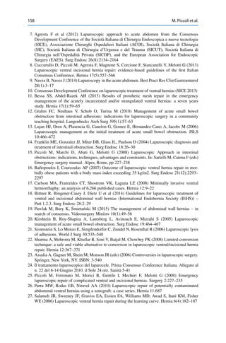

![12Emergency Laparoscopy for Abdominal
Trauma
Salomone Di Saverio, Arianna Birindelli, Andrea Biscardi,
Matteo Mandrioli, Luca Di Donato, Antonio Tarasconi,
Roberto Cirocchi, Carlo Coniglio, Giovanni Gordini,
and Gregorio Tugnoli
12.1 Introduction
Although the first experiences of trauma laparoscopy were reported in the early
1920s [1], the technique did not become immediately popular due to concerns on the
effects of pneumoperitoneum on such delicate patients and because of the relative
inexperience in this procedure and limited laparoscopic skills among the surgeons.
Starting from the second part of the twentieth century, laparoscopy has been
increasingly used in elective surgery, and, from the beginning of the twenty-first
century, it has been introduced also in emergency surgery [2] and finally in trauma
surgery.
Trauma patients are particularly frail since most of them have multiple organ
injuries. Several advantages of the minimally invasive approach such as decreased
postoperative pain and less adhesion formation, faster recovery and shorter length of
Author’s Contribution: AB and SDS wrote the chapter; AB searched and revised the literature; SDS
has described the surgical techniques and performed all laparoscopic procedures; SDS provided
patients materials and pictures; all authors critically revised the chapter and gave the final approval
of manuscript.
S. Di Saverio, MD, FACS, FRCS ( ) • A. Birindelli, MD • A. Biscardi, MD • M. Mandrioli, MD
• L. Di Donato, MD • A. Tarasconi, MD • G. Tugnoli, MD, PhD
Maggiore Hospital Regional Emergency Surgery and Trauma Center – Bologna Local Health
District, AUSL Bologna, Bologna, Italy
e-mail: salo75@inwind.it; salomone.disaverio@gmail.com
R. Cirocchi, MD
Department of Surgery, Hospital of Terni, University of Perugia, Perugia, Italy
C. Coniglio, MD • G. Gordini, MD, PhD, MPH
Trauma ICU, Trauma Center – Department of Emergency, Maggiore Hospital – Bologna Local
Health District, Bologna, Italy
© Springer International Publishing Switzerland 2016
F. Agresta et al. (eds.), Emergency Laparoscopy,
DOI 10.1007/978-3-319-29620-3_12
161](https://image.slidesharecdn.com/emergencylaparoscopy-191115144149/85/Emergency-laparoscopy-172-320.jpg)
![162 S. Di Saverio et al.
hospital stay, earlier return to work and daily physical activities, fewer postoperative
wound infections, incisional hernias, and better cosmetic results are already well
known and are easily reproducible also in the setting of trauma patients. More-
over, in the injured patients the use of minimally invasive surgery is particularly
valuable since it is able to decrease the systemic inflammatory response with lower
risk of secondary organ injury (especially lungs and kidneys) [3, 4]. Diagnostic
laparoscopy seems to significantly reduce the rate of negative laparotomies [5–14],
having high sensitivity, specificity, and accuracy [6, 7, 9, 13, 15, 16], especially
in penetrating abdominal trauma, with better cost-effectiveness when compared to
open surgery [12, 13, 17].
Despite these potential benefits, laparoscopy has not yet gained widespread
acceptance within the trauma community and its role in this area is still under
strong debate. The most relevant criticisms have been raised are about the longer
time needed in laparoscopy to set up the equipment, enter the abdomen, and control
the injury but also the risk of missed injuries that are particularly high if the surgeon
has minimal experience in minimally invasive surgery.
Provided a careful selection of patients and advanced laparoscopic surgical
training, this technique can be then extremely useful in trauma patients both as a
diagnostic and, eventually, a therapeutic tool.
12.2 Diagnostic Laparoscopy
Laparoscopy may be an accurate diagnostic tool for abdominal trauma, having
nowadays replaced Diagnostic Peritoneal Lavage (DPL) being much more accurate
and representing as well a minimally invasive alternative to exploratory laparotomy.
Laparoscopy revealed to be particularly useful in the diagnosis of intra-
abdominal organ injuries for hemodynamically stable trauma patients with doubtful
clinical or imaging findings and it has been already demonstrated to be able to
greatly decrease the rate of nontherapeutic laparotomies, with obvious benefits for
both the patient’s health and cosmesis and the economy, in terms of morbidity,
length of stay, and return to work [6, 9, 11, 12, 14, 17–23]. Despite the clear
advantages of the procedure, it is very important to perform it only in correctly
selected patients and by experienced and laparoscopically skilled surgeons [7, 9,
10, 18, 19, 24, 25].
12.2.1 Patient Selection
The first step in order to safely perform a diagnostic laparoscopy in trauma patients
is the correct selection of candidates.
First of all, the absolute need of hemodynamic stability is widely accepted,
in terms of selecting as candidates for trauma laparoscopy those patients
having hypovolemic shock no higher than ATLS class I (see Table 12.1) or
patients fully responders to initial small-volume fluid bolus administration. Class](https://image.slidesharecdn.com/emergencylaparoscopy-191115144149/85/Emergency-laparoscopy-173-320.jpg)
![12 Emergency Laparoscopy for Abdominal Trauma 163
Table 12.1 ATLS classification of hemorrhagic shock
Class I Class II Class III Class IV
Blood loss (ml) <750 750–1500 1500–2000 >2000
Pulse rate (per min) <100 >100 >120 >140
Blood pressure Normal Normal Decreased Decreased
Pulse pressure
(mmHg)
Normal Decreased Decreased Decreased
Respiratory rate (per
min)
14–20 20–30 30–40 >40
Urine output (ml/h) >30 20–30 5–15 Negligible
Central venous
system (mental status)
Slightly anxious Mildly anxious Anxious confused Lethargic
II–III–IV hypovolemic shock patients, transient or nonresponders to fluid
resuscitation, septic shock patients, or those with cardiorespiratory contraindi-
cations to pneumoperitoneum are not suitable for laparoscopy. Another absolute
contraindication to laparoscopy is a severe traumatic brain injury, since it has been
reported that pneumoperitoneum may increase the intracerebral pressure [26].
FAST US followed by a total body CT scan is recommended in every stable
patient with blunt abdominal trauma, or with penetrating abdominal trauma after
gunshot wound or after stab wound on the lower quadrants or on the back, in order
to assess the site and extent of injuries. However, it has been demonstrated that both
US and CT scan have low sensitivity and specificity in diagnosing diaphragmatic,
mesentery, and hollow viscus injuries [27, 28], with significant negative impact on
patient outcome. On the other hand, laparoscopy has been reported to be highly
sensitive and specific as well as accurate, in the presence of appropriate surgical
skills [7, 9, 13, 19].
Diagnostic laparoscopy is indicated either in blunt or penetrating abdominal
trauma, in case of CT scan equivocal findings or discrepancy between the clinical
examination and the imaging, the so-called unclear abdomen, but also in case of
unclear source of potentially active bleeding.
Another common indication to diagnostic laparoscopy in blunt trauma is the
finding of unexplained free abdominal fluid with the suspicion of a hollow viscus
injury or a mesenteric laceration on a CT scan.
In case of penetrating trauma, laparoscopy can be used to investigate the
presence of abdominal free fluid in the absence of parenchymal injuries and to
rule out the suspicion of peritoneal penetration (Fig. 12.1), hollow viscus injury,
or diaphragmatic injury (Fig. 12.2). A further indication for diagnostic laparoscopy
in penetrating abdominal trauma is the suspicion of an occult diaphragmatic
injury, even in the presence of a spleen or liver injury successfully managed with
observation C/ angioembolization.
In case of a stable patient with isolated penetrating abdominal trauma after stab
wound of the anterior and upper quadrants and a suspected diaphragmatic injury,
the CT scan can even be avoided and a straight diagnostic laparoscopy may be
performed.](https://image.slidesharecdn.com/emergencylaparoscopy-191115144149/85/Emergency-laparoscopy-174-320.jpg)
![164 S. Di Saverio et al.
Fig. 12.1 Laparoscopic
diagnosis of peritoneal
violation after a penetrating
left flank trauma
(intraoperative picture
courtesy from Dr. S. Di
Saverio surgical procedures
logbook)
Fig. 12.2 Laparoscopic
diagnosis of a left
diaphragmatic tear after an
abdominal penetrating
trauma, note the thoracic
drain (intraoperative picture
courtesy from Dr. S. Di
Saverio surgical procedures
logbook)
In a later stage, laparoscopy can be useful to diagnose a post-traumatic acute
pancreatitis development.
In pediatric trauma, where the physical examination is often unreliable and the
radiation exposure should be avoided or minimized, diagnostic laparoscopy can be
particularly useful and safe. Similar indications can be seen in pregnant patients and
in geriatric or mentally impaired.
Large vessel, retroperitoneal, and renal injuries should not be diagnosed laparo-
scopically as their diagnosis is easier and more reliable with three-phase contrast-
enhanced CT scan, including urogram.
12.2.2 Surgical Technique
Many studies have already stated the high efficacy of diagnostic laparoscopy in
precisely recognizing the abdominal injuries, as well as significantly reducing the
rate of nontherapeutic laparotomies for trauma up to 73 % [15]. However, in order
to perform a correct and safe diagnosis and avoid missed injuries, it is extremely
important that the surgeon is properly trained and confident in laparoscopy [7, 9,
10, 18, 19, 24, 25]. Both the presence of appropriate support and collaboration of
the operating room staff and the availability of proper theater equipment play an
important role in performing such procedures.](https://image.slidesharecdn.com/emergencylaparoscopy-191115144149/85/Emergency-laparoscopy-175-320.jpg)
![12 Emergency Laparoscopy for Abdominal Trauma 165
Apart from occasional experiences of local anesthesia with intravenous sedation
(“awake laparoscopy”) in emergency department [29], diagnostic laparoscopy for
trauma is usually performed under general anesthesia.
The patient should be in a supine position, the same as for a trauma open
approach in order to be prepared for immediate conversion to laparotomy or/and
thoracotomy, in case of abdominal injuries that cannot be treated laparoscopically
or sudden onset of hemodynamic instability or suspicion of thoracic injuries. In
addition, as for every laparoscopic procedure, the patient should be fully secured and
tight with belts on every side, in order to be able to safely achieve a moderate tilt in
all directions and shift of the abdominal organs during the exploration to guarantee
an adequate laparoscopic intraoperative view and exposure. A further reason for
using supine position with legs straight and extended is because trauma patients
often have concomitant orthopedic fractures involving the pelvis and/or the long
bones of the extremities.
The first access is best achieved at the navel with an open technique (we
do not recommend using Verres needle in trauma patients who may have a
distended bowel) using a 10/12 mm trocar. Pneumoperitoneum should be slowly
and progressively established, avoiding hyperpressure (reaching no more than 12–
14 mmHg), and insufflation should be suddenly interrupted in case of abrupt rise
in respiratory pressure, blood hypotension, or tachycardia. If the patient’s stability
rapidly restores, laparoscopy can be carefully and slowly resumed. Either 30ı
or
0ı
laparoscope can be used in diagnostic procedures, whereas 30ı
scope is better
performing in case of operative laparoscopy, in our experience. Once preliminary
exploration of the abdominal cavity has shown no urgent need to convert to
laparotomy, we suggest to insert two further trocars (5 or 12 mm), preferably
bladeless, under direct vision in left-upper and lower quadrants, in order to start
a systematic exploration of the abdomen and run the entire bowel. Two more trocars
in the right quadrants can be added in order to explore the left quadrants, if needed.
In penetrating injuries, peritoneal violation (Fig. 12.1) and diaphragmatic injuries
(Fig. 12.2) can be reliably determined with laparoscopy. A diaphragmatic injury can
occur also after a blunt thoracoabdominal trauma (Fig. 12.3). Similarly to what is
usually done in the open approach, the peritoneal cavity is examined systematically,
Fig. 12.3 Laparoscopic
diagnosis of a right
diaphragmatic large tear after
an abdominal blunt trauma;
the thoracic drain can be seen
in the chest (intraoperative
picture courtesy from Dr. S.
Di Saverio surgical
procedures logbook)](https://image.slidesharecdn.com/emergencylaparoscopy-191115144149/85/Emergency-laparoscopy-176-320.jpg)
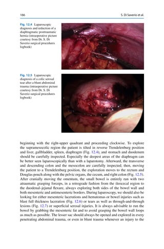
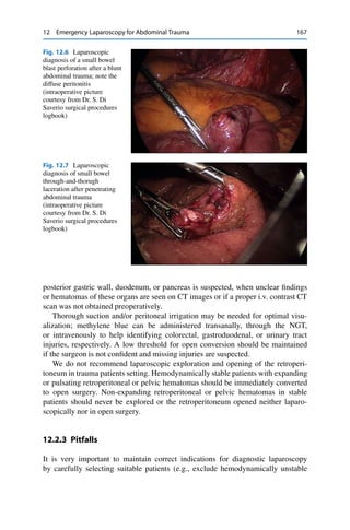
![168 S. Di Saverio et al.
patients) but also performing this procedure only in the presence of a properly
trained and experienced operator and having carefully considered the accuracy of
diagnostic alternatives such as CT scan and angiography.
Diagnostic laparoscopy-related complications are reported up to 2.8 % of patients
[7]. The most common complication of laparoscopy is tension pneumothorax due
to pneumoperitoneum in patients with unexpected diaphragmatic injuries. This
condition can be easily treated with the suture of the injury and placement of
a thoracostomy tube. Other complications might be iatrogenic perforation of a
hollow viscus, laceration of a solid organ, and vascular injuries during trocar
insertion or abdominal exploration and bowel manipulation. Another possible,
although rare, complication is gas embolism in patients with intra-abdominal
venous injuries, especially in liver lacerations; the transperitoneal absorption of
carbon dioxide (which may cause metabolic and hemodynamic changes such as
acidosis, cardiac suppression, atelectasis, subcutaneous emphysema, and increased
intracranial pressure) may lead to life-threatening consequences.
Missing injuries are the most feared complication of diagnostic laparoscopy
and range between 0 % [8] and 77 % [10]: its rate is directly proportional to the
surgeon’s laparoscopic expertise and it can be significantly reduced if the operator
adheres to a standardized laparoscopic exploration technique and does not hesitate
to convert to open surgery if he/she does not feel confident in the diagnostic
procedure performed.
12.3 Therapeutic Laparoscopy
Once the diagnosis is achieved, either by CT scan or by diagnostic laparoscopy, the
possibility of performing a laparoscopic treatment depends on the type and extent
of the injuries, on the patient’s hemodynamic stability, and, last but not least, on
the surgeon’s technical laparoscopic skills. The laparoscopic treatment of traumatic
injuries provides all the advantages of the minimally invasive approach. These
benefits are particularly remarkable in trauma patients. In these patients in fact,
inflicting less trauma by using laparoscopy may represent a significant potential for
decreasing the further release of proinflammatory elements with secondary injury to
lungs and kidneys, in addition to what is already caused by their primary traumatic
injury [3, 4].
12.3.1 Patient Selection
As already stated in the section on diagnostic laparoscopy, also in therapeutic
laparoscopy the first condition required is the absolute patient’s hemodynamic
stability, the absence of severe cardiorespiratory contraindications to laparoscopy,
and the absence of severe brain traumatic injury. Further conditions for using
therapeutic laparoscopy in approaching abdominal trauma are the experience of the
surgeon with advanced laparoscopic skills and being confident in his/her ability to](https://image.slidesharecdn.com/emergencylaparoscopy-191115144149/85/Emergency-laparoscopy-179-320.jpg)
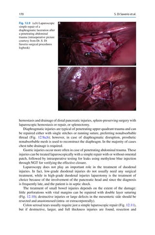
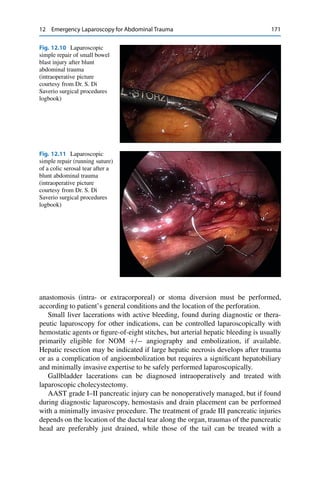
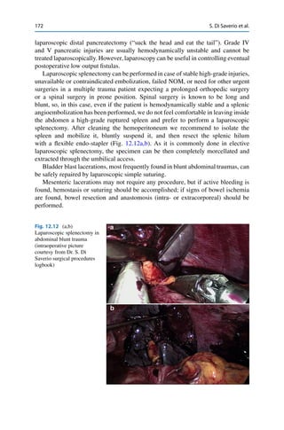
![12 Emergency Laparoscopy for Abdominal Trauma 173
Minor vascular injuries can be laparoscopically treated with coagulation, hemo-
statics, hemoclips, or suturing.
12.3.3 Pitfalls
Before attempting a laparoscopic treatment of arterial bleeding, we suggest to
always keep in mind the chance of better treating this type of injuries with
angioembolization, if available, and eventually consider a laparoscopic approach
only if this first option fails and if the patient remains perfectly hemodynamically
stable. Large vessel injuries are usually associated with hemodynamic instability
and there is no role at all for laparoscopy in such patients.
Once again, we recommend avoiding to explore laparoscopically the retroperi-
toneal traumatic injuries.
The operator should be particularly careful avoiding iatrogenic vascular or
intestinal injuries, especially when handling small bowel loops or using coagulating
devices which can cause intestinal thermal injuries. We recommend to convert to
open surgery as soon as the operator feels he/she loses the control of the injuries
or is not sure of the complete and safe assessment of the whole abdomen. The rate
of treatment success without iatrogenic injuries is directly related to the surgeon’s
experience.
12.4 Literature Review
In recent years, trauma laparoscopy has increasingly gained interest and a large
number of studies have been published in the literature. However, due to the acute
and complex type of patients, most of the series are retrospective and the data quality
is limited and inhomogeneous.
Only two RCTs on trauma patients were ever performed: the first one compared
laparoscopy with peritoneal lavage and found higher diagnostic specificity in the
laparoscopic group [30], while the second one found a significant reduction of
nontherapeutic laparotomy rate (p D .016) and hospital length of stay (p D .049) in
patients with abdominal penetrating trauma managed with diagnostic laparoscopy
if compared to laparotomy [5].
The most widely accepted and reported indication to diagnostic laparoscopy is
the assessment of occult diaphragmatic injury in penetrating left-upper quadrant
traumas, since radiological findings are usually not reliable in predicting diaphrag-
matic injury, which occur in up to 30 % of such patients [31]. This type of injury
can usually be successfully repaired laparoscopically [32].
In the retrospective study by Chestovich et al., with a total of 518 patients with
penetrating trauma, in the laparoscopic group the length of stay has been found to
be shorter (p < .001) and there were no missed injuries; on the other hand, wound
infections (p D .002) and ileus and small bowel obstruction (p D .018) were more
common with the open procedure [8].](https://image.slidesharecdn.com/emergencylaparoscopy-191115144149/85/Emergency-laparoscopy-184-320.jpg)
![174 S. Di Saverio et al.
Similar results were reported on a large Russian population who underwent
laparoscopic surgery for penetrating trauma, with quicker recovery time, less pain,
shorter hospital stay, and lower complication rate (p < .0001) compared to open
surgery, with no missed injuries [12].
According to the European Association for Endoscopic Surgery, laparoscopy
may be useful in hemodynamically stable patients with abdominal trauma (with
a B and C grade of recommendation for penetrating and blunt trauma, respectively)
[33].
Diagnostic laparoscopy has been included in the Eastern Association for the
Surgery of Trauma Guidelines in penetrating abdominal trauma, when there is a
suspicion of intraperitoneal penetration or diaphragmatic injury (Class 2 recom-
mendation) [34].
On the other hand, conflicting results are from a multicenter, institutional review
board-approved prospective study, which enrolled 160 stable patients with anterior
abdominal stab wounds. In this study, only nine laparoscopic explorations were
performed and it did not appear that the minimally invasive approach had a role
in management of this kind of patients and was not cost-effective [35]. Also in
the Leppaniemi prospective randomized study of 43 patients with abdominal stab
wounds, no statistically significant difference in the total hospital costs of the
two strategies was found [5], but a retrospective cost analysis comparing the total
hospital costs of exploratory laparotomy versus diagnostic laparotomy in 37 patients
with penetrating abdominal trauma showed that laparoscopy was 1136 AC cheaper
than exploratory laparotomy [17].
One large series (131 patients) of both penetrating and blunt abdominal traumas
from a Level 1 Trauma Center confirmed the diagnostic efficacy and safety of
laparoscopy, with a rate of nontherapeutic laparotomy for patients sustaining
penetrating vs. blunt injury of 1.8 % and nil, respectively [20].
The largest study ever published analyzed the United States National Trauma
Data Bank between 2007 and 2010 and identified 4755 patients, with either
penetrating or blunt trauma, who underwent a diagnostic laparoscopy at 467 trauma
centers. A therapeutic laparoscopic intervention was performed in 916 (19.3 %)
patients and the conversion to open surgery rate was 20.2 % (961 patients), of which
581 laparotomies were therapeutic. The most common laparoscopic procedure
performed was repair of the diaphragm, followed by gastrostomies, large bowel
repairs, small bowel repairs, liver laceration repairs, splenectomies, and small and
large bowel resections. Mortality was not statistically different between the open
and the laparoscopic groups, but patients undergoing laparoscopic surgery had a
significantly shorter length of stay (5 vs. 6 days; p < .001) [23].
A recent systematic review on laparoscopy in penetrating abdominal trauma
showed a range of sensitivity from 66.7 % to 100 %, specificity from 33.3 %
to 100 %, and accuracy from 50 % to 100 % and confirmed its role in avoiding
negative laparotomy, decreasing morbidity, shortening hospital stay, and increasing
cost-effectiveness [16].
In a previous systematic review by O’Malley et al. including 51 studies (13
prospective) with 2569 patients who underwent diagnostic laparoscopy for penetrat-](https://image.slidesharecdn.com/emergencylaparoscopy-191115144149/85/Emergency-laparoscopy-185-320.jpg)
![12 Emergency Laparoscopy for Abdominal Trauma 175
ing abdominal trauma, 44 % were positive for intra-abdominal injuries, 13.8 % had
a therapeutic laparoscopy, and 58.3 % were spared a nontherapeutic laparotomy.
Overall, 72 patients (2.8 %) suffered complications, there were three mortalities
(0.1 %), and 83 missed injuries (3.2 %). Sensitivity ranged from 66.7 to 100 %,
specificity from 33.3 to 100 %, and accuracy from 50 to 100 %. Twenty-three of
the 51 studies reported sensitivity, specificity, and accuracy of 100 %. The authors
concluded that laparoscopy may have an important role in selected patients with
penetrating abdominal trauma, especially with diaphragmatic injuries, and that the
surgeon expertise plays an important role [7]. According to Lin et al., the surgeon’s
ability and experience is described as at least 20 laparoscopic procedures per month
in acute care patients [6].
The systematic review on the use of laparoscopy in blunt abdominal trauma by
Nicolau included 66 articles for a total of 343 patients and demonstrated a 0 %
missed injury rate [36].
The use of laparoscopy for trauma in specific conditions like pregnancy and
pediatric patients has been also reported in the last years [37–40], but deserves
further investigation.
In conclusion, the current evidence shows that laparoscopy in selected hemody-
namically stable trauma patients can significantly decrease nontherapeutic laparo-
tomy rates, length of stay, and hospital costs and is highly sensitive and specific
with very low missed injury rates, but adherence to a standardized laparoscopic
examination technique and operator experience with routine use of laparoscopy
in elective or acute care practice strongly influence positive results of minimally
invasive surgery in trauma. Provided adequate surgical advanced skills, laparoscopy
has been reported with impressive results in the treatment of a breadth of abdominal
injuries [23].
To optimize results, the laparoscopy for trauma patients should be added to
diagnostic and treatment algorithms at institutions where appropriate levels of
laparoscopic expertise are available.
References
1. Short AR (1925) The uses of celioscopy. BMJ 2:254–255
2. Di Saverio S (2014) Emergency laparoscopy: a new emerging discipline for treating abdominal
emergencies attempting to minimize costs and invasiveness and maximize outcomes and
patients’ comfort. J Trauma Acute Care Surg 77(2):338–350
3. Balogh ZJ, Reumann MK, Gruen RL, Mayer-Kuckuk P, Schuetz MA, Harris IA, Gabbe BJ,
Bhandari M (2012) Advances and future directions for management of trauma patients with
musculoskeletal injuries. Lancet 380(9847):1109Y1119
4. Manfredi AA, Rovere-Querini P (2010) The mitochondrion–a Trojan horse that kicks off
inflammation? N Engl J Med 362(22):2132–2134
5. Leppaniemi A, Haapiainen R (2003) Diagnostic laparoscopy in abdominal stab wounds: a
prospective, randomized study. J Trauma 55(4):636–645
6. Lin HF, Wu JM, Tu CC et al (2010) Value of diagnostic and therapeutic laparoscopy for
abdominal stab wounds. World J Surg 34(7):1653–1662](https://image.slidesharecdn.com/emergencylaparoscopy-191115144149/85/Emergency-laparoscopy-186-320.jpg)
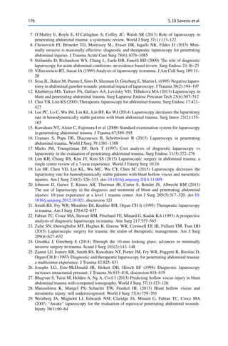
![13Update in Laparoscopic Approach to Acute
Mesenteric Ischemia
Gianfranco Cocorullo, Nicola Falco, Tommaso Fontana,
Roberta Tutino, Giuseppe Salamone, and Gaspare Gulotta
13.1 Introduction
The laparoscopic approach to Acute Mesenteric Ischemia is today unclear and less
debated (AMI).
There are in fact no clinical evidences on this particular focus and only few
articles can be found in several databases (pubmed, cochrane library, etc.), and the
problem concerns both diagnostic and therapeutic utilization of the procedure.
These considerations were already taken into account in 2012 EAES guidelines
where the Grade of Recommendation (GoR) of laparoscopy in AMI was low in both
diagnostic and therapeutic aspects.
According to the new Oxford Classification [1], the use of laparoscopy in patients
with suspicious or diagnosed AMI presents a weak GoR.
13.2 Epidemiology
AMI is an uncommon but serious disease often associated with a bad prognosis,
accounting <1 in every 1000 hospital admissions. Women are more affected than
men with 3:1 ratio. Epidemiological study attributes this difference to higher
longevity of females vs. males. Clinical risk factors may provide some clues for
AMI diagnosis. Patients with a history of atrial fibrillation, recent myocardial
infarction, congestive heart failure, or peripheral arterial embolism have an higher
risk for Superior Mesenteric Artery (SMA) embolism. History of postprandial
abdominal pain, weight loss, and food intolerance in patients with acute abdominal
G. Cocorullo ( ) • N. Falco • T. Fontana • R. Tutino • G. Salamone • G. Gulotta
Department of Surgical Oncological and Stomatological Sciences, Unit of General
and Emergency Surgery, University of Palermo, Palermo, Italy
e-mail: gianfranco.cocorullo@unipa.it
© Springer International Publishing Switzerland 2016
F. Agresta et al. (eds.), Emergency Laparoscopy,
DOI 10.1007/978-3-319-29620-3_13
179](https://image.slidesharecdn.com/emergencylaparoscopy-191115144149/85/Emergency-laparoscopy-189-320.jpg)
![180 G. Cocorullo et al.
pain could indicate an acute thrombotic occlusion of SMA. AMI in fact is associated
with occlusion of SMA for arterial embolism or thrombosis in 67.2 % of cases,
while mesenteric venous thrombosis occurs in 15.7 %. Only in remaining patients
it is possible to find the third form of AMI without vessel obstruction, the so-called
nonocclusive mesenteric ischemia (NOMI) [2].
13.3 Diagnosis
The diagnosis of AMI represent a very difficult diagnostic enigma; clinical markers
are often aspecific but according to the literature severe and diffuse abdominal pain,
bowel emptying, risk factor of embolism, arterial or venous thrombosis, and history
of prior undiagnosed attacks of abdominal angina associated with specific laboratory
tests (persistent pH < 7.34; plasmatic lactate level: 2.1 mmol/L, WBC: 12,000/mmc)
could be highly suggestive of AMI [3].
According to the EAES guidelines and the latest articles of International
literature, the gold standard for diagnosis of AMI is multidetector CT Angiography
(CTA) with a sensibility of 93.3 % and a specificity of 95.9 % (Fig. 13.1) [4, 5].
In a recent review of more than 6000 patients, Acosta [5] underlines the
secondary role of explorative laparotomy/laparoscopy to confirm cases of AMI
without signs of SMA occlusion at CTA. Through the abdominal exploration, in
fact, the surgeon could notice bowel aspect, its dilatation and peristaltism, the
presence of evident pulsations in the mesenteric arcade arteries, and the bleeding
from cut surfaces; the surgeon, moreover, could observe bowel wall necrosis and/or
peritonitis in late form of AMI (Fig. 13.2).
Only few additional reports have been found on the diagnostic role of explorative
laparoscopy. The laparoscopic diagnosis of AMI is clearly influenced by the
stage of bowel damage, the etiology of the ischemia, and the experience of the
surgeon. In fact, the ability to recognize signs of bowel ischemia depends on the
Fig. 13.1 CT angiography](https://image.slidesharecdn.com/emergencylaparoscopy-191115144149/85/Emergency-laparoscopy-190-320.jpg)
![13 Update in Laparoscopic Approach to Acute Mesenteric Ischemia 181
Fig. 13.2 Ischemic bowel
Fig. 13.3 Laparoscopic
abdominal exploration
single operator’s specific medical records and on the level of his learning curve,
considering also that a low pressure of CO2 pneumoperitoneum is advised if there
is a suspicious AMI (Fig. 13.3).
In this way, since the first application of fluorescein proposed in 1993 by Kam
and Scheeres to increase the sensibility of laparoscopy, several studies on canine or
porcine models were proposed [6]. However, only few evidences are collected from
these studies and in particular Paral Jiri et al. (2007) confirmed on porcine model
the feasibility of this procedure using the specific optical system in the follow-up of
AMI patients. Moreover, there are no trials or perspective studies on humans. The
article of Paral represents the last important experience in the use of fluorescein, but](https://image.slidesharecdn.com/emergencylaparoscopy-191115144149/85/Emergency-laparoscopy-191-320.jpg)
![182 G. Cocorullo et al.
the related level of evidence (LE) in the EAES guidelines, which was 2b in 2012, has
been changed to LE 3 according to 2011 Oxford classification [7]. Unfortunately, in
the last 2 years no studies have been produced on the primary diagnostic role of the
laparoscopy and so the procedure maintains a weak GoR.
Most studies, instead, underline the possible role of laparoscopy with or without
fluorescein in second-look exploration as “damage control surgery” [3, 5]. Nev-
ertheless, in 2012, was published a study [3] that underlines the feasibility of a
diagnostic laparoscopy in AMI in patients with Aortic Dissection (AoD). According
to Tshomba Y et al that in patients with suspicious clinical signs of AMI without
peritonitis if there are no conditions for rapid execution of CTA or a previous
CTA had been not conclusive, an early diagnosis might be better than a clearer
but late diagnosis. In this way, laparoscopy can reduce the number of unnecessary
laparotomies overall in elderly critically ill patients. The study of Tshomba et al. [3]
confirms that the best of laparoscopy in AMI diagnosis is the second look with the
possibility of bedside use (directly in ICU). The author observed 202 consecutive
AoD treated in the same center (71 acute type A AoD; 131 acute and chronic type
B AoD) and only in 17 patients AMI was suspected. Diagnostic laparoscopy (DL)
was performed in nine cases, in three cases during the medical treatment and in six
patients after surgical or endovascular revascularization. In Tshomba’s experience
eight of nine DL were negative, but in one case AMI was diagnostic and the patient
underwent revascularization in emergency. This experience is important, but it is
based on few patients and so more studies need to confirm the feasibility and
safeness of DL in AMI secondary to AoD; according to Tshomba the diagnostic
power of DL increases in the second look overall in patients with aortic dissection
type B (chronic type). In these cases, in fact, usually little time has been spent
between starting of symptoms related to ischemic event and the evidence of bowel
wall ischemia; therefore, it is known that sensitivity of diagnostic laparoscopy
increases 24 hours after ischemic event. Moreover, according to the same article
laparoscopy may be a feasible alternative to CTA in patients with kidney failure or
other contraindications to injection of iodate CT contrast medium [3].
13.4 Treatment
Wyers in 2010 underlines that the first treatment of AMI is bowel revascularization.
The second step is the reassessment of bowel viability. If possible the time of 20
or 30 min after revascularization should be spent before decision making about via-
bility to evaluate signs of adequate perfusion as mesenteric vessel pulsation, normal
color and appearance of the bowel serosa, peristalsis, and bleeding from cut surfaces
[2]. Moreover, if an explorative diagnostic laparotomy (or laparoscopy) is performed
as the first diagnostic step, an on-table SMA angiography should be performed at the
same time. If no vascular surgeon is available, resection of obvious necrotic bowel
should be performed and after the abdomen closure the patient should be transported
to a vascular surgical center [5]. Main techniques to resolve the SMA occlusion
are open SMA embolectomy for SMA embolism; other therapeutic options are](https://image.slidesharecdn.com/emergencylaparoscopy-191115144149/85/Emergency-laparoscopy-192-320.jpg)
![13 Update in Laparoscopic Approach to Acute Mesenteric Ischemia 183
hybrid or endovascular stenting in acute thrombotic occlusions, medical therapy
with vasodilators and high dose of EBPM for thrombotic case, NOMI, and venous
infarction [2].
In a limited number of cases when primary laparoscopy access is feasible
and revascularization has been performed with endovascular access, it is possible
to evaluate bowel perfusion laparoscopically and to perform at the same time a
laparoscopical bowel resection of residual ischemic segments. The possibility of
performing a “one-step anastomosis” will be evaluated each time by the surgeon,
depending on the bowel presentation and the performance status (ASA score) of the
patient. In nonpermissive patients, the surgeon should choose a two-step operation
differing in performing anastomosis [8]. However, as suggested by the experience
of Meriggi F et al. in their recent (2011) case report, laparoscopic primary access
overall in AoD is an important tool for leading therapeutic decision and timing
[9] (LE 4). It is clear that unfortunately there are no strong evidences about the
therapeutic role of laparoscopy in AMI because only few experiences are published
referring to a single center or more frequently to a single case report. More resources
and efforts are needed in the evaluation of laparoscopic usefulness in AMI.
13.5 Follow-Up and Damage Control Surgery
Following the rule of damage control surgery a CTA is indicated for patients who
underwent a stent positioning and it should be performed after the revascularization.
The monitoring of restored bowel viability (second look) should be repeated two
times in the postoperative time. Moreover, the second look is frequently performed
also after surgical resection of necrotic bowel because an extension of ischemia is
not rare in these patients and it requires further bowel resection. According to the
literature in both cases a laparoscopic close exploration should represent a valid and
more feasible technique than to perform an unsutured abdomen closure (Bogotà
Bag, VA system, etc.) [5].
13.6 Conclusions
The gold standard for the primary diagnosis is CTA and laparoscopy has a limited
rule. Fluorescein does not increase significantly the sensibility and specificity of
the laparoscopy. The necessity to perform a low-pressure laparoscopy limits the
diagnostic power of the procedure and reduces the feasibility and the diffusion of
its execution. Nevertheless, the literature underlines the feasibility of a diagnostic
laparoscopy in AMI after AoD when in the presence of suggestive clinical signs of
AMI without peritonitis there are no conditions for the rapid execution of CTA or
when a previous CTA had been nonconclusive. Laparoscopy may result in a feasible
alternative to CTA also in patients with IRC that contraindicates injection of iodate
CT contrast medium. Furthermore, laparoscopic primary access overall in AoD is
an important tool for leading therapeutic decision and timing. The main rule of](https://image.slidesharecdn.com/emergencylaparoscopy-191115144149/85/Emergency-laparoscopy-193-320.jpg)
![14Anaesthesia and Emergency Laparoscopy
Riccardo Ragazzi, Marco Tartaglione, Savino Spadaro,
Antonio Gioia, and Carlo Alberto Volta
14.1 The Pathophysiology of Pneumoperitoneum
Pneumoperitoneum [1] is the main cause of all the systemic pathophysiologi-
cal changes that take place during laparoscopic surgery. Understanding normal
variations allows us to understand some conditions in which it is advisable to
avoid or interrupt a laparoscopic procedure and, secondly, to understand which
anaesthesiological strategies are useful in managing critical patients.
To induce pneumoperitoneum, carbon dioxide is usually preferred over other
gases [2] (azote, nitric oxide, helium, argon, or oxygen) due to its high level of
solubility in the circulatory system, together with its inability to feed combustion, its
low peritoneal absorption, and the availability of a chemical buffer in our bodies—
bicarbonate—thatneutralises it and allows it to be eliminated through the respiratory
system.
When inducing pneumoperitoneum, carbon dioxide is insufflated into the peri-
toneal cavity at a speed of 4–6 l/min and a pressure of 10–20 mmHg. It is later
maintained by a constant flow of 200–400 ml/min.
‘Carboperitoneum’ can be defined as a physiological condition, which is mainly
responsible for mechanical changes, like the increase of intra-abdominal pressure
(IAP), and chemical changes linked to insufflated CO2 [3]. Recognising the
physiological effects of carboperitoneum, which are negligible in generally healthy
patients, can help reduce complications and make laparoscopy a safer technique,
even for patients with serious respiratory and/or circulatory comorbidities.
R. Ragazzi • M. Tartaglione • S. Spadaro • A. Gioia • C.A. Volta ( )
Anaesthesia and Intensive Care Unit, University of Ferrara, Ferrara, Italy
e-mail: carloalberto.volta@unife.it
© Springer International Publishing Switzerland 2016
F. Agresta et al. (eds.), Emergency Laparoscopy,
DOI 10.1007/978-3-319-29620-3_14
185](https://image.slidesharecdn.com/emergencylaparoscopy-191115144149/85/Emergency-laparoscopy-195-320.jpg)
![186 R. Ragazzi et al.
14.1.1 Cardiovascular Effects
Increased intra-abdominal pressure alters venous return, systemic vascular resis-
tance (SVR), and cardiac function. With IAP values of less than 10 mmHg venous
return increases: splanchnic circulation is compressed, with a consequent increase
in the circulating haematic volume and therefore in cardiac output. With an IAP of
10–20 mmHg vena cava compression reduces the venous return and consequently
diminishes cardiac output. Systemic vascular resistance is increased both due to
the mechanical effect of increased IAP and increase in circulating catecholamines
(adrenaline and noradrenaline). Arterial pressure is unaltered, or there may be slight
hypertension, as this is determined by the product of systemic vascular resistance
greatly increased by slightly diminished cardiac output, in the case of normal
circulating volume.
Under normal working pressure during laparoscopy (10–15 mmHg of IAP),
related to a slight reduction in venous return, there is always an increase in systemic
vascular resistance which, in an euvolemic patient, tends to result in increased
arterial pressure compared to the subject’s normal values.
With levels of IAP greater than 15–20 mmHg, there is a drastic reduction
in venous return caused by the compression of the inferior vena cava and the
surrounding collateral vessels, which leads to marked hypotension and diminished
cardiac output.
As well as IAP, haemodynamic alterations can also be affected by hypercapnia,
caused by the insufflation of carbon dioxide, if followed by acidosis. A fall in
myocardial contractility and a direct vasodilatory effect [4] can be seen in the case
of moderate or severe hypercapnia (PaCO2 greater than 60 mmHg), while the effects
are modest at lower values.
Tachyarrhythmia can arise due to the increase of the haematic concentration of
CO2 and of catecholamine.
Finally, haemodynamic stability is influenced by the effect of the patient’s
position: falls in arterial pressure in the reverse Trendelemburg position and
increases when the patient is in the Trendelemburg position are much more marked,
the more the patient is hypovolemic and has a low cardio-circulatory reserve.
14.1.2 Effects on the Respiratory System
The main changes caused by pneumoperitoneum are in pulmonary volumes,
pulmonary compliance, the resistance of the airways, and the alteration of the
ventilation/perfusion ratio.
The diaphragm is pushed upwards and there is a reduction in its range, with a
reduction in pulmonary volumes. Pulmonary compliance diminishes by 35–40 %
[5], while the resistance of the airways increases. There is a reduction in functional
residual capacity (FRC), with the possible closure of the small airways and
pulmonary atelectasis. Raising the diaphragm promotes ventilation at the apex of the](https://image.slidesharecdn.com/emergencylaparoscopy-191115144149/85/Emergency-laparoscopy-196-320.jpg)
![14 Anaesthesia and Emergency Laparoscopy 187
lungs, instead of the base, causing a possible alteration of the ventilation/perfusion
ratio (V/Q). These conditions can aggravate the intrapulmonary shunt until it creates
a condition of hypoxemia. The carbon dioxide absorbed in circulation through
peritoneum causes hypercapnia; the absence of adjustment in pulmonary ventilation
causes vasoconstriction of the pulmonary circulation and a condition of acidosis
of varying severity. During an operation, performed with controlled mechanical
ventilation, hypercapnia is contrasted by increasing alveolar ventilation by 10–25 %
[6].
In patients with cardiopulmonary diseases who normally have an altered V/Q
ratio, arterial CO2 can be further increased, with a consequent increase in the
gradient (normally between 2 and 9 mmHg) between PaCO2 and CO2 expired
(EtCO2) [7]. It is advisable for these patients to have invasive monitoring of PaCO2
arterial cannulation for blood samples during the operation, to avoid a state of
hypercapnia that is not detected using only EtCO2.
14.1.3 The Effects of Pneumoperitoneum on Flow Limitation
and the Use of PEEP
In a healthy patient, the pulmonary volume at the end of expiration corresponds
to the point of equilibrium of the respiratory system, i.e. to the functional residual
capacity (FRC). Inducing anaesthesia involves a reduction in FRC, which is further
reduced by the presence of pneumoperitoneum. The consequence is a gradual
reduction of expiratory flows to the point in which they become the same as the
maximum, therefore creating an expiratory flow limitation (EFL). If EFL occurs,
there will also be instability in the small airways, which represents a powerful
proinflammatory mechanism.
The management of ventilation during laparoscopic procedures is a fundamental
element in performing an operation without respiratory complications.
For example, it is useful to apply ‘extrinsic’ PEEP (PEEPe), generally of a value
equal to or above 5 cm H2O, able to produce a ‘volume effect’, increasing FRC and
a ‘pressure effect’ to contrast flow limitation [8].
The benefits of PEEPe include the prevention of the closure of the small airways,
to avoid alveolar collapse at the end of expiration and, as a result, atelectasis: the
action of recruitment on non-ventilated but perfused alveoli and the reduction of
perfusion in non-ventilated areas improve the ventilation/perfusion ratio and reduce
pulmonary shunt [9].
14.1.4 Neurological Effects
Hypercapnia, increased systemic vascular resistance, increased intra-abdominal
pressure, and phases of the operation that involve the use of the Trendelenburg
position are all cofactors that can cause an increase in intracranial pressure (ICP)
and therefore a decrease in cerebral perfusion pressure [10] (CPP). The latter also](https://image.slidesharecdn.com/emergencylaparoscopy-191115144149/85/Emergency-laparoscopy-197-320.jpg)
![188 R. Ragazzi et al.
depends upon an eventual reduction in cardiac output. Therefore, an increase in ICP
should be taken into account during laparoscopic surgery.
14.1.5 Effects on the Renal Apparatus
Increased intra-abdominal pressure increases renal vascular resistances and reduces
the glomerular filtration rate, reducing renal function and diminishing diuresis, often
creating oliguria. The effect is aggravated by a reduction in cardiac output.
14.1.6 Intraoperative Positions
The different positions of a patient and variations in the inclinations of the operating
table can cause further physiopathological changes during laparoscopy or increase
the effects already mentioned. The most widely used positions in laparoscopy are
the Trendelenburg position (head down) and the reverse Trendelenburg position
(head up).
In the first position there is a cephalic movement of the internal organs, with
increased pressure exerted on the diaphragm. The effects on respiration include
reduced FRC, a reduced ventilation/perfusion ratio (due to the increase in perfusion
of spaces that are no longer opportunely ventilated), and an increased risk of atelec-
tasis, i.e. an accentuation of the effects already foreseen for pneumoperitoneum
alone [11]. These effects are more marked in obese patients and in subjects with
a limited cardiorespiratory reserve, as well as in elderly and high-risk patients.
The cardiovascular effects include an increase in venous return from the limbs,
with a consequent increase in cardiac output. In patients who are not affected by
cardiovascular diseases, the compensatory vasodilatation that results cancels out
the consequences of this effect, altering arterial pressure only minimally. Attention
should also be paid to possible movement upward of the carina of trachea, which
may cause dislocation of the tracheal tube, whose end may locate itself in a main
bronchus [12].
In the ‘head up’ position, the caudal movement of the internal organs reduces
pressure on the diaphragm, increasing the current volume to equal the flow and
improving respiratory function. It, therefore, produces the opposite cardiovascular
effects. There is a reduction in venous return, cardiac output, and arterial pressure
and also an increase in peripheral and pulmonary vascular resistance [13–15]. The
lower limbs may be subject to venous stasis during long surgery. Greater attention
must be paid to hypovolemic patients or cardiovascular compromised patients.
14.2 Contraindications
An absolute contraindication for laparoscopy is haemodynamic instability.](https://image.slidesharecdn.com/emergencylaparoscopy-191115144149/85/Emergency-laparoscopy-198-320.jpg)
![14 Anaesthesia and Emergency Laparoscopy 189
The Consensus Statement of the Italian Society of Endoscopic Surgery (SICE)
2012 on a laparoscopic approach to the acute abdomen, in accordance with the
2006 Guidelines of the European Association for Endoscopic Surgery (EAES),
stresses that a necessary condition for a patient to undergo laparoscopic procedures
is ‘stable haemodynamic and respiratory parameters’. Patients admitted to hospital
with ‘major trauma’ or ‘serious’ comorbidity become eligible for laparoscopic
surgery if there is lasting stability of these parameters after reanimation and/or
intensive medical care [16].
A patient defined as ‘haemodynamically unstable’ does not respond or responds
only temporarily to an initial infusion of liquids if in a condition of shock.
Shock is defined as a condition of hypoperfusion in body tissue that leads to a
state of hypoxia in cells and tissues due to the reduced amount of oxygen and/or
increased consumption of oxygen or its inadequate use, necessary for maintaining
the aerobic metabolism [17].
In order to reach a diagnosis of clinical shock, the following haemodynamic and
biochemical parameters must be considered [18]:
1) The presence of systemic arterial hypotension (systolic arterial pressure of less
than 90 mmHg or mean arterial pressure of less than 70 mmHg) with associated
tachycardia (cardiac frequency of over 90 beats/min).
2) Clinical signs of hypoperfusion in body tissue, which can be seen at a cuta-
neous level (cold, sweaty, or cyanotic skin), at a renal level (diuresis of less
than 0.5/kg/h or anuria), and at a neurological level (an altered mental state,
obtundation of the senses, disorientation, and mental confusion).
3) An increase in serum lactates (over 1.5 mmol/L), indicating an alteration in the
cellular metabolism of O2
In patients in a state of shock a vital factor is the speed with which a source of
blood loss or contamination is localised and controlled, and the presence of an expert
surgeon and a trained team. When the time needed to prepare for laparoscopy may
put the patient’s life at risk, a laparotomic technique is preferable, to reduce delaying
the operation as much as possible [19].
In more detail, according to other bibliographical sources, laparoscopy has
contraindications in
– Patients with polytrauma, who may have multiple intrabdominal wounds, who
would need complex laparoscopic procedures, with a consequent increase in the
duration of these laparoscopies [19].
– Patients admitted for abdominal trauma, who have peritonitis with haemody-
namic instability, haemorrhagic shock, or evisceration, who must undergo a
laparotomy immediately [20].](https://image.slidesharecdn.com/emergencylaparoscopy-191115144149/85/Emergency-laparoscopy-199-320.jpg)
![190 R. Ragazzi et al.
The following conditions should be regarded as absolute (or if opportunely
treated and evaluated relative) contraindications:
• Patients with non-correctable hypercapnia over 50 mmHg must be carefully
evaluated [20].
• Patients with traumatic brain injury or neurological conditions in which intracra-
nial pressure (ICP) must be carefully evaluated, as the induction of pneumoperi-
toneum causes an increase in intracranial pressure [21]. As well as the factors
responsible for an increase in ICP already discussed, an extreme Trendelenburg
position may cause a further increase in cerebral blood flow and intracranial
pressure [22].
In these patients, IAP must be kept as low as possible, sudden increases in IAP
must be avoided, and ICP monitoring should be evaluated [23].
• Patients with severe myopia or a detached retina. The increase in intraocular
pressure (IOP) in the extreme Trendelenburg position, combined with the
physiopathological effects caused by pneumoperitoneum and the effects of drugs
used in general anaesthesia [24], may cause a drop in retinal perfusion and, in
some cases, an increased risk of ischemic retinopathy. It has been demonstrated
that an induction of general anaesthesia with propofol and maintenance through
administering propofol under Totally Intravenous Anaesthesia, or TIVA, can
control the increase of IOP during laparoscopic surgery [25].
The following may be regarded as relative contraindications:
• In glaucoma, the effects on ocular pressure do not seem to be clinically
significant, but further confirmation is needed [26].
• An increase has been observed in sickling crises in a series of patients affected
by Sickle-Cell disease who underwent laparoscopic cholecystectomy. In fact,
insufflated CO2 causes respiratory acidosis which, combined with the increase in
IAP and consequent hypoventilation, may cause a sickling crisis due to acidosis
and hypoxemia [27]. Both these effects, typical of pneumoperitoneum, can
increase the risk of developing acute thoracic syndrome (ACS) in these patients
[28]. It is, therefore, advisable that great attention is paid during the operation:
using a medium-low working pressure (8–11 mmHg), extending the hospital stay
beyond 3 days to identify possible ACS on the second or third day, mobilising
the patient early, carrying out effective pain checks, and monitoring the patient’s
respiratory function to prevent pulmonary complications [29].
• Cardiac pathology in patients who undergo laparoscopic surgery is not an
absolute contraindication if an adequate surgical and anaesthesiological strategy
is adopted [30].
• Traumatic haemoperitoneum is a relative contraindication in terms of the
patient’s haemodynamics. Traumatic haemoperitoneum is not in itself a possible
cause of haemorrhagic shock. The SICE Consensus Statement also underlines](https://image.slidesharecdn.com/emergencylaparoscopy-191115144149/85/Emergency-laparoscopy-200-320.jpg)
![14 Anaesthesia and Emergency Laparoscopy 191
that the induction of pneumoperitoneum must be slow, progressive, and carried
out carefully. In the event of a sudden drop in blood pressure and an increase in
respiratory pressure, insufflation of CO2 must be stopped or pressure values
reduced [31]. It should be underlined that the surgeon should not hesitate
to convert the procedure into an exploratory laparotomy if he or she is not
certain that there are no occult lesions [32]. It should also be underlined that in
traumatised patients a minimally invasive approach may be useful and safe, as it
has less potential risk than a negative laparotomy [33], but if shock occurs the
possibility of converting the laparoscopy should be considered.
14.3 Considerations and Management of High-Risk Patients
14.3.1 Assessment of a ‘High-Risk’ Patient
Due to an increase in the average age in Europe (with the population over 65
increased by 16–18 %, and a forecast of 28.7 % by 2080) [34] and life expectancy
in Italy (79.4 years for men and 84.5 for women 2013), there has been an
increase in cases of comorbidity, prior abdominal surgery, and patients undergoing
pharmaceutical combination therapy.
Those patients who have one or more of these characteristics are classified as
‘high risk’:
• Age > 80
• BMI > 30 kg/m2
• Previous major pelvic or abdominal surgery
• ASA > 2
In the guidelines cited above some suggestions were made to manage the
physiological effects of pneumoperitoneum in patients at greater risk during an
operation [23].
The most pronounced cardiovascular effects can be seen when pneumoperi-
toneum is induced, with haemodynamic effects that are virtually irrelevant in ASA
I–II patients with 12–14 mmHg of carboperitoneum, while they are relevant in
ASA III–IV patients, who need at least invasive measurement of arterial pressure.
They also require adequate preoperative volemic filling and beta-blockers [23].
If it is a long operation, intermittent pneumatic compression of the lower limbs
should also be considered. Low-pressure insufflation should be maintained in the
case of cardiac dysfunction. In the case of cardiopulmonary diseases, intra- and
postoperative monitoring of haematic gases is recommended [23].
In ASA I–II patients a high IAP has no significant effects on renal, splanchnic,
or hepatic perfusion, while in patients with hepatic or renal perfusion that is already
compromised, medium-low pressure should be used and adequate volemic filling is
recommended before and during the operation.](https://image.slidesharecdn.com/emergencylaparoscopy-191115144149/85/Emergency-laparoscopy-201-320.jpg)
![192 R. Ragazzi et al.
14.3.2 Patients with Serious Heart Disease
‘Heart disease is not an absolute contraindication for laparoscopic surgery’
according to the guidelines of the SICE 2012 Consensus Statement [16].
Laparoscopy is a safe technique that can be performed without absolute con-
traindications even in patients with terminal heart disease: the literature contains
a recent description of a laparoscopic cholecystectomy in a patient with a left
ventricular assist device (LVAD) awaiting a heart transplant [35].
14.3.3 Serious COPD
The main consequence of pre-existing pulmonary diseases before a laparoscopic
operation is an increased risk of developing pulmonary complications.
These complications partly depend on the development of atelectasis under
general anaesthetic, to which postoperative hypoventilation, a reduction in current
volume, and residual functional capacity and altered pulmonary expansion con-
tribute. There is a 4–10 % rate of COPD in Europe and therefore approximately
one patient in ten is affected.
In urgent operations for these patients, an arterial blood gas (ABG) checking
for haemoglobin before and after the operation and monitoring PaCO2 and PaO2
is essential. In elective surgery, a preoperative spirometric assessment is also
fundamental. This investigation studies the patient’s functional respiratory reserve.
14.3.4 Ventilatory Pulmonary Protection Strategies
All that is needed to contrast the increase of PaCO2 and acidosis is moderate
mechanical hyperventilation [36].
During assisted ventilation of the patient, a volume of air (or pressure) that is
excessive for the lung during positive pressure ventilation may result in ventilator-
induced lung injury (VILI), with possible rare but serious complications such as the
rupture of the alveolar septum, pneumomediastinium, subcutaneous emphysema,
pneumothorax, and/or air embolisms. This is often related to long surgery. The
rupture of the alveolar septum can also occur with normal volumes and pressures in
lungs with bullous disease.
Protective ventilation is born of a need to correct hypercapnia, increasing minute
volume, but at the same time avoiding excessively high insufflation volumes and
pressures that may cause lung damage. This is why mechanical ventilation that is
correctly set and personalised is an important fact that the surgeon–anaesthetist team
must be involved in and assess in order to decide whether or to interrupt or continue
a surgical procedure when problems of ventilation arise.](https://image.slidesharecdn.com/emergencylaparoscopy-191115144149/85/Emergency-laparoscopy-202-320.jpg)
![14 Anaesthesia and Emergency Laparoscopy 193
14.3.5 Obese Patients
Obese patients are usually classified as ‘critical’ patients. As is known, obese
patients have an increased risk of postoperative complications like infection at the
site of surgery, dehiscence, pulmonary embolism, and renal insufficiency [37].
Respiratory function is seriously altered in pathologically obese patients
(BMI 40 kg/m2
) who undergo laparoscopy. The main causes are the
supine position, muscular paralysis due to the use of muscle relaxants, and
pneumoperitoneum [38]. Reduced functional residual capacity and increased
closing volume of the airways with consequent atelectasis increase the risk of
postoperative respiratory complications and prolong hospital stays [39].
Perioperative risk can be increased by various associated comorbidities, like
cardiocirculatory diseases and diabetes.
Elective laparoscopic bariatric surgery is widely practised and the knowledge
acquired through this elective surgery for obese patients should be explored and
evaluated also when the patient is operated on for other urgent reasons.
Physiologically, these patients have an IAP 2–3 times higher than healthy
patients. During laparoscopy, they can absorb more CO2 at a systemic level and
therefore need more time to eliminate it [40].
Increased IAP also increases venous stasis more than in a normal patient; it
reduces portal blood flow with a transitory increase in liver enzymes, reduces
diuresis, diminishes pulmonary compliance, increases pressure in the airways, and
obstructs cardiac function, reducing output due to an increase in systemic vascular
resistance.
The patient therefore needs more intraoperative intervention to optimise mechan-
ical ventilation. Devices for intermittent pneumatic compression of the lower limbs
can be used to reduced venous stasis, the reverse Trendelenburg position plays
a part in reducing operation time, and, finally, stabilising intravascular volume is
beneficial.
In patients with previous cardiopulmonary diseases, invasive haemodynamic
monitoring is needed using at least a radial arterial catheter to continually measure
blood gas and arterial pressure [40].
It has been demonstrated that an alveolar recruitment manoeuvre carried out
every 10 min with PEEP maintained at 10 cm/H2O is able to improve oxygenation
and levels of PaCO2 in obese patients undergoing laparoscopic surgery for gastric
bands [41].
However, no standardised and validated procedure for carrying out the recruit-
ment manoeuvre has been demonstrated. Various random trials have been conducted
using different values of controlled pressure, PEEP, and different dynamics of
administration [42]. Even the most recent results appear diverse and, as a conse-
quence, the meta-analysis has different results.
One possible technique is to use pressure-controlled ventilation (PCV) for
periods of 2–3 min with PCV of 15 cm H2O and incremental increases of PEEP
up to 25–30 cm H2O [42].](https://image.slidesharecdn.com/emergencylaparoscopy-191115144149/85/Emergency-laparoscopy-203-320.jpg)
![194 R. Ragazzi et al.
Ventilation strategies for lung protection in elective surgery in obese patients
undergoing laparoscopic procedures can result in a comparable respiratory outcome
to that seen in non-obese patients and can prevent VILI, even if there are greater
complications and morbility.
14.3.6 Very Elderly Patients and ‘Frailty’
Old age and correlated comorbidities in patients are the main prognostic factors
related to surgical results, but not to complications [43].
A recent Italian study [44] on a group of elderly patients who had undergone
a cholecystectomy (some as emergency surgery and others with various levels
of urgency) confirmed the safety and effectiveness of the laparoscopic procedure
and underlined the absence of statistically significant differences between the
laparotomic group in terms of operation time, conversion rate, and the incidence
of complications.
Frailty is an emerging concept that indicates a reduction in psychological
and physical performance, resistance to stress, and in functional physiological
reserves. A definition according to physiopathologicalparameters takes into account
unintentional weight loss, tiredness, reduction in physical activity, a reduction in
grip strength, and in walking speed [45].
This increases with age and is inversely proportional to the result of the surgical
procedure and to morbility and postoperative mortality [46]. ‘High risk’ and ‘frailty’
are therefore two closely linked but not corresponding terms. All ‘frail’ patients
should be considered ‘high risk’, but not all ‘high-risk’ patients are frail.
There is insufficient evidence in the literature to indicate or contraindicate one
surgical technique over another.
14.4 Intraoperative Complications of Laparoscopy
The incidence of intraoperative complications depends on many factors: the most
important ones are the patient’s ASA score, the eventual urgency of the operation,
intraoperative problems (blood loss and duration), and the experience of the
professionals (surgeons and anaesthetists) [47].
There is currently little data in the literature on morbility and mortality and
what exists is contradictory. In emergency surgery there is 10 % mortality, while
in elective surgery there is 2.6 % mortality [47].
Below is a description of the typical intraoperative complications of laparoscopic
procedures that often require a decision on the part of the surgeon–anaesthetistteam.](https://image.slidesharecdn.com/emergencylaparoscopy-191115144149/85/Emergency-laparoscopy-204-320.jpg)
![14 Anaesthesia and Emergency Laparoscopy 195
14.4.1 Subcutaneous Emphysema
This is due to the insufflation of CO2 in subcutaneous tissue, both due to accidental
causes and as a collateral effect following laparoscopic procedures that involve it.
It can be identified by a crackling in the area involved, increased pressure in
the airways, and/or an increase in ETCO2 and PaCO2 values. It is associated with
hypercapnia and respiratory acidosis.
In the case of accidental insufflation [48], for example difficult insertion of the
Veress needle, a passage can be created between the peritoneal cavity and subcuta-
neous tissues, producing a low-pressure channel that allows CO2 to accumulate and
then absorbed.
In other cases, during operations to repair inguinal hernias through laparoscopy,
the diffusion of CO2 from the peritoneal cavity may reach the mediastinum, due to
the passage of gas through congenital lesions of the diaphragm, with the formation
of pneumomediastinum and then subcutaneous emphysema in the cervical region.
Another cause of subcutaneous emphysema in the cervical region may be the
opening of the peritoneum above the diaphragmatic hiatus during laparoscopic
fundoplication to repair a hiatus hernia with the passage of high-pressure CO2 in
the mediastinum and then the cervical region.
In many cases, no action is involved during anaesthesiology management, as the
emphysema spontaneously disappears as soon as the abdomen is deflated.
It should always be suspected during the procedure if there is an increase in
EtCO2.
Recommendations [48] for managing complications include:
• Repeated tests on the abdominal wall to check for the accumulation of gas.
• Ceasing the administration of nitrous oxide (N2O) if in use, as it could favour the
subcutaneous movement of gas.
• Using a CO2 absorber.
• Increasing ventilation to contrast hypercapnia.
• Rapidly ending pneumoperitoneum and stopping CO2 insufflation if the patient
is unstable.
• Considering other causes of hypercapnia (bronchial intubation, pulmonary
embolism of CO2, malignant hyperthermia, or pneumothorax)
When the increase in the total volume of CO2 expired (VCO2) becomes such that
hypercapnia can no longer be corrected by modifying ventilation, the laparoscopy
should be interrupted to allow CO2 to be eliminated and hypercapnia to be resolved.
The procedure can begin again later using a reduced IAP. The patient should be
mechanically ventilated until the hypercapnia is resolved [7].
If signs of cardiopulmonary instability persist, however, it is sometimes neces-
sary to revert to an open procedure [49].
Subcutaneous emphysema, even in the cervical region, is not a contraindication
for tracheal extubation at the end of the laparoscopy [50], but should be carried out](https://image.slidesharecdn.com/emergencylaparoscopy-191115144149/85/Emergency-laparoscopy-205-320.jpg)
![196 R. Ragazzi et al.
after laryngoscopy or using protective techniques (positioning of tube exchangers)
or should be deferred if the emphysema is massive, by placing the patient in
intensive care.
14.4.2 Gas Embolism
This is the most dangerous complication that can occur during a laparoscopy. It can
be caused by direct insufflation of CO2 into a vein due to accidental insertion of the
Veress needle or trocar, or may be due to the insufflation of gas in an abdominal
organ [51].
It usually occurs when pneumoperitoneum is induced and the risk is greater in
patients who have previously undergone abdominal surgery.
Intravascular insufflation of high-pressure carbon dioxide can cause an embolism
in the vena cava and the right atrium of such a size that it obstructs venous
return, immediately causing cardiac output to diminish and possible circulatory
collapse [52].
Literature also contains cases of paradoxical embolism following the reopening
of a foramen ovale, caused by increased pressure of the right ventricle [53].
In the case of small embolisms (with the insufflation of CO2 up to 0.5 ml/kg), the
use of a precordial Doppler probe or transesophageal echocardiography can confirm
an early diagnosis.
In the case of larger embolisms (with insufflation of 2 ml/kg), intense hypoten-
sion may occur, together with an increase in central venous pressure, cyanosis,
alterations in heart sounds, ECG changes in right leads, and arrhythmia leading
to asystole in the most serious cases. It causes a V/Q mismatch, an increase in dead
space, and hypoxemia [7].
Initially, there is a sudden increase of ETCO2 that then diminishes with the
reduction in cardiac output and pulmonary blood flow.
When gas embolism occurs, CO2 insufflation should be stopped and the abdomen
should be distended. If nitrous oxide is being administered this should be stopped.
The patient should be placed in the Trendelenburg position lying on the left side, to
allow gas to reach the apex of the right ventricle and thus avoid the entrance of the
pulmonary artery. The patient should be hyperventilated with 100 % FiO2 to correct
the hypoxemia and promote the excretion of CO2.
If these measures are not enough, it may necessary to position a central venous
catheter or a catheter in the pulmonary artery to aspirate the gas. External cardiac
massage may help break up the embolism into a bubble of a smaller diameter.
In the case of a massive embolism a cardiopulmonary bypass may be per-
formed [54].
If a cerebral embolism of CO2 is suspected, consider hyperbaric therapy [53].
Once the patient has been stabilised, pneumoperitoneum can carefully be created
once more [54].
If the signs of cardiopulmonary imbalance remain, it may be necessary to revert
to an open procedure [53].](https://image.slidesharecdn.com/emergencylaparoscopy-191115144149/85/Emergency-laparoscopy-206-320.jpg)
![14 Anaesthesia and Emergency Laparoscopy 197
14.4.3 Capnothorax
Embryonic residues between the serous membranes are potential pathways for
communication between the peritoneal cavity, pleural cavity, and pericardium.
These may open due to the increase in IAP as a result of pneumoperitoneum.
Furthermore, defects or week spots in the diaphragm in the oesophageal hiatus or
aortic hiatus may allow the passage of gas into the thorax. Pleural lesions around
the gastroesophageal junction can also be possible complications of laparoscopic
procedures. The spontaneous rupture of an existing emphysematic bubble is also a
possibility. The same subcutaneous emphysema in the cervical region, which has
previously been treated, can develop to generate a pneumothorax, as can all of
the conditions listed above, with possible serious respiratory and haemodynamic
alternations: a reduction in toracopulmonary compliance, an increase in pressure
in the airways, an increase in ETCO2 and PaCO2, diminished SpO2, hypotension,
and—in the most serious cases—cardiac arrest [55].
As this is caused by CO2, which is highly diffusible, if there is no associated
thoracic trauma it will resolve itself spontaneously within 30–60 min [7].
If pneumothorax occurs due to the rupture of a pre-existing bubble, avoid PEEP
and insert a chest tube.
14.5 Intraoperative Monitoring
Patients must be monitored according to SIAARTI [56] guidelines for any type
of laparoscopic surgery carried out under general or loco-regional anaesthetic.
Furthermore, special attention must be paid to certain aspects of monitoring during
laparoscopic surgery:
• Capnometry. During general anaesthetic and deep sedation, patient ventilation
must be continually monitored through capnometry. Clinical signs such as
respiratory range, respiratory frequency, and auscultation of the chest can be
combined with instrumental monitoring. Therefore, as well as ETCO2, other
fundamental parameters are those provided by the ventilator, such as maximum
pressure of the airways and plateau pressure.
For example, in patients with altered cardiorespiratory functions, it may be
necessary to monitor arterial blood pressure through an arterial cannula, also in
order to frequently check arterial blood gas data, as the PaCO2/EtCO2 ratio may
change. An arterial cannula is therefore recommended for ASA III, IV, and obese
patients [56].](https://image.slidesharecdn.com/emergencylaparoscopy-191115144149/85/Emergency-laparoscopy-207-320.jpg)
![198 R. Ragazzi et al.
14.6 Postoperative Complications
The main postoperative complications, according to Bottger [47], seen in 12 %
of patients treated, are heart failure, cerebrovascular complications, prolonged
mechanical ventilation, the need for intensive care, bronchopneumonic and urinary
infections. Cardiac complications and the need for prolonged mechanical ventilation
are significantly linked to the patient’s ASA, the length of the operation, and the
need for transfusions.
On the basis of shorter postoperative recovery, reduced occurrence of complica-
tions and lower structural costs, Luz Moreira suggests a laparoscopic rather than a
laparotomic approach to colectomies in ASA III–IV patients [57].
14.6.1 Postoperative Pulmonary Complications
In the immediate postoperative period, respiratory frequency and ETCO2 in sponta-
neous respiration in a patient who has undergone laparoscopy are greater than in an
open procedure. Exogenous CO2 absorbed during pneumoperitoneum can create a
greater risk of hypercapnia,including in the postoperative phase. The clinical picture
is aggravated by the residual effects of anaesthetics and diaphragmatic dysfunction
[58], which prevent the necessary ventilatory requirements of the patients, with the
risk of not only hypercapnia but also respiratory insufficiency.
High-risk patients (the obese [59], elderly [60], smokers, and COPD [61]) suffer
a greater reduction in expiratory volumes after a laparoscopy and are slower to
recover pulmonary function than healthy subjects.
However, there is a great deal of literature to demonstrate that a laparoscopic pro-
cedure is associated with less alteration of pulmonary function in the postoperative
period than a laparotomy [62, 63].
14.6.2 Deep Vein Thrombosis
Practice guidelines on pneumoperitoneum by the European Association for Endo-
scopic Surgery (EAES) in 2002 [26] state that during a laparoscopic procedure
the reverse Trendelenburg position and increased IAP created during pneumoperi-
toneum cause a reduction in venous return in the lower extremities of over 40 %,
with an increase in pressure in the femoral vein. This is why during long operations
intraoperative devices are recommended for sequential pneumatic compression of
the lower limbs, in order to reduce venous stasis.](https://image.slidesharecdn.com/emergencylaparoscopy-191115144149/85/Emergency-laparoscopy-208-320.jpg)
![15Imaging in Emergency Surgery
Claudia Rossi, Mariateresa Martino, Roberto Ronza, Roberto Rizzati,
and Mariano Scaglione
15.1 Acute Cholecystitis
15.1.1 Rationale
• Helpful to diagnosis
• Assessment severity of disease
• To detect the cause: obstructive or non-obstructive
• Identify complications
• Differential diagnosis
• Follow-up: monitor treatment response
15.1.2 US
• First imaging examination in suspected biliary disease
• Useful to early diagnosis of acute uncomplicated cholecystistis
• Helpful to recognize the cause of disease [1, 2]
C. Rossi • M. Martino • R. Ronza
Dipartimento di Internistica, Clinica e Sperimentale “Magrassi-Lanzara” Seconda Università
degli Studi di Napoli, Napoli, Italy
e-mail: cla-rossi@hotmail.it
R. Rizzati
Dipartimento di Radiologia, Ospedale Sant’Anna, Azienda Universitaria – Ospedaliera di Ferrara,
Ferrara, Italy
M. Scaglione ( )
Dipartimento di Radiologia, PO Pineta Grande, Castel Volturno, Italy
e-mail: mscaglione@tiscali.it
© Springer International Publishing Switzerland 2016
F. Agresta et al. (eds.), Emergency Laparoscopy,
DOI 10.1007/978-3-319-29620-3_15
203](https://image.slidesharecdn.com/emergencylaparoscopy-191115144149/85/Emergency-laparoscopy-212-320.jpg)
![204 C. Rossi et al.
• First signs of complications: gangrenouscholecystitis, emphysematous cholecys-
titis, perforation of the gallbladder, Mirizzi’s syndome
• Follow-up
15.1.2.1 US Findings
• Distension of the gallbladder lumen [2]
• Gallbladder wall thickening with a“ binary” shape
• Obstructing stone in the gallbladder neck or cystic duct
• US Murphy sign
• Hyperemic wall at color—Doppler
• Pericholecystic fluid
• Gaseous parietal, intraluminal, or extraparietal collection
• Aerobilia: hyperchoic images
• Intraluminal hemorrhage: hyperchoic areas
• Poor definition of the gallbladder
15.1.2.2 Advantages
• High accuracy
• Relative sensibility to study the main biliary duct
15.1.2.3 Disadvantages
• Less sensitivity in recognizing complications of acute cholecystitis
• Limitation to differentiate sludge and gallbladder wall thickening
• Less sensitivity to study the main pancreatic duct
• Frequent pitfalls: stones, duodenal artifacts, and other non-inflammatory condi-
tions
• Obese patient
• Operator dependent
15.1.3 CT
• Second-line modality for acute cholecystitis
• Helpful to recognize early local and systemic complications
• Contrast-enhancement CT (CECT) is the “gold standard” in cases of acute
abdomen [3]
• High sensitivity of CECT to detect peryglandular inflammatory changes [3, 4]
15.1.3.1 CT Findings
• Cholelithiasis: sensivity <70 %
• Gallbladder distension
• Gallbladder wall thickening
• Mural or mucosal hyperenhancement (halo sign)
• Pericholecystic fluid and inflammatory fat stranding
• Enhancement of the adjacent liver parenchyma due to reactive hyperaemia [3, 4]](https://image.slidesharecdn.com/emergencylaparoscopy-191115144149/85/Emergency-laparoscopy-213-320.jpg)
![15 Imaging in Emergency Surgery 205
• Complications [5]:
– Gangrenous cholecystitis: sensitivity <30 %, specificity >95 %, intraluminal
gas, irregularity of the gallbladder wall, abscess.
– Emphysematous cholecystitis: site (intraparietal, intra-extraluminal) and large
quantities of gas.
– Gallbladder perforation: wall defect, pericholecystic fluid, and extraluminal
stones.
– Cholecysto-enteric fistula: most common with duodenum and colon. The
passage of a stone is possible, through the fistula, in the intestinal lumen with
secondary intestinal obstruction (Gallstone ileus).
15.1.3.2 Advantages
• Overview to evaluate local or systemic complications
• High specificity to evaluate acute inflammation with hyperenhancement of the
wall, after contrast agent injection
15.1.3.3 Disadvantages
• Less sensitivity to show gallstones into gallbladder or bilary ducts
• Less sensitivity and low resolution to study gallbladder and biliary tree
15.1.4 MRI
• Magnetic Resonance (MR) and especially MRCP are the gold standard in cases
of suspected acute biliary tract disease [6, 7]
• To confirm diagnosis of acute cholecystitis
• MRCP is useful to study the gallbladder and biliary ducts, particularly neck of
gallbladder, and cystic duct
• To rule out coexisting bile duct stones in patients with known or suspected acute
cholecysititis
• Provide same morphologic alterations as CT (i.e., inflammatory changes in the
gallbladder wall and pericholecystic fat)
• More accurate than CT to evaluate other complications such as cholangitis,
obstructive jaundice, and hemobilia [6]
15.1.4.1 MR Findings
• Gallbladder wall thickening: hyperintense signal (seq T2W FS)—very specific
sign for the diagnosis of acute cholecystitis
• Pericholecystic and perihepatic fluid (C sign)
• Gallbladder distension
• Hyperenhancement of the wall: hyperintense signal (seq T1WFS post-contrast)
• Inflammatory fat stranding
• Complications:
– Empyema: distended gallbladder by pus (hypointense signal in T2W)](https://image.slidesharecdn.com/emergencylaparoscopy-191115144149/85/Emergency-laparoscopy-214-320.jpg)
![206 C. Rossi et al.
– Hemorrhagic cholecystitis: intraluminal and intramural hemorrhage
(hypointense signal in T1W)
– Emphysematous cholecystitis: parietal necrosis and endoluminal gas
– Gallbladder perforation: wall discontinuity (seq T1W-T2W FS), greater
sensitivity than TC [6, 7]
15.1.4.2 Advantages
• Accurate study of the biliary tree and main pancreatic duct
• Search for gallstone in the gallbladder neck or into cystic duct
• Differential diagnosis between acute and chronic cholecystitis
• Preferable in young adults and females
• No use of ionizing radiation
• High spatial resolution
• Multiplanar and multiparametric
15.1.4.3 Disadvantages
• To rule out systemic complications
• Motion and breathing artifacts
• Unavailability and expensive
• Long scanning time
15.1.5 Differential Diagnosis
• Choledocholithiasis
• Pancreatitis
• Peptic ulcer disease
• Acute hepatitis
• Liver abscess
15.2 Acute Pancreatitis
15.2.1 Rationale
• To rule out pancreatitis
• Early diagnosis and proper management: conservative treatment or surgery
• Assessment of severity of disease and complications
• To detect the cause
• Differential diagnosis
• Follow-up: monitor treatment response](https://image.slidesharecdn.com/emergencylaparoscopy-191115144149/85/Emergency-laparoscopy-215-320.jpg)
![15 Imaging in Emergency Surgery 207
15.2.2 CT
• CECT is the “ gold standard” for the diagnosis of acute pancreatitis or to exclude
other causes of abdominal pain (i.e., cholecystitis, bowel infarction, perforated
ulcer, and other abdominal emergency) [8]
• Helpful to confirm the clinical hypothesis of acute pancreatitis [8, 9]
• Evaluate complications: peripancreatic collections and extrapancreatic necrosis.
15.2.2.1 CT Findings
Interstitial edematous pancreatitis-mild: no organ failure and no local or systemic
complications
• Focal or diffuse parenchymal enlargement with normal enhancement of the entire
pancreatic gland
• Changes of the pancreatic density because of edema
• Indistinct pancreatic margins owing to inflammation
• Surrounding retroperitoneal fat stranding
• No fluid collections; no necrosis of the pancreatic parenchyma [8]
Necrotizing pancreatitis: protracted clinical course, local complications, and high
mortality
• Indistinct organ contour
• Alteration changes of the internal structure of the pancreatic parenchyma
• Pancreatic gland appears inhomogeneous and enlarged
• Focal areas of pancreatic necrosis
• Diffuse necrosis of the pancreatic gland
• Focal areas of hemorrhage
• Surrounding retroperitoneal fat stranding: larger than in interstitial oedematous
pancreatitis
• Necrosis of peripancreatic tissue and pancreatic parenchyma (most common)
• Necrosis of only extrapancreatic tissue (less common)
• Necrosis of pancreatic parenchyma without peripancreatic collection [8]
15.2.2.2 Complications
• Early: acute fluid collection, hemorrhage,vascular complications, infected necro-
sis
• Late: pseudocysts, pancreatic/peripancreatic abscess [9]
– Peripancreatic collections
(A) Acute peripancreatic fluid collection (APFC)
Early collection; can remain sterile or develop infection
Purely liquid, homogeneous: low density at CT](https://image.slidesharecdn.com/emergencylaparoscopy-191115144149/85/Emergency-laparoscopy-216-320.jpg)
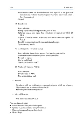
![15 Imaging in Emergency Surgery 209
15.2.2.3 Advantages
• High sensivity and sensibility for diagnosis
• Differential diagnosis
• Evaluate complications: grading, quality, and quantity of pancreatic collections
• Careful study of the pancreas and adjacent organs
• Follow-up
• Availability
• Non-invasiveness and speed
15.2.2.4 Disadvantages
• Relative role to ascertain the acute pancreatitis (cholelithiasis, pancreas divisum)
• Low sensitivity in the study of the biliary tree and pancreatic ducts
• Low accuracy for differential diagnosis between necrosis and infected necrosis
• May be normal in mild cases
• Use of ionizing radiations
15.2.3 US
• High sensivity to recognize the cause of acute pancreatitis: obstructive or non-
obstructive
• May reveal a normal-appearing gland in milder cases
• Follow-up
15.2.3.1 US Findings
• Focal or diffuse enlargement of the gland, with heterogeneous or low echogenic-
ity
• Cause of the acute pancreatitis: cholelitiasis and gallstones into biliary ducts [10]
• Pancreatic and peripancreatic collection: anechoic or complex fluid mixed with
solid components
• Used to monitor the patient with acute pancreatitis
• Guide percutaneous or endoscopic drainage for the collections
• Vascular complications (portal venous thrombosis)
• Hemorrhage
15.2.3.2 Advantages
• Cause of pancreatitis: gallstones and cholelitiasis
• Follow-up
• Availability
• Non-invasiveness
• Fast and cheap
• No use of ionizing radiations](https://image.slidesharecdn.com/emergencylaparoscopy-191115144149/85/Emergency-laparoscopy-218-320.jpg)
![210 C. Rossi et al.
15.2.3.3 Disadvantages
• Limited field of view of pancreas
• Obese patients
• Investigator’s experience
15.2.4 MRI
• Superior than CT for characterization of fluid and/or solid necrotic areas into
pancreatic parenchyma and peripancreatic tissue [10]
• Evaluate the integrity of the biliary system and pancreatic duct [10]
• To detect the cause of the acute pancreatitis
• Evaluate local complications [11]
15.2.4.1 MRI Findings
Interstizial Edematous Pancreatitis
• Enlarged pancreas or part of it: hypontense signal on T1W; hyperintense signal
on T2W—T1 fat suppression post-contrast
• Peripancreatic inflammation: hyperintense signal on T2W-FS
• Cause of acute pancreatitis: gallstones and cholelitiasis on MRCP [10, 11]
Necrotizing Pancreatitis
• Necrotic areas: nonenhancing areas on T1W-FS post-contrast (particularly
important is arterial phase) [11]
• Pancreatic and peripancreatic fluid collections
• Differential diagnosis between necrosis and infected necrosis
• Hemorrhage: areas hyperintense on T1W (subacute phase)
15.2.4.2 Advantages
• Accurate study of the biliary tree, pancreatic ducts, and pancreatic morphology
[10]
• High spatial resolution
• Multiplanar and multiparametric study
• No use of ionizing radiation
15.2.4.3 Disadvantages
• Motion and breathing artifacts
• Relative unavailability in most hospitals and expensive
• Long scanning time
15.2.5 Differential Diagnosis
• Pancreatic ductal adenocarcinoma
• Autoimmune pancreatitis](https://image.slidesharecdn.com/emergencylaparoscopy-191115144149/85/Emergency-laparoscopy-219-320.jpg)
![15 Imaging in Emergency Surgery 211
• Peptic ulcer disease with posterior perforation
• Pancreatic lymphoma (diffuse pattern)
15.3 Acute Appendicitis
15.3.1 Rationale
• To rule out appendicitis
• Early diagnosis and proper management: conservative treatment or surgery
• Assessment severity of disease and complications
• To detect the cause: obstructive or non-obstructive [12]
• Differential diagnosis
• Follow-up
15.3.2 CT
• CECT is the “Gold standard”, with high sensivity (98 %) and specificity (97 %)
for the diagnosis of acute appendicitis [13, 14]
• Exclude alternative causes of abdominal pain, especially on right lower quadrant
[12]
• Identify complicated appendicitis
15.3.2.1 CECT Findings
• Dilated appendix with distended lumen [13, 14]
• Increased intraluminal fluid
• Appendiceal wall thickening
• Hyperenhancement of the appendiceal wall
• Appendicolith
• Thickening of the caecal wall: caecal bar sign or arrowhead sign
• Enlarged mesenteric lymphonodes
• Periappendiceal inflammation: stranding of the adjacent fat and thickening of the
lateroconal fascia or mesoappendix
• Extraluminal fluid [13, 14]
• Complications: inflammatory phlegmon, abscess, perforation, and generalized
peritonitis
15.3.2.2 Advantages
• Differential diagnosis
• Image reconstruction and post-processing to maximize diagnostic efficiency and
accuracy
• Identify complications
• Follow-up](https://image.slidesharecdn.com/emergencylaparoscopy-191115144149/85/Emergency-laparoscopy-220-320.jpg)
![212 C. Rossi et al.
15.3.2.3 Disadvantages
• Use of ionizing radiations
15.3.3 US
• First imaging examination in suspected acute appendicitis, particularly in young
adults and females
• High safety and accuracy (lower than CT) [15]
• Graded-compression US allows successful examination of a patient who may
have peritoneal irritation [15]
• Follow-up, in cases of nonsurgical right low pain [16]
15.3.3.1 US Findings
• Aperistaltic, noncompressible, dilated appendix
• US-McBurney’s sign
• Appendicolith
• Distinct appendiceal wall layers
• Hyperechogenic pericaecal fat [16]
• Periappendiceal fluid
• Target appearance (axial section)
• Appendiceal abscess [16]
15.3.3.2 Advantages
• Differential diagnosis with gynecologic disorders
• Initial imaging approach especially in children, young people and women of
childbearing age [15]
• Available
• Repeatable
• Non-invasiveness
• Fast and cheap
• Follow-up
15.3.3.3 Disadvantages
• Operator dependent
• Obese patients
• Difficult detection of the appendix in atypical sites
15.3.4 Differential Diagnosis
• Gynecologic conditions](https://image.slidesharecdn.com/emergencylaparoscopy-191115144149/85/Emergency-laparoscopy-221-320.jpg)
![15 Imaging in Emergency Surgery 213
15.4 Gynecologic Disorders
15.4.1 Rationale
• Identify causes of gynecological emergencies [17, 18]
• Differentiate surgical from non surgical gynecological emergencies
• Differential diagnosis with alternative causes of pelvic pain
15.4.2 US
First technique used for acute gynecologic emergencies
15.4.2.1 US Findings
• Free intrapelvic fluid
• Uterine echogenicity and enlargement
• Ovaries enlargement and echogenicity with multiple peripheric follicles
• Liquid or solid masses
• Ovaries displaced from its location with torsion of ovarian vessels [19]
• Hemorrhagic ovarian cyst or hemorrhagic corpus luteum
15.4.2.2 Advantages
• Provides an overview of uterus and ovaries
• Ability to evaluate the relationship of uterus and ovaries with the adjacent organs
• Possibility to use PD analysis to evaluate vascularization
15.4.2.3 Disadvantages
• Low spatial resolution because of deep position of genital organs
• Limited in women with abundant subcutaneous fat
15.4.3 Endovaginal US
It requires dedicated intracavitary transducers; technique of choice for acute gyne-
cologic emergencies [20].
15.4.3.1 EVUS Findings
• Optimal visualization of endometrium and myometrium
• Size and location of ectopic pregnancy [21]
• Hemorrhagic ovarian cyst or hemorrhagic corpus luteum
• Presence of complications of PID such as tubo-ovaric abscess](https://image.slidesharecdn.com/emergencylaparoscopy-191115144149/85/Emergency-laparoscopy-222-320.jpg)
![214 C. Rossi et al.
15.4.3.2 Advantages
• Ability to perform a survey in obese women or with abdominal scars
• High spatial resolution
• High sensitivity for visualization of internal cavity
15.4.3.3 Disadvantages
• Changes of endometrium with ormonal phases
• May be difficult to differentiate benignant from malignant pathologies
15.4.4 MRI
MRI is a second technique used:
• To confirm or exclude US diagnosis
• To evaluate complications
15.4.4.1 MRI Findings
• Enlargement of uterus with alterations of signal
• Identification of endometrioma and its rupture [22]
• Hemorrhagic cyst associated with hemoperitoneum such as breaking of hemor-
rhagic corpus luteum, or ovarian cyst
• Characterization of masses such as liquid, solid, or mixed
• Differential diagnosis between acute from chronic bleeding
• Characterization of masses of pelvic inflammatory disease and its complications,
such as tubo-ovaric abscess
15.4.4.2 Advantages
• High specificity for characterization and differential diagnosis of adnexial masses
• Allows a detailed anatomy
• High panoramicity with possibility to individuate extra-pelvic pathologies caus-
ing acute pelvic pain
15.4.4.3 Disadvantages
• Long scanning time
• Lower sensitivity than EVUS for visualization of internal cavity
15.5 Nonspecific Abdominal Pain
15.5.1 Rationale
• To rule out the cause of nonspecific pain
• To differentiate medical from surgical diseases
• To avoid unnecessary laparotomies](https://image.slidesharecdn.com/emergencylaparoscopy-191115144149/85/Emergency-laparoscopy-223-320.jpg)
![15 Imaging in Emergency Surgery 215
15.5.2 X-Ray Abdomen
X-ray abdomen is the first imaging technique [23];
15.5.2.1 X-Ray Findings
• Signs of pneumoperitoneum;
• Air-fluid levels, intestinal morphological changes, and behaviors;
• Signs of small- and large-bowel obstruction (LBO) or Ogilvie’s syndrome
• Fecal stasis.
15.5.2.2 Advantages
• Provides an overview of abdominal cavity
15.5.2.3 Disadvantages
• Serial plain films help understand interstinal behaviours
• Low sensitivity to identify cause
15.5.3 CECT
CECT may provide additional information compared to plain film [24, 25]
15.5.3.1 CECT Findings
• May identify medical and surgical causes of abdominal pain
• To evaluate the enhancement of solid organ and hollow viscera
• To assess abdominal vessels
• To assess presence and characterization of masses
15.5.3.2 Advantages
• May identify both medical and surgical causes of abdominal pain [26]
• High prognostic negative value [27]
15.5.3.3 Disadvantages
• Use of ionizing radiations
15.6 Perforated Gastro-duodenal Ulcer
15.6.1 Rationale
• Evidence of free intraperitoneal air
• Definition of location of perforation](https://image.slidesharecdn.com/emergencylaparoscopy-191115144149/85/Emergency-laparoscopy-224-320.jpg)
![216 C. Rossi et al.
15.6.2 X-Ray Abdomen
First imaging exam. For positivity of exam, 1–2 ml of free gas are sufficient. At least
3 projections are needed:
• Postero-anterior and latero-lateral in orthostatism
• Postero-anterior in left lateral decubitus
• Anterior-posterior in supine decubitus
15.6.2.1 X-Ray Findings
• Free gas
• Presence of free air in perihepatic, perisplenic, along the course of falciform
ligament or round ligament, or in Morrison space is indicative of perforation of
gastroduodenal ulcera [28]
15.6.2.2 Advantages
• Fast and cheap
• Repeatable
• Performed also in supine patients
15.6.2.3 Disadvantages
• Use of ionizing radiations
15.6.3 CT
Most sensitivity method to research free gas [29, 30].
15.6.3.1 CT Findings
• Free air intraperitoneal
• Associated signs of paralitic ileus (horizontal loops, air-fluid levels)
• Spreading extraluminal contrast medium (rare, need oral contrast administration)
• Discontinuity and thickening intestinal wall (rare)
• Free intraperitoneal fluid
15.6.3.2 Advantages
• Sensitivity of 99 % in identify pneumoperitoneum
• It is able to recognize also small gas bubbles not visible to X-Ray abdomen
• Can recognize the site of perforation [31, 32]
15.6.3.3 Disadvantages
• Initial CT study may be negative for free air (covered perforation).](https://image.slidesharecdn.com/emergencylaparoscopy-191115144149/85/Emergency-laparoscopy-225-320.jpg)
![15 Imaging in Emergency Surgery 217
15.7 Large-Bowel Obstruction
15.7.1 Rationale
• To rule out LBO
• Identification of the cause
• Severity of LBO and possible complications
• Differential diagnosis
15.7.2 X-Ray Abdomen
• It is the first imaging study in patient suspected of having LBO
• two views at least in critical patient (supine position Antero-Posterior and Lateral
views)
• When possible in both dependent (supine and prone) and nondependent (upright
decubitus)
• The sensitivity of abdominal radiography for the detection of LBO is 84 %,
and the specificity is only 72 % as a result of difficult to distinguish between
obstruction and colonic pseudo-obstruction [33, 34]
15.7.2.1 X-Ray Findings
• Colon may be glossily dilated proximal to the site of obstruction “transition
zone” with paucity or absence of gas distal to the obstruction site [35]
• Air-fluid levels
• Pneumatosis (unusual and difficult to detect)
• Portal venous gas (unusual and difficult to detect)
• Pneumoperitoneum
• Abnormal location of colonic segments (volvulus)
15.7.3 CT
• CECT is the gold standard for the evaluation of LBO
• Well-tolerated, rapid imaging study
• High sensitivity (96 %) and a specificity (93 %) [36, 37]
15.7.3.1 CT Findings
• Dilated large bowel proximal to the transition point and collapsed bowel distal to
the site of obstruction [38]
Colon Carcinoma LBO
– Asymmetric and short-segment wall thickening](https://image.slidesharecdn.com/emergencylaparoscopy-191115144149/85/Emergency-laparoscopy-226-320.jpg)
![218 C. Rossi et al.
– Enhancing soft tissue narrowing the colonic lumen
– “Shoulder sign,” central necrosis in a large obstructive mass
– Pericolonic fat infiltration (differential diagnosis with diverticulitis)
– Lymph nodes larger than 1 cm
Sigmoid Volvulus
– “Coffee bean” sign, apposition of the medial walls of the dilated loops forms
the cleft of the bean
– “Bird beak” sign, tapering transition point
– The “inverted U” sign, dilated sigmoid extends into the upper quadrant
– “Northern exposure” sign, repositioning of the sigmoid colon out of the pelvis
– Absence of rectal gas;
– The point of twisting and the vessels (Whirl sign) [39, 40].
Cecal Volvulus
– Radiological findings
Marked distension of cecum
Bird beak sign, bowel tapers and meets at the site of twist
The whirl sign
Small-bowel dilatation, in incompetent valve
Signs of ischemia: wall thickening, mural hypoenhancement, pneumatosis
Diverticulitis
– Segmental, symmetric bowel wall thickening with hyperemia in a longer
portion than colonic cancer
Pericolonic inflammation and fat stranding
Extramural abscesses or perforation as complications
Fluid in peritoneal spaces
Vascular engorgement [41]
Intussusception
– CT can identify the cause in more than 80 %. Colonic carcinoma is the most
frequent cause in adults
Intraluminal Contents
– In old or weak patients, fecal impaction is frequent.
Very rare are hernias, inflammatory bowel disease, adhesions, or external
compression.
Acute colonic pseudo obstruction (ACPO) or Ogilvie syndrome is a pseudo-
obstruction without an exact pathophysiology. Distinguishing between LBO and
pseudo-obstruction is challenging. On abdominal radiographs, colonic distension,
involving the cecum, ascending colon, and transverse colon, is the hallmark of
ACPO, but not pathognomonic. Repositioning the patient in right lateral decubitus
results in air filling the distal colon.
Contrast enema may be made to evaluate a transition point. If diagnosis is
unclear a CT may allow characterization of the entire large bowel with or without a
transition point [42].](https://image.slidesharecdn.com/emergencylaparoscopy-191115144149/85/Emergency-laparoscopy-227-320.jpg)
![15 Imaging in Emergency Surgery 219
15.8 Small-Bowel Obstruction
15.8.1 Rationale
• To rule out small-bowel obstruction (SBO);
• To identify the severity of SBO and possible complications
• Determination of the transition point
• Identification of the cause of SBO
• Differential diagnosis
A diagnostic algorithm for evaluation of patients with suspected SBO is shown
in Fig. 15.1 [43, 44].
15.8.2 X-Ray
• First imaging examination
• Its accuracy varies from 50 % to 70 % [33]
• At least three projections (supine, prone, and lateral decubitus)
15.8.2.1 Findings
• Dilated small bowel
• Dilated stomach
• Absence of rectal gas
• Multiple air-fluid levels (upright or left lateral decubitus radiography)
15.8.2.2 Advantages
• Availability
• Rapidity
• Non-invasiveness
• Follow-up
15.8.2.3 Disadvantages
• Rare identification of the cause [45]
15.8.3 US
• Not used in the United states, but it is frequently used in many other countries
where the availability of CT is limited and expertise in sonography is high. Useful
adjunct with plain film](https://image.slidesharecdn.com/emergencylaparoscopy-191115144149/85/Emergency-laparoscopy-228-320.jpg)
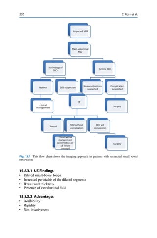
![15 Imaging in Emergency Surgery 221
• Presence or absence of peristalsis
• Presence of extraluminal fluid
15.8.3.3 Disadvantages
• Operator dependent
• Limitation in gas-containing structures
• Obese habit
15.8.4 CT
• CECT is the gold standard
• Confirm or exclude diagnosis of SBO
• Evaluation of severity and complications
• Identifies the cause [46]
15.8.4.1 Findings
• Small-bowel dilatation and colon not dilated
• Transition point from dilated to non-dilated small bowel
• Air-fluid levels
• Colon decompressed
CT Findings of Ischemia
– Bowel wall thickening
– Fluid in mesentery or peritoneal cavity
– Mesenteric edema
– Abnormal bowel wall enhancement (increased or decreased)
– Occlusion of mesenteric vessels
– Engorged mesenteric veins
– Whirl sign
– Pneumatosis [47, 48]
15.8.4.2 Advantages
• Availability
• Non-invasiveness
• Fast exam
• Excellent evaluation of bowel wall, its vessels, and adjacent mesentery
15.8.4.3 Disadvantages
• No other disadvantage out of the use of ionizing radiation](https://image.slidesharecdn.com/emergencylaparoscopy-191115144149/85/Emergency-laparoscopy-230-320.jpg)
![222 C. Rossi et al.
15.9 Hernias
15.9.1 Rationale
• To evaluate the presence and the type of hernia
• To rule out the complications
• To guide the management
• Follow-up
15.9.2 US
• First-line imaging study for evaluation of non-acute adult groin hernia
• Frequently used in many European centers where the expertise in sonography is
high
15.9.2.1 US Findings
• They differentiate between the multiple types of hernia and their contents
• Dilated small-bowel loops proximal to the hernia or in the sac
• Increased peristalsis of the dilated segments
• Bowel wall thickness
• Presence of extraluminal fluid in the sac or in peritoneal space
• Decreased vascularization.
15.9.2.2 Advantages
• Dynamic investigation assessment with and without Valsalva maneuver
• No ionizing radiation
• Evaluation of vessels flow
15.9.2.3 Disadvantages
• Operator dependent
• Limitation in gas-containing structures
• Obese habit
• Evaluation of internal hernias [49].
15.9.3 CT
• First-line imaging study in evaluation of hernia complications
• Fast and panoramic compared with other modalities](https://image.slidesharecdn.com/emergencylaparoscopy-191115144149/85/Emergency-laparoscopy-231-320.jpg)
![15 Imaging in Emergency Surgery 223
Imaging findings differ among the multiple types of hernia, but some signs are
common to the same complication [50, 51]. The most common complications:
CT Findings of Bowel Obstruction
• Dilated bowel proximal to the hernia;
• Normal caliber or reduced caliber distal to the hernia
• Tapering of the afferent end efferent loop, at the hernia neck
• Dilation of herniated bowel loops
• Air-fluid levels in bowel proximal to the hernia
• Fecalization of small bowel proximal to obstruction [52, 53]
CT Findings of Incarceration
• Imaging can only suggest the diagnosis, when a small abdominal defect and a
narrow neck are visualized.
• Incarceration predisposes to obstruction, ischemia, inflammation, or other com-
plications
CT Findings of Strangulation
• Dilated fluid filled loops;
• U- or C-shaped loops of bowel entrapped within hernia sac
• Afferent and efferent have a “beak bird” signs in the transition point
• Wall thickening
• Abnormal mural hypo- or hyperattenuation and enhancement
• Mesenteric vessel engorgement
• Fat obliteration
• Mesenteric haziness
• Ascites and fluid within the sac [54]
CT Findings of Surgically Repaired Hernias Complications
• Hernia recurrence
• Fluid collections: contain serous fluid (seroma) or blood products (hematoma)
are frequently in immediate postoperative period
• Infection: imaging confirms the presence and define the location of volume of
collection and to guide drainage
• Mesh-related complications: inflammatory reactions and fibrosis;
• Testicular complications: testicular vessel ligation and testicular ischemia are the
most feared complication and of inguinal hernia repair; ultrasound is the modality
of choice for the study of vessel flow [55].
15.9.3.1 Advantages
• Availability
• Non-invasiveness
• The short duration of the examination
• Provide anatomic detail of abdominal wall and hernia sac and neck
• Accurate identification of hernia contents](https://image.slidesharecdn.com/emergencylaparoscopy-191115144149/85/Emergency-laparoscopy-232-320.jpg)
![224 C. Rossi et al.
• Differential diagnosis with other abdominal masses
• Evaluation of acute phase and complications
• Useful to evaluate surgically repaired abdominal wall hernias and their compli-
cations
15.9.3.2 Disadvantages
• Limited role in dynamic evaluation (Valsalva maneuver).
• Limited role in vessel flow study.
15.9.4 MRI
• MRI should be considered as possible alternative in young patients with symp-
toms, but only few studies support its use in wall hernia complications.
• Accurate view of musculoskeletal structures
• Very rare clinical use
• One indication is for pregnancy [56].
15.10 Abdominal Trauma
15.10.1 Rationale
• Early assessment and organ injury grading
• To guide patient toward appropriate management: NOM, intervention radiology,
or surgery
• Follow-up
15.10.2 CT
• CECT is the “Gold standard” for early diagnosis, proper management, and
outcome [57]
• Indication for hemodynamically stable or semi-stable patients [57, 58]
• Tailored protocols according to the mechanism: Abdominal CT survey to be
incorporated into “Whole body CT protocols” for high-energy trauma (i.e., motor
vehicle accidents)—two phases
• Conversely in direct or compressive trauma only one contrast-enhancement
phase for abdominal evaluation can be performed—one phase
• Fast overview of the entire abdomen, skeleton, and vessels in a single scan
acquisition
• “Whole body CT” is the fastest radiological investigation, for “complete” body
coverage in multitrauma patient [59]
• Evaluation degree and extent of solid organ injuries [60, 61]
• Follow-up](https://image.slidesharecdn.com/emergencylaparoscopy-191115144149/85/Emergency-laparoscopy-233-320.jpg)
![15 Imaging in Emergency Surgery 225
15.10.2.1 CT Findings
• Hemoperitoneum or retroperitoneum
• Peritoneal or retroperitonel free fluid: mild (one peritoneal space), moderate (<3
peritoneal spaces), or massive (all peritoneal spaces) [57, 60]
• Pneumoperitoneum or retroperitoneum
• Sentinel clot sign (45–70 HU): blood located to the source of the hemorrhage
[61]
• Nonclotted blood (30–40 HU): flow freely between peritoneal spaces
• Differentiate recent bleeding and chronic bleeding
• High sensivity for detecting small amounts of free peritoneal fluid
• Assessment degree of severity solid organ injuries
• Extent of injuries: diameter, length, depth [62]
• Capsular and parenchymal hematoma: hyperdense on acute/subacute phase (40–
60 HU)
• Parenchymal lacerations: hypodense linear or starry area [60, 61]
• Differentiation between contained vascular injury (pseudoaneurysm, arterio-
venous fistula) and arterial or venous active bleeding
• Posttraumatic hypoperfusion: hypovolemic shock complex, parenchymal infarct,
shock bowel [7]
15.10.2.2 Advantages
• Proper management after abdominal trauma: treatment conservative or surgery
• Diagnosis of life-threatening injuries
• Image reconstruction and post-processing to maximize diagnostic efficiency and
accuracy
• Follow-up
• Availability and speed
• Non-invasiveness
15.10.2.3 Disadvantages
• Questionable specificity for diaphragmatic, mesenteric, and hollow organ injuries
• Overestimation of solid organ injury
15.10.3 US-FAST
• Diagnostic modality of choice for evaluation of hemodynamically unstable
patients
• Presence or absence of free peritoneal fluid in five spaces: pericardium, hepa-
torenal fossa, left subphrenic space, right and left paracolic gutters, and pouch of
Douglas [63]
• Different role compared to CT in the clinical algorithm](https://image.slidesharecdn.com/emergencylaparoscopy-191115144149/85/Emergency-laparoscopy-234-320.jpg)
![226 C. Rossi et al.
15.10.3.1 Advantages
• Presence or absence of free peritoneal fluid
• Quantity and quality of peritoneal fluid
• Follow-up
• Available
• Non-invasiveness
• Fast and cheap
15.10.3.2 Disadvantages
• Is not predictor for a tailored surgery
• Does not indicate the need for intervention
• Rarely useful in bowel, pancreatic, and renal injuries
• No evaluation of retroperitoneal spaces
• Obese patient
• Investigator’s experience
15.11 Mesenteric Ischemia
15.11.1 Rationale
• To rule out mesenteric ischemia;
• To identify the type of ischemia [64]:
– Arterial ischemia: occlusion of AMS from embolic or thrombotic origin
– Vein ischemia: occlusion of VMS
– Hypoperfusion non-occlusive ischemia (NOMI)
• Differential diagnosis
15.11.2 CECT
CECT is the technique of reference with a sensitivity of 90 % [65].
15.11.2.1 CECT Findings
To evaluate [66]
• Mesentery
• Mesenteric vessels
• Thickness and enhancement of bowel loops
• Peritoneal cavity
15.11.2.2 CECT: Mesenteric Findings
• In arterial occlusion: bloodless for intense vasoconstriction
• In NOMI: bloodless for the reduced blood flow
• In vein occlusion: edematous for failure outflow](https://image.slidesharecdn.com/emergencylaparoscopy-191115144149/85/Emergency-laparoscopy-235-320.jpg)
![15 Imaging in Emergency Surgery 227
15.11.2.3 CECT: Mesenteric Vessel Findings
• In arterial occlusion: reduction of diameter or endoluminal defects [67];
• In vein occlusion: increase of diameter for venous engorgement
• In NOMI: reduction of diameter without endoluminal defect
15.11.2.4 CECT: Bowel loop Findings
• In arterial occlusion: thin loop; contrast-enhancement reduced or absent, or target
sign in case of reperfusion
• In vein occlusion: thickened loop; contrast-enhancement increased, or target sign
• In NOMI: thin loop; contrast-enhancement reduced or absent, or target sign in
case of riperfusion
15.11.2.5 Advantages [68, 69]
• Evaluate thrombotic or embolic origin of ischemia
• Identify type of acute ischemia;
• Evaluate late signs, such as intestinal, portal, and mesenteric pneumatosis or
pneumoretroperitoneum.
• Necessary for early detection of NOMI with signs of organ hypoperfusion
• Assess signs of spastic, hypotonic, and paralytic ileus
15.11.2.6 Disadvantages
• Use of ionizing radiation
References
1. Smith EA, Dillman JR, Elsayes KM et al (2009) Cross-sectional imaging of acute and chronic
gallbladder inflammatory disease. AJR Am J Roentgenol 192(1):188–196
2. Mirvis SE, Whitley NO, Miller JW (1986) The diagnosis of acute acalculous cholecystitis: a
comparison of sonography, scintigraphy and CT. AJR Am J Roentgenol 147:1171–1175
3. Mirvis SE, Whitley NO, Miller JW (1987) CT diagnosis of acalculous cholecystitis. J Comput
Assist Tomogr 11(1):83–87
4. Bennett GL, Rusinek H, Lisi V et al (2002) CT findings in acute gangrenous cholecystitis. AJR
Am J Roentgenol 178(2):275–281
5. Shakespear JS, Shaaban AM, Rezvani M (2010) CT findings of acute cholecystitis and its
complications. AJR Am J Roentgenol 194:6
6. Park MS, Yu JS, Kim YH et al (1998) Acute cholecystitis: comparison of MR cholangiography
and US. Radiology 209:781–785
7. Altun E, Semelka RC, Elias J Jr et al (2007) Acute cholecystitis: MR findings and differentia-
tion from chronic cholecystitis. Radiology 244:174–183
8. Balthazar EJ (2002) Acute pancreatitis: assessment of severity with clinical and CT evaluation.
Radiology 223:603–613
9. O’Connor OJ, McWilliams S, Maher MM (2011) Imaging of acute pancreatitis. AJR Am J
Roentgenol 197:2
10. Scaglione M, Casciani E, Pinto A et al (2008) Imaging assessment of acute pancreatitis: a
review. Semin Ultrasound CT MR 29:322–340
11. Miller FH, Keppke AL, Dalal K (2004) MRI of pancreatitis and its complications: Part 1, Acute
pancreatitis. AJR Am J Roentgenol 183:1637–1644](https://image.slidesharecdn.com/emergencylaparoscopy-191115144149/85/Emergency-laparoscopy-236-320.jpg)
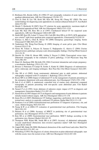
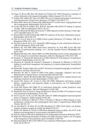
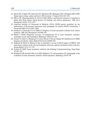
![16Emergency Laparoscopy: The Nurse’s Point
of View
Monica Rolfo, Serena Langella, Valeria Esposito, Antonio Valenti,
Angela Di Marco, and Alessandro Ferrero
16.1 Introduction
Laparoscopic surgery (LS) is increasingly used in the emergency setting [1]. It
is of particular interest and efficacy for the diagnosis and treatment of acute
conditions. In fact, emergency laparoscopy can be safely undertaken if there is an
appropriate patient selection, the surgical team is adequately experienced, and there
are sufficient resources to allow potentially more complex operations.
The advantages of LS are well established: better postoperative outcomes and
pain management and significant reduction in the duration of hospital stay because
of earlier recovery with decreasing of health and social costs [1].
The application of laparoscopy in emergency surgery involves several organiza-
tion problems.
Firstly, surgeons should have large experience in laparoscopic techniques. Sec-
ondly, nurses should be trained in the management of surgical patients with acute
diseases and all operators in the operating room (OR) must be confident with every
technology and device required in LS.
Finally, the perioperative environment is multidimensional, dynamic, and com-
posed of multidisciplinary teams. Moreover, providers of all specialties are facing
with caring of patients with multiple risk factors in a complex condition. Even
with a comprehensive knowledge of the most current strategies, the application of
good communication skills within the surgical team remains a fundamental tool
to achieve best practices. In this light, nurses contribute to the overall success
M. Rolfo, RN ( )
Division of General and Oncology Surgery, Mauriziano “Umberto I” Hospital, Turin, Italy
Italian College of Nurses (IPASVI), Turin, Italy
e-mail: mrolfo@mauriziano.it
S. Langella, MD •V. Esposito, RN • A. Valenti, RN • A. Di Marco, RN • A. Ferrero, MD
Division of General and Oncology Surgery, Mauriziano “Umberto I” Hospital, Turin, Italy
© Springer International Publishing Switzerland 2016
F. Agresta et al. (eds.), Emergency Laparoscopy,
DOI 10.1007/978-3-319-29620-3_16
231](https://image.slidesharecdn.com/emergencylaparoscopy-191115144149/85/Emergency-laparoscopy-240-320.jpg)
![232 M. Rolfo et al.
of the multidisciplinary management assuring a “communication bridge” among
providers, patients, and caregivers.
It is well known that nurses play an important role in providing preoperative edu-
cation, perioperative care, and postoperative evaluation [2]. Moreover, postoperative
course is not only influenced by physiologic outcomes of disease but also addresses
to the biopsychosocial responses on the health-illness continuum that are necessary
to improve the quality of life even in emergent surgery.
In recent years, nursing responsibilities for decision-making in patients care
have been expanded [3, 4]. The evolution of nursing is related to certain levels
of education and practice to fill the important roles within the multi-professional
team in the surgical area. In this light, the nurse leader should create learning
opportunities and promote educational programs in order to improve the nurse
practice in specialized surgical teams.
16.2 Preoperative Phase
16.2.1 Physical Preparation: Technical Skills
Before surgical interventions there are different preoperative procedures and activ-
ities in order to prepare the patient and his family for surgery. In case of emergent
surgery this act must be done quickly because of acute condition.
Patient assessment and the definition of a care plan are designed to meet the phys-
ical, psychological, educational, social, and cultural needs. These aspects are the
cornerstone of nursing activity. Another key point is the collaboration with the mem-
bers of the surgical team in order to identify the best treatment for each case, accord-
ing to clinical evidences and guidelines [5], and to define the future allocation of the
patient in areas with different levels of intensity in the postoperative care (Fig. 16.1).
In the emergency setting, nurse should give importance to the areas of risk
with major clinical implications [6]. Different scales of risk assessment have been
reported [7]. These scales allow the use of rapid response to perform appropriate
postoperative cares with a multidisciplinary approach.
Preoperative nurse management is targeted mainly to prevent infections and
reduce the risk of perioperative complications. Based on the results obtained
in reducing catheter correlate infections and pneumonia in patients artificially
ventilated, the “care bundles” have been introduced. The concept of a care bundle
was developed from evidence documenting that a structured approach performing
3–5 evidence-based collective interventions could improve patients’ outcomes [8].
The care bundle approach is common to many guidelines for the prevention of
surgical site infection (SSI). SSI is a well-known complication of general surgery
occurring in about 3 % of all surgical procedures and in up to 20 % of patients
undergoing emergency intra-abdominal procedures [9]. SSI adversely affects patient
outcomes and healthcare costs. In a recent study, overall SSI rate was 4.09 % and
colorectal surgery was associated with the highest SSI rate (9.43 %), in this setting
was identified as an independent predictor [10]. Concerning the emergency LS, a
mini-invasive approach resulted in a protective factor for SSI [11].](https://image.slidesharecdn.com/emergencylaparoscopy-191115144149/85/Emergency-laparoscopy-241-320.jpg)
![16 Emergency Laparoscopy: The Nurse’s Point of View 233
CLINICAL PATHWAY EMERGENCY SURGERY
SURGICAL
DEPARTMENT
EMERGENCY
DEPARTMENT
MEDICAL
DEPARTMENT
DIAGNOSTIC
ASSESSMENT
SURGERY EXITNO
THERAPEUTIC
PHYSICAL
PSYCHOLOGICAL
SURGICAL
PREPARATION
YES
SURGICAL ROOM
LOW CARE
SURGERY
HIGH CARE
SURGERY
MEDIUM CARE
SURGERY
CLINICAL MONITORING
PROTOCOLS AND PROCEDURES CARE
PROCEDURES DISCHARGE
SURGICAL REHABILITATION
P.A.C.U.
Post anestesia
care unit
Fig. 16.1 Graphic representation of the clinical pathway in emergency surgery
A care bundle approach to SSI includes methods for preoperative removal of
hair (when appropriate), rational antibiotic prophylaxis, avoidance of perioperative
hypothermia, management of perioperative blood glucose, and effective skin prepa-
ration [12].
The skin of the surgical site must appear clean [13] and there is the possibility
that the patient should be helped to meet the need for hygiene and showering without
antiseptic. In particular, attention should be given to the cleanness of umbilical area.
In fact, after LS it is described the infection of the umbilical site access with different
rates depending on the type of intervention [15].
Regarding hair removal, the current evidences are in favor of trichotomy only
in cases where the hairs in the area of incision could affect the operation [16].
Hairs should be removed with an electric shaver suitable for the purpose or with
depilatory creams while the use of hand razor remains strongly discouraged. There
is no specific report on the impact of trichotomy in emergency LS, but we could
speculate that a possible conversion to laparotomy may justify the hair removal of
the entire abdominal area. Removing jewelry, prosthesis for dentures, hearing and
eye, and nail polish is always recommended.
At admission, patients should be evaluated for the risk of thromboembolism
through appropriate flow chart. Operative time, prolonged pneumoperitoneum, and
complexity of the surgical procedure and the surgical site are some of the risk factors
to be considered in addition to the individual patient risks [17]. The nurse must](https://image.slidesharecdn.com/emergencylaparoscopy-191115144149/85/Emergency-laparoscopy-242-320.jpg)
![234 M. Rolfo et al.
collaborate with other professionals for the identification of risk and the correct
application of therapeutic program. The embolic thrombus prophylaxis may have
to pair the use of graduated compression stockings. As alternative, the use of
intermittent pneumatic compression is recommended when clinically indicated [18].
Finally, concerning intestinal surgery, stomal site marking by a wound ostomy
nurse should be advocated in patient at high risk of anastomic leak. This practice
improves selection of stoma site but also introduces the patient and their family to
stoma education empowering postoperative independence [19].
16.2.2 Clinical Monitoring
The assessment of clinical condition is carried out through the monitoring of
vital signs: blood pressure, heart rate and breathing, pain, blood sugar level, pulse
oximetry, and temperature [20]. The nurse puts the vital signs in relation to the
patient comorbidities, surgical diagnosis assumed, age, and symptoms. Different
tools are available to determine the risk of adverse events related to the deterioration
of vital signs, such as the Early Warning Score [21]. Each altered parameter should
be promptly referred to the physician and included in the documentation of the
patient. This way, alterations could be quickly corrected improving postoperative
outcomes. In particular, it is strongly recommended to obtain an optimal glycemic
control since preoperative period [22] and a rapid correction of dehydration,
especially in the elderly [23]. The evaluation of vital signs must be repeated at
regular intervals until the transfer to the operating room.
Depending on the clinical condition, it could be necessary to place a nasogastric
tube, urinary catheter, and to find one or more venous access (peripheral or central)
before surgery.
The condition of fasting does not ever preclude the intake of preoperative oral
therapy. This aspect is crucial to prevent possible postoperative consequences in
particular in case of cardiac medications [24].
Patients under age or mental deficiency should always be accompanied by a
caregiver. Before the transfer to the operating room (OR), nurse should verify that
all these elements of the path are correct using a checklist [25]. When required by
critical clinical conditions, patient is accompanied to the OR by nurse and/or doctor.
Finally, dignity, comfort, and privacy of the patient should always be respected at
all times and in every transfer of operational setting.
16.3 Operative Phase
The OR staff members participate in the intraoperative and postoperative stages
of the multimodal approach to emergency LS. The OR is a complex reality that
includes many operators with different roles and specialties often involved in critical
situation. Furthermore, all providers must be confident with the highly available
technologies. In fact, the team experience is proven to have a beneficial impact on](https://image.slidesharecdn.com/emergencylaparoscopy-191115144149/85/Emergency-laparoscopy-243-320.jpg)
![16 Emergency Laparoscopy: The Nurse’s Point of View 235
overall surgery time [25]. In particular, the nursing team plays a highly important
role in the success and speed of emergency LS.
The main objective of the nurse who accepts the patient in the OR is to
understand the psychological state [26]. This way, an appropriate counseling offers
the possibility to humanize the environment. During the conversation, nurses
provide to minimize every source of stress, worry, and anxiety. This approach
could be restricted in case of emergent surgery because of critical acute conditions
compromising the conscious state.
Upon arrival to the OR, the circulating nurse will interview and identify the
patient. At least two patient identifiers are used, and they should check medical
record number, name, and date of birth. When appropriate, it is imperative for the
patient to confirm the correct laterality (right or left side) of the injured organ. As
suggested by others, the operative site could be marked while the patient is awake
and alert and before sedative medication is administered.
After the patient is secured to the OR table with a safety belt, antithrombotic
stockings and knee- or thigh-high sequential compression devices are placed on
the lower extremities, to prevent venous stasis and reduce the risk of deep vein
thrombosis and pulmonary embolism.
An indwelling urinary drainage catheter is inserted by either the circulating nurse
or surgeon to decompress the bladder. When required, prophylactic intravenous
antibiotics should be administered within 60 min from the initial skin incision. After
the induction of general anesthesia, the patient is endotracheally intubated and an
orogastric tube is placed to decompress the stomach. An arterial pressure line may
be established for hemodynamic monitoring.
It has been demonstrated that a surgical safety checklist may reduce morbidity
and mortality [27]. Haynes BN and colleagues collected data from a large prospec-
tive database on clinical processes and outcomes after noncardiac surgery. The rate
of death was 1.5 % before the checklist was introduced and decreased to 0.8 %
afterward (P D 0.003). Similarly, inpatient complications occurred in 11.0 % of
patients at baseline and in 7.0 % after introduction of the checklist (P < 0.001) [28].
The main steps of surgical check list are conducted by nurse, anesthetist, and
surgeon through three phases: Sign IN (before induction of anesthesia), Time OUT
(before skin incision), and Sign OUT (before patient leave OR) according to WHO
recommendations [29].
OR usually used for LS are often not designed for this purpose. Hence, all
instrumental devices (screen, light, sources, electrocoagulators, insufflator) are
located on dedicated trays. Because all these instruments take space, it is necessary
to have room appositely prepared for this type of surgery, especially in emergency.
Moreover, it is necessary to know highly technical details of sterilization method-
ology for all devices, which require more attention than conventional surgical
instruments.
In this context, it appears to be fundamental a strict cooperation among all
providers of surgical team. In order to facilitate the correct development of
surgical procedures it is mandatory to adopt standardized protocols and checklists](https://image.slidesharecdn.com/emergencylaparoscopy-191115144149/85/Emergency-laparoscopy-244-320.jpg)
![236 M. Rolfo et al.
concerning preparation of OR, anesthetic machine, surgical bed, and laparoscopic
column [30].
For the OR nurses, proper positioning is one of the key responsibilities to
protect the safety and skin integrity of the patient under anesthesia. As reported
[31], positioning of the patient was found to be particularly difficult for the prone
(43.8 %), lithotomy (53.4 %), and lateral (65.5 %) positions. Moreover, lack of
positioning competences and equipment for arm support, standardized equipment
for leg support, and standard sizes of OR beds seemed to complicate positioning.
During LS, nurse must give attention to the control of pneumoperitoneum. It is
well known that CO2 insufflation could cause complications related to increased
abdominal pressure and CO2 retention. In order to avoid such complications,
pressure limit of insufflator is 12 cm H2O. Moreover, flow rate of CO2 insufflation
must be modulated during procedure starting slow at the beginning of operation and
than progressively augmented [32].
An experienced OR nurse knows that any laparoscopic emergent procedure has
the potential open conversion due to bleeding, unanticipated organ injury, or failure
to progress as in the case of excessive intra-abdominal adhesions. Appropriate
instrumentations and supplies necessary for laparotomy should be readily available
in the room, but may be kept unopened and uncounted unless their use is required.
During the procedure, the OR nurse must be vigilant to potential intraoperative
complications. Laparoscopic instruments are placed on a mayo stand with the
handles facing the surgeon to expedite transfer to the surgeon hands. A variety
of laparoscopic thermal energy devices are also available. These devices have
been shown to significantly decrease blood loss and operative time. Furthermore,
hemostatic agents must be readily available to the surgical team should bleeding
occur. Experienced circulating nurses can often anticipate the surgeon needs as they
closely follow the steps of the procedure. During the procedure, the circulating nurse
also monitors the level of the CO2 tank and has reserve tanks available.
At the end of operation, the nurse assesses the patient’s skin and temperature.
The patient is then transported to the postanesthesia care unit (PACU). In the
PACU, nurses play a key role in patient pain management and satisfaction. In fact,
even at the early postoperative stage, nurses can directly affect the reduction of
postoperative infections and ileus and facilitate the patient’s safe and timely hospital
stay. Interestingly, it has been reported [33] that the length of stay in the PACU
correlates with pain intensity, nausea, and vomiting on arrival.
16.4 Postoperative Phase: From Hospitality to Discharge
16.4.1 Acceptance from Operating Room: Clinical Monitoring
Patients who have undergone emergency surgery are at risk for poor outcomes
comparing to elective surgery [34]. The principal objectives of the immediate
postoperative care are prevention of number and severity of complications [35]
and restoring the physiological balance. Early detection and timely management](https://image.slidesharecdn.com/emergencylaparoscopy-191115144149/85/Emergency-laparoscopy-245-320.jpg)
![16 Emergency Laparoscopy: The Nurse’s Point of View 237
of complications may reduce morbidity and mortality. After surgery, nurse first
detects and evaluates signs and symptoms of the three major vital systems (nervous,
respiratory, and cardiovascular). Afterward, the accommodation and the comfort of
the patient are ensured. At this point, nurse starts recognition, measurement, and
recording of clinical parameters: blood pressure, heart and respiratory rate, oxime-
try, glycemic index, pain, diuresis, characteristics and quantity of surgical drains
(if placed), level of consciousness, body temperature, surgical wound, the presence
of medical devices, and any symptoms reported by the patient. After LS particular
importance covers the control of the presence of subcutaneous emphysema [36]. It
is recommended to empty the urinary catheter and drains for precise postoperative
monitoring. The nurse must verify the clinical documentation to check the surgical
procedure and the type of anesthesia performed, any intraoperative problems,
medications used, medical history and preoperative interventions performed, and
not least the therapeutic prescriptions to be administered. All elements collected
must lead the nurse to plan the subsequent appropriate care. Vital signs and clinical
parameters of interest should be monitored every 1–2 h for at least the first 8 h.
Moreover, nurse must continually check that the connections of the drainage pipes
are correct and securely connected and the drains are working properly identifying
significant bleeding or any appearance suggestive of complications (presence of
bile, intestinal fluid, pus, urine air).
Fundamental to the success of any model of team-based care is the skill, relia-
bility, and expertise of the individual members that comprise the team, including
nurses. Additionally, the definition of a standard of care and the adoption of
shared protocols and therapeutic procedures (defined by surgical team) may allow
rapid intervention for the management of major alterations and most common
postoperative symptoms (hypertensive crisis, hyperthermia, nausea or vomiting,
pain, hyperglycemia). The postoperative delirium is one of the most common
adverse events after emergency surgery especially in the elderly [37].The surgical
approach (laparoscopic versus open) does not seem to influence this condition.
Nevertheless, in the emergency setting nurses are frequently facing with patients
affected by cognitive dysfunction. In this regard, the use of screening tools, such as
The Nursing Delirium Screening Scale [38], lead to early recognition. This way,
the interventions required are readily performed reducing the risk of injury and
accelerating the return to normality.
16.4.2 Surgical Rehabilitation
After the initial 24 h, vital signs can be monitored every 4–8 h if the patient is stable.
The incision and dressing should be monitored for the amount of drainage and signs
of infection. The surgeon may order a dressing change during the first postoperative
day; this should be done using sterile technique.
The patient should be monitored for any evidence of potential complications,
such as leg edema, redness and pain (deep vein thrombosis), shortness of breath
(bronchopneumonia and pulmonary embolism), dehiscence and infection of the](https://image.slidesharecdn.com/emergencylaparoscopy-191115144149/85/Emergency-laparoscopy-246-320.jpg)
![238 M. Rolfo et al.
incision, or ileus (intestinal obstruction), heart problems. The surgeon should be
notified immediately if any of these occurs.
Particularly, in patients at higher risk of complication, rehabilitation exer-
cises to prevent respiratory and thromboembolic complications should start early.
When appropriate, preoperative education and maintaining previous levels of self-
sufficiency increase the effectiveness of rehabilitative interventions [39]. The main
objective of prevention is early mobilization from the bed [40]. If the patient
is conscious, cooperative, and clinically stable mobilization can occur after only
few hours after surgery, even after complex procedures [41]. Patients with limited
mobility must be mobilized passively alternating on both sides several times a day,
even for the prevention of pressure lesions, which is one of the main areas for action
for nurses in this phase [42]. LS, even if performed in emergency, allows a rapid
return to the levels of self-sufficiency when postoperative rehabilitation is rapid
and constant [43]. Historically, the assumption of liquids and foods happens to the
restarting of the intestinal functionality; nevertheless, this attitude was questioned
by early rehabilitation programs in surgery having highlighted the absence of the
benefit of fasting in various postoperative surgical procedures [41]. Few data are
available about this aspect in emergency surgery, but a similar strategy should
probably be chosen especially for minor procedures [44].
16.4.3 Therapeutic Education: The Role of the Nurse
The preoperative education is an important aspect of preparation for many surgical
procedures. In particular, it has positive effects on fear [45] and anxiety [46] even
prior to emergency surgery [47]. The contents of preoperative education may vary
in different contexts, but generally include a presentation on the procedures and
perioperative care, with respect to potential complications, management of the
pain and other symptoms, device management, the mobilization, and respiration
[48]. In the emergency setting, clinical conditions and degree of urgency may not
enable a structured educational intervention. As previously reported [49], giving
information just before surgery in urgency increases stress in anxious patients.
In fact, education is best given at hospital admission. Nevertheless, therapeutic
education on postoperative process is a high priority for patients and family
members anticipating temporary changing needs as a result of the surgery. This
stage of education usually happens preoperatively, preparing timely the patients at
discharge. This way, the discharge is not an isolated event, but it becomes part of a
process which begins at admission and actively involves patients and family during
hospitalization [50].
However, for patients who underwent emergency surgery all the steps of this
intervention are accomplished during the postoperative course.
A well-structured educational plan is based on the quality of given information,
to reassure patients that initial fears and anxieties are a normal part of the healing
process. It has been demonstrated that this approach could positively affect expe-](https://image.slidesharecdn.com/emergencylaparoscopy-191115144149/85/Emergency-laparoscopy-247-320.jpg)
![16 Emergency Laparoscopy: The Nurse’s Point of View 239
rience of postoperative pain, decrease preoperative state of anxiety, and promote
exercise to prevent postoperative complications [51].
After surgery, nurse starts giving practical daily information as to the care of the
surgical wound, both after laparoscopic and open surgery, diet, return to work, limits
on activities, bowel movements, management of an eventual therapeutic regimen,
and pain control. Before the discharge, other useful information should be given
concerning the follow-up, such as the date, time, and location of their visits [52].
The best results are obtained associating oral information with other aids such as
booklets or other information instrument. The delivery of written material has the
advantage of ensuring a standardization of the educational process reducing the risk
of the variability information based on different communication skills, knowledge,
and resources of nurses.
The multidisciplinary team (surgeons, anesthetists, nurses, nutritionists, dieti-
tians, and respiratory therapists) collaborates in the educational process. In this
scenario, the role of nurse is essential giving proper planning instructions and
educating patient and family.
16.4.4 Pain Management
The laparoscopic approach often results in lesser postoperative pain, shorter anal-
gesia requirement, and earlier recovery. Nevertheless, in some cases early pain after
laparoscopy can be comparable or even more severe than after open surgery. Pain
intensity is maximal during the first postoperative hours and some patients need
opioid analgesics to control it. Laparoscopy implicates insufflation of the abdominal
cavity with carbon dioxide, and the remaining gas after pneumoperitoneum is one
of the basics for postoperative pain, often described in the shoulder area and that
it may persist for up to 72 h. However, pain after laparoscopic surgery is derived
from multiple origins: nociceptive, incisional, and visceral pain; inflammatory pain
in response to tissue trauma; and neuropathic pain in association with distension-
induced neuropraxia. For this reason, a multimodal pain management protocol
should be preferred. Moreover, persistent pain after laparoscopic surgery has also
been reported. This condition could be partially explained by the influence of
sociocultural and individual factors. Thus, these aspects should also be considered.
Patient counseling on pain and pain management is important because it may
increase the compliance and adherence to the selected analgesic treatments. More-
over, reaching patients early, even before surgery, leads to influence psychological
variables such as presurgical anxiety. In fact, the impact of psychological interven-
tion on the prevention of persistent pain is yet to be demonstrated, but this approach
seems to be promising in terms of outcomes in the acute postsurgical time period
in combination with multimodal preventive analgesia [53]. As previously reported
by Fernandez et al., an acceptance-based psychological intervention along with the
usual presurgical protocol for a laparoscopic cholecystectomy will produce healthier
reactions to postsurgical pain and will reduce anxiety, duration of postsurgical
hospitalization, and demand of analgesics. Unfortunately, data on the management](https://image.slidesharecdn.com/emergencylaparoscopy-191115144149/85/Emergency-laparoscopy-248-320.jpg)
![240 M. Rolfo et al.
of pain after laparoscopic emergency surgery are lacking. In this subset of patients,
nurse can provide counseling mainly in the postoperative period [54]. There are
multiple measures available to assess pain in adults. Each measure has its own
strengths and weaknesses. Both the Visual Analog Scale (VAS) for Pain and the
Numeric Rating Scale (NRS) for Pain are unidimensional single-item scales that
provide an estimate of patients’ pain intensity [55]. They are easy to administer,
complete, and score. In particular, the pain NRS may be preferred for the patient
care because of simpler scoring and its ability to be administered both verbally
and in writing. Furthermore, by using specific pain assessment methods adapted for
sensory and cognitive impairment, and language difficulties, pain can be managed
effectively for most patients. During the postoperative course, nurses could apply
strategies to involve patients in pain decision-making, which include educating
patients to inform nurses when analgesia is required, offering patients’ choices
between analgesia, and evaluating the effectiveness of the treatments. Moreover,
a collaborative relationship between nurses and clinicians improves communication
to address complex pain management decisions. Finally, postoperative management
requires substantial communication and collaboration between all members of the
healthcare team to be successful.
In conclusion, the laparoscopic approach to emergency surgery is gaining
widespread agreement. Nowadays, a multidisciplinary approach is mandatory for
the successful management of patients who underwent emergency LS. The peri-
operative nursing in this setting is constantly changing and it requires knowledge,
technical skills, leadership, and excellent communication abilities. In order to
respond to the changing needs of the surgical patient, the nurse must constantly
add knowledge and skills. These objectives could be accomplished through self-
education practices and participating in educational opportunities.
References
1. Harji DP, Griffiths B, Burke D et al (2014) Systematic review of emergency laparoscopic
colorectal resection. Br J Surg 101(1):e126–e133
2. Stobinski JX (2008) Perioperative nursing competency. AORN J 88(3):417–418, 421–436
3. Decreto Ministeriale 14 settembre 1994, nı
739, GU 9 gennaio 1995 nı
6: “Regolamento
concernente l’individuazione della figura e del relativo profilo professionale dell’infermiere”
4. Legge 10 agosto 2000, nı
251 GU 6 settembre 2000 nı
208: “Disciplina delle professioni
infermieristiche, tecniche, della riabilitazione, della prevenzione nonché della professione
ostetrica”
5. Møller TP, Münter KH, Østergaard D et al (2015) Exploring challenges and solutions in the
preparation of surgical patients. Dan Med J 62:A5141
6. Visser A, Geboers B, Gouma DJ et al (2015) Predictors of surgical complications: a systematic
review. Surgery 158(1):58–65
7. Carpenter CR, Shelton E, Fowler S et al (2015) Risk factors and screening instruments
to predict adverse outcomes for undifferentiated older emergency department patients: a
systematic review and meta-analysis. Acad Emerg Med 22(1):1–21
8. Tanner J, Padley W, Assadian O et al (2015) Do surgical care bundles reduce the risk of surgical
site infection in patients undergoing colorectal surgery? A systematic review and cohort meta-
analysis of 8,515 patients. Surgery 158(1):66–77](https://image.slidesharecdn.com/emergencylaparoscopy-191115144149/85/Emergency-laparoscopy-249-320.jpg)
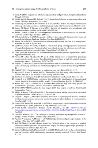
![17Emergency Laparoscopy in the Elderly
From the Evidence to the Philosophy
Gian Luca Baiocchi, Luca Arru, Federico Gheza, Carlo Bergamini,
Gabriele Anania, Fabio Cesare Campanile, Mauro Zago,
Nazario Portolani, and Ferdinando Agresta
17.1 Introduction
The increasing age of the population is an ineluctable phenomenon, observed both
in rich and emerging countries [1]. It is believed that the number of people older than
65 will be higher than those of children under 5 within a few years [2]. The number
of octogenarians has risen by more than 50 % over the last 15 years [3]. People aged
85 and older will increase from 2.8 % in 2011 to 7.8 % in 2050, with an average
life expectancy of 85.3 years for males and 90.5 years for females [1], with obvious
implications, both for the health policies and from a clinical standpoint.
G.L. Baiocchi ( ) • F. Gheza • N. Portolani
Surgical Clinic, Department of Clinical and Experimental Sciences, University of Brescia, Via
Cicognini, 6, 25127 Brescia, Italy
e-mail: gianluca.baiocchi@unibs.it
L. Arru
Service de Chirurgie Générale et Mini-Invasive, Centre Hospitalier de Luxembourg, Luxembourg
City, Luxembourg, UK
C. Bergamini
SOD Chirurgia Generale 1, d’Urgenza e Mini-invasiva – AOU Careggi, Firenze, Italy
G. Anania
Department of Morfology, Surgery and Experimental Medicine, School of Medicine, University
of Ferrara, Ferrara, Italy
F.C. Campanile
General Surgery Department, Ospedale Andosilla, Civita Castellana, VT, Italy
M. Zago
General Surgery Department, Minimally Invasive Surgery Unit, Policlinico San Pietro, Ponte San,
Pietro, BG, Italy
F. Agresta
Department of General Surgery, Azienda ULSS19 del Veneto, Adria (RO), Italy
© Springer International Publishing Switzerland 2016
F. Agresta et al. (eds.), Emergency Laparoscopy,
DOI 10.1007/978-3-319-29620-3_17
243](https://image.slidesharecdn.com/emergencylaparoscopy-191115144149/85/Emergency-laparoscopy-252-320.jpg)
![244 G.L. Baiocchi et al.
Healthcare needs increase with age both in the elective and emergency setting
[4]. However, while in the former the decision-making process can result from a
comprehensive assessment, shared with the patients and their family, the urgency-
emergency setting does not allow such a peaceful, quiet, and “settled” evaluation [5,
6]. The surgeon who deals with emergency surgery needs proper tools to quickly
take the right decisions, based on solid data about the chances of success, and about
the expectations of life after a successful procedure [7].
The elderly patient specificities heavily influence the diagnostic and therapeutic
pathway. The diagnostic process is often more difficult and sometimes totally
inconclusive, because of communication difficulties and peculiar biochemical and
radiographic changes [8]. A retrospective Mayo Clinic analysis on 231 elderly
patients with abdominal pain shows that temperature, physical examination, and
basic blood tests have limited sensitivity in discriminating the surgical cases
[9]. Another recent article reports that 75 % of elderly patients evaluated in the
emergency room for abdominal pain were discharged with a final diagnosis of NSAP
(nonspecific abdominal pain) [10]. In addition, the mortality is high, up to 44 % (for
the most part due to abdominal and pulmonary septic complications) in a study
that included only patients more than 80 years old [11]. In the series published by
Arenal et al., mortality was 7 %, 8 %, and 9 % for appendectomy, cholecystectomy,
and parietal surgery [3].
In this context, the use of minimally invasive surgical techniques is characterized
by extreme uncertainty. While on one side a pragmatic approach considers as a
good surgical practice a laparotomy for quick and essential damage control, leaving
aside any aesthetic consideration; on the other side, it is likely that the principles
of minimally invasive procedures would find especially in frail patients a very good
indication. The emergency surgeon must quickly decide if and how to operate an
elderly patient, and needs to know the clinical data about benefits and limitations of
a minimally invasive approach.
This chapter aims to provide the emergency surgeon with the bibliographic
elements on which to base its decisions [12], with specific reference to the use of the
laparoscopic approach in urgent abdominal diseases in the elderly and frail patients
(anyway, the simple chronological age is a parameter by itself inadequate to define
a patient at increased risk; thus, in this chapter the term “old/frail” will be used).
The following questions are answered:
• Who’s the old/frail patient?
• What are the clinical pictures of potential emergency abdominal surgery in
old patients? When is it right not to operate an old/frail patient despite life-
threatening condition?
• Should a minimally invasive approach be a priori ruled out in old/frail patients?
• May a minimally invasive approach be beneficial in old/frail patients?
• What are the essential conditions to approach acute abdomen in old/frail patients
by minimally invasive surgery?
• Should the indication for laparotomy conversion be different in old/frail patients?](https://image.slidesharecdn.com/emergencylaparoscopy-191115144149/85/Emergency-laparoscopy-253-320.jpg)
![17 Emergency Laparoscopy in the Elderly 245
• What are the acute abdominal diseases potentially treatable by laparoscopy in
old/frail patients?
• Outcomes analysis
17.2 Who Is the Old Patient?
Though it is now accepted that the chronological age determines the clinical choices
less than the so-called biological age, it is possible to identify some demographic
cutoff and group patients into homogeneous risk classes. In particular, it is defined
“old-young” a patient aged 70–80, “old” a patient aged 80–90, and “old-old” a
patient over the age of 90 years. From a medical perspective, however, the most
relevant definition for clinical choices is that of “frail” patients. It is defined by
this way: a clinical situation that predisposes to increased morbidity and mortality,
assessed over some parameters that can be easily determined such as the speed of
walking, the level of physical activity, and cognitive capacity [13, 14]. There are no
clinical scores validated for frail patients with acute abdominal disease to be treated
surgically. However, it may be useful to take into account some other scores, initially
designed for different clinical settings [15, 16]. This score takes into account the
following parameters: anorexia, weight loss, BMI, motility, neuropsychological
problems (dementia, depression), number of daily medications, chronological age,
and comparison with peers.
17.3 What Are the Clinical Pictures of Potential Emergency
Abdominal Surgery in Frail Patients?
At least one-fourth of the patients with acute abdomen are more than 50 years
[17]. In this group of patients, the diagnostic path is often aggressive and is often
completed with a CT scan [18]. CT scan is generally characterized by a good
ability to identify the source of the problem. Several retrospective papers have
investigated the incidence of abdominal diseases requiring urgent surgical treatment
in elderly patients. In principle, 40–50 % of cases are obstructive syndromes, 30–
40 % peritonitis, 5 % visceral ischemia, and 10 % hemorrhagic issues. In particular,
inguinal hernia and colorectal cancer are the most frequent causes of obstruction;
perforated acute diverticulitis and acute cholecystitis are the most frequent causes
of peritonitis, while acute appendicitis is less frequent. It must also be taken
into account that in the abovementioned retrospective Spanish series, 53 % of
cholecystitis and 44 % of appendicitis were gangrenous [3]. Acute appendicitis
is characterized by delay in treatment, high perforation rate, and unfavorable
outcome [19]. Colorectal obstructions represent a significant cause of morbidity and
mortality: a retrospective study in a tertiary referral university center included 292
patients operated on in the period 2000–2006. The overall mortality was 15 % and
morbidity was 35 % [20]. Conversely, the incidence of small bowel obstruction does
not seem to be age dependent. Finally, trauma requiring surgery is generally related](https://image.slidesharecdn.com/emergencylaparoscopy-191115144149/85/Emergency-laparoscopy-254-320.jpg)
![246 G.L. Baiocchi et al.
to a low-speed impact (fall in the bathroom or in the garden), while traffic accidents
are rare [3]. Two studies, retrospectively targeting patients >80 and >90 years old,
confirm that incarcerated hernias, colorectal cancer, and peritonitis from perforated
diverticulitis or gangrenous cholecystitis are the leading causes of surgery in elderly
patients [11, 21]. In another recent retrospective study including 456 patients older
than 65 years who underwent emergency surgery between 2006 and 2009 in Finland,
32 %, 13 %, 11 %, and 11 %, respectively, had acute cholecystitis, incarcerated
hernia, colorectal cancer, and acute appendicitis [22].
17.4 When Is It Right Not to Operate a Frail Patient Despite
Life-Threatening Condition?
Answering to this question is extremely complex, and there are few scientific studies
that have attempted to justify the answer on strong clinical data [2]. In the decision,
clinical issues (chance of recovery, procedure-related risks, available therapeutic
alternatives), ethical aspects (who decides that a patient should die?), philosophical
observations (which life is worth living?), and health policy considerations (what
resources is it right to employ against a limited life expectancy?) need to be
considered. Therefore, in this area it is really difficult to give appropriate instruments
for clinical choices. We should evaluate, in order: (1) the degree of patient
consciousness (if dignity of life equals to a clear conscience, and the propriety
of informed consent requires formally a conscious signature, it is rather hard to
justify invasive procedures in a frail unconscious patient); (2) the “quod vitam”
prognosis, besides the acute disease for which the patient is actually observed (with
particular reference to oncologic, cardiac, and pulmonary terminal diseases: it is
hard to justify a life-saving intervention in a patient with a few months prognosis);
(3) the success rate of the procedure (with particular reference to abdominal sepsis
and severe ischemia); and (4) the wish of the patients and families. There have been
some attempts to identify preoperative data helpful to determine the real impact
associated with urgent intervention [23]; globally, it is known that, in the last period
of life, intensive treatments often prolong the suffering of the patient [24], and the
hospitalization, even more in the ICU, worsens his quality of life and restricts the
psychosocial well-being of the patient and his family [5].
17.5 Should a Minimally Invasive Approach Be a Priori Ruled
Out in Frail Patients?
The answer to this question is very easy: no. No anesthetic or surgical elements,
based on frailty only, formally contraindicate a laparoscopic procedure. It is,
however, true that in everyday practice this indication is often disregarded: in the
series published by Ukkonen of 460 patients aged over 65, 80.7 % had a straight
laparotomy [22]. This is in apparent conflict with an already broad literature,](https://image.slidesharecdn.com/emergencylaparoscopy-191115144149/85/Emergency-laparoscopy-255-320.jpg)
![17 Emergency Laparoscopy in the Elderly 247
testifying that even complex surgical procedures can be performed, in the elective
setting, with equal safety in elderly patients [25–27].
17.6 May a Minimally Invasive Approach Be Beneficial in Frail
Patients?
Also the answer to this question is very easy: yes. While considerations related
to cosmetics clearly lose their value in frail patients, the benefits of a minimally
invasive approach in terms of postoperative pain, wound infections, mobilization,
transit, and nutrition resumption could be of even greater relevance in these patients.
In a retrospective Korean study, 30 patients over 65 who underwent laparoscopic
appendectomy were compared to 47 patients undergoing an open procedure for
acute appendicitis: the incidence of complications was strongly in favor of the
laparoscopic group (7 % vs. 32 %, P < 0.01), while all the other parameters,
including costs, were equal [28]. Another paper on the same subject considered
a much larger population (65,464 elderly patients with acute appendicitis) and
concluded that for both perforated and non-perforated appendicitis, the laparoscopic
approach allowed a reduction of complications (15 % vs. 23 % and 36 % vs.
46 %, in laparoscopic vs. open appendectomy, in perforated and non-perforated
appendicitis, respectively), mortality, costs, and length of stay (3.0 vs. 4.8 and 5.8 vs.
8.7 days, respectively) [29]. However, also negative trials were published. Shin and
colleagues, for example, evaluated the incidence of postoperative delirium in 130
patients undergoing open and laparoscopic (elective) gastrectomy. The evaluation
was conducted through perspective, dedicated questionnaires, administered in the
third postoperative day. Though the incidence and severity of delirium (31 % vs.
41 %) and the cognitive impairment rate (38 % vs. 42 %) were all in favor of
the laparoscopic approach, none of these differences was significant. This may
obviously be due just to a limited sample size [30]. Indeed, other studies have
suggested, but not proven, an advantage associated with the laparoscopic approach
on postoperative cognitive function [31].
17.7 What Are the Essential Conditions to Approach Acute
Abdomen in Frail Patients by Minimally Invasive Surgery?
Undoubtedly, among these cannot be missed: (a) the patient suitability to anes-
thesia:, laparoscopy involves well-known pathophysiological changes, due to both
the pneumoperitoneum (decreased venous return and increased respiratory pres-
sures) and the extreme positions required for the proper conduct of the operation
(trendelenburg or lateral rotation). Such changes may be particularly significant
in frail patients [32]. (b) The availability of proper materials (the logistics in
emergency may be different from those in the elective setting, for the presence
of an unaccustomed auxiliary team, unusual operating room, or instruments). (c)
The presence of a surgical team with sufficient expertise in minimally invasive](https://image.slidesharecdn.com/emergencylaparoscopy-191115144149/85/Emergency-laparoscopy-256-320.jpg)
![248 G.L. Baiocchi et al.
procedures. Indeed, in the frail patients it is of paramount importance not to accept
technical compromises that could make surgery more difficult, slower, and less
effective. In younger patients, the advantages of minimally invasive approach,
also aesthetic, could make a greater fatigue and a prolongation of the surgical
times justified. On the contrary, the antiplatelet therapy is not considered among
the essential conditions: the hypothesis that it increases the bleeding risks just in
laparoscopy and not in laparotomy has been largely disavowed [33].
17.8 Should the Indication for Laparotomy Conversion Be
Different in Frail Patients?
Generally speaking, yes. Besides the obvious and universally accepted reasons for
conversion in laparotomy (bleeding difficult to control, jatrogenic injury difficult to
repair, judgment of substantial ineffectiveness of the procedure), in frail patients the
element to which it must be paid greater attention is the duration of the intervention.
The operation should be converted to laparotomy when the presumed operative time
for laparoscopy is significantly longer than for laparotomy and this could have a
negative adverse impact on the condition of the patient. Given a protracted operative
time of 30–60 min more than that of a presumptive open surgery, we suggest to
cautiously convert into laparotomy and proceed with an effective conclusion of the
intervention. In the retrospective Finnish study, the conversion rate was of 25.9 %,
thus considerably higher than that expected in the general population [22].
17.9 What Are the Acute Abdominal Diseases Potentially
Treatable by Laparoscopy in Frail Patients?
Certainly, it is cholecystectomy for acute cholecystitis, which keeps the known
benefits of minimally invasive surgery and does not differ in anything from the
already accepted rules of laparoscopic surgery in young patients. It is likely
appendectomy, especially in relation to a greater diagnostic accuracy, which takes
into account the fact that acute appendicitis is relatively rare in the elderly,
while numerous alternative diseases could be the cause of symptoms [34]. In
the same way, it is the conservative therapy by washing-drainage of complicated
Hinchey stage 3 acute diverticulitis. Considering that in elderly patients a terminal
colostomy has lesser chance of being converted in the following months [35], the
simple peritoneal washing and drainage may be an attractive option; instead, in
Hinchey stage IV, laparoscopy is formally contraindicated in elderly patients, and
laparotomic resection-anastomosis protected by lateral oostomy would be preferable
to the Hartmann procedure (of course if the general conditions allow a longer lasting
intervention). A prospective randomized study of 62 patients aged over 60 years
has shown that the rate of recanalization is higher with primary anastomosis and
diversion by loop ileostomy [36]. The perforation of peptic ulcer is a not as a great
indication to laparoscopy: the concern for a malignant nature of the ulcer, at least in](https://image.slidesharecdn.com/emergencylaparoscopy-191115144149/85/Emergency-laparoscopy-257-320.jpg)
![17 Emergency Laparoscopy in the Elderly 249
the stomach, and the reported limited effectiveness of laparoscopic repair compared
to open repair, make this intervention to be proposed with less conviction [37]. The
same applies to the small bowel occlusion: the difficult standardization of times and
results of laparoscopic viscerolysis makes the price to pay to laparoscopy (potential
less efficacy, increased surgical time, possible underestimation of suffering intestinal
loop, and increased iatrogenic injury) not justifiable in frail patients. The indications
for the laparoscopic approach to strangulated inguinal and ventral hernia do not
change in the elderly. On the contrary, minimally invasive surgery in our opinion
should not be considered in nonspecific abdominal pain (which must be ruled out
with imaging techniques without the concerns on radiotoxicity rightfully arising for
young patients) [38], in gynecological pathology (virtually absent in women after
menopause), and in blunt abdominal trauma. In the same way, it appears to make
no sense to treat obstructing colorectal cancer with a laparoscopic approach. Since
the strategy of colonic stent as a bridge to elective surgery was recently questioned
because of high morbidity and mortality associated with iatrogenic perforation [39],
the best strategy for frail patients remains at the moment the Hartmann procedure,
to be done quickly and effectively by laparotomy. The alternative of primary
anastomosis with diversion may be considered, similarly to diverticular peritonitis.
Rare anecdotal cases of laparoscopic management of postsurgical complications
are, however, described also in elderly patients (Shadis, for example, report a case
of laparoscopic treatment of anastomotic fistula in an 86-year-old patient [40]). A
special discussion should be reserved to the case of mesenteric ischemia, because
of the high incidence of this pathology in the elderly. Although the clinical scenario
in which all the conditions making a laparoscopic exploration useful for settling the
hypothesis of intestinal ischemia are very limited, this strategy, potentially feasible
also in critically ill patients in the ICU, still looks more appealing in the frail patient,
and it deserves to be taken into account.
17.10 Outcomes
The expected mortality after emergency abdominal surgery in elderly/frail patients
is obviously high. In the series of Ukkonen mortality is reported as high as 14.2 %
at 30 days, with 31.9 % morbidity [22]. Adverse prognostic factors were age,
atrial fibrillation, low BMI, laparoscopic surgery, ASA 3 or 4, and a history of
cancer. Similar data are reported by the National Surgical Quality Improvement
Project (30-day mortality and morbidity 22.3 % and 42.1 %, respectively) [41]
and by the Spanish series (all-cause mortality 22 %, 24 % for patients >80 years
[3]). ASA score, the delay between the onset of symptoms and treatment, and the
chance to make only a palliative treatment (e.g., defunctioning stoma) were negative
prognostic factors in this paper. An Italian study still under peer review shows that
only one out of four patients >90 years operated in urgency is alive 2 years later.
Thus, in the overall management of the frail patient undergoing emergency
surgery, it takes maximum relevance a perioperative management paying specific
attention to the need of these patients. In particular, the prevention of postoperative](https://image.slidesharecdn.com/emergencylaparoscopy-191115144149/85/Emergency-laparoscopy-258-320.jpg)
![250 G.L. Baiocchi et al.
delirium (which affects 35–70 % of elderly patients after gastrointestinal surgery)
[30], the adequate (opioid-sparing) pain therapy, and a proper communication
strategy represent essential elements to restore the patient’s living conditions
antecedents to the acute event [42]. The problem is indeed not only to save
the patient life but also to look at the following life conditions. As concerns
rehabilitation, a number of papers reported as indicators for recovery of everyday
autonomy a number of scores for daily activity. Lawrence and colleagues in a
prospective cohort study of 372 consecutive patients >60 year old showed that, after
major abdominal surgery, the return to previous autonomy takes up to 3 months
for normal daily activities and up to 6 months for more complex intellectual task.
Preoperative depression is another problem with very difficult treatment [43].
17.11 Conclusions
The more protective system for the emergency surgeon who should take the decision
to approach an elderly patient by open or laparoscopic surgery in the emergency
setting would provide a simple list of essential requirements, in the absence of
even one of which the indication shall not to be considered correct. We propose the
following flow chart, which lends itself to a practical use to guide a cumbersome and
complex choice, often to be taken in a short time and without the decision support
of other colleagues.
1. The patient is conscious?
2. The patient has a life perspective, regardless of the urgent disease, of at least 3
months?
3. The current disease could be treated surgically?
4. If not treated in emergency, it would have clinically meaningful consequences?
5. The patient and family require and accept the intervention?
6. If it was not a frail patient, the approach would be minimally invasive?
7. The suspected disease is appendicitis, cholecystitis, Hinchey 3 diverticulitis
(accepted indication)? Is it peptic ulcer, incisional or other incarcerated hernia,
suspected intestinal ischemia (doubtful indication)?
8. The anesthesiologist agrees to the use of laparoscopy?
9. Are there environmental conditions for safe operation?
10. Has the team experience of minimally invasive surgery?
11. The expected duration of the intervention is not greater than 60 min than the
expected duration of the open intervention?
If the answer to all these items is YES, the operation can be performed by
laparoscopy.](https://image.slidesharecdn.com/emergencylaparoscopy-191115144149/85/Emergency-laparoscopy-259-320.jpg)
![17 Emergency Laparoscopy in the Elderly 251
References
1. Ageing well: a global priority (2012) Lancet 379(9823):1274. http://dx.doi.org/10.1016/
S0140-6736(12)60518-2
2. Søreide K, Desserud KF (2015) Emergency surgery in the elderly: the balance between
function, frailty, fatality and futility. Scand J Trauma Resusc Emerg Med 23:10. doi:10.1186/
s13049-015-0099-x
3. Arenal JJ, Bengoechea-Beeby M (2003) Mortality associated with emergency abdominal
surgery in the elderly. Can J Surg 46:111–116
4. Laurell H, Hansson LE, Gunnarsson U (2006) Acute abdominal pain among elderly patients.
Gerontology 52(6):339–344, Epub 2006 Aug 11
5. Cooper Z, Courtwright A, Karlage A, Gawande A, Block S (2014) Pitfalls in communication
that lead to nonbeneficial emergency surgery in elderly patients with serious illness: description
of the problem and elements of a solution. Ann Surg 260(6):949–957. doi:10.1097/SLA.
0000000000000721
6. Pappas A, Toutouni H, Gourgiotis S, Seretis C, Koukoutsis I, Chrysikos I, Gemenetzis G,
Matzoukas I, Karavitis G, Lagoudianakis E (2013) Comparative approach to non-traumatic
acute abdominal pain between elderly and non-elderly in the emergency department: a study
in rural Greece. J Clin Med Res 5(4):300–304. doi:10.4021/jocmr1424w, Epub 2013 Jun 21
7. National Confidential Enquiry into Patient Outcome and Death (NCEPOD). An age old
problem: a review of the care received by elderly patients undergoing surgery. The National
Confidential Enquiry into Patient Outcome and Death. 2010info@ncepod.org.uk, http://www.
ncepod.org.uk/
8. Launay-Savary MV, Rainfray M, Dubuisson V (2015) Emergency gastrointestinal surgery in
the elderly. J Visc Surg. pii: S1878-7886(15)00123-X. doi:10.1016/j.jviscsurg.2015.09.016
[Epub ahead of print]
9. Parker JS, Vukov LF, Wollan PC (1996) Abdominal pain in the elderly: use of temperature and
laboratory testing to screen for surgical disease. Fam Med 28(3):193–197
10. Tran BK, Espolio Desbaillet Y, Groebli Y, Della SV (2012) Abdominal pain among elderly
patients in the emergency department. RevMed Suisse 8:1548–1552
11. Green G, Shaikh I, Fernandes R, Wegstapel H (2013) Emergency laparotomy in octogenarians:
a 5-year study of morbidity and mortality. World J Gastrointest Surg 5:216–221
12. OCEBM Levels of Evidence Working Group. The Oxford 2011 levels of evidence. Oxford
Centre for Evidence-Based Medicine. http://www.cebm.net/index.aspx?o=5653
13. Fried LP, Tangen CM, Walston J et al (2001) Frailty in older adults: evidence for a phenotype.
J Gerontol A Biol Sci 56:M146–M156
14. Sternberg SA, Wershof Schwartz A, Karunananthan S, Bergman H, Mark Clarfield A (2011)
The identification of frailty: a systematic literature review. J Am Geriatr Soc 59:2129–2138
15. Soubeyran P, Bellera C, Goyard J et al (2011) Validation of the G-8 screening tool in
geriatriconcology: the ONCODAGE project. J Clin Oncol 2001:29 (suppl; abstr 9001; 2011
ASCO Annual Meeting)
16. Bellera CA, Rainfray M, Mathoulin-Pélissier S et al (2012) Screening older cancer patients:
first evaluation of the G-8 geriatric screening tool. Ann Oncol 23:2166–2172
17. Hastings RS, Powers RD (2011) Abdominal pain in the ED: a 35 year retrospective. Am J
Emerg Med 29(7):711–716
18. Reginelli A, Russo A, Pinto A, Stanzione F, Martiniello C, Cappabianca S, Brunese L, Squillaci
E (2014) The role of computed tomography in the preoperative assessment of gastrointestinal
causes of acute abdomen in elderly patients. Int J Surg 12(Suppl 2):S181–S186. doi:10.1016/j.
ijsu.2014.08.345, Epub 2014 Aug 23
19. Kraemer M, Franke C, Ohmann C, Yang Q, Acute Abdominal Pain Study Group (2000) Acute
appendicitis in late adulthood: incidence, presentation, and outcome. Results of a prospective
multicenter acute abdominal pain study and a review of the literature. Langenbecks Arch Surg
385(7):470–481](https://image.slidesharecdn.com/emergencylaparoscopy-191115144149/85/Emergency-laparoscopy-260-320.jpg)
![252 G.L. Baiocchi et al.
20. McGillicuddy EA, Schuster KM, Davis KA, Longo WE (2009) Factors predicting morbidity
and mortality in emergency colorectal procedures in elderly patients. Arch Surg 144(12):1157–
1162. doi:10.1001/archsurg.2009.203
21. Racz J, Dubois L, Katchky A, Wall W (2012) Elective and emergency abdominal surgery in
patients 90 years of age or older. Can J Surg 55:322–328
22. Ukkonen M, Kivivuori A, Rantanen T, Paajanen H (2015) Emergency abdominal operations in
the elderly: a multivariate regression analysis of 430 consecutive patients with acute abdomen.
World J Surg 39(12):2854–2861
23. Amrock LG, Neuman MD, Lin HM, Deiner S (2014) Can routine preoperative data predict
adverse outcomes in the elderly? Development and validation of a simple risk model
incorporating a chart-derived frailty score. J Am Coll Surg 219(4):684–694. doi:10.1016/j.
jamcollsurg.2014.04.018, Epub 2014 Jun 3
24. Wright AA, Zhang B, Ray A et al (2008) Associations between end-of-life discussions,
patient mental health, medical care near death, and caregiver bereavement adjustment. JAMA
300:1665–1673
25. Koh FH, Wong J, Tan JK, Tan KK, Cheong WK, Lieske B (2015) Laparoscopic colorectal
surgery is safe and benefits octogenarian patients with malignant disease: a matched case-
control study comparing laparoscopic and open colorectal surgery. Int J Colorectal Dis
30(7):963–968. doi:10.1007/s00384-015-2252-4, Epub 2015 May 24
26. Grailey K, Markar SR, Karthikesalingam A, Aboud R, Ziprin P, Faiz O (2013) Laparoscopic
versus open colorectal resection in the elderly population. Surg Endosc 27(1):19–30. doi:10.
1007/s00464-012-2414-1, Epub 2012 Jun 30
27. Kurian AA, Suryadevara S, Vaughn D et al (2010) Laparoscopic colectomy in octogenarians
and nonagenarians: a preferable option to open surgery? J Surg Educ 67(3):161–166 [PubMed]
28. Baek HN, Jung YH, Hwang YH (2011) Laparoscopic versus open appendectomy for appen-
dicitis in elderly patients. J Korean Soc Coloproctol 27(5):241–245. doi:10.3393/jksc.2011.27.
5.241, Epub 2011 Oct 31
29. Masoomi H, Mills S, Dolich MO, Ketana N, Carmichael JC, Nguyen NT, Stamos MJ (2012)
Does laparoscopic appendectomy impart an advantage over open appendectomy in elderly
patients? World J Surg 36(7):1534–1539. doi:10.1007/s00268-012-1545-9
30. Shin Y-H, Kim D-K, Jeong H-J (2015) Impact of surgical approach on postoperative delirium
in elderly patients undergoing gastrectomy: laparoscopic versus open approaches. Korean J
Anesthesiol 68(4):379–385. doi:10.4097/kjae.2015.68.4.379
31. Tan CB, Ng J, Jeganathan R, Kawai F, Pan CX, Pollock S, Turner J, Cohen S, Chorost M (2015)
Cognitive changes after surgery in the elderly: does minimally invasive surgery influence the
incidence of postoperative cognitive changes compared to open colon surgery? Dement Geriatr
Cogn Disord 39(3–4):125–131. doi:10.1159/000357804, Epub 2014 Nov 27
32. Peden CJ (2011) Emergency surgery in the elderly patient: a quality improvement approach.
Anaesthesia 66:440–445
33. Joseph B, Rawashdeh B, Aziz H, Kulvatunyou N, Pandit V, Jehangir Q, O’Keeffe T, Tang A,
Green DJ, Friese RS, Rhee P (2015) An acute care surgery dilemma: emergent laparoscopic
cholecystectomy in patients on aspirin therapy. Am J Surg 209(4):689–694. doi:10.1016/j.
amjsurg.2014.04.014, Epub 2014 Jun 27
34. Omari AH, Khammash MR, Qasaiheh GR et al (2014) Acute appendicitis in the elderly: risk
factors for perforation. World J Emerg Surg 9:1–6
35. Salem L, Flum DR (2004) Primary anastomosis or Hartmann’s procedure for patients with
diverticular peritonitis? A systematic review. Dis Colon Rectum 47:1953–1964
36. Oberkofler CE, Rickenbacher A, Raptis DA et al (2012) A multicenter randomized clinical trial
of primary anastomosis or Hartmann’s procedure for perforated left colonic diverticulitis with
purulent or fecal peritonitis. Ann Surg 256:819–826, discussion 826–827
37. Mouly C, Chati R, Scotté M, Regimbeau JM (2013) Therapeutic management of perforated
gastroduodenal ulcer: literature review. J Visc Surg 150:333–340](https://image.slidesharecdn.com/emergencylaparoscopy-191115144149/85/Emergency-laparoscopy-261-320.jpg)