SlideShare a Scribd company logo
1 of 23
Download to read offline
+
www.hysteroscopy.info
1
INSIDE THIS ISSUEH
Welcome 1
Histeroscopy Pictures 2
Uterine Septum
Interview of the month 3
Luca Mencaglia
Brief Review 5
Diagnosis and treatment of
endocervical lesions
Conundrums 10
Hysteroscopy & Ultrasound
Hysteroscopy Basic 11
Uterine Cervix
Original Article 14
Hysteroscopy Simulator
Hysteroscopy Devices 18
Gynko Sheath
Resident's Corner 21
Narrow Band Imaging (NBI)
Luiz Cavalcanti de Albuquerque Neto
Nov-Dec 2016 | vol. 2 | issue 6
ysteroscopy in Brazil
Modern hysteroscopy came to Brazil in the late 80s.
In this initial period, colleagues with relevant roles, Kleber Morais and Walter Pace,
disciples of Jacques Hamou, who returned to Brazil after a training period in France.
Since then, due to the interest aroused by the new technique, we have been visited by
Jacques Hamou, especially Luca Mencaglia and Carlo Tantini more often. They have
recently received many Brazilian colleagues in Firenze for training.
In 1995, I organized the 1st. Italo– Brazilian Symposium of Gynaecological Endoscopy,
which aroused unique interest and in this opportunity we were visited by more than 10
Italian colleagues who helped us a lot in the spread and interpretation of the method.
These events are bi annual and we will be holding the 11th. Symposium in 2017.
It has been more than 20 years of Italo– Brazilian Symposiums.
Here in the events in SĂŁo Paulo, we have been frequently visited by renowned
Hysteroscopists such as Jacques Hamou, Luca Mencaglia, Carlo Tantini, Ramon
Labastida, Steffano Bettocchi, Pietro Litta, Carlo De Angelis, Sebastiano Pace, Attillio Di
Spiezio and Giuseppe Bigatti among the best known Hysteroscopists.
Since the beginning of 2000, with the invaluable assistance of Professor Luca Mencaglia,
we have organized practical courses of Office and Surgical Hysteroscopy, six annual
hands-on courses with outpatient and surgical practices in Brazil. For all of them we have
received almost 2.000 students which is a significant number!
At the same time , we have organized other similar hands-on courses (European Institute
of Endoscopic Sciences) in other countries in South America.
In Uruguay, with the support of Prof. Agustin Rubal, in Argentina with Dr. Leopoldo
Videla, in Santiago - Chile with the help of Prof. Christian Miranda and in Venezuela with
the help of our dear Prof. Alfonso Arias, who has been a mandatory presence in all our
events in Brazil and other countries of the continent for several years.
With strong belief in the method, we are aware of its diagnostic,
surgical and social importance. We fully support the initiative to
hold this next event in Barcelona.
I might say that all roads will lead to Barcelona in May 2017!
Hysteroscopyc Greetings!!!!
TEAM COODINATOR
SPAIN
L. Alonso
EDITORIAL COMMITTEE
SPAIN
E. Cayuela
L. Nieto
ITALY
G. Gubbini
A. S. LaganĂ 
USA
J. Carugno
L. Bradley
MEXICO
J. Alanis-Fuentes
PORTUGAL
J. Metello
ARGENTINA
A. M. Gonzalez
VENEZUELA
J. Jimenez
SCIENTIFIC
COMMITTEE
A. Tinelli (Ita)
O. Shawki (Egy)
A. Ăšbeda (Spa)
A. Arias (Ven)
M. Rodrigo (Spa)
A. Di Spiezio Sardo (Ita)
E. de la Blanca (Spa)
A. Favilli (Ita)
M. Bigozzi (Arg)
S. Haimovich (Spa)
R. Lasmar (Bra)
A. Garcia (USA)
N. Malhotra (Ind)
J. Dotto (Arg)
I. Alkatout (Ger)
R. Manchanda (Ind)
M. Medvediev (Ukr)
M. Elessawy (Ger)
All rights reserved.
The responsibility of the signed
contributions is primarily of the authors
and does not necessarily reflect the views
of the editorial or scientific committees.
HYSTEROSCOPY
PICTURES
www.hysteroscopy.info
2
The septate uterus is the result of a failure in the process of resorption of the medial
septum that can range from a minimal septum in the uterine fundus to a complete one,
dividing the uterine cavity completely or even asociating double cervix and vaginal
septum. The proposed mechanism by which the uterine septum regresses is apoptosis, the
presence of Blc-2 protein protects from apoptosis. The absence of Blc-2 in the embryonal
uterine septum may indicate a lack of protection from apoptosis in this area. On the other
hand different studies have allowed the identification of several genes that can be
implicated in the development of uterine malformations
In an interesting study comparing the use of resectoscope to VersaPoint in 63 women for
the treatment of septum (Resectoscope or Versapoint for hysteroscopic metroplasty. Int J
Obstet Gynaecol 2008 Apr; 101 (1):39-42) Dr. Litta concludes that the VersaPoint system
does not require cervical dilation, avoiding potential cervical incompetence, cervical
lacerations and uterine perforation related to the dilation process. The use of VersaPoint is
a safe alternative to the use of resectoscope, especially in nulligravid or patients with
cervical stenosis.
If you are interested in sharing your cases or have a hysteroscopy image that
you consider unique and want to share, send it to hysteronews@gmail.com
Superficial vaginal
endometriotic implant
Detailed aspect of the cystic
area with retained blood
Detailed uterine septum Hysteroscopic metroplasty
with bipolar system
Nov-Dec 2016 | vol. 2 | issue 6
3
www.hysteroscopy.info
INTERVIEW WITH...
A entire life devoted to endoscopy and always a leader in
progress and development. His daily work in favor of
hysteroscopy, has contributed in making hysteroscopy a routine
technique.
Luca Mencaglia
Direttore U.O.C.
P.O.3 USL Toscana Sud Est
Centro di PMA Ospedale Santa
Margherita La Fratta
Firenze, Italy
One of your first publications in the early 80's was about the
experimental application of the hysteroscopy. What has changed since
then?
Today we can say that hysteroscopy is no longer an experimental
technique but a routine technique. Actually is not possible to work without
it. Any modern gynecology service must have both diagnostic and surgical
hysteroscopy. In particular, before to start any treatment, the modern
diagnostic hysteroscopy should be performed in all patients with abnomal
uterine bleeding (AUB) and in all infertile patients. In Europe these two
categories represents about 65% of requests for gynecological
examinations. Concerning the surgical hysteroscopy, some surgeries can
only be performed by hysteroscopy. In particular, the treatment of uterine
septum and intracavitary pathology, as endometrial polyps and
submucosal fibroids, can be performed only and exclusively by
hysteroscopy.
Is the role of hysteroscopy in the premalignant lesions
underestimated?
Today diagnostic hysteroscopy, in association with endometrial biopsy
under visual control, allows a precise diagnosis not only for histological
type, but also for localization of endometrial preneoplastic and neoplastic
lesions. I want to remember that 14% of pre-neoplastic and neoplastic
endometrial lesions are focal lesions and therefore can be diagnosed only
by visual inspection using hysteroscopy. A precise diagnosis of
endometrial hyperplasia, with proper histological staging in its various
forms (simple and complex and with or without cytologic atypia) is
essential for proper treatment. It should be emphasized that today the
hyperplasia treatment is based on a combination of therapy with
progesterone and endometrial resection. This type of treatment has also
been used, by our group, in selected cases of complex endometrial
hyperplasia with cytologic atypia and focal endometrial cancer in young
patients which desire a pregnancy.
”14% of pre-neoplastic and neoplastic endometrial
lesions are focal lesions and therefore can be diagnosed
only by visual inspection using hysteroscopy”
Nov-Dec 2016 | vol. 2 | issue 6
4
www.hysteroscopy.info
Has the debate about hysteroscopy and fertility finished or is just starting?
We can definitely state that diagnostic hysteroscopy is an indisputable step for diagnosis of female
infertility. So is necessary to perform a diagnostic hysteroscopy before starting any fertility treatment. The
role of hysteroscopy surgery in infertile patients appear basic. Wherever an intracavitary disease
(malformations, polyps, fibroids) is detected, the treatment should be done exclusively by hysteroscopy. If
anything, the application of hysteroscopy as an experimental method in the study of the endometrium before
implantation is still debated.
Do you think that there is a growing interest about hysteroscopy?
There is no doubt that hysteroscopy is a technological and conceptual evolving technique. For me it is
absolutely clear that a modern gynecology service can not be exist without an efficient diagnostic and
surgical hysteroscopy. Today in Italy approximately 15% of gynecologists are able to perform a diagnostic
hysteroscopy and only 5% can perform a good surgical hysteroscopy. These numbers, although high
compared to other European countries, have surely to grow for the diffusion of this technique in gynecology
world.
What's your vision about the future of the hysteroscopy?
The hysteroscopy is a simple technique, painless and totally outpatient almost comparable with a
transvaginal ultrasound. Gynecologists have to keep in mind that diagnostic hysteroscopy can be heplfull for
“give a look" to the uterine cavity, this should be easy and simple as the modern hysteroscopy is.
Do you have any advice for the young physician that is starting out in the world of gynecologic
minimally invasive surgery?
I suggest to young gynecologists, which work in minimally invasive surgery or in pure diagnostics, to learn
as soon as possible the diagnostic hysteroscopy. It is known that to properly manage diagnostic hysteroscopy
is necessary to perform al least 50 hysteroscopies. On the other hand the use of surgical hysteroscopy is more
complex and is reserved to those who wants to pursue an experience in the field of minimally invasive surgery
for which this technique is essential and indispensable. In other words, who works in gynecological
laparoscopy and mininvasive surgery has to become an expert even in surgical hysteroscopy.
”There is no doubt that hysteroscopy is a technological
and conceptual evolving technique.”
” I suggest to young gynecologists to learn as
soon as possible the diagnostic hysteroscopy”
Nov-Dec 2016 | vol. 2 | issue 6
www.hysteroscopy.info
5
Diagnosis and Treatment of Endocervical Lesions by
Cervicoscopy. Use and Accuracy of Hysteroscopic
Endocervical Canal Biopsy.
MarĂ­a Alejandra Brito PĂ©rez. Consulta de GinecologĂ­a. Unidad MĂ©dico QuirĂşrgica NORESTE.
Caracas, Venezuela.
Brief Review
INTRODUCTION
The endocervical canal is lined by columnar epithelium on a basement membrane. There are also endocervical glands,
and its extension is influenced by age, parity and hormonal status. The Squamous Columnar Union (SCJ) is in constant
change throughout the life of the woman, there is a constant transformation from columnar to squamous epithelium,
which is known as squamous metaplasia. Lesions of the cervix can be produced by trauma (tears, holes or ulcerations),
infection: such as those caused by sexually transmitted agents (Chlamydia trachomatis, Neisseria gonorrhea,
Trichomonas, herpes virus, or HPV), and neoplasms, that can be benign represented by endocervical polyps or fibroids,
endometriosis, or malignant.
For analysis of premalignant cervical lesions, we use the 2001 Bethesda System, which organizes cervical cells from
normal to carcinoma in situ, both originated from squamous cells, and from the glandular epithelium. In 1975, for the
first time, the possible association between HPV and cervical cancer was noted, and 8 years later Zur Hausen and then
Walmoeers and Bosch demonstrated the possibility to isolate the virus from cervical biopsies.
Research in this area aims to eliminate intraepithelial neoplasia to avoid progression. The hysteroscope allows precise
and direct evaluation of internal genitalia by a trained provider. It represents a powerful tool in the diagnosis of benign
pathologies, pre-malignant and malignant lesions located in the endocervical canal and endometrial cavity. The gold
standard for the diagnosis of premalignant and malignant cervical pathology, is the pap smear followed by colposcopy
and biopsy. In this study we aim to assess the use of hysteroscopy, and its undeniable utility in observing the
endocervical canal, deeper than the area reached by the gynecologist with a colposcope.
OBJECTIVE
To examine the use of hysteroscopy evaluation of the endocervical canal as complementary technique for diagnosis
and treatment of endocervical pathology.
SETTING
Prospective, observational-descriptive study of a group of patients referred for hysteroscopy between August 2015
and January 2016. Endocervical lesions were described. Gynecological Medical Surgical Unit NORTHEAST. Caracas
Venezuela.
46 patients were included, aged between 28 and 65 years, who were referred to the consultation of hysteroscopy
evaluation. The indications for the evaluation were:
Infertility
Suspected endometrial pathology ultrasound
Abnormal vaginal bleeding
Cervical cytology with squamous or glandular cells (AGUS / ASCUS)
Lost IUD threads
Suspected Mullerian malformations
Nov-Dec 2016 | vol. 2 | issue 6
www.hysteroscopy.info
6
METHOD
Each of the patients underwent colposcopy followed by hysteroscopy of the endocervical canal (endo-cervicoscopy).
After performing hysteroscopy, acetic acid 5% was placed in the endocervical canal. A second hysteroscopy was then
performed. Patients were divided in 3 groups:
GROUP A: patients without lesions seen at endocervicoscopy.
GROUP B: Patients with lesions seen at endocervicoscopy.
GROUP C: Patients with lesions seen at endocervicoscopy after acetic acid administration.
RESULTS
The most frequent indication for hysteroscopy was abnormal endometrial ultrasound findings (45.65%) followed by
abnormal uterine bleeding.
Group A: 32.6% of patients had sinequia of the cervical canal followed by endocervical poyp in a 26.08%.
Group B: 21 (45.6%) patients had proliferative lesions seen on endocervicoscopy.
Group C, 11 patients, which represented 24% of the study population, who had lesions present at endocervicoscopy
after acetic acid was applied. Of the 11 patients in this group, 10 had pathologic findings associated with HPV on
biopsy.
DISCUSSION
Colposcopic diagnosis of endocervical lesions is a challenge, due to the technical difficulty to evaluate the
endocervical canal that is not visible to the examiner. The use of the hysteroscope could simplify the visualization of
cervical lesions located deep in the endocervical canal.
Squamous adenocarcinoma of the cervix accounts for 20% of cervical cancers.The metaplastic epithelium of the
cervix and glandular epithelium SCJ (squamous columnar junction) is susceptible to the Human Papilloma Virus.
Lesions frequently extend up to the endocervical canal.
We found 11 patients with acetowhite lesions deep in the endocervical canal seen during endocerviscopy that were not
seen on colposcopy. Of these 10 (90.0%) had HPV changes on pathology. This should call reflection about the role of
hysteroscopy in the evaluation of premalignant lesions of the cervix.
CONCLUSIONS
Endocervicoscopy (Hysteroscopic evaluation of the endocervical canal) is a safe tool to evaluate cervical
premalignant lesions located deep in the endocervical canal not seen during colposcopy due to their location. Further
studies of this innovative technique with a larger group of patients is encourage.
Nov-Dec 2016 | vol. 2 | issue 6
Cervicoscopy with acetic acid
(5%) in patients with cervical
lesions
Cervicoscopy with acetic
acid 5%
Acetowhite and
papilomatosis
lesions
Without atypia
Acetowhite and
papilomatosis
lesions
Negative
www.hysteroscopy.info
7
Endocervical canal and internal os
without acetic acid.
Hysteroscopy Newsletter
Hysteroscopy Newsletter Hysteroscopy Newsletter
Hysteroscopy Newsletter Hysteroscopy Newsletter Hysteroscopy Newsletter
Endocervical thick adhesion close
to the internal os without acetic
acid.
Endocervical polyp without acetic
acid.
Endocervical canal (Plicae
palmatae) without acetic acid
Papilloma like lesion in the
endocervical canal after application
of acetic acid
Papillary like lesion in the
endocervical canal. Note the thin
vascularity inside the papillae. After
application acetic acid.
Papillary like lesion in the uterine
isthmus after application of acetic
acid.
Papilloma like lesion in the
endocervical canal after application
of acetic acid. It reveals the
endocervical glandular epithelium.
Nov-Dec 2016 | vol. 2 | issue 6
www.hysteroscopy.info
8
DID YOU KNOW...?
The intravasation syndrome is characterized by hyponatremia, with
consequent headache, sleepiness, scotomas, restlessness, and
nausea.
A randomized trial found that intracervical injection of dilute
vasopresin solution (0,05 U/ml)can reduce the force needed to
dilate the cervix
Nov-Dec 2016 | vol. 2 | issue 6
www.hysteroscopy.info
9
Hysteroscopy
H. Van Der Pas, B. Van
Herendael and L.G. Keith
Springer
Reprint of the original 1st ed. 1983
Year 2013; 252 pages
Because of increasing worldwide
interest in the subject, a decision was
made to coordinate the papers
presented at the First European
Symposium on Hysteroscopy, held in
Antwerp, Belgium, in September
1982, into a text on hysteroscopy,
including its indications, techniques,
and complications. The Organising
Committee called on Dr. L. G. Keith
of Northwestern University, Chicago,
illinois USA, to edit all of the papers
and to prepare them for publication as
a multi-author textbook rather than as
a haphazard collection of the
proceedings of a meeting. The
speakers at the Symposium were all
eminent European hysteroscopists;
the most up-to-date information was
presented, assuring the current nature
of the scientific data.
WHAT'S YOUR
DIAGNOSIS?
Sometimes, when performing hysteroscopy, it is
important to pay attention to every corner of the
uterus, as Vasari stated «cerca trova», «he who
seeks finds»
Answer to the previous issue:
Vascularization of the implantation area of RPOC.
Hysteroscopy Newsletter
Nov-Dec 2016 | vol. 2 | issue 6
www.hysteroscopy.info
10
Hysteroscopy Conundrums
Hysteroscopy & Ultrasound
Lookforus:hysteroscopygroupinLinkedIn
Do you usually perform an ultrasound after a hysteroscopy? Do you think
it is necessary? Do you use 3D ultrasound in the diagnosis of uterine
malformations?
Ultrasound is performed only after a known incomplete
myomectomy for monitoring of the residual fibroid tissue and prior to performing a second
hysteroscopy if indicated. To evaluate mullerian malformations I usually use 2D unltrasound and
MRI prior to hysteroscopy, but I believe 3D ultrasound is a valid option to consider.
Nov-Dec 2016 | vol. 2 | issue 6
www.hysteroscopy.info
11
HYSTEROSCOPY
BASIC
Uterine Cervix
The name cervix ​​derives from the Latin word “Cervic” meaning "neck". It represents the lower portion of
the uterus and communicates the uterine cavity with the vagina. It has a cylindrical shape with a length of
about 3 cm and a diameter of about 2 cm. The uterine cervix has an opening to the vagina called the “external
os” (EO). In the area of ​​division between the cervix and uterine body lies a fibromuscular area called the
“internal os” (IO). The area located between EO and IO is called the “endocervical canal”, which has a
fusiform shape and an oval cross section, the endocervial canal has a diameter ranging between 3 and 10
millimiters.
The normal development of the uterus involves some changes happening over the mullerian ducts to form
the cervix, uterus, fallopian tubes and upper vagina through the processes of differentiation, migration, fusion
and canalization
The cervix has an intravaginal portion (portio vaginalis) and a supravaginal portion that is usually larger.
Structurally, the cervix is ​​composed of 85% extracellular matrix and 15% smooth muscle tissue. The
extracellular matrix is ​​made mainly of collagen, elastin and proteoglycans cells of smooth muscle and
fibroblasts, epithelial cells and blood vessels.
Hysteroscopy Newsletter
Nov-Dec 2016 | vol. 2 | issue 6
www.hysteroscopy.info
12
The hard consistency of the cervix is ​​derived from its
collagen component, which is the predominant protein
found in the extracellular matrix. The ratio of connective
tissue and smooth muscle is not evenly distributed through
the entire length of the cervix, the distal portion has more
connective tissue than the upper portion, where there is
more smooth muscle. Smooth muscle fibers are
predominantly located at the level of ​​the IO.
The epithelium of the cervix in its intravaginal portion
corresponds to squamous epithelium. It changes to
columnar epithelium in the endocervical canal. The area of
transition between the two epithelia corresponds to the
squamocolumnar junction also known as the
transformation zone.
The arrangement of the epithelium at the level of the
cervical canal is made of longitudinal ridges along the
canal, this is called "plica palmatae". On top of the
longitudinales ridges there are also oblique branches that
give it the appearance of tree branches that is also called
"arbor vitae".
The cervix remains anchored in place by the uterosacral
ligaments and the lateral or cardinal ligaments. The two
uterosacral ligaments extend from the posterior cervix to
the sacrum. The lateral ligaments, also called “cardinal
ligaments” or “Mackenrodt ligaments” target the pelvic
sidewall, which also consitute the base of the broad
ligament.
The vascular blood supply of the cervix is provided
mainly by the uterine artery. When the uterine artery reach
the level of the uterine isthmus it is divided into two
terminal branches, the cervicovaginal branch, which
supplies the lower portion of the cervix and the ascending
branch of the uterine artery, which branches out to be
responsible for the irrigation of the upper portion of the
uterus. The venous drainage of the cervix is ​​performed by
capillaries that join to form the uterine venous plexus that
drains into the ipsilateral uterine vein.
The sensory innervation of the uterus is maily provided
by two nerves. The Franken-Hauer plexus (S2-S4) which
collects the sensitivity of the cervix and lower uterine body.
The upper uterine corpus and uterine fundus receive
innervation through the infundibulopelvic ligament from
the ovarian plexus.
Hysteroscopy Newsletter
Nov-Dec 2016 | vol. 2 | issue 6
www.hysteroscopy.info
13
CongresSINTERNATIONAL
APAGE and TAMIG Annual
Congress
Taipei, Taiwan |Nov 3-6|2016
European Society of
Gynaecological Oncology
(ESGO)
Vienna, Austria | Nov 4-7| 2017
The 24th World Congress on
Controversies in Obstetrics,
Gynecology & Infertility
Amsterdam, Netherlands |Nov 10-13|2016
45th AAGL Global Congress
Orlando, Florida |Nov 14-18|2016
3rd
International Conference on
Gynecology & Obstetrics
Dubai, EAU Nov 24-26 |2016
3rd
Global Congress on
Hysteroscopy
Barcelona, Spain May 2-5 |2017
RCOG World Congress 2017
Cape Town, South Africa | Mar 20-22 |2017
World Congress on Recurrent
Pregnancy Loss
Cannes, France | Jan 19-22|2017
Pelvic Anatomy and
Gynecologic Surgery
Las Vegas, USA | Dec 8-10|2016
Congresso Brasileiro de
Reprodução Humana
Sao Paulo, Brasil| Nov 3-5|2016
All India Congress of
Obstetrics & Gynaeology
Ahmadabad, India| Jan 25-29|2017
Congress of the European
Society of Gynecology
Barcelona Spain | Oct 18-21|2017
Nov-Dec 2016 | vol. 2 | issue 6
14
www.hysteroscopy.info
Introduction
Hysteroscopy is a minimally invasive technique for the assessment and treatment of intrauterine pathologies.
The development of new instruments and the introduction of fellowship programs for the training of
residents in minimally invasive gynecological surgery have encouraged the usage and implementation of
diagnostic and operative hysteroscopy in routine clinical practice.
In comparison with laparoscopy, hysteroscopic skills are often assumed to be less difficult to obtain. The
teaching of hysteroscopic interventions has received little attention thus far, focusing mainly on the
development of physical models and box simulators, while the demand for well-structured hysteroscopy
training is expanding rapidly.
Existing training models range from cow uteri and bladders to virtual reality; however, a Dutch survey
revealed that lack of simulation training during residency was the leading factor that could be enhanced for
optimal acquisition of hysteroscopic skills.
Objective
The primary objective of our study was to test the construct validity of the HystSim™ hysteroscopic
simulator to determine whether simulation training can improve the acquisition of hysteroscopic skills
regardless of the previous levels of experience of the participants. The secondary objective was to analyze the
performance of a selected task, using specially designed scoring charts to help reduce the learning curve for
both novices and experienced surgeons.
Kiel endoscopy school experience with integration of
hysteroscopy simulator in training Courses
Mohamed Elessawy, N. Maass, Ibrahim Alkatout
Department of Gynecology and Obstetrics, University Hospitals Schleswig-Holstein, Kiel, Germany
Original Article
Nov-Dec 2016 | vol. 2 | issue 6
Design
The teaching of hysteroscopic intervention has received
only scant attention, focusing mainly on the development
of physical models and box simulators. This encouraged
our working group to search for a suitable hysteroscopic
simulator module and to test its validation. We decided to
use the HystSim™ hysteroscopic simulator, which is one
of the few such simulators that has already completed a
validation process, with high ratings for both realism and
training capacity. As a testing tool for our study, we
selected the myoma resection task. We analyzed the
results using the multimetric score system suggested by
HystSim™, allowing a more precise interpretation of the
results.
Setting
Between June 2014 and May 2015, our group collected
data on 57 participants of minimally invasive surgical
training courses at the Kiel School of Gynecological
Endoscopy, Department of Gynecology and Obstetrics,
University Hospitals Schleswig-Holstein, Campus Kiel.
The novice group consisted of 42 medical students and
residents with no prior experience in hysteroscopy, while
the expert group consisted of 15 participants with more
than 2 years of experience of advanced hysteroscopy
operations.
Results
The overall results demonstrated that all participants
attained significant improvements between their pre- and
post-tests, independent of their previous levels of
experience (P< 0,002). Those in the expert group
demonstrated statistically significant, superior scores in
the pre- and post-tests (P=0.001, P=0.006) Figure 1.
With regard to visualization and ergonomics, the novices
showed a better pre-test value than the experts; however,
the experts were able to improve significantly during the
post-test. These precise findings demonstrated that the
multimetric scoring system achieved several important
objectives, including clinical relevance, critical relevance,
and training motivation.
Conclusion
All participants demonstrated improvements in their
hysteroscopic skills, proving an adequate construct
validation of the HystSim™. Using the multimetric
scoring system enabled a more accurate analysis of the
performance of the participants independent of their levels
of experience which could be an important key for
streamlining the learning curve.
15
www.hysteroscopy.info
Figure 1a Mean ± standard deviation between the two
groups in the pre- and post-tests for the overall result
The novice group scored a mean (±standard deviations) of
54.95 (85.06) points in the pre-test and 147.81 (97.79) in the
post-test, while the experts scored a mean (±standard
deviations) of 52.40 (92.77) in the pre-test and 206.60 (97.21)
in the post-test.
Figure 1b Improvement between post- and pre-test
mean ± standard deviation between the two groups
Both the novices and experts improved significantly, scoring a
mean difference (±standard deviations) between the pre- and
post-tests of 90.32 (122.89) and 154.20 (148.34), respectively
Nov-Dec 2016 | vol. 2 | issue 6
www.hysteroscopy.info
16
WWW:HYSTEROSCOPY2017.COM
LIMITED PLACES !!
Register now to ensure your participation
Nov-Dec 2016 | vol. 2 | issue 6
www.twitter.com/hysteronews
HYSTEROscopy group
Hysteroscopy newsletter
www.facebook.com/hysteronews
17
www.hysteroscopy.info
Some Pictures
Dr. Luis Alonso and Prof. Sergio Haimovich chairs of the
Global Congress Hysteroscopy in a great meeting with Prof.
Bruno Van herendael, presidente of the ISGE and the famous
Prof. Stefano Bettocchi, during the ESGE Congress in Brussels.
Including more friends to the Global Congress Hysteroscopy.
Grigoris Grimbizis (ESGE), Stefano Bettocchi (ISGE),
Attilio Di Spiezio Sardo (ESGE), Luis Alonso (cochair #GCOH2017),
Sergio Haimovich (chair #GCOH2017) and Ricardo Lasmar
(organizing committte #GCOH2017).
Cooper Surgical will be present at the
Global Congress Hysteroscopy next May 2017 with
the Endosee device. Thank you Jamie Planner and
Emma Dauncey.
Prof. Hua Duan from China and Prof. Chyi-Long Lee
Chairman of Board of Trustees APAGE. The Asia-Pacific
Association for Gynecologic Endoscopy and Minimally
Invasive Therapy is also supporting the GlobalCongress on
Hysteroscopy.
Nov-Dec 2016 | vol. 2 | issue 6
18
www.hysteroscopy.info
DEVICES
HYSTEROSCOPY
GynkoSheath
The new single-use sheath which makes diagnostic hysteroscopy of 3mm!
Made of medical polyurethane plastic, the new cover GYNKO provides the gynecologist a useful tool to perform
diagnostic hysteroscopy of only 3mm.
GYNKO is compatible to the optical commonly used in hysteroscopy 0 and 30 degrees (Storz type) and incorporates a
working channel for instruments up 7Fr in conjunction with a channel for irrigation and a protective cover for the
camera.
GYNKO will allow to perform the biopsy of the endometrium as well as the removal of small polyps.
With this new system, you can multiply the hysteroscopic procedures without having to repeatedly sterilize the entire
system.
Risks of Virus Transmission During Diagnostic Hysteroscopy
J Am Assoc Gynecol Laparosc. 1996 Aug;3(4, Supplement):S30.
Mencaglia L, Tiso E, Tantini C, Bianchi R.
The high frequency of asymptomatic carriers of viral infections represents a major risk for transmission. Viral agents can be transmitted
through blood or biologic fluids during diagnostic hysteroscopy. Routine disinfection methods to clean hysteroscopes cannot be
considered adequate to prevent transmission. Hepatitis C and B viruses require 1-hour sterilization in glutaric aldehyde or gas
sterilization (ethylene oxide) to be decontaminated. Human immunodeficiency virus decontamination of instruments is easier and safer.
Blood test screening of patients positive for hepatitis B and C should be performed in all candidates for endoscopic gynecologic
procedures to avoid the possibility of virus transmission.
www.gyneworld.com
Nov-Dec 2016 | vol. 2 | issue 6
19
www.hysteroscopy.info
HIGHLIGHT ARTICLES
Published on different medias
OBJECTIVE: Investigate a novel office hysteroscopic tubal catheterization therapeutic method for proximal
tubal occlusion.
STUDY DESIGN: Prospective cohort study in a tertiary referral center. We evaluated the procedure on a
group of 27 patients that were referred to our unit for proximal tubal occlusion demonstrated by
hysterosalpingography, 9 (33.3%) of them with primary infertility and 18 of them (66.6%) with secondary
infertility. The intervention included the usage of the modified Novy cornual cannulation set which was
inserted through a 5F working cannel during an office operative hysteroscopy, followed by fallopian tube
irrigation with saline-air mixture under ultrasonographic imaging.
RESULTS: Our series revealed no complication during or after the procedure; anesthesia was not required.
One patient lost from follow-up. Of the remaining 26, 10 patients (38.4%) conceived either spontaneously or
with treatment by clomiphene or gonadotropine associated with intrauterine insemination. The median time to
conception was 5 months (range 4-17).
CONCLUSION: We therefore concluded that office hysteroscopic tubal catheterization is a simple (without
anesthesia required) option for the treatment of patients suffering from proximal tubal occlusion. Fertility
outcomes in our series are comparable to other treatments options for tubal catheterization. Therefore, tubal
catheterization should not delay the assisted reproducted techniques if indicated but we propose to include it in
a global integrated approach.
Hysteroscopically guided transvaginal ultrasound tubal catheterization-a novel office
procedure.
Cohen SB, Bouaziz J, Jakobson-Setton A, Goldenberg M, Schiff E, Orvieto R, Shulman A.
Eur J Obstet Gynecol Reprod Biol. 2016 Sep;204:113-6
OBJECTIVE: Type 3 myomas are intramural within contact with the endometrium but lack any cavity
deformation. There is no guideline for management of symptomatic type 3 myoma. The aim of this study was
to evaluate the feasibility of symptomatic type 3 myoma hysteroscopic resection.
METHOD: This retrospective study included symptomatic women (mainly pain, infertility or bleeding) who
obtained an operative hysteroscopy for type 3 symptomatic myoma from June 2010 to December 2014 in the
gynaecological unit of a teaching hospital.
RESULTS: Thirteen women with an operative resection using bipolar electrosurgery of type 3 myoma during
the study period (June 2010 to December 2014) were included in the study. Two women had a hysterectomy 6
and 12 months after the procedure and one woman had an open myomectomy 30 months after the procedure
for the recurrence of abnormal bleeding. Postoperative office hysteroscopy show a postoperative synechiae in
3 women out of 8. Incomplete resection was also obtained in 3 women out of 8.
CONCLUSIONS: Hysteroscopic resection is a potential alternative to traditional surgery for type 3 myoma.
This procedure must be confined to skilled surgeons because it is a difficult procedure. A postoperative office
hysteroscopy is recommended in women of reproductive age.
Hysteroscopic resection of type 3 myoma: a new challenge?
Capmas P, Voulgaropoulos A, Legendre G, Pourcelot AG, Fernandez H.
Eur J Obstet Gynecol Reprod Biol. 2016 Oct;205:165-9
Hysteroscopic resection of type 3 myoma: a new challenge?
Capmas P1, Voulgaropoulos A2, Legendre G2, Pourcelot AG3, Fernandez H4.
Eur J Obstet Gynecol Reprod Biol. 2016 Oct;205:165-9
Nov-Dec 2016 | vol. 2 | issue 6
20
www.hysteroscopy.info
HYSTEROSCOPY IN THE NET
HYSTEROWEB
If you are a lover or an expert of hysteroscopy, Hysteroyou is your new professional interactive world.
You will have a room with a view on hysteroscopy: you will share opinions, clinical cases, procedures, photos
and videos. All events and technological news on hysteroscopy will be daily updated and shared. And if this is
not enough you will have all specialised assistance needed from companies’ experts for all your “technical”
problems concerning hysteroscopic instruments.
The new portal developed by Prof. Stefano Bettochi, allows the exchange of knowledge between
hysteroscopists around the world. Get up and enjoy Hysteroyou network. The world is waiting more
hysteroscopy.
http://www.hysteroyou.com/
Nov-Dec 2016 | vol. 2 | issue 6
21
www.hysteroscopy.info
Resident'sCORNER
Reliability of hysteroscopy combined with narrow band imaging
(NBI) technology
Paula Masó Marrodán. Hysteroscopy Unit. Hospital del Mar, Barcelona.España
Narrow-band imaging (NBI) or "optical narrowband filter" is a novel system of endoscopic visualization in
real time which significantly improves visualization of the vascularity and the tissue surface, allowing better
characterization of abnormal findings. Applies a technology based on narrowing of the white light spectrum
commonly used endoscopically through a system of filters. Thus, this system allows two wavelengths
(415nm blue light and 540nm green light) to reach the surface of the tissue and reflected back on a chip that
captures the image. Both wavelengths correspond to peaks in the absorption spectrum of the oxidized
hemoglobin light, fact that allows to appreciate more clearly the vascular network in tissue surface. Blue light
penetrates tissues poorly (0'17mm) and as a result allows to see the superficial vascular networks, while
green light penetrates deeper and shows sub-epithelial vessels. When both are combined you can observe an
image of the tissue surface with high contrast.
This technology can detect irregular vascular patterns, can be a helpful tool in the early detection of
malignant lesions.
The first data of this system date from 2001, it was described by Gono and Sano at the National Center of
Tokyo and created by the technology company Olympus.
So far, this technology has had its medical application mainly in the field of gastroenterology, where it is
used to detect lesions such as Barrett's esophagus, esophageal cancer, premalignant and malignant stomach
and colon lesions, or small bowel lesions through the use of NBI in upper endoscopy and colonoscopy.
In the last two years it has extended its use and is beginning to be used for the detection of urothelial
carcinoma of the bladder, and especially for detecting recurrences of this neoplasm in monitoring patients.
In the field of gynecology, there is work using this technology, especially for detection of endometrial
pathology. There are reported cases of use of NBI for the detection of peritoneal implants in borderline
ovarian tumors and peritoneal implants in cervical carcinoma, with promising results (Fanfani F. et al).
A.Polyp B. Myoma C. Endometrial Hyperplasia
Nov-Dec 2016 | vol. 2 | issue 6
22
www.hysteroscopy.info Nov-Dec 2016 | vol. 2 | issue 6
Diagnostic hysteroscopy is currently considered the gold
standard for the evaluation of the uterine cavity, with high
sensitivity (94.2%), specificity (88.8%), negative predictive
value (96.3%) and positive predictive value (83.1%). However,
it has lower performance for the detection of diseases such as
hyperplasia, with a sensitivity of 70%, specificity of 91.6%,
negative predictive value of 94.3% and a positive predictive
value of 60.6%.
Thus there is the need for additional tests to increase the
sensitivity of detection of lesions such as hyperplasia, usually
performing biopsy with an increased risk of complications.
Cicinelli et al. in 2009 (Journal of Minimally Invasive
Gynecology) suggests that the NBI technology improves the
diagnostic accuracy of hysteroscopy, increasing the sensitivity
from 77% (with normal white light) to 90% (p <0.0001), and
the positive predictive value from 77% to 99% (p> 0.0001).
This was also replicated by other investigators. Doctor R.
Tinelli, describing the improvement in sensitivity with the use
of NBI in the detection of endometrial cancer compared to
using white light (93% vs 81%, p <0.05 ) and improved
sensitivity (82% vs 56%, p <0.05) and positive predictive
value (79% vs 71%, p <0.05) for the detection of low-risk
hyperplasia, and improved sensitivity (60% vs 20%, p <0.05)
and positive predictive value (67% vs 25%, p <0.05) for the
detection of high-risk hyperplasia, without differences in the
negative predictive value, specificity and accuracy. Moreover,
Kisu Iori et al. associated the NBI technology with flexible
hysteroscopy, also obtaining increased sensitivity for the
detection of atypical endometrial hyperplasia and endometrial
cancer.
Angiogenic intensity of tissues could have a prognostic role
in endometrial malignancy. In 2006, Ingunn et al published a
paper on the importance of vascular proliferation for clinical
development of endometrial cancer, finding that such
proliferation is the most potent angiogenic prognostic marker
in the progression of this malignancy. This factor could be
detected with the use of NBI and allow early diagnosis and
prognostic information of this pathology.
These encouraging results of the studies published to date
make us think that the application of NBI in hysteroscopy has
a promising future in the detection of endometrial malignant
lesions, allowing early detection and characterization of the
malignant lesions.
EnhacedvascularizationBaseofpolypEndometrialCancer
In recent years we have witnessed the remarkable progress that has happened in the
field of gynecological endoscopy. Technological development has led to a huge
improvement in image quality and miniaturization of surgical devices. New
techniques and methods have emerged. This process is consistently advancing, very
dynamically, and still has quite a way to go.
Within the field of gynecological endoscopy, hysteroscopy is booming. More and
more gynecologists want to learn and develop this technique in order to include it in
their daily clinical practice. As a result, you can also see a simultaneous increase in
the practice of hysteroscopy carried out in all hospitals in the world.
Almost two years ago a group of gynecologists wanted to share their passion for
hysteroscopy with the world and so we created the first international publication
dedicated exclusively to the hysteroscopy and this was done through a LINKEDIN
group: Hysteroscopy.
This Hysteroscopy Newsletter brought the first international group of experts in
hysteroscopy together, completely independent from all other international societies.
In our desire to further the dissemination of hysteroscopy and to establish it
independent of laparoscopy, we decided to organize the first Global Congress on
Hysteroscopy, which will be held in Barcelona next year between the 2nd and 5th
May, 2017.
To bring toguether the best hysteroscopists, experts in different subjects of the
hysteroscopy under the same roof it is something almost impossible. To gather
around 1500 Hysteroscopy lovers in one congress is simply crazy. But this is our
dream. As doctors we have to look for the best knowledge and to know the different
techniques to help our patients. A Global Congress on Hysteroscopy will achieve this
purpose.
This conference has been organized by hysteroscopists from different countries and a
network of regional representatives, who form the link between the organizing
committee and the various national societies. Social media has been the perfect
channel to bring toghether the most important hysteroscopists from all around the
world and helped us to build the First Global Congress on Hysteroscopy. Most
relevant International Societies of gynecological surgery will support this congress.
Please, help us to spread the Word about this event. If you are a Hysteroscopy Lover,
share this newsletter with your colleagues, your residents, in your Clinic or hospital.
EVERYBODY must know about this congress. Your help is needed to spread the
hysteroscopy worldwide!!
thank you all for making this possible!!!
Luis Alonso
HYSTEROSCOPY
Editorial teaM
www.medtube.net
Hysteroscopy newsletter
HYSTEROscopy group
Hysteroscopy newsletter
www.twitter.com/hysteronews
www.facebook.com/hysteronews
FIND US ON
Hysteroscopy Newsletter
is an opened forum to all
professionals who want
to contribute with their
knowledge and even
share their doubts with a
word-wide gynecological
community
www.hysteroscopy.info
23
Nov-Dec 2016 | vol. 2 | issue 6

More Related Content

What's hot

Vaginoplasty. An update and future concern
Vaginoplasty. An update and future concernVaginoplasty. An update and future concern
Vaginoplasty. An update and future concernMahmoud Abdel-Aleem
 
Hysteroscopy newsletter vol 3 issue 4 english
Hysteroscopy newsletter vol 3 issue 4 englishHysteroscopy newsletter vol 3 issue 4 english
Hysteroscopy newsletter vol 3 issue 4 englishLuis Alonso Pacheco
 
Infertility Hysteroscopy
Infertility HysteroscopyInfertility Hysteroscopy
Infertility Hysteroscopyguest9dc181
 
Hysteroscopy pre IVF is it neccessary ??
Hysteroscopy pre IVF is it neccessary ??Hysteroscopy pre IVF is it neccessary ??
Hysteroscopy pre IVF is it neccessary ??NARENDRA MALHOTRA
 
A comparative study of fine needle aspiration cytology, trucut biopsy and his...
A comparative study of fine needle aspiration cytology, trucut biopsy and his...A comparative study of fine needle aspiration cytology, trucut biopsy and his...
A comparative study of fine needle aspiration cytology, trucut biopsy and his...iosrjce
 
What is the role of hysteroscopy for the management of women undergoing IVF?
What is the role of hysteroscopy for the management of women undergoing IVF?�What is the role of hysteroscopy for the management of women undergoing IVF?�
What is the role of hysteroscopy for the management of women undergoing IVF?Ulun UluÄź
 
Laparoscopy and fertility
Laparoscopy and fertilityLaparoscopy and fertility
Laparoscopy and fertilitySundar Narayanan
 
Role of hysteroscopy and laparoscopy in ivf
Role of hysteroscopy and laparoscopy in  ivfRole of hysteroscopy and laparoscopy in  ivf
Role of hysteroscopy and laparoscopy in ivfPoonam Loomba
 
Africa Women Health Extension Summit 2019
Africa Women Health Extension Summit 2019Africa Women Health Extension Summit 2019
Africa Women Health Extension Summit 2019Zafar Khan
 
Complete obstetrics ultrasound radiology training nchanji nkeh keneth
Complete obstetrics ultrasound  radiology training nchanji nkeh kenethComplete obstetrics ultrasound  radiology training nchanji nkeh keneth
Complete obstetrics ultrasound radiology training nchanji nkeh kenethNchanji Nkeh Keneth
 
Cosmetic and asthetic gynaecology
Cosmetic and asthetic gynaecologyCosmetic and asthetic gynaecology
Cosmetic and asthetic gynaecologyNARENDRA MALHOTRA
 
Fertility Preserving Hysteroscopic Surgery
Fertility Preserving Hysteroscopic SurgeryFertility Preserving Hysteroscopic Surgery
Fertility Preserving Hysteroscopic SurgerySujoy Dasgupta
 
Complications in laparoscopic surgery
Complications in laparoscopic surgeryComplications in laparoscopic surgery
Complications in laparoscopic surgerymostafa hegazy
 
oocyte pickup ESHRE2019
oocyte pickup ESHRE2019oocyte pickup ESHRE2019
oocyte pickup ESHRE2019Aboubakr Elnashar
 

What's hot (16)

Vaginoplasty. An update and future concern
Vaginoplasty. An update and future concernVaginoplasty. An update and future concern
Vaginoplasty. An update and future concern
 
Hysteroscopy newsletter vol 3 issue 4 english
Hysteroscopy newsletter vol 3 issue 4 englishHysteroscopy newsletter vol 3 issue 4 english
Hysteroscopy newsletter vol 3 issue 4 english
 
Infertility Hysteroscopy
Infertility HysteroscopyInfertility Hysteroscopy
Infertility Hysteroscopy
 
Hysteroscopy pre IVF is it neccessary ??
Hysteroscopy pre IVF is it neccessary ??Hysteroscopy pre IVF is it neccessary ??
Hysteroscopy pre IVF is it neccessary ??
 
A comparative study of fine needle aspiration cytology, trucut biopsy and his...
A comparative study of fine needle aspiration cytology, trucut biopsy and his...A comparative study of fine needle aspiration cytology, trucut biopsy and his...
A comparative study of fine needle aspiration cytology, trucut biopsy and his...
 
What is the role of hysteroscopy for the management of women undergoing IVF?
What is the role of hysteroscopy for the management of women undergoing IVF?�What is the role of hysteroscopy for the management of women undergoing IVF?�
What is the role of hysteroscopy for the management of women undergoing IVF?
 
Laparoscopy and fertility
Laparoscopy and fertilityLaparoscopy and fertility
Laparoscopy and fertility
 
Role of hysteroscopy and laparoscopy in ivf
Role of hysteroscopy and laparoscopy in  ivfRole of hysteroscopy and laparoscopy in  ivf
Role of hysteroscopy and laparoscopy in ivf
 
Africa Women Health Extension Summit 2019
Africa Women Health Extension Summit 2019Africa Women Health Extension Summit 2019
Africa Women Health Extension Summit 2019
 
Complete obstetrics ultrasound radiology training nchanji nkeh keneth
Complete obstetrics ultrasound  radiology training nchanji nkeh kenethComplete obstetrics ultrasound  radiology training nchanji nkeh keneth
Complete obstetrics ultrasound radiology training nchanji nkeh keneth
 
Cosmetic and asthetic gynaecology
Cosmetic and asthetic gynaecologyCosmetic and asthetic gynaecology
Cosmetic and asthetic gynaecology
 
Fertility Preserving Hysteroscopic Surgery
Fertility Preserving Hysteroscopic SurgeryFertility Preserving Hysteroscopic Surgery
Fertility Preserving Hysteroscopic Surgery
 
Complications in laparoscopic surgery
Complications in laparoscopic surgeryComplications in laparoscopic surgery
Complications in laparoscopic surgery
 
oocyte pickup ESHRE2019
oocyte pickup ESHRE2019oocyte pickup ESHRE2019
oocyte pickup ESHRE2019
 
Aesthetic vaginoplasty
Aesthetic vaginoplastyAesthetic vaginoplasty
Aesthetic vaginoplasty
 
Tubal patency tests
Tubal patency testsTubal patency tests
Tubal patency tests
 

Similar to Hysteroscopy newsletter vol 2 issue 6 english def

Vol 1 issue 4 eng definitivo
Vol 1 issue 4 eng definitivoVol 1 issue 4 eng definitivo
Vol 1 issue 4 eng definitivoLuis Alonso Pacheco
 
Hysteroscopy newsletter vol 3 issue 3 english
Hysteroscopy newsletter vol 3 issue 3 englishHysteroscopy newsletter vol 3 issue 3 english
Hysteroscopy newsletter vol 3 issue 3 englishLuis Alonso Pacheco
 
Hysteroscopy newsletter vol 3 issue 2 english
Hysteroscopy newsletter vol 3 issue 2 englishHysteroscopy newsletter vol 3 issue 2 english
Hysteroscopy newsletter vol 3 issue 2 englishLuis Alonso Pacheco
 
Hysteroscopy newsletter vol2 issue 4 english
Hysteroscopy newsletter vol2 issue 4 englishHysteroscopy newsletter vol2 issue 4 english
Hysteroscopy newsletter vol2 issue 4 englishLuis Alonso Pacheco
 
Vol 1 issue 6 eng definitivo
Vol 1 issue 6 eng definitivoVol 1 issue 6 eng definitivo
Vol 1 issue 6 eng definitivoLuis Alonso Pacheco
 
Updates in surgery
Updates in surgeryUpdates in surgery
Updates in surgerymostafa hegazy
 
Hysteroscopy in management of AUB in women with intact hymen by Associate Pro...
Hysteroscopy in management of AUB in women with intact hymen by Associate Pro...Hysteroscopy in management of AUB in women with intact hymen by Associate Pro...
Hysteroscopy in management of AUB in women with intact hymen by Associate Pro...Dr. Aisha M Elbareg
 
Role of hysteroscopy in infertility Management
Role of hysteroscopy in infertility Management  Role of hysteroscopy in infertility Management
Role of hysteroscopy in infertility Management Azuka Chinweokwu Ezeike
 
laparoscopy in gynaecology nursing students
laparoscopy in gynaecology  nursing studentslaparoscopy in gynaecology  nursing students
laparoscopy in gynaecology nursing studentsThangamjayarani
 
Laparoscopy in gynecology
Laparoscopy in gynecologyLaparoscopy in gynecology
Laparoscopy in gynecologyChaithanya Malalur
 
Surgical treatment of infertility: pre and post - Dr. Abayomi Ajayi
Surgical treatment of infertility: pre and post - Dr. Abayomi AjayiSurgical treatment of infertility: pre and post - Dr. Abayomi Ajayi
Surgical treatment of infertility: pre and post - Dr. Abayomi Ajayiabayomi ajayi
 
Intrauterine Contraceptive Device (IUCD-Cu T380a) Migration-A Case Report_ Cr...
Intrauterine Contraceptive Device (IUCD-Cu T380a) Migration-A Case Report_ Cr...Intrauterine Contraceptive Device (IUCD-Cu T380a) Migration-A Case Report_ Cr...
Intrauterine Contraceptive Device (IUCD-Cu T380a) Migration-A Case Report_ Cr...Crimsonpublishers-IGRWH
 
Laparoscopy for ovarian tumours in in pregnancy
Laparoscopy for ovarian tumours in  in pregnancy  Laparoscopy for ovarian tumours in  in pregnancy
Laparoscopy for ovarian tumours in in pregnancy Niranjan Chavan
 
Office hysteroscopy and infertility ..alaa hassanin
Office hysteroscopy and infertility ..alaa hassaninOffice hysteroscopy and infertility ..alaa hassanin
Office hysteroscopy and infertility ..alaa hassaninDr-Alaa Hassanin
 
Cesarienne_WEB_UK_2020_3.pdf
Cesarienne_WEB_UK_2020_3.pdfCesarienne_WEB_UK_2020_3.pdf
Cesarienne_WEB_UK_2020_3.pdfVenanceeKasese
 
Effect of uterine massage to women during third stage of labor on preventing ...
Effect of uterine massage to women during third stage of labor on preventing ...Effect of uterine massage to women during third stage of labor on preventing ...
Effect of uterine massage to women during third stage of labor on preventing ...Alexander Decker
 
Hysteroscopic metroplasty
Hysteroscopic metroplasty Hysteroscopic metroplasty
Hysteroscopic metroplasty Hesham Gaber
 
Obstetrical Ultrasound Examination and Biochemical Markers as Contemporary To...
Obstetrical Ultrasound Examination and Biochemical Markers as Contemporary To...Obstetrical Ultrasound Examination and Biochemical Markers as Contemporary To...
Obstetrical Ultrasound Examination and Biochemical Markers as Contemporary To...Rustem Celami
 

Similar to Hysteroscopy newsletter vol 2 issue 6 english def (20)

Vol 1 issue 4 eng definitivo
Vol 1 issue 4 eng definitivoVol 1 issue 4 eng definitivo
Vol 1 issue 4 eng definitivo
 
Hysteroscopy newsletter vol 3 issue 3 english
Hysteroscopy newsletter vol 3 issue 3 englishHysteroscopy newsletter vol 3 issue 3 english
Hysteroscopy newsletter vol 3 issue 3 english
 
Hysteroscopy newsletter vol 3 issue 2 english
Hysteroscopy newsletter vol 3 issue 2 englishHysteroscopy newsletter vol 3 issue 2 english
Hysteroscopy newsletter vol 3 issue 2 english
 
Hysteroscopy newsletter vol2 issue 4 english
Hysteroscopy newsletter vol2 issue 4 englishHysteroscopy newsletter vol2 issue 4 english
Hysteroscopy newsletter vol2 issue 4 english
 
Vol 1 issue 6 eng definitivo
Vol 1 issue 6 eng definitivoVol 1 issue 6 eng definitivo
Vol 1 issue 6 eng definitivo
 
Updates in surgery
Updates in surgeryUpdates in surgery
Updates in surgery
 
Hysteroscopy in management of AUB in women with intact hymen by Associate Pro...
Hysteroscopy in management of AUB in women with intact hymen by Associate Pro...Hysteroscopy in management of AUB in women with intact hymen by Associate Pro...
Hysteroscopy in management of AUB in women with intact hymen by Associate Pro...
 
Role of hysteroscopy in infertility Management
Role of hysteroscopy in infertility Management  Role of hysteroscopy in infertility Management
Role of hysteroscopy in infertility Management
 
laparoscopy in gynaecology nursing students
laparoscopy in gynaecology  nursing studentslaparoscopy in gynaecology  nursing students
laparoscopy in gynaecology nursing students
 
Laparoscopy in gynecology
Laparoscopy in gynecologyLaparoscopy in gynecology
Laparoscopy in gynecology
 
Surgical treatment of infertility: pre and post - Dr. Abayomi Ajayi
Surgical treatment of infertility: pre and post - Dr. Abayomi AjayiSurgical treatment of infertility: pre and post - Dr. Abayomi Ajayi
Surgical treatment of infertility: pre and post - Dr. Abayomi Ajayi
 
Intrauterine Contraceptive Device (IUCD-Cu T380a) Migration-A Case Report_ Cr...
Intrauterine Contraceptive Device (IUCD-Cu T380a) Migration-A Case Report_ Cr...Intrauterine Contraceptive Device (IUCD-Cu T380a) Migration-A Case Report_ Cr...
Intrauterine Contraceptive Device (IUCD-Cu T380a) Migration-A Case Report_ Cr...
 
Diagnostic-Laparoscopy.pdf
Diagnostic-Laparoscopy.pdfDiagnostic-Laparoscopy.pdf
Diagnostic-Laparoscopy.pdf
 
Laparoscopy for ovarian tumours in in pregnancy
Laparoscopy for ovarian tumours in  in pregnancy  Laparoscopy for ovarian tumours in  in pregnancy
Laparoscopy for ovarian tumours in in pregnancy
 
Diagnostic Laparoscopy
Diagnostic LaparoscopyDiagnostic Laparoscopy
Diagnostic Laparoscopy
 
Office hysteroscopy and infertility ..alaa hassanin
Office hysteroscopy and infertility ..alaa hassaninOffice hysteroscopy and infertility ..alaa hassanin
Office hysteroscopy and infertility ..alaa hassanin
 
Cesarienne_WEB_UK_2020_3.pdf
Cesarienne_WEB_UK_2020_3.pdfCesarienne_WEB_UK_2020_3.pdf
Cesarienne_WEB_UK_2020_3.pdf
 
Effect of uterine massage to women during third stage of labor on preventing ...
Effect of uterine massage to women during third stage of labor on preventing ...Effect of uterine massage to women during third stage of labor on preventing ...
Effect of uterine massage to women during third stage of labor on preventing ...
 
Hysteroscopic metroplasty
Hysteroscopic metroplasty Hysteroscopic metroplasty
Hysteroscopic metroplasty
 
Obstetrical Ultrasound Examination and Biochemical Markers as Contemporary To...
Obstetrical Ultrasound Examination and Biochemical Markers as Contemporary To...Obstetrical Ultrasound Examination and Biochemical Markers as Contemporary To...
Obstetrical Ultrasound Examination and Biochemical Markers as Contemporary To...
 

More from Luis Alonso Pacheco

Hysteroscopy newsletter app
Hysteroscopy newsletter appHysteroscopy newsletter app
Hysteroscopy newsletter appLuis Alonso Pacheco
 
Hysteroscopy newsletter vol 3 issue 4 spanish
Hysteroscopy newsletter vol 3 issue 4 spanishHysteroscopy newsletter vol 3 issue 4 spanish
Hysteroscopy newsletter vol 3 issue 4 spanishLuis Alonso Pacheco
 
Hysteroscopy newsletter vol 3 issue 3 spanish
Hysteroscopy newsletter vol 3 issue 3 spanishHysteroscopy newsletter vol 3 issue 3 spanish
Hysteroscopy newsletter vol 3 issue 3 spanishLuis Alonso Pacheco
 
Hysteroscopy newsletter vol 3 issue 2 spanish
Hysteroscopy newsletter vol 3 issue 2 spanish Hysteroscopy newsletter vol 3 issue 2 spanish
Hysteroscopy newsletter vol 3 issue 2 spanish Luis Alonso Pacheco
 
Hysteroscopy newsletter vol 3 issue 1 spanish
Hysteroscopy newsletter vol 3 issue 1 spanishHysteroscopy newsletter vol 3 issue 1 spanish
Hysteroscopy newsletter vol 3 issue 1 spanishLuis Alonso Pacheco
 
Hysteroscopy newsletter vol 2 issue 6 spanish def
Hysteroscopy newsletter vol 2 issue 6 spanish defHysteroscopy newsletter vol 2 issue 6 spanish def
Hysteroscopy newsletter vol 2 issue 6 spanish defLuis Alonso Pacheco
 
Hysteroscopy newsletter vol 2 issue 5 spanish
Hysteroscopy newsletter vol 2 issue 5 spanishHysteroscopy newsletter vol 2 issue 5 spanish
Hysteroscopy newsletter vol 2 issue 5 spanishLuis Alonso Pacheco
 
Hysteroscopy newsletter vol 2 issue 4 spanish
Hysteroscopy newsletter vol 2 issue 4 spanishHysteroscopy newsletter vol 2 issue 4 spanish
Hysteroscopy newsletter vol 2 issue 4 spanishLuis Alonso Pacheco
 
Hysteroscopy newsletter vol 2 issue 3 spanish
Hysteroscopy newsletter vol 2 issue 3 spanishHysteroscopy newsletter vol 2 issue 3 spanish
Hysteroscopy newsletter vol 2 issue 3 spanishLuis Alonso Pacheco
 
Hysteroscopy newsletter vol 2 issue 2 spanish
Hysteroscopy newsletter vol 2 issue 2 spanishHysteroscopy newsletter vol 2 issue 2 spanish
Hysteroscopy newsletter vol 2 issue 2 spanishLuis Alonso Pacheco
 
Hysteroscopy newsletter vol 2 issue 1 spanish
Hysteroscopy newsletter vol 2 issue 1 spanishHysteroscopy newsletter vol 2 issue 1 spanish
Hysteroscopy newsletter vol 2 issue 1 spanishLuis Alonso Pacheco
 
Hysteroscopy newsletter vol 1 issue 5 español spanish
Hysteroscopy newsletter vol 1 issue 5 español spanishHysteroscopy newsletter vol 1 issue 5 español spanish
Hysteroscopy newsletter vol 1 issue 5 español spanishLuis Alonso Pacheco
 
Vol 1 issue 4 esp definitivo
Vol 1 issue 4 esp definitivoVol 1 issue 4 esp definitivo
Vol 1 issue 4 esp definitivoLuis Alonso Pacheco
 
Vol 1 issue 3 esp definitivo
Vol 1 issue 3 esp definitivoVol 1 issue 3 esp definitivo
Vol 1 issue 3 esp definitivoLuis Alonso Pacheco
 
Vol 1 issue 2 esp definitivo
Vol 1 issue 2 esp definitivoVol 1 issue 2 esp definitivo
Vol 1 issue 2 esp definitivoLuis Alonso Pacheco
 
Hysteroscopy newsletter esp
Hysteroscopy newsletter espHysteroscopy newsletter esp
Hysteroscopy newsletter espLuis Alonso Pacheco
 

More from Luis Alonso Pacheco (16)

Hysteroscopy newsletter app
Hysteroscopy newsletter appHysteroscopy newsletter app
Hysteroscopy newsletter app
 
Hysteroscopy newsletter vol 3 issue 4 spanish
Hysteroscopy newsletter vol 3 issue 4 spanishHysteroscopy newsletter vol 3 issue 4 spanish
Hysteroscopy newsletter vol 3 issue 4 spanish
 
Hysteroscopy newsletter vol 3 issue 3 spanish
Hysteroscopy newsletter vol 3 issue 3 spanishHysteroscopy newsletter vol 3 issue 3 spanish
Hysteroscopy newsletter vol 3 issue 3 spanish
 
Hysteroscopy newsletter vol 3 issue 2 spanish
Hysteroscopy newsletter vol 3 issue 2 spanish Hysteroscopy newsletter vol 3 issue 2 spanish
Hysteroscopy newsletter vol 3 issue 2 spanish
 
Hysteroscopy newsletter vol 3 issue 1 spanish
Hysteroscopy newsletter vol 3 issue 1 spanishHysteroscopy newsletter vol 3 issue 1 spanish
Hysteroscopy newsletter vol 3 issue 1 spanish
 
Hysteroscopy newsletter vol 2 issue 6 spanish def
Hysteroscopy newsletter vol 2 issue 6 spanish defHysteroscopy newsletter vol 2 issue 6 spanish def
Hysteroscopy newsletter vol 2 issue 6 spanish def
 
Hysteroscopy newsletter vol 2 issue 5 spanish
Hysteroscopy newsletter vol 2 issue 5 spanishHysteroscopy newsletter vol 2 issue 5 spanish
Hysteroscopy newsletter vol 2 issue 5 spanish
 
Hysteroscopy newsletter vol 2 issue 4 spanish
Hysteroscopy newsletter vol 2 issue 4 spanishHysteroscopy newsletter vol 2 issue 4 spanish
Hysteroscopy newsletter vol 2 issue 4 spanish
 
Hysteroscopy newsletter vol 2 issue 3 spanish
Hysteroscopy newsletter vol 2 issue 3 spanishHysteroscopy newsletter vol 2 issue 3 spanish
Hysteroscopy newsletter vol 2 issue 3 spanish
 
Hysteroscopy newsletter vol 2 issue 2 spanish
Hysteroscopy newsletter vol 2 issue 2 spanishHysteroscopy newsletter vol 2 issue 2 spanish
Hysteroscopy newsletter vol 2 issue 2 spanish
 
Hysteroscopy newsletter vol 2 issue 1 spanish
Hysteroscopy newsletter vol 2 issue 1 spanishHysteroscopy newsletter vol 2 issue 1 spanish
Hysteroscopy newsletter vol 2 issue 1 spanish
 
Hysteroscopy newsletter vol 1 issue 5 español spanish
Hysteroscopy newsletter vol 1 issue 5 español spanishHysteroscopy newsletter vol 1 issue 5 español spanish
Hysteroscopy newsletter vol 1 issue 5 español spanish
 
Vol 1 issue 4 esp definitivo
Vol 1 issue 4 esp definitivoVol 1 issue 4 esp definitivo
Vol 1 issue 4 esp definitivo
 
Vol 1 issue 3 esp definitivo
Vol 1 issue 3 esp definitivoVol 1 issue 3 esp definitivo
Vol 1 issue 3 esp definitivo
 
Vol 1 issue 2 esp definitivo
Vol 1 issue 2 esp definitivoVol 1 issue 2 esp definitivo
Vol 1 issue 2 esp definitivo
 
Hysteroscopy newsletter esp
Hysteroscopy newsletter espHysteroscopy newsletter esp
Hysteroscopy newsletter esp
 

Recently uploaded

Call Girls Service Chennai Jiya 7001305949 Independent Escort Service Chennai
Call Girls Service Chennai Jiya 7001305949 Independent Escort Service ChennaiCall Girls Service Chennai Jiya 7001305949 Independent Escort Service Chennai
Call Girls Service Chennai Jiya 7001305949 Independent Escort Service ChennaiNehru place Escorts
 
Call Girls Kanakapura Road Just Call 7001305949 Top Class Call Girl Service A...
Call Girls Kanakapura Road Just Call 7001305949 Top Class Call Girl Service A...Call Girls Kanakapura Road Just Call 7001305949 Top Class Call Girl Service A...
Call Girls Kanakapura Road Just Call 7001305949 Top Class Call Girl Service A...narwatsonia7
 
Mumbai Call Girls Service 9910780858 Real Russian Girls Looking Models
Mumbai Call Girls Service 9910780858 Real Russian Girls Looking ModelsMumbai Call Girls Service 9910780858 Real Russian Girls Looking Models
Mumbai Call Girls Service 9910780858 Real Russian Girls Looking Modelssonalikaur4
 
Call Girls Service Jaipur Grishma WhatsApp ❤8445551418 VIP Call Girls Jaipur
Call Girls Service Jaipur Grishma WhatsApp ❤8445551418 VIP Call Girls JaipurCall Girls Service Jaipur Grishma WhatsApp ❤8445551418 VIP Call Girls Jaipur
Call Girls Service Jaipur Grishma WhatsApp ❤8445551418 VIP Call Girls Jaipurparulsinha
 
Russian Call Girls in Pune Riya 9907093804 Short 1500 Night 6000 Best call gi...
Russian Call Girls in Pune Riya 9907093804 Short 1500 Night 6000 Best call gi...Russian Call Girls in Pune Riya 9907093804 Short 1500 Night 6000 Best call gi...
Russian Call Girls in Pune Riya 9907093804 Short 1500 Night 6000 Best call gi...Miss joya
 
Call Girls Hsr Layout Just Call 7001305949 Top Class Call Girl Service Available
Call Girls Hsr Layout Just Call 7001305949 Top Class Call Girl Service AvailableCall Girls Hsr Layout Just Call 7001305949 Top Class Call Girl Service Available
Call Girls Hsr Layout Just Call 7001305949 Top Class Call Girl Service Availablenarwatsonia7
 
Ahmedabad Call Girls CG Road 🔝9907093804 Short 1500 💋 Night 6000
Ahmedabad Call Girls CG Road 🔝9907093804  Short 1500  💋 Night 6000Ahmedabad Call Girls CG Road 🔝9907093804  Short 1500  💋 Night 6000
Ahmedabad Call Girls CG Road 🔝9907093804 Short 1500 💋 Night 6000aliya bhat
 
Call Girl Surat Madhuri 7001305949 Independent Escort Service Surat
Call Girl Surat Madhuri 7001305949 Independent Escort Service SuratCall Girl Surat Madhuri 7001305949 Independent Escort Service Surat
Call Girl Surat Madhuri 7001305949 Independent Escort Service Suratnarwatsonia7
 
Call Girls Frazer Town Just Call 7001305949 Top Class Call Girl Service Avail...
Call Girls Frazer Town Just Call 7001305949 Top Class Call Girl Service Avail...Call Girls Frazer Town Just Call 7001305949 Top Class Call Girl Service Avail...
Call Girls Frazer Town Just Call 7001305949 Top Class Call Girl Service Avail...narwatsonia7
 
Bangalore Call Girls Marathahalli đź“ž 9907093804 High Profile Service 100% Safe
Bangalore Call Girls Marathahalli đź“ž 9907093804 High Profile Service 100% SafeBangalore Call Girls Marathahalli đź“ž 9907093804 High Profile Service 100% Safe
Bangalore Call Girls Marathahalli đź“ž 9907093804 High Profile Service 100% Safenarwatsonia7
 
Call Girl Koramangala | 7001305949 At Low Cost Cash Payment Booking
Call Girl Koramangala | 7001305949 At Low Cost Cash Payment BookingCall Girl Koramangala | 7001305949 At Low Cost Cash Payment Booking
Call Girl Koramangala | 7001305949 At Low Cost Cash Payment Bookingnarwatsonia7
 
VIP Mumbai Call Girls Hiranandani Gardens Just Call 9920874524 with A/C Room ...
VIP Mumbai Call Girls Hiranandani Gardens Just Call 9920874524 with A/C Room ...VIP Mumbai Call Girls Hiranandani Gardens Just Call 9920874524 with A/C Room ...
VIP Mumbai Call Girls Hiranandani Gardens Just Call 9920874524 with A/C Room ...Garima Khatri
 
Call Girls Hebbal Just Call 7001305949 Top Class Call Girl Service Available
Call Girls Hebbal Just Call 7001305949 Top Class Call Girl Service AvailableCall Girls Hebbal Just Call 7001305949 Top Class Call Girl Service Available
Call Girls Hebbal Just Call 7001305949 Top Class Call Girl Service Availablenarwatsonia7
 
Call Girl Service Bidadi - For 7001305949 Cheap & Best with original Photos
Call Girl Service Bidadi - For 7001305949 Cheap & Best with original PhotosCall Girl Service Bidadi - For 7001305949 Cheap & Best with original Photos
Call Girl Service Bidadi - For 7001305949 Cheap & Best with original Photosnarwatsonia7
 
Call Girl Lucknow Mallika 7001305949 Independent Escort Service Lucknow
Call Girl Lucknow Mallika 7001305949 Independent Escort Service LucknowCall Girl Lucknow Mallika 7001305949 Independent Escort Service Lucknow
Call Girl Lucknow Mallika 7001305949 Independent Escort Service Lucknownarwatsonia7
 
Vip Call Girls Anna Salai Chennai 👉 8250192130 ❣️💯 Top Class Girls Available
Vip Call Girls Anna Salai Chennai 👉 8250192130 ❣️💯 Top Class Girls AvailableVip Call Girls Anna Salai Chennai 👉 8250192130 ❣️💯 Top Class Girls Available
Vip Call Girls Anna Salai Chennai 👉 8250192130 ❣️💯 Top Class Girls AvailableNehru place Escorts
 
VIP Call Girls Pune Vrinda 9907093804 Short 1500 Night 6000 Best call girls S...
VIP Call Girls Pune Vrinda 9907093804 Short 1500 Night 6000 Best call girls S...VIP Call Girls Pune Vrinda 9907093804 Short 1500 Night 6000 Best call girls S...
VIP Call Girls Pune Vrinda 9907093804 Short 1500 Night 6000 Best call girls S...Miss joya
 
Call Girl Bangalore Nandini 7001305949 Independent Escort Service Bangalore
Call Girl Bangalore Nandini 7001305949 Independent Escort Service BangaloreCall Girl Bangalore Nandini 7001305949 Independent Escort Service Bangalore
Call Girl Bangalore Nandini 7001305949 Independent Escort Service Bangalorenarwatsonia7
 
Call Girls In Andheri East Call 9920874524 Book Hot And Sexy Girls
Call Girls In Andheri East Call 9920874524 Book Hot And Sexy GirlsCall Girls In Andheri East Call 9920874524 Book Hot And Sexy Girls
Call Girls In Andheri East Call 9920874524 Book Hot And Sexy Girlsnehamumbai
 
Call Girls Jp Nagar Just Call 7001305949 Top Class Call Girl Service Available
Call Girls Jp Nagar Just Call 7001305949 Top Class Call Girl Service AvailableCall Girls Jp Nagar Just Call 7001305949 Top Class Call Girl Service Available
Call Girls Jp Nagar Just Call 7001305949 Top Class Call Girl Service Availablenarwatsonia7
 

Recently uploaded (20)

Call Girls Service Chennai Jiya 7001305949 Independent Escort Service Chennai
Call Girls Service Chennai Jiya 7001305949 Independent Escort Service ChennaiCall Girls Service Chennai Jiya 7001305949 Independent Escort Service Chennai
Call Girls Service Chennai Jiya 7001305949 Independent Escort Service Chennai
 
Call Girls Kanakapura Road Just Call 7001305949 Top Class Call Girl Service A...
Call Girls Kanakapura Road Just Call 7001305949 Top Class Call Girl Service A...Call Girls Kanakapura Road Just Call 7001305949 Top Class Call Girl Service A...
Call Girls Kanakapura Road Just Call 7001305949 Top Class Call Girl Service A...
 
Mumbai Call Girls Service 9910780858 Real Russian Girls Looking Models
Mumbai Call Girls Service 9910780858 Real Russian Girls Looking ModelsMumbai Call Girls Service 9910780858 Real Russian Girls Looking Models
Mumbai Call Girls Service 9910780858 Real Russian Girls Looking Models
 
Call Girls Service Jaipur Grishma WhatsApp ❤8445551418 VIP Call Girls Jaipur
Call Girls Service Jaipur Grishma WhatsApp ❤8445551418 VIP Call Girls JaipurCall Girls Service Jaipur Grishma WhatsApp ❤8445551418 VIP Call Girls Jaipur
Call Girls Service Jaipur Grishma WhatsApp ❤8445551418 VIP Call Girls Jaipur
 
Russian Call Girls in Pune Riya 9907093804 Short 1500 Night 6000 Best call gi...
Russian Call Girls in Pune Riya 9907093804 Short 1500 Night 6000 Best call gi...Russian Call Girls in Pune Riya 9907093804 Short 1500 Night 6000 Best call gi...
Russian Call Girls in Pune Riya 9907093804 Short 1500 Night 6000 Best call gi...
 
Call Girls Hsr Layout Just Call 7001305949 Top Class Call Girl Service Available
Call Girls Hsr Layout Just Call 7001305949 Top Class Call Girl Service AvailableCall Girls Hsr Layout Just Call 7001305949 Top Class Call Girl Service Available
Call Girls Hsr Layout Just Call 7001305949 Top Class Call Girl Service Available
 
Ahmedabad Call Girls CG Road 🔝9907093804 Short 1500 💋 Night 6000
Ahmedabad Call Girls CG Road 🔝9907093804  Short 1500  💋 Night 6000Ahmedabad Call Girls CG Road 🔝9907093804  Short 1500  💋 Night 6000
Ahmedabad Call Girls CG Road 🔝9907093804 Short 1500 💋 Night 6000
 
Call Girl Surat Madhuri 7001305949 Independent Escort Service Surat
Call Girl Surat Madhuri 7001305949 Independent Escort Service SuratCall Girl Surat Madhuri 7001305949 Independent Escort Service Surat
Call Girl Surat Madhuri 7001305949 Independent Escort Service Surat
 
Call Girls Frazer Town Just Call 7001305949 Top Class Call Girl Service Avail...
Call Girls Frazer Town Just Call 7001305949 Top Class Call Girl Service Avail...Call Girls Frazer Town Just Call 7001305949 Top Class Call Girl Service Avail...
Call Girls Frazer Town Just Call 7001305949 Top Class Call Girl Service Avail...
 
Bangalore Call Girls Marathahalli đź“ž 9907093804 High Profile Service 100% Safe
Bangalore Call Girls Marathahalli đź“ž 9907093804 High Profile Service 100% SafeBangalore Call Girls Marathahalli đź“ž 9907093804 High Profile Service 100% Safe
Bangalore Call Girls Marathahalli đź“ž 9907093804 High Profile Service 100% Safe
 
Call Girl Koramangala | 7001305949 At Low Cost Cash Payment Booking
Call Girl Koramangala | 7001305949 At Low Cost Cash Payment BookingCall Girl Koramangala | 7001305949 At Low Cost Cash Payment Booking
Call Girl Koramangala | 7001305949 At Low Cost Cash Payment Booking
 
VIP Mumbai Call Girls Hiranandani Gardens Just Call 9920874524 with A/C Room ...
VIP Mumbai Call Girls Hiranandani Gardens Just Call 9920874524 with A/C Room ...VIP Mumbai Call Girls Hiranandani Gardens Just Call 9920874524 with A/C Room ...
VIP Mumbai Call Girls Hiranandani Gardens Just Call 9920874524 with A/C Room ...
 
Call Girls Hebbal Just Call 7001305949 Top Class Call Girl Service Available
Call Girls Hebbal Just Call 7001305949 Top Class Call Girl Service AvailableCall Girls Hebbal Just Call 7001305949 Top Class Call Girl Service Available
Call Girls Hebbal Just Call 7001305949 Top Class Call Girl Service Available
 
Call Girl Service Bidadi - For 7001305949 Cheap & Best with original Photos
Call Girl Service Bidadi - For 7001305949 Cheap & Best with original PhotosCall Girl Service Bidadi - For 7001305949 Cheap & Best with original Photos
Call Girl Service Bidadi - For 7001305949 Cheap & Best with original Photos
 
Call Girl Lucknow Mallika 7001305949 Independent Escort Service Lucknow
Call Girl Lucknow Mallika 7001305949 Independent Escort Service LucknowCall Girl Lucknow Mallika 7001305949 Independent Escort Service Lucknow
Call Girl Lucknow Mallika 7001305949 Independent Escort Service Lucknow
 
Vip Call Girls Anna Salai Chennai 👉 8250192130 ❣️💯 Top Class Girls Available
Vip Call Girls Anna Salai Chennai 👉 8250192130 ❣️💯 Top Class Girls AvailableVip Call Girls Anna Salai Chennai 👉 8250192130 ❣️💯 Top Class Girls Available
Vip Call Girls Anna Salai Chennai 👉 8250192130 ❣️💯 Top Class Girls Available
 
VIP Call Girls Pune Vrinda 9907093804 Short 1500 Night 6000 Best call girls S...
VIP Call Girls Pune Vrinda 9907093804 Short 1500 Night 6000 Best call girls S...VIP Call Girls Pune Vrinda 9907093804 Short 1500 Night 6000 Best call girls S...
VIP Call Girls Pune Vrinda 9907093804 Short 1500 Night 6000 Best call girls S...
 
Call Girl Bangalore Nandini 7001305949 Independent Escort Service Bangalore
Call Girl Bangalore Nandini 7001305949 Independent Escort Service BangaloreCall Girl Bangalore Nandini 7001305949 Independent Escort Service Bangalore
Call Girl Bangalore Nandini 7001305949 Independent Escort Service Bangalore
 
Call Girls In Andheri East Call 9920874524 Book Hot And Sexy Girls
Call Girls In Andheri East Call 9920874524 Book Hot And Sexy GirlsCall Girls In Andheri East Call 9920874524 Book Hot And Sexy Girls
Call Girls In Andheri East Call 9920874524 Book Hot And Sexy Girls
 
Call Girls Jp Nagar Just Call 7001305949 Top Class Call Girl Service Available
Call Girls Jp Nagar Just Call 7001305949 Top Class Call Girl Service AvailableCall Girls Jp Nagar Just Call 7001305949 Top Class Call Girl Service Available
Call Girls Jp Nagar Just Call 7001305949 Top Class Call Girl Service Available
 

Hysteroscopy newsletter vol 2 issue 6 english def

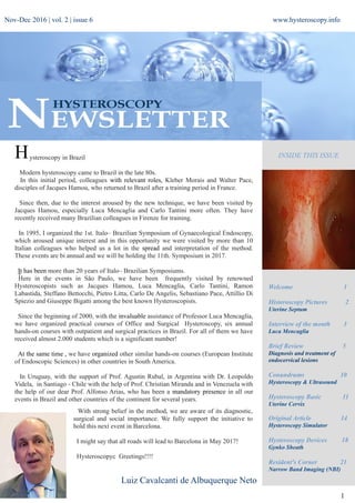
  • 1. + www.hysteroscopy.info 1 INSIDE THIS ISSUEH Welcome 1 Histeroscopy Pictures 2 Uterine Septum Interview of the month 3 Luca Mencaglia Brief Review 5 Diagnosis and treatment of endocervical lesions Conundrums 10 Hysteroscopy & Ultrasound Hysteroscopy Basic 11 Uterine Cervix Original Article 14 Hysteroscopy Simulator Hysteroscopy Devices 18 Gynko Sheath Resident's Corner 21 Narrow Band Imaging (NBI) Luiz Cavalcanti de Albuquerque Neto Nov-Dec 2016 | vol. 2 | issue 6 ysteroscopy in Brazil Modern hysteroscopy came to Brazil in the late 80s. In this initial period, colleagues with relevant roles, Kleber Morais and Walter Pace, disciples of Jacques Hamou, who returned to Brazil after a training period in France. Since then, due to the interest aroused by the new technique, we have been visited by Jacques Hamou, especially Luca Mencaglia and Carlo Tantini more often. They have recently received many Brazilian colleagues in Firenze for training. In 1995, I organized the 1st. Italo– Brazilian Symposium of Gynaecological Endoscopy, which aroused unique interest and in this opportunity we were visited by more than 10 Italian colleagues who helped us a lot in the spread and interpretation of the method. These events are bi annual and we will be holding the 11th. Symposium in 2017. It has been more than 20 years of Italo– Brazilian Symposiums. Here in the events in SĂŁo Paulo, we have been frequently visited by renowned Hysteroscopists such as Jacques Hamou, Luca Mencaglia, Carlo Tantini, Ramon Labastida, Steffano Bettocchi, Pietro Litta, Carlo De Angelis, Sebastiano Pace, Attillio Di Spiezio and Giuseppe Bigatti among the best known Hysteroscopists. Since the beginning of 2000, with the invaluable assistance of Professor Luca Mencaglia, we have organized practical courses of Office and Surgical Hysteroscopy, six annual hands-on courses with outpatient and surgical practices in Brazil. For all of them we have received almost 2.000 students which is a significant number! At the same time , we have organized other similar hands-on courses (European Institute of Endoscopic Sciences) in other countries in South America. In Uruguay, with the support of Prof. Agustin Rubal, in Argentina with Dr. Leopoldo Videla, in Santiago - Chile with the help of Prof. Christian Miranda and in Venezuela with the help of our dear Prof. Alfonso Arias, who has been a mandatory presence in all our events in Brazil and other countries of the continent for several years. With strong belief in the method, we are aware of its diagnostic, surgical and social importance. We fully support the initiative to hold this next event in Barcelona. I might say that all roads will lead to Barcelona in May 2017! Hysteroscopyc Greetings!!!!
  • 2. TEAM COODINATOR SPAIN L. Alonso EDITORIAL COMMITTEE SPAIN E. Cayuela L. Nieto ITALY G. Gubbini A. S. LaganĂ  USA J. Carugno L. Bradley MEXICO J. Alanis-Fuentes PORTUGAL J. Metello ARGENTINA A. M. Gonzalez VENEZUELA J. Jimenez SCIENTIFIC COMMITTEE A. Tinelli (Ita) O. Shawki (Egy) A. Ăšbeda (Spa) A. Arias (Ven) M. Rodrigo (Spa) A. Di Spiezio Sardo (Ita) E. de la Blanca (Spa) A. Favilli (Ita) M. Bigozzi (Arg) S. Haimovich (Spa) R. Lasmar (Bra) A. Garcia (USA) N. Malhotra (Ind) J. Dotto (Arg) I. Alkatout (Ger) R. Manchanda (Ind) M. Medvediev (Ukr) M. Elessawy (Ger) All rights reserved. The responsibility of the signed contributions is primarily of the authors and does not necessarily reflect the views of the editorial or scientific committees. HYSTEROSCOPY PICTURES www.hysteroscopy.info 2 The septate uterus is the result of a failure in the process of resorption of the medial septum that can range from a minimal septum in the uterine fundus to a complete one, dividing the uterine cavity completely or even asociating double cervix and vaginal septum. The proposed mechanism by which the uterine septum regresses is apoptosis, the presence of Blc-2 protein protects from apoptosis. The absence of Blc-2 in the embryonal uterine septum may indicate a lack of protection from apoptosis in this area. On the other hand different studies have allowed the identification of several genes that can be implicated in the development of uterine malformations In an interesting study comparing the use of resectoscope to VersaPoint in 63 women for the treatment of septum (Resectoscope or Versapoint for hysteroscopic metroplasty. Int J Obstet Gynaecol 2008 Apr; 101 (1):39-42) Dr. Litta concludes that the VersaPoint system does not require cervical dilation, avoiding potential cervical incompetence, cervical lacerations and uterine perforation related to the dilation process. The use of VersaPoint is a safe alternative to the use of resectoscope, especially in nulligravid or patients with cervical stenosis. If you are interested in sharing your cases or have a hysteroscopy image that you consider unique and want to share, send it to hysteronews@gmail.com Superficial vaginal endometriotic implant Detailed aspect of the cystic area with retained blood Detailed uterine septum Hysteroscopic metroplasty with bipolar system Nov-Dec 2016 | vol. 2 | issue 6
  • 3. 3 www.hysteroscopy.info INTERVIEW WITH... A entire life devoted to endoscopy and always a leader in progress and development. His daily work in favor of hysteroscopy, has contributed in making hysteroscopy a routine technique. Luca Mencaglia Direttore U.O.C. P.O.3 USL Toscana Sud Est Centro di PMA Ospedale Santa Margherita La Fratta Firenze, Italy One of your first publications in the early 80's was about the experimental application of the hysteroscopy. What has changed since then? Today we can say that hysteroscopy is no longer an experimental technique but a routine technique. Actually is not possible to work without it. Any modern gynecology service must have both diagnostic and surgical hysteroscopy. In particular, before to start any treatment, the modern diagnostic hysteroscopy should be performed in all patients with abnomal uterine bleeding (AUB) and in all infertile patients. In Europe these two categories represents about 65% of requests for gynecological examinations. Concerning the surgical hysteroscopy, some surgeries can only be performed by hysteroscopy. In particular, the treatment of uterine septum and intracavitary pathology, as endometrial polyps and submucosal fibroids, can be performed only and exclusively by hysteroscopy. Is the role of hysteroscopy in the premalignant lesions underestimated? Today diagnostic hysteroscopy, in association with endometrial biopsy under visual control, allows a precise diagnosis not only for histological type, but also for localization of endometrial preneoplastic and neoplastic lesions. I want to remember that 14% of pre-neoplastic and neoplastic endometrial lesions are focal lesions and therefore can be diagnosed only by visual inspection using hysteroscopy. A precise diagnosis of endometrial hyperplasia, with proper histological staging in its various forms (simple and complex and with or without cytologic atypia) is essential for proper treatment. It should be emphasized that today the hyperplasia treatment is based on a combination of therapy with progesterone and endometrial resection. This type of treatment has also been used, by our group, in selected cases of complex endometrial hyperplasia with cytologic atypia and focal endometrial cancer in young patients which desire a pregnancy. ”14% of pre-neoplastic and neoplastic endometrial lesions are focal lesions and therefore can be diagnosed only by visual inspection using hysteroscopy” Nov-Dec 2016 | vol. 2 | issue 6
  • 4. 4 www.hysteroscopy.info Has the debate about hysteroscopy and fertility finished or is just starting? We can definitely state that diagnostic hysteroscopy is an indisputable step for diagnosis of female infertility. So is necessary to perform a diagnostic hysteroscopy before starting any fertility treatment. The role of hysteroscopy surgery in infertile patients appear basic. Wherever an intracavitary disease (malformations, polyps, fibroids) is detected, the treatment should be done exclusively by hysteroscopy. If anything, the application of hysteroscopy as an experimental method in the study of the endometrium before implantation is still debated. Do you think that there is a growing interest about hysteroscopy? There is no doubt that hysteroscopy is a technological and conceptual evolving technique. For me it is absolutely clear that a modern gynecology service can not be exist without an efficient diagnostic and surgical hysteroscopy. Today in Italy approximately 15% of gynecologists are able to perform a diagnostic hysteroscopy and only 5% can perform a good surgical hysteroscopy. These numbers, although high compared to other European countries, have surely to grow for the diffusion of this technique in gynecology world. What's your vision about the future of the hysteroscopy? The hysteroscopy is a simple technique, painless and totally outpatient almost comparable with a transvaginal ultrasound. Gynecologists have to keep in mind that diagnostic hysteroscopy can be heplfull for “give a look" to the uterine cavity, this should be easy and simple as the modern hysteroscopy is. Do you have any advice for the young physician that is starting out in the world of gynecologic minimally invasive surgery? I suggest to young gynecologists, which work in minimally invasive surgery or in pure diagnostics, to learn as soon as possible the diagnostic hysteroscopy. It is known that to properly manage diagnostic hysteroscopy is necessary to perform al least 50 hysteroscopies. On the other hand the use of surgical hysteroscopy is more complex and is reserved to those who wants to pursue an experience in the field of minimally invasive surgery for which this technique is essential and indispensable. In other words, who works in gynecological laparoscopy and mininvasive surgery has to become an expert even in surgical hysteroscopy. ”There is no doubt that hysteroscopy is a technological and conceptual evolving technique.” ” I suggest to young gynecologists to learn as soon as possible the diagnostic hysteroscopy” Nov-Dec 2016 | vol. 2 | issue 6
  • 5. www.hysteroscopy.info 5 Diagnosis and Treatment of Endocervical Lesions by Cervicoscopy. Use and Accuracy of Hysteroscopic Endocervical Canal Biopsy. MarĂ­a Alejandra Brito PĂ©rez. Consulta de GinecologĂ­a. Unidad MĂ©dico QuirĂşrgica NORESTE. Caracas, Venezuela. Brief Review INTRODUCTION The endocervical canal is lined by columnar epithelium on a basement membrane. There are also endocervical glands, and its extension is influenced by age, parity and hormonal status. The Squamous Columnar Union (SCJ) is in constant change throughout the life of the woman, there is a constant transformation from columnar to squamous epithelium, which is known as squamous metaplasia. Lesions of the cervix can be produced by trauma (tears, holes or ulcerations), infection: such as those caused by sexually transmitted agents (Chlamydia trachomatis, Neisseria gonorrhea, Trichomonas, herpes virus, or HPV), and neoplasms, that can be benign represented by endocervical polyps or fibroids, endometriosis, or malignant. For analysis of premalignant cervical lesions, we use the 2001 Bethesda System, which organizes cervical cells from normal to carcinoma in situ, both originated from squamous cells, and from the glandular epithelium. In 1975, for the first time, the possible association between HPV and cervical cancer was noted, and 8 years later Zur Hausen and then Walmoeers and Bosch demonstrated the possibility to isolate the virus from cervical biopsies. Research in this area aims to eliminate intraepithelial neoplasia to avoid progression. The hysteroscope allows precise and direct evaluation of internal genitalia by a trained provider. It represents a powerful tool in the diagnosis of benign pathologies, pre-malignant and malignant lesions located in the endocervical canal and endometrial cavity. The gold standard for the diagnosis of premalignant and malignant cervical pathology, is the pap smear followed by colposcopy and biopsy. In this study we aim to assess the use of hysteroscopy, and its undeniable utility in observing the endocervical canal, deeper than the area reached by the gynecologist with a colposcope. OBJECTIVE To examine the use of hysteroscopy evaluation of the endocervical canal as complementary technique for diagnosis and treatment of endocervical pathology. SETTING Prospective, observational-descriptive study of a group of patients referred for hysteroscopy between August 2015 and January 2016. Endocervical lesions were described. Gynecological Medical Surgical Unit NORTHEAST. Caracas Venezuela. 46 patients were included, aged between 28 and 65 years, who were referred to the consultation of hysteroscopy evaluation. The indications for the evaluation were: Infertility Suspected endometrial pathology ultrasound Abnormal vaginal bleeding Cervical cytology with squamous or glandular cells (AGUS / ASCUS) Lost IUD threads Suspected Mullerian malformations Nov-Dec 2016 | vol. 2 | issue 6
  • 6. www.hysteroscopy.info 6 METHOD Each of the patients underwent colposcopy followed by hysteroscopy of the endocervical canal (endo-cervicoscopy). After performing hysteroscopy, acetic acid 5% was placed in the endocervical canal. A second hysteroscopy was then performed. Patients were divided in 3 groups: GROUP A: patients without lesions seen at endocervicoscopy. GROUP B: Patients with lesions seen at endocervicoscopy. GROUP C: Patients with lesions seen at endocervicoscopy after acetic acid administration. RESULTS The most frequent indication for hysteroscopy was abnormal endometrial ultrasound findings (45.65%) followed by abnormal uterine bleeding. Group A: 32.6% of patients had sinequia of the cervical canal followed by endocervical poyp in a 26.08%. Group B: 21 (45.6%) patients had proliferative lesions seen on endocervicoscopy. Group C, 11 patients, which represented 24% of the study population, who had lesions present at endocervicoscopy after acetic acid was applied. Of the 11 patients in this group, 10 had pathologic findings associated with HPV on biopsy. DISCUSSION Colposcopic diagnosis of endocervical lesions is a challenge, due to the technical difficulty to evaluate the endocervical canal that is not visible to the examiner. The use of the hysteroscope could simplify the visualization of cervical lesions located deep in the endocervical canal. Squamous adenocarcinoma of the cervix accounts for 20% of cervical cancers.The metaplastic epithelium of the cervix and glandular epithelium SCJ (squamous columnar junction) is susceptible to the Human Papilloma Virus. Lesions frequently extend up to the endocervical canal. We found 11 patients with acetowhite lesions deep in the endocervical canal seen during endocerviscopy that were not seen on colposcopy. Of these 10 (90.0%) had HPV changes on pathology. This should call reflection about the role of hysteroscopy in the evaluation of premalignant lesions of the cervix. CONCLUSIONS Endocervicoscopy (Hysteroscopic evaluation of the endocervical canal) is a safe tool to evaluate cervical premalignant lesions located deep in the endocervical canal not seen during colposcopy due to their location. Further studies of this innovative technique with a larger group of patients is encourage. Nov-Dec 2016 | vol. 2 | issue 6 Cervicoscopy with acetic acid (5%) in patients with cervical lesions Cervicoscopy with acetic acid 5% Acetowhite and papilomatosis lesions Without atypia Acetowhite and papilomatosis lesions Negative
  • 7. www.hysteroscopy.info 7 Endocervical canal and internal os without acetic acid. Hysteroscopy Newsletter Hysteroscopy Newsletter Hysteroscopy Newsletter Hysteroscopy Newsletter Hysteroscopy Newsletter Hysteroscopy Newsletter Hysteroscopy Newsletter Hysteroscopy Newsletter Hysteroscopy Newsletter Hysteroscopy Newsletter Endocervical thick adhesion close to the internal os without acetic acid. Endocervical polyp without acetic acid. Endocervical canal (Plicae palmatae) without acetic acid Papilloma like lesion in the endocervical canal after application of acetic acid Papillary like lesion in the endocervical canal. Note the thin vascularity inside the papillae. After application acetic acid. Papillary like lesion in the uterine isthmus after application of acetic acid. Papilloma like lesion in the endocervical canal after application of acetic acid. It reveals the endocervical glandular epithelium. Nov-Dec 2016 | vol. 2 | issue 6
  • 8. www.hysteroscopy.info 8 DID YOU KNOW...? The intravasation syndrome is characterized by hyponatremia, with consequent headache, sleepiness, scotomas, restlessness, and nausea. A randomized trial found that intracervical injection of dilute vasopresin solution (0,05 U/ml)can reduce the force needed to dilate the cervix Nov-Dec 2016 | vol. 2 | issue 6
  • 9. www.hysteroscopy.info 9 Hysteroscopy H. Van Der Pas, B. Van Herendael and L.G. Keith Springer Reprint of the original 1st ed. 1983 Year 2013; 252 pages Because of increasing worldwide interest in the subject, a decision was made to coordinate the papers presented at the First European Symposium on Hysteroscopy, held in Antwerp, Belgium, in September 1982, into a text on hysteroscopy, including its indications, techniques, and complications. The Organising Committee called on Dr. L. G. Keith of Northwestern University, Chicago, illinois USA, to edit all of the papers and to prepare them for publication as a multi-author textbook rather than as a haphazard collection of the proceedings of a meeting. The speakers at the Symposium were all eminent European hysteroscopists; the most up-to-date information was presented, assuring the current nature of the scientific data. WHAT'S YOUR DIAGNOSIS? Sometimes, when performing hysteroscopy, it is important to pay attention to every corner of the uterus, as Vasari stated «cerca trova», «he who seeks finds» Answer to the previous issue: Vascularization of the implantation area of RPOC. Hysteroscopy Newsletter Nov-Dec 2016 | vol. 2 | issue 6
  • 10. www.hysteroscopy.info 10 Hysteroscopy Conundrums Hysteroscopy & Ultrasound Lookforus:hysteroscopygroupinLinkedIn Do you usually perform an ultrasound after a hysteroscopy? Do you think it is necessary? Do you use 3D ultrasound in the diagnosis of uterine malformations? Ultrasound is performed only after a known incomplete myomectomy for monitoring of the residual fibroid tissue and prior to performing a second hysteroscopy if indicated. To evaluate mullerian malformations I usually use 2D unltrasound and MRI prior to hysteroscopy, but I believe 3D ultrasound is a valid option to consider. Nov-Dec 2016 | vol. 2 | issue 6
  • 11. www.hysteroscopy.info 11 HYSTEROSCOPY BASIC Uterine Cervix The name cervix ​​derives from the Latin word “Cervic” meaning "neck". It represents the lower portion of the uterus and communicates the uterine cavity with the vagina. It has a cylindrical shape with a length of about 3 cm and a diameter of about 2 cm. The uterine cervix has an opening to the vagina called the “external os” (EO). In the area of ​​division between the cervix and uterine body lies a fibromuscular area called the “internal os” (IO). The area located between EO and IO is called the “endocervical canal”, which has a fusiform shape and an oval cross section, the endocervial canal has a diameter ranging between 3 and 10 millimiters. The normal development of the uterus involves some changes happening over the mullerian ducts to form the cervix, uterus, fallopian tubes and upper vagina through the processes of differentiation, migration, fusion and canalization The cervix has an intravaginal portion (portio vaginalis) and a supravaginal portion that is usually larger. Structurally, the cervix is ​​composed of 85% extracellular matrix and 15% smooth muscle tissue. The extracellular matrix is ​​made mainly of collagen, elastin and proteoglycans cells of smooth muscle and fibroblasts, epithelial cells and blood vessels. Hysteroscopy Newsletter Nov-Dec 2016 | vol. 2 | issue 6
  • 12. www.hysteroscopy.info 12 The hard consistency of the cervix is ​​derived from its collagen component, which is the predominant protein found in the extracellular matrix. The ratio of connective tissue and smooth muscle is not evenly distributed through the entire length of the cervix, the distal portion has more connective tissue than the upper portion, where there is more smooth muscle. Smooth muscle fibers are predominantly located at the level of ​​the IO. The epithelium of the cervix in its intravaginal portion corresponds to squamous epithelium. It changes to columnar epithelium in the endocervical canal. The area of transition between the two epithelia corresponds to the squamocolumnar junction also known as the transformation zone. The arrangement of the epithelium at the level of the cervical canal is made of longitudinal ridges along the canal, this is called "plica palmatae". On top of the longitudinales ridges there are also oblique branches that give it the appearance of tree branches that is also called "arbor vitae". The cervix remains anchored in place by the uterosacral ligaments and the lateral or cardinal ligaments. The two uterosacral ligaments extend from the posterior cervix to the sacrum. The lateral ligaments, also called “cardinal ligaments” or “Mackenrodt ligaments” target the pelvic sidewall, which also consitute the base of the broad ligament. The vascular blood supply of the cervix is provided mainly by the uterine artery. When the uterine artery reach the level of the uterine isthmus it is divided into two terminal branches, the cervicovaginal branch, which supplies the lower portion of the cervix and the ascending branch of the uterine artery, which branches out to be responsible for the irrigation of the upper portion of the uterus. The venous drainage of the cervix is ​​performed by capillaries that join to form the uterine venous plexus that drains into the ipsilateral uterine vein. The sensory innervation of the uterus is maily provided by two nerves. The Franken-Hauer plexus (S2-S4) which collects the sensitivity of the cervix and lower uterine body. The upper uterine corpus and uterine fundus receive innervation through the infundibulopelvic ligament from the ovarian plexus. Hysteroscopy Newsletter Hysteroscopy Newsletter Hysteroscopy Newsletter Nov-Dec 2016 | vol. 2 | issue 6
  • 13. www.hysteroscopy.info 13 CongresSINTERNATIONAL APAGE and TAMIG Annual Congress Taipei, Taiwan |Nov 3-6|2016 European Society of Gynaecological Oncology (ESGO) Vienna, Austria | Nov 4-7| 2017 The 24th World Congress on Controversies in Obstetrics, Gynecology & Infertility Amsterdam, Netherlands |Nov 10-13|2016 45th AAGL Global Congress Orlando, Florida |Nov 14-18|2016 3rd International Conference on Gynecology & Obstetrics Dubai, EAU Nov 24-26 |2016 3rd Global Congress on Hysteroscopy Barcelona, Spain May 2-5 |2017 RCOG World Congress 2017 Cape Town, South Africa | Mar 20-22 |2017 World Congress on Recurrent Pregnancy Loss Cannes, France | Jan 19-22|2017 Pelvic Anatomy and Gynecologic Surgery Las Vegas, USA | Dec 8-10|2016 Congresso Brasileiro de Reprodução Humana Sao Paulo, Brasil| Nov 3-5|2016 All India Congress of Obstetrics & Gynaeology Ahmadabad, India| Jan 25-29|2017 Congress of the European Society of Gynecology Barcelona Spain | Oct 18-21|2017 Nov-Dec 2016 | vol. 2 | issue 6
  • 14. 14 www.hysteroscopy.info Introduction Hysteroscopy is a minimally invasive technique for the assessment and treatment of intrauterine pathologies. The development of new instruments and the introduction of fellowship programs for the training of residents in minimally invasive gynecological surgery have encouraged the usage and implementation of diagnostic and operative hysteroscopy in routine clinical practice. In comparison with laparoscopy, hysteroscopic skills are often assumed to be less difficult to obtain. The teaching of hysteroscopic interventions has received little attention thus far, focusing mainly on the development of physical models and box simulators, while the demand for well-structured hysteroscopy training is expanding rapidly. Existing training models range from cow uteri and bladders to virtual reality; however, a Dutch survey revealed that lack of simulation training during residency was the leading factor that could be enhanced for optimal acquisition of hysteroscopic skills. Objective The primary objective of our study was to test the construct validity of the HystSim™ hysteroscopic simulator to determine whether simulation training can improve the acquisition of hysteroscopic skills regardless of the previous levels of experience of the participants. The secondary objective was to analyze the performance of a selected task, using specially designed scoring charts to help reduce the learning curve for both novices and experienced surgeons. Kiel endoscopy school experience with integration of hysteroscopy simulator in training Courses Mohamed Elessawy, N. Maass, Ibrahim Alkatout Department of Gynecology and Obstetrics, University Hospitals Schleswig-Holstein, Kiel, Germany Original Article Nov-Dec 2016 | vol. 2 | issue 6
  • 15. Design The teaching of hysteroscopic intervention has received only scant attention, focusing mainly on the development of physical models and box simulators. This encouraged our working group to search for a suitable hysteroscopic simulator module and to test its validation. We decided to use the HystSim™ hysteroscopic simulator, which is one of the few such simulators that has already completed a validation process, with high ratings for both realism and training capacity. As a testing tool for our study, we selected the myoma resection task. We analyzed the results using the multimetric score system suggested by HystSim™, allowing a more precise interpretation of the results. Setting Between June 2014 and May 2015, our group collected data on 57 participants of minimally invasive surgical training courses at the Kiel School of Gynecological Endoscopy, Department of Gynecology and Obstetrics, University Hospitals Schleswig-Holstein, Campus Kiel. The novice group consisted of 42 medical students and residents with no prior experience in hysteroscopy, while the expert group consisted of 15 participants with more than 2 years of experience of advanced hysteroscopy operations. Results The overall results demonstrated that all participants attained significant improvements between their pre- and post-tests, independent of their previous levels of experience (P< 0,002). Those in the expert group demonstrated statistically significant, superior scores in the pre- and post-tests (P=0.001, P=0.006) Figure 1. With regard to visualization and ergonomics, the novices showed a better pre-test value than the experts; however, the experts were able to improve significantly during the post-test. These precise findings demonstrated that the multimetric scoring system achieved several important objectives, including clinical relevance, critical relevance, and training motivation. Conclusion All participants demonstrated improvements in their hysteroscopic skills, proving an adequate construct validation of the HystSim™. Using the multimetric scoring system enabled a more accurate analysis of the performance of the participants independent of their levels of experience which could be an important key for streamlining the learning curve. 15 www.hysteroscopy.info Figure 1a Mean ± standard deviation between the two groups in the pre- and post-tests for the overall result The novice group scored a mean (±standard deviations) of 54.95 (85.06) points in the pre-test and 147.81 (97.79) in the post-test, while the experts scored a mean (±standard deviations) of 52.40 (92.77) in the pre-test and 206.60 (97.21) in the post-test. Figure 1b Improvement between post- and pre-test mean ± standard deviation between the two groups Both the novices and experts improved significantly, scoring a mean difference (±standard deviations) between the pre- and post-tests of 90.32 (122.89) and 154.20 (148.34), respectively Nov-Dec 2016 | vol. 2 | issue 6
  • 16. www.hysteroscopy.info 16 WWW:HYSTEROSCOPY2017.COM LIMITED PLACES !! Register now to ensure your participation Nov-Dec 2016 | vol. 2 | issue 6
  • 17. www.twitter.com/hysteronews HYSTEROscopy group Hysteroscopy newsletter Hysteroscopy newsletter www.facebook.com/hysteronews 17 www.hysteroscopy.info Some Pictures Dr. Luis Alonso and Prof. Sergio Haimovich chairs of the Global Congress Hysteroscopy in a great meeting with Prof. Bruno Van herendael, presidente of the ISGE and the famous Prof. Stefano Bettocchi, during the ESGE Congress in Brussels. Including more friends to the Global Congress Hysteroscopy. Grigoris Grimbizis (ESGE), Stefano Bettocchi (ISGE), Attilio Di Spiezio Sardo (ESGE), Luis Alonso (cochair #GCOH2017), Sergio Haimovich (chair #GCOH2017) and Ricardo Lasmar (organizing committte #GCOH2017). Cooper Surgical will be present at the Global Congress Hysteroscopy next May 2017 with the Endosee device. Thank you Jamie Planner and Emma Dauncey. Prof. Hua Duan from China and Prof. Chyi-Long Lee Chairman of Board of Trustees APAGE. The Asia-Pacific Association for Gynecologic Endoscopy and Minimally Invasive Therapy is also supporting the GlobalCongress on Hysteroscopy. Nov-Dec 2016 | vol. 2 | issue 6
  • 18. 18 www.hysteroscopy.info DEVICES HYSTEROSCOPY GynkoSheath The new single-use sheath which makes diagnostic hysteroscopy of 3mm! Made of medical polyurethane plastic, the new cover GYNKO provides the gynecologist a useful tool to perform diagnostic hysteroscopy of only 3mm. GYNKO is compatible to the optical commonly used in hysteroscopy 0 and 30 degrees (Storz type) and incorporates a working channel for instruments up 7Fr in conjunction with a channel for irrigation and a protective cover for the camera. GYNKO will allow to perform the biopsy of the endometrium as well as the removal of small polyps. With this new system, you can multiply the hysteroscopic procedures without having to repeatedly sterilize the entire system. Risks of Virus Transmission During Diagnostic Hysteroscopy J Am Assoc Gynecol Laparosc. 1996 Aug;3(4, Supplement):S30. Mencaglia L, Tiso E, Tantini C, Bianchi R. The high frequency of asymptomatic carriers of viral infections represents a major risk for transmission. Viral agents can be transmitted through blood or biologic fluids during diagnostic hysteroscopy. Routine disinfection methods to clean hysteroscopes cannot be considered adequate to prevent transmission. Hepatitis C and B viruses require 1-hour sterilization in glutaric aldehyde or gas sterilization (ethylene oxide) to be decontaminated. Human immunodeficiency virus decontamination of instruments is easier and safer. Blood test screening of patients positive for hepatitis B and C should be performed in all candidates for endoscopic gynecologic procedures to avoid the possibility of virus transmission. www.gyneworld.com Nov-Dec 2016 | vol. 2 | issue 6
  • 19. 19 www.hysteroscopy.info HIGHLIGHT ARTICLES Published on different medias OBJECTIVE: Investigate a novel office hysteroscopic tubal catheterization therapeutic method for proximal tubal occlusion. STUDY DESIGN: Prospective cohort study in a tertiary referral center. We evaluated the procedure on a group of 27 patients that were referred to our unit for proximal tubal occlusion demonstrated by hysterosalpingography, 9 (33.3%) of them with primary infertility and 18 of them (66.6%) with secondary infertility. The intervention included the usage of the modified Novy cornual cannulation set which was inserted through a 5F working cannel during an office operative hysteroscopy, followed by fallopian tube irrigation with saline-air mixture under ultrasonographic imaging. RESULTS: Our series revealed no complication during or after the procedure; anesthesia was not required. One patient lost from follow-up. Of the remaining 26, 10 patients (38.4%) conceived either spontaneously or with treatment by clomiphene or gonadotropine associated with intrauterine insemination. The median time to conception was 5 months (range 4-17). CONCLUSION: We therefore concluded that office hysteroscopic tubal catheterization is a simple (without anesthesia required) option for the treatment of patients suffering from proximal tubal occlusion. Fertility outcomes in our series are comparable to other treatments options for tubal catheterization. Therefore, tubal catheterization should not delay the assisted reproducted techniques if indicated but we propose to include it in a global integrated approach. Hysteroscopically guided transvaginal ultrasound tubal catheterization-a novel office procedure. Cohen SB, Bouaziz J, Jakobson-Setton A, Goldenberg M, Schiff E, Orvieto R, Shulman A. Eur J Obstet Gynecol Reprod Biol. 2016 Sep;204:113-6 OBJECTIVE: Type 3 myomas are intramural within contact with the endometrium but lack any cavity deformation. There is no guideline for management of symptomatic type 3 myoma. The aim of this study was to evaluate the feasibility of symptomatic type 3 myoma hysteroscopic resection. METHOD: This retrospective study included symptomatic women (mainly pain, infertility or bleeding) who obtained an operative hysteroscopy for type 3 symptomatic myoma from June 2010 to December 2014 in the gynaecological unit of a teaching hospital. RESULTS: Thirteen women with an operative resection using bipolar electrosurgery of type 3 myoma during the study period (June 2010 to December 2014) were included in the study. Two women had a hysterectomy 6 and 12 months after the procedure and one woman had an open myomectomy 30 months after the procedure for the recurrence of abnormal bleeding. Postoperative office hysteroscopy show a postoperative synechiae in 3 women out of 8. Incomplete resection was also obtained in 3 women out of 8. CONCLUSIONS: Hysteroscopic resection is a potential alternative to traditional surgery for type 3 myoma. This procedure must be confined to skilled surgeons because it is a difficult procedure. A postoperative office hysteroscopy is recommended in women of reproductive age. Hysteroscopic resection of type 3 myoma: a new challenge? Capmas P, Voulgaropoulos A, Legendre G, Pourcelot AG, Fernandez H. Eur J Obstet Gynecol Reprod Biol. 2016 Oct;205:165-9 Hysteroscopic resection of type 3 myoma: a new challenge? Capmas P1, Voulgaropoulos A2, Legendre G2, Pourcelot AG3, Fernandez H4. Eur J Obstet Gynecol Reprod Biol. 2016 Oct;205:165-9 Nov-Dec 2016 | vol. 2 | issue 6
  • 20. 20 www.hysteroscopy.info HYSTEROSCOPY IN THE NET HYSTEROWEB If you are a lover or an expert of hysteroscopy, Hysteroyou is your new professional interactive world. You will have a room with a view on hysteroscopy: you will share opinions, clinical cases, procedures, photos and videos. All events and technological news on hysteroscopy will be daily updated and shared. And if this is not enough you will have all specialised assistance needed from companies’ experts for all your “technical” problems concerning hysteroscopic instruments. The new portal developed by Prof. Stefano Bettochi, allows the exchange of knowledge between hysteroscopists around the world. Get up and enjoy Hysteroyou network. The world is waiting more hysteroscopy. http://www.hysteroyou.com/ Nov-Dec 2016 | vol. 2 | issue 6
  • 21. 21 www.hysteroscopy.info Resident'sCORNER Reliability of hysteroscopy combined with narrow band imaging (NBI) technology Paula MasĂł Marrodán. Hysteroscopy Unit. Hospital del Mar, Barcelona.España Narrow-band imaging (NBI) or "optical narrowband filter" is a novel system of endoscopic visualization in real time which significantly improves visualization of the vascularity and the tissue surface, allowing better characterization of abnormal findings. Applies a technology based on narrowing of the white light spectrum commonly used endoscopically through a system of filters. Thus, this system allows two wavelengths (415nm blue light and 540nm green light) to reach the surface of the tissue and reflected back on a chip that captures the image. Both wavelengths correspond to peaks in the absorption spectrum of the oxidized hemoglobin light, fact that allows to appreciate more clearly the vascular network in tissue surface. Blue light penetrates tissues poorly (0'17mm) and as a result allows to see the superficial vascular networks, while green light penetrates deeper and shows sub-epithelial vessels. When both are combined you can observe an image of the tissue surface with high contrast. This technology can detect irregular vascular patterns, can be a helpful tool in the early detection of malignant lesions. The first data of this system date from 2001, it was described by Gono and Sano at the National Center of Tokyo and created by the technology company Olympus. So far, this technology has had its medical application mainly in the field of gastroenterology, where it is used to detect lesions such as Barrett's esophagus, esophageal cancer, premalignant and malignant stomach and colon lesions, or small bowel lesions through the use of NBI in upper endoscopy and colonoscopy. In the last two years it has extended its use and is beginning to be used for the detection of urothelial carcinoma of the bladder, and especially for detecting recurrences of this neoplasm in monitoring patients. In the field of gynecology, there is work using this technology, especially for detection of endometrial pathology. There are reported cases of use of NBI for the detection of peritoneal implants in borderline ovarian tumors and peritoneal implants in cervical carcinoma, with promising results (Fanfani F. et al). A.Polyp B. Myoma C. Endometrial Hyperplasia Nov-Dec 2016 | vol. 2 | issue 6
  • 22. 22 www.hysteroscopy.info Nov-Dec 2016 | vol. 2 | issue 6 Diagnostic hysteroscopy is currently considered the gold standard for the evaluation of the uterine cavity, with high sensitivity (94.2%), specificity (88.8%), negative predictive value (96.3%) and positive predictive value (83.1%). However, it has lower performance for the detection of diseases such as hyperplasia, with a sensitivity of 70%, specificity of 91.6%, negative predictive value of 94.3% and a positive predictive value of 60.6%. Thus there is the need for additional tests to increase the sensitivity of detection of lesions such as hyperplasia, usually performing biopsy with an increased risk of complications. Cicinelli et al. in 2009 (Journal of Minimally Invasive Gynecology) suggests that the NBI technology improves the diagnostic accuracy of hysteroscopy, increasing the sensitivity from 77% (with normal white light) to 90% (p <0.0001), and the positive predictive value from 77% to 99% (p> 0.0001). This was also replicated by other investigators. Doctor R. Tinelli, describing the improvement in sensitivity with the use of NBI in the detection of endometrial cancer compared to using white light (93% vs 81%, p <0.05 ) and improved sensitivity (82% vs 56%, p <0.05) and positive predictive value (79% vs 71%, p <0.05) for the detection of low-risk hyperplasia, and improved sensitivity (60% vs 20%, p <0.05) and positive predictive value (67% vs 25%, p <0.05) for the detection of high-risk hyperplasia, without differences in the negative predictive value, specificity and accuracy. Moreover, Kisu Iori et al. associated the NBI technology with flexible hysteroscopy, also obtaining increased sensitivity for the detection of atypical endometrial hyperplasia and endometrial cancer. Angiogenic intensity of tissues could have a prognostic role in endometrial malignancy. In 2006, Ingunn et al published a paper on the importance of vascular proliferation for clinical development of endometrial cancer, finding that such proliferation is the most potent angiogenic prognostic marker in the progression of this malignancy. This factor could be detected with the use of NBI and allow early diagnosis and prognostic information of this pathology. These encouraging results of the studies published to date make us think that the application of NBI in hysteroscopy has a promising future in the detection of endometrial malignant lesions, allowing early detection and characterization of the malignant lesions. EnhacedvascularizationBaseofpolypEndometrialCancer
  • 23. In recent years we have witnessed the remarkable progress that has happened in the field of gynecological endoscopy. Technological development has led to a huge improvement in image quality and miniaturization of surgical devices. New techniques and methods have emerged. This process is consistently advancing, very dynamically, and still has quite a way to go. Within the field of gynecological endoscopy, hysteroscopy is booming. More and more gynecologists want to learn and develop this technique in order to include it in their daily clinical practice. As a result, you can also see a simultaneous increase in the practice of hysteroscopy carried out in all hospitals in the world. Almost two years ago a group of gynecologists wanted to share their passion for hysteroscopy with the world and so we created the first international publication dedicated exclusively to the hysteroscopy and this was done through a LINKEDIN group: Hysteroscopy. This Hysteroscopy Newsletter brought the first international group of experts in hysteroscopy together, completely independent from all other international societies. In our desire to further the dissemination of hysteroscopy and to establish it independent of laparoscopy, we decided to organize the first Global Congress on Hysteroscopy, which will be held in Barcelona next year between the 2nd and 5th May, 2017. To bring toguether the best hysteroscopists, experts in different subjects of the hysteroscopy under the same roof it is something almost impossible. To gather around 1500 Hysteroscopy lovers in one congress is simply crazy. But this is our dream. As doctors we have to look for the best knowledge and to know the different techniques to help our patients. A Global Congress on Hysteroscopy will achieve this purpose. This conference has been organized by hysteroscopists from different countries and a network of regional representatives, who form the link between the organizing committee and the various national societies. Social media has been the perfect channel to bring toghether the most important hysteroscopists from all around the world and helped us to build the First Global Congress on Hysteroscopy. Most relevant International Societies of gynecological surgery will support this congress. Please, help us to spread the Word about this event. If you are a Hysteroscopy Lover, share this newsletter with your colleagues, your residents, in your Clinic or hospital. EVERYBODY must know about this congress. Your help is needed to spread the hysteroscopy worldwide!! thank you all for making this possible!!! Luis Alonso HYSTEROSCOPY Editorial teaM www.medtube.net Hysteroscopy newsletter HYSTEROscopy group Hysteroscopy newsletter www.twitter.com/hysteronews www.facebook.com/hysteronews FIND US ON Hysteroscopy Newsletter is an opened forum to all professionals who want to contribute with their knowledge and even share their doubts with a word-wide gynecological community www.hysteroscopy.info 23 Nov-Dec 2016 | vol. 2 | issue 6