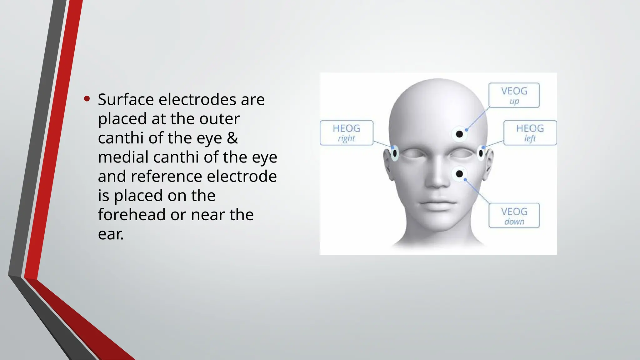
Electro-oculography
&
Electroretinography
-By Dr. N. Revanth
Specific Learning Objectives:
• Definitions of Electro-oculography (EOG) & Electroretinogram
(ERG).
• Different types of EOG & ERG.
• Test procedure.
• Normal patterns & interpretations.
• Advantages & limitations.
Electro-oculography:
Definition:
• The electrooculogram (EOG) measures the cornea-positive
standing potential relative to the back of the eye by attaching skin
electrodes outside the eye near the lateral and medial canthus.
Introduction:
• EOG measures the potential difference between the cornea and
the retina ( mainly retina pigment epithelium) during eye
movements.
• Cornea and retina maintain a constant electrical potential
difference creating a constant “ocular dipole”.
• When the eyes move, this potential difference changes.
• The EOG is the method by which these minor electrical potential
differences are captured by placing electrodes on the surface.
History:
• Elwin Marg first described & named EOG.
• Geoffrey Arden is the first to use and compare the amplitudes of
electric potentials under light and dark-adapted states, hence the
name “ Arden Ratio”.
• EOG became one of the first techniques to provide non-invasive
insight into the functional status of the RPE.
Test procedure:
• The patient should be in stable indoor lighting for at least 30 mins
before the test.
• Should not be exposed to any strong illumination during the rest
period.
• The patient should be dilated.
• He/she should be told to remain still except for eye movement
during the test procedure.
• Surface electrodes are
placed at the outer
canthi of the eye &
medial canthi of the eye
and reference electrode
is placed on the
forehead or near the
ear.
• The patient alternates the gaze between two fixed points and the
voltage change during the eye movements is recorded.
• The difference in potential is called standing potential.
• It indirectly measures the transepithelial potential of retinal
pigment epithelium(RPE).
Arden Ratio:
• Dark trough/DT: During 15 mins of dark adaptation the standing
potential usually reaches a minimum level, called dark trough.
• Light peak/LP: During 15 mins of light adaptation the standing
potential usually reaches its highest value at 7-12 min called light
peak.
• The light peak: dark trough(usually preferred) ratio is referred to
as the Arden ratio.
• Normal wave forms in EOG during light & dark adaptations
Image source: Donnell J. Creel’s ERG & EOG Clinical applications
Interpretation:
• Light peak-to-dark trough ratio of > 1.8 or 180% is normal and less
than 1.5 or 150% is abnormal, in people below 60 years.
• In people aged above 60 yrs, >1.7 is considered normal.
Advantages and limitations:
• simple., non-invasive and provides information about retinal
pigment epithelium.
• Limitations include, it cannot provide information about retinal
functional beyond the RPE.
Electroretinography
Definition:
• In electroretinography (ERG), changes in the resting potential of
the eye when induced by the stimulation of the eye with a light
stimulus are measured.
• The flash of light elicits a biphasic waveform recordable at cornea
when placed an electrode.
History:
• Ragnar Granit is the first to identify that ERG waveform reflected
different components of retinal activity, particularly rods and
cones.
Introduction:
• ERG measures the electrical responses of various retinal cells,
particularly rods and cones to light stimuli.
• Photoreceptor cells generate electrical signals in response to light
stimuli.
Test procedure:
• Pupils are dilated first, station may be set to dark or light
adaptation depending on the test that would like to be performed.
• Contact lens electrode is placed on cornea and reference
electrodes are placed on the skin.
• Flashes or patterns of light are presented ti evoke retinal
responses.
Electrode placements:
• Waveform:
Image source: Parsons diseases of the Eye- 23rd
edition.
Types of ERG:
• 1). Pattern ERG
• 2). Multifocal ERG
• 3). Full field ERG.
• Rods are tested in dark adaptation with a blue light stimulus-
Scotopic ERG
• Cones are tested in bright light or flickering light stimulus-
Photopic ERG..
Advantages and limitations:
• Provides detailed functional assessment of retinal health.
• Also can identify a wide range of retinal disorders.
• Limitations include, it requires specialized equipment & can be
uncomfortable to patients due to the use of corneal electrodes.
Summary:
•EOG: Provides insight into RPE function and eye movement control.
•ERG: Offers a detailed evaluation of retinal cell function, aiding in
diagnosing various retinal conditions.
References:
• Parsons Diseases of the Eye- 23rd
edition.
• The electroretinogram and electrooculogram: clinical applications
by Donnell J. Creel.
Electrocculography & Electroretinography.pptx

Electrocculography & Electroretinography.pptx

  • 1.
  • 2.
    Specific Learning Objectives: •Definitions of Electro-oculography (EOG) & Electroretinogram (ERG). • Different types of EOG & ERG. • Test procedure. • Normal patterns & interpretations. • Advantages & limitations.
  • 3.
    Electro-oculography: Definition: • The electrooculogram(EOG) measures the cornea-positive standing potential relative to the back of the eye by attaching skin electrodes outside the eye near the lateral and medial canthus.
  • 4.
    Introduction: • EOG measuresthe potential difference between the cornea and the retina ( mainly retina pigment epithelium) during eye movements. • Cornea and retina maintain a constant electrical potential difference creating a constant “ocular dipole”. • When the eyes move, this potential difference changes. • The EOG is the method by which these minor electrical potential differences are captured by placing electrodes on the surface.
  • 5.
    History: • Elwin Margfirst described & named EOG. • Geoffrey Arden is the first to use and compare the amplitudes of electric potentials under light and dark-adapted states, hence the name “ Arden Ratio”. • EOG became one of the first techniques to provide non-invasive insight into the functional status of the RPE.
  • 6.
    Test procedure: • Thepatient should be in stable indoor lighting for at least 30 mins before the test. • Should not be exposed to any strong illumination during the rest period. • The patient should be dilated. • He/she should be told to remain still except for eye movement during the test procedure.
  • 7.
    • Surface electrodesare placed at the outer canthi of the eye & medial canthi of the eye and reference electrode is placed on the forehead or near the ear.
  • 8.
    • The patientalternates the gaze between two fixed points and the voltage change during the eye movements is recorded. • The difference in potential is called standing potential. • It indirectly measures the transepithelial potential of retinal pigment epithelium(RPE).
  • 9.
    Arden Ratio: • Darktrough/DT: During 15 mins of dark adaptation the standing potential usually reaches a minimum level, called dark trough. • Light peak/LP: During 15 mins of light adaptation the standing potential usually reaches its highest value at 7-12 min called light peak. • The light peak: dark trough(usually preferred) ratio is referred to as the Arden ratio.
  • 10.
    • Normal waveforms in EOG during light & dark adaptations Image source: Donnell J. Creel’s ERG & EOG Clinical applications
  • 11.
    Interpretation: • Light peak-to-darktrough ratio of > 1.8 or 180% is normal and less than 1.5 or 150% is abnormal, in people below 60 years. • In people aged above 60 yrs, >1.7 is considered normal.
  • 12.
    Advantages and limitations: •simple., non-invasive and provides information about retinal pigment epithelium. • Limitations include, it cannot provide information about retinal functional beyond the RPE.
  • 13.
    Electroretinography Definition: • In electroretinography(ERG), changes in the resting potential of the eye when induced by the stimulation of the eye with a light stimulus are measured.
  • 14.
    • The flashof light elicits a biphasic waveform recordable at cornea when placed an electrode.
  • 15.
    History: • Ragnar Granitis the first to identify that ERG waveform reflected different components of retinal activity, particularly rods and cones.
  • 16.
    Introduction: • ERG measuresthe electrical responses of various retinal cells, particularly rods and cones to light stimuli. • Photoreceptor cells generate electrical signals in response to light stimuli.
  • 17.
    Test procedure: • Pupilsare dilated first, station may be set to dark or light adaptation depending on the test that would like to be performed. • Contact lens electrode is placed on cornea and reference electrodes are placed on the skin. • Flashes or patterns of light are presented ti evoke retinal responses.
  • 18.
  • 19.
    • Waveform: Image source:Parsons diseases of the Eye- 23rd edition.
  • 20.
    Types of ERG: •1). Pattern ERG • 2). Multifocal ERG • 3). Full field ERG.
  • 21.
    • Rods aretested in dark adaptation with a blue light stimulus- Scotopic ERG • Cones are tested in bright light or flickering light stimulus- Photopic ERG..
  • 22.
    Advantages and limitations: •Provides detailed functional assessment of retinal health. • Also can identify a wide range of retinal disorders. • Limitations include, it requires specialized equipment & can be uncomfortable to patients due to the use of corneal electrodes.
  • 23.
    Summary: •EOG: Provides insightinto RPE function and eye movement control. •ERG: Offers a detailed evaluation of retinal cell function, aiding in diagnosing various retinal conditions.
  • 24.
    References: • Parsons Diseasesof the Eye- 23rd edition. • The electroretinogram and electrooculogram: clinical applications by Donnell J. Creel.

Editor's Notes

  • #19 Time: Implicit times. Amplitude.