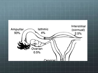
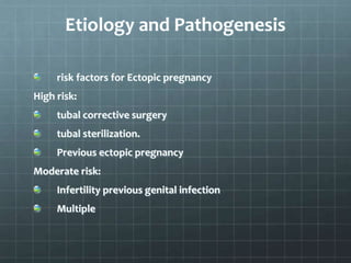
ECTOPIC PREGNANCY
Dr Michieka Nyariki
is which the fertilized ovum is implanted and develops
outside the normal uterine endometrial cavity.
Implantation sites.
Implantation sites.
Tubal commonests 90%
ampulla
isthumus
Intundibulum
Interstitial
Ovarian
Abdominal
Uterine:
Cervical
Cornual
Etiology and Pathogenesis
risk factors for Ectopic pregnancy
High risk:
tubal corrective surgery
tubal sterilization.
Previous ectopic pregnancy
Moderate risk:
Infertility previous genital infection
Multiple
Slight Risk:
Previous pelvic/abdominal surgery
Generally risk factors can be grouped as mechanical
and functional factors but may overlap.
Mechanical Factors.
They prevent passage of facilitated ovum into
the uterine cavity, e.g. prior tubal surgery coufers
the highest risk. After one ectopic pregnancy the
chance of another is 7 to 15%
Previous salpingitis causes agglutination of the
mucosal folds with narrowing a formation of
pockets also causes reduced ciliation due to
infection. Clamydia tracomatis infection is the
commonest.
In utero exposure to dietylstilbestrol predisposes to
developmental tubal abnormalities.
Previous Cesarean Section.
Functional Factors
Cause delay passage of the fertilized ovum into the
uterine cavity e.g. progesterone only contraceptives,
IUCD, post-ovulatory high-dose estrogen to prevent
pregnancy e.g. ECPs, ovulation induction.
Assisted reproduction
Ectopic Pregnancy increase follow gamate
intrafallopian transfer (GIFT) and In Vitro Fertilization
(IVF)
Failed Contraception
Induce in IUCDs, tubal sterilization, progesterone
only
Epidemiology
There has been a increase in both the absolute no and
rate of ectopic pregnancy.
Non-white women have above 1.4 times more chances
than white.
Causes for increase Ectopic
Pregnancy Rates
High prevalence of STIs.
Earlier diagnosis with sensitive x-rays for chronic
gonadotropin and TVS
Induced abortion followed by infection
High use of ARTs
Tubal surgery
Mortality
This has decreased due to immediate diagnosis and
time management.
Anatomical Changes
Zygote Implantation
Fertilised ovum burrows the epithelium and zygote
comes to lie in the muscular wall because the tube
lacks a submucosa. Proliferative trophoblast invades
and erodes the subject mucosants opening maternal
vessels. embryo and fetus in an ectopic pregnancy is
often absent or stunted.
Uterine Changes
Some changes associated with early normal
pregnancy.
Endometrial Changes
Enlarged epithelial with in nuclei that are
hypatrophic, hyperchromadic cellular and
irregular shaped Aria-stella reaction.
External bleeding normally occurs in cases of
tubal pregnancy and is uterine origin from
degeneration and slough of the uterine decidue.
Natural History of Tubal
Pregnancy
Tubal Abortion:
Common in ampullary tubal pregnancy.
Tubal rapture:
As a rule rapture in the 1st few weeks pregnancy is in the
isthimic portion of the tube. Rupture is usually
spontaneous or can be caused by trauma also associated
with coitus, with intraperitoneal rupture, the entire
rupture may be extruded from the tube or if the vent is
small profuse hemorrhage may occur.
Concentrates if small may be reabsorbed or if large
remain in cul-de-sac for years as an encapsulated mass or
even become captured to form a lithopedion.
Abdominal Pregnancy
Follows rupture or abortion
Broad-ligament Pregnancy
Implantation is towards mesosalphinx and based
between folds of broad ligament.
Interstitial Pregnancy
Implantation with the tubal segment penetrates the
uterine wall results in an interstilial or cornual
pregnancy. Haemorrhage can be fatal rapidly
Heterotrophic gestation – Ectopic pregnancy co-exist
with IUP.
Clinical and Laboratory features of Tubal Pregnancy.
Clinical diverse and depends on whether rupture has
occurred
In classical cases here, vaginal bleeding amenorrhea/lower abdominal pain,
others – vasomotor disturbances e.g. vertigo, syncope
Tender abdomen on palpation and vaginal exams elicits cervical motion
tenderness
Post fornix may bulge because of blood in the cul-de-sac tender boggy mass
Diaphragmatic Irritation with pain at shoulder/neck.
With rupture signs/symptoms of shock.
Before rupture – vital signs are normal. if bleeding continues hypotension
tachycardia
Culdocentesis simple technique for identity haemopert
Laboratory tests
Hb, haemotocrit, leucocyte count.
Chronic Gonadotropin Assays;
BHCG – markedly reduced concentrations compared
with normal pregnancy.
Urinary pregnancy tests.
Serum progesterone – single progesterone can be to
establish presence of developing pregnancy
Ultrasound Imaging.
Abdominal
Vaginal
Surgical Diagnosis
Curettage to differentiate between threatened
abortion an incomplete and tubal pregnancy
Laparascopy
Laparatomy
Treatment and prognosis of tubal pregnancy.
Technical advances with earlier diagnosis and
treatment of high risk when women have
allowed definitive management of an unruptured
ectopic pregnancy even before clinical
symptoms.
Surgical Management
laparascopy is preferred over laparatomy unless
patient is unstable even though reproductive
outcome is similar. Laparoscopy is more cost-
effective.
Tubal surgery is divided
into:
Conservative tubal salvage e.g. salpingostomy,
salpingotomy and expression of pregnancy
Radical surgery – salpingectomy
Medical Treatment
Use of methotrexate.
. ECTOPIC GESTATION.ppt. Obstetrics and gyn

. ECTOPIC GESTATION.ppt. Obstetrics and gyn

  • 1.
  • 2.
    is which thefertilized ovum is implanted and develops outside the normal uterine endometrial cavity.
  • 3.
    Implantation sites. Implantation sites. Tubalcommonests 90% ampulla isthumus Intundibulum Interstitial Ovarian Abdominal Uterine: Cervical Cornual
  • 5.
    Etiology and Pathogenesis riskfactors for Ectopic pregnancy High risk: tubal corrective surgery tubal sterilization. Previous ectopic pregnancy Moderate risk: Infertility previous genital infection Multiple
  • 6.
    Slight Risk: Previous pelvic/abdominalsurgery Generally risk factors can be grouped as mechanical and functional factors but may overlap.
  • 7.
    Mechanical Factors. They preventpassage of facilitated ovum into the uterine cavity, e.g. prior tubal surgery coufers the highest risk. After one ectopic pregnancy the chance of another is 7 to 15% Previous salpingitis causes agglutination of the mucosal folds with narrowing a formation of pockets also causes reduced ciliation due to infection. Clamydia tracomatis infection is the commonest.
  • 8.
    In utero exposureto dietylstilbestrol predisposes to developmental tubal abnormalities. Previous Cesarean Section.
  • 9.
    Functional Factors Cause delaypassage of the fertilized ovum into the uterine cavity e.g. progesterone only contraceptives, IUCD, post-ovulatory high-dose estrogen to prevent pregnancy e.g. ECPs, ovulation induction.
  • 10.
    Assisted reproduction Ectopic Pregnancyincrease follow gamate intrafallopian transfer (GIFT) and In Vitro Fertilization (IVF) Failed Contraception Induce in IUCDs, tubal sterilization, progesterone only
  • 11.
    Epidemiology There has beena increase in both the absolute no and rate of ectopic pregnancy. Non-white women have above 1.4 times more chances than white.
  • 12.
    Causes for increaseEctopic Pregnancy Rates High prevalence of STIs. Earlier diagnosis with sensitive x-rays for chronic gonadotropin and TVS Induced abortion followed by infection High use of ARTs Tubal surgery
  • 13.
    Mortality This has decreaseddue to immediate diagnosis and time management.
  • 14.
    Anatomical Changes Zygote Implantation Fertilisedovum burrows the epithelium and zygote comes to lie in the muscular wall because the tube lacks a submucosa. Proliferative trophoblast invades and erodes the subject mucosants opening maternal vessels. embryo and fetus in an ectopic pregnancy is often absent or stunted.
  • 15.
    Uterine Changes Some changesassociated with early normal pregnancy. Endometrial Changes Enlarged epithelial with in nuclei that are hypatrophic, hyperchromadic cellular and irregular shaped Aria-stella reaction. External bleeding normally occurs in cases of tubal pregnancy and is uterine origin from degeneration and slough of the uterine decidue.
  • 16.
    Natural History ofTubal Pregnancy Tubal Abortion: Common in ampullary tubal pregnancy. Tubal rapture: As a rule rapture in the 1st few weeks pregnancy is in the isthimic portion of the tube. Rupture is usually spontaneous or can be caused by trauma also associated with coitus, with intraperitoneal rupture, the entire rupture may be extruded from the tube or if the vent is small profuse hemorrhage may occur. Concentrates if small may be reabsorbed or if large remain in cul-de-sac for years as an encapsulated mass or even become captured to form a lithopedion.
  • 17.
    Abdominal Pregnancy Follows ruptureor abortion Broad-ligament Pregnancy Implantation is towards mesosalphinx and based between folds of broad ligament.
  • 18.
    Interstitial Pregnancy Implantation withthe tubal segment penetrates the uterine wall results in an interstilial or cornual pregnancy. Haemorrhage can be fatal rapidly Heterotrophic gestation – Ectopic pregnancy co-exist with IUP. Clinical and Laboratory features of Tubal Pregnancy. Clinical diverse and depends on whether rupture has occurred
  • 19.
    In classical caseshere, vaginal bleeding amenorrhea/lower abdominal pain, others – vasomotor disturbances e.g. vertigo, syncope Tender abdomen on palpation and vaginal exams elicits cervical motion tenderness Post fornix may bulge because of blood in the cul-de-sac tender boggy mass Diaphragmatic Irritation with pain at shoulder/neck. With rupture signs/symptoms of shock. Before rupture – vital signs are normal. if bleeding continues hypotension tachycardia Culdocentesis simple technique for identity haemopert
  • 20.
    Laboratory tests Hb, haemotocrit,leucocyte count. Chronic Gonadotropin Assays; BHCG – markedly reduced concentrations compared with normal pregnancy. Urinary pregnancy tests. Serum progesterone – single progesterone can be to establish presence of developing pregnancy
  • 21.
  • 22.
    Surgical Diagnosis Curettage todifferentiate between threatened abortion an incomplete and tubal pregnancy Laparascopy Laparatomy Treatment and prognosis of tubal pregnancy. Technical advances with earlier diagnosis and treatment of high risk when women have allowed definitive management of an unruptured ectopic pregnancy even before clinical symptoms.
  • 23.
    Surgical Management laparascopy ispreferred over laparatomy unless patient is unstable even though reproductive outcome is similar. Laparoscopy is more cost- effective.
  • 24.
    Tubal surgery isdivided into: Conservative tubal salvage e.g. salpingostomy, salpingotomy and expression of pregnancy Radical surgery – salpingectomy Medical Treatment Use of methotrexate.