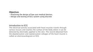
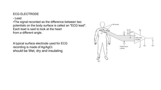
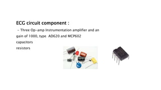
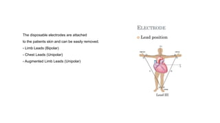
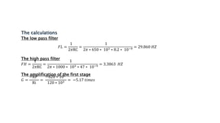
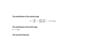
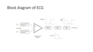
ECG circuit
design project
BACKGROUNDECG
works mostly by detecting and amplifying
changes on the that are caused when the
in the heart muscle is charged and spread
heart beat. This is detected as tiny rises and
voltage between two electrodes placed
heart.
Abstract
This research deals with the design of
that measures heart signals.
The design is based on discrete electronics
cost of the system.
The system consists of 4-op-amp instrumentation
high-pass filter, and low pass filter .
The ECG system is tested using medical ECG electrodes
signals were some recorded from volunteers.
Objectives
- Practicing the design of low cost medical devices.
- Design and testing of ECG system using discrete
Introduction to ECG
The electrical activity associated with heart muscles travels through
various tissues and reaches the surface of the body where it can be
detected by electrodes applied to the skin. The record obtained from
the depolarization and repolarization voltages of the heart muscle is
called an electrocardiogram or ECG
What electrical signals are recorded by the
ECG?
the ECG signal is comprised of multiple sources. The recording is
made through electrodes on the skin, which capture more than just
activity of the heart. The primary electrical components captured
myocardium, muscle, skin-electrode interface, and external
The common frequencies of the important components on the ECG:
Heart rate: 0.67 – 5 Hz (i.e. 40 – 300 bpm)
P-wave: 0.67 – 5 Hz
QRS: 10 – 50 Hz
T-wave: 1 – 7 Hz
High frequency potentials: 100 – 500 Hz
The common frequencies of the artifact and noise on the ECG:
Muscle: 5 – 50 Hz
Respiratory: 0.12 – 0.5 Hz (e.g. 8 – 30 bpm)
External electrical: 50 or 60 Hz (A/C mains or line frequency)
Other electrical: typically >10 Hz (muscle stimulators, strong magnetic
fields, pacemakers with impedance monitoring)
ECG SIGNAL
- bio-signal typical specifications:
- low differential voltage from 0.4 to 3 mV
- high common-mode rejection ratio level
- low frequency range
- high noise
•Artifacts (disturbances) can have many causes.
Common causes are
•Movement:
Sudden movement
Baseline drift
ECG ELECTRODE
- Lead
•The signal recorded as the difference between two
potentials on the body surface is called an "ECG lead".
Each lead is said to look at the heart
from a different angle.
A typical surface electrode used for ECG
recording is made of Ag/AgCl.
should be Wet, dry and insulating
ECG circuit component :
- Three Op-amp Instrumentation amplifier and an
gain of 1000, type AD620 and MCP602
capacitors
resistors
The disposable electrodes are attached
to the patients skin and can be easily removed.
- Limb Leads (Bipolar)
- Chest Leads (Unipolar)
- Augmented Limb Leads (Unipolar)
Low-pass filters
on the ECG are used to remove high frequency muscle artifact and external
typically attenuate only the amplitude of higher frequency ECG components.
has a noticeable affect on the QRS complex, epsilon, and J-waves but do
signals.
High-pass filters
remove low-frequency components such as motion artifact, respiratory
Unlike low-pass filters, analog high pass filters do not attenuate much of
analog high-pass filters suffer from phase shift affecting the first 5 to 10
means that a 0.5 Hz high pass filter, which is a lower frequency than the
can affect frequencies up to 5 Hz!
The low pass filter
𝐹𝐿 =
1
2𝜋RC
=
1
2𝜋 ∗ 650 ∗ 103 ∗ 8.2 ∗ 10−9
= 29.860 𝐻𝑍
The high pass filter
𝐹𝐻 =
1
2𝜋RC
=
1
2𝜋 ∗ 1000 ∗ 103 ∗ 47 ∗ 10−9
= 3.3863 𝐻𝑍
The amplification of the first stage
𝐺 =
−RF
Ri
=
−620 ∗ 103
120 ∗ 103 = −5.17 𝑡𝑖𝑚𝑒𝑠
The amplification of the second stage
𝐺 =
−RF
Ri
=
−620 ∗ 103
120 ∗ 103
= −5.17 𝑡𝑖𝑚𝑒𝑠
The amplification of the third stage
G = 1 𝑡𝑖𝑚𝑒
The resonant frequency
Block diagram of ECG

ECG (1).pptx

  • 1.
  • 3.
    BACKGROUNDECG works mostly bydetecting and amplifying changes on the that are caused when the in the heart muscle is charged and spread heart beat. This is detected as tiny rises and voltage between two electrodes placed heart.
  • 4.
    Abstract This research dealswith the design of that measures heart signals. The design is based on discrete electronics cost of the system. The system consists of 4-op-amp instrumentation high-pass filter, and low pass filter . The ECG system is tested using medical ECG electrodes signals were some recorded from volunteers.
  • 5.
    Objectives - Practicing thedesign of low cost medical devices. - Design and testing of ECG system using discrete Introduction to ECG The electrical activity associated with heart muscles travels through various tissues and reaches the surface of the body where it can be detected by electrodes applied to the skin. The record obtained from the depolarization and repolarization voltages of the heart muscle is called an electrocardiogram or ECG
  • 6.
    What electrical signalsare recorded by the ECG? the ECG signal is comprised of multiple sources. The recording is made through electrodes on the skin, which capture more than just activity of the heart. The primary electrical components captured myocardium, muscle, skin-electrode interface, and external
  • 7.
    The common frequenciesof the important components on the ECG: Heart rate: 0.67 – 5 Hz (i.e. 40 – 300 bpm) P-wave: 0.67 – 5 Hz QRS: 10 – 50 Hz T-wave: 1 – 7 Hz High frequency potentials: 100 – 500 Hz The common frequencies of the artifact and noise on the ECG: Muscle: 5 – 50 Hz Respiratory: 0.12 – 0.5 Hz (e.g. 8 – 30 bpm) External electrical: 50 or 60 Hz (A/C mains or line frequency) Other electrical: typically >10 Hz (muscle stimulators, strong magnetic fields, pacemakers with impedance monitoring)
  • 8.
    ECG SIGNAL - bio-signaltypical specifications: - low differential voltage from 0.4 to 3 mV - high common-mode rejection ratio level - low frequency range - high noise
  • 9.
    •Artifacts (disturbances) canhave many causes. Common causes are •Movement: Sudden movement Baseline drift
  • 10.
    ECG ELECTRODE - Lead •Thesignal recorded as the difference between two potentials on the body surface is called an "ECG lead". Each lead is said to look at the heart from a different angle. A typical surface electrode used for ECG recording is made of Ag/AgCl. should be Wet, dry and insulating
  • 11.
    ECG circuit component: - Three Op-amp Instrumentation amplifier and an gain of 1000, type AD620 and MCP602 capacitors resistors
  • 12.
    The disposable electrodesare attached to the patients skin and can be easily removed. - Limb Leads (Bipolar) - Chest Leads (Unipolar) - Augmented Limb Leads (Unipolar)
  • 13.
    Low-pass filters on theECG are used to remove high frequency muscle artifact and external typically attenuate only the amplitude of higher frequency ECG components. has a noticeable affect on the QRS complex, epsilon, and J-waves but do signals. High-pass filters remove low-frequency components such as motion artifact, respiratory Unlike low-pass filters, analog high pass filters do not attenuate much of analog high-pass filters suffer from phase shift affecting the first 5 to 10 means that a 0.5 Hz high pass filter, which is a lower frequency than the can affect frequencies up to 5 Hz!
  • 14.
    The low passfilter 𝐹𝐿 = 1 2𝜋RC = 1 2𝜋 ∗ 650 ∗ 103 ∗ 8.2 ∗ 10−9 = 29.860 𝐻𝑍 The high pass filter 𝐹𝐻 = 1 2𝜋RC = 1 2𝜋 ∗ 1000 ∗ 103 ∗ 47 ∗ 10−9 = 3.3863 𝐻𝑍 The amplification of the first stage 𝐺 = −RF Ri = −620 ∗ 103 120 ∗ 103 = −5.17 𝑡𝑖𝑚𝑒𝑠
  • 15.
    The amplification ofthe second stage 𝐺 = −RF Ri = −620 ∗ 103 120 ∗ 103 = −5.17 𝑡𝑖𝑚𝑒𝑠 The amplification of the third stage G = 1 𝑡𝑖𝑚𝑒 The resonant frequency
  • 16.