Direct and indirectpathway striatum
Sensory motor Cortex
putamen
GPm
Thalamus
SN r SN c
STN
Caudate nucleus
GPe
Motor Cortex
Nigro striatal
pathway striatal
pallidal
pathway
D1 D2
DA
Not all circuits are shown here
4.
Direct and indirectpathway striatum
Sensory motor Cortex
putamen
GPm
Thalamus
SN r SN c
STN
Caudate nucleus
GPe
Motor Cortex
Nigro striatal
pathway striatal
pallidal
pathway
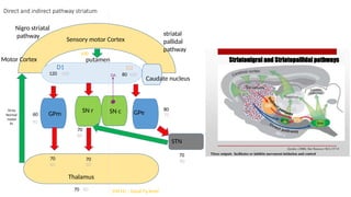
70
80
60
70
70
70
80
80
60
90
60 60
70
70
120 80
100
100
100 Hz – basal Fq level
D1 D2
100
70 Hz
Normal
motor
fn
DA
5.
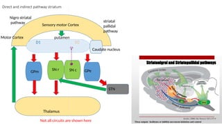
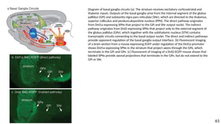
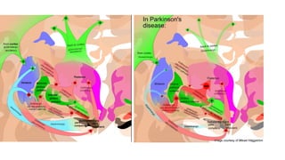
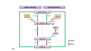
Diagram of basalganglia circuits (a). The striatum receives excitatory corticostriatal and
thalamic inputs. Outputs of the basal ganglia arise from the internal segment of the globus
pallidus (GPi) and substantia nigra pars reticulata (SNr), which are directed to the thalamus,
superior colliculus and pendunculopontine nucleus (PPN). The direct pathway originates
from Drd1a expressing SPNs that project to the GPi and SNr output nuclei. The indirect
pathway originates from Drd2 expressing SPNs that project only to the external segment of
the globus pallidus (GPe), which together with the subthalamic nucleus (STN) contains
transynaptic circuits connecting to the basal output nuclei. The direct and indirect pathways
provide opponent regulation of the basal ganglia output interface. (b) Fluorescent imaging
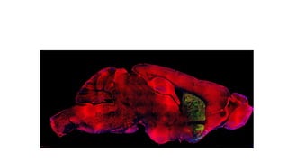
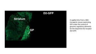
of a brain section from a mouse expressing EGFP under regulation of the Drd1a promoter
shows Drd1a expressing SPNs in the striatum that project axons through the GPe, which
terminate in the GPi and GPe. (c) Fluorescent of imaging of a Drd2-EGFP mouse shows that
labeled SPNs provide axonal projections that terminate in the GPe, but do not extend to the
GPi or SNr.
63
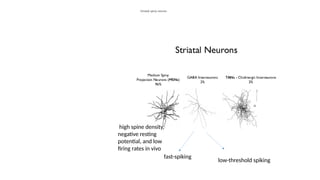
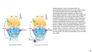
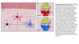
Synaptic plasticity inthe striatum(A) Simplified
schematic of striatal neurons and their
interconnections. Cortical pyramidal neurons
(green) project to striatal interneurons (INTs)
and medium spiny neurons (MSNs) of the
direct (blue) and indirect (red) pathways.
Interneurons also form synapses on medium
spiny neurons. Rectangles highlight potential
sites of synaptic plasticity that could alter
striatal output from MSNs. Corticostriatal
synapses on direct and indirect pathway MSNs
are expanded at right. (B) Indirect-pathway
spines contain dopamine D2 receptors (D2R),
group I mGluRs (mGluR1/5), and L-type
voltage-sensitive calcium channels (VSCCs),
which synergistically mobilize endocannabinoid
(eCB) release that can induce presynaptic LTD
by acting at cannabinoid receptors (CB1R). (C)
Direct-pathway spines contain dopamine D1
receptors (D1R), group I mGluRs, and L-type
VSCCs. Endocannabinoid-dependent LTD
reportedly occurs at direct-pathway MSNs
under conditions in which D1 receptors are not
activated.
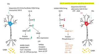
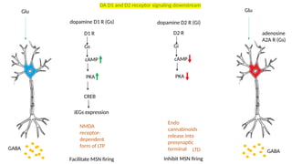
D1
D1 R
Gs
cAMP
PKA
D2 R
Gi
cAMP
PKA
GABA
CREB
GABA
IEGsexpression
Glu Glu
dopamine D1 R (Gs) dopamine D2 R (Gi)
Facilitate MSN firing Inhibit MSN firing
Endo
cannabinoids
release into
presynaptic
terminal
NMDA
receptor-
dependent
form of LTP
LTD
DA D1 and D2 receptor signaling downstream
adenosine
A2A R (Gs)
Editor's Notes
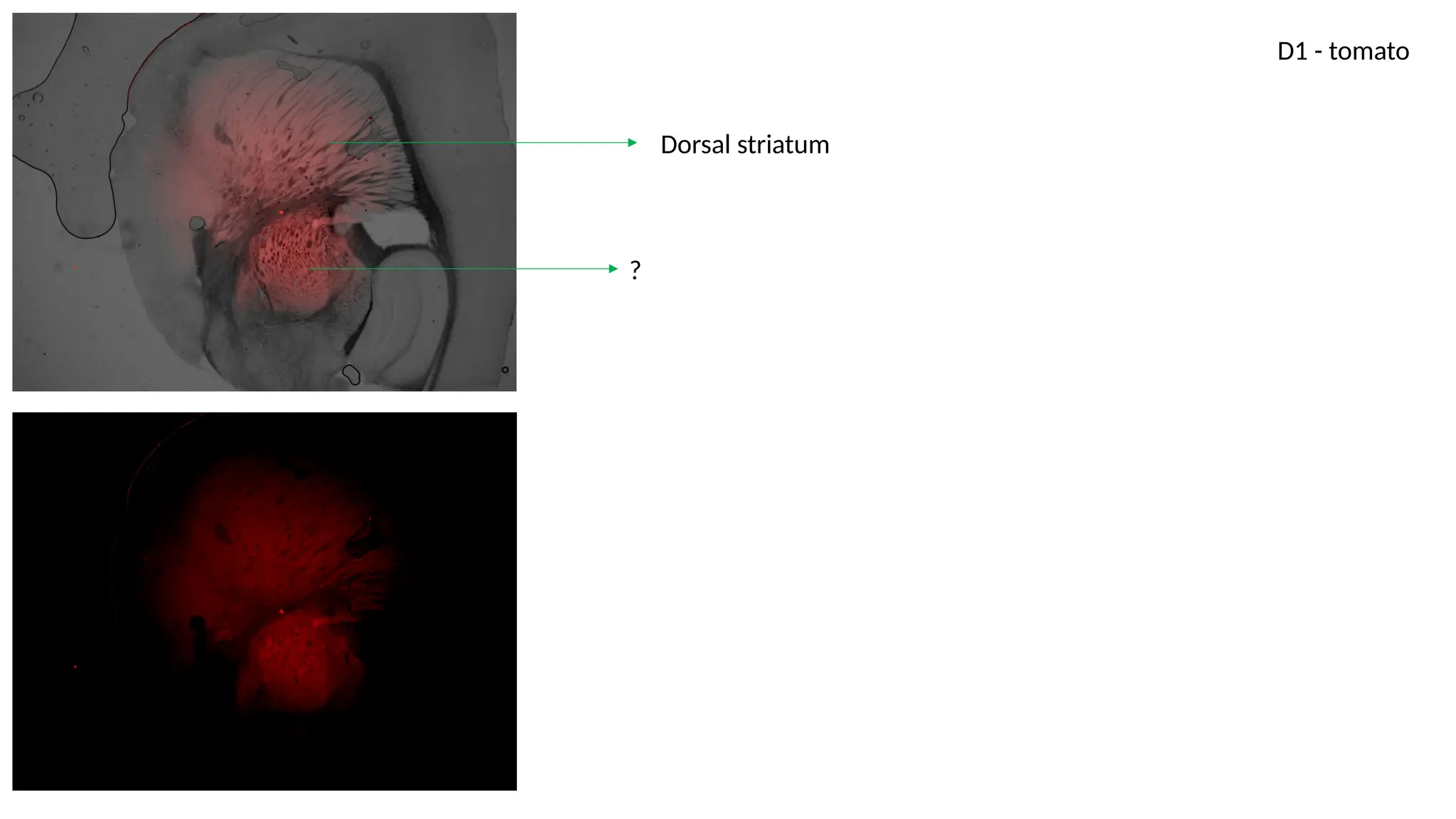
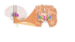
#3 Dorsal striatum – Putamen and Caudate nucleus
Ventral striatum – nucleus accumbens and olfactory tubercle
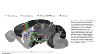
The primary input nucleus is the striatum, which receives excitatory afferents from the cortex and thalamus, as well as dense innervation from midbrain dopamine neurons, and represents a major site of synaptic plasticity in the basal ganglia.
The output from the striatum to downstream basal ganglia nuclei thus reflects a complex interplay between intrinsic properties of MSNs and their excitatory and inhibitory synaptic inputs.
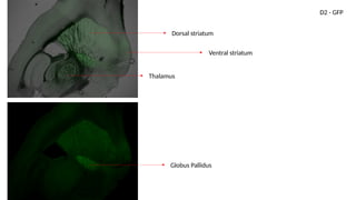
#4 Dorsal striatum – Putamen and Caudate nucleus
Ventral striatum – nucleus accumbens and olfactory tubercle
The primary input nucleus is the striatum, which receives excitatory afferents from the cortex and thalamus, as well as dense innervation from midbrain dopamine neurons, and represents a major site of synaptic plasticity in the basal ganglia. The output from the striatum to downstream basal ganglia nuclei thus reflects a complex interplay between intrinsic properties of MSNs and their excitatory and inhibitory synaptic inputs.
Normal – enhanced activity in D1 MSN, suppressed activity in D2 MSN
PD – suppressed activity in D1 MSN, enhanced activity in D2 MSN
#10 Striatal plasticity alters the transfer of information throughout basal ganglia circuits and may represent a key neural substrate for adaptive motor control and procedural memory.
#16 There are several postsynaptic membrane proteins that are required to elicit eCB release sufficient to induce indirect pathway eCB-LTD: group I (Gq-coupled) metabotropic glutamate receptors (mGluRs), Ltype
voltage-gated calcium channels (L-VGCCs), and dopamine D2 receptors
that a GTPaseactivating
protein called Regulator of G-protein Signaling 4 (RGS4) links D2 and A2A
signaling to group I mGluR signaling.
#18 Striatal plasticity alters the transfer of information throughout basal ganglia circuits and may represent a key neural substrate for adaptive motor control and procedural memory.