Diopsys Visual Electrophysiology Suite - Product Guide
•
0 likes•193 views

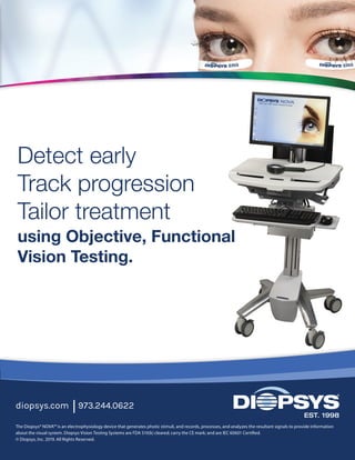
Diopsys offers eye care professionals the ability to detect early, track progression, and tailor treatment using objective, functional vision testing.

Recommended
Recommended
More Related Content
What's hot
What's hot (20)
Similar to Diopsys Visual Electrophysiology Suite - Product Guide
Similar to Diopsys Visual Electrophysiology Suite - Product Guide (20)
Recently uploaded
Recently uploaded (20)
Diopsys Visual Electrophysiology Suite - Product Guide
- 1. •• Roll-Cart Visual Electrophysiology Suite •• Adjustable height workspace •• Adjustable height patient monitor 28 in. 25 in. to 54 in. 20 in. •• Tabletop Visual Electrophysiology Suite •• Wireless keyboard with mouse 16 in. 19 in 14 in. •• Carry case Diopsys® ffERG System •• Scalable to create complete visual electrophysiology suite 16.5 in. 13 in. 16 in. The Diopsys® NOVA™ is an electrophysiology device that generates photic stimuli, and records, processes, and analyzes the resultant signals to provide information about the visual system. Diopsys Vision Testing Systems are FDA 510(k) cleared; carry the CE mark; and are IEC 60601 Certified. © Diopsys, Inc. 2019. All Rights Reserved. Detect early Track progression Tailor treatment using Objective, Functional Vision Testing. diopsys.com 973.244.0622 of the Flash Electroretinogram in Primary Open Angle Glaucoma. Invest. Ophthalmol. Vis. Sci. 2001;42(2):514-522. 8. Chen H, et al. The photopic negative response of flash ERG in nonproliferative diabetic retinopathy. Doc Ophthalmol (2008) 117: 129. 9. Banitt MR, et al. Progressive Loss of Retinal Ganglion Cell Function Precedes Structural Loss by Several Years in Glaucoma Suspects. Invest. Ophthalmol. Vis. Sci. 2013;54(3):2346-2352. 10. Ozkiris A. Pattern electroretinogram changes after intravitreal bevacizumab injection for diabetic macular edema. Doc Ophthalmol 2010;120:243-50. 11. Naismith et al. Optical coherence tomography is less sensitive than visual evoked potentials in optic neuritis. Neurology. 2009 Jul 7;73(1):46-52. 12. Simon J, et al. A New Visual Evoked Potential System for Vision Screening in Infants and Young Children. Journal of AAPOS. 8.6 (2004): 549-554. 13. McKerral et al. Visual and Cognitive Information Processing after Traumatic Brain Injury: VEP and ERP Studies. Invest Ophthalmol Vis Sci 2002;43: E-Abstract 1803. 14. Hood DC, et al. ISCEV Standard for clinical multifocal electroretinography (2011 edition). Doc Ophthalmol 124:1–13. 15. Dettoraki M, Moschos MM. The Role of Multifocal Electroretinography in the Assessment of Drug-Induced Retinopathy: A Review of the Literature. Ophthalmic Res 2016;56:169–177. 16. Talamini CL, et al. Abnormal multifocal ERG findings in patients with normal-appearing retinal anatomy. Doc Ophthalmol 2011;123(3):187-192. 17. Marmor M, et al. Recommendations on Screening for Chloroquine and Hydroxychloroquine Retinopathy (2016 Revision). Ophthalmology 2016;123:6:1386-1394. Three unique platforms. Complete visual electrophysiology suite.
- 2. Diopsys® PERG Pattern Electroretinography Module Provides objective, functional information on the performance of retinal ganglion cells. PERG has been recognized as an effective test in helping doctors to diagnose and manage disease including:9-10 •• glaucoma •• diabetic macular edema Diopsys® mfERG Multifocal Electroretinography Module Provides objective information about localized retinal function to help recognize the first signs of drug-induced retinopathy.14-15 In some cases, retinal dysfunction may occur before structural abnormalities, requiring a robust functional testing method to detect retinal toxicity early.15-17 Diopsys® VEP Visual Evoked Potential Module Provides objective information on the functional integrity of the entire visual system, from the anterior segment of the eye to the visual cortex. VEP is often used to help doctors diagnose and manage neuro- visual disorders such as:11-13 •• optic neuritis •• amblyopia •• vision problems due to TBI Diopsys® ffERG Full Field Electroretinography Module Multi- and Fixed Luminance Flicker Provides objective, functional information about global retinal health and is clinically effective in helping doctors manage retinal disorders like:1-5 •• diabetic retinopathy •• central retinal vein occlusion •• uveitis •• retinal concerns obscured by media opacities Photopic Negative Response (PhNR) Provides objective information to help evaluate optic nerve and retinal disease affecting retinal ganglion cell function, including glaucoma.6-7 Diopsys® Chromatic Flash Vision Screener Designed to quickly and easily screen diabetic patients for early retinal dysfunction before retinopathy.8 1. Yasuda S, et al. Flicker electroretinograms before and after intravitreal ranibizumab injection in eyes with central retinal vein occlusion. Acta Ophthalmol. 2015;93:e465-8. 2. Moschos MM, et al. Electrophysiological examination in uveitis: a review of the literature. Clin Ophthalmol. 2014;8:199-214. 3. Larsson J, Andréasson S. Photopic 30 Hz flicker ERG as a predictor for Rubeosis in central retinal vein occlusion. Br J Ophthalmol. 2001;85:683-5. 4. Ratanapakorn T, et al. Effect of cataract on electroretinographic response. J Med Assoc Thai. 2010 Oct;93(10):1196-9. 5. Holm K, et al. Peripheral retinal function assessed with 30-Hz flicker seems to improve after treatment with Lucentis in patients with diabetic macular oedema. Doc Ophthalmol. 2015;131:43-51. 6. North RV, et al. Electrophysiological Evidence of Early Functional Damage in Glaucoma and Ocular Hypertension. Invest. Ophthalmol. Vis. Sci. 2010;51(2):1216-1222. 7. Viswanathan S, et al. The Photopic Negative Response
- 3. Diopsys® PERG Pattern Electroretinography Module Provides objective, functional information on the performance of retinal ganglion cells. PERG has been recognized as an effective test in helping doctors to diagnose and manage disease including:9-10 •• glaucoma •• diabetic macular edema Diopsys® mfERG Multifocal Electroretinography Module Provides objective information about localized retinal function to help recognize the first signs of drug-induced retinopathy.14-15 In some cases, retinal dysfunction may occur before structural abnormalities, requiring a robust functional testing method to detect retinal toxicity early.15-17 Diopsys® VEP Visual Evoked Potential Module Provides objective information on the functional integrity of the entire visual system, from the anterior segment of the eye to the visual cortex. VEP is often used to help doctors diagnose and manage neuro- visual disorders such as:11-13 •• optic neuritis •• amblyopia •• vision problems due to TBI Diopsys® ffERG Full Field Electroretinography Module Multi- and Fixed Luminance Flicker Provides objective, functional information about global retinal health and is clinically effective in helping doctors manage retinal disorders like:1-5 •• diabetic retinopathy •• central retinal vein occlusion •• uveitis •• retinal concerns obscured by media opacities Photopic Negative Response (PhNR) Provides objective information to help evaluate optic nerve and retinal disease affecting retinal ganglion cell function, including glaucoma.6-7 Diopsys® Chromatic Flash Vision Screener Designed to quickly and easily screen diabetic patients for early retinal dysfunction before retinopathy.8 1. Yasuda S, et al. Flicker electroretinograms before and after intravitreal ranibizumab injection in eyes with central retinal vein occlusion. Acta Ophthalmol. 2015;93:e465-8. 2. Moschos MM, et al. Electrophysiological examination in uveitis: a review of the literature. Clin Ophthalmol. 2014;8:199-214. 3. Larsson J, Andréasson S. Photopic 30 Hz flicker ERG as a predictor for Rubeosis in central retinal vein occlusion. Br J Ophthalmol. 2001;85:683-5. 4. Ratanapakorn T, et al. Effect of cataract on electroretinographic response. J Med Assoc Thai. 2010 Oct;93(10):1196-9. 5. Holm K, et al. Peripheral retinal function assessed with 30-Hz flicker seems to improve after treatment with Lucentis in patients with diabetic macular oedema. Doc Ophthalmol. 2015;131:43-51. 6. North RV, et al. Electrophysiological Evidence of Early Functional Damage in Glaucoma and Ocular Hypertension. Invest. Ophthalmol. Vis. Sci. 2010;51(2):1216-1222. 7. Viswanathan S, et al. The Photopic Negative Response
- 4. •• Roll-Cart Visual Electrophysiology Suite •• Adjustable height workspace •• Adjustable height patient monitor 28 in. 25 in. to 54 in. 20 in. •• Tabletop Visual Electrophysiology Suite •• Wireless keyboard with mouse 16 in. 19 in 14 in. •• Carry case Diopsys® ffERG System •• Scalable to create complete visual electrophysiology suite 16.5 in. 13 in. 16 in. The Diopsys® NOVA™ is an electrophysiology device that generates photic stimuli, and records, processes, and analyzes the resultant signals to provide information about the visual system. Diopsys Vision Testing Systems are FDA 510(k) cleared; carry the CE mark; and are IEC 60601 Certified. © Diopsys, Inc. 2019. All Rights Reserved. Detect early Track progression Tailor treatment using Objective, Functional Vision Testing. diopsys.com 973.244.0622 of the Flash Electroretinogram in Primary Open Angle Glaucoma. Invest. Ophthalmol. Vis. Sci. 2001;42(2):514-522. 8. Chen H, et al. The photopic negative response of flash ERG in nonproliferative diabetic retinopathy. Doc Ophthalmol (2008) 117: 129. 9. Banitt MR, et al. Progressive Loss of Retinal Ganglion Cell Function Precedes Structural Loss by Several Years in Glaucoma Suspects. Invest. Ophthalmol. Vis. Sci. 2013;54(3):2346-2352. 10. Ozkiris A. Pattern electroretinogram changes after intravitreal bevacizumab injection for diabetic macular edema. Doc Ophthalmol 2010;120:243-50. 11. Naismith et al. Optical coherence tomography is less sensitive than visual evoked potentials in optic neuritis. Neurology. 2009 Jul 7;73(1):46-52. 12. Simon J, et al. A New Visual Evoked Potential System for Vision Screening in Infants and Young Children. Journal of AAPOS. 8.6 (2004): 549-554. 13. McKerral et al. Visual and Cognitive Information Processing after Traumatic Brain Injury: VEP and ERP Studies. Invest Ophthalmol Vis Sci 2002;43: E-Abstract 1803. 14. Hood DC, et al. ISCEV Standard for clinical multifocal electroretinography (2011 edition). Doc Ophthalmol 124:1–13. 15. Dettoraki M, Moschos MM. The Role of Multifocal Electroretinography in the Assessment of Drug-Induced Retinopathy: A Review of the Literature. Ophthalmic Res 2016;56:169–177. 16. Talamini CL, et al. Abnormal multifocal ERG findings in patients with normal-appearing retinal anatomy. Doc Ophthalmol 2011;123(3):187-192. 17. Marmor M, et al. Recommendations on Screening for Chloroquine and Hydroxychloroquine Retinopathy (2016 Revision). Ophthalmology 2016;123:6:1386-1394. Three unique platforms. Complete visual electrophysiology suite.