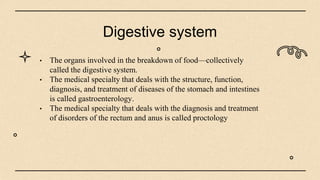
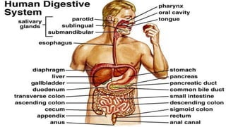
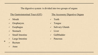
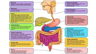
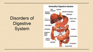
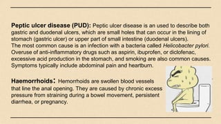
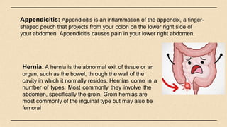
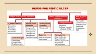
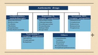
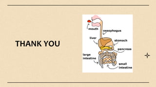
Motility OF SMALL INTESTINE, Malabsorbtion and diarrhoea.pptxWallerianDegenration
The small intestine has two main functions: to mix chyme with bile and pancreatic juices to aid in digestion and absorption of nutrients, and to propel intestinal contents toward the large intestine. Around 10 liters of fluid reach the small intestine daily, of which only 1-2 liters pass into the large intestine as the rest is absorbed. The small intestine exhibits various motility patterns like pendular, segmental, peristaltic and antiparistaltic movements to help with mixing and propulsion of contents. When the lumen is empty between meals, motility changes to the interdigestive migrating motor complex. Malabsorption can result from issues that impair these motor, secretory, digestive or absorpt
The presentation includes the parts and function of our digestive system as well as the process of the parts. Moreover, the presentation includes some diseases in digestive system.
Demonstrate knowledge of a particular body system. Describe the bodily components of the system; describe the pathology of the system; and teach word components and abbreviations.
overview of the digestive system and diseases of itShatha Almahmoud
overview of the digestive system and disorders (disease) of it.
King Saud University, college of applied medical sciences, CLS 224
Anatomy and physiology
Shatha Almahmoud
The document provides an overview of the digestive system, including its main organs and their functions. It describes the mouth, esophagus, stomach, small intestine, large intestine, liver, gallbladder and pancreas. It explains that food is broken down mechanically and chemically by these organs, through processes like digestion and absorption, into nutrients that can be used by the body's cells. Key enzymes produced by organs like the stomach, pancreas and intestines help break down carbohydrates, proteins and fats during digestion.
The alimentary canal is a long, muscular tube that includes the mouth, esophagus, stomach, small intestine, large intestine and anus. Food is broken down mechanically and chemically as it passes through these structures. Digestion begins in the mouth and stomach and nutrients are absorbed in the small intestine. The large intestine absorbs water and waste is stored in the rectum before exiting through the anus. Gastrointestinal disorders like GERD, gastritis, PUD, IBS, hemorrhoids, Crohn's disease and ulcerative colitis can affect digestion and cause various symptoms. Treatment depends on the specific condition but may include lifestyle changes, medications and surgery.
The document provides an overview of the digestive system, including the structures and functions of the mouth, esophagus, stomach, small intestine, large intestine, and related organs. It discusses common digestive issues like GERD, gastritis, peptic ulcers, IBS, hemorrhoids, Crohn's disease, and ulcerative colitis. Treatment options mentioned include changes to diet and lifestyle, medications to reduce inflammation or treat infections, and in some cases surgery.
Peptic Ulcer complications By Abdullah Farooqi GM20-148.pptxshiv847105
The document discusses complications that can arise from peptic ulcers, including bleeding, perforation, and obstruction. Bleeding occurs when ulcers erode blood vessels, which can lead to vomiting blood or black stools. Perforation happens when an ulcer eats through the stomach or intestinal wall, causing severe abdominal pain and potentially shock. Obstruction develops from scarring that narrows the digestive tract and blocks food passage.
This document provides an overview of common diseases of the digestive system. It begins with an introduction to the digestive system and its basic processes. Then it lists and describes several common pathologies/diseases that can affect the digestive system, including appendicitis, ascites, cirrhosis of the liver, diverticular disease, dysentery, fistula, GERD, hematochezia, hemorrhoids, hernia, inflammatory bowel disease, Crohn's disease, ulcerative colitis, irritable bowel syndrome, jaundice, obesity, morbid obesity, polyps, polyposis, and peptic ulcers. It also provides some ICD-10-CM coding guidelines related to complications,
Acute infectious diarrhea is usually caused by faecal–oral transmission of bacteria or their toxins, viruses or parasites. It is typically short-lived and presents with acute diarrhea, sometimes with vomiting, as the predominant symptom. Clinical assessment involves evaluating the history of illness, examining the patient for dehydration, and investigating stool and blood samples. Management focuses on fluid replacement to treat dehydration as well as controlling symptoms, while antibiotics are usually not needed except for specific invasive bacterial infections.
The pancreas is a glandular organ that produces hormones and digestive enzymes. It is located behind the stomach and connected to nearby organs. Under a microscope, it contains clusters of endocrine cells that produce hormones like insulin and clusters of exocrine cells that produce digestive enzymes. The enzymes help digest carbohydrates, proteins, and lipids in the small intestine. Diseases like chronic pancreatitis, cystic fibrosis, and pancreatic cancer can impair the pancreas's functioning and cause digestive issues. Treatment involves enzyme supplements, diet changes, and surgery in some cases.
Gastro intestinal diseases( sample study of peptic ulcer)saugat lamsal
Gastrointestinal diseases refer to diseases involving the gastrointestinal tract and accessory organs of digestion. Common causes include food allergies or intolerance, poor diet, viral or bacterial infection, inflammation, autoimmune diseases, structural abnormalities, genetic factors, lifestyle choices, medication side effects, and cancer. Common signs and symptoms include constipation, diarrhea, stomach pain, bleeding, ulcers, and vomiting or nausea. Common gastrointestinal diseases are gastroesophageal reflux disease, peptic ulcers, stomach flu, inflammatory bowel disease, irritable bowel syndrome, constipation, hemorrhoids, and diverticular disease.
Formation of hard, pebble and stone like structure mainly made up of cholesterol in gall bladder is called cholelithiasis.
Know more about cholelithiasis
This document discusses peptic ulcer disease (PUD), including its causes, types, symptoms, diagnosis, and treatment. PUD is characterized by erosion of the GI mucosa from stomach acid and pepsin. It commonly affects the lower esophagus, stomach, and duodenum. The two main types are gastric and duodenal ulcers. Symptoms include abdominal pain, nausea, and vomiting. Diagnosis involves endoscopy and tests for H. pylori bacteria. Treatment focuses on reducing stomach acid with PPIs or H2 blockers, eradicating H. pylori, and protecting the mucosa. Complications can include bleeding, perforation, and obstruction if not properly treated.
The document provides an overview of renal and gastrointestinal systems. It describes the kidney's role in filtering blood and regulating homeostasis. The nephron is identified as the kidney's functional unit where filtration occurs. Other kidney functions include hormone release. The gastrointestinal system digests and processes food through mechanical movement, enzymes, acid and hormones. Specific sections of the GI tract are described including the stomach, small intestine and large intestine. Three case studies are presented and diagnoses of kidney stones, peptic ulcer, and diverticulitis are provided based on presented findings and symptoms.
The document provides an overview of the gastrointestinal system, including:
- It describes the anatomy and functions of the GI tract from mouth to anus, as well as related organs like the liver and pancreas.
- It lists 100 key facts about the structure and function of different parts of the GI system, including the mouth, esophagus, stomach, and liver.
- It explains that understanding the basic science of the normal GI system is important for learning about GI diseases.
Pancreatic Disease :- The pancreas is a narrow, flat organ about six inches long, with a head, middle, and tail section. It is located below the liver, between the stomach and the spine, and its head section connects to the duodenum. Inside the pancreas, small ducts (tubes) feed fluids produced by the pancreas into the pancreatic duct. This larger duct carries the fluids down the length of the pancreas, from the tail to the head, and into the duodenum.
The common bile duct also runs through the head section of the pancreas, carrying bile from the liver and gall bladder into the small intestine. The bile duct and the pancreatic duct usually join just before entering the duodenum and so have a common opening into the small intestine.
The pancreas consists of two kinds of tissues:
Exocrine — which make powerful enzymes to digest fats, proteins, and carbohydrates. The enzymes normally are created and carried to the duodenum in an inactive form, then activated as needed. Exocrine tissue also makes bicarbonates that work to neutralize stomach acids.
Endocrine — which produce the hormones insulin and glucagon and release them into the blood stream. These hormones regulate glucose transport into the body’s cells and are crucial for energy production.
Pancreatic Disease
Diseases of the Pancreas:
1. Pancreatitis: Pancreatitis is an inflammation of the pancreas. It is caused when the digestive enzymes from the exocrine pancreas become activated inside of the pancreas, instead of in the duodenum, and start “digesting” the pancreas itself. It usually presents with abdominal pain and can cause nausea and vomiting. Two Types of Pancreatitis
Acute pancreatitis : The most common cause of acute pancreatitis is blockage of the pancreatic duct by gallstones. Secretions can back up in the pancreas and cause permanent damage in just a few hours. Acute pancreatitis often presents with raised levels of pancreatic enzymes in the blood. The abdominal pain in acute pancreatitis is often severe. The disease may even lead to internal bleeding and infection and can be life-threatening.
Chronic pancreatitis: chronic or persistent abdominal pain and may or may not present with raised pancreatic enzymes. It develops gradually, often results in slow destruction of the pancreas. The main causes of chronic pancreatitis are gall bladder disease (ductal obstruction) and alcoholism. Other causes of chronic pancreatitis include cystic fibrosis, hypercalcemia, hyperlipidemia, some drugs, and autoimmune conditions.
2. Pancreatic Insufficiency: Pancreatic insufficiency is the inability of the pancreas to produce and/or transport enough digestive enzymes to break down food in the intestine and allow its absorption. occurs as the result of progressive pancreatic damage – It is most frequently associated with cystic fibrosis in children and with chronic pancreatitis in adults.
3. Pancreatic Cancer: Main Causes of Pancreatic cancer are chronic pancreatitis, and exposure to some industrial
Peptic ulcers form when the lining of the stomach or duodenum is corroded by acidic digestive juices. Common symptoms include abdominal pain relieved by food or antacids. While acid contributes to ulcer formation, infection with H. pylori bacteria is now believed to be the leading cause. Other risk factors include NSAID use, smoking, alcohol, and stress. Complications can include bleeding, perforation, and narrowing or obstruction of the stomach outlet. Endoscopy allows visualization and biopsy of ulcers, while treatment aims to eliminate H. pylori infection and reduce acid secretion.
P. Kolala presented on peptic ulcer disease. PUD is caused by an imbalance between gastric acid production and mucosal defenses, resulting in ulcers in the esophagus, stomach, or duodenum. Risk factors include H. pylori infection, NSAID use, smoking, and stress. Symptoms include epigastric pain relieved by food or antacids. Treatment involves eradicating H. pylori with antibiotics, reducing acid with PPIs, and surgery if complications occur or medications fail. Nursing care focuses on pain management, diet, medication administration and monitoring for complications.
Anatomy and Physiology of the GI System, Hormonal Control, Upper GI Tract Disorders, Gallbladder Disorders, Liver Disorders, Pancreatic Disorders, Lower GI Tract Disorders
This document discusses the dietary management of various gastrointestinal diseases including diarrhea, constipation, gastritis, and peptic ulcers. It describes the anatomy and functions of the digestive system. It defines different types of constipation and diarrhea and their causes. Dietary recommendations are provided for different GI conditions, focusing on fluid and fiber intake, meal patterns, and avoiding irritating foods. Medical treatments including drugs and H. pylori eradication therapies are also summarized.
This document discusses acute and chronic diarrhea, their causes and management. Acute diarrhea is usually short-lived and self-limiting, often resulting from food poisoning or bacterial infection. Chronic diarrhea lasts longer than 2 weeks and requires investigation to determine the cause, such as irritable bowel syndrome, inflammatory bowel disease, malabsorption syndromes, or infections. Malabsorption is defined as the defective absorption of nutrients and can result in deficiencies. Causes of malabsorption discussed include celiac disease, tropical sprue, bacterial overgrowth, and surgical resections.
1. The pancreas is an elongated organ located in the abdominal cavity behind the stomach. It has three parts - head, body, and tail.
2. The pancreas has both exocrine and endocrine functions. Exocrine functions include producing pancreatic juice containing enzymes that digest carbohydrates, proteins, and fats. Endocrine functions include production of insulin, glucagon, and somatostatin by islets of Langerhans cells.
3. Pancreatitis is inflammation of the pancreas that can be acute or chronic. Acute pancreatitis symptoms include severe abdominal pain and its causes include gallstones and alcohol use. Chronic pancreatitis involves long-term inflammation that destroys the pancreas over
symptoms, causes and risk factors pacreatitisLankeSuneetha
The pancreas is an organ located in the upper abdomen that has both exocrine and endocrine functions. The exocrine function involves secreting enzymes like amylase and lipase to help digest food. The endocrine function involves secreting hormones like insulin and glucagon to regulate blood glucose. Pancreatitis is inflammation of the pancreas that can be acute or chronic. Acute pancreatitis is sudden inflammation that usually resolves within days, while chronic pancreatitis causes permanent damage over many years. Both types are often caused by gallstones blocking the pancreatic duct or heavy alcohol use damaging the pancreas over time. Symptoms include abdominal pain, nausea, and digestive issues. Diagnosis involves blood tests of pancreatic enzymes and imaging tests
Peptic ulcer disease is characterized by erosion of the GI mucosa due to gastric acid and pepsin. Ulcers most commonly form in the lower esophagus, stomach, and duodenum. The development of ulcers involves disruption of the mucosal barrier that normally protects the GI tract from acid. Common complications include hemorrhage, perforation, and gastric outlet obstruction. Diagnosis involves endoscopy to identify ulcers and tests for H. pylori infection.
The document summarizes the key parts and functions of the digestive system, including:
- The alimentary canal extends from the mouth to anus and includes organs like the esophagus, stomach, and intestines. Accessory organs include the liver, gallbladder and pancreas.
- The mouth, salivary glands, teeth and tongue are involved in mechanical digestion. The stomach contains acids and enzymes that begin chemical digestion.
- The small intestine further breaks down food and the large intestine absorbs water before waste is excreted from the anus. Common disorders of these organs include ulcers, infections, and cancers.
Gastro Oesophagal Reflux Disease GERD.pptxSudipta Roy
This document provides information on gastroesophageal reflux disease (GERD). It defines GERD as when stomach acid leaks up into the esophagus. Risk factors include smoking, large meals, and hiatal hernia. Symptoms include heartburn and acid taste in the mouth. Diagnosis involves endoscopy and pH testing. Treatment includes lifestyle changes like weight loss and elevation of the head, as well as medications like antacids, H2 blockers, and proton pump inhibitors.
share - Lions, tigers, AI and health misinformation, oh my!.pptxTina Purnat
• Pitfalls and pivots needed to use AI effectively in public health
• Evidence-based strategies to address health misinformation effectively
• Building trust with communities online and offline
• Equipping health professionals to address questions, concerns and health misinformation
• Assessing risk and mitigating harm from adverse health narratives in communities, health workforce and health system
More Related Content
Similar to DIGESTIVE SYSTEM. PPT human anatomy and physiology
This document provides an overview of common diseases of the digestive system. It begins with an introduction to the digestive system and its basic processes. Then it lists and describes several common pathologies/diseases that can affect the digestive system, including appendicitis, ascites, cirrhosis of the liver, diverticular disease, dysentery, fistula, GERD, hematochezia, hemorrhoids, hernia, inflammatory bowel disease, Crohn's disease, ulcerative colitis, irritable bowel syndrome, jaundice, obesity, morbid obesity, polyps, polyposis, and peptic ulcers. It also provides some ICD-10-CM coding guidelines related to complications,
Acute infectious diarrhea is usually caused by faecal–oral transmission of bacteria or their toxins, viruses or parasites. It is typically short-lived and presents with acute diarrhea, sometimes with vomiting, as the predominant symptom. Clinical assessment involves evaluating the history of illness, examining the patient for dehydration, and investigating stool and blood samples. Management focuses on fluid replacement to treat dehydration as well as controlling symptoms, while antibiotics are usually not needed except for specific invasive bacterial infections.
The pancreas is a glandular organ that produces hormones and digestive enzymes. It is located behind the stomach and connected to nearby organs. Under a microscope, it contains clusters of endocrine cells that produce hormones like insulin and clusters of exocrine cells that produce digestive enzymes. The enzymes help digest carbohydrates, proteins, and lipids in the small intestine. Diseases like chronic pancreatitis, cystic fibrosis, and pancreatic cancer can impair the pancreas's functioning and cause digestive issues. Treatment involves enzyme supplements, diet changes, and surgery in some cases.
Gastro intestinal diseases( sample study of peptic ulcer)saugat lamsal
Gastrointestinal diseases refer to diseases involving the gastrointestinal tract and accessory organs of digestion. Common causes include food allergies or intolerance, poor diet, viral or bacterial infection, inflammation, autoimmune diseases, structural abnormalities, genetic factors, lifestyle choices, medication side effects, and cancer. Common signs and symptoms include constipation, diarrhea, stomach pain, bleeding, ulcers, and vomiting or nausea. Common gastrointestinal diseases are gastroesophageal reflux disease, peptic ulcers, stomach flu, inflammatory bowel disease, irritable bowel syndrome, constipation, hemorrhoids, and diverticular disease.
Formation of hard, pebble and stone like structure mainly made up of cholesterol in gall bladder is called cholelithiasis.
Know more about cholelithiasis
This document discusses peptic ulcer disease (PUD), including its causes, types, symptoms, diagnosis, and treatment. PUD is characterized by erosion of the GI mucosa from stomach acid and pepsin. It commonly affects the lower esophagus, stomach, and duodenum. The two main types are gastric and duodenal ulcers. Symptoms include abdominal pain, nausea, and vomiting. Diagnosis involves endoscopy and tests for H. pylori bacteria. Treatment focuses on reducing stomach acid with PPIs or H2 blockers, eradicating H. pylori, and protecting the mucosa. Complications can include bleeding, perforation, and obstruction if not properly treated.
The document provides an overview of renal and gastrointestinal systems. It describes the kidney's role in filtering blood and regulating homeostasis. The nephron is identified as the kidney's functional unit where filtration occurs. Other kidney functions include hormone release. The gastrointestinal system digests and processes food through mechanical movement, enzymes, acid and hormones. Specific sections of the GI tract are described including the stomach, small intestine and large intestine. Three case studies are presented and diagnoses of kidney stones, peptic ulcer, and diverticulitis are provided based on presented findings and symptoms.
The document provides an overview of the gastrointestinal system, including:
- It describes the anatomy and functions of the GI tract from mouth to anus, as well as related organs like the liver and pancreas.
- It lists 100 key facts about the structure and function of different parts of the GI system, including the mouth, esophagus, stomach, and liver.
- It explains that understanding the basic science of the normal GI system is important for learning about GI diseases.
Pancreatic Disease :- The pancreas is a narrow, flat organ about six inches long, with a head, middle, and tail section. It is located below the liver, between the stomach and the spine, and its head section connects to the duodenum. Inside the pancreas, small ducts (tubes) feed fluids produced by the pancreas into the pancreatic duct. This larger duct carries the fluids down the length of the pancreas, from the tail to the head, and into the duodenum.
The common bile duct also runs through the head section of the pancreas, carrying bile from the liver and gall bladder into the small intestine. The bile duct and the pancreatic duct usually join just before entering the duodenum and so have a common opening into the small intestine.
The pancreas consists of two kinds of tissues:
Exocrine — which make powerful enzymes to digest fats, proteins, and carbohydrates. The enzymes normally are created and carried to the duodenum in an inactive form, then activated as needed. Exocrine tissue also makes bicarbonates that work to neutralize stomach acids.
Endocrine — which produce the hormones insulin and glucagon and release them into the blood stream. These hormones regulate glucose transport into the body’s cells and are crucial for energy production.
Pancreatic Disease
Diseases of the Pancreas:
1. Pancreatitis: Pancreatitis is an inflammation of the pancreas. It is caused when the digestive enzymes from the exocrine pancreas become activated inside of the pancreas, instead of in the duodenum, and start “digesting” the pancreas itself. It usually presents with abdominal pain and can cause nausea and vomiting. Two Types of Pancreatitis
Acute pancreatitis : The most common cause of acute pancreatitis is blockage of the pancreatic duct by gallstones. Secretions can back up in the pancreas and cause permanent damage in just a few hours. Acute pancreatitis often presents with raised levels of pancreatic enzymes in the blood. The abdominal pain in acute pancreatitis is often severe. The disease may even lead to internal bleeding and infection and can be life-threatening.
Chronic pancreatitis: chronic or persistent abdominal pain and may or may not present with raised pancreatic enzymes. It develops gradually, often results in slow destruction of the pancreas. The main causes of chronic pancreatitis are gall bladder disease (ductal obstruction) and alcoholism. Other causes of chronic pancreatitis include cystic fibrosis, hypercalcemia, hyperlipidemia, some drugs, and autoimmune conditions.
2. Pancreatic Insufficiency: Pancreatic insufficiency is the inability of the pancreas to produce and/or transport enough digestive enzymes to break down food in the intestine and allow its absorption. occurs as the result of progressive pancreatic damage – It is most frequently associated with cystic fibrosis in children and with chronic pancreatitis in adults.
3. Pancreatic Cancer: Main Causes of Pancreatic cancer are chronic pancreatitis, and exposure to some industrial
Peptic ulcers form when the lining of the stomach or duodenum is corroded by acidic digestive juices. Common symptoms include abdominal pain relieved by food or antacids. While acid contributes to ulcer formation, infection with H. pylori bacteria is now believed to be the leading cause. Other risk factors include NSAID use, smoking, alcohol, and stress. Complications can include bleeding, perforation, and narrowing or obstruction of the stomach outlet. Endoscopy allows visualization and biopsy of ulcers, while treatment aims to eliminate H. pylori infection and reduce acid secretion.
P. Kolala presented on peptic ulcer disease. PUD is caused by an imbalance between gastric acid production and mucosal defenses, resulting in ulcers in the esophagus, stomach, or duodenum. Risk factors include H. pylori infection, NSAID use, smoking, and stress. Symptoms include epigastric pain relieved by food or antacids. Treatment involves eradicating H. pylori with antibiotics, reducing acid with PPIs, and surgery if complications occur or medications fail. Nursing care focuses on pain management, diet, medication administration and monitoring for complications.
Anatomy and Physiology of the GI System, Hormonal Control, Upper GI Tract Disorders, Gallbladder Disorders, Liver Disorders, Pancreatic Disorders, Lower GI Tract Disorders
This document discusses the dietary management of various gastrointestinal diseases including diarrhea, constipation, gastritis, and peptic ulcers. It describes the anatomy and functions of the digestive system. It defines different types of constipation and diarrhea and their causes. Dietary recommendations are provided for different GI conditions, focusing on fluid and fiber intake, meal patterns, and avoiding irritating foods. Medical treatments including drugs and H. pylori eradication therapies are also summarized.
This document discusses acute and chronic diarrhea, their causes and management. Acute diarrhea is usually short-lived and self-limiting, often resulting from food poisoning or bacterial infection. Chronic diarrhea lasts longer than 2 weeks and requires investigation to determine the cause, such as irritable bowel syndrome, inflammatory bowel disease, malabsorption syndromes, or infections. Malabsorption is defined as the defective absorption of nutrients and can result in deficiencies. Causes of malabsorption discussed include celiac disease, tropical sprue, bacterial overgrowth, and surgical resections.
1. The pancreas is an elongated organ located in the abdominal cavity behind the stomach. It has three parts - head, body, and tail.
2. The pancreas has both exocrine and endocrine functions. Exocrine functions include producing pancreatic juice containing enzymes that digest carbohydrates, proteins, and fats. Endocrine functions include production of insulin, glucagon, and somatostatin by islets of Langerhans cells.
3. Pancreatitis is inflammation of the pancreas that can be acute or chronic. Acute pancreatitis symptoms include severe abdominal pain and its causes include gallstones and alcohol use. Chronic pancreatitis involves long-term inflammation that destroys the pancreas over
symptoms, causes and risk factors pacreatitisLankeSuneetha
The pancreas is an organ located in the upper abdomen that has both exocrine and endocrine functions. The exocrine function involves secreting enzymes like amylase and lipase to help digest food. The endocrine function involves secreting hormones like insulin and glucagon to regulate blood glucose. Pancreatitis is inflammation of the pancreas that can be acute or chronic. Acute pancreatitis is sudden inflammation that usually resolves within days, while chronic pancreatitis causes permanent damage over many years. Both types are often caused by gallstones blocking the pancreatic duct or heavy alcohol use damaging the pancreas over time. Symptoms include abdominal pain, nausea, and digestive issues. Diagnosis involves blood tests of pancreatic enzymes and imaging tests
Peptic ulcer disease is characterized by erosion of the GI mucosa due to gastric acid and pepsin. Ulcers most commonly form in the lower esophagus, stomach, and duodenum. The development of ulcers involves disruption of the mucosal barrier that normally protects the GI tract from acid. Common complications include hemorrhage, perforation, and gastric outlet obstruction. Diagnosis involves endoscopy to identify ulcers and tests for H. pylori infection.
The document summarizes the key parts and functions of the digestive system, including:
- The alimentary canal extends from the mouth to anus and includes organs like the esophagus, stomach, and intestines. Accessory organs include the liver, gallbladder and pancreas.
- The mouth, salivary glands, teeth and tongue are involved in mechanical digestion. The stomach contains acids and enzymes that begin chemical digestion.
- The small intestine further breaks down food and the large intestine absorbs water before waste is excreted from the anus. Common disorders of these organs include ulcers, infections, and cancers.
Gastro Oesophagal Reflux Disease GERD.pptxSudipta Roy
This document provides information on gastroesophageal reflux disease (GERD). It defines GERD as when stomach acid leaks up into the esophagus. Risk factors include smoking, large meals, and hiatal hernia. Symptoms include heartburn and acid taste in the mouth. Diagnosis involves endoscopy and pH testing. Treatment includes lifestyle changes like weight loss and elevation of the head, as well as medications like antacids, H2 blockers, and proton pump inhibitors.
Similar to DIGESTIVE SYSTEM. PPT human anatomy and physiology (20)
share - Lions, tigers, AI and health misinformation, oh my!.pptxTina Purnat
• Pitfalls and pivots needed to use AI effectively in public health
• Evidence-based strategies to address health misinformation effectively
• Building trust with communities online and offline
• Equipping health professionals to address questions, concerns and health misinformation
• Assessing risk and mitigating harm from adverse health narratives in communities, health workforce and health system
These lecture slides, by Dr Sidra Arshad, offer a quick overview of the physiological basis of a normal electrocardiogram.
Learning objectives:
1. Define an electrocardiogram (ECG) and electrocardiography
2. Describe how dipoles generated by the heart produce the waveforms of the ECG
3. Describe the components of a normal electrocardiogram of a typical bipolar lead (limb II)
4. Differentiate between intervals and segments
5. Enlist some common indications for obtaining an ECG
6. Describe the flow of current around the heart during the cardiac cycle
7. Discuss the placement and polarity of the leads of electrocardiograph
8. Describe the normal electrocardiograms recorded from the limb leads and explain the physiological basis of the different records that are obtained
9. Define mean electrical vector (axis) of the heart and give the normal range
10. Define the mean QRS vector
11. Describe the axes of leads (hexagonal reference system)
12. Comprehend the vectorial analysis of the normal ECG
13. Determine the mean electrical axis of the ventricular QRS and appreciate the mean axis deviation
14. Explain the concepts of current of injury, J point, and their significance
Study Resources:
1. Chapter 11, Guyton and Hall Textbook of Medical Physiology, 14th edition
2. Chapter 9, Human Physiology - From Cells to Systems, Lauralee Sherwood, 9th edition
3. Chapter 29, Ganong’s Review of Medical Physiology, 26th edition
4. Electrocardiogram, StatPearls - https://www.ncbi.nlm.nih.gov/books/NBK549803/
5. ECG in Medical Practice by ABM Abdullah, 4th edition
6. Chapter 3, Cardiology Explained, https://www.ncbi.nlm.nih.gov/books/NBK2214/
7. ECG Basics, http://www.nataliescasebook.com/tag/e-c-g-basics
One health condition that is becoming more common day by day is diabetes.
According to research conducted by the National Family Health Survey of India, diabetic cases show a projection which might increase to 10.4% by 2030.
Integrating Ayurveda into Parkinson’s Management: A Holistic ApproachAyurveda ForAll
Explore the benefits of combining Ayurveda with conventional Parkinson's treatments. Learn how a holistic approach can manage symptoms, enhance well-being, and balance body energies. Discover the steps to safely integrate Ayurvedic practices into your Parkinson’s care plan, including expert guidance on diet, herbal remedies, and lifestyle modifications.
These lecture slides, by Dr Sidra Arshad, offer a simplified look into the mechanisms involved in the regulation of respiration:
Learning objectives:
1. Describe the organisation of respiratory center
2. Describe the nervous control of inspiration and respiratory rhythm
3. Describe the functions of the dorsal and respiratory groups of neurons
4. Describe the influences of the Pneumotaxic and Apneustic centers
5. Explain the role of Hering-Breur inflation reflex in regulation of inspiration
6. Explain the role of central chemoreceptors in regulation of respiration
7. Explain the role of peripheral chemoreceptors in regulation of respiration
8. Explain the regulation of respiration during exercise
9. Integrate the respiratory regulatory mechanisms
10. Describe the Cheyne-Stokes breathing
Study Resources:
1. Chapter 42, Guyton and Hall Textbook of Medical Physiology, 14th edition
2. Chapter 36, Ganong’s Review of Medical Physiology, 26th edition
3. Chapter 13, Human Physiology by Lauralee Sherwood, 9th edition
Muktapishti is a traditional Ayurvedic preparation made from Shoditha Mukta (Purified Pearl), is believed to help regulate thyroid function and reduce symptoms of hyperthyroidism due to its cooling and balancing properties. Clinical evidence on its efficacy remains limited, necessitating further research to validate its therapeutic benefits.
Adhd Medication Shortage Uk - trinexpharmacy.comreignlana06
The UK is currently facing a Adhd Medication Shortage Uk, which has left many patients and their families grappling with uncertainty and frustration. ADHD, or Attention Deficit Hyperactivity Disorder, is a chronic condition that requires consistent medication to manage effectively. This shortage has highlighted the critical role these medications play in the daily lives of those affected by ADHD. Contact : +1 (747) 209 – 3649 E-mail : sales@trinexpharmacy.com
Clinic ^%[+27633867063*Abortion Pills For Sale In Tembisa Central19various
Clinic ^%[+27633867063*Abortion Pills For Sale In Tembisa Central Clinic ^%[+27633867063*Abortion Pills For Sale In Tembisa CentralClinic ^%[+27633867063*Abortion Pills For Sale In Tembisa CentralClinic ^%[+27633867063*Abortion Pills For Sale In Tembisa CentralClinic ^%[+27633867063*Abortion Pills For Sale In Tembisa Central
Local Advanced Lung Cancer: Artificial Intelligence, Synergetics, Complex Sys...Oleg Kshivets
Overall life span (LS) was 1671.7±1721.6 days and cumulative 5YS reached 62.4%, 10 years – 50.4%, 20 years – 44.6%. 94 LCP lived more than 5 years without cancer (LS=2958.6±1723.6 days), 22 – more than 10 years (LS=5571±1841.8 days). 67 LCP died because of LC (LS=471.9±344 days). AT significantly improved 5YS (68% vs. 53.7%) (P=0.028 by log-rank test). Cox modeling displayed that 5YS of LCP significantly depended on: N0-N12, T3-4, blood cell circuit, cell ratio factors (ratio between cancer cells-CC and blood cells subpopulations), LC cell dynamics, recalcification time, heparin tolerance, prothrombin index, protein, AT, procedure type (P=0.000-0.031). Neural networks, genetic algorithm selection and bootstrap simulation revealed relationships between 5YS and N0-12 (rank=1), thrombocytes/CC (rank=2), segmented neutrophils/CC (3), eosinophils/CC (4), erythrocytes/CC (5), healthy cells/CC (6), lymphocytes/CC (7), stick neutrophils/CC (8), leucocytes/CC (9), monocytes/CC (10). Correct prediction of 5YS was 100% by neural networks computing (error=0.000; area under ROC curve=1.0).
2. Digestive system
• The organs involved in the breakdown of food—collectively
called the digestive system.
• The medical specialty that deals with the structure, function,
diagnosis, and treatment of diseases of the stomach and intestines
is called gastroenterology.
• The medical specialty that deals with the diagnosis and treatment
of disorders of the rectum and anus is called proctology
3.
4. The digestive system is divided into two groups of organs
The Accessory Digestive Organs
Teeth
Tongue
Salivary Glands
Liver
Gallbladder
Pancreas
The Gastrointestinal Tract (GIT)
Mouth
Oropharynx
Esophagus
Stomach
Small Intestine
Large Intestine
Rectum
Anus
5. Digestive system secretes
around 7L of water, buffer,
enzymes into the lumen
The food is mixed with digestive
secretions. The movement of
food in GIT is termed as
peristalsis movement
Stages of the digestive process
Process of eating food or
water by mouth
Mechanical digestion- breaking down
of food particles by teeth to mixing
with digestive enzymes
Chemical digestion – carbohydrates,
proteins, fats are breakdown into micro
molecules by digestive enzymes
The absorbed substances
pass into blood or lymph and
circulate to cells throughout
the body.
The unabsorbed food
particles are eliminated from
body through the process of
defecation through anus.
Ingestion Secretion Mixing and
propulsion
Digestion
Absorption Defecation
8. Peritonitis: A common cause of peritonitis, an acute inflammation of
the peritoneum, is contamination of the peritoneum by infectious
microbes, which can result from accidental or surgical wounds in the
abdominal wall, or from perforation or rupture of abdominal organs.
Mumps: it is an inflammation and enlargement of the parotid glands
accompanied by moderate fever, malaise (general discomfort), and
extreme pain in the throat, especially when swallowing sour foods or
acidic juices.
Pyloric stenosis: It is a narrowing of the pyloric sphincter that must
be corrected surgically. The hallmark symptom is vomiting the spraying
of liquid vomitus some distance from the infant.
9. Gastroesophageal reflux disease: If the lower esophageal sphincter
fails to close adequately after food has entered the stomach, the stomach
contents can reflux into the inferior portion of the esophagus. This
condition is known as Gastroesophageal reflux disease (gerd).
Hydrochloric acid (hcl) from the stomach contents can irritate the
esophageal wall, resulting in a Burning sensation that is called heartburn
because it is experienced in a region very near the heart.
Liver cirrhosis: Cirrhosis of liver refers to inflammation and damage of
parenchyma of liver. It results in degeneration of hepatic cells and
dysfunction of liver.
Pancreatitis:Inflammation of the pancreas, as may occur in association
with alcohol abuse or chronic gallstones, is called pancreatitis
10. Gallstones: If bile contains either insufficient bile salts or lecithin or excessive
cholesterol, the cholesterol may crystallize to form gallstones. As they grow in
size and number, gallstones may cause obstruction to the flow of bile from the
gallbladder into the duodenum.
Diarrhea: Symptoms of diarrhea include frequent, loose, watery stools (feces)
which are usually accompanied by an urgent need to go to the toilet. Abdominal
pain or cramping may also occur, and sometimes nausea or vomiting. Viruses or
bacterial infection also leads diarrhea.
Vomiting: vomit is the contents of the stomach are
forcefully expelled through the mouth, usually involuntarily.
Infection from bacteria, viruses or micro-organisms causes
of vomiting
11.
12. Crohn's disease: Crohn’s disease is a chronic bowel disease that causes
patches of inflammation in the GI tract anywhere between the mouth and the
anus, although the area where the small intestine joins the large intestine is
most commonly affected. Symptoms may include diarrhea that persists for
several weeks, abdominal pain and weight loss.
Ulcerative colitis: it affects only the innermost lining of the colon. Although
the colon is the only part of the bowel affected, the whole of the colon is
inflamed. Symptoms are similar to Crohn’s disease and include diarrhea, rectal
bleeding or bloody stools, abdominal pain, tiredness, and loss of appetite.
Malabsorption syndrome: it refers to a number of different conditions in
which the small intestine is unable to absorb nutrients, such as proteins,
carbohydrates, fats, vitamins or minerals.
13. Peptic ulcer disease (PUD): Peptic ulcer disease is an used to describe both
gastric and duodenal ulcers, which are small holes that can occur in the lining of
stomach (gastric ulcer) or upper part of small intestine (duodenal ulcers).
The most common cause is an infection with a bacteria called Helicobacter pylori.
Overuse of anti-inflammatory drugs such as aspirin, ibuprofen, or diclofenac,
excessive acid production in the stomach, and smoking are also common causes.
Symptoms typically include abdominal pain and heartburn.
Haemorrhoids: Hemorrhoids are swollen blood vessels
that line the anal opening. They are caused by chronic excess
pressure from straining during a bowel movement, persistent
diarrhea, or pregnancy.
14. Appendicitis: Appendicitis is an inflammation of the appendix, a finger-
shaped pouch that projects from your colon on the lower right side of
your abdomen. Appendicitis causes pain in your lower right abdomen.
Hernia: A hernia is the abnormal exit of tissue or an
organ, such as the bowel, through the wall of the
cavity in which it normally resides. Hernias come in a
number of types. Most commonly they involve the
abdomen, specifically the groin. Groin hernias are
most commonly of the inguinal type but may also be
femoral