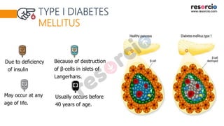
The PPT on Diabetes Mellites. Check out the presentation right away!
To download original, editable and non-watermark PowerPoint Presentation check,
https://resorcio.com/products/diabetes-mellites
To watch more similar PPTs visit www.resorcio.com
Call Girls Service Navi Mumbai Samaira 8617697112 Independent Escort Service ...
Diabetes Mellites
1.
2.
3.
4.
5.
6. CLASSIFICATION OF DIABETES MELLITUS
Several forms due to different causes.
DIABETES
Primary Secondary
Unrelated to
another disease.
Damage or disease of
pancreas by another
disease or factor.
8. TYPE I DIABETES
MELLITUS
Due to deficiency
of insulin
Because of destruction
of β-cells in islets of
Langerhans.
May occur at any
age of life.
Usually occurs before
40 years of age.
9. TYPE I DIABETES MELLITUS
May be associated with
acidosis or ketosis.
Affected persons require insulin
injection.
Also called insulin-dependent
diabetes mellitus (IDDM).
It develops at infancy or
childhood, it is called juvenile
diabetes.
Develops rapidly and progresses
at a rapid phase.
Not associated with obesity
01
02
03
04
05
06
10. CAUSES OF TYPE I
DIABETES MELLITUS
Degeneration of β-cells in islets
of Langerhans of pancreas.
Destruction of β-cells by viral
infection
Congenital disorder of β-cells
Destruction of β-cells during
autoimmune diseases due to
development of antibodies
against β-cells
11.
12. OTHER FORMS OF TYPE 1 DIABETES
MELLITUS
Latent autoimmune
diabetes in adults
Maturity onset diabetes in
young individuals (MODY):
01
02
13. LATENT AUTOIMMUNE DIABETES IN ADULTS
Since pancreas takes
longer period to stop
secreting insulin.
LADA or slow onset
diabetes
Slow onset and slow
progress than IDDM
It occurs in later
life after 35 years.
Difficult to distinguish
LADA from type II
diabetes mellitus
01 02 03 04 05
14. 02
03
01
MATURITY ONSET DIABETES IN
YOUNG INDIVIDUALS (MODY):
Due to hereditary defects
in insulin secretion.
Rare inherited form
of diabetes mellitus
Occurs before 25
years.
15. TYPE
II
DIABETES
MELLITUS
Insulin resistance (failure of insulin
receptors to give response to insulin).
Body is unable to use insulin.
About 90% of diabetic patients have
type II diabetes mellitus
16. Only some forms of
Type II diabetes
require insulin.
Can be controlled by
oral hypoglycemic
drugs.
Also called non
insulin dependent
diabetes mellitus
(NIDDM).
May or may not be
associated with
ketosis.
Often it is associated
with obesity.
TYPE II DIABETES
MELLITUS
17. Structure and function of β-cells and
blood level of insulin are normal.
TYPE II DIABETES
MELLITUS
Insulin receptors may be less, absent
or abnormal, resulting in insulin
resistance.
18. Genetic
disorders
COMMON CAUSES OF INSULIN RESISTANCE
Lifestyle changes such
as bad eating habits
and physical inactivity,
leading to obesity
Stress.
01
02
03
20. 1
2
3
4
Due to
Hormones secreted during pregnancy
Obesity and lifestyle before and
during pregnancy.
GESTATIONAL DIABETES
Occurs during pregnancy.
Usually, diabetes disappears after
delivery of child.
High risk of development of type
II diabetes later.
21. 1
2
3
PRE-DIABETES
Also called chemical, subclinical,
latent or borderline diabetes.
Stage between normal condition and
diabetes.
Person does not show overt
(observable) symptoms of diabetes
22. PRE-
DIABETES
There is an
increase in
blood glucose
level.
Affected persons
are at a high risk
of developing
type II diabetes
mellitus.
Though pre-
diabetes is
reversible
25. CAUSES OF SECONDARY DIABETES MELLITUS
(high iron content in body causing
damage of organs)
02 DAMAGE OF PANCREAS DUE TO DISORDERS
Cystic fibrosis
Chronic
pancreatitis
Hemochromatosis
26. CAUSES OF SECONDARY DIABETES
MELLITUS
Pancreatectomy (surgical removal)
Liver diseases such as hepatitis C
and fatty liver
Autoimmune diseases such as celiac
disease
Excessive use of drugs like antihypertensive
drugs (beta blockers and diuretics), steroids,
oral contraceptives, chemotherapy drugs, etc.
Excessive intake of alcohol and
opiates.
27. CAUSES OF SECONDARY DIABETES MELLITUS
Increased blood glucose level (300 to 400
mg/dL) due to reduced utilization by tissue.
Mobilization of fats from adipose
tissue for energy purpose, leading to
elevated fatty acid content in blood.
This causes deposition of fat on
wall of arteries and development
of atherosclerosis.
Depletion of proteins from
tissues.
01
03 02
28. SIGNS AND SYMPTOMS OF DIABETES
MELLITUS
Glucosuria
01
Osmotic diuresis
02
03
Polyuria
Polydipsia
04
05
Polyphagia
Asthenia 06
29. SIGNS AND SYMPTOMS OF DIABETES
MELLITUS
Acidosis
07
08
09
10
11
Acetone breathing
Kussmaul Breathing
Circulatory shock
Coma
30. GLUCOSURIA
Loss of glucose in urine. Normally, glucose does
not appear in urine.
Glucose level rises above 180
mg/dL in blood, glucose appears
in urine.
It is renal threshold level
for glucose.
01
31.
32. OSMOTIC
DIURESIS
Diuresis caused by osmotic effects.
02
It leads to polyuria and polydipsia.
Excess glucose in renal tubules develops osmotic effect.
Osmotic effect decreases reabsorption of water from renal
tubules, resulting in diuresis.
35. POLYDIPSIA
Increase in water intake.
Excess loss of water decreases water content
and increases salt content in body.
This stimulates thirst center in hypothalamus.
Thirst center, in turn increases intake of water.
04
37. ASTHENIA
It also occurs due to utilization of proteins for
energy in absence of glucose utilization.
06 Loss of strength.
Body becomes very weak because of this.
Protein depletion caused by lack of insulin.
Decrease in protein synthesis and increase in
protein breakdown, resulting in protein depletion.
1
3
2
4
5
38. ACIDOSIS 07
During insulin deficiency,
glucose cannot be utilized by
peripheral tissues for energy.
So, a large amount of
fat is broken down to
release energy.
39. 01
02
03
ACIDOSIS 07
It causes formation of excess
ketoacids, leading to acidosis.
One more reason for acidosis is
that ketoacids are excreted in
combination with sodium ions
through urine (ketonuria).
Sodium is exchanged for hydrogen
ions, which diffuse from renal
tubules into ECF adding to acidosis.
40. ACETONE BREATHING
08
In cases of severe ketoacidosis, acetone is
expired in expiratory air, giving
characteristic acetone or fruity breath odor.
It is a life-threatening condition of severe
diabetes.
41.
42. KUSSMAUL
BREATHING
09
Increase in rate and depth of
respiration caused by severe
acidosis.
CIRCULATORY
SHOCK
10
Osmotic diuresis leads to
dehydration
It occurs only in severe diabetes.
43.
44. COMA 11
Due to Kussmaul breathing,
large amount of carbon dioxide
is lost during expiration.
It leads to drastic reduction in
concentration of bicarbonate ions
causing severe acidosis and coma.
45. COMA 11
It occurs in severe cases of
diabetes mellitus.
Increase in blood glucose level
develops hyperosmolarity of plasma
which also leads to coma.
Hyperosmolar
coma :
severe cases
46. COMPLICATIONS OF DIABETES MELLITUS
Prolonged hyperglycemia cause
dysfunction and injury of many
tissues, resulting in some
complications.
Development of complications is
directly proportional to degree and
duration of hyperglycemia.
47. Patients with well controlled diabetes
can postpone onset or reduce rate of
progression of these complications.
Chronic hyperglycemia affects
blood vessels, resulting in vascular
complications like atherosclerosis.
COMPLICATIONS OF DIABETES MELLITUS
48. Vascular complications are responsible for development of most of
complications of diabetes such as:
VASCULAR COMPLICATIONS
Cardiovascular complications
Diabetic Retinopathy
Diabetic Nephropathy
Diabetic Neuropathy
50. Degenerative
changes in retina
called diabetic
retinopathy
VASCULAR COMPLICATIONS
Degenerative
changes in kidney
known as diabetic
nephropathy
Diabetic Retinopathy Diabetic Nephropathy
Degeneration of
autonomic and
peripheral nerves
called diabetic
neuropathy.
Diabetic Neuropathy.
51. A B
C D
Fasting blood glucose
DIAGNOSTIC TESTS FOR DIABETES MELLITUS
Postprandial blood
glucose
Glucose tolerance
test (GTT)
Glycosylated
(glycated) hemoglobin.
Monitor glycemic control of persons
already diagnosed with diabetes mellitus.
52. Occurs in conditions
like pre-diabetes
ABNORMAL RESPONSE IN
DIAGNOSTIC TESTS
There is an increased fasting
blood glucose level or impaired
(decreased) glucose tolerance.
01 02
54. Treated by exogenous
insulin.
TYPE I DIABETES
MELLITUS
Since insulin is a polypeptide, it is
degraded in GI tract if taken orally.
So, it is generally administered
by subcutaneous injection.
55. Patients with
longstanding severe
diabetes mellitus may
require a combination
of oral hypoglycemic
drugs with insulin to
control hyperglycemia.
TYPE II DIABETES
MELLITUS
Treated by oral
hypoglycemic drugs.
57. INSULIN SECRETAGOGUES
These drugs decrease blood glucose level by
stimulating insulin secretion from β-cells.
Sulfonylureas (tolbutamide, gluburide,
glipizide, etc.) are commonly available insulin
secretagogues.
58. INSULIN
SENSITIZERS
These drugs decrease blood glucose level by
facilitating insulin action in target tissues.
Examples are
Biguanides (metformin)
Thiazolidinediones (pioglitazone and rosiglitazone)
59. ALPHA GLUCOSIDASE INHIBITORS
These drugs
control blood
glucose level by
inhibiting α-
glucosidase.
This intestinal enzyme
is responsible for
conversion of dietary
and other complex
carbohydrates into
glucose and other
monosaccharides,
which can be absorbed
from intestine.
Examples of α-
glucosidase inhibitors
are acarbose and
meglitol