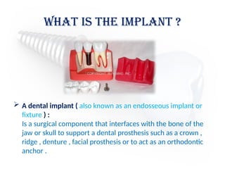
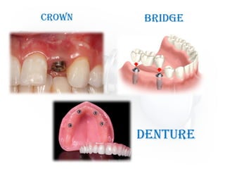
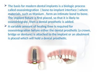
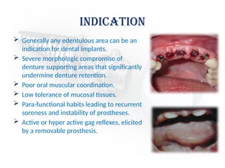
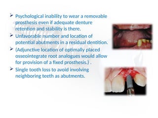
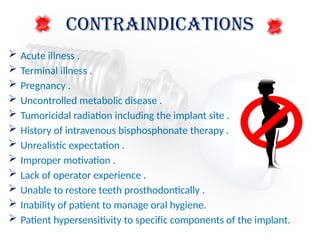
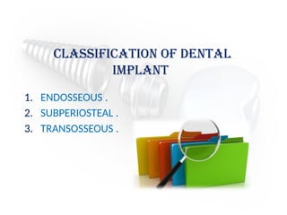
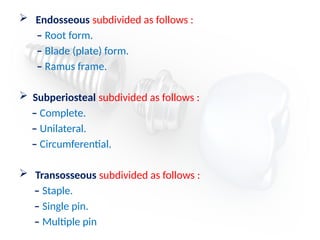
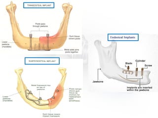
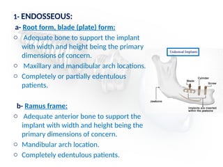
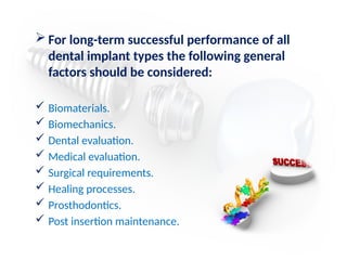
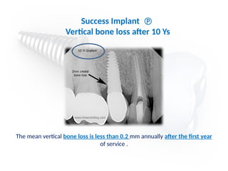
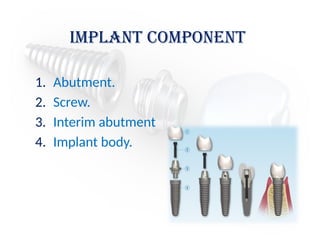
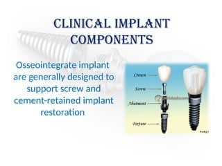
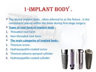
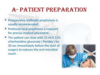
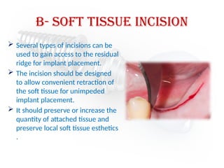
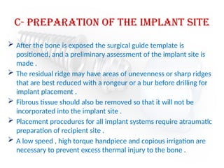
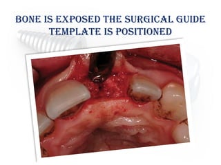
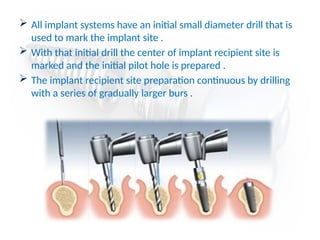
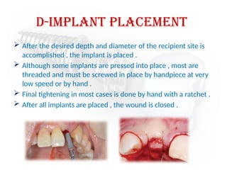
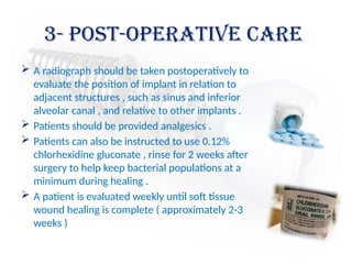
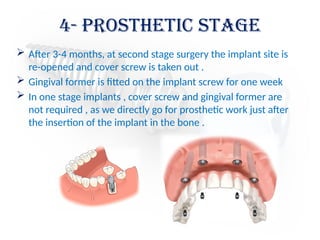
Dental Implant
Presented by :
Ghadah Abulqumsan
Noor Abdurrahman
Reham Al Tayep
What is the implant ?
 A dental implant ( also known as an endosseous implant or
fixture ) :
Is a surgical component that interfaces with the bone of the
jaw or skull to support a dental prosthesis such as a crown ,
ridge , denture , facial prosthesis or to act as an orthodontic
anchor .
Crown Bridge
Denture
 The basis for modern dental implants is a biologic process
called osseointegration ( bone-to-implant interface ) where
materials, such as titanium , form an intimate bond to bone.
The implant fixture is first placed, so that it is likely to
osseointegrate, then a dental prosthetic is added.
 A variable amount of healing time is required for
osseointegration before either the dental prosthetic (a crown,
bridge or denture) is attached to the implant or an abutment
is placed which will hold a dental prosthetic.
Indication
 Generally any edentulous area can be an
indication for dental implants.
 Severe morphologic compromise of
denture supporting areas that significantly
undermine denture retention.
 Poor oral muscular coordination.
 Low tolerance of mucosal tissues.
 Para-functional habits leading to recurrent
soreness and instability of prostheses.
 Active or hyper active gag reflexes, elicited
by a removable prosthesis.
 Psychological inability to wear a removable
prosthesis even if adequate denture
retention and stability is there.
 Unfavorable number and location of
potential abutments in a residual dentition.
 (Adjunctive location of optimally placed
osseointegrate root analogues would allow
for provision of a fixed prosthesis.) .
 Single tooth loss to avoid involving
neighboring teeth as abutments.
CONTRAINDICATIONS
 Acute illness .
 Terminal illness .
 Pregnancy .
 Uncontrolled metabolic disease .
 Tumoricidal radiation including the implant site .
 History of intravenous bisphosphonate therapy .
 Unrealistic expectation .
 Improper motivation .
 Lack of operator experience .
 Unable to restore teeth prosthodontically .
 Inability of patient to manage oral hygiene.
 Patient hypersensitivity to specific components of the implant.
Classification of dental
implant
1. ENDOSSEOUS .
2. SUBPERIOSTEAL .
3. TRANSOSSEOUS .
 Endosseous subdivided as follows :
– Root form.
– Blade (plate) form.
– Ramus frame.
 Subperiosteal subdivided as follows :
– Complete.
– Unilateral.
– Circumferential.
 Transosseous subdivided as follows :
– Staple.
– Single pin.
– Multiple pin
INDICATIONS FOR EACH IMPLANT TYPE ARE
SPECIFIED BELOW:
1- ENDOSSEOUS:
a- Root form, blade (plate) form:
o Adequate bone to support the implant
with width and height being the primary
dimensions of concern.
o Maxillary and mandibular arch locations.
o Completely or partially edentulous
patients.
b- Ramus frame:
o Adequate anterior bone to support the
implant with width and height being the
primary dimensions of concern.
o Mandibular arch location.
o Completely edentulous patients.
2- SUBPERIOSTEAL:
( complete, unilateral, circumferential ):
o Atrophy of bone but with adequate bone to support the
implant.
o Maxillary and mandibular arch locations.
o Completely and partially edentulous patients.
o Stable bone for support.
3- TRANSOSSEOUS:
( staple, single pin, multiple pin ):
o Adequate anterior bone to support the implant with width and
height being the primary dimensions of concern.
o Anterior mandibular arch location.
o Completely and partially edentulous patients.
 For long-term successful performance of all
dental implant types the following general
factors should be considered:
 Biomaterials.
 Biomechanics.
 Dental evaluation.
 Medical evaluation.
 Surgical requirements.
 Healing processes.
 Prosthodontics.
 Post insertion maintenance.
Generally Accepted Implant
Success Criteria
1. The individual unattached implant is immobile when tested
clinically .
2. No evidence of pri-implant radiolucency is present , as
assessed on an undistorted radiograph .
3. The mean vertical bone loss is less than 0.2 mm annually
after the first year of service .
4. No persistent pain , discomfort , or infection is attributable
to the implant .
5. The implant design does not preclude placement of a crown
or prosthesis with an appearance that is satisfactory to the
patient and the dentist .
The mean vertical bone loss is less than 0.2 mm annually after the first year
of service .
Success Implant 
Vertical bone loss after 10 Ys
Implant component
1. Abutment.
2. Screw.
3. Interim abutment
4. Implant body.
Clinical implant
components
Osseointegrate implant
are generally designed to
support screw and
cement-retained implant
restoration
1-Implant body .
 The dental implant body , often referred to as the fixture , is the
component placed within the bone during first-stage surgery.
 Types of root form of implant body :
1. Threaded root form
2. Non-threaded root form
 The main categories of implant body :
1. Titanium screw .
2. Hydroxyapatite-coated screw
3. Titanium plasma-sprayed cylinder
4. Hydroxyapatite-coated cylinder
Types of root form of
implant body
2- Healing Screw
 During the healing phase after first stage surgery , a screw is
normally placed in superior aspect of the fixture .
 The screw is usually low in profile to facilitate the suturing of
soft tissue in two stage implant or minimize loading in the one
stage implant
 A second stage surgery , the screw is removed and placed by
subsequent components .
3- Interim Abutment
 Interim abutment are dome-shaped screws placed after
second stage surgery and before insertion of prosthesis .
 The abutments range in length from 2-10 mm and project
through the soft tissue into the oral cavity .
 The abutments may screw directly into the fixture or, in some
systems , onto the abutment immediately after second stage
surgery
4- Abutments
 Abutments are the component of the implant system that
screw directly into the implant .
 Abutments eventually support the prosthesis in screw-
retained restoration , in as much as they accept the retaining
screw of prosthesis .
 For cement retained restorations, abutments may be shaped
like a conventional crown preparation .
 The walls of abutments are usually smooth ,
polished , and straight – sided .
 The length range from 1-10 mm
 In nonesthetic areas , 1-2 mm of titanium should be
allowed to penetrate the soft tissue to maximize the
patient’s ability to clean the prosthesis
 The choice of abutment size depends on the vertical
distance between the fixture base and opposing
dentition , the existing sulcular depth , and the
esthetic requirements in the area being restored .
 For acceptable appearance , an anterior maxillary
crown may require 2-3 mm of subgingival porcelain
at the facial gingival margin to create the proper
emergence profile and appearance , whereas
fixtures in the posterior maxilla or mandible may
have margin termination at or below the gingival
crest .
5- Impression Coping
 Impression coping facilitate transfer of the of intraoral
location of the implant or abutment to a similar position on
the laboratory cast .
 Impression coping may screw into the implant or onto the
abutment and are customarily subdivided into fixture types or
abutments types .
 Types of impression coping :
1. Transfer ( indirect ) .
2. Pickup ( direct ) .
6- implant analog
 Implant analogs are made to represent
exactly the top of the implant fixture or
the abutment in the laboratory cast .
 Therefore , analogs can be classified as
fixture analogs and abutment analogs .
 Both types of analog screw directly into
the impression coping after it has been
removed from the mouth , and the
joined components are returned to the
impression before pouring .

Abutment + Transfer r + Analog + Healing Cup
7- waxing sleeve
 Waxing sleeves are attached to the abutment by the
relating screw on the laboratory model .
 The sleeves eventually become part of prosthesis .
 In non segmented implant crowns , the sleeves are
attached directly to the implant body analog in the
cast .
8- prosthesis retaining screw
 prosthesis retaining screws
penetrate the fixed restoration and
secure it to the abutment .
 The screws are tightened with a
screwdriver and attach non-
segmented crowns to the body of
the implant .
Basic Surgical procedure
1- before Implant placement
Successful implant placement relies on adequate bone
quantity and quality .
The availability of bone at the implant site is based on
many variables .
Some of variables controlled by the dentist include use
of :
1. Atraumatic extraction techniques .
2. Preservation of the bony socket .
3. Selection of interim prosthesis.
4. Timing of implant placement .
2- implant placement
a) Patient preparation
b) Soft tissue incision
c) Preparation of implant site
d) Implant placement
a- Patient preparation
 Preoperative antibiotic prophylaxis is
usually recommended .
 Profound local anesthesia is required
for precise implant placement .
 The patient can rinse with 15 ml 0.12%
chlorhexidine gluconate ( Peridex ) for
30 sec immediately before the start of
surgery to reduces the oral microbial
count .
b- soft tissue Incision
 Several types of incisions can be
used to gain access to the residual
ridge for implant placement.
 The incision should be designed
to allow convenient retraction of
the soft tissue for unimpeded
implant placement.
 It should preserve or increase the
quantity of attached tissue and
preserve local soft tissue esthetics
.
c- preparation of the implant site
 After the bone is exposed the surgical guide template is
positioned, and a preliminary assessment of the implant site is
made .
 The residual ridge may have areas of unevenness or sharp ridges
that are best reduced with a rongeur or a bur before drilling for
implant placement .
 Fibrous tissue should also be removed so that it will not be
incorporated into the implant site .
 Placement procedures for all implant systems require atraumatic
preparation of recipient site .
 A low speed , high torque handpiece and copious irrigation are
necessary to prevent excess thermal injury to the bone .
bone is exposed the surgical guide
template is positioned
 All implant systems have an initial small diameter drill that is
used to mark the implant site .
 With that initial drill the center of implant recipient site is
marked and the initial pilot hole is prepared .
 The implant recipient site preparation continuous by drilling
with a series of gradually larger burs .
d-implant placement
 After the desired depth and diameter of the recipient site is
accomplished , the implant is placed .
 Although some implants are pressed into place , most are
threaded and must be screwed in place by handpiece at very
low speed or by hand .
 Final tightening in most cases is done by hand with a ratchet .
 After all implants are placed , the wound is closed .
3- post-operative care
 A radiograph should be taken postoperatively to
evaluate the position of implant in relation to
adjacent structures , such as sinus and inferior
alveolar canal , and relative to other implants .
 Patients should be provided analgesics .
 Patients can also be instructed to use 0.12%
chlorhexidine gluconate , rinse for 2 weeks after
surgery to help keep bacterial populations at a
minimum during healing .
 A patient is evaluated weekly until soft tissue
wound healing is complete ( approximately 2-3
weeks )
If the patient wears a tissue borne denture
over the area of implant placement , the
denture can be relined with soft liner after
one week and may be worn .
Interim partial dentures or orthodontic
retainers with an attached pontic may be worn
immediately but must be contoured to avoid
soft tissue loading over the implant site .
 After 3-4 months, at second stage surgery the implant site is
re-opened and cover screw is taken out .
 Gingival former is fitted on the implant screw for one week
 In one stage implants , cover screw and gingival former are
not required , as we directly go for prosthetic work just after
the insertion of the implant in the bone .
4- prosthetic stage
Contemporary Oral and Maxillofacial Surgery
5th
ed ( Ch:14 )
http://www.slideshare.net/
indiandentalacademy/indications-contra-
indications-of-isp
Reference
Dental Implants Basic Overview Presentation

Dental Implants Basic Overview Presentation

  • 1.
    Dental Implant Presented by: Ghadah Abulqumsan Noor Abdurrahman Reham Al Tayep
  • 2.
    What is theimplant ?  A dental implant ( also known as an endosseous implant or fixture ) : Is a surgical component that interfaces with the bone of the jaw or skull to support a dental prosthesis such as a crown , ridge , denture , facial prosthesis or to act as an orthodontic anchor .
  • 3.
  • 4.
     The basisfor modern dental implants is a biologic process called osseointegration ( bone-to-implant interface ) where materials, such as titanium , form an intimate bond to bone. The implant fixture is first placed, so that it is likely to osseointegrate, then a dental prosthetic is added.  A variable amount of healing time is required for osseointegration before either the dental prosthetic (a crown, bridge or denture) is attached to the implant or an abutment is placed which will hold a dental prosthetic.
  • 5.
    Indication  Generally anyedentulous area can be an indication for dental implants.  Severe morphologic compromise of denture supporting areas that significantly undermine denture retention.  Poor oral muscular coordination.  Low tolerance of mucosal tissues.  Para-functional habits leading to recurrent soreness and instability of prostheses.  Active or hyper active gag reflexes, elicited by a removable prosthesis.
  • 6.
     Psychological inabilityto wear a removable prosthesis even if adequate denture retention and stability is there.  Unfavorable number and location of potential abutments in a residual dentition.  (Adjunctive location of optimally placed osseointegrate root analogues would allow for provision of a fixed prosthesis.) .  Single tooth loss to avoid involving neighboring teeth as abutments.
  • 7.
    CONTRAINDICATIONS  Acute illness.  Terminal illness .  Pregnancy .  Uncontrolled metabolic disease .  Tumoricidal radiation including the implant site .  History of intravenous bisphosphonate therapy .  Unrealistic expectation .  Improper motivation .  Lack of operator experience .  Unable to restore teeth prosthodontically .  Inability of patient to manage oral hygiene.  Patient hypersensitivity to specific components of the implant.
  • 8.
    Classification of dental implant 1.ENDOSSEOUS . 2. SUBPERIOSTEAL . 3. TRANSOSSEOUS .
  • 9.
     Endosseous subdividedas follows : – Root form. – Blade (plate) form. – Ramus frame.  Subperiosteal subdivided as follows : – Complete. – Unilateral. – Circumferential.  Transosseous subdivided as follows : – Staple. – Single pin. – Multiple pin
  • 11.
    INDICATIONS FOR EACHIMPLANT TYPE ARE SPECIFIED BELOW:
  • 12.
    1- ENDOSSEOUS: a- Rootform, blade (plate) form: o Adequate bone to support the implant with width and height being the primary dimensions of concern. o Maxillary and mandibular arch locations. o Completely or partially edentulous patients. b- Ramus frame: o Adequate anterior bone to support the implant with width and height being the primary dimensions of concern. o Mandibular arch location. o Completely edentulous patients.
  • 13.
    2- SUBPERIOSTEAL: ( complete,unilateral, circumferential ): o Atrophy of bone but with adequate bone to support the implant. o Maxillary and mandibular arch locations. o Completely and partially edentulous patients. o Stable bone for support.
  • 14.
    3- TRANSOSSEOUS: ( staple,single pin, multiple pin ): o Adequate anterior bone to support the implant with width and height being the primary dimensions of concern. o Anterior mandibular arch location. o Completely and partially edentulous patients.
  • 15.
     For long-termsuccessful performance of all dental implant types the following general factors should be considered:  Biomaterials.  Biomechanics.  Dental evaluation.  Medical evaluation.  Surgical requirements.  Healing processes.  Prosthodontics.  Post insertion maintenance.
  • 16.
    Generally Accepted Implant SuccessCriteria 1. The individual unattached implant is immobile when tested clinically . 2. No evidence of pri-implant radiolucency is present , as assessed on an undistorted radiograph . 3. The mean vertical bone loss is less than 0.2 mm annually after the first year of service . 4. No persistent pain , discomfort , or infection is attributable to the implant . 5. The implant design does not preclude placement of a crown or prosthesis with an appearance that is satisfactory to the patient and the dentist .
  • 17.
    The mean verticalbone loss is less than 0.2 mm annually after the first year of service . Success Implant  Vertical bone loss after 10 Ys
  • 18.
    Implant component 1. Abutment. 2.Screw. 3. Interim abutment 4. Implant body.
  • 19.
    Clinical implant components Osseointegrate implant aregenerally designed to support screw and cement-retained implant restoration
  • 20.
    1-Implant body . The dental implant body , often referred to as the fixture , is the component placed within the bone during first-stage surgery.  Types of root form of implant body : 1. Threaded root form 2. Non-threaded root form  The main categories of implant body : 1. Titanium screw . 2. Hydroxyapatite-coated screw 3. Titanium plasma-sprayed cylinder 4. Hydroxyapatite-coated cylinder
  • 21.
    Types of rootform of implant body
  • 22.
    2- Healing Screw During the healing phase after first stage surgery , a screw is normally placed in superior aspect of the fixture .  The screw is usually low in profile to facilitate the suturing of soft tissue in two stage implant or minimize loading in the one stage implant  A second stage surgery , the screw is removed and placed by subsequent components .
  • 23.
    3- Interim Abutment Interim abutment are dome-shaped screws placed after second stage surgery and before insertion of prosthesis .  The abutments range in length from 2-10 mm and project through the soft tissue into the oral cavity .  The abutments may screw directly into the fixture or, in some systems , onto the abutment immediately after second stage surgery
  • 24.
    4- Abutments  Abutmentsare the component of the implant system that screw directly into the implant .  Abutments eventually support the prosthesis in screw- retained restoration , in as much as they accept the retaining screw of prosthesis .  For cement retained restorations, abutments may be shaped like a conventional crown preparation .
  • 25.
     The wallsof abutments are usually smooth , polished , and straight – sided .  The length range from 1-10 mm  In nonesthetic areas , 1-2 mm of titanium should be allowed to penetrate the soft tissue to maximize the patient’s ability to clean the prosthesis  The choice of abutment size depends on the vertical distance between the fixture base and opposing dentition , the existing sulcular depth , and the esthetic requirements in the area being restored .  For acceptable appearance , an anterior maxillary crown may require 2-3 mm of subgingival porcelain at the facial gingival margin to create the proper emergence profile and appearance , whereas fixtures in the posterior maxilla or mandible may have margin termination at or below the gingival crest .
  • 26.
    5- Impression Coping Impression coping facilitate transfer of the of intraoral location of the implant or abutment to a similar position on the laboratory cast .  Impression coping may screw into the implant or onto the abutment and are customarily subdivided into fixture types or abutments types .  Types of impression coping : 1. Transfer ( indirect ) . 2. Pickup ( direct ) .
  • 27.
    6- implant analog Implant analogs are made to represent exactly the top of the implant fixture or the abutment in the laboratory cast .  Therefore , analogs can be classified as fixture analogs and abutment analogs .  Both types of analog screw directly into the impression coping after it has been removed from the mouth , and the joined components are returned to the impression before pouring .  Abutment + Transfer r + Analog + Healing Cup
  • 28.
    7- waxing sleeve Waxing sleeves are attached to the abutment by the relating screw on the laboratory model .  The sleeves eventually become part of prosthesis .  In non segmented implant crowns , the sleeves are attached directly to the implant body analog in the cast .
  • 29.
    8- prosthesis retainingscrew  prosthesis retaining screws penetrate the fixed restoration and secure it to the abutment .  The screws are tightened with a screwdriver and attach non- segmented crowns to the body of the implant .
  • 30.
  • 31.
    1- before Implantplacement Successful implant placement relies on adequate bone quantity and quality . The availability of bone at the implant site is based on many variables . Some of variables controlled by the dentist include use of : 1. Atraumatic extraction techniques . 2. Preservation of the bony socket . 3. Selection of interim prosthesis. 4. Timing of implant placement .
  • 32.
    2- implant placement a)Patient preparation b) Soft tissue incision c) Preparation of implant site d) Implant placement
  • 33.
    a- Patient preparation Preoperative antibiotic prophylaxis is usually recommended .  Profound local anesthesia is required for precise implant placement .  The patient can rinse with 15 ml 0.12% chlorhexidine gluconate ( Peridex ) for 30 sec immediately before the start of surgery to reduces the oral microbial count .
  • 34.
    b- soft tissueIncision  Several types of incisions can be used to gain access to the residual ridge for implant placement.  The incision should be designed to allow convenient retraction of the soft tissue for unimpeded implant placement.  It should preserve or increase the quantity of attached tissue and preserve local soft tissue esthetics .
  • 35.
    c- preparation ofthe implant site  After the bone is exposed the surgical guide template is positioned, and a preliminary assessment of the implant site is made .  The residual ridge may have areas of unevenness or sharp ridges that are best reduced with a rongeur or a bur before drilling for implant placement .  Fibrous tissue should also be removed so that it will not be incorporated into the implant site .  Placement procedures for all implant systems require atraumatic preparation of recipient site .  A low speed , high torque handpiece and copious irrigation are necessary to prevent excess thermal injury to the bone .
  • 36.
    bone is exposedthe surgical guide template is positioned
  • 37.
     All implantsystems have an initial small diameter drill that is used to mark the implant site .  With that initial drill the center of implant recipient site is marked and the initial pilot hole is prepared .  The implant recipient site preparation continuous by drilling with a series of gradually larger burs .
  • 38.
    d-implant placement  Afterthe desired depth and diameter of the recipient site is accomplished , the implant is placed .  Although some implants are pressed into place , most are threaded and must be screwed in place by handpiece at very low speed or by hand .  Final tightening in most cases is done by hand with a ratchet .  After all implants are placed , the wound is closed .
  • 39.
    3- post-operative care A radiograph should be taken postoperatively to evaluate the position of implant in relation to adjacent structures , such as sinus and inferior alveolar canal , and relative to other implants .  Patients should be provided analgesics .  Patients can also be instructed to use 0.12% chlorhexidine gluconate , rinse for 2 weeks after surgery to help keep bacterial populations at a minimum during healing .  A patient is evaluated weekly until soft tissue wound healing is complete ( approximately 2-3 weeks )
  • 40.
    If the patientwears a tissue borne denture over the area of implant placement , the denture can be relined with soft liner after one week and may be worn . Interim partial dentures or orthodontic retainers with an attached pontic may be worn immediately but must be contoured to avoid soft tissue loading over the implant site .
  • 41.
     After 3-4months, at second stage surgery the implant site is re-opened and cover screw is taken out .  Gingival former is fitted on the implant screw for one week  In one stage implants , cover screw and gingival former are not required , as we directly go for prosthetic work just after the insertion of the implant in the bone . 4- prosthetic stage
  • 42.
    Contemporary Oral andMaxillofacial Surgery 5th ed ( Ch:14 ) http://www.slideshare.net/ indiandentalacademy/indications-contra- indications-of-isp Reference

Editor's Notes

  • #4 Osseointegration derives from the Greek osteon, bone, and the Latin integrare, to make whole. The term refers to the direct structural and functional connection between living bone and the surface of a load-bearing artificial implant. Osseointegration has enhanced the science of medical bone and joint replacement techniques as well as dental implants and improving prosthetics for amputees.