
Recommended
More Related Content
What's hot
What's hot (20)
Similar to Cryptococcal lung infections
Similar to Cryptococcal lung infections (20)
Recently uploaded
Recently uploaded (20)
Cryptococcal lung infections
- 1. Cryptococcal Lung Infections Kate Skolnik, MDa , Shaunna Huston, PhDb , Christopher H. Mody, MDc,d, * INTRODUCTION Cryptococcus remains one of the leading causes of acquired immunodeficiency syndrome (AIDS)- related deaths, and is among the most common fungal pathogens worldwide.1 Cryptococcus de- mands global attention because of the high mortal- ity, unique geographic distribution, and its propensity to cause severe and rapidly progressive disease in both healthy and immunosuppressed in- dividuals.1,2 Cryptococcus neoformans, which mainly infects immunosuppressed individuals, and Cryptococcus gattii, which has a high propen- sity to infect healthy individuals, are the 2 major species of Cryptococcus that are associated with mortality.1 Arguably, the greatest virulence factor for both species is the cryptococcal polysaccha- ride capsule, which helps evade immune detec- tion.3 The organism often leads to respiratory infection but can also disseminate to other organs, in particular to the meninges causing meningoen- cephalitis, which is associated with poor clinical outcomes.2,4 Current diagnostics are based on detection of antigen and culture techniques, and treatment recommendations are tailored based on fungal susceptibility, location, and severity of disease. Research is advancing technologies to enable more effective and efficient diagnostics for early detection along with improved prophylactic and therapeutic regimes. Disclosure Statement: The authors have nothing to disclose. a Division of Respirology, Department of Internal Medicine, Rockyview General Hospital, University of Calgary, Respirology Offices, 7007 14th Street Southwest, Calgary, Alberta T2V 1P9, Canada; b Department of Physi- ology and Pharmacology, Health Research Innovation Centre, University of Calgary, Room 4AA08, 3330 Hospi- tal Drive Northwest, Calgary, Alberta T2N 4N1, Canada; c Department of Microbiology and Infectious Diseases, Health Research Innovation Centre, University of Calgary, Room 4AA14, 3330 Hospital Drive Northwest, Cal- gary, Alberta T2N 4N1, Canada; d Department of Internal Medicine, Health Research Innovation Centre, Uni- versity of Calgary, Room 4AA14, 3330 Hospital Drive Northwest, Calgary, Alberta T2N 4N1, Canada * Corresponding author. Health Research Innovation Centre, Room 4AA14, 3330 Hospital Drive Northwest, Calgary, Alberta T2N 4N1, Canada. E-mail address: cmody@ucalgary.ca KEYWORDS Cryptococcus Cryptococcosis Fungal lung infection Endemic mycoses Treatment KEY POINTS Cryptococcus infections have high morbidity and mortality rates worldwide, particularly in the context of immune suppression and central nervous system involvement. Cryptococcus neoformans can be found globally and predominantly affects the immune- suppressed individual, whereas Cryptococcus gattii is endemic to certain regions and often affects immune-competent individuals. Most individuals with cryptococcal pulmonary infection present with symptoms; however, they may be mild and nonspecific, making timely diagnosis challenging. Pulmonary nodules or focal consolidation is the most common radiographic finding with Cryptococcus. Azoles (specifically fluconazole) and amphotericin B (in severe disease) are key cryptococcal ther- apies. There is no role for echinocandins. Clin Chest Med 38 (2017) 451–464 http://dx.doi.org/10.1016/j.ccm.2017.04.007 0272-5231/17/Ó 2017 Elsevier Inc. All rights reserved. chestmed.theclinics.com
- 2. EPIDEMIOLOGY General Cryptococcal Epidemiology Among the 37 species of Cryptococcus, only a few are human pathogens.5 C neoformans is characterized as an opportunistic pathogen because it commonly causes infections in individ- uals with impaired immunity. C neoformans causes more than 1 million infections in AIDS pa- tients annually, and, unfortunately, most of these patients will die within a few months of diag- nosis.2 By contrast, C gattii has a more distinct geographic range and is more likely to affect healthy individuals leading to its characterization as an endemic mycosis.6,7 C gattii is classically located in tropical and subtropical areas of the world such as Australia and Papua New Guinea, but more recently it has emerged as an endemic mycosis on the west coast of Canada and the United States.7 Rarely, Cryptococcus laurentii causes infection in individuals with compromised immunity8–10 and even less often in patients who are immune competent.11,12 Immunosuppression is a significant risk factor, and the prevalence of cryptococcal infection significantly increased dur- ing the emergence of AIDS.13 Currently, in devel- oped countries with effective antiretroviral therapies, the prevalence of opportunistic crypto- coccal infections has decreased, but Crypto- coccus continues to affect those with other defects in cell-mediated immunity. Infections also occur in healthy individuals, especially if exposed in endemic areas.14,15 Although Cryptococcus causes both endemic and opportunistic mycosis, transmission of Crypto- coccus occurs directly from the environment rather than from human to human.16–18 Although Crypto- coccus can cause infection in animals such as cats, dogs, ferrets, cockatiels, parrots, llamas, hors- es, and marine mammals,19,20 direct transmission from these species is exceptionally rare. The pri- mary environmental sources of C gattii are trees, such as eucalyptus, Douglas-fir, red cedar, and Garry oak, among others,16,17,21 but it can also be foundin the soil, water, and air in endemic regions.22 C gattii has been isolated from the environment mainlyinAustralia,NewZealand,PapaNewGuinea, Central America, parts of South America, and the Pacific Northwest of North America including Van- couver Island.18,21,23–25 Interestingly, C gattii has also been identified in the Mediterranean basin on various trees, including olive and eucalyptus, which marks the first detection of the species in Europe.26 In contrast, C neoformans is found worldwide, with the primary source being pigeon excrement, although certain subtypes are more common in particular regions of the world.16,18 C neoformans has also been linked to other bird species such as magpies and cockatiels.27,28 Additionally, there seems to be a seasonality to C gattii infections in some regions, which has not been noted with C neoformans.29 Cryptococcal Subtypes It is important to differentiate between subtypes of C neoformans and C gattii for clinical purposes. C neoformans is divided into 2 major groups known as C neoformans var grubii (serotype A) and C neoformans var neoformans (serotype D).30 Addi- tionally, a hybrid serotype, AD, has also been iden- tified as a clinically significant subtype.30 C neoformans and C gattii have also more recently been subdivided into 4 molecular types, termed VNI, VNII, VNIII, VNIV and VGI, VGII, VGIII, VGIV, respectively, which may lead to further species distinctions.30,31 C neoformans var grubii and C neoformans var neoformans have important differ- ences in their genomes and replication methods,30 which have clinical implications, as certain sero- types and molecular types may be more likely to respond to treatment.31 STRUCTURE AND LIFE CYCLE Cryptococci are eukaryotic organisms that belong to the phylum Basidiomycota of filamentous fungi.32 The cryptococcal cell is enveloped by a cell membrane, cell wall, and a characteristic polysac- charide capsule.4 The capsule is unique to Crypto- coccus and sets it apart from other pathogenic fungi.33 It is composed of glucuronoxylomannan (GXM), which is the predominant polysaccharide, GalXM (galactoxylomannan), and mannopro- teins.34,35 Structural differences in GXM allow for the identification of Cryptococcus based on sero- type.36 The fungal cell wall consists of chitin and b-glucans (which provide structural integrity), pores (which allow for the movement of important mole- cules and transport vesicles), melanin (which pro- tects the cell from oxidative stress), and proteins that serve a variety of cellular functions.37–39 Crypto- coccal cells can be identified by light microscopy but require special stains such as mucicarmine or Periodic-acid Schiff to identify the capsule or silver stains to identify the cell wall.40 Cryptococcus is also readily identified with India ink staining, in which exclusion of the stain in the perimeter of the fungal cell is indicative of the fungal capsule (Fig. 1).41,42 Cryptococcus species are able to reproduce asexually through simple budding, and sexually, through the mating of a- and a-mating types.43,44 Asexual reproduction is more common and oc- curs in the human host.43,45 Sexual reproduction Skolnik et al 452
- 3. provides greater genetic diversity and, with this, the potential for increased virulence.46 Both mat- ing strategies have the potential to increase ge- netic diversity.45,46 The reproductive strategy is also influenced by Cryptococcus variety. For example, less than half of C neoformans var grubii are fertile, whereas most C neoformans var neofor- mans are capable of sexual reproduction.30,47 Interestingly, the highly virulent C gattii subtype VGIIa, which is responsible for the outbreak on Vancouver Island, seems to have emerged specif- ically from same-sex reproduction, suggesting that different types of reproduction may provide environmental adaptation and virulence.45 PATHOGENESIS The primary risk factor for C gattii infection is expo- sure from environmental sources in endemic re- gions. It is unclear why most C gattii infections occur in healthy individuals.48 By contrast, C neo- formans infection is more common in immunosup- pressed patients, in particular, those with defective cell-mediated immunity such as those with human immunodeficiency virus (HIV), idiopathic CD4 lym- phopenia, organ transplant, hematologic malig- nancies or those receiving immune-suppressive medications (including systemic steroids, biolo- gic agents, chemotherapy, and other cytotoxic therapies).16,49 Although it is much less common, individuals with a milder degree of immune sup- pression (such as splenectomy, cirrhosis, diabetes mellitus, systemic lupus erythematosus, and preg- nancy) have an increased risk of getting C neofor- mans infections.50–53 Independent of the type of cryptococcal infec- tion or the primary organ involved, the fungus typi- cally enters the body through the lungs.54 After entering, the organism undergoes changes to a phenotype that facilitates invasive infection of the host.54 Although C neoformans takes advantage of natural weak points in the immune system, C gattii is postulated to have additional unique viru- lence factors that allow it to evade the immune system.3,55–57 C neoformans infections may also occur owing to the reactivation of latent infection, similar to tuberculosis.57–60 In one study, antibodies to Cryp- tococcus were found in most individuals in the borough of the Bronx, New York, independent of prior exposure or HIV status, suggesting that exposure is surprisingly common.61 Additionally, years after emigrants from Africa moved to France, infections developed with organisms of an African phenotype rather than a French phenotype, sug- gesting that organisms were dormant in these hosts for years.58 There is evidence that anti- cryptococcal antibodies (and Cryptococcus) can persist in the body for years without symptom development.58,61 However, when impairment in immunity occurs, as with solid-organ transplanta- tion or AIDS, dormant Cryptococcus may reacti- vate and develop into an active fungal infection.58,60 By contrast, C gattii infections seem to occur de novo,62 although it must be noted that clinical disease may occur months or years after exposure.63,64 VIRULENCE FACTORS The distinctive polysaccharide capsule of Crypto- coccus spp. acts as a key virulence factor.4,37 The polysaccharide capsule prevents phagocy- tosis, allowing the organism to bypass the host innate immune response.65 In addition, GXM is known to directly interfere with T lymphocyte function,66 whereas GalXM has been shown to induce cytokine dysregulation and T lymphocyte apoptosis,67 and both GXM and GalXM are capable of stimulating macrophage apoptosis.68 In addition to the polysaccharide capsule, Cryp- tococcus has several virulence factors that act as antioxidants, neutralizing host innate immune mol- ecules and preventing oxidative damage to the cell.69 These antioxidants include mannitol,70 superoxide dismutase,71 thioredoxin reductase,72 and pigments such as melanin.69 Melanin also binds to antibiotics, which may confer antifungal resistance.73 Additional virulence factors target the host by various mechanisms including destabi- lization of the cell membrane (as with b1 phospho- lipase),74 degradation of host proteins (as with metalloproteinases),75 and altering pH (via ure- ase).76 The a-mating type of C gattii is also a viru- lence factor.77 C gattii likely has additional virulence factors that allow it to cause invasive infection in hosts with intact immune systems; Fig. 1. Cryptococcus species as visualized by India ink staining. Arrows indicate the fungal cell capsule. Cryptococcal Lung Infections 453
- 4. however, the exact nature of these factors is yet to be discovered. CLINICAL OUTCOMES OF CRYPTOCOCCAL PULMONARY INFECTION Clinical Manifestations of Cryptococcal Pulmonary Infection Cryptococcal infection can produce a range of manifestations from minimal symptoms to severe life-threatening disease.78 Cryptococcus most often leads to pulmonary75,76 and central nervous system (CNS) disease,79,80 the latter being a ma- jor source of morbidity and mortality. However, Cryptococcus can also present as infection in the bone,78 skin,81 eyes,82 and prostate83,84 and may even cause disseminated disease and cryp- tococcemia.85,86 These findings may occur in isolation or in association with pulmonary Crypto- coccus infection. Interestingly, these presenta- tions can occur in both immune-suppressed and immune-competent individuals, although cryptococcemia is rare in those with intact immunity. An individual’s underlying risk factors may affect the manifestations of cryptococcal disease. For example, those with pre-existing lung disease are more likely to have significant pulmonary infec- tion,78 whereas those with HIV or organ transplant and healthy subjects are more likely to have CNS infection.78,87–89 Lung involvement occurs in 28% to 64% of HIV-negative individuals infected with Cryptococcus52,90 compared with less than 5% of those co-infected with Cryptococcus and HIV.90 By contrast, C gattii tends to cause more pulmonary disease and is generally seen in immune-competent individuals.91 Furthermore, the prevalence of pulmonary cryptococcal infec- tion may be underestimated, as individuals can have asymptomatic pulmonary nodules that never come to medical attention.92 Cryptococcal pulmonary infection can cause vague symptoms and a wide spectrum of disease severity including minimal or no symptoms in a subset of individuals.93 Consequently, early diag- nosis may be challenging, and cryptococcal dis- ease may be overlooked in the setting of subtle symptoms. Most patients with cryptococcal lung infection are clinically symptomatic.94–96 When present, pulmonary symptoms can include dry cough, dyspnea, chest tightness, and fever, with cough and fever being the most common (Ta- ble 1).4,48,95 Mediastinal or hilar lymphadenopathy may occur in both immune-competent and immune-suppressed individuals, which may be a solitary finding or associated with other pulmonary manifestations.97,98 Those with lymphadenopathy may present with symptoms secondary to compression of adjacent structures if the adenop- athy is severe. Cryptococcoma, a granulomatous mass harboring the fungus, may be mistaken for lung cancer on imaging94,99–102 and has been described more frequently in infections caused by C gattii in immune-competent patients99,100 but can also occur with C neoformans.52 Rarely, endobronchial infection can occur.101,102 In some cases, severe pulmonary disease leading to respi- ratory failure can be seen, and acute respiratory distress syndrome has occurred in immune- compromised individuals with pulmonary crypto- coccosis.103,104 A miliary tuberculosislike presen- tation with Cryptococcus in an AIDS patient has also been reported but is exceedingly rare.105,106 Importantly, although immune-compromised indi- viduals, such as those with HIV, are less likely to manifest with pulmonary symptoms, when pre- sent, the manifestations can be very severe. Radiographic Findings of Cryptococcal Pulmonary Infection Radiographic findings can include pulmonary nod- ules (single or multiple), lobar or segmental consol- idation, nodular or reticulonodular interstitial pattern, ground glass opacities, masses, hilar/ mediastinal lymphadenopathy, and even pleural effusions.52,85,93,94,97,98,105 Cryptococcal pleural effusions are typically limited to severely immune- suppressed individuals, as is seen with AIDS or ideopathic CD4 lymphopenia (Fig. 2). Reticular or reticulonodular interstitial changes are the most frequent radiographic presentation in HIV, whereas dense nodules, consolidation, or intersti- tial changes seem to be the most common findings in the immune-competent population.52,85,107,108 Table 1 Potential manifestations of pulmonary cryptococcal infection Symptoms Radiographic Findings None Fevera Dry cougha Dyspnea Chest pain/ discomfort Malaise Single or multiple pulmo- nary nodulesa Consolidation (lobar or segmental)a Nodular interstitial pattern Reticular interstitial pattern Ground glass opacities Mass Mediastinal/hilar lymphadenopathy Pleural effusions a Most common. Skolnik et al 454
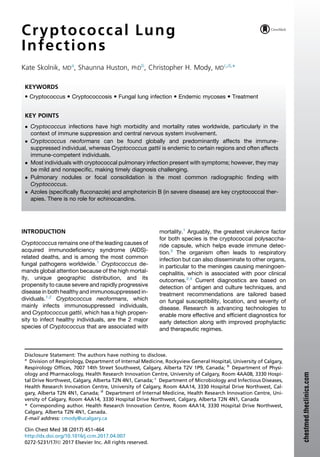
- 5. Overall, about half of all cryptococcal infections present with chest radiograph findings, with some variability depending on cryptococcal subtype and host immune status (see Table 1). In one study of predominantly C neoformans–affected individuals, 66% of those without HIV had an abnormal chest radiograph, compared with 40% of those with HIV.96 In those with HIV and pulmonary C neofor- mans infection, infiltrates or several small nodules were most commonly seen.96 Cryptococcal pul- monary involvement was confirmed in only 58% and 14%, respectively.96 In comparison, a Colom- bian study focusing on C gattii (with a minority of individuals having concurrent HIV) found abnormal chest radiographs in 15% of individuals with known C gattii and only half of them had pulmo- nary involvement.109 However, these numbers are likely an underestimate, as only 40% of sub- jects underwent chest imaging. A separate study of HIV-negative individuals had findings that fell between those of the aforementioned studies, with 36% having pulmonary involvement.78 Computed tomography scanning would likely improve the sensitivity for detecting crypto- coccal-related changes and should be pursued when radiographs are not helpful, but clinical sus- picion for Cryptococcus persists. DIAGNOSIS Testing for Cryptococcus should be considered in anyone with risk factors or exposure history and persistent pulmonary symptoms or radiographic findings despite treatment of common bacterial pathogens. Diagnosis of cryptococcal pulmonary infections usually requires a combination of micro- biologic and radiographic investigations. Although microbiologic tests are the key to diagnosis, pulmonary imaging may bring subclinical infection to the clinicians’ attention and suggest Crypto- coccus as part of a differential diagnosis (see Table 1). As part of the diagnostic process, there should be an assessment for possible extrapulmonary complications of disease; in particular, neurologic involvement should be considered and investigated.49 Cryptococcus can be identified by various methods, including fungal culture, special stains, and immunohistochemical tests that identify cryptococcal antigen.6,110 Fungal culture may be obtained by plating respiratory samples on selec- tive media. Cryptococcus grows best in tem- peratures between 30 C and 35 C,111 and environmental concentrations decrease with hott- er weather.112 Cryptococcus can be identified by exclusion of India ink stain in the immediate cell perimeter; however, this method cannot differen- tiate between viable and dead fungal cells (see Fig. 1).6 Alternatively, the diagnosis may be made by detection of cryptococcal polysaccha- ride antigen in body fluids by various immunologic techniques.113,114 In respiratory secretions, the la- tex agglutination assay has a sensitivity between 93% and 100% and specificity of 93% to 98%,113 whereas enzyme-linked immunosorbent assay has a sensitivity of 99% and specificity of 97% for Cryptococcus.114 These results are similar to assays performed on cerebrospinal fluid samples.115,116 However, the latex agglutination assay was found to be slightly more sensitive than enzyme-linked immunosorbent assay for blood and cerebrospinal fluid samples from a larger study.116 The lateral flow assay is the new- est test for cryptococcal antigen with good yield and has been adopted at most centers because it is easier to use.115,117 An important downfall of these assays is suboptimal sensitivity in detecting C gattii. In one study, 40% of C gattii were missed despite the application of all 3 tests.118 Neverthe- less, lateral flow assay seems to have the best sensitivity for C gattii and has largely replaced other methods.119 Furthermore, although the enzyme-linked immunosorbent assay, latex agglu- tination assay, and lateral flow assay are generally highly sensitive and specific, none of the tests can differentiate viable from dead fungal cells.6 In addi- tion, DNA amplification has largely replaced canavanine-glycine-bromothymol blue selective media for further identification and confirmation of the presence of C gattii.120 If the aforementioned Fig. 2. Computed tomography scan of drowned lung in an adult with idiopathic CD4 lymphopenia second- ary to invasive cryptococcal pneumonia and associ- ated pleural effusion. Cryptococcal Lung Infections 455
- 6. tests are nondiagnostic, but clinical suspicion for Cryptococcus remains high because of exposure history, further diagnostic testing may be required. In some situations, a video-assisted thoraco- scopic biopsy or a surgical wedge resection may be considered if a pulmonary nodule is growing and cannot be accessed by bronchoscopy or percutaneous biopsy.93 Surgical intervention may be important in individuals with risk factors for lung cancer or immune suppression. These biopsy and surgical specimens should be sent for fungal cultures in addition to histopathology. TREATMENT Treatment Indications Antifungal treatment may not be required for everyone with Cryptococcus that is isolated from the lungs. In some individuals, Cryptococcus may simply be colonizing the airways, and patients remain asymptomatic. Occasionally, cryptococcal pulmonary nodules may also not require treatment if involvement is minimal and patients have no symptoms.48 However, immune-suppressed indi- viduals with pulmonary Cryptococcus should be offered antifungal therapy.48,121 Therapy is partic- ularly important in those with impaired cell- mediated immunity, such as those receiving immunosuppressive therapy after organ trans- plant, those with hematologic malignancies, those receiving chemotherapy or biologic agents, and those with long-term prednisone use. There is also a lower threshold to treat asymptomatic indi- viduals with mild immune impairment, such as diabetes or severe renal failure. In addition, immune-competent individuals with mild to severe symptomatic pulmonary cryptococcal infection should be treated.48,121 One must be mindful that some immune-competent individuals may have impaired immunity in the future; therefore, it is essential that those with previously identified Cryptococcus be followed up carefully for signs of reactivation and active infection. Treatment of Cryptococcus neoformans Pulmonary Disease Recommended treatment regimens for crypto- coccal pulmonary infection are summarized in Table 2. Immune-suppressed individuals (spe- cifically, those infected with HIV or have had organ transplant) with mild cryptococcal pulmonary dis- ease or asymptomatic colonization (Cryptococcus isolated from respiratory specimens in the absence of symptoms or radiographic findings) can be treated with fluconazole alone. However, CNS or disseminated disease must be first ruled out.48,121 The standard dose of fluconazole is 400 mg once daily for 6 to 12 months (longer dura- tion preferred).48,121 In the setting of severe cryp- tococcal lung infection or milder lung disease with concurrent disseminated or CNS infection, more aggressive therapy is warranted.48,121 In these cases, the induction treatment consists of intravenous amphotericin B deoxycholate (AmB; 0.7–1 mg/kg/d) with intravenous or oral flucytosine (100 mg/kg/d) for 2 weeks followed by fluconazole, 400 mg once daily (OD) for a minimum of 8 weeks.48,121–124 Alternative induction regimens that can be considered in the absence of flucyto- sine (if it is not available or not tolerated) include a longer duration of AmB or increasing the dose and duration of fluconazole (see Table 2).48,121 Liposomal AmB should be used in place of the usual deoxycholate formulation in those at risk of renal dysfunction, which is often the case for trans- plant patients. Of note, fluconazole has superior efficacy against Cryptococcus compared with other azoles. Doses as high as 1200 mg daily have been used with minimal toxicity, and it has excellent CNS penetration.48,121 Consequently, alternate azoles are reserved for resistant isolates or rare patients with fluconazole intolerance.121 Af- ter induction, maintenance therapy (or secondary prophylaxis) should be continued with fluconazole, 200 mg/d, in organ transplants or other forms of severe immunosuppression for 6 to 12 months.121 In contrast, secondary prophylaxis should be continued until a CD4 count of 200 or greater is sustained for at least 3 months in HIV patients on antiretroviral therapy.48,121 In those with HIV, anti- retroviral therapy should commence 5 weeks after initiation of cryptococcal treatment to reduce the risk of immune reconstitution inflammatory syn- drome, which can be particularly severe and detri- mental with CNS cryptococcal infection.121,122,125 The evidence behind what has now become standard therapy is derived from a few key studies. A landmark treatment trial for crypto- coccal meningitis in AIDS patients found that combined AmB (0.7 mg/kg/d) and flucytosine (100 mg/kg/d) for 2 weeks had a higher likelihood of achieving negative cryptococcal cultures compared with AmB alone.123 Furthermore, pa- tients who were placed on fluconazole for the following 8 weeks had a statistically significant lower fungal burden than those on itraconazole (regardless of flucytosine use).123 Additional studies have validated these findings124,126 and also demonstrated lower risk of relapse with flucy- tosine use,126 although there was no correspond- ing difference in mortality.127 In immunocompetent individuals with mild-to- moderate pulmonary disease (based on combina- tion of symptoms and radiographic findings) the Skolnik et al 456
- 7. Table 2 Summary of treatment regimens for pulmonary cryptococcal infection Form of Disease Treatment Comments Immune suppressedd 1 mild/moderate lung infection Fluconazolea 400 mg po OD for 6–12 mo Secondary prophylaxis required; fluconazole 200 mg po OD Immunosuppressed (HIV1 and CD4100/mm3 ) 1 positive cryptococcal serum antigen Fluconazole 800 mg/d for 2 weeks, followed by 400 mg/d for 8 weeks Secondary prophylaxis required Immune suppressed 1 severe lung infection and/or extrapulmonary infection AmB 0.7–1.0 mg/kg/d IV 1 flucytosineb 100 mg/kg/d for 2 wk followed by fluconazole 400 mg po OD for 8 wk Alternatives 1. Liposomal Amphotericin B (3–6 mg/kg IV daily) or amphotericin B lipid complex (5 mg/kg IV daily)1 flucytosineb 100 mg/kg/d for 2 wk in those with renal failure or chronic kidney disease followed by fluconazole 400 mg po OD for 8 wk 2. AmB 0.7–1.0 mg/kg/d IV 1 flucytosine 100 mg/kg/d for 6–10 wk 3. AmB 0.7–1.0 mg/kg/d IV for 6–10 wk 4. AmB 0.7–1.0 mg/kg/d IV and fluconazole 800 mg OD for 2 wk then fluconazole 800 mg OD for 8 wk 5. Fluconazole 1200 mg OD 1 flucytosine 100 mg/kg OD for 6 wk 6. Fluconazole 1200–2000 mg OD for 10–12 wk Secondary prophylaxis required; fluconazole 200 mg po OD Immune competent 1 mild/moderate lung infection Fluconazolea 400 mg po OD for 6 mo Alternatives 1. Itraconazole 200 mg po bid for 6 mo 2. Voriconazole 200 mg po bid for 6 mo 3. Posaconazole delayed release (DR) 300 mg po bid once then 300 mg OD for 6 mo (guided by drug levels) 4. Fluconazole 400 mg po bid for 6 moc No secondary prophylaxis Immune competent 1 severe lung infection and/or extrapulmonary infection AmB 0.7–1.0 mg/kg/d IV flucytosine 100 mg/kg/d for 2 wk followed by fluconazole 400 mg po OD for 8 wk Alternatives Same as C neoformans treatment in severe lung or disseminated disease in the immune suppressed No secondary prophylaxis C gattii 1 mild/moderate lung infection Same as C neoformans treatment in mild lung disease in immune suppressed but may require longer duration of therapy No secondary prophylaxis C gattii 1 severe lung infection and/or extrapulmonary infection Same as C neoformans treatment in severe lung or disseminated disease in immune suppressed but may require longer duration of therapy No secondary prophylaxis Abbreviations: bid, twice a day; IV, intravenous; po, orally. a Fluconazole may be replaced with one of the following if it is unavailable or contraindicated: itraconazole, 200 mg po bid; voriconazole, 200 mg po bid; or posaconazole delayed release (DR) 300 mg po bid once then 300 mg OD for 6 mo (guided by drug levels) (but fluconazole is preferred as first line). b Flucytosine is given in 4 divided doses. c If not responding to standard dosing fluconazole. d Immune-suppressed individualsinclude those with HIV, organ transplant,hematologicmalignancy, andonimmunesup- pressing medications. Treatment regimen may require extension if inadequate response with standard duration. Data from Limper AH, Knox KS, Sarosi GA, et al. An official American Thoracic Society statement: treatment of fungal infections in adult pulmonary and critical care patients. Am J Respir Crit Care Med 2011;183(1):96–128; and Perfect JR, Dismukes WE, Dromer F, et al. Clinical practice guidelines for the management of cryptococcal disease: 2010 update by the Infectious Diseases Society of America. Clin Infect Dis 2010;50(3):291–322. 457
- 8. mainstay of therapy is daily fluconazole for 6 months (see Table 2).48,121 Treatment for severe pulmonary cryptococcal disease or milder lung disease with disseminated or extrapulmonary infection is similar to immunocompromised pa- tients, where parenteral AmB should be used for at least the first 2 weeks of treatment, and flucyto- sine is strongly recommended (see Table 2).48,121 The remainder of treatment may be completed using 8 to 10 weeks of a daily azole or 6 to 10 addi- tional weeks of AmB.48,121 As with immune- suppressed patients, maintenance therapy is advised. Treatment of Cryptococcus gattii Pulmonary Disease The treatment for mild C gattii pulmonary disease, such as a single pulmonary cryptococcoma (nodule) is the same as for C neoformans.121 How- ever, for severe C gattii pulmonary infection, a longer induction regimen is recommended (4– 6 weeks of AmB and flucytosine) and longer main- tenance therapy (up to 18 months of fluconazole therapy). The longer regimen is thought to be necessary because C gattii tends to form multiple or large cryptococcomas, which may be more difficult to penetrate with antifungal agents.121 There is also a recommendation against the use of interferon therapy in C gattii lung infections but greater use of thoracic surgery compared with C neoformans.121 Treatment Challenges and Complications Clinicians must maintain a high index of suspicion for CNS infection regardless of the patient’s im- mune status, as symptoms may be subtle. Conse- quently, a lumbar puncture is usually performed in immunosuppressed individuals and in many immunocompetent individuals with cryptococcal lung infection, unless the extent of disease is limited, such as a nodule with negative serum cryptococcal antigen.120 Surgical intervention may also be required if there are complications caused by compression of vital structures or persistent focal infection despite optimal medical therapy, which tends to occur more frequently with C gattii. Occasionally, severe cryptococcal infection can be associated with a profound inflammatory response and potentially life threatening acute res- piratory distress syndrome. In these circum- stances, a short (approximately 1 week) course of oral corticosteroids is suggested, despite mini- mal evidence to support this practice.48,121 Notably, dexamethasone was recently found to have no benefit in patients with cryptococcal meningitis.128 Adjunctive interferon-g may also be considered in those with severe C neoformans infection.48,121 Finally, it is important to monitor complications from therapy in the form of renal toxicity (with AmB) or hepatotoxicity (with azoles, including high dose fluconazole). Treatment Failure and Resistant Cryptococcal Infection The therapeutic goal is resolution of clinical symp- toms and radiographic findings with eradication of viable organisms. Residual radiographic abnor- malities may be present despite microbiologic cure and persistent positive cryptococcal antigen. However, this should not necessarily be deemed a treatment failure if the patient exhibits resolution of clinical symptoms. Reasons for treatment failure may include drug intolerance, drug resistance, inaccessibility to key drugs (as with flucytosine in developing countries), suboptimal drug concen- trations, and surgical complications. Of particular concern, Cryptococcus strains resistant to key antifungals have emerged in recent years and require adjustment of standard thera- peutic regimens. In the setting of fluconazole resis- tance or intolerance, an alternative azole121 can be trialed. Itraconazole is one option, although it sac- rifices the superior CNS penetration and lower toxicity of fluconazole.121 Similar outcomes have been observed with voriconazole and AmB/high- dose fluconazole induction for cryptococcal meningitis.129 In the setting of AmB resistance/ intolerance, flucytosine and high-dose fluconazole (800 mg/d to 1200 mg/d) have been used for in- duction.121 Flucytosine-resistant strains may be managed with AmB and high-dose fluconazole. Antifungal susceptibility testing should be consid- ered treatment failure or when relapse occurs in the patient. Drug level monitoring (specifically azoles) may also be helpful in cases slow to respond to treatment. Prophylaxis Primary cryptococcal prophylaxis should be initi- ated in those with HIV and continued until the CD4 count is 200 or greater for at least 3 months.48,121 In contrast, guidelines do not routinely recommend primary cryptococcal pro- phylaxis in transplant patients; however, screening for cryptococcal antigen may be considered in areas with a high incidence of cryptococcal infec- tion and with primary prophylaxis in those who are positive for cryptococcal antigen.121 Nevertheless, most lung transplant centers have some form of antifungal prophylaxis directly after trans- plant (although not necessarily cryptococcal Skolnik et al 458
- 9. specific).130 Secondary cryptococcal prophylaxis with daily fluconazole should be initiated at the time of completion of induction therapy as dis- cussed above.48,121 PROGNOSIS Despite improved diagnostic techniques and treat- ment options, cryptococcal mortality remains high even in developed countries (25% to 35%), with a range between 7% and 65%.52,73,85,89,90,131,132 Mortality with invasive cryptococcal infections is substantial regardless of immune status, type of organ involvement, or species (C neoformans vs C gattii).52,85,90 However, certain modifying factors seem to increase mortality risk, such as CNS involvement2,132 and cryptococcemia.132,133 The presence of organ failure or hematologic malig- nancy has variable effects on cryptococcal mortal- ity.132,133 Interestingly, investigators found that there was no statistically significant difference in deaths between HIV-positive and HIV-negative pa- tients with C neoformans infection in one study.48 NEW DEVELOPMENTS AND FUTURE DIRECTIONS Research priorities in Cryptococcus include devel- oping antifungal treatment regimens with lower toxicity and better efficacy. Recent advances include the development of novel conjugates or delivery methods for AmB to improve drug delivery and efficacy, and lower toxicity. A preparation has been described that combines AmB with polyeth- ylene glycol, which resulted in a compound that had comparable in vitro efficacy against C neofor- mans but 2 times less toxicity than AmB.134 A novel delivery method through encapsulated AmB in a biodegradable fibrin microsphere has been described.135 This AmB-fibrin microsphere not only led to sustained drug release but was also significantly more efficacious at reducing cryptococcal burden and survival in mice compared with AmB alone.135 Additionally, adjuncts have been developed to augment standard anticryptococcal therapy. Because iron is required for microbial growth, inhi- bition of iron uptake has been examined in Crypto- coccus.136 Lai and colleagues136 found that several iron chelators such as lactoferrin signifi- cantly improved AmB efficacy in vitro. However, some chelators interfered with azole therapy indi- cating that further work will be needed.136 Novel compounds, such as quercetin and rutin, have also been investigated as adjuncts to AmB.137 Both agents improved in vitro AmB efficacy, but only quercetin reduced AmB-related cell toxicity. There continues to be much interest in devel- oping a cryptococcal vaccine. Approaches include cryptococcal proteins, polysaccharide, and whole avirulent or genetically modified organisms.138–140 Eliciting antibody-mediated immunity and cell- mediated immunity has shown promise and we await clinical trials.141,142 Finally, novel antifungal agents and cryptococcal molecular targets are also under investiga- tion.143,144 Interestingly, sertraline had comparable efficacy to fluconazole against Cryptococcus in vitro and in murine models.143 Highly effective non–flucytosine-containing regimens are also of interest, as this drug is not available in many coun- tries. The combination of terbinafine with flucona- zole is one such example with excellent in vitro activity against C neoformans compared with other regimens.31 Invasive cryptococcal infections contribute to a high global burden of morbidity and mortality. Although diagnosis and therapy has improved over the years, there is still much work to be done to optimize Cryptococcus management and improve mortality. Ongoing research is essen- tial to tackle these important problems. REFERENCES 1. Vallabhaneni S, Mody RK, Walker T, et al. The global burden of fungal diseases. Infect Dis Clin North Am 2016;30(1):1–11. 2. Park BJ, Wannemuehler KA, Marston BJ, et al. Esti- mation of the current global burden of cryptococcal meningitis among persons living with HIV/AIDS. AIDS 2009;23(4):525–30. 3. Huston SM, Ngamskulrungroj P, Xiang RF, et al. Cryptococcus gattii capsule blocks Surface Recognition required for dendritic cell maturation independent of internalization and antigen pro- cessing. J Immunol 2016;196(3):1259–71. 4. Li SS, Mody CH. Cryptococcus. Proc Am Thorac Soc 2010;7(3):186–96. 5. Findley K, Rodriguez-Carres M, Metin B, et al. Phy- logeny and phenotypic characterization of patho- genic Cryptococcus species and closely related saprobic taxa in the Tremellales. Eukaryot Cell 2009;8(3):353–61. 6. Huston SM, Mody CH. Cryptococcosis: an emerging respiratory mycosis. Clin Chest Med 2009;30(2):253–64, vi. 7. Galanis E, Macdougall L, Kidd S, et al. Epidemi- ology of Cryptococcus gattii, British Columbia, Canada, 1999-2007. Emerg Infect Dis 2010;16(2): 251–7. 8. Mittal N, Vatsa S, Minz A. Fatal meningitis by Cryp- tococcus laurentii in a post-partum woman: a mani- festation of immune reconstitution inflammatory Cryptococcal Lung Infections 459
- 10. syndrome. Indian J Med Microbiol 2015;33(4): 590–3. 9. Neves RP, Lima Neto RG, Leite MC, et al. Crypto- coccus laurentii fungaemia in a cervical cancer pa- tient. Braz J Infect Dis 2015;19(6):660–3. 10. Conti F, Spinelli FR, Colafrancesco S, et al. Acute longitudinal myelitis following Cryptococcus lau- rentii pneumonia in a patient with systemic lupus erythematosus. Lupus 2015;24(1):94–7. 11. Banerjee P, Haider M, Trehan V, et al. Crypto- coccus laurentii fungemia. Indian J Med Microbiol 2013;31(1):75–7. 12. Molina-Leyva A, Ruiz-Carrascosa JC, Leyva- Garcia A, et al. Cutaneous Cryptococcus laurentii infection in an immunocompetent child. Int J Infect Dis 2013;17(12):e1232–3. 13. Coker RJ. Cryptococcal infection in AIDS. Int J STD AIDS 1992;3(3):168–72. 14. Dromer F, Mathoulin S, Dupont B, et al. Comparison of the efficacy of amphotericin B and fluconazole in the treatment of cryptococcosis in human immuno- deficiency virus-negative patients: retrospective analysis of 83 cases. French Cryptococcosis Study Group. Clin Infect Dis 1996;22(Suppl 2):S154–60. 15. Maziarz EK, Perfect JR. Cryptococcosis. Infect Dis Clin North Am 2016;30(1):179–206. 16. Gugnani HC, Mitchell TG, Litvintseva AP, et al. Isolation of Cryptococcus gattii and Cryptococcus neoformans var. grubii from the flowers and bark of Eucalyptus trees in India. Med Mycol 2005;43(6): 565–9. 17. Escandon P, Quintero E, Granados D, et al. Isola- tion of Cryptococcus gattii serotype B from detritus of Eucalyptus trees in Colombia. Biomedica 2005; 25(3):390–7. 18. Pfeiffer T, Ellis D. Environmental isolation of Crypto- coccus neoformans gattii from California. J Infect Dis 1991;163(4):929–30. 19. Stephen C, Lester S, Black W, et al. Multispecies outbreak of cryptococcosis on southern Vancouver Island, British Columbia. Can Vet J 2002;43(10): 792–4. 20. MacDougall L, Kidd SE, Galanis E, et al. Spread of Cryptococcus gattii in British Columbia, Canada, and detection in the Pacific Northwest, USA. Emerg Infect Dis 2007;13(1):42–50. 21. Pfeiffer TJ, Ellis DH. Environmental isolation of Cryp- tococcus neoformans var. gattii from Eucalyptus ter- eticornis. J Med Vet Mycol 1992;30(5):407–8. 22. Mak S, Klinkenberg B, Bartlett K, et al. Ecological niche modeling of Cryptococcus gattii in British Columbia, Canada. Environ Health Perspect 2010;118:653–8. 23. Campbell LT, Currie BJ, Krockenberger M, et al. Clonality and recombination in genetically differen- tiated subgroups of Cryptococcus gattii. Eukaryot Cell 2005;4(8):1403–9. 24. Firacative C, Roe CC, Malik R, et al. MLST and whole-genome-based population analysis of Cryp- tococcus gattii VGIII links clinical, veterinary and environmental strains, and reveals divergent sero- type specific sub-populations and distant ances- tors. PLoS Negl Trop Dis 2016;10(8):e0004861. 25. Bartlett KH, Cheng PY, Duncan C, et al. A decade of experience: Cryptococcus gattii in British Columbia. Mycopathologia 2012;173(5–6):311–9. 26. Cogliati M, D’Amicis R, Zani A, et al. Environmental distribution of Cryptococcus neoformans and C. gattii around the Mediterranean basin. FEMS Yeast Res 2016;16(4) [pii:fow086]. 27. Lagrou K, Van Eldere J, Keuleers S, et al. Zoonotic transmission of Cryptococcus neoformans from a magpie to an immunocompetent patient. J Intern Med 2005;257(4):385–8. 28. Shrestha RK, Stoller JK, Honari G, et al. Pneumonia due to Cryptococcus neoformans in a patient receiving infliximab: possible zoonotic transmis- sion from a pet cockatiel. Respir Care 2004;49(6): 606–8. 29. Chen YC, Chang SC, Shih CC, et al. Clinical fea- tures and in vitro susceptibilities of two varieties of Cryptococcus neoformans in Taiwan. Diagn Mi- crobiol Infect Dis 2000;36(3):175–83. 30. Desnos-Ollivier M, Patel S, Raoux-Barbot D, et al. Cryptococcosis serotypes impact outcome and provide evidence of Cryptococcus neoformans Speciation. MBio 2015;6(3):e00311. 31. Reichert-Lima F, Busso-Lopes AF, Lyra L, et al. Evaluation of antifungal combination against Cryp- tococcus spp. Mycoses 2016;59(9):585–93. 32. Kidd SE, Chow Y, Mak S, et al. Characterization of environmental sources of the human and animal pathogen Cryptococcus gattii in British Columbia, Canada, and the Pacific Northwest of the United States. Appl Environ Microbiol 2007;73(5): 1433–43. 33. Perfect JR, Casadevall A. Cryptococcosis. Infect Dis Clin North Am 2002;16(4):837–74, v–vi. 34. Bose I, Reese AJ, Ory JJ, et al. A yeast under cover: the capsule of Cryptococcus neoformans. Eukaryot Cell 2003;2(4):655–63. 35. Doering TL. How sweet it is! Cell wall biogenesis and polysaccharide capsule formation in Crypto- coccus neoformans. Annu Rev Microbiol 2009; 63:223–47. 36. Kwon-Chung KJ, Varma A. Do major species con- cepts support one, two or more species within Cryptococcus neoformans? FEMS Yeast Res 2006;6(4):574–87. 37. Wang Y, Aisen P, Casadevall A. Cryptococcus neo- formans melanin and virulence: mechanism of ac- tion. Infect Immun 1995;63(8):3131–6. 38. Adams DJ. Fungal cell wall chitinases and gluca- nases. Microbiology 2004;150(Pt 7):2029–35. Skolnik et al 460
- 11. 39. Longo LV, Nakayasu ES, Pires JH, et al. Character- ization of lipids and proteins associated to the cell wall of the acapsular mutant Cryptococcus neofor- mans Cap 67. J Eukaryot Microbiol 2015;62(5): 591–604. 40. Makino Y, Nishiyama O, Sano H, et al. Cavitary pul- monary cryptococcosis with an Aspergillus fungus ball. Intern Med 2014;53(23):2737–9. 41. Mora DJ, Fortunato LR, Andrade-Silva LE, et al. Cytokine profiles at admission can be related to outcome in AIDS patients with cryptococcal menin- gitis. PLoS One 2015;10(3):e0120297. 42. Cohen J. Comparison of the sensitivity of three methods for the rapid identification of Crypto- coccus neoformans. J Clin Pathol 1984;37:332–4. 43. Wickes BL, Mayorga ME, Edman U, et al. Dimor- phism and haploid fruiting in Cryptococcus neofor- mans: association with the alpha-mating type. Proc Natl Acad Sci U S A 1996;93(14):7327–31. 44. McClelland CM, Chang YC, Varma A, et al. Unique- ness of the mating system in Cryptococcus neofor- mans. Trends Microbiol 2004;12(5):208–12. 45. Fraser JA, Heitman J. Sex, MAT, and the evolution of fungal virulence. In: Heitman J, Filler GF, Edwards JEJ, et al, editors. Molecular principles of fungal pathogenesis. Washington, DC: ASM Press; 2006. p. 13–33. 46. Sun S, Heitman J. From two to one: unipolar sexual reproduction. Fungal Biol Rev 2015;29(3–4):118–25. 47. Wang X, Darwiche S, Heitman J. Sex-induced silencing operates during opposite sex and unisex- ual reproduction in Cryptococcus neoformans. Ge- netics 2013;193(4):1163–74. 48. Limper AH, Knox KS, Sarosi GA, et al. An official American Thoracic Society statement: treatment of fungal infections in adult pulmonary and critical care patients. Am J Respir Crit Care Med 2011; 183(1):96–128. 49. Pagano L, Fianchi L, Leone G. Fungal pneumonia due to molds in patients with hematological malig- nancies. J Chemother 2006;18(4):339–52. 50. Qazzafi Z, Thiruchunapalli D, Birkenhead D, et al. Invasive Cryptococcus neoformans infection in an asplenic patient. J Infect 2007;55(6):566–8. 51. Singh N, Husain S, De Vera M, et al. Cryptococcus neoformans infection in patients with cirrhosis, including liver transplant candidates. Medicine (Baltimore) 2004;83(3):188–92. 52. Kiertiburanakul S, Wirojtananugoon S, Pracharktam R, et al. Cryptococcosis in human im- munodeficiency virus-negative patients. Int J Infect Dis 2006;10(1):72–8. 53. Nath R, Laskar B, Ahmed J, et al. Cryptococcus neoformans var. grubii Infection in HIV-Seronega- tive Patients from Northeast India: report of two cases with review of literature. Mycopathologia 2016;181(3–4):315–21. 54. Kronstad JW, Attarian R, Cadieux B, et al. Expand- ing fungal pathogenesis: Cryptococcus breaks out of the opportunistic box. Nat Rev Microbiol 2011; 9(3):193–203. 55. Cheng PY, Sham A, Kronstad JW. Cryptococcus gattii isolates from the British Columbia cryptococ- cosis outbreak induce less protective inflammation in a murine model of infection than Cryptococcus neoformans. Infect Immun 2009;77(10):4284–94. 56. Huston SM, Li SS, Stack D, et al. Cryptococcus gattii is killed by dendritic cells, but evades adap- tive immunity by failing to induce dendritic cell maturation. J Immunol 2013;191(1):249–61. 57. Ngamskulrungroj P, Chang Y, Roh J, et al. Differ- ences in nitrogen metabolism between Crypto- coccus neoformans and C. gattii, the two etiologic agents of cryptococcosis. PLoS One 2012;7(3):e34258. 58. Garcia-Hermoso D, Janbon G, Dromer F. Epidemi- ological evidence for dormant Cryptococcus neo- formans infection. J Clin Microbiol 1999;37(10): 3204–9. 59. Dromer F, Ronin O, Dupont B. Isolation of Crypto- coccus neoformans var. gattii from an Asian patient in France: evidence for dormant infection in healthy subjects. J Med Vet Mycol 1992;30(5):395–7. 60. Saha DC, Goldman DL, Shao X, et al. Serologic ev- idence for reactivation of cryptococcosis in solid- organ transplant recipients. Clin Vaccine Immunol 2007;14(12):1550–4. 61. Chen LC, Goldman DL, Doering TL, et al. Antibody response to Cryptococcus neoformans proteins in rodents and humans. Infect Immun 1999;67(5): 2218–24. 62. Lindberg J, Hagen F, Laursen A, et al. Crypto- coccus gattii risk for tourists visiting Vancouver Is- land, Canada. Emerg Infect Dis 2007;13(1):178–9. 63. Levy PY, Habib G, Reynaud-Gaubert M, et al. Peri- cardial effusion due to Cryptococcus neoformans in a patient with cystic fibrosis following lung trans- plantation. Int J Infect Dis 2008;12(4):452. 64. Johannson KA, Huston SM, Mody CH, et al. Cryp- tococcus gattii pneumonia. CMAJ 2012;184(12): 1387–90. 65. Del Poeta M. Role of phagocytosis in the virulence of Cryptococcus neoformans. Eukaryot Cell 2004; 3(5):1067–75. 66. Syme RM, Spurrell JC, Amankwah EK, et al. Pri- mary dendritic cells phagocytose Cryptococcus neoformans via mannose receptors and Fcgamma receptor II for presentation to T lymphocytes. Infect Immun 2002;70(11):5972–81. 67. Pericolini E, Cenci E, Monari C, et al. Cryptococcus neoformans capsular polysaccharide component galactoxylomannan induces apoptosis of human T- cells through activation of caspase-8. Cell Microbiol 2006;8(2):267–75. Cryptococcal Lung Infections 461
- 12. 68. Villena SN, Pinheiro RO, Pinheiro CS, et al. Capsular polysaccharides galactoxylomannan and glucuro- noxylomannan from Cryptococcus neoformans induce macrophage apoptosis mediated by Fas ligand. Cell Microbiol 2008;10(6):1274–85. 69. Kwon-Chung KJ, Rhodes JC. Encapsulation and melanin formation as indicators of virulence in Cryptococcus neoformans. Infect Immun 1986; 51(1):218–23. 70. Suvarna K, Bartiss A, Wong B. Mannitol-1-phos- phate dehydrogenase from Cryptococcus neo- formans is a zinc-containing long-chain alcohol/ polyol dehydrogenase. Microbiology 2000; 146(Pt 10):2705–13. 71. Jacobson ES, Jenkins ND, Todd JM. Relationship between superoxide dismutase and melanin in a pathogenic fungus. Infect Immun 1994;62(9): 4085–6. 72. Wong B, Perfect JR, Beggs S, et al. Production of the hexitol D-mannitol by Cryptococcus neoformans in vitro and in rabbits with experi- mental meningitis. Infect Immun 1990;58(6): 1664–70. 73. Nosanchuk JD, Casadevall A. The contribution of melanin to microbial pathogenesis. Cell Microbiol 2003;5(4):203–23. 74. Ghannoum MA. Potential role of phospholipases in virulence and fungal pathogenesis. Clin Microbiol Rev 2000;13(1):122–43. 75. Supasorn O, Sringkarin N, Srimanote P, et al. Matrix metalloproteinases contribute to the regulation of chemokine expression and pulmonary inflamma- tion in Cryptococcus infection. Clin Exp Immunol 2016;183(3):431–40. 76. Cox GM, Mukherjee J, Cole GT, et al. Urease as a virulence factor in experimental cryptococcosis. Infect Immun 2000;68(2):443–8. 77. Phadke SS, Feretzaki M, Clancey SA, et al. Unisex- ual reproduction of Cryptococcus gattii. PLoS One 2014;9(10):e111089. 78. Pappas PG, Perfect JR, Cloud GA, et al. Crypto- coccosis in human immunodeficiency virus- negative patients in the era of effective azole ther- apy. Clin Infect Dis 2001;33(5):690–9. 79. Cabello Ubeda A, Fortes Alen J, Gadea I, et al. Cryptococcal meningoencephalitis. Epidemiology and mortality risk factors in pre- and post-HAART era. Med Clin (Barc) 2016;146(9):397–401 [in Spanish]. 80. Guevara-Campos J, Gonzalez-Guevara L, Urbez- Cano J, et al. Cryptococcus neoformans meningo- encephalitis in immunocompetent schoolchildren. Invest Clin 2009;50(2):231–9. 81. Hoang JK, Burruss J. Localized cutaneous Crypto- coccus albidus infection in a 14-year-old boy on etanercept therapy. Pediatr Dermatol 2007;24(3): 285–8. 82. Sheu SJ, Chen YC, Kuo NW, et al. Endogenous cryptococcal endophthalmitis. Ophthalmology 1998;105(2):377–81. 83. Shah VB, Patil PA, Agrawa V, et al. Primary crypto- coccal prostatitis–rare occurrence. J Assoc Physi- cians India 2012;60:57–9. 84. de Lima MA, dos Santos JA, Lazo J, et al. Crypto- coccus infection limited to the prostate in an AIDS patient with disseminated mycobacteriosis. A nec- ropsy report. Rev Soc Bras Med Trop 1997;30(6): 501–5. 85. Jean SS, Fang CT, Shau WY, et al. Cryptococcae- mia: clinical features and prognostic factors. QJM 2002;95(8):511–8. 86. Rachadi H, Senouci K, Lyagoubi M, et al. Multiple facial nodules revealing disseminated cryptococ- cosis in an immunocompetent patient. Ann Derma- tol Venereol 2016;143(4):289–94. 87. Nasri H, Kabbani S, Bou Alwan M, et al. Retrospec- tive study of cryptococcal meningitis with elevated minimum inhibitory concentration to fluconazole in immunocompromised patients. Open Forum Infect Dis 2016;3(2):ofw076. 88. McCarthy KM, Morgan J, Wannemuehler KA, et al. Population-based surveillance for cryptococcosis in an antiretroviral-naive South African province with a high HIV seroprevalence. AIDS 2006; 20(17):2199–206. 89. Lomes NR, Melhem MS, Szeszs MW, et al. Cryp- tococcosis in non-HIV/non-transplant patients: a Brazilian case series. Med Mycol 2016;54(7): 669–76. 90. Jongwutiwes U, Sungkanuparph S, Kiertiburanakul S. Comparison of clinical features and survival between cryptococcosis in human immunodeficiency virus (HIV)-positive and HIV-negative patients. Jpn J Infect Dis 2008;61(2):111–5. 91. Phillips P, Galanis E, MacDougall L, et al. Longitu- dinal clinical findings and outcome among patients with Cryptococcus gattii infection in British Columbia. Clin Infect Dis 2015;60(9):1368–86. 92. Sweeney DA, Caserta MT, Korones DN, et al. A ten- year- old boy with a pulmonary nodule secondary to Cryptococcus neoformans: case report and re- view of the literature. Pediatr Infect Dis J 2003; 22(12):1089–93. 93. Jang DW, Jeong I, Kim SJ, et al. Pulmonary crypto- coccosis that mimicked rheumatoid nodule in rheu- matoid arthritis lesion. Tuberc Respir Dis (Seoul) 2014;77(6):266–70. 94. Haddad N, Cavallaro MC, Lopes MP, et al. Pulmo- nary cryptococcoma: a rare and challenging diag- nosis in immunocompetent patients. Autops Case Rep 2015;5(2):35–40. 95. Nadrous HF, Antonios VS, Terrell CL, et al. Pulmo- nary cryptococcosis in nonimmunocompromised- patients. Chest 2003;124(6):2143–7. Skolnik et al 462
- 13. 96. Chan M, Lye D, Win MK, et al. Clinical and microbi- ological characteristics of cryptococcosis in Singapore: predominance of Cryptococcus neofor- mans compared with Cryptococcus gattii. Int J Infect Dis 2014;26:110–5. 97. Wong M, Loong F, Khong PL, et al. Mediastinal cryptococcosis masquerading as therapy- refractory lymphoma. Ann Hematol 2011;90(5): 601–2. 98. Vawda F, Maharajh J, Naidoo K. Massive crypto- coccal lymphadenopathy in an immunocompetent pregnant patient. Br J Radiol 2008;81(962):e53–6. 99. Oliveira Fde M, Severo CB, Guazzelli LS, et al. Cryptococcus gattii fungemia: report of a case with lung and brain lesions mimicking radiological features of malignancy. Rev Inst Med Trop Sao Paulo 2007;49(4):263–5. 100. Prasad KT, Sehgal IS, Shivaprakash MR, et al. Un- common mycosis in a patient with diabetes. BMJ Case Rep 2016;2016 [pii:bcr2016214453]. 101. Zhou Q, Hu B, Shao C, et al. A case report of pul- monary cryptococcosis presenting as endobron- chial obstruction. J Thorac Dis 2013;5(4):E170–3. 102. Nakashima K, Akamatsu H, Endo M, et al. Endo- bronchial cryptococcosis induced by Crypto- coccus gattii mimicking metastatic lung cancer. Respirol Case Rep 2014;2(3):108–10. 103. Gunda DW, Bakshi FA, Rambau P, et al. Pulmonary cryptococcosis presenting as acute severe respi- ratory distress in a newly diagnosed HIV patient in Tanzania: a case report. Clin Case Rep 2015; 3(9):749–52. 104. Orsini J, Blaak C, Tam E, et al. Disseminated cryp- tococcal infection resulting in acute respiratory distress syndrome (ARDS) as the initial clinical pre- sentation of AIDS. Intern Med 2016;55(8):995–8. 105. Rigby AL, Glanville AR. Miliary pulmonary crypto- coccosis in an HIV-positive patient. Am J Respir Crit Care Med 2012;186(2):200–1. 106. Shimoda M, Saraya T, Tsujimoto N, et al. Fatal disseminated cryptococcosis resembling miliary tuberculosis in a patient with HIV infection. Intern Med 2014;53(15):1641–4. 107. Friedman EP, Miller RF, Severn A, et al. Crypto- coccal pneumonia in patients with the acquired im- munodeficiency syndrome. Clin Radiol 1995; 50(11):756–60. 108. Balloul E, Couderc LJ, Molina JM, et al. Pulmonary cryptococcosis during HIV infection. 15 cases. Rev Mal Respir 1997;14(5):365–70. 109. Lizarazo J, Escandon P, Agudelo CI, et al. Retro- spective study of the epidemiology and clinical manifestations of Cryptococcus gattii infections in Colombia from 1997-2011. PLoS Negl Trop Dis 2014;8(11):e3272. 110. McTaggart L, Richardson SE, Seah C, et al. Rapid identification of Cryptococcus neoformans var. grubii, C. neoformans var. neoformans, and C. gat- tii by use of rapid biochemical tests, differential media, and DNA sequencing. Journal of Clinical Microbiology 2011;49(7):2522–7. 111. Johnston SA, Voelz K, May RC. Cryptococcus neo- formans Thermotolerance to avian body tempera- ture is sufficient for extracellular growth but not intracellular survival in macrophages. Sci Rep 2016;6:20977. 112. Uejio CK, Mak S, Manangan A, et al. Climatic influ- ences on Cryptococcus gattii populations, Vancou- ver Island, Canada, 2002-2004. Emerg Infect Dis 2015;21(11):1989–96. 113. Baughman RP, Rhodes JC, Dohn MN, et al. Detec- tion of cryptococcal antigen in bronchoalveolar lavage fluid: a prospective study of diagnostic util- ity. Am Rev Respir Dis 1992;145(5):1226–9. 114. Gade W, Hinnefeld SW, Babcock LS, et al. Com- parison of the PREMIER cryptococcal antigen enzyme immunoassay and the latex agglutination assay for detection of cryptococcal antigens. J Clin Microbiol 1991;29(8):1616–9. 115. Ji S, Ni L, Zhang J, et al. Value of three capsular an- tigen detection methods in diagnosis and efficacy assessment in patients with cryptococcal meningo- encephalitis. Zhonghua Yi Xue Za Zhi 2015;95(46): 3733–6. 116. Panackal AA, Dekker JP, Proschan M, et al. Enzyme immunoassay versus latex agglutination cryptococcal antigen assays in adults with non- HIV-related cryptococcosis. J Clin Microbiol 2014; 52(12):4356–8. 117. Mamuye AT, Bornstein E, Temesgen O, et al. Point- of care testing for cryptococcal disease among hospitalized human immunodeficiency virus-in- fected adults in Ethiopia. Am J Trop Med Hyg 2016;95(4):786–92. 118. Tintelnot K, Hagen F, Han CO, et al. Pitfalls in sero- logical diagnosis of Cryptococcus gattii infections. Med Mycol 2015;53(8):874–9. 119. Vidal JE, Boulware DR. Lateral flow assay for cryp- tococcal antigen: an important advance to improve the continuum of HIV care and reduce cryptococcal meningitis-related mortality. Rev Inst Med Trop Sao Paulo 2015;57(Suppl 19):38–45. 120. Klein KR, Hall L, Deml SM, et al. Identification of Cryptococcus gattii by use of L-canavanine glycine bromothymol blue medium and DNA sequencing. J Clin Microbiol 2009;47(11):3669–72. 121. Perfect JR, Dismukes WE, Dromer F, et al. Clinical practice guidelines for the management of crypto- coccal disease: 2010 update by the infectious dis- eases society of America. Clin Infect Dis 2010; 50(3):291–322. 122. Boulware DR, Meya DB, Muzoora C, et al. Timing of antiretroviral therapy after diagnosis of crypto- coccal meningitis. N Engl J Med 2014;370:2487–98. Cryptococcal Lung Infections 463
- 14. 123. van der Horst CM, Saag MS, Cloud GA, et al. Treat- ment of cryptococcal meningitis associated with the acquired immunodeficiency syndrome. N Engl J Med 1997;337:15–21. 124. Brouwer AE, Rajanuwong A, Chierakul W, et al. Combination antifungal therapies for HIV associ- ated cryptococcal meningitis: a randomised trial. Lancet 2004;363(9423):1764–7. 125. Jarvis JN, Bicanic T, Loyse A, et al. Determinants of mortality in a combined cohort of 501 patients with HIV-associated Cryptococcal meningitis: implica- tions for improving outcomes. Clin Infect Dis 2014;58(5):736–45. 126. Dromer F, Bernede-Bauduin C, Guillemot D, et al. Major role for amphotericin B-flucytosine combina- tion in severe cryptococcosis. PLoS One 2008;3(8): e2870. 127. Bicanic T, Wood R, Meintjes G, et al. High-dose amphotericin B with flucytosine for the treatment of cryptococcal meningitis in HIV-infected patients: a randomized trial. Clin Infect Dis 2008;47(1): 123–30. 128. Beardsley J, Wolbers M, Kibengo FM, et al. Adjunctive dexamethasone in HIV-associated cryptococcal meningitis. N Engl J Med 2016; 374(6):542–54. 129. Loyse A, Wilson D, Meintjes G, et al. Comparison of the early fungicidal activity of high-dose flucona- zole, voriconazole, and flucytosine as second-line drugs given in combination with amphotericin B for the treatment of HIV associated cryptococcal meningitis. Clin Infect Dis 2012;54(1):121–8. 130. Avery RK. Antifungal prophylaxis in lung transplan- tation. Semin Respir Crit Care Med 2011;32(6): 717–26. 131. Aye C, Henderson A, Yu H, et al. Cryptococcosis- the impact of delay to diagnosis. Clin Microbiol Infect 2016;22(7):632–5. 132. Pappas PG. Cryptococcal infections in non-HIV- infected patients. Trans Am Clin Climatol Assoc 2013;124:61–79. 133. Brizendine KD, Baddley JW, Pappas PG. Predic- tors of mortality and differences in clinical features among patients with Cryptococcosis according to immune status. PLoS One 2013;8(3):e60431. 134. Tan TR, Hoi KM, Zhang P, et al. Characterization of a polyethylene glycol amphotericin B conjugate loaded with free amb for improved antifungal effi- cacy. PLoS One 2016;11(3):e0152112. 135. Khan AA, Jabeen M, Alanazi AM, et al. Antifungal efficacy of amphotericin B encapsulated fibrin microsphere for treating Cryptococcus neoformans infection in Swiss albino mice. Braz J Infect Dis 2016;20(4):342–8. 136. Lai YW, Campbell LT, Wilkins MR, et al. Synergy and antagonism between iron chelators and anti- fungal drugs in Cryptococcus. Int J Antimicrob Agents 2016;48(4):388–94. 137. Oliveira VM, Carraro E, Auler ME, et al. Quercetin and rutin as potential agents antifungal against Cryptococcus spp. Braz J Biol 2016;76(4):1029–34. 138. Wormley FL Jr, Perfect JR, Steele C, et al. Protec- tion against cryptococcosis by using a murine gamma interferon-producing Cryptococcus neofor- mans strain. Infect Immun 2007;75(3):1453–62. 139. Chaturvedi AK, Hameed RS, Wozniak KL, et al. Vaccine-mediated immune responses to experi- mental pulmonary Cryptococcus gattii infection in mice. PLoS One 2014;9(8):e104316. 140. Specht CA, Lee CK, Huang H, et al. Protection against Experimental Cryptococcosis following vaccination with glucan particles containing Cryp- tococcus alkaline extracts. MBio 2015;6(6): e01905–15. 141. Casadevall A, Pirofski LA. Immunoglobulins in de- fense, pathogenesis, and therapy of fungal dis- eases. Cell Host Microbe 2012;11(5):447–56. 142. Leopold Wager CM, Wormley FL Jr. Is develop- ment of a vaccine against Cryptococcus neo- formans feasible? PLoS Pathog 2015;11(6): e1004843. 143. Trevino-Rangel Rde J, Villanueva-Lozano H, Her- nandez-Rodriguez P, et al. Activity of sertraline against Cryptococcus neoformans: in vitro and in vivo assays. Med Mycol 2016;54(3):280–6. 144. Park YD, Sun W, Salas A, et al. Identification of mul- tiple cryptococcal fungicidal drug targets by combined gene dosing and drug affinity respon- sive targetstability screening. MBio 2016;7(4): e01073–116. Skolnik et al 464