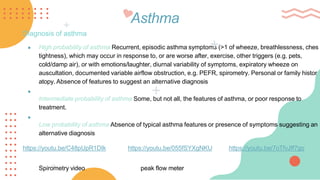
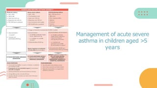
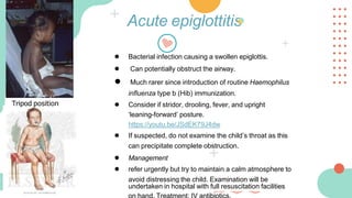
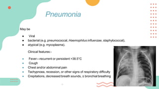
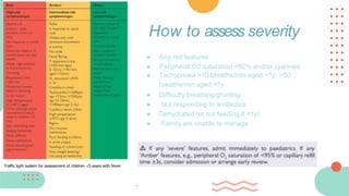
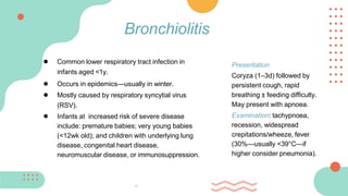
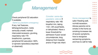
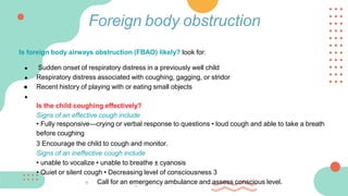
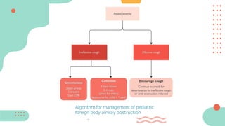
Cough & SOB in
children
AL-Muntaha Mohamed Al-Amri
OMCF-18-120
1 Explore the history of
cough & dyspnea in
children
Perform relevant
examination
3
Discuss causes &
differential diagnosis of
cough & dyspnea in
children
SLOs
Select appropriate
investigations
4
2
6
5 Construct appropriate
management plan
Identify patients suitable for
referral
A 9-year old boy came in with his mother to the A&E with
complaints of cough, shortness of breath and chest pain.
● What questions would you ask?
● What examination would you perform?
History & Examination
Causes of SOB
● Asthma
● Croup
● Epiglottitis
● Pneumonia
● Bronchiolitis
● Foreign body obstruction
Asthma
● Childhood asthma affects 75% of children in the UK.
● Peak age of onset is 5y.
Asthma
Diagnosis of asthma
● High probability of asthma Recurrent, episodic asthma symptoms (>1 of wheeze, breathlessness, ches
tightness), which may occur in response to, or are worse after, exercise, other triggers (e.g. pets,
cold/damp air), or with emotions/laughter, diurnal variability of symptoms, expiratory wheeze on
auscultation, documented variable airflow obstruction, e.g. PEFR, spirometry. Personal or family histor
atopy. Absence of features to suggest an alternative diagnosis
●
Intermediate probability of asthma Some, but not all, the features of asthma, or poor response to
treatment.
●
Low probability of asthma Absence of typical asthma features or presence of symptoms suggesting an
alternative diagnosis
https://youtu.be/C48pUpR1DIk https://youtu.be/055fSYXgNKU https://youtu.be/7oTfvJff7go
Spirometry video peak flow meter
Stepwise management of asthma in
children aged <5 y
Stepwise management of asthma
in children aged 5-12 y
https://youtu.be/Y52QUekiG5U
Management of acute severe
asthma in children aged 2-5
years
Management of acute severe
asthma in children aged >5
years
• Severe exacerbation of asthma or severe URTI—E
● Unexpected clinical findings, e.g. focal signs, abnormal voice or cry,
dysphagia, inspiratory stridor—E/U
● Persistent wet or productive cough—U/S
● Faltering growth (failure to thrive)—U/S
● Diagnosis unclear or in doubt—U/S/R
● Failure to respond to conventional treatment (particularly inhaled
corticosteroids >400 micrograms/d or frequent use of steroid
tablets)—S
● excessive vomiting or posseting—S/R
● Symptoms present from birth or perinatal lung problem—S/R
● FH of unusual chest disease—R • Nasal polyps—R
● Parental anxiety or need for reassurance—R
Reasons for referral
Acute epiglottitis
● Bacterial infection causing a swollen epiglottis.
● Can potentially obstruct the airway.
● Much rarer since introduction of routine Haemophilus
influenza type b (Hib) immunization.
● Consider if stridor, drooling, fever, and upright
‘leaning-forward’ posture.
https://youtu.be/JSdEK79J4dw
● If suspected, do not examine the child’s throat as this
can precipitate complete obstruction.
● Management
● refer urgently but try to maintain a calm atmosphere to
avoid distressing the child. Examination will be
undertaken in hospital with full resuscitation facilities
Tripod position
May be
● Viral
● bacterial (e.g. pneumococcal, Haemophilus influenzae, staphylococcal),
● atypical (e.g. mycoplasma).
Clinical features:-
● Fever—recurrent or persistent >38.5°C
● Cough
● Chest and/or abdominal pain
● Tachypnoea, recession, or other signs of respiratory difficulty
● Crepitations, decreased breath sounds, ± bronchial breathing
Pneumonia
● Any red features
● Peripheral O2 saturation <92% and/or cyanosis
● Tachypnoea >70 breaths/min aged <1y; >50
breaths/min aged >1y
● Difficulty breathing/grunting
● Not responding to antibiotics
● Dehydrated (or not feeding if <1y)
● Family are unable to manage
How to assess severity
Traffic light system for assessment of children <5 years with fever
● Provide advice about management of fever and hydration
● Well children with mild symptoms of LRTI may not require antibiotics
● If prescribing antibiotics, amoxicillin is first-line; consider a macrolide if
penicillin allergic, Mycoplasma or Chlamydia is suspected as the cause of
the infection, or in addition to amoxicillin if first-line treatment is ineffective.
Use co-amoxiclav as first-line if pneumonia associated with influenza
● Advise parents to seek further medical review if no better in <48h (sooner if
any ‘Amber’ features) or worse in the interim
Prevention
Pneumococcal vaccination is part of the routine childhood vaccination
programme and is given at 2, 4, and 12mo
Management
● Common lower respiratory tract infection in
infants aged <1y.
● Occurs in epidemics—usually in winter.
● Mostly caused by respiratory syncytial virus
(RSV).
● Infants at increased risk of severe disease
include: premature babies; very young babies
(<12wk old); and children with underlying lung
disease, congenital heart disease,
neuromuscular disease, or immunosuppression.
Bronchiolitis
Presentation
Coryza (1–3d) followed by
persistent cough, rapid
breathing ± feeding difficulty.
May present with apnoea.
Examination: tachypnoea,
recession, widespread
crepitations/wheeze, fever
(30%—usually <39°C—if
higher consider pneumonia).
Check peripheral O2 saturation
if available.
Admit as paediatric emergency
If any ‘red’ features ,
lethargy/exhaustion, looks
seriously unwell, marked
intercostal recession, grunting,
respiratory rate >70
breaths/min, cyanosis,
peripheral O2 saturation <92%
or apnoeic episode(s) (reported
or observed)
Management
Consider same-day
paediatric referral If
respiratory rate >60
breaths/ min, taking
<1⁄2—3⁄4 usual feeds,
or dehydrated. Have a
lower threshold for
admission if poor social
circumstances, long
distance from hospital,
and/or if high risk infant
Home management Is
safe if feeding well,
no/mild recession.
Advise parents not to
smoke in the home as
smoking increases risk
of severe symptoms,
how to recognize
worsening symptoms,
and how to call for help
Prognosis
Most recover in <14d, up to 50% wheeze with subsequent URTIs.
Prevention
Palivizumab (a monoclonal antibody) decreases RSV infection rate and severity. It
is consultant initiated and given IM to premature babies, and those at increased
risk from RSV infection due to other co-morbidities, before the RSV season and
then monthly until the end of the season.
Is foreign body airways obstruction (FBAO) likely? look for:
● Sudden onset of respiratory distress in a previously well child
● Respiratory distress associated with coughing, gagging, or stridor
● Recent history of playing with or eating small objects
●
Is the child coughing effectively?
Signs of an effective cough include
• Fully responsive—crying or verbal response to questions • loud cough and able to take a breath
before coughing
3 Encourage the child to cough and monitor.
Signs of an ineffective cough include
• unable to vocalize • unable to breathe ± cyanosis
• Quiet or silent cough • Decreasing level of consciousness 3
○ Call for an emergency ambulance and assess conscious level.
Foreign body obstruction
Algorithm for management of pediatric
foreign body airway obstruction
A 6 month old baby on 15 of December, came to A&E with rapid
breathing, persistent cough, nasal discharge and difficulty in feeding.
On auscultation crepitations were heard.
What is your diagnosis?
Who are at increased risk of developing this disease?
Oxford handbook of general practice- 5th edition
Google images
References

cough%20%26%20SOB%20in%20children.pptx

  • 1.
    Cough & SOBin children AL-Muntaha Mohamed Al-Amri OMCF-18-120
  • 2.
    1 Explore thehistory of cough & dyspnea in children Perform relevant examination 3 Discuss causes & differential diagnosis of cough & dyspnea in children SLOs Select appropriate investigations 4 2 6 5 Construct appropriate management plan Identify patients suitable for referral
  • 3.
    A 9-year oldboy came in with his mother to the A&E with complaints of cough, shortness of breath and chest pain. ● What questions would you ask? ● What examination would you perform? History & Examination
  • 4.
    Causes of SOB ●Asthma ● Croup ● Epiglottitis ● Pneumonia ● Bronchiolitis ● Foreign body obstruction
  • 5.
    Asthma ● Childhood asthmaaffects 75% of children in the UK. ● Peak age of onset is 5y.
  • 6.
    Asthma Diagnosis of asthma ●High probability of asthma Recurrent, episodic asthma symptoms (>1 of wheeze, breathlessness, ches tightness), which may occur in response to, or are worse after, exercise, other triggers (e.g. pets, cold/damp air), or with emotions/laughter, diurnal variability of symptoms, expiratory wheeze on auscultation, documented variable airflow obstruction, e.g. PEFR, spirometry. Personal or family histor atopy. Absence of features to suggest an alternative diagnosis ● Intermediate probability of asthma Some, but not all, the features of asthma, or poor response to treatment. ● Low probability of asthma Absence of typical asthma features or presence of symptoms suggesting an alternative diagnosis https://youtu.be/C48pUpR1DIk https://youtu.be/055fSYXgNKU https://youtu.be/7oTfvJff7go Spirometry video peak flow meter
  • 7.
    Stepwise management ofasthma in children aged <5 y
  • 8.
    Stepwise management ofasthma in children aged 5-12 y https://youtu.be/Y52QUekiG5U
  • 9.
    Management of acutesevere asthma in children aged 2-5 years
  • 10.
    Management of acutesevere asthma in children aged >5 years
  • 11.
    • Severe exacerbationof asthma or severe URTI—E ● Unexpected clinical findings, e.g. focal signs, abnormal voice or cry, dysphagia, inspiratory stridor—E/U ● Persistent wet or productive cough—U/S ● Faltering growth (failure to thrive)—U/S ● Diagnosis unclear or in doubt—U/S/R ● Failure to respond to conventional treatment (particularly inhaled corticosteroids >400 micrograms/d or frequent use of steroid tablets)—S ● excessive vomiting or posseting—S/R ● Symptoms present from birth or perinatal lung problem—S/R ● FH of unusual chest disease—R • Nasal polyps—R ● Parental anxiety or need for reassurance—R Reasons for referral
  • 12.
    Acute epiglottitis ● Bacterialinfection causing a swollen epiglottis. ● Can potentially obstruct the airway. ● Much rarer since introduction of routine Haemophilus influenza type b (Hib) immunization. ● Consider if stridor, drooling, fever, and upright ‘leaning-forward’ posture. https://youtu.be/JSdEK79J4dw ● If suspected, do not examine the child’s throat as this can precipitate complete obstruction. ● Management ● refer urgently but try to maintain a calm atmosphere to avoid distressing the child. Examination will be undertaken in hospital with full resuscitation facilities Tripod position
  • 13.
    May be ● Viral ●bacterial (e.g. pneumococcal, Haemophilus influenzae, staphylococcal), ● atypical (e.g. mycoplasma). Clinical features:- ● Fever—recurrent or persistent >38.5°C ● Cough ● Chest and/or abdominal pain ● Tachypnoea, recession, or other signs of respiratory difficulty ● Crepitations, decreased breath sounds, ± bronchial breathing Pneumonia
  • 14.
    ● Any redfeatures ● Peripheral O2 saturation <92% and/or cyanosis ● Tachypnoea >70 breaths/min aged <1y; >50 breaths/min aged >1y ● Difficulty breathing/grunting ● Not responding to antibiotics ● Dehydrated (or not feeding if <1y) ● Family are unable to manage How to assess severity Traffic light system for assessment of children <5 years with fever
  • 15.
    ● Provide adviceabout management of fever and hydration ● Well children with mild symptoms of LRTI may not require antibiotics ● If prescribing antibiotics, amoxicillin is first-line; consider a macrolide if penicillin allergic, Mycoplasma or Chlamydia is suspected as the cause of the infection, or in addition to amoxicillin if first-line treatment is ineffective. Use co-amoxiclav as first-line if pneumonia associated with influenza ● Advise parents to seek further medical review if no better in <48h (sooner if any ‘Amber’ features) or worse in the interim Prevention Pneumococcal vaccination is part of the routine childhood vaccination programme and is given at 2, 4, and 12mo Management
  • 16.
    ● Common lowerrespiratory tract infection in infants aged <1y. ● Occurs in epidemics—usually in winter. ● Mostly caused by respiratory syncytial virus (RSV). ● Infants at increased risk of severe disease include: premature babies; very young babies (<12wk old); and children with underlying lung disease, congenital heart disease, neuromuscular disease, or immunosuppression. Bronchiolitis Presentation Coryza (1–3d) followed by persistent cough, rapid breathing ± feeding difficulty. May present with apnoea. Examination: tachypnoea, recession, widespread crepitations/wheeze, fever (30%—usually <39°C—if higher consider pneumonia).
  • 17.
    Check peripheral O2saturation if available. Admit as paediatric emergency If any ‘red’ features , lethargy/exhaustion, looks seriously unwell, marked intercostal recession, grunting, respiratory rate >70 breaths/min, cyanosis, peripheral O2 saturation <92% or apnoeic episode(s) (reported or observed) Management Consider same-day paediatric referral If respiratory rate >60 breaths/ min, taking <1⁄2—3⁄4 usual feeds, or dehydrated. Have a lower threshold for admission if poor social circumstances, long distance from hospital, and/or if high risk infant Home management Is safe if feeding well, no/mild recession. Advise parents not to smoke in the home as smoking increases risk of severe symptoms, how to recognize worsening symptoms, and how to call for help
  • 18.
    Prognosis Most recover in<14d, up to 50% wheeze with subsequent URTIs. Prevention Palivizumab (a monoclonal antibody) decreases RSV infection rate and severity. It is consultant initiated and given IM to premature babies, and those at increased risk from RSV infection due to other co-morbidities, before the RSV season and then monthly until the end of the season.
  • 19.
    Is foreign bodyairways obstruction (FBAO) likely? look for: ● Sudden onset of respiratory distress in a previously well child ● Respiratory distress associated with coughing, gagging, or stridor ● Recent history of playing with or eating small objects ● Is the child coughing effectively? Signs of an effective cough include • Fully responsive—crying or verbal response to questions • loud cough and able to take a breath before coughing 3 Encourage the child to cough and monitor. Signs of an ineffective cough include • unable to vocalize • unable to breathe ± cyanosis • Quiet or silent cough • Decreasing level of consciousness 3 ○ Call for an emergency ambulance and assess conscious level. Foreign body obstruction
  • 20.
    Algorithm for managementof pediatric foreign body airway obstruction
  • 21.
    A 6 monthold baby on 15 of December, came to A&E with rapid breathing, persistent cough, nasal discharge and difficulty in feeding. On auscultation crepitations were heard. What is your diagnosis? Who are at increased risk of developing this disease?
  • 22.
    Oxford handbook ofgeneral practice- 5th edition Google images References