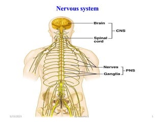
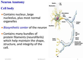
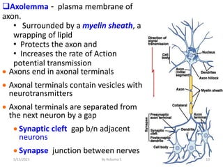
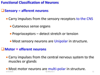
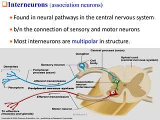
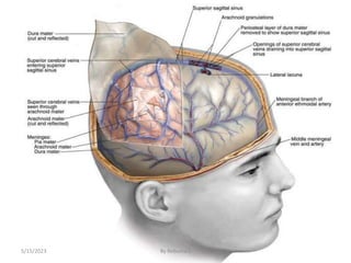
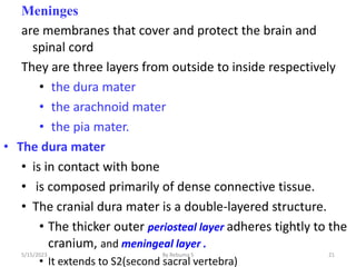
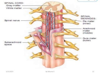
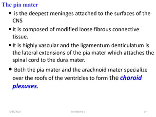
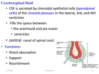
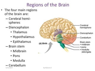
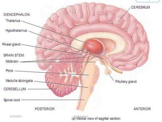
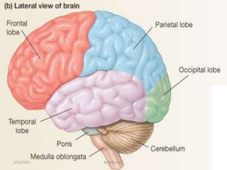
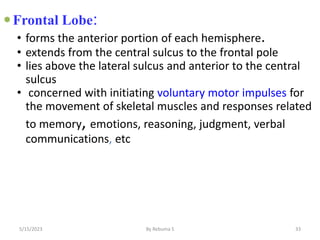
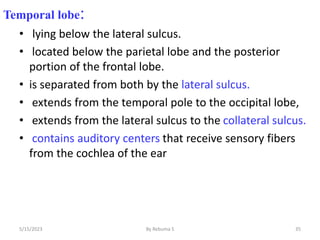
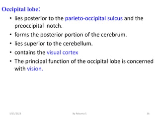
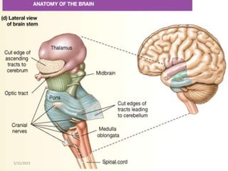
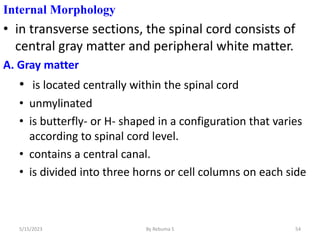
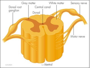
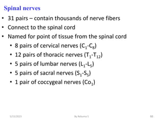
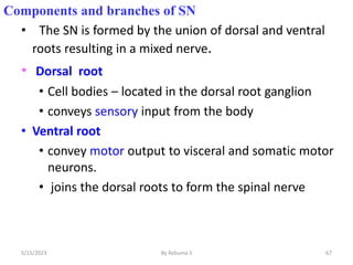
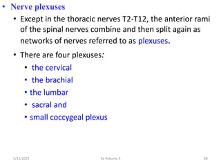
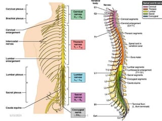
The nervous system is one of the most complex body systems, consisting of the central nervous system (brain and spinal cord) and peripheral nervous system. The CNS contains billions of neurons and neuroglia that communicate via electrical and chemical signals. The brain is protected by meninges and cerebrospinal fluid. Neurons are specialized cells that conduct electrical signals, while neuroglia provide support. The nervous system is organized into the forebrain, midbrain, and hindbrain. The cerebrum controls higher functions and is divided into four lobes. The diencephalon includes the thalamus, hypothalamus, and epithalamus.
The nervous system is one of the most complex body systems, accounting for about 3% of body weight. It is composed of the central nervous system (brain and spinal cord) and peripheral nervous system. The CNS contains billions of neurons and neuroglia that allow for sensory input, integration of information, and motor output responses. Within the CNS, the brain is the largest organ and contains regions like the cerebrum, diencephalon, and cerebellum that are involved in higher functions. The peripheral nervous system connects the CNS to sensory receptors and effector organs through nerves and ganglia.
THIS PRESENTATION IS UPLOADED TO HELP THE EDUCATOR OF MEDICAL, NURSING & ALLIE HEALTH SCIENCES TO TEACH THEIR STUDENTS ABOUT THE NERVOUS SYSTEM. IT WILL ALSO CREATE AWARENESS AMONG THE COMMON PEOPLE REGARDING NERVOUS SYSTEM.
The nervous system is comprised of neurons and glial cells. Neurons transmit electro-chemical signals while glial cells provide support. The brain and spinal cord are surrounded by three layers of meninges and bathed in cerebrospinal fluid. The brain is divided into four lobes which control different functions and is separated into two hemispheres connected by the corpus callosum. The peripheral nervous system connects the brain and spinal cord to the rest of the body and controls both voluntary and involuntary functions.
The central nervous system develops from the ectoderm and forms the neural tube which will become the brain and spinal cord. The neural tube develops brain vesicles which become the forebrain, midbrain and hindbrain. The hindbrain further develops into the cerebellum, pons and medulla. Neuroepithelial cells form the inner layer and produce neuroblasts which migrate outward to form the mantle layer and later the gray matter. Glial cells and neurons develop from progenitor cells. The spinal cord develops basal and alar plates which form motor and sensory areas. Spinal nerves form from ventral motor and dorsal sensory roots. The central nervous system continues developing after birth with myelination of axons.
The nervous system is divided structurally into the central nervous system (CNS) and peripheral nervous system (PNS). The CNS consists of the brain and spinal cord, which act as integrating and command centers. The PNS consists of nerves that extend from the CNS and includes the cranial and spinal nerves. Functionally, the nervous system is divided into sensory and motor systems, with the motor system further divided into somatic and autonomic systems. Neurons are specialized cells that transmit nerve impulses, while neuroglia provide support and protection for neurons.
The nervous system controls and coordinates functions throughout the body using electrical signals called nerve impulses. It has three main parts - the central nervous system (brain and spinal cord), and the peripheral nervous system which includes sensory neurons, motor neurons, and interneurons. Sensory neurons detect stimuli and send signals to the central nervous system. Motor neurons carry signals from the central nervous system to muscles and glands. Interneurons connect neurons within the central nervous system. Neurons communicate via neurotransmitters released at synapses between neurons.
Hi Guys, this PPT covers Nervous System for class 10th, Tamil Nadu state board.
To access similar content click the Blogger link below:
https://konjampadipoma.blogspot.com/
This document provides an overview of the nervous system, including its objectives, organization, key components, and functions. It discusses the central nervous system (CNS), peripheral nervous system (PNS), and autonomic nervous system (ANS). The CNS is made up of the brain and spinal cord. The PNS includes cranial nerves, spinal nerves, and ganglia. Neurons and neuroglia are the main cell types. Neurons transmit nerve impulses while neuroglia provide support. The nervous system has sensory, integrative, and motor functions to detect stimuli and control the body's responses.
The nervous system has three main functions: sensory, integration, and motor. The nervous system is divided into the central nervous system (CNS) and peripheral nervous system (PNS). There are two main types of neural cells: neurons, which process and transmit information, and neuroglia (also called glial cells or glia), which support and protect neurons. Neurons can be classified structurally as unipolar, bipolar, or multipolar, and functionally as sensory, motor, or interneurons. Communication between neurons occurs at synapses, which can be chemical or electrical. The CNS is divided into gray matter, containing cell bodies, and white matter, containing myelinated axons.
Cellular organization of the nervous systemDavis Mburu
This document summarizes the cellular organization of the nervous system. It describes the main cell types: neurons, which are the basic functional units, and neuroglia, which provide support. Neurons have a cell body, dendrites, and an axon. Neuroglia include astrocytes, oligodendrocytes, microglia, and ependymal cells. Astrocytes regulate the neuronal microenvironment and form part of the blood-brain barrier. Oligodendrocytes and Schwann cells myelinate axons to increase conduction speed. The document also notes that glial cells can give rise to brain tumors since they continue to divide in adulthood, unlike most neurons.
Neurons are the basic structural and functional units of the nervous system. They have extensions called dendrites that receive signals and an axon that transmits signals. Neurons communicate with each other through electrochemical processes at synapses. The three main parts of a neuron are the dendrites, cell body, and axon. The cell body contains organelles like the nucleus, Golgi apparatus, endoplasmic reticulum, and mitochondria that support neuronal functions. Dendrites receive signals and axons transmit signals long distances via myelinated sheaths to synapse with other neurons. Neurons vary in structure and function depending on their role in the nervous system.
The nervous system is divided into the central nervous system (CNS) and peripheral nervous system (PNS). The CNS consists of the brain and spinal cord and is protected by three layers of tissue called meninges. Within the CNS, gray matter contains neurons and white matter contains myelinated axons. Neuroglia provide support to neurons in the CNS. The PNS connects the CNS to the rest of the body and is composed of nerves and ganglia. Neurons are the basic functional units and come in several types defined by their dendrites and axons.
The nervous system detects changes inside and outside the body and maintains homeostasis. It has two main parts: the central nervous system (CNS) consisting of the brain and spinal cord, and the peripheral nervous system (PNS) consisting of nerves outside the CNS. The nervous system consists of neurons which communicate via electrical and chemical signals. Neurons have a cell body, axon, and dendrites. The nervous system regulates both voluntary and involuntary functions through the somatic and autonomic nervous systems.
The document provides an overview of the nervous system, including its main components and cell types. It discusses the four main tissues of the body before focusing on nervous tissue. There are two main parts to the nervous system: the central nervous system comprising the brain and spinal cord, and the peripheral nervous system which communicates information between the body and central nervous system. The peripheral nervous system contains somatic and autonomic divisions. Nervous tissue consists of neurons, glial cells, and nerve fibers. Neurons receive, transmit and process stimuli via their cell body, dendrites and axon. Glial cells provide support and protection. Communication occurs at synapses between neurons.
The nervous system consists of two main parts - the central nervous system (CNS) and the peripheral nervous system (PNS). The CNS contains the brain and spinal cord, while the PNS contains nerves that connect the CNS to the rest of the body. The structures that make up the nervous system include neurons, neuroglia, the brain, spinal cord, cranial and spinal nerves. Neurons are specialized to conduct electrical signals, while neuroglia provide support and insulation to the neurons. The spinal cord is protected within the bony vertebral column and surrounded by three layers of meninges.
The document discusses the anatomy and histology of the central nervous system. It describes the different types of neurons, their classification based on structure and function. It also discusses the supporting glial cells like astrocytes, oligodendrocytes, microglia and ependymal cells. It explains the structure and function of synapses and myelin sheath formation in the CNS.
There are two main cell types in nervous tissue: neurons and neuroglia. Neurons are excitable cells that transmit electrical signals down axons and dendrites. They have a cell body containing organelles and long processes. Neuroglia, also called glial cells, support and protect neurons. The four main types of neuroglia in the central nervous system are astrocytes, microglia, ependymal cells, and oligodendrocytes. Astrocytes connect neurons to blood vessels and regulate the neuronal environment. Microglia monitor neuronal health and act as phagocytes. Ependymal cells line cavities and circulate cerebrospinal fluid. Oligodendrocytes wrap myelin
control system in humans, neurons, types of neurons, nerves, human nervous system, CNS, PNS, ANS, Brain, parts of brain, spinal cord, functions of spinal cord, reflex arc, PNS, ANS,
The document discusses the structure and function of the nervous system. It describes how the nervous system is divided into the central nervous system (CNS), which includes the brain and spinal cord, and the peripheral nervous system (PNS). Neurons are the basic functional units that transmit signals in the nervous system. The document outlines the main parts of neurons including the cell body, dendrites, axon, and myelin sheath. It also describes the different types of neurons and specialized cells that support neurons called neuroglial cells.
This document provides an overview of the structure and function of the nervous system. It begins by outlining learning objectives related to defining different parts of the nervous system and describing neurons and glia. It then discusses the organization of the central and peripheral nervous systems. Specific sections describe the structure and function of the brain, spinal cord, neurons, glia and different regions of the brain like the cerebrum and cerebellum. The document concludes by discussing protection of the central nervous system through meninges and cerebrospinal fluid.
The reticular formation is a region in the brainstem that contains a network of neurons and fibers. It receives sensory information from ascending and descending tracts and has connections with other brain regions. It contains four important neuronal systems - the gigantocellular nuclei, substantia nigra, locus ceruleus, and raphe nuclei. The reticular formation plays roles in consciousness, sleep, sensory regulation, motor control, and autonomic functions through these neuronal systems.
The nervous system is divided into the central nervous system (CNS) and peripheral nervous system (PNS). The CNS contains the brain and spinal cord, while the PNS contains nerves that connect the CNS to sensory organs and effector organs. The basic functional unit of the nervous system is the neuron, which transmits electrochemical signals. The nervous system also contains neuroglial cells that provide support to neurons. Sensory neurons carry signals to the CNS, motor neurons carry signals from the CNS, and interneurons connect sensory and motor neurons within the CNS.
Objective of the study:- Structure of a typical Neuron, Classification of Neuron based on Polarity, on conduction direction, on neurotransmitters released, on their shape, Glial cells, major type of Glial cells present in CNS and PNS and their functions.
5-hydroxytryptamine or 5-HT or Serotonin is a neurotransmitter that serves a range of roles in the human body. It is sometimes referred to as the happy chemical since it promotes overall well-being and happiness.
It is mostly found in the brain, intestines, and blood platelets.
5-HT is utilised to transport messages between nerve cells, is known to be involved in smooth muscle contraction, and adds to overall well-being and pleasure, among other benefits. 5-HT regulates the body's sleep-wake cycles and internal clock by acting as a precursor to melatonin.
It is hypothesised to regulate hunger, emotions, motor, cognitive, and autonomic processes.
Osteoporosis - Definition , Evaluation and Management .pdfJim Jacob Roy
Osteoporosis is an increasing cause of morbidity among the elderly.
In this document , a brief outline of osteoporosis is given , including the risk factors of osteoporosis fractures , the indications for testing bone mineral density and the management of osteoporosis
Cell Therapy Expansion and Challenges in Autoimmune DiseaseHealth Advances
There is increasing confidence that cell therapies will soon play a role in the treatment of autoimmune disorders, but the extent of this impact remains to be seen. Early readouts on autologous CAR-Ts in lupus are encouraging, but manufacturing and cost limitations are likely to restrict access to highly refractory patients. Allogeneic CAR-Ts have the potential to broaden access to earlier lines of treatment due to their inherent cost benefits, however they will need to demonstrate comparable or improved efficacy to established modalities.
In addition to infrastructure and capacity constraints, CAR-Ts face a very different risk-benefit dynamic in autoimmune compared to oncology, highlighting the need for tolerable therapies with low adverse event risk. CAR-NK and Treg-based therapies are also being developed in certain autoimmune disorders and may demonstrate favorable safety profiles. Several novel non-cell therapies such as bispecific antibodies, nanobodies, and RNAi drugs, may also offer future alternative competitive solutions with variable value propositions.
Widespread adoption of cell therapies will not only require strong efficacy and safety data, but also adapted pricing and access strategies. At oncology-based price points, CAR-Ts are unlikely to achieve broad market access in autoimmune disorders, with eligible patient populations that are potentially orders of magnitude greater than the number of currently addressable cancer patients. Developers have made strides towards reducing cell therapy COGS while improving manufacturing efficiency, but payors will inevitably restrict access until more sustainable pricing is achieved.
Despite these headwinds, industry leaders and investors remain confident that cell therapies are poised to address significant unmet need in patients suffering from autoimmune disorders. However, the extent of this impact on the treatment landscape remains to be seen, as the industry rapidly approaches an inflection point.
share - Lions, tigers, AI and health misinformation, oh my!.pptxTina Purnat
• Pitfalls and pivots needed to use AI effectively in public health
• Evidence-based strategies to address health misinformation effectively
• Building trust with communities online and offline
• Equipping health professionals to address questions, concerns and health misinformation
• Assessing risk and mitigating harm from adverse health narratives in communities, health workforce and health system
The skin is the largest organ and its health plays a vital role among the other sense organs. The skin concerns like acne breakout, psoriasis, or anything similar along the lines, finding a qualified and experienced dermatologist becomes paramount.
Histololgy of Female Reproductive System.pptxAyeshaZaid1
Dive into an in-depth exploration of the histological structure of female reproductive system with this comprehensive lecture. Presented by Dr. Ayesha Irfan, Assistant Professor of Anatomy, this presentation covers the Gross anatomy and functional histology of the female reproductive organs. Ideal for students, educators, and anyone interested in medical science, this lecture provides clear explanations, detailed diagrams, and valuable insights into female reproductive system. Enhance your knowledge and understanding of this essential aspect of human biology.
Kosmoderma Academy, a leading institution in the field of dermatology and aesthetics, offers comprehensive courses in cosmetology and trichology. Our specialized courses on PRP (Hair), DR+Growth Factor, GFC, and Qr678 are designed to equip practitioners with advanced skills and knowledge to excel in hair restoration and growth treatments.
Clinic ^%[+27633867063*Abortion Pills For Sale In Tembisa Central19various
Clinic ^%[+27633867063*Abortion Pills For Sale In Tembisa Central Clinic ^%[+27633867063*Abortion Pills For Sale In Tembisa CentralClinic ^%[+27633867063*Abortion Pills For Sale In Tembisa CentralClinic ^%[+27633867063*Abortion Pills For Sale In Tembisa CentralClinic ^%[+27633867063*Abortion Pills For Sale In Tembisa Central
Does Over-Masturbation Contribute to Chronic Prostatitis.pptxwalterHu5
In some case, your chronic prostatitis may be related to over-masturbation. Generally, natural medicine Diuretic and Anti-inflammatory Pill can help mee get a cure.
8 Surprising Reasons To Meditate 40 Minutes A Day That Can Change Your Life.pptxHolistified Wellness
We’re talking about Vedic Meditation, a form of meditation that has been around for at least 5,000 years. Back then, the people who lived in the Indus Valley, now known as India and Pakistan, practised meditation as a fundamental part of daily life. This knowledge that has given us yoga and Ayurveda, was known as Veda, hence the name Vedic. And though there are some written records, the practice has been passed down verbally from generation to generation.
2. Introduction
• The nervous system is one of the smallest and the
most complex of the 11 body systems.
• The nervous system is an intricate, highly organized network of
billions of neurons and more neuroglia.
• It accounts 2 kg ,about 3% of total body weight
• The nervous system include the brain, cranial nerves and their
branches, the spinal cord, spinal nerves and their branches, ganglia,
enteric plexuses, and sensory receptors
• The brain - which contains about 100 billion neurons.
• Twelve pairs (right and left) of cranial nerves, numbered I through XII,
emerge from the base of the brain.
• Nervous are bundle of axon , connective tissue and blood vessels
2
5/15/2023 By Rebuma S
3. Spinal cord contains about 100 million neurons.
Thirty-one (31) pairs
pairs of spinal nerves emerge from the spinal cord, each
serving a specific region on the right or left side of the
body.
Ganglia singular is( ganglion) are small masses of nervous
tissue , that are located outside the brain and spinal cord.
the walls of organs of the gastrointestinal tract, extensive net-
works of neuron, enteric plexus,help regulate the digestive
system.
sensory receptor is neurons (specialized cells) that monitor
changes in the internal or external environment,
such as photoreceptors in the retina of the eye
5/15/2023 By Rebuma S 3
4. Nervous system
Nervous Tissue
• Consist of 2 types of cells
1. Neurons
• Functional, signal conducting cells
2. Neuroglia
• It doesn’t conduct impulse
• Supporting cells & nutrition
Neurons = nerve cells , the functional and
structural unit of the nervous system
Specialized to conduct information from one
part of the body to another
4
5/15/2023 By Rebuma S
5. Major regions of neurons
Cell body (soma) – contain
nucleus other organells and
metabolic center of the cell
One or more slender processes
– fibers that extend from the cell
body (dendrites and axons)
Dendrites An input region –
receive impulse to wards the cell
body
Axon conducting message ,
away from the cell body
A secretory (output) region
(axon terminal) 5
5/15/2023 By Rebuma S
6. Neuron Anatomy
Cell body
Contains nucleus ,large
nucleolus, plus most normal
organelles
Biosynthetic center of the neuron
Contains many bundles of
protein filaments (neurofibrils)
which help maintain the shape,
structure, and integrity of the
cell.
6
5/15/2023 By Rebuma S
7. Nervous system
CNS:
Gray matter cell bodies and unmylenated fibers
Nuclei – clusters of cell bodies
White matter tracts, collections of myelinated fibers
PNS:
Ganglia – clusters of cell bodies
Nerves-collections of myelinated fibers
• Dendrites and Axons
Dendrites are thin, branched processes whose main
function is to receive incoming signals
Axons are conduct impulses away from the cell body 7
5/15/2023 By Rebuma S
8. 8
Structural classification of neurons
• Structurally, neurons are classified according to the number
of processes extending from the cell body
Multipolar- neurons usually have several dendrites and one
axon
• Most neurons in the brain and spinal cord
Bipolar -neurons have one main dendrite and one axon
• They are found in the retina of the eye, in the inner ear, and
in the olfactory (to smell) area of the brain.
Unipolar- neurons have dendrites and one axon that are
• fused together to form a continuous process that emerges
from the cell body
5/15/2023 By Rebuma S
10. Axolemma - plasma membrane of
axon.
• Surrounded by a myelin sheath, a
wrapping of lipid
• Protects the axon and
• Increases the rate of Action
potential transmission
Axons end in axonal terminals
Axonal terminals contain vesicles with
neurotransmitters
Axonal terminals are separated from
the next neuron by a gap
Synaptic cleft gap b/n adjacent
neurons
Synapse junction between nerves
10
5/15/2023 By Rebuma S
11. Functional Classification of Neurons
Sensory = afferent neurons
Carry impulses from the sensory receptors to the CNS
Cutaneous sense organs
Proprioceptors – detect stretch or tension
Most sensory neurons are Unipolar in structure.
Motor = efferent neurons
Carry impulses from the central nervous system to the
muscles or glands
Most motor neurons are multi-polar in structure.
11
5/15/2023 By Rebuma S
12. Interneurons (association neurons)
Found in neural pathways in the central nervous system
b/n the connection of sensory and motor neurons
Most interneurons are multipolar in structure.
12
5/15/2023 By Rebuma S
13. Neuroglia
• Neuroglia are the supporting cells of the central nervous
system .
• These cells are non -excitable and undergo mitotic division.
• The neuroglial cells are of two types - macroglial and
microglial cells.
• The macroglial cells include Astrocytes ,Oligodendrocytes
and Ependymal cells .
• The macroglial cells are derived from the embryonic
neuroectoderm and the microglial cells are mesodermal in
origin.
13
5/15/2023 By Rebuma S
14. Are supporting nerve cells
6 types of supporting cells (neuroglia)
• 4 &2are found in the CNS and
PNS
Astrocytes
• Star-shaped, abundant, and
versatile
• Guide the migration of
developing neurons
• Function in nutrient transfer
• Support neurons; protect
neurons from harmful
substances; help maintain
prope chemical environment
for generation of nerve
impulses 14
5/15/2023 By Rebuma S
16. • Microglia -Protect CNS cells from disease by
engulfing invading microbes; migrate to areas of injured
nerve tissue where they clear away debris of dead cells.
• Specialized immune cells that act as the macrophages of
the CNS
• the main phagocytic cell and antigen-presenting cells in the
CNS.
• Ependymal Cells
Some are ciliated which facilitates the movement of
cerebrospinal fluid and circulation
• Oligodendrocytes
Produce the myelin sheath which provides the electrical
insulation for certain neurons in the CNS 16
5/15/2023 By Rebuma S
17. Neuroglia in the PNS
1. Satellite cells
• Surround clusters of neuronal cell bodies in the PNS
• Support neurons and regulate exchange of material
b/n neural cell bodies and intertistial fluid
2. Schwann cells
• Form myelin sheaths around the axon of larger
nerve fibers in the PNS.
• Vital to neuronal regeneration
17
5/15/2023 By Rebuma S
18. Organization of the Nervous System
• is divided into
• the Central Nervous System (CNS)
• the brain
• spinal cord
• the Peripheral Nervous System (PNS)
• Includes all nervous tissue
outside the CNS
• Component of PNS
• the cranial nerves and their
branches
• the spinal nerves and their
branches, ganglia and sensory
receptors
18
5/15/2023 By Rebuma S
19. What covers and protects the CNS ?
• The entire delicate CNS is protected by:
• a bony -cranial bone & vertebrae
• The menings,
• The cerebrospinal fluid (CSF)
19
5/15/2023 By Rebuma S
21. Meninges
are membranes that cover and protect the brain and
spinal cord
They are three layers from outside to inside respectively
• the dura mater
• the arachnoid mater
• the pia mater.
• The dura mater
• is in contact with bone
• is composed primarily of dense connective tissue.
• The cranial dura mater is a double-layered structure.
• The thicker outer periosteal layer adheres tightly to the
cranium, and meningeal layer .
• It extends to S2(second sacral vertebra)
5/15/2023 By Rebuma S 21
23. • The spinal dura mater is single layered and is similar to the
menigeal layer of the cranial dura mater.
The arachnoid mater
is the middle of the three meninges.
The subarachnoid space is located between the
arachnoid mater and the pia mater.
The subarachnoid space contains the CSF
5/15/2023 By Rebuma S 23
24. The pia mater
is the deepest meninges attached to the surfaces of the
CNS
It is composed of modified loose fibrous connective
tissue.
It is highly vascular and the ligamentum denticulatum is
the lateral extensions of the pia mater which attaches the
spinal cord to the dura mater.
Both the pia mater and the arachnoid mater specialize
over the roofs of the ventricles to form the choroid
plexuses.
24
5/15/2023 By Rebuma S
25. Cerebrospinal fluid
• CSF is secreted by choroidal epithelial cells (ependymal
cells) of the choroid plexuses in the lateral, 3rd, and 4th
ventricles
• Fills the space between
• the arachnoid and pia mater
• ventricles
• central canal of spinal cord.
• Functions:
• Shock absorption
• Support
• Nourishment
25
5/15/2023 By Rebuma S
28. 28
• Based on embryological dev’t the brain subdivided in to
three as:
• the forebrain (cerebrum and diencephalons)
• midbrain
• hindbrain (cerebellum, pons, and medulla).
The Cerebrum
• is the largest portion of the brain.
• located in the region of the telencephalon
• It accounts for about 80%
• is responsible for the higher mental functions including
memory and reason
• consists of
• the right hemispheres & left hemispheres
5/15/2023 By Rebuma S
29. 29
The two hemisphere are connected internally by the
corpus callosum
Each hemisphere contains a central cavity called the
lateral ventricle
5/15/2023 By Rebuma S
30. • The cerebrum consists of two layers.
• cerebral cortex
• The surface layer
• is composed of gray matter( nerve cell bodies.)
• has numerous folds and grooves called convolutions.
• The elevated folds of the convolutions are the cerebral
gyri (singular, gyrus)
• the grooves are the cerebral sulci (singular, sulcus)
• white matter
• Beneath the cerebral cortex
• constitutes the second layer.
30
5/15/2023 By Rebuma S
31. Lobes of cerebrum
• Each cerebral hemisphere is subdivided into four lobes by
deep sulci called fissures
• The central sulcus (fissure of Rolando)
• b/n the frontal lobe and the parietal lobe.
• The lateral sulcus (fissure of Sylvius)
• b/n the frontal and temporal lobes
31
5/15/2023 By Rebuma S
33. 33
Frontal Lobe:
• forms the anterior portion of each hemisphere.
• extends from the central sulcus to the frontal pole
• lies above the lateral sulcus and anterior to the central
sulcus
• concerned with initiating voluntary motor impulses for
the movement of skeletal muscles and responses related
to memory, emotions, reasoning, judgment, verbal
communications, etc
5/15/2023 By Rebuma S
34. 34
Parietal lobe:
• extends from the central sulcus to the occipital lobe
• lies superior to the temporal lobe,
• posterior to the central sulcus.
function : receives and
• integrates sensory information
5/15/2023 By Rebuma S
35. Temporal lobe:
• lying below the lateral sulcus.
• located below the parietal lobe and the posterior
portion of the frontal lobe.
• is separated from both by the lateral sulcus.
• extends from the temporal pole to the occipital lobe,
• extends from the lateral sulcus to the collateral sulcus.
• contains auditory centers that receive sensory fibers
from the cochlea of the ear
35
5/15/2023 By Rebuma S
36. Occipital lobe:
• lies posterior to the parieto-occipital sulcus and the
preoccipital notch.
• forms the posterior portion of the cerebrum.
• lies superior to the cerebellum.
• contains the visual cortex
• The principal function of the occipital lobe is concerned
with vision.
36
5/15/2023 By Rebuma S
37. Functions of Cerebrum:
• Frontal
• voluntary motor functions
• planning, mood, smell and social judgment
• Parietal
• receives and integrates sensory information
• Occipital
• visual center of brain
• Temporal
• areas for hearing, smell, learning, memory, emotional
behavior
37
5/15/2023 By Rebuma S
38. Special Senses
• Organs of special senses project to specialized regions of
the brain
• Taste - lower end of parietal lobe
• Smell - medial temporal lobe and inferior frontal lobe
• Vision - occipital lobe
• Hearing - superior temporal lobe
• Equilibrium - cerebellum and lateral and central sulcus
• Language
• Includes reading, writing, speaking and understanding
words.
Wernicke area
• permits recognition of spoken and written language and
creates plan of speech
38
5/15/2023 By Rebuma S
39. Diencephalon
• is the second subdivision of the forebrain and
• Divide in to:
• thalamus
• hypothalamus
• the epithalamus
• The 3rd ventricle forms a cavity on the median plane within
the diencephalon
39
5/15/2023 By Rebuma S
41. a. Thalamus:
• a paired, large, avoid mass of gray matter, constituting
nearly 80% of the diencephalon.
• Each portion is located immediately below the
respective lateral ventricle.
• Its principal function is to act as a relay center for all
sensory impulses, except smell(olfactory).
41
5/15/2023 By Rebuma S
42. b. Hypothalamus:
• a small portion of the diencephalon which forms the
floor and part of the lateral walls of the third ventricle.
• The hypothalamus performs numerous vital functions
• hormone secretion (pituitary)
• autonomic NS control
• thermoregulation (thermostat)
• food & water intake (hunger & satiety)
• sleep & circadian rhythms
• memory (mammillary bodies)
• emotional behavior
• anger, aggression, fear,
• pleasure, sex drive, orgasm 42
5/15/2023 By Rebuma S
43. c. Epithalamus:
• the dorsal portion of the diencephalon that includes a
thin roof over the third ventricle.
• The pineal gland extends outward from the posterior
end of the epithalamus.
• The inside lining of the roof consists of a vascular
choroids plexus where CSF is produced.
43
5/15/2023 By Rebuma S
44. Pituitary gland (hypophysis)
• is supported by the sella turcica of the sphenoid bone
• positioned on the inferior aspect of the diencephalon
and is attached to the hypothalamus by a stalk-like
structure called the infundibulum.
• The pituitary gland is divided into
• anterior portion (adenohypophysis )
• posterior portion ( the neurohypophysis)
44
5/15/2023 By Rebuma S
45. 3. The brain stem
• The brain stem contains nuclei for autonomic functions
of the body and their connecting tracts.
• It is the portion of the brain that attaches to the spinal
cord it :
• includes
• the midbrain
• pons
• medulla oblongata
45
5/15/2023 By Rebuma S
47. b. Pons
• b/n the midbrain and the medulla oblongata .
it has two respiratory centers of the pons are called the
apneustic and the pneumotaxic areas.
Medulla oblongata
• function as autonomic centers for controlling vital visceral
functions include
• Cardiac center- adjusts rate & force of heart beat
• Vasomotor center -adjusts blood vessel diameter
• Respiratory centers- control rate & depth of breathing
• Reflex centers- for coughing, sneezing, gagging, swallowing,
vomiting, salivation, sweating, movements of tongue &
head
47
5/15/2023 By Rebuma S
49. Cerebellum
• is the second lager structure in the brain.
• occupies the inferior and posterior aspect of the cranial
cavity.
• attached to the brain stem by three paired bundles of
nerve fibers called cerebellar peduncles
• function
• Balance
• coordinating skeletal muscle contractions by recruiting
precise motor units within muscles.
• Impulses for voluntary muscular movement originate in the
cerebral cortex and are coordinated by the cerebellum
49
5/15/2023 By Rebuma S
50. con.t…
• The cerebellum can be permanently damaged by trauma
or stroke or temporarily affected by drugs such as
alcohol.
• These alterations can produce ataxia is a disturbance in
balance
50
5/15/2023 By Rebuma S
51. Spinal cord
• extends through the vertebral canal and surrounded by
three meninges
• provides a means of neural communication to and from the
brain through tracts of white matter.
• These are:
• Ascending tracts
• Descending tracts
• serves as a center for spinal reflexes
51
5/15/2023 By Rebuma S
52. • Location
• in adults, from the foramen magnum to the lower
border of the first lumbar vertebra or (L2)
• in newborns, it extends to the third lumbar(L3)
vertebra.
• is continuous with the medulla oblongata at the
spinomedullary junction.
52
5/15/2023 By Rebuma S
54. Internal Morphology
• in transverse sections, the spinal cord consists of
central gray matter and peripheral white matter.
A. Gray matter
• is located centrally within the spinal cord
• unmylinated
• is butterfly- or H- shaped in a configuration that varies
according to spinal cord level.
• contains a central canal.
• is divided into three horns or cell columns on each side
54
5/15/2023 By Rebuma S
56. 1. Dorsal/ posterior horn
• receives and processes sensory input
2. Lateral horn
• receives viscerosensory input.
• is found between the dorsal and ventral horns.
3. Ventral/anterior horn
• contains predominately motor nuclei.
• is found at all levels.
56
5/15/2023 By Rebuma S
58. B . the white matter
• Tracts of the spinal cord
• are divided into ascending and descending pathways.
• Ascending spinal Tracts
• Represent functional pathways that convey sensory
information from soma or viscera to higher levels of
the neural axis.
• Descending spinal Tracts
• are concerned with somatic and visceral motor
activities.
• It is mylinated ,with myelin sheath
58
5/15/2023 By Rebuma S
59. PNS -subdivided into
I. Somatic nervous system
Consist of :
Sensory neurons- convey information from somatic
receptors and special senses to CNS
Motor neurons –conduct impulses from CNS to skeletal
muscle only- voluntarly
II. The Autonomic Nervous System (ANS).
Consist of:
Sensory neurons- convey information from autonomic
sensory receptors, located primarily in visceral organs to
CNS
Motor neurons- conduct impulse from CNS to smooth
muscle, cardiac muscle and glands- involuntary 59
5/15/2023 By Rebuma S
60. The peripheral nervous system.
• is that portion of the nervous system outside the CNS.
• The PNS functions to convey impulses to and from the
brain or spinal cord.
• The nerves of the PNS are classified as
• cranial nerves (I-XII) pairs and
• spinal nerve (31) pairs
60
5/15/2023 By Rebuma S
61. The motor part of the ANS consists of two branches
• sympathetic division
• parasympathetic division.
With few exceptions, effectors receive nerves from both
divisions, and usually they have opposing actions
61
Sympathetic Nervous System
“Fight or Flight”
Exercise, excitement,
emergency, embarrassment
and,ejaculation
Parasympathetic Nervous System
“Rest and Digest”
Digestion, defecation, and
diuresis and erection of clitoris
and penis
5/15/2023 By Rebuma S
62. Cranial nerves
• There are 12 pairs of cranial nerves
• The cranial nerves are designated by roman numerals
• Their names indicate the structures innervated or the
principal functions of the nerves
I Olfactory nerve – sensory for smell
II Optic nerve – sensory for vision
III Oculomotor nerve – motor fibers to eye muscles
IV Trochlear – motor fiber to eye muscles
V Trigeminal nerve – sensory for the face; motor fibers to
chewing muscles
62
5/15/2023 By Rebuma S
63. VI Abducens nerve – motor fibers to eye muscles
VII Facial nerve – sensory for taste; motor fibers to the face
VIII Vestibulocochlear nerve – sensory for balance and
hearing
IX Glossopharyngeal nerve – sensory for taste; motor fibers
to the pharynx
X Vagus nerves – sensory and motor fibers for pharynx,
larynx, and viscera
XI Accessory nerve – motor fibers to neck and upper back
XII Hypoglossal nerve – motor fibers to tongue
63
5/15/2023 By Rebuma S
65. 65
No Nerve Type Function
1 Olfactory Sensory Smell
2 Optic Sensory Vision
3 Oculomotor Motor To all muscles of the eye except two: (superior
oblique and lateral rectus)
4 Trochlear Motor To one eye muscle (superior oblique)
5 Trigeminal
Ophtalmic
Maxillary
Mandibular
Mixed: mainly, sensory; small motor
part
-Sensory to mouth, face and
anterior ½ of scalp
- Motor to muscles of mastication.
6 Abducent Motor To one eye muscle (lateral rectus)
7 Facial Mixed: motor, sensory, and
parasympathetic
-Motor to muscles of the face
-Parasympathetic to certain glands in the bead.
-Sensory: taste to anterior 2/3rd of tongue.
8 Auditory
(stato-acoustic)
Sensory (a)Hearing (cochlear part)
(b)Equilibrium (vestibular part)
9 Glossopharyngeal Mixed: motor, sensory, and
parasympathetic
-Sensory for pharynx and tongue
-Motor to one muscle of pharynx (stylopharyngeus)
-Parasympathetic to the parotid gland.
10 Vagus (including
cranial accessory)
Mixed: motor, sensory and
parasympathetic
-Motor to the muscles of the pharynx (except
stylopharyngeus), larynx and palate
-Parasympathetic and sensory to the structures in
the thorax and abdomen.
11 Spinal Accessory Motor To 2 important muscles of the neck:
sternocleidomastoid and trapezius.
12 Hypoglossal Motor To all muscles of the tongue (except palatoglossus)
5/15/2023 By Rebuma S
66. 66
Spinal nerves
• 31 pairs – contain thousands of nerve fibers
• Connect to the spinal cord
• Named for point of tissue from the spinal cord
• 8 pairs of cervical nerves (C1-C8)
• 12 pairs of thoracic nerves (T1-T12)
• 5 pairs of lumbar nerves (L1-L5)
• 5 pairs of sacral nerves (S1-S5)
• 1 pair of coccygeal nerves (Co1)
5/15/2023 By Rebuma S
67. Components and branches of SN
• The SN is formed by the union of dorsal and ventral
roots resulting in a mixed nerve.
• Dorsal root
• Cell bodies – located in the dorsal root ganglion
• conveys sensory input from the body
• Ventral root
• convey motor output to visceral and somatic motor
neurons.
• joins the dorsal roots to form the spinal nerve
67
5/15/2023 By Rebuma S
68. • Nerve plexuses
• Except in the thoracic nerves T2-T12, the anterior rami
of the spinal nerves combine and then split again as
networks of nerves referred to as plexuses.
• There are four plexuses:
• the cervical
• the brachial
• the lumbar
• sacral and
• small coccygeal plexus
68
5/15/2023 By Rebuma S
70. 70
Cervical plexus (8pairs)
formed by the anterior rami of the nerves C1 - C4 and a
portion of C5.
Branches of this innervate the skin and muscles of the
neck, and portions of the head and upper part of the
shoulders.
E.g. Fibers from C3, C4, and C5 unite to form the phrenic
nerve (C3-C5) which innervates the diaphragm thereby
causing it to contract during inspiration
5/15/2023 By Rebuma S
71. 71
Brachial plexus
It’s formed by the anterior rami of the nerves C5 – C8 and
T1
Five major nerves
• the axillary- deltoid , teres minor
• radial- posterior compartment of the arm
• musculocutaneus- anterior compartments of the arm
•ulnar -innervates muscles of anterior medial
compartements of the arm
median nerves- anterior compartments of the arm and
some portion of the hand
5/15/2023 By Rebuma S
72. 72
Lumbar plexus
It’s formed by the anterior rami of spinal nerves (L1 - L4)
Branches from this innervate structures of
• the lower part of abdomen
• anterior and medial potions of the lower limb.
E.g. femoral nerve innervates the anterior muscles of the
thigh
• obturator nerve innervates the medial adductor muscles
of the thigh
5/15/2023 By Rebuma S
73. 73
Pudendal , gluteal, sciatic nerves are forms the sacral plexus
The sciatic nerve
is the largest branch of the sacral plexus and is the largest
nerve in the body.
It’s composed of two nerves-
the tibial
common fibular nerves.
Tibial nerve
innervates most of the posterior thigh and leg muscles
and many of the plantar muscles.
Common fibular nerve
innervates the anterior and lateral muscles of the leg
and foot
5/15/2023 By Rebuma S