• Cholangitis refersto inflammation of the bile ducts with or without accompanying
infection. When intermittent or persistent inflammation lasts 6 months or more, the
condition is classified as chronic cholangitis.
• Otherwise, it is considered an acute cholangitis. Cholangitis can also be classified
according to the inciting agent, e.g. complete mechanical obstruction, which is the
leading cause of acute cholangitis, longstanding partial mechanical blockage, or
immune-mediated bile duct damage that results in chronic cholangitis.
• The typical features of AC, including fever, colicky upper abdominal pain, and
tenderness, are most often due to choledocholithiasis. These symptoms may also
follow
• interventions, e.g. biliary-enteric
• anastomotic stenosis,
• or indwelling biliary stent malfunction.
• Non-bacterial,e.g. immune-mediated
6.
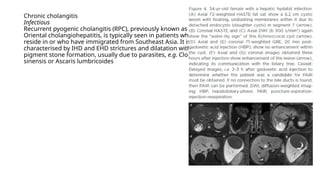
Chronic cholangitis
Infectious
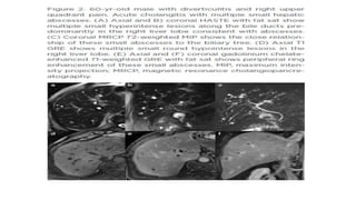
Recurrent pyogeniccholangitis (RPC), previously known as
Oriental cholangiohepatitis, is typically seen in patients who
reside in or who have immigrated from Southeast Asia. It is
characterised by IHD and EHD strictures and dilatation with
pigment stone formation, usually due to parasites, e.g. Clonorchis
sinensis or Ascaris lumbricoides
7.
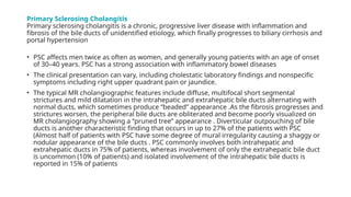
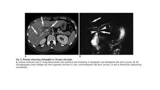
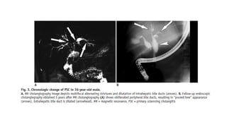
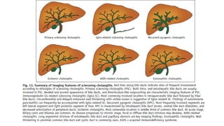
Primary Sclerosing Cholangitis
Primarysclerosing cholangitis is a chronic, progressive liver disease with inflammation and
fibrosis of the bile ducts of unidentified etiology, which finally progresses to biliary cirrhosis and
portal hypertension
• PSC affects men twice as often as women, and generally young patients with an age of onset
of 30–40 years. PSC has a strong association with inflammatory bowel diseases
• The clinical presentation can vary, including cholestatic laboratory findings and nonspecific
symptoms including right upper quadrant pain or jaundice.
• The typical MR cholangiographic features include diffuse, multifocal short segmental
strictures and mild dilatation in the intrahepatic and extrahepatic bile ducts alternating with
normal ducts, which sometimes produce “beaded” appearance .As the fibrosis progresses and
strictures worsen, the peripheral bile ducts are obliterated and become poorly visualized on
MR cholangiography showing a “pruned tree” appearance . Diverticular outpouching of bile
ducts is another characteristic finding that occurs in up to 27% of the patients with PSC
(Almost half of patients with PSC have some degree of mural irregularity causing a shaggy or
nodular appearance of the bile ducts . PSC commonly involves both intrahepatic and
extrahepatic ducts in 75% of patients, whereas involvement of only the extrahepatic bile duct
is uncommon (10% of patients) and isolated involvement of the intrahepatic bile ducts is
reported in 15% of patients
8.
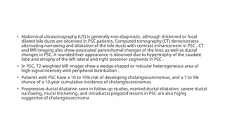
• Abdominal ultrasonography(US) is generally non-diagnostic, although thickened or focal
dilated bile ducts are observed in PSC patients. Computed tomography (CT) demonstrates
alternating narrowing and dilatation of the bile ducts with contrast enhancement in PSC . CT
and MR imaging also show associated parenchymal changes of the liver, as well as ductal
changes in PSC. A rounded liver appearance is observed due to hypertrophy of the caudate
lobe and atrophy of the left lateral and right posterior segments in PSC .
• In PSC, T2-weighted MR images show a wedge-shaped or reticular heterogeneous area of
high-signal intensity with peripheral distribution .
• Patients with PSC have a 10 to 15% risk of developing cholangiocarcinomas, and a 7 to 9%
chance of a 10-year cumulative incidence of cholangiocarcinomas
• Progressive ductal dilatation seen in follow-up studies, marked ductal dilatation, severe ductal
narrowing, mural thickening, and intraductal polypoid lesions in PSC are also highly
suggestive of cholangiocarcinoma
11.
Secondary Sclerosing Cholangitis
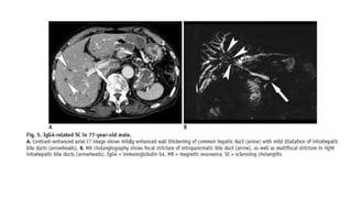
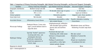
•IgG4-Related Sclerosing Cholangitis
• Immunoglobulin G4-SC is bile duct involvement of IgG4- related systemic disease (IgG4-RD) .
After the pancreas, bile ducts are the second most common organ, involved with IgG4-RD.
Patients with IgG4-SC are predominantly males in their 60s (mean age, 63 years) . Patients
with IgG4-SC commonly present with obstructive jaundice. Like other IgG4-related diseases,
the serum IgG4 is frequently elevated.
• Abdominal US has limited value for diagnosing IgG4- SC, although it may show thickening of
the bile duct and gallbladder. In cases of IgG4-SC, cross-sectional imaging, such as CT or MR
imaging, demonstrates long-segmental, symmetrical, circumferential wall thickening and
delayed contrast enhancement of the involved bile ducts
• Differentiation of IgG4-SC from other types of sclerosing cholangitis, especially from PSC, is
clinically important as IgG4-SC shows a dramatic response to steroid therapy . Patients with
PSC are generally younger and less symptomatic than those with IgG4-related disease .
13.
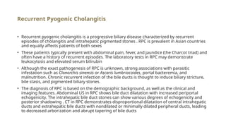
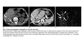
Recurrent Pyogenic Cholangitis
•Recurrent pyogenic cholangitis is a progressive biliary disease characterized by recurrent
episodes of cholangitis and intrahepatic pigmented stones . RPC is prevalent in Asian countries
and equally affects patients of both sexes
• These patients typically present with abdominal pain, fever, and jaundice (the Charcot triad) and
often have a history of recurrent episodes. The laboratory tests in RPC may demonstrate
leukocytosis and elevated serum bilirubin
• Although the exact pathogenesis of RPC is unknown, strong associations with parasitic
infestation such as Clonorchis sinensis or Ascaris lumbriocoides, portal bacteremia, and
malnutrition. Chronic recurrent infection of the bile ducts is thought to induce biliary stricture,
bile stasis, and pigmented biliary stones.
• The diagnosis of RPC is based on the demographic background, as well as the clinical and
imaging features. Abdominal US in RPC shows bile duct dilatation with increased periportal
echogenicity. The intrahepatic bile duct stones can show various degrees of echogenicity and
posterior shadowing . CT in RPC demonstrates disproportional dilatation of central intrahepatic
ducts and extrahepatic bile ducts with nondilated or minimally dilated peripheral ducts, leading
to decreased arborization and abrupt tapering of bile ducts
18.
Ischemic Cholangitis
• Ischemiccholangitis is defined as ischemia-induced bile duct injuries due to various causes.
The bile ducts are vulnerable to ischemic injuries as the blood supply to the bile ducts
depends completely on the arterial supply in contrast to the hepatic parenchyma, which has a
dual blood supply from the hepatic artery and the portal vein
• Among the various conditions that compromise the arterial supply and can cause ischemic
cholangitis, iatrogenic causes including liver transplantation, hepatic arterial infusion of
chemotherapeutic agents, and vessel injury during biliary or pancreatic surgery constitute the
most common etiology
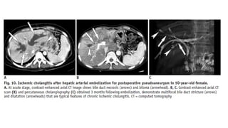
• At the acute stage of ischemic cholangitis, the patients usually present with fever, abdominal
pain, jaundice, and biliary sepsis. At this stage, the radiologic findings of biliary casts include
intraductal filling defects in the dilated bile duct showing high signal intensity on
nonenhanced T1-weighed MR images . At the acute stage of ischemic cholangitis, biliary casts
appear similar to intraductal stones. However, they usually differ in their shapes, as biliary
casts appear linear or have a branching pattern, whereas stones are usually oval or round . In
severe ischemic injury, bile duct necrosis presents as tubular low density or intensity
structures along the portal tracts on enhanced CT or MR images
20.
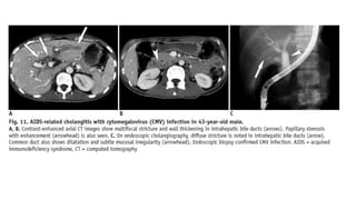
AIDS-Related Cholangitis
• AIDS-relatedcholangitis typically affects advanced HIV-infected patients with markedly
decreased immune function (CD4 count < 100/mm3
) . The suggested mechanisms of AIDS-
related cholangitis include randomly occurring infections involving the bile ducts,
• ischemia, autonomic nerve injury, and direct invasion of bile duct epithelium by the HIV itself .
The most common pathogens in AIDS-related cholangitis are cytomegalovirus,
Cryptosporidium parvum, Mycobacterium avium complex, and herpes simplex virus, although
no definite pathogen is identified in almost 50% of these patients
• On CT and MR imaging, enhanced wall thickening of the extrahepatic bile duct and the
intrahepatic bile ducts can be seen in AIDS-related cholangitis . Cholangiographic findings in
AIDS-related cholangitis include multifocal strictures and dilatation of intrahepatic and
extrahepatic bile ducts resembling those seen in patients with PSC.
22.
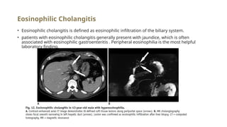
Eosinophilic Cholangitis
• Eosinophiliccholangitis is defined as eosinophilic infiltration of the biliary system.
• patients with eosinophilic cholangitis generally present with jaundice, which is often
associated with eosinophilic gastroenteritis . Peripheral eosinophilia is the most helpful
laboratory finding,
23.
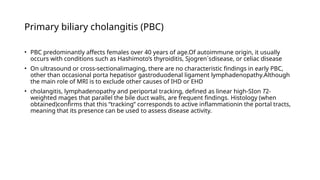
Primary biliary cholangitis(PBC)
• PBC predominantly affects females over 40 years of age.Of autoimmune origin, it usually
occurs with conditions such as Hashimoto’s thyroiditis, Sjogren´sdisease, or celiac disease
• On ultrasound or cross-sectionalimaging, there are no characteristic findings in early PBC,
other than occasional porta hepatisor gastroduodenal ligament lymphadenopathy.Although
the main role of MRI is to exclude other causes of IHD or EHD
• cholangitis, lymphadenopathy and periportal tracking, defined as linear high-SIon T2-
weighted mages that parallel the bile duct walls, are frequent findings. Histology (when
obtained)confirms that this “tracking” corresponds to active inflammationin the portal tracts,
meaning that its presence can be used to assess disease activity.
25.
Systemic Approach forSclerosing Cholangitis
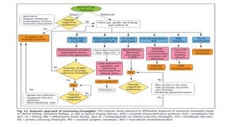
• Before arriving at the differential diagnosis of sclerosing cholangitis, it is critical to exclude
malignant bile duct strictures, especially when a single segmental bile duct wall thickening
and/or stricture is detected. The following image features favor malignant biliary strictures
rather than sclerosing cholangitis: a narrowed segment with hyperenhancement relative to
the liver seen during the portal-venous phase; long length involved (> 12 mm); prominent bile
duct thickening (> 3 mm); an indistinct outer margin; luminal irregularity; and asymmetry .
After excluding malignant biliary strictures, the next step is to distinguish between PSC and
secondary sclerosing cholangitis .