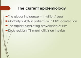
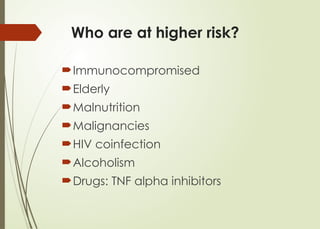
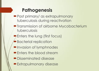
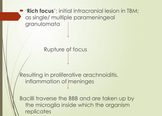
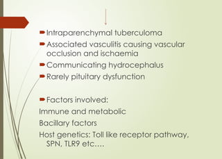
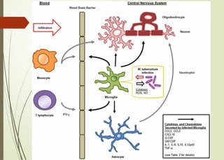
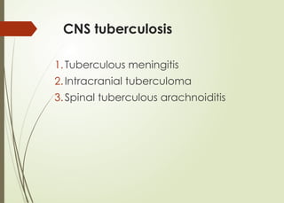
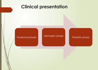
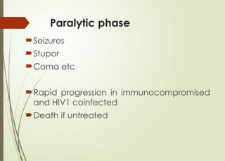
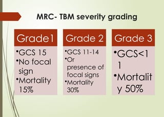
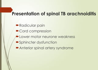
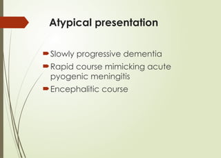
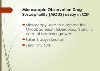
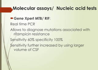
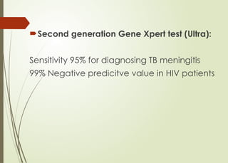
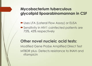
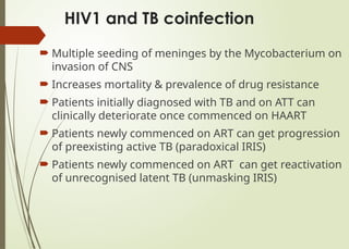
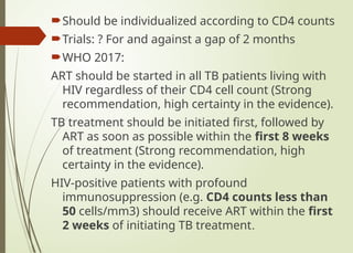
CNS Tuberculosis
Y.Nilukshana
Outline
The change in the epidemiology
Pathogenesis
Clinical presentation
Microbiological diagnosis
Radiological diagnosis
WHO recommendations
The challenges encountered
Novel drugs at the horizon
Prognostic markers
The current epidemiology
The global incidence > 1 milllion/ year
Mortality > 40% in patients with HIV1 coinfection
The rapidly escalating prevalence of HIV
Drug resistant TB meningitis is on the rise
Who are at higher risk?
Immunocompromised
Elderly
Malnutrition
Malignancies
HIV coinfection
Alcoholism
Drugs: TNF alpha inhibitors
Pathogenesis
Post primary/ as extrapulmonary
tuberculosis during reactivation
Transmission of airborne Mycobacterium
tuberculosis
Enters the lung (first focus)
Bacterial replication
Invasion of lymphnodes
Enters the blood stream
Disseminated disease
Extrapulmonary disease
 ‘Rich focus’: initial intracranial lesion in TBM;
as single/ multiple parameningeal
granulomata
Rupture of focus
Resulting in proliferative arachnoiditis,
inflammation of meninges
Bacilli traverse the BBB and are taken up by
the microglia inside which the organism
replicates
Intraparenchymal tuberculoma
Associated vasculitis causing vascular
occlusion and ischaemia
Communicating hydrocephalus
Rarely pituitary dysfunction
Factors involved:
Immune and metabolic
Bacillary factors
Host genetics: Toll like receptor pathway,
SPN, TLR9 etc….
CNS tuberculosis
1.Tuberculous meningitis
2.Intracranial tuberculoma
3.Spinal tuberculous arachnoiditis
Clinical presentation
Prodromal phase
Meningitic phase
Paralytic phase
Prodromal phase
2-3/ 52 duration
Low grade fever
Lassitude
Headache
Personality changes etc
Meningitic phase
Protactable headache
Meningism
Confusion
Vomiting
Cranial nerve lesions and long tract
signs
50% develop V, III cranial nerve palsies!
10% develop hemi/ paraparesis
Choroidal tubercles in miliary TB
Paralytic phase
Seizures
Stupor
Coma etc
Rapid progression in immunocompromised
and HIV1 coinfected
Death if untreated
MRC- TBM severity grading
Grade1
•GCS 15
•No focal
sign
•Mortality
15%
Grade 2
•GCS 11-14
•Or
presence of
focal signs
•Mortality
30%
Grade 3
•GCS<1
1
•Mortalit
y 50%
Presentation of spinal TB arachnoiditis
Radicular pain
Cord compression
Lower motor neurone weakness
Sphincter dysfunction
Anterior spinal artery syndrome
Atypical presentation
Slowly progressive dementia
Rapid course mimicking acute
pyogenic meningitis
Encephalitic course
Therapeutic paradox
In females with illness of a shorter duration
Worsening of the clinical signs ie fever,
mental state just after the commencement
of ATT.
Well known even in HIV negative patients
Seen in 1/3 of patients with TB meningitis
CSF: lymphocytic switches to neutrophilic!
CSF findings
WBC 150-1000/ μL lymphocytic predominant;
can be mixed
Neutrophilic in:
Early stages
HIV1 coinfection
IRIS (Immune Reconstitution Syndrome)
Protein 0.8-2.0g/dL
CSF glucose/ RBS < 0.5 in 90% patients
Low glucose and high lactate predict a
poorer prognosis
Novel markers in CSF: TNF, IL1В, VEGF
Microbiological diagnosis
ZN Staining:
Detects the bacilli if > 10,000/mL
Sensitivity in cavitatory PTB 30%
Paucibacillary TB, TB meningitis 10-20%
How to increase the ZN stain sensitivity?
 CSF > 10 mL
 Preferrably the fluid removed last from the
lumbar puncture
 centrifuged at 3000 g
 Examine the smear of the clot/ sediment of CSF
 If not clotted: add 95% alcohol 2 ml
 Apply 0.02 ml of the centrifuged deposit onto the
slide to < 1cm diameter
 Examine under 200-500 HPF
 By > one observer
 For > 30 minutes!
Mycobacterium TB culture of
CSF
More sensitive than the microscopy
Takes 10 days in liquid medium and 8
weeks in solid medium
Ideal laboratory safety settings are
warranted
Microscopic Observation Drug
Susceptibility (MODS) assay in CSF
Microscopy used to diagnose the
Mycobacterium tuberculous ‘specific
cord’ of bacterial growth
Takes 6 days duration
Sensitivity 65%
Molecular assays/ Nucleic acid tests
Gene Xpert MTB/ RIF:
Real time PCR
Allows to diagnose mutations associated with
rifampicin resistance
Sensitivity 60% specificity 100%
Sensitivity further increased by using larger
volume of CSF
Second generation Gene Xpert test (Ultra):
Sensitivity 95% for diagnosing TB meningitis
99% Negative predicitve value in HIV patients
Mycobacterium tuberculous
glycolipid lipoarabinomannan in CSF
Uses LFA (Lateral Flow Assay) or ELISA
Sensitivity in HIV1 coinfected patients are
75%, 43% respectively
Other novel nucleic acid tests:
Modified Gene Probe Amplified Direct Test
MTBDR plus- Detects resistance to INAH and
rifampicin
What is the place of ADA in
CSF?
No clear cut off values to
differentiate from acute pyogenic
meningitis
Radiological diagnosis
Gadolinium enhanced MRI is superior to CT
DWI imaging for early diagnosis of cerebral
ischaemia, borderzone necrosis
Marked basilar enhancement correlates
with vasculitis and also predicts infarctions
CT angiography useful as supraclinoid parts
of internal carotid artery, proximal parts of
anterior and middle cerebral arteries are
commonly affected
Gadolinum MRI
What to look for?
Cerebral oedema
Basilar arachnoiditis
Tuberculoma:
Initial low/ isodense regions becoming
hyperdense with ring enhancement
Mostly disappear with treatment; may get
calcified
Hydrocephalus
Infarctions
Mimickers
 Fungal, viral meningoencephalitis
 Parameningeal infections like sphenoidal
sinusitis
 Partially treated meningitis
 Neurosyphilis
 Carcinomatous meningitis
 Neurosarcoid
 Neurobrucellosis
 Neurocysticercosis
CNS toxoplasmosis
Challeneges in radiological
diagnosis
30% patients in early stages, MRC
grade1 will have normal CT and 15%
have normal MRI
Basal enhancement is less prominent
in presence of HIV1 with low CD4
counts
>50% patients have concomitant TB
spine which is commonly overlooked*
50% have a normal CXR
AntiTB therapy
Intensive phase: HRZE for two months
Continuation phase: HR for 10 months
Treatment of tuberculoma for a total
duration of 18 months
The place of glucocorticoids
In patients with tuberculous meningitis, an
initial adjuvant corticosteroid therapy with
dexamethasone or prednisolone tapered
over 6-8 weeks should be used (Strong
recommendation, moderate certainty in the
evidence).
Dexamethasone 0.3-0.4mg/kg/day for 2
weeks and taper over total of 8 weeks
Prednisolone 60 mg/day for 2 weeks and
taper over 8 weeks
Pros and cons of adjuvant steroid
therapy
Reduces intracerebral inflammation, inhibits
the production of cytokines by microglia,
astrocytes and improves outcome
Lack of evidence in HIV coinfected
patients
No difference in the longterm neurological
disability in either groups
? Does not prevent brain infarctions
2 small scale trial evidence suggests
addition of aspirin to reduce infarctions
Urgent indications for steroids
Progressive neurological deterioration
Encephalitis with CSF opening pressure
> 400 mmwater, clinical/ CT evidence
of cerebral oedema
Therapeutic paradox
CT revealing marked basilar
enhancement, moderate to advanced
hydrocephalus
Tuberculoma with disproportionate
oedema
IRIS
Other general measures
Monitor sodium, oxygenations,
temperature, intracranial pressure
Bedside transcranial doppler to
assess for vascular compromise
Hypertonic saline is equally effective
as mannitol
Pitfalls in using routine ATT
regime in CNS TB
 CSF concentration of rifampicin is 10-20% of that of
plasma
 *? Need to use ‘ Hyperintense regime’ ( ie> 10mg/kg)
for a better mortality benefit
 Ethambutol hardly crosses the BBB even in the
presence of inflammation (CSF concentration <20%)
 Contribution of ethambutol for a better outcome is
minimal even in the presence of INAH resistance
 Streptomycin poorly penetrates once the inflammation
subsides
 Higher prevalence of resistance to Streptomycin
 Vestibular and ototoxic effects of Streptomycin
 Second line drugs to be considered in patients with
resistance to rifampicin
Drugs at the horizon
Ethionamide: better penetration than
ethambutol and streptomycin
Fluoroquinolones: Levofloxacin, Moxifloxacin
have good penetration and highly active
against drug susceptible as well as many drug
resistant bacilli
Trial evidence of improved survival with usage
of high dose rifampicin (15mg/kg) with
levofloxacin as fifth agent
Thalidomide useful in tuberculoma invading
the optic chiasm
Infliximab has similar evidence to Thalidomide
Predictors of poor prognosis
Delayed diagnosis and delayed
treatment
Advanced disease
Resistance to ATT
HIV coinfection
Complications: ischaemia, hydrocephalus
Hyponatraemia
HIV1 and TB coinfection
 Multiple seeding of meninges by the Mycobacterium on
invasion of CNS
 Increases mortality & prevalence of drug resistance
 Patients initially diagnosed with TB and on ATT can
clinically deteriorate once commenced on HAART
 Patients newly commenced on ART can get progression
of preexisting active TB (paradoxical IRIS)
 Patients newly commenced on ART can get reactivation
of unrecognised latent TB (unmasking IRIS)
Early ART
Early ATT
Should be individualized according to CD4 counts
Trials: ? For and against a gap of 2 months
WHO 2017:
ART should be started in all TB patients living with
HIV regardless of their CD4 cell count (Strong
recommendation, high certainty in the evidence).
TB treatment should be initiated first, followed by
ART as soon as possible within the first 8 weeks
of treatment (Strong recommendation, high
certainty in the evidence).
HIV-positive patients with profound
immunosuppression (e.g. CD4 counts less than
50 cells/mm3) should receive ART within the first
2 weeks of initiating TB treatment.
References
 Guidelines for treatment of drug-susceptible
tuberculosis and patient care, WHO 2017 update
 National TB manual
 Tuberculous meningitis nature reviews 2017
 Treatment of Drug-Susceptible Tuberculosis
Guidelines CDC 2016 updates
Thank you

Central Nervous System Tuberculosis.pptx

  • 1.
  • 2.
    Outline The change inthe epidemiology Pathogenesis Clinical presentation Microbiological diagnosis Radiological diagnosis WHO recommendations The challenges encountered Novel drugs at the horizon Prognostic markers
  • 3.
    The current epidemiology Theglobal incidence > 1 milllion/ year Mortality > 40% in patients with HIV1 coinfection The rapidly escalating prevalence of HIV Drug resistant TB meningitis is on the rise
  • 4.
    Who are athigher risk? Immunocompromised Elderly Malnutrition Malignancies HIV coinfection Alcoholism Drugs: TNF alpha inhibitors
  • 5.
    Pathogenesis Post primary/ asextrapulmonary tuberculosis during reactivation Transmission of airborne Mycobacterium tuberculosis Enters the lung (first focus) Bacterial replication Invasion of lymphnodes Enters the blood stream Disseminated disease Extrapulmonary disease
  • 6.
     ‘Rich focus’:initial intracranial lesion in TBM; as single/ multiple parameningeal granulomata Rupture of focus Resulting in proliferative arachnoiditis, inflammation of meninges Bacilli traverse the BBB and are taken up by the microglia inside which the organism replicates
  • 7.
    Intraparenchymal tuberculoma Associated vasculitiscausing vascular occlusion and ischaemia Communicating hydrocephalus Rarely pituitary dysfunction Factors involved: Immune and metabolic Bacillary factors Host genetics: Toll like receptor pathway, SPN, TLR9 etc….
  • 9.
    CNS tuberculosis 1.Tuberculous meningitis 2.Intracranialtuberculoma 3.Spinal tuberculous arachnoiditis
  • 10.
  • 11.
    Prodromal phase 2-3/ 52duration Low grade fever Lassitude Headache Personality changes etc
  • 12.
    Meningitic phase Protactable headache Meningism Confusion Vomiting Cranialnerve lesions and long tract signs 50% develop V, III cranial nerve palsies! 10% develop hemi/ paraparesis
  • 13.
  • 14.
    Paralytic phase Seizures Stupor Coma etc Rapidprogression in immunocompromised and HIV1 coinfected Death if untreated
  • 15.
    MRC- TBM severitygrading Grade1 •GCS 15 •No focal sign •Mortality 15% Grade 2 •GCS 11-14 •Or presence of focal signs •Mortality 30% Grade 3 •GCS<1 1 •Mortalit y 50%
  • 16.
    Presentation of spinalTB arachnoiditis Radicular pain Cord compression Lower motor neurone weakness Sphincter dysfunction Anterior spinal artery syndrome
  • 17.
    Atypical presentation Slowly progressivedementia Rapid course mimicking acute pyogenic meningitis Encephalitic course
  • 18.
    Therapeutic paradox In femaleswith illness of a shorter duration Worsening of the clinical signs ie fever, mental state just after the commencement of ATT. Well known even in HIV negative patients Seen in 1/3 of patients with TB meningitis CSF: lymphocytic switches to neutrophilic!
  • 19.
    CSF findings WBC 150-1000/μL lymphocytic predominant; can be mixed Neutrophilic in: Early stages HIV1 coinfection IRIS (Immune Reconstitution Syndrome) Protein 0.8-2.0g/dL CSF glucose/ RBS < 0.5 in 90% patients Low glucose and high lactate predict a poorer prognosis Novel markers in CSF: TNF, IL1В, VEGF
  • 20.
    Microbiological diagnosis ZN Staining: Detectsthe bacilli if > 10,000/mL Sensitivity in cavitatory PTB 30% Paucibacillary TB, TB meningitis 10-20%
  • 21.
    How to increasethe ZN stain sensitivity?  CSF > 10 mL  Preferrably the fluid removed last from the lumbar puncture  centrifuged at 3000 g  Examine the smear of the clot/ sediment of CSF  If not clotted: add 95% alcohol 2 ml  Apply 0.02 ml of the centrifuged deposit onto the slide to < 1cm diameter  Examine under 200-500 HPF  By > one observer  For > 30 minutes!
  • 22.
    Mycobacterium TB cultureof CSF More sensitive than the microscopy Takes 10 days in liquid medium and 8 weeks in solid medium Ideal laboratory safety settings are warranted
  • 23.
    Microscopic Observation Drug Susceptibility(MODS) assay in CSF Microscopy used to diagnose the Mycobacterium tuberculous ‘specific cord’ of bacterial growth Takes 6 days duration Sensitivity 65%
  • 24.
    Molecular assays/ Nucleicacid tests Gene Xpert MTB/ RIF: Real time PCR Allows to diagnose mutations associated with rifampicin resistance Sensitivity 60% specificity 100% Sensitivity further increased by using larger volume of CSF
  • 25.
    Second generation GeneXpert test (Ultra): Sensitivity 95% for diagnosing TB meningitis 99% Negative predicitve value in HIV patients
  • 26.
    Mycobacterium tuberculous glycolipid lipoarabinomannanin CSF Uses LFA (Lateral Flow Assay) or ELISA Sensitivity in HIV1 coinfected patients are 75%, 43% respectively Other novel nucleic acid tests: Modified Gene Probe Amplified Direct Test MTBDR plus- Detects resistance to INAH and rifampicin
  • 27.
    What is theplace of ADA in CSF? No clear cut off values to differentiate from acute pyogenic meningitis
  • 28.
    Radiological diagnosis Gadolinium enhancedMRI is superior to CT DWI imaging for early diagnosis of cerebral ischaemia, borderzone necrosis Marked basilar enhancement correlates with vasculitis and also predicts infarctions CT angiography useful as supraclinoid parts of internal carotid artery, proximal parts of anterior and middle cerebral arteries are commonly affected
  • 30.
  • 32.
    What to lookfor? Cerebral oedema Basilar arachnoiditis Tuberculoma: Initial low/ isodense regions becoming hyperdense with ring enhancement Mostly disappear with treatment; may get calcified Hydrocephalus Infarctions
  • 33.
    Mimickers  Fungal, viralmeningoencephalitis  Parameningeal infections like sphenoidal sinusitis  Partially treated meningitis  Neurosyphilis  Carcinomatous meningitis  Neurosarcoid  Neurobrucellosis  Neurocysticercosis
  • 34.
  • 35.
    Challeneges in radiological diagnosis 30%patients in early stages, MRC grade1 will have normal CT and 15% have normal MRI Basal enhancement is less prominent in presence of HIV1 with low CD4 counts >50% patients have concomitant TB spine which is commonly overlooked* 50% have a normal CXR
  • 36.
    AntiTB therapy Intensive phase:HRZE for two months Continuation phase: HR for 10 months Treatment of tuberculoma for a total duration of 18 months
  • 37.
    The place ofglucocorticoids In patients with tuberculous meningitis, an initial adjuvant corticosteroid therapy with dexamethasone or prednisolone tapered over 6-8 weeks should be used (Strong recommendation, moderate certainty in the evidence). Dexamethasone 0.3-0.4mg/kg/day for 2 weeks and taper over total of 8 weeks Prednisolone 60 mg/day for 2 weeks and taper over 8 weeks
  • 38.
    Pros and consof adjuvant steroid therapy Reduces intracerebral inflammation, inhibits the production of cytokines by microglia, astrocytes and improves outcome Lack of evidence in HIV coinfected patients No difference in the longterm neurological disability in either groups ? Does not prevent brain infarctions 2 small scale trial evidence suggests addition of aspirin to reduce infarctions
  • 39.
    Urgent indications forsteroids Progressive neurological deterioration Encephalitis with CSF opening pressure > 400 mmwater, clinical/ CT evidence of cerebral oedema Therapeutic paradox CT revealing marked basilar enhancement, moderate to advanced hydrocephalus Tuberculoma with disproportionate oedema IRIS
  • 40.
    Other general measures Monitorsodium, oxygenations, temperature, intracranial pressure Bedside transcranial doppler to assess for vascular compromise Hypertonic saline is equally effective as mannitol
  • 41.
    Pitfalls in usingroutine ATT regime in CNS TB  CSF concentration of rifampicin is 10-20% of that of plasma  *? Need to use ‘ Hyperintense regime’ ( ie> 10mg/kg) for a better mortality benefit  Ethambutol hardly crosses the BBB even in the presence of inflammation (CSF concentration <20%)  Contribution of ethambutol for a better outcome is minimal even in the presence of INAH resistance  Streptomycin poorly penetrates once the inflammation subsides  Higher prevalence of resistance to Streptomycin  Vestibular and ototoxic effects of Streptomycin  Second line drugs to be considered in patients with resistance to rifampicin
  • 42.
    Drugs at thehorizon Ethionamide: better penetration than ethambutol and streptomycin Fluoroquinolones: Levofloxacin, Moxifloxacin have good penetration and highly active against drug susceptible as well as many drug resistant bacilli Trial evidence of improved survival with usage of high dose rifampicin (15mg/kg) with levofloxacin as fifth agent Thalidomide useful in tuberculoma invading the optic chiasm Infliximab has similar evidence to Thalidomide
  • 43.
    Predictors of poorprognosis Delayed diagnosis and delayed treatment Advanced disease Resistance to ATT HIV coinfection Complications: ischaemia, hydrocephalus Hyponatraemia
  • 44.
    HIV1 and TBcoinfection  Multiple seeding of meninges by the Mycobacterium on invasion of CNS  Increases mortality & prevalence of drug resistance  Patients initially diagnosed with TB and on ATT can clinically deteriorate once commenced on HAART  Patients newly commenced on ART can get progression of preexisting active TB (paradoxical IRIS)  Patients newly commenced on ART can get reactivation of unrecognised latent TB (unmasking IRIS)
  • 45.
  • 46.
    Should be individualizedaccording to CD4 counts Trials: ? For and against a gap of 2 months WHO 2017: ART should be started in all TB patients living with HIV regardless of their CD4 cell count (Strong recommendation, high certainty in the evidence). TB treatment should be initiated first, followed by ART as soon as possible within the first 8 weeks of treatment (Strong recommendation, high certainty in the evidence). HIV-positive patients with profound immunosuppression (e.g. CD4 counts less than 50 cells/mm3) should receive ART within the first 2 weeks of initiating TB treatment.
  • 48.
    References  Guidelines fortreatment of drug-susceptible tuberculosis and patient care, WHO 2017 update  National TB manual  Tuberculous meningitis nature reviews 2017  Treatment of Drug-Susceptible Tuberculosis Guidelines CDC 2016 updates
  • 49.