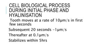
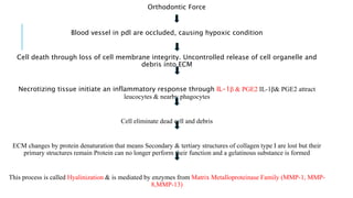
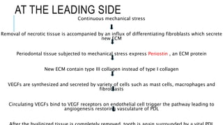
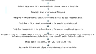
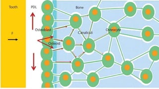
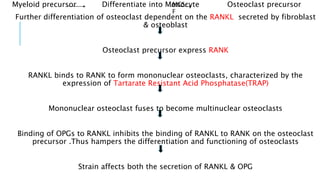
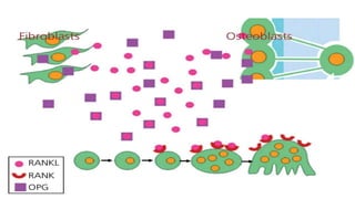
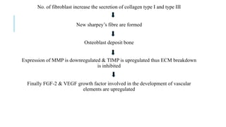
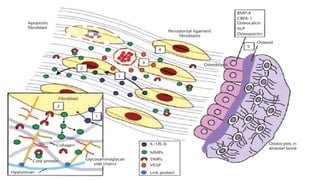
CELL BIOLOGICAL PROCESS
DURING ORTHODONTIC TOOTH
MOVEMENT
Guided by:
Dr Rajan K Mahindra
Dr Rakesh Mohode
Dr Govind Suryavanshi
Presented by- Sneha
Shah
CONTENTS
•Process of hyalinization.
•How osteoclast recruited at site of resorption.
•Process of alveolar bone resorption.
•Changes occurring on the trailing side.
CELL BIOLOGICAL PROCESS
DURING INITIAL PHASE AND
HYALINISATION
Tooth moves at a rate of 10µm/s in first
few seconds
Subsequent 20 seconds -1µm/s
Thereafter at 0.1µm/s
Stabilizes within 5hrs
PROCESS OF HYALINIZATION
Orthodontic Force
Blood vessel in pdl are occluded, causing hypoxic condition
Cell death through loss of cell membrane integrity. Uncontrolled release of cell organelle and
debris into ECM
Necrotizing tissue initiate an inflammatory response through IL-1β & PGE2 IL-1β& PGE2 attract
leucocytes & nearby phagocytes
Cell eliminate dead cell and debris
ECM changes by protein denaturation that means Secondary & tertiary structures of collagen type I are lost but their
primary structures remain Protein can no longer perform their function and a gelatinous substance is formed
This process is called Hyalinization & is mediated by enzymes from Matrix Metalloproteinase Family (MMP-1, MMP-
8,MMP-13)
AT THE LEADING SIDE
Continuous mechanical stress
Removal of necrotic tissue is accompanied by an influx of differentiating fibroblasts which secrete
new ECM
Periodontal tissue subjected to mechanical stress express Periostin , an ECM protein
New ECM contain type III collagen instead of type I collagen
VEGFs are synthesized and secreted by variety of cells such as mast cells, macrophages and
fibroblasts
Circulating VEGFs bind to VEGF receptors on endothelial cell trigger the pathway leading to
angiogenesis restoring vasculature of PDL
CELL BIOLOGICAL PROCESS
DURING TOOTH MOVEMENT
Orthodontic force
Induces negative strain at leading side and positive strain at trailing side
Results in strain of periodontal fibroblast
Integrins by which fibroblast are attached to the ECM can act as a force transducer
Fluid flow in PDL & canalicular network in the alveolar bone is induced
Fluid flow induces strain in the cell membrane of fibroblasts, osteoblast, & osteocyte
Osteoblast and periodontal fibroblast contribute to activation of cells by integrin mediated strain transmission to
the cytoskeleton & subsequent induction of expression of a variety factors & cytokines
These factors such as FGF, IGF-1, IL-1α, IL-β,IL-6& TNFα
Mediate the differentiation of precursors into osteoblast and osteoclast
,
HOW ARE OSTEOCLAST
RECRUITED AT THE SITE OF
RESORPTION ?
Myeloid precursor Differentiate into Monocyte Osteoclast precursor
Further differentiation of osteoclast dependent on the RANKL secreted by fibroblast
& osteoblast
Osteoclast precursor express RANK
RANKL binds to RANK to form mononuclear osteoclasts, characterized by the
expression of Tartarate Resistant Acid Phosphatase(TRAP)
Mononuclear osteoclast fuses to become multinuclear osteoclasts
Binding of OPGs to RANKL inhibits the binding of RANKL to RANK on the osteoclast
precursor .Thus hampers the differentiation and functioning of osteoclasts
Strain affects both the secretion of RANKL & OPG
MCS-
F
RESORPTION OF ALVEOLAR BONE
For functioning of osteoclast they should be attached to
mineralised bone matrix through αVβ3 integrin
This is only possible when the osteoblasts, osteoid& non-mineralized bone matrix
covering the surface of alveolar bone are removed.
ECM of the osteoid is degraded through the action of MMP’s (MMP1, 8 , 13 &14)
Osteoblast disappear by apoptosis induced by binding of TNF-α to its receptor
TNFR1&TNFR2
Combined osteoblast apoptosis &ECM degradation leads to exposure of mineralized
bone matrix which serves as landing site for osteoclasts
Osteoclasts moves by chemotaxis & attach the bone by αVβ3 integrin ,
connecting osteoclast to RGD peptides in bone matrix
Upon adhesion to bone osteoclast polarize & reorganize their cytoskeleton to
generate a ring like F-actin rich structures sealing zone that isolates howship’s
lacunae from the surroundings
Inside the sealing zone ruffled border is formed
Isolated area becomes acidic through influx of H+ ATPase mediated proton
pump this favours dissolution of bone minerals
In addition, the lysosomal enzyme, CathepsinK , &MMP -9 are secreted into
howship’s lacunae
TRAILING SIDE
PDL is widened
Accompanied by positive strain in the ECM
Acute inflammatory reaction
Results in increase in IL-1β, IL-10, PGE2 &TGF-β
Subsequent increase in OPG and decrease in RANKL by
osteoblast and fibroblast
No. of fibroblast increase the secretion of collagen type I and type III
New sharpey’s fibre are formed
Osteoblast deposit bone
Expression of MMP is downregulated & TIMP is upregulated thus ECM breakdown
is inhibited
Finally FGF-2 & VEGF growth factor involved in the development of vascular
elements are upregulated
Diagnosis and Management of Malocclusion And
Dentofacial Deformities, O.P.
 Kharbanda, Third Edition.Contemporary
Orthodontics, William R. Proffit. Sixth Edition
Biological Mechanisms of Tooth Movement- JaapC.
Maltha , Vinod Krrishnan , Anne Marie Kuijpers-
Jagtman.
THANK
YOU

Cell biological process during real tooth movement.pptx

  • 1.
    CELL BIOLOGICAL PROCESS DURINGORTHODONTIC TOOTH MOVEMENT Guided by: Dr Rajan K Mahindra Dr Rakesh Mohode Dr Govind Suryavanshi Presented by- Sneha Shah
  • 2.
    CONTENTS •Process of hyalinization. •Howosteoclast recruited at site of resorption. •Process of alveolar bone resorption. •Changes occurring on the trailing side.
  • 3.
    CELL BIOLOGICAL PROCESS DURINGINITIAL PHASE AND HYALINISATION Tooth moves at a rate of 10µm/s in first few seconds Subsequent 20 seconds -1µm/s Thereafter at 0.1µm/s Stabilizes within 5hrs
  • 4.
  • 5.
    Orthodontic Force Blood vesselin pdl are occluded, causing hypoxic condition Cell death through loss of cell membrane integrity. Uncontrolled release of cell organelle and debris into ECM Necrotizing tissue initiate an inflammatory response through IL-1β & PGE2 IL-1β& PGE2 attract leucocytes & nearby phagocytes Cell eliminate dead cell and debris ECM changes by protein denaturation that means Secondary & tertiary structures of collagen type I are lost but their primary structures remain Protein can no longer perform their function and a gelatinous substance is formed This process is called Hyalinization & is mediated by enzymes from Matrix Metalloproteinase Family (MMP-1, MMP- 8,MMP-13)
  • 6.
    AT THE LEADINGSIDE Continuous mechanical stress Removal of necrotic tissue is accompanied by an influx of differentiating fibroblasts which secrete new ECM Periodontal tissue subjected to mechanical stress express Periostin , an ECM protein New ECM contain type III collagen instead of type I collagen VEGFs are synthesized and secreted by variety of cells such as mast cells, macrophages and fibroblasts Circulating VEGFs bind to VEGF receptors on endothelial cell trigger the pathway leading to angiogenesis restoring vasculature of PDL
  • 7.
  • 8.
    Orthodontic force Induces negativestrain at leading side and positive strain at trailing side Results in strain of periodontal fibroblast Integrins by which fibroblast are attached to the ECM can act as a force transducer Fluid flow in PDL & canalicular network in the alveolar bone is induced Fluid flow induces strain in the cell membrane of fibroblasts, osteoblast, & osteocyte Osteoblast and periodontal fibroblast contribute to activation of cells by integrin mediated strain transmission to the cytoskeleton & subsequent induction of expression of a variety factors & cytokines These factors such as FGF, IGF-1, IL-1α, IL-β,IL-6& TNFα Mediate the differentiation of precursors into osteoblast and osteoclast ,
  • 10.
    HOW ARE OSTEOCLAST RECRUITEDAT THE SITE OF RESORPTION ?
  • 11.
    Myeloid precursor Differentiateinto Monocyte Osteoclast precursor Further differentiation of osteoclast dependent on the RANKL secreted by fibroblast & osteoblast Osteoclast precursor express RANK RANKL binds to RANK to form mononuclear osteoclasts, characterized by the expression of Tartarate Resistant Acid Phosphatase(TRAP) Mononuclear osteoclast fuses to become multinuclear osteoclasts Binding of OPGs to RANKL inhibits the binding of RANKL to RANK on the osteoclast precursor .Thus hampers the differentiation and functioning of osteoclasts Strain affects both the secretion of RANKL & OPG MCS- F
  • 13.
    RESORPTION OF ALVEOLARBONE For functioning of osteoclast they should be attached to mineralised bone matrix through αVβ3 integrin This is only possible when the osteoblasts, osteoid& non-mineralized bone matrix covering the surface of alveolar bone are removed. ECM of the osteoid is degraded through the action of MMP’s (MMP1, 8 , 13 &14) Osteoblast disappear by apoptosis induced by binding of TNF-α to its receptor TNFR1&TNFR2 Combined osteoblast apoptosis &ECM degradation leads to exposure of mineralized bone matrix which serves as landing site for osteoclasts
  • 14.
    Osteoclasts moves bychemotaxis & attach the bone by αVβ3 integrin , connecting osteoclast to RGD peptides in bone matrix Upon adhesion to bone osteoclast polarize & reorganize their cytoskeleton to generate a ring like F-actin rich structures sealing zone that isolates howship’s lacunae from the surroundings Inside the sealing zone ruffled border is formed Isolated area becomes acidic through influx of H+ ATPase mediated proton pump this favours dissolution of bone minerals In addition, the lysosomal enzyme, CathepsinK , &MMP -9 are secreted into howship’s lacunae
  • 16.
    TRAILING SIDE PDL iswidened Accompanied by positive strain in the ECM Acute inflammatory reaction Results in increase in IL-1β, IL-10, PGE2 &TGF-β Subsequent increase in OPG and decrease in RANKL by osteoblast and fibroblast
  • 17.
    No. of fibroblastincrease the secretion of collagen type I and type III New sharpey’s fibre are formed Osteoblast deposit bone Expression of MMP is downregulated & TIMP is upregulated thus ECM breakdown is inhibited Finally FGF-2 & VEGF growth factor involved in the development of vascular elements are upregulated
  • 19.
    Diagnosis and Managementof Malocclusion And Dentofacial Deformities, O.P.  Kharbanda, Third Edition.Contemporary Orthodontics, William R. Proffit. Sixth Edition Biological Mechanisms of Tooth Movement- JaapC. Maltha , Vinod Krrishnan , Anne Marie Kuijpers- Jagtman.
  • 20.