SlideShare a Scribd company logo
Oncogenic Ras differentially regulates metabolism and
anoikis in extracellular matrix-detached cells
JA Mason1
, CA Davison-Versagli1
, AK Leliaert1
, DJ Pape1
, C McCallister1
, J Zuo1
, SM Durbin1
, CL Buchheit1
, S Zhang1
and ZT Schafer*1
In order for cancer cells to survive during metastasis, they must overcome anoikis, a caspase-dependent cell death process
triggered by extracellular matrix (ECM) detachment, and rectify detachment-induced metabolic defects that compromise cell
survival. However, the precise signals used by cancer cells to facilitate their survival during metastasis remain poorly understood.
We have discovered that oncogenic Ras facilitates the survival of ECM-detached cancer cells by using distinct effector pathways to
regulate metabolism and block anoikis. Surprisingly, we find that while Ras-mediated phosphatidylinositol (3)-kinase signaling
is critical for rectifying ECM-detachment-induced metabolic deficiencies, the critical downstream effector is serum and
glucocorticoid-regulated kinase-1 (SGK-1) rather than Akt. Our data also indicate that oncogenic Ras blocks anoikis by
diminishing expression of the phosphatase PHLPP1 (PH Domain and Leucine-Rich Repeat Protein Phosphatase 1), which
promotes anoikis through the activation of p38 MAPK. Thus, our study represents a novel paradigm whereby oncogene-initiated
signal transduction can promote the survival of ECM-detached cells through divergent downstream effectors.
Cell Death and Differentiation advance online publication, 26 February 2016; doi:10.1038/cdd.2016.15
Cancer metastasis, the spread of cancer cells to distant parts
of the body, accounts for ~ 90% of cancer-related deaths and
represents an inherently difficult clinical challenge.1,2
It has
become clear that for successful metastasis to occur, cells
must overcome a caspase-dependent cell death mechanism,
anoikis, which is triggered by detachment from the extra-
cellular matrix (ECM).3
In addition to anoikis evasion, cancer
cells must also contend with anoikis-independent cellular
alterations that can compromise cellular viability.4
Chief
among these alterations are metabolic deficiencies that are
induced by ECM detachment.5–7
These metabolic alterations
involve deficiencies in ATP generation, elevated levels of
reactive oxygen species, and the induction of autophagy.6,8,9
Although recent studies have begun to unravel the strategies
used by cancer cells to ameliorate metabolic deficiencies
during ECM detachment,10
the signal-transduction cascades
responsible for regulating metabolism during ECM detach-
ment in cancer cells remain almost entirely unexplored.
The activation of oncogenic signaling pathways is critical to
anchorage-independent growth and ultimately to the survival
of a variety of distinct cancer cell types during ECM
detachment.4,11
Presumably, this oncogenic signaling is also
necessary for resolving the aforementioned ECM-deta-
chment-induced metabolic deficiencies. ErbB2 overexpres-
sion in mammary epithelial cells results in a stimulation of
phosphatidylinositol (3)-kinase (PI(3)K)/Akt signaling to pro-
mote glucose uptake and ATP generation.6
These data raise
the question as to how cancer cells that lack ErbB2 over-
expression rectify metabolic deficiencies during ECM detach-
ment. Does activation of other oncogenic signaling pathways
facilitate ATP generation during ECM detachment, and are
similar downstream effectors used? The design of novel
chemotherapeutic approaches to eliminate ECM-detached
cancer cells requires a better understanding of the signal-
transduction cascades that regulate metabolism during ECM
detachment.
The Ras oncogene is mutated in ~ 30% of all human
cancers, typically resulting in constitutive activation via
mutations in codon 12, 13, or 61.12–14
Given the frequency
with which Ras mutations arise in cancers, it seems reason-
able to speculate that Ras signaling may be involved in
facilitating the survival of ECM-detached cells. Indeed, pre-
vious studies in intestinal epithelial cells suggest that Ras
activation can promote anoikis evasion,15
although significant
ambiguities exist with regard to the precise downstream
signaling that is involved.16
Once activated, Ras is well known
to activate ERK and Akt, which have well-documented roles in
promoting cell survival in cancer cells.12
More specifically, the
ability of Ras to modulate metabolism during ECM detachment
would be consistent with its known capabilities to activate PI(3)
K and the proficiency with which PI(3)K signaling promotes
glucose uptake and ATP generation.17
Given this and the
aforementioned studies demonstrating that ErbB2 promotes
ATP generation during ECM detachment by activating PI(3)K
and Akt,6
it seems reasonable to speculate that Ras promotes
metabolic activity through a similar signal-transduction
cascade.
Here, we uncover a surprising and novel signal-transduction
pathway operating downstream of oncogenic Ras to promote
the survival of ECM-detached cancer cells. Interestingly,
1
Department of Biological Sciences, University of Notre Dame, Notre Dame, IN, USA
*Corresponding author: ZT Schafer, Department of Biological Sciences, University of Notre Dame, 222 Galvin Life Science Center, Notre Dame, IN 46556, USA. Tel: +1 574
631 0875; Fax: +1 574 631 7413; E-mail: zschafe1@nd.edu
Received 09.7.15; revised 09.12.15; accepted 25.1.16; Edited by S Fulda
Abbreviations: ECM, extracellular matrix; PI(3)K, phosphatidylinositol (3)-kinase; SGK-1, serum and glucocorticoid-regulated kinase-1; PHLPP1, PH domain and leucine
rich repeat protein phosphatase; poly-HEMA, poly-(2-hydroxyethyl methacrylate); EGF, epidermal growth factor; DAPI, 4,6-diamidino-2-phenylindone; RPPA, reverse
phase protein array; INT-violet, iodonitrotetrazolium chloride; STS, staurosporine; GAPDH, glyceraldehyde-3-phosphate dehydrogenase; EV, empty vector
Cell Death and Differentiation (2016), 1–12
& 2016 Macmillan Publishers Limited All rights reserved 1350-9047/16
www.nature.com/cdd
rather than relying on PI(3)K/Akt to promote ATP generation,
we have found that Ras overcomes ECM-detachment-
induced ATP deficiencies through the activation of a distinct
PI(3)K effector, serum and glucocorticoid-regulated kinase-1
(SGK-1). Furthermore, anoikis resistance in cells harboring
Ras mutations relies on the downregulation of the PHLPP1
(PH Domain and Leucine-Rich Repeat Protein Phosphatase
1) phosphatase. Despite the well-known proclivity of PHLPP1
to dephosphorylate and deactivate Akt, we have discovered
that PHLPP1 mediates anoikis through the activation of the
p38 MAPK pathway. These data identify novel downstream
targets that could be used for the development of chemother-
apeutic approaches aimed at antagonizing the ability of Ras to
eliminate deficits in ATP generation and block anoikis.
Furthermore, our data substantially refine the current under-
standing of Ras signaling to suggest that Ras-mediated signal
transduction can promote the survival of ECM-detached cells
through divergent downstream effectors.
Results
Oncogenic Ras promotes ATP generation through a PI(3)
K-dependent, Akt-independent signaling pathway in
ECM-detached cells. To examine the ability of oncogenic
Ras to promote ATP generation and cell viability during ECM
detachment, we engineered MCF-10A cells to stably express
constitutively active H-Ras (G12V, hereafter referred to as
10A HrasG12V) or K-Ras (G12V, hereafter referred to as 10A
KrasG12V). Indeed, hyperactive Ras promotes ATP genera-
tion in ECM-detached (but not ECM-attached) cells
(Figure 1a). This corresponds to an increase in the viability
of ECM-detached cells (Figure 1b). Given that PI(3)K signal-
ing is well known to operate downstream of Ras,6
we
investigated if PI(3)K also operates to promote ATP genera-
tion. As expected, inhibition of PI(3)K signaling compromises
ATP production in a dose-dependent manner in both 10A
HrasG12V and 10A KrasG12V cells (Figure 1c). However,
when examining phosphorylation of the PI(3)K effector Akt,
we surprisingly observe minimal changes in Akt (S473)
phosphorylation (Figure 1c). To confirm the efficacy of the
inhibitor, we blocked PI(3)K signaling in MCF-10A cells
overexpressing insulin growth factor receptor 1 (10A IGF-1R)
and observed the expected loss of Akt phosphorylation
(Figure 1d). These data therefore suggest that ATP genera-
tion in ECM-detached cells downstream of oncogenic Ras is
occurring through a PI(3)K-dependent, but Akt-independent
mechanism.
SGK-1 activation is sufficient to promote ATP production
and cell survival during ECM detachment. Given that Akt
does not appear to be a relevant mediator of ATP generation
downstream of PI(3)K (in the presence of oncogenic Ras)
during ECM detachment, we assessed the contribution of
SGK-1, another AGC family kinase known to operate down-
stream of PI(3)K.6
SGK-1 shares significant sequence
identity with Akt and has previously been linked to the
regulation of glucose transporter localization.6,18–20
In our
10A HrasG12V and 10A KrasG12V cells, we see a decrease
in SGK-1 activation upon PI(3)K inhibition as measured by
phosphorylation status of Sek1 at serine 80 (Figure 1e), a
canonical downstream SGK-1 target.21
In addition, we
observe an increase in the phosphorylation of Sek1 in the
presence of oncogenic Ras mutations (Figure 1f). This
increase in Sek1 phosphorylation downstream of oncogenic
Ras occurs in both ECM-detached and -attached cells with
similar kinetics (Supplementary Figures S1A and S1B).
To examine if SGK-1 is sufficient to promote ATP generation
and survival during ECM detachment, we generated MCF-10A
cells that express constitutively active SGK-1 (S422D,
hereafter referred to as 10A S422D). Overexpression and
constitutive activation of SGK-1 was confirmed via immuno-
blotting for SGK-1 and phospho (p)-Sek1 (Figure 2a). When
grown in ECM-detached conditions, we observe an increase in
glucose uptake (Figure 2b), ATP production (Figure 2c), and
cellular viability (Figure 2d) in 10A S422D cells. This
increase in productive metabolism and ATP generation in
ECM-detached 10A S422D cells does not seem to be a result
of alterations in SGK-1 localization (Supplementary
Figure S2A) and occurs in both glucose-rich and -limiting
conditions (Supplementary Figure S2B). Furthermore, using a
3D cell culture model of mammary acini where the hollow
lumen is normally generated by ECM-detachment-induced
cell death,6
we find that constitutive activation of SGK-1 can
promote luminal filling and cell survival (Figure 2e). Interest-
ingly, constitutively active SGK-1 does not influence caspase
activation in either ECM-attached or -detached cells
(Figure 2f), suggesting that the effects of SGK-1 activation
on ATP generation and viability are independent of anoikis.
To ascertain the importance of SGK-1 kinase activity for the
observed changes in ATP generation and cell viability during
ECM detachment, we expressed kinase-dead SGK-1
(K127M) in MCF-10A cells (hereafter referred to as 10A
K127M). Immunoblotting for p-Sek1 confirmed the lack of
SGK-1 kinase activity in these cells (Figure 2g). In contrast to
constitutively active SGK-1 (S422D), the expression of SGK-1
(K127M) does not promote ATP production or cell viability in
ECM-detached cells (Figures 2h and i). Thus, our data
suggest that the SGK-1-mediated increase in ATP generation
and cell viability is a direct consequence of SGK-1 kinase
activity.
SGK-1 is required for the promotion of ATP generation
downstream of oncogenic Ras during ECM detachment.
Given that SGK-1 kinase activity is sufficient to promote ATP
generation during ECM detachment, we next investigated the
role of SGK-1 downstream of oncogenic Ras during ECM
detachment. Indeed, 10A HrasG12V and 10A KrasG12V
cells have elevated SGK-1 activity (Figure 1f), ATP production
(Figures 1a and c), glucose uptake (Figure 3a), and cellular
viability (Figure 1b). To more directly examine the role of
SGK-1 in Ras-mediated regulation of ATP generation, we
eliminated SGK-1 expression (and concomitant activity;
Figure 3b) in 10A HrasG12V cells using short hairpin RNA
(shRNA) (10A HrasG12V shSGK-1). Elimination of SGK-1 in
these cells results in a loss of glucose uptake (Figure 3c),
diminished ATP generation (Figure 3d), and a loss of cell
viability (Figure 3e). Interestingly, the loss of SGK-1 signaling
is only detrimental to ECM-detached cells, as SGK-1
knockdown does not alter ATP production in ECM-attached
Ras-mediated inhibition of death during detachment
JA Mason et al
2
Cell Death and Differentiation
cells (Figure 3d). As we observed in our 10A S422D cells, the
loss of ATP production in 10A HrasG12V shSGK-1 cells
occurs in both glucose-rich and -limiting conditions
(Supplementary Figure S3A).
To further validate the shRNA data, we used a pharmaco-
logical inhibitor of SGK-1 kinase activity, EMD638683, to
assess the contribution of SGK-1 kinase activity to ATP
generation and viability in 10A HrasG12V cells. Successful
inhibition of SGK-1 in these cells was confirmed via immuno-
blotting for p-Sek1 (Figure 3f). As expected, pharmacological
inhibition of SGK-1 kinase activity resulted in a decrease in ATP
production (Figure 3g) and viability (Figure 3h) in ECM-
detached cells. Furthermore, neither the shRNA-mediated
reduction in SGK-1 nor the inhibition of SGK-1 kinase activity
altered caspase activation (Supplementary Figures S3B and
C). These data suggest that, analogous to the results observed
GAPDH
2510DMSO
p-Akt
10A IGF-1R
μM LY294002
50
37
N.S.
p = 4.74 x 10
-5
p = 8.23 x 10
-6
p = 5.75 x 10
-5
p = 0.0001
p = 1.49 x 10⁻⁵
p = 5.53 x 10
-5
p = 0.0003
p = 3.44 x 10⁻⁵
p = 6.55 x 10
-6
0
0.2
0.4
0.6
0.8
1
1.2
1.4
1.6
EV
10A
DMSO 5uM 10uM 25uM DMSO 5uM 10uM 25uM
LY294002 LY294002
10A HrasG12V 10A KrasG12V
ATPFoldChange(a.u.)
p = 0.0010
p = 0.0015
p-Akt
GAPDH
10A HrasG12V
DMSO 5 10 25
10A KrasG12V
DMSO 5 10 25 μM LY294002
50
37
p-Akt
GAPDH
10A HrasG12V
Ctrl 10
10A KrasG12V
Ctrl 5 10
50
37p-Sek1
μM LY294002
37
20
50
37
37
Ras
p-Akt
p-Sek1
10A
EV
HrasG
12V
KrasG
12V
EV
10A
FLAG
p-Sek1
α-tubulin
p-Akt
β-actin
50
37
20
KrasG
12V
EV
10A
37
50
GAPDH
0
0.2
0.4
0.6
0.8
1
1.2
EV HrasG12V KrasG12V EV HrasG12V KrasG12V
ATPFoldChange(a.u.) N.S.
150
p-Akt
10A
EV
IG
F-1R
IGF-1R
β-tubulin
50
50
p = 5.56 x 10
-7
p = 2.34 x 10
-7
0
0.5
1
1.5
2
2.5
3
3.5
CellViabilityFoldChange(a.u.)
EV HrasG12V KrasG12V
10A
Attached
10A
Detached
10A
Figure 1 Oncogenic Ras promotes ATP generation through a PI(3)K-dependent, Akt-independent signaling axis. (a) ATP levels were measured using CellTiter Glo in the
indicated cells 24 h after plating in either attachment or detachment (poly-HEMA). (b) Cell viability was measured after 24 h in detached conditions in the indicated cells using
AlamarBlue. (c) ATP levels were measured using the ATP Determination Kit in the indicated cells 24 h after plating in detachment. Detached cells were treated with the indicated
dose of LY294002 (PI(3)K inhibitor) for 24 h. Lysates were prepared and immunoblotted for p-Akt and GAPDH. (d and e) Detached cells were treated with the indicated dose of
LY294002 for 24 h. Lysates were prepared and then underwent immunoblotting for IGF-1R, p-Akt, p-Sek1, GAPDH, or β-tubulin. (f) The indicated (ECM-detached) cells were
immunoblotted for Ras, p-Sek1, p-Akt, GAPDH, FLAG-tag (on K-Ras G12V), α-tubulin, or β-actin. All data are presented as mean ± S.D. and statistical significance was
calculated using a two-tailed t-test. Fold change is calculated as a ratio compared with empty vector (EV). a.u., Arbitrary units
Ras-mediated inhibition of death during detachment
JA Mason et al
3
Cell Death and Differentiation
in 10A S422D cells, loss of ATP generation and viability in
ECM-detached cells with constitutively active Ras mutations
occurs independently of anoikis induction.
To extend our studies into cancer cell lines with
endogenous-activating Ras mutations, we used a colorectal
cancer cell line (HCT116), which contains a G13D mutation in
K-Ras.22
HCT116 cells were engineered to have reduced
SGK-1 (hereafter HCT116 shSGK-1 cells) and knockdown
was confirmed via immunoblotting for SGK-1 and p-Sek1
(Figure 4a). In line with our data in MCF-10A cells, HCT116
shSGK-1 cells have diminished glucose uptake (Figure 4b),
lower ATP generation (Figure 4c), and reduced viability during
ECM detachment (Figure 4d). Furthermore, the loss of ATP
generation in HCT116 shSGK-1 cells occurs in both glucose-
rich and -limiting conditions. (Supplementary Figure S4A). The
loss of SGK-1 signaling also leads to a significant reduction in
anchorage-independent growth in HCT116 cells (Figure 4e).
Building on these findings, we treated HCT116 cells with the
SGK-1 kinase inhibitor (EMD638683) (Figure 4f) and dis-
covered a decrease in ATP generation (Figure 4g) and viability
(Figure 4h) in ECM-detached cells. In line with our data in
MCF-10A cells, the loss of ATP generation and viability
observed during ECM detachment in HCT116 shSGK-1 cells
and HCT116 cells treated with the SGK-1 inhibitor
occurs independently of alterations in caspase activation
(Supplementary Figures S4B and S4C). These data suggest
EV
S422D
10A
p-Sek1
GAPDH
50
37
0
0.2
0.4
0.6
0.8
1
1.2
1.4
1.6
1.8
EV S422D
10A
GlucoseUptake
FoldChange(a.u.)
p = 0.0069
0
0.2
0.4
0.6
0.8
1
1.2
1.4
1.6
1.8
EV S422D
10A
ATPFoldChange(a.u.)
p = 2.33 x 10
-5
Clear
Mostly
Clear
Mostly
Filled
Filled
DAPI Laminin V
10A
3%
57%40%
EV
n = 63 n = 70
6%
28%
33%
33%
S422D
N.S.
0
1
2
3
4
5
6
7
8
9
10
DMSO
10A EV
DMSO STS DMSO
10A EV
DMSO STS
10A S422D 10A S422D
Attached Detached
Caspase-3/7Activity
FoldChange(a.u.)
N.S.
SGK-1
GAPDH
EV
S422D
10A
37
37
EV
S422D
10A
K127M
p-Sek1
SGK-1
GAPDH
37
37
50
0
0.2
0.4
0.6
0.8
1
1.2
1.4
1.6
1.8
S422D K127M
EV SGK-1
10A
ATPFoldChange(a.u.)
p = 3.43 x 10
-6
p = 1.63 x 10
-5
0
0.2
0.4
0.6
0.8
1
1.2
1.4
1.6
1.8
S422D K127M
EV SGK-1
10A
CellViability
FoldChange(a.u.)
p = 1.61 x 10
-4
p = 1.19 x 10⁻⁴
p = 1.83 x 10⁻⁴
0
0.2
0.4
0.6
0.8
1
1.2
1.4
1.6
1.8
EV S422D
10A
CellViability
FoldChange(a.u.)
Figure 2 SGK-1 kinase signaling is sufficient to promote metabolic maintenance and survival in ECM-detached cells independently of modulating caspase-3/7 activity. (a)
The indicated (ECM-detached) cells underwent immunoblotting for SGK-1, p-Sek1 or GAPDH. (b) Glucose uptake levels were measured in detached MCF-10A and 10A S422D
cells after 24 h using the Amplex Red Glucose Assay Kit. (c) ATP levels were measured in detached MCF-10A cells and 10A S422D cells after 24 h using the ATP Determination
Kit. (d) Cell viability was measured via AlamarBlue after 24 h in detached MCF-10A cells and 10A S422D cells. (e) MCF-10A and 10A S422D cells were plated in Matrigel and
allowed to form acini for 7 days. Acini were stained with DAPI (blue) and anti-Laminin 5 (red). Acini were scored as clear, mostly clear, mostly filled, or filled. Representative
images of these categories are shown above at × 40. (f) Caspase-3/7 activity was measured in MCF-10A and 10A S422D cells in attachment (left) and detachment (right) after
24 h using the CaspaseGlo 3/7 assay. 10A S422D cells were treated with 1 μM STS at the time of plating as a control for cell death. (g) The indicated (ECM-detached) cells
underwent immunoblotting for SGK-1, p-Sek1, and GAPDH. (h) ATP levels were measured using the ATP Determination Kit following 24 h of ECM detachment in the indicated
cells. (i) Cell viability was measured via AlamarBlue after in the indicated cells following 24 h of detachment. All data are presented as mean ± S.D. and statistical significance was
calculated using a two-tailed t-test. Fold change is calculated as a ratio compared with empty vector (EV). a.u., Arbitrary units
Ras-mediated inhibition of death during detachment
JA Mason et al
4
Cell Death and Differentiation
that the ability of SGK-1 kinase activity to promote the survival
during ECM detachment is also present in cancer cells with
endogenous-activating mutations in Ras.
Oncogenic Ras downregulates PHLPP1 to evade anoikis.
The survival of cancer cells during ECM detachment requires
rectifying ECM-detachment-induced loss of ATP generation
(accomplished in this case by Ras-mediated SGK-1 activa-
tion) and blocking caspase activation (anoikis).4
Given that
we did not discern a role for Ras-mediated SGK-1 signaling in
anoikis regulation (Figure 2f and Supplementary Figures
S3B, S3C, S4B and S4C), we hypothesized that oncogenic
Ras may use a distinct signaling pathway to block anoikis.
Indeed, oncogenic Ras (either H-Ras or K-Ras) can
effectively inhibit caspase-3/7 activation (Figure 5a) and
promote cell viability (Figure 5b) during ECM detachment.
These changes in caspase activation are specific to
ECM-detached cells, as the introduction of oncogenic Ras
does not inherently alter caspase activation in ECM-attached
cells (Figure 5a). While contemplating Ras-mediated signal-
ing pathways that may be responsible for this anoikis
inhibition, we revisited our surprising results from Figure 1c:
0
0.2
0.4
0.6
0.8
1
1.2
1.4
1.6
EV KrasG12V
10A
GlucoseUptake
FoldChange(a.u.)
0
0.2
0.4
0.6
0.8
1
1.2
1.4
1.6
1.8
EV HrasG12V
10A
GlucoseUptake
FoldChange(a.u.)
0
0.2
0.4
0.6
0.8
1
1.2
shEV shSGK-1
10A HrasG12V
GlucoseUptake
FoldChange(a.u.)
0
0.2
0.4
0.6
0.8
1
1.2
shEV shSGK-1
10A HrasG12VCellViability
FoldChange(a.u.)
0
0.2
0.4
0.6
0.8
1
1.2
DMSO EMD638683
10A HrasG12V
ATPFoldChange(a.u.)
0
0.2
0.4
0.6
0.8
1
1.2
DMSO EMD638683
10A HrasG12V
CellViability
FoldChange(a.u.)
0
0.2
0.4
0.6
0.8
1
1.2
shEV shSGK-1
10A HrasG12V
ATPFoldChange(a.u.)
Attached
0
0.2
0.4
0.6
0.8
1
1.2
shEV shSGK-1
10A HrasG12V
ATPFoldChange(a.u.)
Detached
β-tubulin
p-Sek1
p = 2.39 x 10
-4 p = 0.0031
37
50
p-Sek1
GAPDH
p = 0.0307p = 4.48 x 10⁻⁵
p = 0.0032
37
37
shEV
shSG
K-1
10A HrasG12V
50
37
SGK-1
GAPDH
shEV
shSG
K-110A HrasG12V
p = 0.0002N.S. p = 4.95 x 10
-4
10A HrasG12V
DM
SO
EM
D638683
Figure 3 SGK-1 is necessary for the promotion of ATP generation in ECM-detached cells harboring oncogenic Ras. (a) Glucose uptake levels were measured at 24 h in the
indicated detached cells using the Amplex Red Glucose Assay Kit. (b) Immunoblotting against SGK-1, p-Sek1 and GAPDH was used to confirm the SGK-1 knockdown
(shSGK-1) in detached 10A HrasG12V cells. (c) Glucose uptake levels were measured using the Amplex Red Glucose Assay Kit at 24 h in detached 10A HrasG12V and
shSGK-1 cells. (d) ATP levels were measured in 10A HrasG12Vand shSGK-1 cells after 24 h in either attachment (left) or detachment (right) using CellTiter Glo. (e) Cell viability
was measured in detached 10A HrasG12V and shSGK-1 cells at 24 h using AlamarBlue. (f) Immunoblotting against p-Sek1 and β-tubulin was used following treatment of
detached cells for 24 h with 25 μM of SGK-1 kinase inhibitor EMD638683 to confirm inhibitor efficacy. (g) ATP levels were measured using the ATP Determination Kit in ECM-
detached 10A HrasG12V cells following 24 h of treatment with 25 μM EMD638683. (h) Cell viability was measured using AlamarBlue in ECM-detached 10A HrasG12V cells after
being treated with 25 μM EMD638683. All data are presented as mean ± S.D. and statistical significance was calculated using a two-tailed t-test. Fold change is calculated as a
ratio compared with empty vector (EV, shEV) or dimethyl sulfoxide (DMSO) (vehicle) treatment. a.u., Arbitrary units
Ras-mediated inhibition of death during detachment
JA Mason et al
5
Cell Death and Differentiation
Akt phosphorylation in cells with oncogenic Ras is only
marginally affected when PI(3)K is inhibited. We reasoned
that this might be due to diminished phosphatase activity and
hence the resulting phosphorylated (and activated) Akt may
have an important role in anoikis inhibition. The phosphatase
PHLPP1 is known to negatively regulate Akt by depho-
sphorylating the hydrophobic motif (S473).23,24
To examine if
PHLPP1 is negatively regulated by Ras, we assessed
PHLPP1 levels in the presence and absence of oncogenic
Ras. Indeed, the expression of oncogenic Ras is sufficient to
lower the quantities of PHLPP1 in MCF-10A cells (Figure 5c).
To examine if changes in PHLPP1 have the capacity to
regulate anoikis, we engineered MCF-10A cells to be
deficient in PHLPP1 (10A shPHLPP1) and confirmed that
PHLPP1 reduction did lead to an increase in Akt phosphor-
ylation (Figure 5d). Reduction of PHLPP1 in MCF-10A cells is
sufficient to inhibit caspase-3/7 activation during ECM
detachment (Figure 5e) and to promote the viability of
ECM-detached cells (Figure 5f). Furthermore, acinar struc-
tures derived from MCF-10A cells deficient in PHLPP1 have
increased luminal filling, which is indicative of enhanced
survival during ECM detachment (Figure 5g).
To examine if the Ras-mediated elimination of PHLPP1 is
necessary for anoikis inhibition, we rescued PHLPP1 expres-
sion in MCF-10A derivatives with activating Ras mutations and
HCT116 cells, and confirmed PHLPP1 expression in each cell
line (Figure 6a). Indeed, when PHLPP1 expression is rescued,
there is a substantive increase in caspase-3/7 activation
(Figure 6b). Furthermore, in HCT116 cells with PHLPP1
overexpression, there is a marked decrease in anchorage-
independent growth in soft agar (Figure 6c). Treatment with
the pancaspase inhibitor z-VAD-fmk rescued anchorage-
independent growth in HCT116 cells engineered to express
PHLPP1 (Figure 6c), but had a negligible effect on HCT116
EV cells. These data suggest that PHLPP1-mediated caspase
activation is responsible for the loss of colony formation in soft
agar. Interestingly, the increase in anoikis induction as a result
of PHLPP1 expression occurs independently of the regulation
of ATP generation (Supplementary Figures S5A, S5B and
S5C).
PHLPP1-mediated rescue of anoikis in cells with Ras
mutations is dependent on p38 MAPK. Much to our
surprise, when we examined the phosphorylation of Akt
following PHLPP1 expression in either 10A HRasG12V or
shEV shSGK-1
0
0.2
0.4
0.6
0.8
1
1.2
shEV shSGK-1
HCT116
CellViability
FoldChange(a.u.)
0
0.2
0.4
0.6
0.8
1
1.2
1.4
shEV shSGK-1
HCT116
ColonyCountFoldChange
0
0.2
0.4
0.6
0.8
1
1.2
DMSO EMD638683
HCT116
ATPFoldChange(a.u.)
0
0.2
0.4
0.6
0.8
1
1.2
DMSO EMD638683
HCT116
CellViability
FoldChange(a.u.)
0
0.2
0.4
0.6
0.8
1
1.2
shEV shSGK-1
HCT116
Attached
ATPFoldChange(a.u.)
0
0.2
0.4
0.6
0.8
1
1.2
shEV shSGK-1
HCT116
Detached
ATPFoldChange(a.u.)
β-tubulin
β-tubulin
0
0.2
0.4
0.6
0.8
1
1.2
shEV shSGK-1
HCT116
GlucoseUptake
FoldChange(a.u.)
p-Sek1 37
50
shEV
shSG
K-1
HCT116
50
SGK-1
HCT116p = 0.0042 p = 0.0247
p = 0.0022 p = 0.0011
N.S. p = 0.0017
HCT116
37p-Sek1
50
p = 0.0027
DM
SO
EM
D638683
Figure 4 SGK-1 is required for ATP maintenance and survival of ECM-detached colon cancer cells. (a) Immunoblotting against SGK-1, p-Sek1 and β -tubulin was used to
confirm the SGK-1 knockdown (shSGK-1) in detached HCT116 cells. (b) Glucose uptake levels were measured using the Amplex Red Glucose Assay Kit in HCT116 and
shSGK-1 cells after 24 h in detachment. (c) ATP levels were measured using CellTiter Glo after 24 h in either attachment (left) or detachment (right) in HCT116 and shSGK-1
cells. (d) Cell viability for HCT116 and shSGK-1 cells was measured via AlamarBlue following 24 h in detachment. (e) HCT116 and shSGK-1 cells were plated in soft agar. After
5 days, images were taken following iodonitrotetrazolium chloride (INT-violet) staining. Colony count was determined using ImageJ. (f) Immunoblotting against p-Sek1 and β-
tubulin was used following treatment of detached cells for 24 h with 25 μM EMD638683 to confirm the efficacy of the inhibitor. (g) ATP levels were measured using the ATP
Determination Kit in ECM-detached HCT116 cells after 24 h of treatment with 25 μM EMD638683. (h) Cell viability was measured in ECM-detached HCT116 cells using
AlamarBlue following 24 h of treatment with 25 μM EMD638683. All data are presented as mean ± S.D. and statistical significance was calculated using a two-tailed t-test. Fold
change is calculated as a ratio compared with empty vector (shEV) or DMSO (vehicle) treatment. a.u., Arbitrary units
Ras-mediated inhibition of death during detachment
JA Mason et al
6
Cell Death and Differentiation
HCT116 cells, we did not observe an appreciable loss of
p-Akt (Figure 7a). This suggested that the ability of PHLPP1
to promote anoikis might be independent of its ability to
dephosphorylate Akt. In support of this possibility, we found
that the levels of FKBP5, which has been shown to function
as a scaffolding protein to target PHLPP1 to Akt,20
are
substantially reduced in the presence of oncogenic Ras
(Figure 7b). This loss of FKBP5 would render PHLPP1
obsolete in its ability to target and dephosphorylate Akt,
suggesting that PHLPP1-mediated anoikis is unlikely to be
due to Akt regulation. Another common target of PHLPP1 is
the ERK/MAPK pathway,25,26
but we observe no change in
p-ERK when PHLPP1 is overexpressed (Figure 7a).
Given that neither Akt nor ERK seem to be the relevant
target of PHLPP1 responsible for anoikis induction, we next
investigated the role of the p38 MAPK pathway in PHLPP1-
mediated anoikis. Recently, Mst-1 has been identified as a
substrate of PHLPP1. Desphosphorylation activates Mst-1
and ultimately leads to stimulation of p38 MAPK activity.25
Indeed, in 10A HrasG12Vand HCT116 cells, the expression of
PHLPP1 results in an increase in p38 MAPK activation
(that can be blocked by the p38 MAPK inhibitor SB203580) as
measured by phosphorylation of the downstream target
MAPKAPK227
(Figure 7c). Furthermore, SB203580 treat-
ment leads to a significant reduction in PHLPP1-mediated
caspase-3/7 activation (Figure 7c). Taken together, these data
PHLPP1 150
shEV
shP
H
LP
P
1
10A
50
37β-actin
p-Akt
0
0.5
1
1.5
2
2.5
3
3.5
4
4.5
5
EV HrasG12V
10A
KrasG12V
CellViability
FoldChange(a.u.) 0
0.2
0.4
0.6
0.8
1
1.2
1.4
shEV shPHLPP1
10A
CellViability
FoldChange(a.u.)
Clear
Mostly
Clear
Mostly
Filled
Filled
10A
10%
35%
52%
19%
20% 36%
25%
3%
DAPI Laminin V
shPHLPP1
n = 59
shEV
n = 58
0
0.5
1
1.5
2
2.5
EV HrasG12V KrasG12V EV HrasG12V KrasG12V
Caspase-3/7Activity
FoldChange(a.u.)
10A
EV
H
rasG
12V
K
rasG
12V
37GAPDH
PHLPP1
150
p = 7.88 x 10
-9
p = 1.97 x 10
-5
p = 1.67 x 10
-4 p = 1.03x 10
-4
p = 1.79 x 10
-4
0
0.5
1
1.5
2
2.5
3
shEV shPHLPP1 shEV shPHLPP1
10A
Attached
10A
Detached
Caspase-3/7Activity
FoldChange(a.u.)
N.S.
p = 0.039
p = 1.01 x 10
-4
p = 7.94 x 10
-4
10A
Attached
10A
Detached
Figure 5 Loss of PHLPP1 leads to anoikis evasion and enhanced viability in ECM-detached cells. (a) Caspase-3/7 activity was measured using the CaspaseGlo 3/7 assay in
the indicated cells following 48 h in either attachment (left) or detachment (right). (b) Cell viability was measured at 48 h in detachment in the indicated cells using AlamarBlue. (c)
The indicated (ECM-detached) cells underwent immunoblotting for PHLPP1 and GAPDH. (d) Detached MCF-10A and 10A shPHLPP1 (shPHLPP1) cells underwent
immunoblotting for PHLPP1 and β-actin. p-Akt levels were measured as a functional control. (e) Caspase-3/7 activity was measured in MCF-10A and shPHLPP1 cells after 48 h
in either attachment (left) or detachment (right) using the CaspaseGlo 3/7 assay. (f) Cell viability was measured using AlamarBlue in MCF-10A and shPHLPP1 cells following 48 h
in detachment. (g) MCF-10A and shPHLPP1 cells were plated in Matrigel and allowed to form acini for 10 days. Acini were stained with DAPI (blue) and anti-Laminin 5 (red), and
scored as clear, mostly clear, mostly filled, or filled. Representative images are shown above at × 40. All data are presented as mean ± S.D. and statistical significance was
calculated using a two-tailed t-test. Fold change is calculated as a ratio compared with empty vector (EV, shEV). a.u., Arbitrary units
Ras-mediated inhibition of death during detachment
JA Mason et al
7
Cell Death and Differentiation
demonstrate that the rescue of anoikis by PHLPP1 in cells
harboring mutations in oncogenic Ras is dependent on
activation of the p38 MAPK pathway.
SGK-1 and PHLPP1 cooperatively function to promote
the survival of ECM-detached cancer cells. Given that our
data support a model by which Ras facilitates cell survival
during ECM detachment through distinct downstream signal-
ing pathways, we sought to investigate if there is any synergy
between SGK-1 and PHLPP1/p38 MAPK in promoting
anchorage-independent growth. To examine this possibility,
we engineered HCT116 shSGK-1 cells to express PHLPP1
(Figure 7d). Indeed, in HCT116 shSGK-1 cells we see a
decrease in anchorage-independent growth that is further
diminished by the addition of PHLPP1 (Figure 7d). These
data suggest that the simultaneous modulation of both
SGK-1 and PHLPP1/p38 MAPK signaling during ECM
detachment can function cooperatively to influence
anchorage-independent growth.
Given the aforementioned data, it is reasonable to surmise
that simultaneous activation of SGK-1 and inhibition of p38
MAPK may be able to enhance the viability of cells treated with
PI(3)K inhibitors. Indeed, overexpression of constitutively
active SGK-1 (S422D) and inhibition of p38 MAPK (with
SB203580) rescues cell death induced by PI(3)K inhibition
(Figure 7e). We next sought to further assess the individual
contributions of the SGK-1 and p38 MAPK pathway. Interest-
ingly, while p38 MAPK inhibition alone is not sufficient to
promote viability of ECM-detached cells upon PI(3)K inhibi-
tion, p38 MAPK inhibition did cooperate with constitutive
activation of SGK-1 to elevate viability (Figure 7f). Taken
together, these data suggest that simultaneous activation of
SGK-1 and inhibition of p38 MAPK can promote the viability of
cells treated with a PI(3)K inhibitor.
We next sought to ascertain if patients afflicted with
colorectal adenocarcinomas containing Ras mutations har-
bored evidence of concomitant regulation of SGK-1 and
PHLPP1. Indeed, in these patients, there is a statistically
significant negative correlation between levels of oncogenic
K-Ras and phosphorylation of p38 MAPK at threonine 180
(Figure 7g). Furthermore, in these same patients, there exists
a statistically significant positive correlation between levels of
oncogenic K-Ras and phosphorylation of p27/KIP1 at threo-
nine 157, a site well known to be phosphorylated by SGK-128
(Figure 7g). In aggregate, these data suggest that differential
and concurrent regulation of ATP production and caspase
activation (anoikis) downstream of oncogenic K-Ras through
α-tubulin α-tubulin β-actin
150
EV
PHLPP1
10A HrasG12V
50
PHLPP1
EV
PHLPP1
10A KrasG12V
150
50
PHLPP1
EV
PHLPP1
HCT116
PHLPP1 150
37
0
0.2
0.4
0.6
0.8
1
1.2
1.4
1.6
1.8
2
EV PHLPP1
10A HrasG12V
Caspase-3/7Activity
FoldChange(a.u.)
0
0.2
0.4
0.6
0.8
1
1.2
1.4
1.6
1.8
EV PHLPP1
HCT116
Caspase-3/7Activity
FoldChange(a.u.)
0
0.5
1
1.5
2
2.5
EV PHLPP1
10A KrasG12V
Caspase-3/7Activity
FoldChange(a.u.)
0
0.2
0.4
0.6
0.8
1
1.2
DMSO z-VAD-fmk DMSO z-VAD-fmk
HCT116 EV HCT116 PHLPP1
ColonyCountFoldChange
p = 0.0001
p = 0.0015
p = 2.82 x 10⁻⁶
p = 3.14 x 10
-7
p = 4.96 x 10
-6
N.S.
Figure 6 PHLPP1 downregulation is necessary for Ras-induced anoikis evasion. (a) The indicated cells were immunoblotted for PHLPP1, α-tubulin, or β-actin. (b) Caspase-
3/7 levels were measured using the CaspaseGlo 3/7 assay in the indicated cells after 48 h in detachment. (c) HCT116 EV and HCT116 PHLPP1 cells treated with 20 μM of
caspase inhibitor z-VAD-fmk (or DMSO vehicle) were plated in soft agar and images were taken after 5 days following staining with INT-violet. z-VAD-fmk or DMSO was added at
plating every day as the cells were fed with new media. Colony count was determined using ImageJ. All data are presented as mean ± S.D. and statistical significance was
calculated using a two-tailed t-test. Fold change is calculated as a ratio compared with empty vector (EV). a.u., Arbitrary units
Ras-mediated inhibition of death during detachment
JA Mason et al
8
Cell Death and Differentiation
50
37p-Erk1/2
α-tubulin
37
50
EV
PHLPP1
10A HrasG12V
p-Akt
β-actin
α-tubulin
EV
PHLPP1
HCT116
p-Akt
GAPDH
p-Erk1/2
50
37
37
50
10A
EV
HrasG
12V
KrasG
12V
50
37
FKBP5
β-actin
p-MAPKAPK2
37
0
0.2
0.4
0.6
0.8
1
1.2
1.4
1.6
1.8
HCT116 EV DMSO SB203580
HCT116 PHLPP1
Caspase-3/7Activity
FoldChange(a.u.)
GAPDH
50
37
EV
DM
SO
HCT116
HCT116
PHLPP1
SB203580
p = 0.0003
p = 0.023
0
0.5
1
1.5
2
2.5
10A Hras
G12V EV
DMSO SB203580
10A HrasG12V PHLPP1
Caspase-3/7Activity
FoldChange(a.u.)
EV
DM
SO
HrasG12V
HrasG12V
PHLPP1
SB203580
50
GAPDH
p-MAPKAPK2
p = 4.76 x 10
-5
p = 0.0017
0
0.2
0.4
0.6
0.8
1
1.2
EV
shEV
EV PHLPP1
shSGK-1
HCT116
ColonyCountFoldChange
p = 0.0003
p = 0.0036
150
37
EV
EV
PHLPP1
HCT116
shSGK-1
HCT116
shEV
β-tubulin
PHLPP1
p-Sek1
50
-2.5
-2
-1.5
-1
-0.5
0
0.5
1
1.5
2
0.5 1 1.5 2
-2
-1.5
-1
-0.5
0
0.5
1
1.5
2
0.5 1 1.5 2
n = 31
n = 31
Oncogenic Kras
Oncogenic Kras
r = 0.47
r = -0.37
phospho-p27/Kip1(T157)phospho-p38MAPK(T180)
0
0.2
0.4
0.6
0.8
1
1.2
1.4
DMSO LY294002 LY294002
SB203580
10A SGK-1
DMSO
10A EV
CellViability
FoldChange(a.u.)
p = 3.48 x 10
-4
p = 5.06 x 10
-6
0
0.2
0.4
0.6
0.8
1
1.2
1.4
1.6
1.8
2
LY294002
DMSO
LY294002
SB203580
LY294002
DMSO
LY294002
SB203580
10A EV 10A SGK-1
CellViability
FoldChange(a.u.)
p = 0.0010
N.S.
p = 1.71 x 10
-4
p = 6.04 x 10
-4
p = 0.0097
*
**
Figure 7 PHLPP1 mediates anoikis through a p38-dependent signaling axis. (a) The indicated (ECM-detached) cells underwent immunoblotting for p-Akt, p-ERK, GAPDH, α-tubulin,
or β-actin. (b) Immunoblotting for FKBP5 and β-actin was performed on the indicated (ECM-detached) cells. (c) The indicated (ECM-detached) cells were treated with SB203580
(p38 inhibitor) at 10 μM (HCT116 PHLPP1) or 20 μM (10A HrasG12V PHLPP1) concentrations. Caspase-3/7 activity levels were measured in the indicated cells using the CaspaseGlo
3/7 assay after 24 h (10A HrasG12V) or 48 h (HCT116) in detachment. Immunoblotting for p-MAPKAPK2 and GAPDH was performed to confirm the efficacy of the inhibitor.
(d) Immunoblotting for PHLPP1, β-tubulin and p-Sek1 was performed on the indicated (ECM-detached) cells. The indicated cells were plated in soft agar and images were taken after
5 days following staining with INT-violet. Colony count was calculated using ImageJ. (e) Cell viability was measured using AlamarBlue in the indicated attached cells following treatment for
24 h with DMSO, 10 μM LY294002 (PI(3)K inhibitor), or 10 μM SB203580 (p38 inhibitor). (f) Cell viability was measured by AlamarBlue in ECM-detached 10A EVand 10A S422D cells
treated with the indicated inhibitor (DMSO, 10 μM LY294002, or 10 μM SB203580) for 24 h. (g) RPPA data for colorectal adenocarcinoma patients was analyzed. (Top) p-p27/Kip1 (T157)
versus oncogenic K-Ras (Pearson’s coefficient=0.47; Po0.01). (Bottom) p-p38 MAPK (T180) versus oncogenic K-Ras (Pearson’s coefficient=−0.37; Po0.05). All data are presented
as mean±S.D. and statistical significance was calculated using a two-tailed t-test. Fold change is calculated as a ratio compared to empty vector (EV, shEV). a.u., Arbitrary units
Ras-mediated inhibition of death during detachment
JA Mason et al
9
Cell Death and Differentiation
SGK-1 and PHLPP1/p38 MAPK is occurring in human
colorectal adenocarcinoma.
Discussion
Given that it is now appreciated that cancer cells must rectify
metabolic defects (e.g. ATP generation) in addition to blocking
caspase activation (anoikis) to survive during ECM deta-
chment,4
it is critically important to understand the precise
molecular mechanisms involved in mediating both anoikis and
metabolism. The data described here outline a unique,
multifaceted signaling pathway operating downstream of
oncogenic Ras to permit anoikis inhibition and stimulate ATP
production (Figure 8). Our data place Ras signaling in contrast
to ErbB2 with regard to metabolic regulation during ECM
detachment, as previous studies have shown that ErbB2
promotes ATP generation through PI(3)K-mediated activation
of Akt (rather than SGK-1).6
These data suggest that the
strategies used by cancer cells to alleviate deleterious meta-
bolic changes (and block anoikis) may vary considerably
across tumor types and contexts.11,29,30
Additional studies
examining the relationship between oncogenic signaling and
metabolism in ECM-detached cancer cells are warranted
to better ascertain the best strategies to facilitate their
elimination.
The identification of SGK-1 as a primary mediator of ATP
generation downstream of oncogenic Ras adds to the growing
body of literature, suggesting that molecules other than Akt
can function downstream of PI(3)K to modulate distinct cellular
functions in cancer cells.18,31
Our studies suggest that ECM-
detached cells with oncogenic Ras mutations could be
compromised by SGK-1 inhibition. Indeed, the development
of SGK-1 antagonists has already commenced in other
cancers,32,33
and our results suggest that similar inhibitors
may be efficacious in eliminating ECM-detached cells with
Ras mutations. Interestingly, our findings are also consistent
with data suggesting that SGK-1 activity can mediate the
sensitivity of cancer cells to targeted therapies.34
It is
noteworthy that other investigators have discovered that
SGK-1 can interact with proteins at the mitochondria and
can inhibit the induction of necrotic cell death.19,35
Our data
implicating SGK-1 in ATP generation may provide additional
insight into these connections between SGK-1 and mitochon-
drial integrity.
Much like SGK-1, interest in understanding PHLPP1
regulation during tumorigenesis has intensified over the past
few years. Studies from a number of labs have now implicated
loss of PHLPP1 activity as an important facet of cancer cell
survival.23,36–38
That being said, despite the fact that PHLPP1
has a predicted Ras-associated domain,39
our study is the first
(to our knowledge) to directly link downregulation of PHLPP1
to oncogenic Ras signaling. In addition, the majority of studies
aimed at understanding PHLPP1 in cancer cells have focused
on PHLPP1’s ability to dephosphorylate Akt. Our data suggest
that PHLPP1-mediated anoikis is not reliant on Akt depho-
sphorylation, but is instead dependent on activation of the p38
MAPK pathway. Our data provide an important and novel
physiological setting by which PHLPP1-mediated activation of
p38 MAPK could be important in the context of tumor
progression.
In fact, our data suggest that the most effective way to
eliminate ECM-detached cells with Ras mutations would be by
stimulating p38 MAPK (and inducing anoikis) while simulta-
neously antagonizing SGK-1 (and blocking ATP generation).
Indeed, our analysis of publically available reverse phase
protein array (RPPA) data from colorectal cancer patients
(Figure 7g) demonstrates a correlation between mutant K-Ras
levels, activation of SGK-1, and inactivation of p38 MAPK.
However, while these data are consistent with the notion that
simultaneous activation of p38 MAPK and inhibition of SGK-1
might be an effective therapeutic strategy, future experiments
aimed at assessing the efficacy of such a regimen will be
critical to the development of novel chemotherapies designed
to eradicate ECM-detached cancer cells.
Materials and Methods
ATP assays. To measure ATP levels in ECM-detached cells, 400 000 cells were
plated in 6-well poly-(2-hydroxyethyl methacrylate)- (poly-HEMA; Sigma-Aldrich,
St. Louis, MO, USA) coated plates for the amount of time indicated in the figure
legends. The ATP Determination Kit (Invitrogen, Carlsbad, CA, USA) was used
following normalization of total protein concentration according to the manufacturer’s
instructions. In LY294002 (Calbiochem, Billerica, MA, USA) and EMD638683
(ApexBio, Houston, TX, USA) experiments, the indicated concentration of inhibitor
was added at the time of plating. All data presented are representative images from
at least three independent replicates.
Caspase-3/7 assays. Cells were plated at a density of 13 000 cells per well
on 96-well poly-HEMA-coated plates. Caspase activation was measured using the
CaspaseGlo 3/7 Assay Kit (Promega, Madison, WI, USA) according to the
manufacturer’s instructions. In p38 (SB203580; Cell Signaling Technologies,
Danvers, MA, USA), SGK-1 kinase (EMD638683), and staurosporine (STS;
Sigma-Aldrich) experiments, the indicated concentration of the inhibitor was added
at the time of plating. For SB203580 experiments, the inhibitor was added every
24 h (12 h for 10A HrasG12V PHLPP1) throughout the duration of the experiment.
All data presented are representative images from at least three independent
replicates.
Glucose
ATP
Akt
PI(3)K
PHLPP1 SGK1
Oncogenic
Ras
FKBP5
p38
MAPKAPK2
P
PP
P
Non-apoptotic
cell death
Apoptotic
cell death
Figure 8 Model for oncogenic Ras-mediated cell survival during ECM
detachment
Ras-mediated inhibition of death during detachment
JA Mason et al
10
Cell Death and Differentiation
Cell culture. MCF-10A cells (ATCC, Manassas, VA, USA) and derivatives were
cultured in Dulbecco’s modified Eagle's medium/F12 (Gibco, Waltham, MA, USA)
supplemented with 5% horse serum (Invitrogen), 20ng/ml epidermal growth factor
(EGF), 10 μg/ml insulin, 500 μg/ml hydrocortisone, 100 ng/ml cholera toxin, and 1%
penicillin/streptomycin. HCT116 cells (ATCC) and derivatives were cultured in
McCoy’s media (Gibco) plus 10% fetal bovine serum (Invitrogen) and 1% penicillin/
streptomycin.
CellTiter Glo assays. Cells were plated at a density of 13 000 cells per well
on either a 96-well plate (attached) or a 96-well poly-HEMA-coated plate (detached)
for the indicated time. ATP levels were measured using the CellTiter Glo Assay
(Promega) according to the manufacturer’s instructions. All data presented are
representative images from at least three independent replicates.
Cell viability assays. Cells were plated at a density of 13 000 cells per well
on 96-well poly-HEMA-coated plates. AlamarBlue reagent (Invitrogen) was added to
each well according to the manufacturer’s instructions. EMD638683, LY294002, and
SB203580 were added at the indicated concentrations at the time of plating. All data
presented are representative images from at least three independent replicates.
Glucose-limiting assays. The indicated cells were plated at 400 000 cells
per well in 6-well poly-HEMA-coated plates for 24 h. MCF-10A derivatives were
plated in standard media (described above) or glucose-free Dulbecco’s modified
Eagle's medium/F12 (US Biologicals, Salem, MA, USA) supplemented with 5%
horse serum (Invitrogen) 20 ng/ml EGF, 10 μg/ml insulin, 500 μg/ml hydrocortisone,
100 ng/ml cholera toxin, 1% penicillin/streptomycin, and 0.4 mM glucose solution
(Gibco). HCT116 derivatives were plated in either standard media (described
above) or glucose-free Dulbecco’s modified Eagle's medium (Gibco) supplemented
with 10% fetal bovine serum (Invitrogen), 1% penicillin/streptomycin, and 0.4 mM
glucose solution (Gibco).
Glucose uptake assays. Glucose uptake was measured in ECM-detached
cells using the Amplex Red Glucose Assay Kit (Invitrogen) according to the
manufacturer’s instructions. Cells were plated at a density of 13 000 cells per well,
and baseline glucose measurements were taken from a media-only control plated at
the same time as the cells. All data presented are representative images from at
least three independent replicates.
Immunoblotting. ECM-detached cells were harvested, washed once with cold
PBS, and lysed in 1% Nonidet P-40 supplemented with protease inhibitors leupeptin
(5 μg/ml), aprotinin (1 μg/ml), and PMSF (1 mM) and the Halt Phosphatase Inhibitor
Mixture (Thermo Scientific, Waltham, MA, USA). Lysates were collected after
spinning for 30 min at 4 °C at 14 000 r.p.m. and normalized by BCA Assay (Pierce
Biotechnology, Waltham, MA, USA). Normalized lysates underwent SDS-PAGE and
transfer/blotting was performed as described previously.10
The following antibodies
were used for western blotting: IGF-1Rβ (Santa Cruz Biotechnology, Dallas, TX,
USA; no. sc-81167), SGK-1 (EMD Millipore, Billerica, MA, USA; no. 07-315), p-Akt
(Cell Signaling Technology; no. 4060), glyceraldehyde-3-phosphate dehydrogenase
(GAPDH) (Cell Signaling Technologies; no. 5174), Ras (Cell Signaling Technology;
no. 3965), p-Sek1 (Abcam, Cambridge, MA, USA; ab39403), DYKDDDDK (FLAG)
tag (Cell Signaling Technology; no. 2368), α-tubulin (Cell Signaling Technology; no.
2144), β-tubulin (Cell Signaling Technology; no. 2146), β-actin (Sigma-Aldrich; no.
A1978), PHLPP (Bethyl Laboratories, Montgomery, TX, USA; no. A304-029A),
p-Erk1/2 (Invitrogen; no. 368800), FKBP5 (Cell Signaling Technology; no. 12210),
and p-MAPKAPK2 (Cell Signaling Technology; no. 3007).
Immunofluorescence. Cells were plated at a density of 50 000 cells per well
in a 6-well plate (attached), or a 6-well poly-HEMA-coated plate (detached) for 24 h.
The cells were then harvested, washed two times with cold PBS, and deposited
onto slides with a Shandon Cytospin3 (Thermo Scientific) at 800 r.p.m. for 5 min.
Cells were then fixed with 4% paraformaldehyde and permeabilized with 0.5% Triton
X-100 in PBS. Cells were washed with 100 mM glycine in PBS three times and
blocked with immunofluorescence (IF) buffer containing 130 mM NaCl, 7 mM
Na2HPO4, 3.5 mM NaH2PO4, 7.7 mM NaH3, 0.1% BSA (Millipore), 1.2% Triton
X-100, and 0.5% Tween-20, supplemented with 10% goat serum (Invitrogen). Slides
were stained with SGK-1 (EMD Millipore, no. 07-315) at a concentration of 1 : 200
in IF buffer. For secondary visualization, AlexaFluor 568 (Invitrogen; no. A11031)
was used at 1 : 200 in IF buffer. Nuclei were stained with 5 μg/ml 4,6-diamidino-2-
phenylindone (DAPI; Invitrogen) and mounted with ProLong Gold Antifade Reagent
(Life Technologies, Carlsbad, CA, USA). Imaging was completed using the Applied
Precision DeltaVision OMX fluorescent microscope (Applied Precision; GE
Healthcare, Issaquah, WA, USA). Images shown are representative images at
× 60 magnification.
Retroviral generation of stable cell lines. The pBABE-Puro-based
retroviral vectors encoding constitutively active H-Ras, constitutively active K-Ras, and
PHLPP1, the pBABE-Neo-based retroviral vectors encoding constitutive active H-Ras
and constitutive active K-Ras, and the pLPCX-Puro-based retroviral vectors encoding
constitutive active SGK-1 and kinase-dead SGK-1 were used to generate stable cell
lines. HEK293T cells were transfected with 0.75 μg target DNA along with the
packaging vector pCLAmpho (0.75 μg) with Lipofectamine 2000 (Life Technologies).
Virus was collected at 48 and 72 h post-transfection, filtered through a 0.45 μm filter
(EMD Millipore), and used for transduction of MCF-10A and HCT116 cells in the
presence of 8 μg/ml polybrene. Stable populations of puromycin-resistant cells were
obtained using 2 μg/ml puromycin (Invivogen, San Diego, CA, USA). Stable
populations of neomycin-based cells were obtained using 300 μg/ml G418 (Nalgene,
Waltham, MA, USA).
shRNA generation of stable cell lines. MISSION shRNA constructs
against SGK-1 (NM_005627; TRCN0000094957, TRCN0000040175) and PHLPP1
(NM_194449; TRCN0000082796) in the puromycin-resistant pLKO.4 vector along
with an empty vector control were purchased from Sigma-Aldrich. HEK293T cells
were transfected with 0.5 μg target DNA along with the packaging vectors pCMV-
D8.9 (0.5 μg) and pCMV-VSV-G (60 ng) using Lipofectamine 2000 and PLUS
reagent (Life Technologies). Virus was collected 24 and 48 h post-transfection and
filtered through a 0.45 μm filter (EMD Millipore), and used for transduction of
MCF-10A and HCT116 cells in the presence of 8 μg/ml polybrene. Stable
populations of cells were selected using 2 μg/ml puromycin (Invivogen).
Soft agar assays. Cells were plated at densities of 20 000–30 000 cells per
well in 1.5 ml of growth media plus 0.4% low-melt agarose (Sigma-Aldrich) and
layered onto a 3 ml bed of growth media with 0.5% low-melt agarose. Cells were fed
daily with 1 ml of growth media, plus the indicated concentration of z-VAD-fmk when
indicated. At the indicated time, growth media was removed and viable colonies
were stained using INT-violet (Sigma-Aldrich). Colony number was determined
using ImageJ (Bethesda, MD, USA). All data presented are representative images
of at least three independent replicates.
Statistics. Statistical significance was determined using a two-tailed t-test. Error
bars represent S.D.
Transient transfection. The indicated cells were transfected with 1.5 μg target
DNA for the pcDNA3-based vectors including an empty vector control and PHLPP1
(Addgene, Cambridge, MA, USA; plasmid no. 22404) using Lipofectamine 2000 and
PLUS reagent (Life Technologies) according to the manufacturer’s instructions. Cells
were plated for soft agar assays and immunoblotting was completed 48 h post-
transfection.
3D cell culture. To generate acini, 3000–4000 cells were plated per well in an
8-well chamber slide on a Matrigel (Lot. no. 9044) bed and allowed to form acinar
structures as described previously40,41
and according to the protocol at http://
brugge.med.harvard.edu/. Overlay media (Dulbecco’s modified Eagle's Medium/F12
supplemented with 2% horse serum, 5 ng/ml EGF, 10 μg/ml insulin, 500 μg/ml
hydrocortisone, 100 ng/ml cholera toxin, 2% Matrigel, and 1% penicillin/
streptomycin) was added at days 0 and 4. Overlay media, consisting of the above
without the 2% Matrigel, was added every 4 subsequent days until fixing. At the
indicated time, acinar structures were harvested with 4% paraformaldehyde and
permeabilized with 0.5% Triton X-100 in PBS. Fixed structures then underwent three
glycine (100 mM) washes in IF buffer (described above). Laminin 5 (Millipore; no.
MAB88198) primary antibody was used at 1 : 150 in IF buffer plus 10% goat serum.
For secondary visualization, AlexaFluor 568 (Invitrogen; no. A11031) was used at
1 : 200 in IF buffer plus 10% goat serum. Nuclei were counterstained with 5 μg/ml
DAPI and slides were mounted using ProLong Gold Antifade Reagent (Waltham,
MA, USA). Luminal filling was examined using a Nikon A1R-MP microscope (Nikon,
Melville, NY, USA) and scored as clear (~0–10% filled), most clear (~10–50% filled),
mostly filled (~50–90% filled), or filled (~90–100% filled). All data are representative
of at least three independent replicates and images are shown at × 40.
Ras-mediated inhibition of death during detachment
JA Mason et al
11
Cell Death and Differentiation
Analysis of RPPA data. R studio version 3.1.3 was used to mine colorectal
adenocarcinoma (TCGA, Provisional) for RPPA clinical data from MD Anderson
Cancer Center (Houston, TX, USA). K-Ras mutations and RPPA data for K-Ras, and
p27 phosphorylated at threonine 157 (p-p27/KIP1 T157) and p38/MAPK phos-
phorylated at threonine 180 (phospho-p38/MAPK T180) data were collected. The data
were parsed into a data set that contained patients with highly expressed oncogenic
K-Ras (samples with positive values of relative K-Ras levels). These patient data were
then used to generate scatter plots in Excel separating K-Ras on the X axis and either
p27_pT157 or p38_pT180 on the Yaxis and a best fit line was drawn for visualization
of the overall trend of relative protein levels. R studio was used to calculate the
Pearson's coefficients (r-values) and P-values based on Fisher's Z-scores (*95%
confidence interval, Po0.05 and **99% confidence interval, Po0.001).
Conflict of Interest
The authors declare no conflict of interest.
Acknowledgements. We thank Raju Rayavarapu, Kelsey Weigel, Mark Hawk,
Meaghan Boyd, Kimberly Hagel, Veronica Schafer, Patricia Champion, Athanasia
Panopoulos, Crislyn D’Souza-Schorey, and the entire Schafer Lab for their
discussions, comments, and experimental assistance. We also express gratitude
to Chris Counter (Duke University) for the KrasG12V plasmid, Suzanne Conzen
(University of Chicago) for the constitutively active and kinase-dead SGK-1 plasmids,
Tianyan Gao (University of Kentucky) for the PHLPP1 plasmid, and Alexandra
Newton (UC San Diego) for the pcDNA3-PHLPP1 plasmid. This work was supported
by a Lee National Denim Day Research Scholar Grant (RSG-14-145-01-CSM) from
the American Cancer Society (to ZTS), a research grant from the Phi Beta Psi
National Project (to ZTS), and a Pathway to Independence Award (5R00CA158066)
from the National Cancer Institute (to SZ). ZTS is also the recipient of a Career
Catalyst Research Grant (CCR14302768) from Susan G Komen.
1. Nguyen DX, Massague J. Genetic determinants of cancer metastasis. Nat Rev Genet 2007;
8: 341–352.
2. Li X, Stevens PD, Yang H, Gulhati P, Wang W, Evers BM et al. The deubiquitination enzyme
USP46 functions as a tumor suppressor by controlling PHLPP-dependent attenuation of Akt
signaling in colon cancer. Oncogene 2013; 32: 471–478.
3. Frisch SM, Francis H. Disruption of epithelial cell–matrix interactions induces apoptosis.
J Cell Biol 1994; 124: 619–626.
4. Buchheit CL, Weigel KJ, Schafer ZT. Cancer cell survival during detachment from the ECM:
multiple barriers to tumour progression. Nat Rev Cancer 2014; 14: 632–641.
5. Buchheit CL, Rayavarapu RR, Schafer ZT. The regulation of cancer cell death and
metabolism by extracellular matrix attachment. Semin Cell Dev Biol 2012; 23: 402–411.
6. Schafer ZT, Grassian AR, Song L, Jiang Z, Gerhart-Hines Z, Irie HY et al. Antioxidant and
oncogene rescue of metabolic defects caused by loss of matrix attachment. Nature 2009;
461: 109–113.
7. Davison CA, Schafer ZT. Keeping a breast of recent developments in cancer metabolism.
Curr Drug Targets 2010; 11: 1112–1120.
8. Avivar-Valderas A, Salas E, Bobrovnikova-Marjon E, Diehl JA, Nagi C, Debnath J et al.
PERK integrates autophagy and oxidative stress responses to promote survival during
extracellular matrix detachment. Mol Cell Biol 2011; 31: 3616–3629.
9. Fung C, Lock R, Gao S, Salas E, Debnath J. Induction of autophagy during extracellular
matrix detachment promotes cell survival. Mol Biol Cell 2008; 19: 797–806.
10. Davison CA, Durbin SM, Thau MR, Zellmer VR, Chapman SE, Diener J et al. Antioxidant
enzymes mediate survival of breast cancer cells deprived of extracellular matrix. Cancer Res
2013; 73: 3704–3715.
11. Buchheit CL, Angarola BL, Steiner A, Weigel KJ, Schafer ZT. Anoikis evasion in
inflammatory breast cancer cells is mediated by Bim-EL sequestration. Cell Death Differ
2015; 22: 1275–1286.
12. Downward J. Targeting RAS signalling pathways in cancer therapy. Nat Rev Cancer 2003; 3:
11–22.
13. Prior IA, Lewis PD, Mattos C. A comprehensive survey of Ras mutations in cancer. Cancer
Res 2012; 72: 2457–2467.
14. Pylayeva-Gupta Y, Grabocka E, Bar-Sagi D. RAS oncogenes: weaving a tumorigenic web.
Nat Rev Cancer 2011; 11: 761–774.
15. McFall A, Ulku A, Lambert QT, Kusa A, Rogers-Graham K, Der CJ. Oncogenic Ras blocks
anoikis by activation of a novel effector pathway independent of phosphatidylinositol
3-kinase. Mol Cell Biol 2001; 21: 5488–5499.
16. Eckert LB,RepaskyGA, UlkuAS,McFallA, Zhou H, Sartor CI et al. Involvement of Ras activation
in human breast cancer cell signaling, invasion, and anoikis. Cancer Res 2004; 64: 4585–4592.
17. Bryant NJ, Govers R, James DE. Regulated transport of the glucose transporter GLUT4. Nat
Rev Mol Cell Biol 2002; 3: 267–277.
18. Bruhn MA, Pearson RB, Hannan RD, Sheppard KE. Second AKT: the rise of SGK in cancer
signalling. Growth Factors 2010; 28: 394–408.
19. Brickley DR, Agyeman AS, Kopp RF, Hall BA, Harbeck MC, Belova L et al. Serum- and
glucocorticoid-induced protein kinase 1 (SGK1) is regulated by store-operated Ca2+
entry and
mediates cytoprotection against necrotic cell death. J Biol Chem 2013; 288: 32708–32719.
20. Park J, Leong ML, Buse P, Maiyar AC, Firestone GL, Hemmings BA. Serum and
glucocorticoid-inducible kinase (SGK) is a target of the PI 3-kinase-stimulated signaling
pathway. EMBO J 1999; 18: 3024–3033.
21. Kim MJ, Chae JS, Kim KJ, Hwang SG, Yoon KW, Kim EK et al. Negative regulation of SEK1
signaling by serum- and glucocorticoid-inducible protein kinase 1. EMBO J 2007; 26: 3075–3085.
22. Brink M, de Goeij AF, Weijenberg MP, Roemen GM, Lentjes MH, Pachen MM et al. K-ras
oncogene mutations in sporadic colorectal cancer in The Netherlands Cohort Study.
Carcinogenesis 2003; 24: 703–710.
23. Newton AC, Trotman LC. Turning off AKT: PHLPP as a drug target. Annu Rev Pharmacol
Toxicol 2014; 54: 537–558.
24. Gao T, Furnari F, Newton AC. PHLPP: a phosphatase that directly dephosphorylates Akt,
promotes apoptosis, and suppresses tumor growth. Mol Cell 2005; 18: 13–24.
25. Qiao M, Wang Y, Xu X, Lu J, Dong Y, Tao W et al. Mst1 is an interacting protein that mediates
PHLPPs' induced apoptosis. Mol Cell 2010; 38: 512–523.
26. Suljagic M, Laurenti L, Tarnani M, Alam M, Malek SN, Efremov DG. Reduced expression of
the tumor suppressor PHLPP1 enhances the antiapoptotic B-cell receptor signal in chronic
lymphocytic leukemia B-cells. Leukemia 2010; 24: 2063–2071.
27. Xu L, Chen S, Bergan RC. MAPKAPK2 and HSP27 are downstream effectors of p38 MAP
kinase-mediated matrix metalloproteinase type 2 activation and cell invasion in human
prostate cancer. Oncogene 2006; 25: 2987–2998.
28. Hong F, Larrea MD, Doughty C, Kwiatkowski DJ, Squillace R, Slingerland JM. mTOR-raptor
binds and activates SGK1 to regulate p27 phosphorylation. Mol Cell 2008; 30: 701–711.
29. Rayavarapu RR, Heiden B, Pagani N, Shaw MM, Shuff S, Zhang S et al. The role of
multicellular aggregation in the survival of ErbB2-positive breast cancer cells during
extracellular matrix detachment. J Biol Chem 2015; 290: 8722–8733.
30. Weigel KJ, Jakimenko A, Conti BA, Chapman SE, Kaliney WJ, Leevy WM et al. CAF-
secreted IGFBPs regulate breast cancer cell anoikis. Mol Cancer Res 2014; 12: 855–866.
31. Gasser JA, Inuzuka H, Lau AW, Wei W, Beroukhim R, Toker A. SGK3 mediates INPP4B-
dependent PI3K signaling in breast cancer. Mol Cell 2014; 56: 595–607.
32. Sherk AB, Frigo DE, Schnackenberg CG, Bray JD, Laping NJ, Trizna W et al. Development of
a small-molecule serum- and glucocorticoid-regulated kinase-1 antagonist and its evaluation
as a prostate cancer therapeutic. Cancer Res 2008; 68: 7475–7483.
33. Skor MN, Wonder EL, Kocherginsky M, Goyal A, Hall BA, Cai Y et al. Glucocorticoid receptor
antagonism as a novel therapy for triple-negative breast cancer. Clin Cancer Res 2013; 19:
6163–6172.
34. Sommer EM, Dry H, Cross D, Guichard S, Davies BR, Alessi DR. Elevated SGK1 predicts
resistance of breast cancer cells to Akt inhibitors. Biochem J 2013; 452: 499–508.
35. O'Keeffe BA, Cilia S, Maiyar AC, Vaysberg M, Firestone GL. The serum- and glucocorticoid-
induced protein kinase-1 (Sgk-1) mitochondria connection: identification of the IF-1 inhibitor
of the F(1)F(0)-ATPase as a mitochondria-specific binding target and the stress-induced
mitochondrial localization of endogenous Sgk-1. Biochimie 2013; 95: 1258–1265.
36. Wang P, Zhou Z, Hu A, Ponte de Albuquerque C, Zhou Y, Hong L et al. Both decreased and
increased SRPK1 levels promote cancer by interfering with PHLPP-mediated
dephosphorylation of Akt. Mol Cell 2014; 54: 378–391.
37. Wen YA, Stevens PD, Gasser ML, Andrei R, Gao T. Downregulation of PHLPP expression
contributes to hypoxia-induced resistance to chemotherapy in colon cancer cells. Mol Cell
Biol 2013; 33: 4594–4605.
38. Cai J, Fang L, Huang Y, Li R, Yuan J, Yang Y et al. miR-205 targets PTEN and PHLPP2 to
augment AKT signaling and drive malignant phenotypes in non-small cell lung cancer.
Cancer Res 2013; 73: 5402–5415.
39. Warfel NA, Newton AC. Pleckstrin homology domain leucine-rich repeat protein phosphatase
(PHLPP): a new player in cell signaling. J Biol Chem 2012; 287: 3610–3616.
40. Mailleux AA, Overholtzer M, Schmelzle T, Bouillet P, Strasser A, Brugge JS. BIM regulates
apoptosis during mammary ductal morphogenesis, and its absence reveals alternative cell
death mechanisms. Dev Cell 2007; 12: 221–234.
41. Debnath J, Muthuswamy SK, Brugge JS. Morphogenesis and oncogenesis of MCF-10A
mammary epithelial acini grown in three-dimensional basement membrane cultures.
Methods 2003; 30: 256–268.
Supplementary Information accompanies this paper on Cell Death and Differentiation website (http://www.nature.com/cdd)
Ras-mediated inhibition of death during detachment
JA Mason et al
12
Cell Death and Differentiation

More Related Content

What's hot

Akt/ Protein kinase B
Akt/ Protein kinase BAkt/ Protein kinase B
Akt/ Protein kinase B
Amy Mehaboob
 
Wong J et al. - PLoS ONE - 2013
Wong J et al. - PLoS ONE - 2013Wong J et al. - PLoS ONE - 2013
Wong J et al. - PLoS ONE - 2013Jacob Wong
 
Complement system Immunoglobulin
Complement system ImmunoglobulinComplement system Immunoglobulin
Complement system Immunoglobulin
GovtRoyapettahHospit
 
Perez Cruz Et All 2003
Perez Cruz Et All 2003Perez Cruz Et All 2003
Perez Cruz Et All 2003
perezcruzisabel
 
Proteolysis
Proteolysis Proteolysis
Proteolysis
Carl Page
 
Research Paper - Ayla Kanber
Research Paper - Ayla KanberResearch Paper - Ayla Kanber
Research Paper - Ayla KanberAyla Kanber
 
Post translational modifications
Post translational modificationsPost translational modifications
Post translational modifications
EmaSushan
 
Post translational modifications
Post translational modificationsPost translational modifications
Post translational modifications
Tejaswini Petkar
 
Strategies for Post-translational Modification (PTM)
Strategies for Post-translational Modification (PTM)Strategies for Post-translational Modification (PTM)
Strategies for Post-translational Modification (PTM)
Creative Proteomics
 
Patten DA et al. - EMBO J - 2014
Patten DA et al. - EMBO J - 2014Patten DA et al. - EMBO J - 2014
Patten DA et al. - EMBO J - 2014Jacob Wong
 
Brief introduction of post-translational modifications (PTMs)
Brief introduction of post-translational modifications (PTMs)Brief introduction of post-translational modifications (PTMs)
Brief introduction of post-translational modifications (PTMs)
Creative Proteomics
 
Complement system
Complement systemComplement system
Complement system
Pawan Kumar
 
Complement system and its regulation
Complement system and its regulation Complement system and its regulation
Complement system and its regulation
Bundelkhand University
 
Post Translational Modification
Post Translational ModificationPost Translational Modification
Post Translational Modification
LubnaKhayal
 
A Review on Protein and Cancer ; Etiology, Metabolism and Management
A Review on Protein and Cancer ; Etiology,  Metabolism and ManagementA Review on Protein and Cancer ; Etiology,  Metabolism and Management
A Review on Protein and Cancer ; Etiology, Metabolism and Management
Abdulrahman Ragab
 
Insulin signaling
Insulin signalingInsulin signaling
Insulin signalingsbarkanic
 
Signalling through ubiquitin ligase
Signalling through ubiquitin ligaseSignalling through ubiquitin ligase
Signalling through ubiquitin ligase
Akumpaul
 
Senior Thesis-Analyzing the interactions between MYOGEF and a component of er...
Senior Thesis-Analyzing the interactions between MYOGEF and a component of er...Senior Thesis-Analyzing the interactions between MYOGEF and a component of er...
Senior Thesis-Analyzing the interactions between MYOGEF and a component of er...Dougan McGrath
 

What's hot (20)

Akt/ Protein kinase B
Akt/ Protein kinase BAkt/ Protein kinase B
Akt/ Protein kinase B
 
Biotech 2012 spring_8_post-trans
Biotech 2012 spring_8_post-transBiotech 2012 spring_8_post-trans
Biotech 2012 spring_8_post-trans
 
Wong J et al. - PLoS ONE - 2013
Wong J et al. - PLoS ONE - 2013Wong J et al. - PLoS ONE - 2013
Wong J et al. - PLoS ONE - 2013
 
Complement system Immunoglobulin
Complement system ImmunoglobulinComplement system Immunoglobulin
Complement system Immunoglobulin
 
Perez Cruz Et All 2003
Perez Cruz Et All 2003Perez Cruz Et All 2003
Perez Cruz Et All 2003
 
Proteolysis
Proteolysis Proteolysis
Proteolysis
 
Research Paper - Ayla Kanber
Research Paper - Ayla KanberResearch Paper - Ayla Kanber
Research Paper - Ayla Kanber
 
Post translational modifications
Post translational modificationsPost translational modifications
Post translational modifications
 
JBC1
JBC1JBC1
JBC1
 
Post translational modifications
Post translational modificationsPost translational modifications
Post translational modifications
 
Strategies for Post-translational Modification (PTM)
Strategies for Post-translational Modification (PTM)Strategies for Post-translational Modification (PTM)
Strategies for Post-translational Modification (PTM)
 
Patten DA et al. - EMBO J - 2014
Patten DA et al. - EMBO J - 2014Patten DA et al. - EMBO J - 2014
Patten DA et al. - EMBO J - 2014
 
Brief introduction of post-translational modifications (PTMs)
Brief introduction of post-translational modifications (PTMs)Brief introduction of post-translational modifications (PTMs)
Brief introduction of post-translational modifications (PTMs)
 
Complement system
Complement systemComplement system
Complement system
 
Complement system and its regulation
Complement system and its regulation Complement system and its regulation
Complement system and its regulation
 
Post Translational Modification
Post Translational ModificationPost Translational Modification
Post Translational Modification
 
A Review on Protein and Cancer ; Etiology, Metabolism and Management
A Review on Protein and Cancer ; Etiology,  Metabolism and ManagementA Review on Protein and Cancer ; Etiology,  Metabolism and Management
A Review on Protein and Cancer ; Etiology, Metabolism and Management
 
Insulin signaling
Insulin signalingInsulin signaling
Insulin signaling
 
Signalling through ubiquitin ligase
Signalling through ubiquitin ligaseSignalling through ubiquitin ligase
Signalling through ubiquitin ligase
 
Senior Thesis-Analyzing the interactions between MYOGEF and a component of er...
Senior Thesis-Analyzing the interactions between MYOGEF and a component of er...Senior Thesis-Analyzing the interactions between MYOGEF and a component of er...
Senior Thesis-Analyzing the interactions between MYOGEF and a component of er...
 

Viewers also liked

Pääkaupunkiseudun matka-aikamatriisi
Pääkaupunkiseudun matka-aikamatriisiPääkaupunkiseudun matka-aikamatriisi
Pääkaupunkiseudun matka-aikamatriisi
Accessibility Research Group / University of Helsinki
 
Couracoeur
CouracoeurCouracoeur
Couracoeurbellju
 
Zherebtsova, Ekaterina-Evaluation FL Report
Zherebtsova, Ekaterina-Evaluation FL ReportZherebtsova, Ekaterina-Evaluation FL Report
Zherebtsova, Ekaterina-Evaluation FL ReportEkaterina Zherebtsova
 
Marketing Automation : One ring to rule them all
Marketing Automation : One ring to rule them allMarketing Automation : One ring to rule them all
Marketing Automation : One ring to rule them all
Pierre-Antoine Thiebaut
 
90539 7570 manual_eia
90539 7570 manual_eia90539 7570 manual_eia
90539 7570 manual_eia
Vasile Nica
 
Jasp 2010 présentation données eqsp
Jasp 2010 présentation données eqspJasp 2010 présentation données eqsp
Jasp 2010 présentation données eqspGroupe_EquiLibre
 
Erepublik presentation
Erepublik presentationErepublik presentation
Erepublik presentationdavid1978L
 
MIC mag pakjes van MIVB vervoeren
MIC mag pakjes van MIVB vervoerenMIC mag pakjes van MIVB vervoeren
MIC mag pakjes van MIVB vervoeren
Thierry Debels
 
MS Excel 2003 Level 2
MS Excel 2003 Level 2MS Excel 2003 Level 2
MS Excel 2003 Level 2Duan Burger
 
SmartLab - English Version
SmartLab - English VersionSmartLab - English Version
SmartLab - English VersionRobson Lisboa
 
Herpes tipo 1 y Virus Varicela Zoster
Herpes tipo 1 y Virus Varicela ZosterHerpes tipo 1 y Virus Varicela Zoster
Herpes tipo 1 y Virus Varicela Zoster
Cesar Celaya
 
Animation mit Goanimate
Animation mit GoanimateAnimation mit Goanimate
Animation mit Goanimate
deutschonline
 
Harmonic ami court
Harmonic ami courtHarmonic ami court
Harmonic ami court
ptiloup
 
M1b Roy
M1b RoyM1b Roy
M1b Roy
edmondvincent
 

Viewers also liked (16)

Pääkaupunkiseudun matka-aikamatriisi
Pääkaupunkiseudun matka-aikamatriisiPääkaupunkiseudun matka-aikamatriisi
Pääkaupunkiseudun matka-aikamatriisi
 
Couracoeur
CouracoeurCouracoeur
Couracoeur
 
Zherebtsova, Ekaterina-Evaluation FL Report
Zherebtsova, Ekaterina-Evaluation FL ReportZherebtsova, Ekaterina-Evaluation FL Report
Zherebtsova, Ekaterina-Evaluation FL Report
 
Marketing Automation : One ring to rule them all
Marketing Automation : One ring to rule them allMarketing Automation : One ring to rule them all
Marketing Automation : One ring to rule them all
 
90539 7570 manual_eia
90539 7570 manual_eia90539 7570 manual_eia
90539 7570 manual_eia
 
Jasp 2010 présentation données eqsp
Jasp 2010 présentation données eqspJasp 2010 présentation données eqsp
Jasp 2010 présentation données eqsp
 
Erepublik presentation
Erepublik presentationErepublik presentation
Erepublik presentation
 
MIC mag pakjes van MIVB vervoeren
MIC mag pakjes van MIVB vervoerenMIC mag pakjes van MIVB vervoeren
MIC mag pakjes van MIVB vervoeren
 
MS Excel 2003 Level 2
MS Excel 2003 Level 2MS Excel 2003 Level 2
MS Excel 2003 Level 2
 
SmartLab - English Version
SmartLab - English VersionSmartLab - English Version
SmartLab - English Version
 
Herpes tipo 1 y Virus Varicela Zoster
Herpes tipo 1 y Virus Varicela ZosterHerpes tipo 1 y Virus Varicela Zoster
Herpes tipo 1 y Virus Varicela Zoster
 
Animation mit Goanimate
Animation mit GoanimateAnimation mit Goanimate
Animation mit Goanimate
 
Harmonic ami court
Harmonic ami courtHarmonic ami court
Harmonic ami court
 
M1b Roy
M1b RoyM1b Roy
M1b Roy
 
Article%20 intelligence%20collective%20(2)
Article%20 intelligence%20collective%20(2)Article%20 intelligence%20collective%20(2)
Article%20 intelligence%20collective%20(2)
 
HPLC
HPLCHPLC
HPLC
 

Similar to CDD 2016

hemichannel makes it a major contributor toionic dysregula
hemichannel makes it a major contributor toionic dysregulahemichannel makes it a major contributor toionic dysregula
hemichannel makes it a major contributor toionic dysregula
SusanaFurman449
 
Klf2 is an essential factor that sustains ground state pluripotency cell st...
Klf2 is an essential factor that sustains ground state pluripotency   cell st...Klf2 is an essential factor that sustains ground state pluripotency   cell st...
Klf2 is an essential factor that sustains ground state pluripotency cell st...
Jia-Chi Yeo, PhD
 
Linking t rna localization with activation
Linking t rna localization with activationLinking t rna localization with activation
Linking t rna localization with activation
Joy Maria Mitchell
 
Cancer cell signaling and its mechanisms.pptx
Cancer cell signaling and its mechanisms.pptxCancer cell signaling and its mechanisms.pptx
Cancer cell signaling and its mechanisms.pptx
pharmanotesready
 
Akt protein kinase b
Akt protein kinase bAkt protein kinase b
Akt protein kinase b
Bhagya Siripalli
 
Molecular mechanisms final
Molecular mechanisms finalMolecular mechanisms final
Molecular mechanisms finalmosaiques
 
Pathways of intracellular signal transduction
Pathways of intracellular signal transductionPathways of intracellular signal transduction
Pathways of intracellular signal transduction
MohamedEramHosen
 
Na f activates map ks and induces apoptosis in odontoblast-like
Na f activates map ks and induces apoptosis in odontoblast-likeNa f activates map ks and induces apoptosis in odontoblast-like
Na f activates map ks and induces apoptosis in odontoblast-likeGanesh Murthi
 
Dissertation final complete1
Dissertation final complete1Dissertation final complete1
Dissertation final complete1Patrick Newton
 
2011 - Cellular inhibitor of apoptosis protein-1 (cIAP1) can regulate E2F1 tr...
2011 - Cellular inhibitor of apoptosis protein-1 (cIAP1) can regulate E2F1 tr...2011 - Cellular inhibitor of apoptosis protein-1 (cIAP1) can regulate E2F1 tr...
2011 - Cellular inhibitor of apoptosis protein-1 (cIAP1) can regulate E2F1 tr...Simon Gemble
 
510 does lxr links lipid metabolism to inflammation, or ppar
510 does lxr links lipid metabolism to inflammation, or ppar510 does lxr links lipid metabolism to inflammation, or ppar
510 does lxr links lipid metabolism to inflammation, or ppar
Society for Heart Attack Prevention and Eradication
 
510 does lxr links lipid metabolism to inflammation, or ppar
510 does lxr links lipid metabolism to inflammation, or ppar510 does lxr links lipid metabolism to inflammation, or ppar
510 does lxr links lipid metabolism to inflammation, or ppar
SHAPE Society
 
meinkoth1993.pdf
meinkoth1993.pdfmeinkoth1993.pdf
meinkoth1993.pdf
SofiaFossatti
 
Carbon monoxide
Carbon monoxideCarbon monoxide
Carbon monoxide
votinhkaka
 

Similar to CDD 2016 (20)

hemichannel makes it a major contributor toionic dysregula
hemichannel makes it a major contributor toionic dysregulahemichannel makes it a major contributor toionic dysregula
hemichannel makes it a major contributor toionic dysregula
 
BBL2
BBL2BBL2
BBL2
 
Klf2 is an essential factor that sustains ground state pluripotency cell st...
Klf2 is an essential factor that sustains ground state pluripotency   cell st...Klf2 is an essential factor that sustains ground state pluripotency   cell st...
Klf2 is an essential factor that sustains ground state pluripotency cell st...
 
Linking t rna localization with activation
Linking t rna localization with activationLinking t rna localization with activation
Linking t rna localization with activation
 
Cancer cell signaling and its mechanisms.pptx
Cancer cell signaling and its mechanisms.pptxCancer cell signaling and its mechanisms.pptx
Cancer cell signaling and its mechanisms.pptx
 
Akt protein kinase b
Akt protein kinase bAkt protein kinase b
Akt protein kinase b
 
Molecular mechanisms final
Molecular mechanisms finalMolecular mechanisms final
Molecular mechanisms final
 
1 Lactaptin
1 Lactaptin1 Lactaptin
1 Lactaptin
 
JBC2
JBC2JBC2
JBC2
 
Pathways of intracellular signal transduction
Pathways of intracellular signal transductionPathways of intracellular signal transduction
Pathways of intracellular signal transduction
 
Na f activates map ks and induces apoptosis in odontoblast-like
Na f activates map ks and induces apoptosis in odontoblast-likeNa f activates map ks and induces apoptosis in odontoblast-like
Na f activates map ks and induces apoptosis in odontoblast-like
 
Dissertation final complete1
Dissertation final complete1Dissertation final complete1
Dissertation final complete1
 
2011 - Cellular inhibitor of apoptosis protein-1 (cIAP1) can regulate E2F1 tr...
2011 - Cellular inhibitor of apoptosis protein-1 (cIAP1) can regulate E2F1 tr...2011 - Cellular inhibitor of apoptosis protein-1 (cIAP1) can regulate E2F1 tr...
2011 - Cellular inhibitor of apoptosis protein-1 (cIAP1) can regulate E2F1 tr...
 
Esv3n3
Esv3n3Esv3n3
Esv3n3
 
510 does lxr links lipid metabolism to inflammation, or ppar
510 does lxr links lipid metabolism to inflammation, or ppar510 does lxr links lipid metabolism to inflammation, or ppar
510 does lxr links lipid metabolism to inflammation, or ppar
 
510 does lxr links lipid metabolism to inflammation, or ppar
510 does lxr links lipid metabolism to inflammation, or ppar510 does lxr links lipid metabolism to inflammation, or ppar
510 does lxr links lipid metabolism to inflammation, or ppar
 
meinkoth1993.pdf
meinkoth1993.pdfmeinkoth1993.pdf
meinkoth1993.pdf
 
Vaillencourt
VaillencourtVaillencourt
Vaillencourt
 
Polymophism in the promoter
Polymophism in the promoterPolymophism in the promoter
Polymophism in the promoter
 
Carbon monoxide
Carbon monoxideCarbon monoxide
Carbon monoxide
 

CDD 2016

  • 1. Oncogenic Ras differentially regulates metabolism and anoikis in extracellular matrix-detached cells JA Mason1 , CA Davison-Versagli1 , AK Leliaert1 , DJ Pape1 , C McCallister1 , J Zuo1 , SM Durbin1 , CL Buchheit1 , S Zhang1 and ZT Schafer*1 In order for cancer cells to survive during metastasis, they must overcome anoikis, a caspase-dependent cell death process triggered by extracellular matrix (ECM) detachment, and rectify detachment-induced metabolic defects that compromise cell survival. However, the precise signals used by cancer cells to facilitate their survival during metastasis remain poorly understood. We have discovered that oncogenic Ras facilitates the survival of ECM-detached cancer cells by using distinct effector pathways to regulate metabolism and block anoikis. Surprisingly, we find that while Ras-mediated phosphatidylinositol (3)-kinase signaling is critical for rectifying ECM-detachment-induced metabolic deficiencies, the critical downstream effector is serum and glucocorticoid-regulated kinase-1 (SGK-1) rather than Akt. Our data also indicate that oncogenic Ras blocks anoikis by diminishing expression of the phosphatase PHLPP1 (PH Domain and Leucine-Rich Repeat Protein Phosphatase 1), which promotes anoikis through the activation of p38 MAPK. Thus, our study represents a novel paradigm whereby oncogene-initiated signal transduction can promote the survival of ECM-detached cells through divergent downstream effectors. Cell Death and Differentiation advance online publication, 26 February 2016; doi:10.1038/cdd.2016.15 Cancer metastasis, the spread of cancer cells to distant parts of the body, accounts for ~ 90% of cancer-related deaths and represents an inherently difficult clinical challenge.1,2 It has become clear that for successful metastasis to occur, cells must overcome a caspase-dependent cell death mechanism, anoikis, which is triggered by detachment from the extra- cellular matrix (ECM).3 In addition to anoikis evasion, cancer cells must also contend with anoikis-independent cellular alterations that can compromise cellular viability.4 Chief among these alterations are metabolic deficiencies that are induced by ECM detachment.5–7 These metabolic alterations involve deficiencies in ATP generation, elevated levels of reactive oxygen species, and the induction of autophagy.6,8,9 Although recent studies have begun to unravel the strategies used by cancer cells to ameliorate metabolic deficiencies during ECM detachment,10 the signal-transduction cascades responsible for regulating metabolism during ECM detach- ment in cancer cells remain almost entirely unexplored. The activation of oncogenic signaling pathways is critical to anchorage-independent growth and ultimately to the survival of a variety of distinct cancer cell types during ECM detachment.4,11 Presumably, this oncogenic signaling is also necessary for resolving the aforementioned ECM-deta- chment-induced metabolic deficiencies. ErbB2 overexpres- sion in mammary epithelial cells results in a stimulation of phosphatidylinositol (3)-kinase (PI(3)K)/Akt signaling to pro- mote glucose uptake and ATP generation.6 These data raise the question as to how cancer cells that lack ErbB2 over- expression rectify metabolic deficiencies during ECM detach- ment. Does activation of other oncogenic signaling pathways facilitate ATP generation during ECM detachment, and are similar downstream effectors used? The design of novel chemotherapeutic approaches to eliminate ECM-detached cancer cells requires a better understanding of the signal- transduction cascades that regulate metabolism during ECM detachment. The Ras oncogene is mutated in ~ 30% of all human cancers, typically resulting in constitutive activation via mutations in codon 12, 13, or 61.12–14 Given the frequency with which Ras mutations arise in cancers, it seems reason- able to speculate that Ras signaling may be involved in facilitating the survival of ECM-detached cells. Indeed, pre- vious studies in intestinal epithelial cells suggest that Ras activation can promote anoikis evasion,15 although significant ambiguities exist with regard to the precise downstream signaling that is involved.16 Once activated, Ras is well known to activate ERK and Akt, which have well-documented roles in promoting cell survival in cancer cells.12 More specifically, the ability of Ras to modulate metabolism during ECM detachment would be consistent with its known capabilities to activate PI(3) K and the proficiency with which PI(3)K signaling promotes glucose uptake and ATP generation.17 Given this and the aforementioned studies demonstrating that ErbB2 promotes ATP generation during ECM detachment by activating PI(3)K and Akt,6 it seems reasonable to speculate that Ras promotes metabolic activity through a similar signal-transduction cascade. Here, we uncover a surprising and novel signal-transduction pathway operating downstream of oncogenic Ras to promote the survival of ECM-detached cancer cells. Interestingly, 1 Department of Biological Sciences, University of Notre Dame, Notre Dame, IN, USA *Corresponding author: ZT Schafer, Department of Biological Sciences, University of Notre Dame, 222 Galvin Life Science Center, Notre Dame, IN 46556, USA. Tel: +1 574 631 0875; Fax: +1 574 631 7413; E-mail: zschafe1@nd.edu Received 09.7.15; revised 09.12.15; accepted 25.1.16; Edited by S Fulda Abbreviations: ECM, extracellular matrix; PI(3)K, phosphatidylinositol (3)-kinase; SGK-1, serum and glucocorticoid-regulated kinase-1; PHLPP1, PH domain and leucine rich repeat protein phosphatase; poly-HEMA, poly-(2-hydroxyethyl methacrylate); EGF, epidermal growth factor; DAPI, 4,6-diamidino-2-phenylindone; RPPA, reverse phase protein array; INT-violet, iodonitrotetrazolium chloride; STS, staurosporine; GAPDH, glyceraldehyde-3-phosphate dehydrogenase; EV, empty vector Cell Death and Differentiation (2016), 1–12 & 2016 Macmillan Publishers Limited All rights reserved 1350-9047/16 www.nature.com/cdd
  • 2. rather than relying on PI(3)K/Akt to promote ATP generation, we have found that Ras overcomes ECM-detachment- induced ATP deficiencies through the activation of a distinct PI(3)K effector, serum and glucocorticoid-regulated kinase-1 (SGK-1). Furthermore, anoikis resistance in cells harboring Ras mutations relies on the downregulation of the PHLPP1 (PH Domain and Leucine-Rich Repeat Protein Phosphatase 1) phosphatase. Despite the well-known proclivity of PHLPP1 to dephosphorylate and deactivate Akt, we have discovered that PHLPP1 mediates anoikis through the activation of the p38 MAPK pathway. These data identify novel downstream targets that could be used for the development of chemother- apeutic approaches aimed at antagonizing the ability of Ras to eliminate deficits in ATP generation and block anoikis. Furthermore, our data substantially refine the current under- standing of Ras signaling to suggest that Ras-mediated signal transduction can promote the survival of ECM-detached cells through divergent downstream effectors. Results Oncogenic Ras promotes ATP generation through a PI(3) K-dependent, Akt-independent signaling pathway in ECM-detached cells. To examine the ability of oncogenic Ras to promote ATP generation and cell viability during ECM detachment, we engineered MCF-10A cells to stably express constitutively active H-Ras (G12V, hereafter referred to as 10A HrasG12V) or K-Ras (G12V, hereafter referred to as 10A KrasG12V). Indeed, hyperactive Ras promotes ATP genera- tion in ECM-detached (but not ECM-attached) cells (Figure 1a). This corresponds to an increase in the viability of ECM-detached cells (Figure 1b). Given that PI(3)K signal- ing is well known to operate downstream of Ras,6 we investigated if PI(3)K also operates to promote ATP genera- tion. As expected, inhibition of PI(3)K signaling compromises ATP production in a dose-dependent manner in both 10A HrasG12V and 10A KrasG12V cells (Figure 1c). However, when examining phosphorylation of the PI(3)K effector Akt, we surprisingly observe minimal changes in Akt (S473) phosphorylation (Figure 1c). To confirm the efficacy of the inhibitor, we blocked PI(3)K signaling in MCF-10A cells overexpressing insulin growth factor receptor 1 (10A IGF-1R) and observed the expected loss of Akt phosphorylation (Figure 1d). These data therefore suggest that ATP genera- tion in ECM-detached cells downstream of oncogenic Ras is occurring through a PI(3)K-dependent, but Akt-independent mechanism. SGK-1 activation is sufficient to promote ATP production and cell survival during ECM detachment. Given that Akt does not appear to be a relevant mediator of ATP generation downstream of PI(3)K (in the presence of oncogenic Ras) during ECM detachment, we assessed the contribution of SGK-1, another AGC family kinase known to operate down- stream of PI(3)K.6 SGK-1 shares significant sequence identity with Akt and has previously been linked to the regulation of glucose transporter localization.6,18–20 In our 10A HrasG12V and 10A KrasG12V cells, we see a decrease in SGK-1 activation upon PI(3)K inhibition as measured by phosphorylation status of Sek1 at serine 80 (Figure 1e), a canonical downstream SGK-1 target.21 In addition, we observe an increase in the phosphorylation of Sek1 in the presence of oncogenic Ras mutations (Figure 1f). This increase in Sek1 phosphorylation downstream of oncogenic Ras occurs in both ECM-detached and -attached cells with similar kinetics (Supplementary Figures S1A and S1B). To examine if SGK-1 is sufficient to promote ATP generation and survival during ECM detachment, we generated MCF-10A cells that express constitutively active SGK-1 (S422D, hereafter referred to as 10A S422D). Overexpression and constitutive activation of SGK-1 was confirmed via immuno- blotting for SGK-1 and phospho (p)-Sek1 (Figure 2a). When grown in ECM-detached conditions, we observe an increase in glucose uptake (Figure 2b), ATP production (Figure 2c), and cellular viability (Figure 2d) in 10A S422D cells. This increase in productive metabolism and ATP generation in ECM-detached 10A S422D cells does not seem to be a result of alterations in SGK-1 localization (Supplementary Figure S2A) and occurs in both glucose-rich and -limiting conditions (Supplementary Figure S2B). Furthermore, using a 3D cell culture model of mammary acini where the hollow lumen is normally generated by ECM-detachment-induced cell death,6 we find that constitutive activation of SGK-1 can promote luminal filling and cell survival (Figure 2e). Interest- ingly, constitutively active SGK-1 does not influence caspase activation in either ECM-attached or -detached cells (Figure 2f), suggesting that the effects of SGK-1 activation on ATP generation and viability are independent of anoikis. To ascertain the importance of SGK-1 kinase activity for the observed changes in ATP generation and cell viability during ECM detachment, we expressed kinase-dead SGK-1 (K127M) in MCF-10A cells (hereafter referred to as 10A K127M). Immunoblotting for p-Sek1 confirmed the lack of SGK-1 kinase activity in these cells (Figure 2g). In contrast to constitutively active SGK-1 (S422D), the expression of SGK-1 (K127M) does not promote ATP production or cell viability in ECM-detached cells (Figures 2h and i). Thus, our data suggest that the SGK-1-mediated increase in ATP generation and cell viability is a direct consequence of SGK-1 kinase activity. SGK-1 is required for the promotion of ATP generation downstream of oncogenic Ras during ECM detachment. Given that SGK-1 kinase activity is sufficient to promote ATP generation during ECM detachment, we next investigated the role of SGK-1 downstream of oncogenic Ras during ECM detachment. Indeed, 10A HrasG12V and 10A KrasG12V cells have elevated SGK-1 activity (Figure 1f), ATP production (Figures 1a and c), glucose uptake (Figure 3a), and cellular viability (Figure 1b). To more directly examine the role of SGK-1 in Ras-mediated regulation of ATP generation, we eliminated SGK-1 expression (and concomitant activity; Figure 3b) in 10A HrasG12V cells using short hairpin RNA (shRNA) (10A HrasG12V shSGK-1). Elimination of SGK-1 in these cells results in a loss of glucose uptake (Figure 3c), diminished ATP generation (Figure 3d), and a loss of cell viability (Figure 3e). Interestingly, the loss of SGK-1 signaling is only detrimental to ECM-detached cells, as SGK-1 knockdown does not alter ATP production in ECM-attached Ras-mediated inhibition of death during detachment JA Mason et al 2 Cell Death and Differentiation
  • 3. cells (Figure 3d). As we observed in our 10A S422D cells, the loss of ATP production in 10A HrasG12V shSGK-1 cells occurs in both glucose-rich and -limiting conditions (Supplementary Figure S3A). To further validate the shRNA data, we used a pharmaco- logical inhibitor of SGK-1 kinase activity, EMD638683, to assess the contribution of SGK-1 kinase activity to ATP generation and viability in 10A HrasG12V cells. Successful inhibition of SGK-1 in these cells was confirmed via immuno- blotting for p-Sek1 (Figure 3f). As expected, pharmacological inhibition of SGK-1 kinase activity resulted in a decrease in ATP production (Figure 3g) and viability (Figure 3h) in ECM- detached cells. Furthermore, neither the shRNA-mediated reduction in SGK-1 nor the inhibition of SGK-1 kinase activity altered caspase activation (Supplementary Figures S3B and C). These data suggest that, analogous to the results observed GAPDH 2510DMSO p-Akt 10A IGF-1R μM LY294002 50 37 N.S. p = 4.74 x 10 -5 p = 8.23 x 10 -6 p = 5.75 x 10 -5 p = 0.0001 p = 1.49 x 10⁻⁵ p = 5.53 x 10 -5 p = 0.0003 p = 3.44 x 10⁻⁵ p = 6.55 x 10 -6 0 0.2 0.4 0.6 0.8 1 1.2 1.4 1.6 EV 10A DMSO 5uM 10uM 25uM DMSO 5uM 10uM 25uM LY294002 LY294002 10A HrasG12V 10A KrasG12V ATPFoldChange(a.u.) p = 0.0010 p = 0.0015 p-Akt GAPDH 10A HrasG12V DMSO 5 10 25 10A KrasG12V DMSO 5 10 25 μM LY294002 50 37 p-Akt GAPDH 10A HrasG12V Ctrl 10 10A KrasG12V Ctrl 5 10 50 37p-Sek1 μM LY294002 37 20 50 37 37 Ras p-Akt p-Sek1 10A EV HrasG 12V KrasG 12V EV 10A FLAG p-Sek1 α-tubulin p-Akt β-actin 50 37 20 KrasG 12V EV 10A 37 50 GAPDH 0 0.2 0.4 0.6 0.8 1 1.2 EV HrasG12V KrasG12V EV HrasG12V KrasG12V ATPFoldChange(a.u.) N.S. 150 p-Akt 10A EV IG F-1R IGF-1R β-tubulin 50 50 p = 5.56 x 10 -7 p = 2.34 x 10 -7 0 0.5 1 1.5 2 2.5 3 3.5 CellViabilityFoldChange(a.u.) EV HrasG12V KrasG12V 10A Attached 10A Detached 10A Figure 1 Oncogenic Ras promotes ATP generation through a PI(3)K-dependent, Akt-independent signaling axis. (a) ATP levels were measured using CellTiter Glo in the indicated cells 24 h after plating in either attachment or detachment (poly-HEMA). (b) Cell viability was measured after 24 h in detached conditions in the indicated cells using AlamarBlue. (c) ATP levels were measured using the ATP Determination Kit in the indicated cells 24 h after plating in detachment. Detached cells were treated with the indicated dose of LY294002 (PI(3)K inhibitor) for 24 h. Lysates were prepared and immunoblotted for p-Akt and GAPDH. (d and e) Detached cells were treated with the indicated dose of LY294002 for 24 h. Lysates were prepared and then underwent immunoblotting for IGF-1R, p-Akt, p-Sek1, GAPDH, or β-tubulin. (f) The indicated (ECM-detached) cells were immunoblotted for Ras, p-Sek1, p-Akt, GAPDH, FLAG-tag (on K-Ras G12V), α-tubulin, or β-actin. All data are presented as mean ± S.D. and statistical significance was calculated using a two-tailed t-test. Fold change is calculated as a ratio compared with empty vector (EV). a.u., Arbitrary units Ras-mediated inhibition of death during detachment JA Mason et al 3 Cell Death and Differentiation
  • 4. in 10A S422D cells, loss of ATP generation and viability in ECM-detached cells with constitutively active Ras mutations occurs independently of anoikis induction. To extend our studies into cancer cell lines with endogenous-activating Ras mutations, we used a colorectal cancer cell line (HCT116), which contains a G13D mutation in K-Ras.22 HCT116 cells were engineered to have reduced SGK-1 (hereafter HCT116 shSGK-1 cells) and knockdown was confirmed via immunoblotting for SGK-1 and p-Sek1 (Figure 4a). In line with our data in MCF-10A cells, HCT116 shSGK-1 cells have diminished glucose uptake (Figure 4b), lower ATP generation (Figure 4c), and reduced viability during ECM detachment (Figure 4d). Furthermore, the loss of ATP generation in HCT116 shSGK-1 cells occurs in both glucose- rich and -limiting conditions. (Supplementary Figure S4A). The loss of SGK-1 signaling also leads to a significant reduction in anchorage-independent growth in HCT116 cells (Figure 4e). Building on these findings, we treated HCT116 cells with the SGK-1 kinase inhibitor (EMD638683) (Figure 4f) and dis- covered a decrease in ATP generation (Figure 4g) and viability (Figure 4h) in ECM-detached cells. In line with our data in MCF-10A cells, the loss of ATP generation and viability observed during ECM detachment in HCT116 shSGK-1 cells and HCT116 cells treated with the SGK-1 inhibitor occurs independently of alterations in caspase activation (Supplementary Figures S4B and S4C). These data suggest EV S422D 10A p-Sek1 GAPDH 50 37 0 0.2 0.4 0.6 0.8 1 1.2 1.4 1.6 1.8 EV S422D 10A GlucoseUptake FoldChange(a.u.) p = 0.0069 0 0.2 0.4 0.6 0.8 1 1.2 1.4 1.6 1.8 EV S422D 10A ATPFoldChange(a.u.) p = 2.33 x 10 -5 Clear Mostly Clear Mostly Filled Filled DAPI Laminin V 10A 3% 57%40% EV n = 63 n = 70 6% 28% 33% 33% S422D N.S. 0 1 2 3 4 5 6 7 8 9 10 DMSO 10A EV DMSO STS DMSO 10A EV DMSO STS 10A S422D 10A S422D Attached Detached Caspase-3/7Activity FoldChange(a.u.) N.S. SGK-1 GAPDH EV S422D 10A 37 37 EV S422D 10A K127M p-Sek1 SGK-1 GAPDH 37 37 50 0 0.2 0.4 0.6 0.8 1 1.2 1.4 1.6 1.8 S422D K127M EV SGK-1 10A ATPFoldChange(a.u.) p = 3.43 x 10 -6 p = 1.63 x 10 -5 0 0.2 0.4 0.6 0.8 1 1.2 1.4 1.6 1.8 S422D K127M EV SGK-1 10A CellViability FoldChange(a.u.) p = 1.61 x 10 -4 p = 1.19 x 10⁻⁴ p = 1.83 x 10⁻⁴ 0 0.2 0.4 0.6 0.8 1 1.2 1.4 1.6 1.8 EV S422D 10A CellViability FoldChange(a.u.) Figure 2 SGK-1 kinase signaling is sufficient to promote metabolic maintenance and survival in ECM-detached cells independently of modulating caspase-3/7 activity. (a) The indicated (ECM-detached) cells underwent immunoblotting for SGK-1, p-Sek1 or GAPDH. (b) Glucose uptake levels were measured in detached MCF-10A and 10A S422D cells after 24 h using the Amplex Red Glucose Assay Kit. (c) ATP levels were measured in detached MCF-10A cells and 10A S422D cells after 24 h using the ATP Determination Kit. (d) Cell viability was measured via AlamarBlue after 24 h in detached MCF-10A cells and 10A S422D cells. (e) MCF-10A and 10A S422D cells were plated in Matrigel and allowed to form acini for 7 days. Acini were stained with DAPI (blue) and anti-Laminin 5 (red). Acini were scored as clear, mostly clear, mostly filled, or filled. Representative images of these categories are shown above at × 40. (f) Caspase-3/7 activity was measured in MCF-10A and 10A S422D cells in attachment (left) and detachment (right) after 24 h using the CaspaseGlo 3/7 assay. 10A S422D cells were treated with 1 μM STS at the time of plating as a control for cell death. (g) The indicated (ECM-detached) cells underwent immunoblotting for SGK-1, p-Sek1, and GAPDH. (h) ATP levels were measured using the ATP Determination Kit following 24 h of ECM detachment in the indicated cells. (i) Cell viability was measured via AlamarBlue after in the indicated cells following 24 h of detachment. All data are presented as mean ± S.D. and statistical significance was calculated using a two-tailed t-test. Fold change is calculated as a ratio compared with empty vector (EV). a.u., Arbitrary units Ras-mediated inhibition of death during detachment JA Mason et al 4 Cell Death and Differentiation
  • 5. that the ability of SGK-1 kinase activity to promote the survival during ECM detachment is also present in cancer cells with endogenous-activating mutations in Ras. Oncogenic Ras downregulates PHLPP1 to evade anoikis. The survival of cancer cells during ECM detachment requires rectifying ECM-detachment-induced loss of ATP generation (accomplished in this case by Ras-mediated SGK-1 activa- tion) and blocking caspase activation (anoikis).4 Given that we did not discern a role for Ras-mediated SGK-1 signaling in anoikis regulation (Figure 2f and Supplementary Figures S3B, S3C, S4B and S4C), we hypothesized that oncogenic Ras may use a distinct signaling pathway to block anoikis. Indeed, oncogenic Ras (either H-Ras or K-Ras) can effectively inhibit caspase-3/7 activation (Figure 5a) and promote cell viability (Figure 5b) during ECM detachment. These changes in caspase activation are specific to ECM-detached cells, as the introduction of oncogenic Ras does not inherently alter caspase activation in ECM-attached cells (Figure 5a). While contemplating Ras-mediated signal- ing pathways that may be responsible for this anoikis inhibition, we revisited our surprising results from Figure 1c: 0 0.2 0.4 0.6 0.8 1 1.2 1.4 1.6 EV KrasG12V 10A GlucoseUptake FoldChange(a.u.) 0 0.2 0.4 0.6 0.8 1 1.2 1.4 1.6 1.8 EV HrasG12V 10A GlucoseUptake FoldChange(a.u.) 0 0.2 0.4 0.6 0.8 1 1.2 shEV shSGK-1 10A HrasG12V GlucoseUptake FoldChange(a.u.) 0 0.2 0.4 0.6 0.8 1 1.2 shEV shSGK-1 10A HrasG12VCellViability FoldChange(a.u.) 0 0.2 0.4 0.6 0.8 1 1.2 DMSO EMD638683 10A HrasG12V ATPFoldChange(a.u.) 0 0.2 0.4 0.6 0.8 1 1.2 DMSO EMD638683 10A HrasG12V CellViability FoldChange(a.u.) 0 0.2 0.4 0.6 0.8 1 1.2 shEV shSGK-1 10A HrasG12V ATPFoldChange(a.u.) Attached 0 0.2 0.4 0.6 0.8 1 1.2 shEV shSGK-1 10A HrasG12V ATPFoldChange(a.u.) Detached β-tubulin p-Sek1 p = 2.39 x 10 -4 p = 0.0031 37 50 p-Sek1 GAPDH p = 0.0307p = 4.48 x 10⁻⁵ p = 0.0032 37 37 shEV shSG K-1 10A HrasG12V 50 37 SGK-1 GAPDH shEV shSG K-110A HrasG12V p = 0.0002N.S. p = 4.95 x 10 -4 10A HrasG12V DM SO EM D638683 Figure 3 SGK-1 is necessary for the promotion of ATP generation in ECM-detached cells harboring oncogenic Ras. (a) Glucose uptake levels were measured at 24 h in the indicated detached cells using the Amplex Red Glucose Assay Kit. (b) Immunoblotting against SGK-1, p-Sek1 and GAPDH was used to confirm the SGK-1 knockdown (shSGK-1) in detached 10A HrasG12V cells. (c) Glucose uptake levels were measured using the Amplex Red Glucose Assay Kit at 24 h in detached 10A HrasG12V and shSGK-1 cells. (d) ATP levels were measured in 10A HrasG12Vand shSGK-1 cells after 24 h in either attachment (left) or detachment (right) using CellTiter Glo. (e) Cell viability was measured in detached 10A HrasG12V and shSGK-1 cells at 24 h using AlamarBlue. (f) Immunoblotting against p-Sek1 and β-tubulin was used following treatment of detached cells for 24 h with 25 μM of SGK-1 kinase inhibitor EMD638683 to confirm inhibitor efficacy. (g) ATP levels were measured using the ATP Determination Kit in ECM- detached 10A HrasG12V cells following 24 h of treatment with 25 μM EMD638683. (h) Cell viability was measured using AlamarBlue in ECM-detached 10A HrasG12V cells after being treated with 25 μM EMD638683. All data are presented as mean ± S.D. and statistical significance was calculated using a two-tailed t-test. Fold change is calculated as a ratio compared with empty vector (EV, shEV) or dimethyl sulfoxide (DMSO) (vehicle) treatment. a.u., Arbitrary units Ras-mediated inhibition of death during detachment JA Mason et al 5 Cell Death and Differentiation
  • 6. Akt phosphorylation in cells with oncogenic Ras is only marginally affected when PI(3)K is inhibited. We reasoned that this might be due to diminished phosphatase activity and hence the resulting phosphorylated (and activated) Akt may have an important role in anoikis inhibition. The phosphatase PHLPP1 is known to negatively regulate Akt by depho- sphorylating the hydrophobic motif (S473).23,24 To examine if PHLPP1 is negatively regulated by Ras, we assessed PHLPP1 levels in the presence and absence of oncogenic Ras. Indeed, the expression of oncogenic Ras is sufficient to lower the quantities of PHLPP1 in MCF-10A cells (Figure 5c). To examine if changes in PHLPP1 have the capacity to regulate anoikis, we engineered MCF-10A cells to be deficient in PHLPP1 (10A shPHLPP1) and confirmed that PHLPP1 reduction did lead to an increase in Akt phosphor- ylation (Figure 5d). Reduction of PHLPP1 in MCF-10A cells is sufficient to inhibit caspase-3/7 activation during ECM detachment (Figure 5e) and to promote the viability of ECM-detached cells (Figure 5f). Furthermore, acinar struc- tures derived from MCF-10A cells deficient in PHLPP1 have increased luminal filling, which is indicative of enhanced survival during ECM detachment (Figure 5g). To examine if the Ras-mediated elimination of PHLPP1 is necessary for anoikis inhibition, we rescued PHLPP1 expres- sion in MCF-10A derivatives with activating Ras mutations and HCT116 cells, and confirmed PHLPP1 expression in each cell line (Figure 6a). Indeed, when PHLPP1 expression is rescued, there is a substantive increase in caspase-3/7 activation (Figure 6b). Furthermore, in HCT116 cells with PHLPP1 overexpression, there is a marked decrease in anchorage- independent growth in soft agar (Figure 6c). Treatment with the pancaspase inhibitor z-VAD-fmk rescued anchorage- independent growth in HCT116 cells engineered to express PHLPP1 (Figure 6c), but had a negligible effect on HCT116 EV cells. These data suggest that PHLPP1-mediated caspase activation is responsible for the loss of colony formation in soft agar. Interestingly, the increase in anoikis induction as a result of PHLPP1 expression occurs independently of the regulation of ATP generation (Supplementary Figures S5A, S5B and S5C). PHLPP1-mediated rescue of anoikis in cells with Ras mutations is dependent on p38 MAPK. Much to our surprise, when we examined the phosphorylation of Akt following PHLPP1 expression in either 10A HRasG12V or shEV shSGK-1 0 0.2 0.4 0.6 0.8 1 1.2 shEV shSGK-1 HCT116 CellViability FoldChange(a.u.) 0 0.2 0.4 0.6 0.8 1 1.2 1.4 shEV shSGK-1 HCT116 ColonyCountFoldChange 0 0.2 0.4 0.6 0.8 1 1.2 DMSO EMD638683 HCT116 ATPFoldChange(a.u.) 0 0.2 0.4 0.6 0.8 1 1.2 DMSO EMD638683 HCT116 CellViability FoldChange(a.u.) 0 0.2 0.4 0.6 0.8 1 1.2 shEV shSGK-1 HCT116 Attached ATPFoldChange(a.u.) 0 0.2 0.4 0.6 0.8 1 1.2 shEV shSGK-1 HCT116 Detached ATPFoldChange(a.u.) β-tubulin β-tubulin 0 0.2 0.4 0.6 0.8 1 1.2 shEV shSGK-1 HCT116 GlucoseUptake FoldChange(a.u.) p-Sek1 37 50 shEV shSG K-1 HCT116 50 SGK-1 HCT116p = 0.0042 p = 0.0247 p = 0.0022 p = 0.0011 N.S. p = 0.0017 HCT116 37p-Sek1 50 p = 0.0027 DM SO EM D638683 Figure 4 SGK-1 is required for ATP maintenance and survival of ECM-detached colon cancer cells. (a) Immunoblotting against SGK-1, p-Sek1 and β -tubulin was used to confirm the SGK-1 knockdown (shSGK-1) in detached HCT116 cells. (b) Glucose uptake levels were measured using the Amplex Red Glucose Assay Kit in HCT116 and shSGK-1 cells after 24 h in detachment. (c) ATP levels were measured using CellTiter Glo after 24 h in either attachment (left) or detachment (right) in HCT116 and shSGK-1 cells. (d) Cell viability for HCT116 and shSGK-1 cells was measured via AlamarBlue following 24 h in detachment. (e) HCT116 and shSGK-1 cells were plated in soft agar. After 5 days, images were taken following iodonitrotetrazolium chloride (INT-violet) staining. Colony count was determined using ImageJ. (f) Immunoblotting against p-Sek1 and β- tubulin was used following treatment of detached cells for 24 h with 25 μM EMD638683 to confirm the efficacy of the inhibitor. (g) ATP levels were measured using the ATP Determination Kit in ECM-detached HCT116 cells after 24 h of treatment with 25 μM EMD638683. (h) Cell viability was measured in ECM-detached HCT116 cells using AlamarBlue following 24 h of treatment with 25 μM EMD638683. All data are presented as mean ± S.D. and statistical significance was calculated using a two-tailed t-test. Fold change is calculated as a ratio compared with empty vector (shEV) or DMSO (vehicle) treatment. a.u., Arbitrary units Ras-mediated inhibition of death during detachment JA Mason et al 6 Cell Death and Differentiation
  • 7. HCT116 cells, we did not observe an appreciable loss of p-Akt (Figure 7a). This suggested that the ability of PHLPP1 to promote anoikis might be independent of its ability to dephosphorylate Akt. In support of this possibility, we found that the levels of FKBP5, which has been shown to function as a scaffolding protein to target PHLPP1 to Akt,20 are substantially reduced in the presence of oncogenic Ras (Figure 7b). This loss of FKBP5 would render PHLPP1 obsolete in its ability to target and dephosphorylate Akt, suggesting that PHLPP1-mediated anoikis is unlikely to be due to Akt regulation. Another common target of PHLPP1 is the ERK/MAPK pathway,25,26 but we observe no change in p-ERK when PHLPP1 is overexpressed (Figure 7a). Given that neither Akt nor ERK seem to be the relevant target of PHLPP1 responsible for anoikis induction, we next investigated the role of the p38 MAPK pathway in PHLPP1- mediated anoikis. Recently, Mst-1 has been identified as a substrate of PHLPP1. Desphosphorylation activates Mst-1 and ultimately leads to stimulation of p38 MAPK activity.25 Indeed, in 10A HrasG12Vand HCT116 cells, the expression of PHLPP1 results in an increase in p38 MAPK activation (that can be blocked by the p38 MAPK inhibitor SB203580) as measured by phosphorylation of the downstream target MAPKAPK227 (Figure 7c). Furthermore, SB203580 treat- ment leads to a significant reduction in PHLPP1-mediated caspase-3/7 activation (Figure 7c). Taken together, these data PHLPP1 150 shEV shP H LP P 1 10A 50 37β-actin p-Akt 0 0.5 1 1.5 2 2.5 3 3.5 4 4.5 5 EV HrasG12V 10A KrasG12V CellViability FoldChange(a.u.) 0 0.2 0.4 0.6 0.8 1 1.2 1.4 shEV shPHLPP1 10A CellViability FoldChange(a.u.) Clear Mostly Clear Mostly Filled Filled 10A 10% 35% 52% 19% 20% 36% 25% 3% DAPI Laminin V shPHLPP1 n = 59 shEV n = 58 0 0.5 1 1.5 2 2.5 EV HrasG12V KrasG12V EV HrasG12V KrasG12V Caspase-3/7Activity FoldChange(a.u.) 10A EV H rasG 12V K rasG 12V 37GAPDH PHLPP1 150 p = 7.88 x 10 -9 p = 1.97 x 10 -5 p = 1.67 x 10 -4 p = 1.03x 10 -4 p = 1.79 x 10 -4 0 0.5 1 1.5 2 2.5 3 shEV shPHLPP1 shEV shPHLPP1 10A Attached 10A Detached Caspase-3/7Activity FoldChange(a.u.) N.S. p = 0.039 p = 1.01 x 10 -4 p = 7.94 x 10 -4 10A Attached 10A Detached Figure 5 Loss of PHLPP1 leads to anoikis evasion and enhanced viability in ECM-detached cells. (a) Caspase-3/7 activity was measured using the CaspaseGlo 3/7 assay in the indicated cells following 48 h in either attachment (left) or detachment (right). (b) Cell viability was measured at 48 h in detachment in the indicated cells using AlamarBlue. (c) The indicated (ECM-detached) cells underwent immunoblotting for PHLPP1 and GAPDH. (d) Detached MCF-10A and 10A shPHLPP1 (shPHLPP1) cells underwent immunoblotting for PHLPP1 and β-actin. p-Akt levels were measured as a functional control. (e) Caspase-3/7 activity was measured in MCF-10A and shPHLPP1 cells after 48 h in either attachment (left) or detachment (right) using the CaspaseGlo 3/7 assay. (f) Cell viability was measured using AlamarBlue in MCF-10A and shPHLPP1 cells following 48 h in detachment. (g) MCF-10A and shPHLPP1 cells were plated in Matrigel and allowed to form acini for 10 days. Acini were stained with DAPI (blue) and anti-Laminin 5 (red), and scored as clear, mostly clear, mostly filled, or filled. Representative images are shown above at × 40. All data are presented as mean ± S.D. and statistical significance was calculated using a two-tailed t-test. Fold change is calculated as a ratio compared with empty vector (EV, shEV). a.u., Arbitrary units Ras-mediated inhibition of death during detachment JA Mason et al 7 Cell Death and Differentiation
  • 8. demonstrate that the rescue of anoikis by PHLPP1 in cells harboring mutations in oncogenic Ras is dependent on activation of the p38 MAPK pathway. SGK-1 and PHLPP1 cooperatively function to promote the survival of ECM-detached cancer cells. Given that our data support a model by which Ras facilitates cell survival during ECM detachment through distinct downstream signal- ing pathways, we sought to investigate if there is any synergy between SGK-1 and PHLPP1/p38 MAPK in promoting anchorage-independent growth. To examine this possibility, we engineered HCT116 shSGK-1 cells to express PHLPP1 (Figure 7d). Indeed, in HCT116 shSGK-1 cells we see a decrease in anchorage-independent growth that is further diminished by the addition of PHLPP1 (Figure 7d). These data suggest that the simultaneous modulation of both SGK-1 and PHLPP1/p38 MAPK signaling during ECM detachment can function cooperatively to influence anchorage-independent growth. Given the aforementioned data, it is reasonable to surmise that simultaneous activation of SGK-1 and inhibition of p38 MAPK may be able to enhance the viability of cells treated with PI(3)K inhibitors. Indeed, overexpression of constitutively active SGK-1 (S422D) and inhibition of p38 MAPK (with SB203580) rescues cell death induced by PI(3)K inhibition (Figure 7e). We next sought to further assess the individual contributions of the SGK-1 and p38 MAPK pathway. Interest- ingly, while p38 MAPK inhibition alone is not sufficient to promote viability of ECM-detached cells upon PI(3)K inhibi- tion, p38 MAPK inhibition did cooperate with constitutive activation of SGK-1 to elevate viability (Figure 7f). Taken together, these data suggest that simultaneous activation of SGK-1 and inhibition of p38 MAPK can promote the viability of cells treated with a PI(3)K inhibitor. We next sought to ascertain if patients afflicted with colorectal adenocarcinomas containing Ras mutations har- bored evidence of concomitant regulation of SGK-1 and PHLPP1. Indeed, in these patients, there is a statistically significant negative correlation between levels of oncogenic K-Ras and phosphorylation of p38 MAPK at threonine 180 (Figure 7g). Furthermore, in these same patients, there exists a statistically significant positive correlation between levels of oncogenic K-Ras and phosphorylation of p27/KIP1 at threo- nine 157, a site well known to be phosphorylated by SGK-128 (Figure 7g). In aggregate, these data suggest that differential and concurrent regulation of ATP production and caspase activation (anoikis) downstream of oncogenic K-Ras through α-tubulin α-tubulin β-actin 150 EV PHLPP1 10A HrasG12V 50 PHLPP1 EV PHLPP1 10A KrasG12V 150 50 PHLPP1 EV PHLPP1 HCT116 PHLPP1 150 37 0 0.2 0.4 0.6 0.8 1 1.2 1.4 1.6 1.8 2 EV PHLPP1 10A HrasG12V Caspase-3/7Activity FoldChange(a.u.) 0 0.2 0.4 0.6 0.8 1 1.2 1.4 1.6 1.8 EV PHLPP1 HCT116 Caspase-3/7Activity FoldChange(a.u.) 0 0.5 1 1.5 2 2.5 EV PHLPP1 10A KrasG12V Caspase-3/7Activity FoldChange(a.u.) 0 0.2 0.4 0.6 0.8 1 1.2 DMSO z-VAD-fmk DMSO z-VAD-fmk HCT116 EV HCT116 PHLPP1 ColonyCountFoldChange p = 0.0001 p = 0.0015 p = 2.82 x 10⁻⁶ p = 3.14 x 10 -7 p = 4.96 x 10 -6 N.S. Figure 6 PHLPP1 downregulation is necessary for Ras-induced anoikis evasion. (a) The indicated cells were immunoblotted for PHLPP1, α-tubulin, or β-actin. (b) Caspase- 3/7 levels were measured using the CaspaseGlo 3/7 assay in the indicated cells after 48 h in detachment. (c) HCT116 EV and HCT116 PHLPP1 cells treated with 20 μM of caspase inhibitor z-VAD-fmk (or DMSO vehicle) were plated in soft agar and images were taken after 5 days following staining with INT-violet. z-VAD-fmk or DMSO was added at plating every day as the cells were fed with new media. Colony count was determined using ImageJ. All data are presented as mean ± S.D. and statistical significance was calculated using a two-tailed t-test. Fold change is calculated as a ratio compared with empty vector (EV). a.u., Arbitrary units Ras-mediated inhibition of death during detachment JA Mason et al 8 Cell Death and Differentiation
  • 9. 50 37p-Erk1/2 α-tubulin 37 50 EV PHLPP1 10A HrasG12V p-Akt β-actin α-tubulin EV PHLPP1 HCT116 p-Akt GAPDH p-Erk1/2 50 37 37 50 10A EV HrasG 12V KrasG 12V 50 37 FKBP5 β-actin p-MAPKAPK2 37 0 0.2 0.4 0.6 0.8 1 1.2 1.4 1.6 1.8 HCT116 EV DMSO SB203580 HCT116 PHLPP1 Caspase-3/7Activity FoldChange(a.u.) GAPDH 50 37 EV DM SO HCT116 HCT116 PHLPP1 SB203580 p = 0.0003 p = 0.023 0 0.5 1 1.5 2 2.5 10A Hras G12V EV DMSO SB203580 10A HrasG12V PHLPP1 Caspase-3/7Activity FoldChange(a.u.) EV DM SO HrasG12V HrasG12V PHLPP1 SB203580 50 GAPDH p-MAPKAPK2 p = 4.76 x 10 -5 p = 0.0017 0 0.2 0.4 0.6 0.8 1 1.2 EV shEV EV PHLPP1 shSGK-1 HCT116 ColonyCountFoldChange p = 0.0003 p = 0.0036 150 37 EV EV PHLPP1 HCT116 shSGK-1 HCT116 shEV β-tubulin PHLPP1 p-Sek1 50 -2.5 -2 -1.5 -1 -0.5 0 0.5 1 1.5 2 0.5 1 1.5 2 -2 -1.5 -1 -0.5 0 0.5 1 1.5 2 0.5 1 1.5 2 n = 31 n = 31 Oncogenic Kras Oncogenic Kras r = 0.47 r = -0.37 phospho-p27/Kip1(T157)phospho-p38MAPK(T180) 0 0.2 0.4 0.6 0.8 1 1.2 1.4 DMSO LY294002 LY294002 SB203580 10A SGK-1 DMSO 10A EV CellViability FoldChange(a.u.) p = 3.48 x 10 -4 p = 5.06 x 10 -6 0 0.2 0.4 0.6 0.8 1 1.2 1.4 1.6 1.8 2 LY294002 DMSO LY294002 SB203580 LY294002 DMSO LY294002 SB203580 10A EV 10A SGK-1 CellViability FoldChange(a.u.) p = 0.0010 N.S. p = 1.71 x 10 -4 p = 6.04 x 10 -4 p = 0.0097 * ** Figure 7 PHLPP1 mediates anoikis through a p38-dependent signaling axis. (a) The indicated (ECM-detached) cells underwent immunoblotting for p-Akt, p-ERK, GAPDH, α-tubulin, or β-actin. (b) Immunoblotting for FKBP5 and β-actin was performed on the indicated (ECM-detached) cells. (c) The indicated (ECM-detached) cells were treated with SB203580 (p38 inhibitor) at 10 μM (HCT116 PHLPP1) or 20 μM (10A HrasG12V PHLPP1) concentrations. Caspase-3/7 activity levels were measured in the indicated cells using the CaspaseGlo 3/7 assay after 24 h (10A HrasG12V) or 48 h (HCT116) in detachment. Immunoblotting for p-MAPKAPK2 and GAPDH was performed to confirm the efficacy of the inhibitor. (d) Immunoblotting for PHLPP1, β-tubulin and p-Sek1 was performed on the indicated (ECM-detached) cells. The indicated cells were plated in soft agar and images were taken after 5 days following staining with INT-violet. Colony count was calculated using ImageJ. (e) Cell viability was measured using AlamarBlue in the indicated attached cells following treatment for 24 h with DMSO, 10 μM LY294002 (PI(3)K inhibitor), or 10 μM SB203580 (p38 inhibitor). (f) Cell viability was measured by AlamarBlue in ECM-detached 10A EVand 10A S422D cells treated with the indicated inhibitor (DMSO, 10 μM LY294002, or 10 μM SB203580) for 24 h. (g) RPPA data for colorectal adenocarcinoma patients was analyzed. (Top) p-p27/Kip1 (T157) versus oncogenic K-Ras (Pearson’s coefficient=0.47; Po0.01). (Bottom) p-p38 MAPK (T180) versus oncogenic K-Ras (Pearson’s coefficient=−0.37; Po0.05). All data are presented as mean±S.D. and statistical significance was calculated using a two-tailed t-test. Fold change is calculated as a ratio compared to empty vector (EV, shEV). a.u., Arbitrary units Ras-mediated inhibition of death during detachment JA Mason et al 9 Cell Death and Differentiation
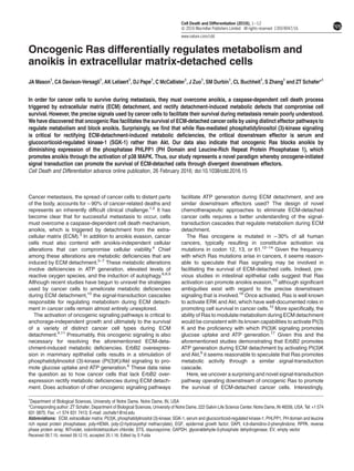
  • 10. SGK-1 and PHLPP1/p38 MAPK is occurring in human colorectal adenocarcinoma. Discussion Given that it is now appreciated that cancer cells must rectify metabolic defects (e.g. ATP generation) in addition to blocking caspase activation (anoikis) to survive during ECM deta- chment,4 it is critically important to understand the precise molecular mechanisms involved in mediating both anoikis and metabolism. The data described here outline a unique, multifaceted signaling pathway operating downstream of oncogenic Ras to permit anoikis inhibition and stimulate ATP production (Figure 8). Our data place Ras signaling in contrast to ErbB2 with regard to metabolic regulation during ECM detachment, as previous studies have shown that ErbB2 promotes ATP generation through PI(3)K-mediated activation of Akt (rather than SGK-1).6 These data suggest that the strategies used by cancer cells to alleviate deleterious meta- bolic changes (and block anoikis) may vary considerably across tumor types and contexts.11,29,30 Additional studies examining the relationship between oncogenic signaling and metabolism in ECM-detached cancer cells are warranted to better ascertain the best strategies to facilitate their elimination. The identification of SGK-1 as a primary mediator of ATP generation downstream of oncogenic Ras adds to the growing body of literature, suggesting that molecules other than Akt can function downstream of PI(3)K to modulate distinct cellular functions in cancer cells.18,31 Our studies suggest that ECM- detached cells with oncogenic Ras mutations could be compromised by SGK-1 inhibition. Indeed, the development of SGK-1 antagonists has already commenced in other cancers,32,33 and our results suggest that similar inhibitors may be efficacious in eliminating ECM-detached cells with Ras mutations. Interestingly, our findings are also consistent with data suggesting that SGK-1 activity can mediate the sensitivity of cancer cells to targeted therapies.34 It is noteworthy that other investigators have discovered that SGK-1 can interact with proteins at the mitochondria and can inhibit the induction of necrotic cell death.19,35 Our data implicating SGK-1 in ATP generation may provide additional insight into these connections between SGK-1 and mitochon- drial integrity. Much like SGK-1, interest in understanding PHLPP1 regulation during tumorigenesis has intensified over the past few years. Studies from a number of labs have now implicated loss of PHLPP1 activity as an important facet of cancer cell survival.23,36–38 That being said, despite the fact that PHLPP1 has a predicted Ras-associated domain,39 our study is the first (to our knowledge) to directly link downregulation of PHLPP1 to oncogenic Ras signaling. In addition, the majority of studies aimed at understanding PHLPP1 in cancer cells have focused on PHLPP1’s ability to dephosphorylate Akt. Our data suggest that PHLPP1-mediated anoikis is not reliant on Akt depho- sphorylation, but is instead dependent on activation of the p38 MAPK pathway. Our data provide an important and novel physiological setting by which PHLPP1-mediated activation of p38 MAPK could be important in the context of tumor progression. In fact, our data suggest that the most effective way to eliminate ECM-detached cells with Ras mutations would be by stimulating p38 MAPK (and inducing anoikis) while simulta- neously antagonizing SGK-1 (and blocking ATP generation). Indeed, our analysis of publically available reverse phase protein array (RPPA) data from colorectal cancer patients (Figure 7g) demonstrates a correlation between mutant K-Ras levels, activation of SGK-1, and inactivation of p38 MAPK. However, while these data are consistent with the notion that simultaneous activation of p38 MAPK and inhibition of SGK-1 might be an effective therapeutic strategy, future experiments aimed at assessing the efficacy of such a regimen will be critical to the development of novel chemotherapies designed to eradicate ECM-detached cancer cells. Materials and Methods ATP assays. To measure ATP levels in ECM-detached cells, 400 000 cells were plated in 6-well poly-(2-hydroxyethyl methacrylate)- (poly-HEMA; Sigma-Aldrich, St. Louis, MO, USA) coated plates for the amount of time indicated in the figure legends. The ATP Determination Kit (Invitrogen, Carlsbad, CA, USA) was used following normalization of total protein concentration according to the manufacturer’s instructions. In LY294002 (Calbiochem, Billerica, MA, USA) and EMD638683 (ApexBio, Houston, TX, USA) experiments, the indicated concentration of inhibitor was added at the time of plating. All data presented are representative images from at least three independent replicates. Caspase-3/7 assays. Cells were plated at a density of 13 000 cells per well on 96-well poly-HEMA-coated plates. Caspase activation was measured using the CaspaseGlo 3/7 Assay Kit (Promega, Madison, WI, USA) according to the manufacturer’s instructions. In p38 (SB203580; Cell Signaling Technologies, Danvers, MA, USA), SGK-1 kinase (EMD638683), and staurosporine (STS; Sigma-Aldrich) experiments, the indicated concentration of the inhibitor was added at the time of plating. For SB203580 experiments, the inhibitor was added every 24 h (12 h for 10A HrasG12V PHLPP1) throughout the duration of the experiment. All data presented are representative images from at least three independent replicates. Glucose ATP Akt PI(3)K PHLPP1 SGK1 Oncogenic Ras FKBP5 p38 MAPKAPK2 P PP P Non-apoptotic cell death Apoptotic cell death Figure 8 Model for oncogenic Ras-mediated cell survival during ECM detachment Ras-mediated inhibition of death during detachment JA Mason et al 10 Cell Death and Differentiation
  • 11. Cell culture. MCF-10A cells (ATCC, Manassas, VA, USA) and derivatives were cultured in Dulbecco’s modified Eagle's medium/F12 (Gibco, Waltham, MA, USA) supplemented with 5% horse serum (Invitrogen), 20ng/ml epidermal growth factor (EGF), 10 μg/ml insulin, 500 μg/ml hydrocortisone, 100 ng/ml cholera toxin, and 1% penicillin/streptomycin. HCT116 cells (ATCC) and derivatives were cultured in McCoy’s media (Gibco) plus 10% fetal bovine serum (Invitrogen) and 1% penicillin/ streptomycin. CellTiter Glo assays. Cells were plated at a density of 13 000 cells per well on either a 96-well plate (attached) or a 96-well poly-HEMA-coated plate (detached) for the indicated time. ATP levels were measured using the CellTiter Glo Assay (Promega) according to the manufacturer’s instructions. All data presented are representative images from at least three independent replicates. Cell viability assays. Cells were plated at a density of 13 000 cells per well on 96-well poly-HEMA-coated plates. AlamarBlue reagent (Invitrogen) was added to each well according to the manufacturer’s instructions. EMD638683, LY294002, and SB203580 were added at the indicated concentrations at the time of plating. All data presented are representative images from at least three independent replicates. Glucose-limiting assays. The indicated cells were plated at 400 000 cells per well in 6-well poly-HEMA-coated plates for 24 h. MCF-10A derivatives were plated in standard media (described above) or glucose-free Dulbecco’s modified Eagle's medium/F12 (US Biologicals, Salem, MA, USA) supplemented with 5% horse serum (Invitrogen) 20 ng/ml EGF, 10 μg/ml insulin, 500 μg/ml hydrocortisone, 100 ng/ml cholera toxin, 1% penicillin/streptomycin, and 0.4 mM glucose solution (Gibco). HCT116 derivatives were plated in either standard media (described above) or glucose-free Dulbecco’s modified Eagle's medium (Gibco) supplemented with 10% fetal bovine serum (Invitrogen), 1% penicillin/streptomycin, and 0.4 mM glucose solution (Gibco). Glucose uptake assays. Glucose uptake was measured in ECM-detached cells using the Amplex Red Glucose Assay Kit (Invitrogen) according to the manufacturer’s instructions. Cells were plated at a density of 13 000 cells per well, and baseline glucose measurements were taken from a media-only control plated at the same time as the cells. All data presented are representative images from at least three independent replicates. Immunoblotting. ECM-detached cells were harvested, washed once with cold PBS, and lysed in 1% Nonidet P-40 supplemented with protease inhibitors leupeptin (5 μg/ml), aprotinin (1 μg/ml), and PMSF (1 mM) and the Halt Phosphatase Inhibitor Mixture (Thermo Scientific, Waltham, MA, USA). Lysates were collected after spinning for 30 min at 4 °C at 14 000 r.p.m. and normalized by BCA Assay (Pierce Biotechnology, Waltham, MA, USA). Normalized lysates underwent SDS-PAGE and transfer/blotting was performed as described previously.10 The following antibodies were used for western blotting: IGF-1Rβ (Santa Cruz Biotechnology, Dallas, TX, USA; no. sc-81167), SGK-1 (EMD Millipore, Billerica, MA, USA; no. 07-315), p-Akt (Cell Signaling Technology; no. 4060), glyceraldehyde-3-phosphate dehydrogenase (GAPDH) (Cell Signaling Technologies; no. 5174), Ras (Cell Signaling Technology; no. 3965), p-Sek1 (Abcam, Cambridge, MA, USA; ab39403), DYKDDDDK (FLAG) tag (Cell Signaling Technology; no. 2368), α-tubulin (Cell Signaling Technology; no. 2144), β-tubulin (Cell Signaling Technology; no. 2146), β-actin (Sigma-Aldrich; no. A1978), PHLPP (Bethyl Laboratories, Montgomery, TX, USA; no. A304-029A), p-Erk1/2 (Invitrogen; no. 368800), FKBP5 (Cell Signaling Technology; no. 12210), and p-MAPKAPK2 (Cell Signaling Technology; no. 3007). Immunofluorescence. Cells were plated at a density of 50 000 cells per well in a 6-well plate (attached), or a 6-well poly-HEMA-coated plate (detached) for 24 h. The cells were then harvested, washed two times with cold PBS, and deposited onto slides with a Shandon Cytospin3 (Thermo Scientific) at 800 r.p.m. for 5 min. Cells were then fixed with 4% paraformaldehyde and permeabilized with 0.5% Triton X-100 in PBS. Cells were washed with 100 mM glycine in PBS three times and blocked with immunofluorescence (IF) buffer containing 130 mM NaCl, 7 mM Na2HPO4, 3.5 mM NaH2PO4, 7.7 mM NaH3, 0.1% BSA (Millipore), 1.2% Triton X-100, and 0.5% Tween-20, supplemented with 10% goat serum (Invitrogen). Slides were stained with SGK-1 (EMD Millipore, no. 07-315) at a concentration of 1 : 200 in IF buffer. For secondary visualization, AlexaFluor 568 (Invitrogen; no. A11031) was used at 1 : 200 in IF buffer. Nuclei were stained with 5 μg/ml 4,6-diamidino-2- phenylindone (DAPI; Invitrogen) and mounted with ProLong Gold Antifade Reagent (Life Technologies, Carlsbad, CA, USA). Imaging was completed using the Applied Precision DeltaVision OMX fluorescent microscope (Applied Precision; GE Healthcare, Issaquah, WA, USA). Images shown are representative images at × 60 magnification. Retroviral generation of stable cell lines. The pBABE-Puro-based retroviral vectors encoding constitutively active H-Ras, constitutively active K-Ras, and PHLPP1, the pBABE-Neo-based retroviral vectors encoding constitutive active H-Ras and constitutive active K-Ras, and the pLPCX-Puro-based retroviral vectors encoding constitutive active SGK-1 and kinase-dead SGK-1 were used to generate stable cell lines. HEK293T cells were transfected with 0.75 μg target DNA along with the packaging vector pCLAmpho (0.75 μg) with Lipofectamine 2000 (Life Technologies). Virus was collected at 48 and 72 h post-transfection, filtered through a 0.45 μm filter (EMD Millipore), and used for transduction of MCF-10A and HCT116 cells in the presence of 8 μg/ml polybrene. Stable populations of puromycin-resistant cells were obtained using 2 μg/ml puromycin (Invivogen, San Diego, CA, USA). Stable populations of neomycin-based cells were obtained using 300 μg/ml G418 (Nalgene, Waltham, MA, USA). shRNA generation of stable cell lines. MISSION shRNA constructs against SGK-1 (NM_005627; TRCN0000094957, TRCN0000040175) and PHLPP1 (NM_194449; TRCN0000082796) in the puromycin-resistant pLKO.4 vector along with an empty vector control were purchased from Sigma-Aldrich. HEK293T cells were transfected with 0.5 μg target DNA along with the packaging vectors pCMV- D8.9 (0.5 μg) and pCMV-VSV-G (60 ng) using Lipofectamine 2000 and PLUS reagent (Life Technologies). Virus was collected 24 and 48 h post-transfection and filtered through a 0.45 μm filter (EMD Millipore), and used for transduction of MCF-10A and HCT116 cells in the presence of 8 μg/ml polybrene. Stable populations of cells were selected using 2 μg/ml puromycin (Invivogen). Soft agar assays. Cells were plated at densities of 20 000–30 000 cells per well in 1.5 ml of growth media plus 0.4% low-melt agarose (Sigma-Aldrich) and layered onto a 3 ml bed of growth media with 0.5% low-melt agarose. Cells were fed daily with 1 ml of growth media, plus the indicated concentration of z-VAD-fmk when indicated. At the indicated time, growth media was removed and viable colonies were stained using INT-violet (Sigma-Aldrich). Colony number was determined using ImageJ (Bethesda, MD, USA). All data presented are representative images of at least three independent replicates. Statistics. Statistical significance was determined using a two-tailed t-test. Error bars represent S.D. Transient transfection. The indicated cells were transfected with 1.5 μg target DNA for the pcDNA3-based vectors including an empty vector control and PHLPP1 (Addgene, Cambridge, MA, USA; plasmid no. 22404) using Lipofectamine 2000 and PLUS reagent (Life Technologies) according to the manufacturer’s instructions. Cells were plated for soft agar assays and immunoblotting was completed 48 h post- transfection. 3D cell culture. To generate acini, 3000–4000 cells were plated per well in an 8-well chamber slide on a Matrigel (Lot. no. 9044) bed and allowed to form acinar structures as described previously40,41 and according to the protocol at http:// brugge.med.harvard.edu/. Overlay media (Dulbecco’s modified Eagle's Medium/F12 supplemented with 2% horse serum, 5 ng/ml EGF, 10 μg/ml insulin, 500 μg/ml hydrocortisone, 100 ng/ml cholera toxin, 2% Matrigel, and 1% penicillin/ streptomycin) was added at days 0 and 4. Overlay media, consisting of the above without the 2% Matrigel, was added every 4 subsequent days until fixing. At the indicated time, acinar structures were harvested with 4% paraformaldehyde and permeabilized with 0.5% Triton X-100 in PBS. Fixed structures then underwent three glycine (100 mM) washes in IF buffer (described above). Laminin 5 (Millipore; no. MAB88198) primary antibody was used at 1 : 150 in IF buffer plus 10% goat serum. For secondary visualization, AlexaFluor 568 (Invitrogen; no. A11031) was used at 1 : 200 in IF buffer plus 10% goat serum. Nuclei were counterstained with 5 μg/ml DAPI and slides were mounted using ProLong Gold Antifade Reagent (Waltham, MA, USA). Luminal filling was examined using a Nikon A1R-MP microscope (Nikon, Melville, NY, USA) and scored as clear (~0–10% filled), most clear (~10–50% filled), mostly filled (~50–90% filled), or filled (~90–100% filled). All data are representative of at least three independent replicates and images are shown at × 40. Ras-mediated inhibition of death during detachment JA Mason et al 11 Cell Death and Differentiation
  • 12. Analysis of RPPA data. R studio version 3.1.3 was used to mine colorectal adenocarcinoma (TCGA, Provisional) for RPPA clinical data from MD Anderson Cancer Center (Houston, TX, USA). K-Ras mutations and RPPA data for K-Ras, and p27 phosphorylated at threonine 157 (p-p27/KIP1 T157) and p38/MAPK phos- phorylated at threonine 180 (phospho-p38/MAPK T180) data were collected. The data were parsed into a data set that contained patients with highly expressed oncogenic K-Ras (samples with positive values of relative K-Ras levels). These patient data were then used to generate scatter plots in Excel separating K-Ras on the X axis and either p27_pT157 or p38_pT180 on the Yaxis and a best fit line was drawn for visualization of the overall trend of relative protein levels. R studio was used to calculate the Pearson's coefficients (r-values) and P-values based on Fisher's Z-scores (*95% confidence interval, Po0.05 and **99% confidence interval, Po0.001). Conflict of Interest The authors declare no conflict of interest. Acknowledgements. We thank Raju Rayavarapu, Kelsey Weigel, Mark Hawk, Meaghan Boyd, Kimberly Hagel, Veronica Schafer, Patricia Champion, Athanasia Panopoulos, Crislyn D’Souza-Schorey, and the entire Schafer Lab for their discussions, comments, and experimental assistance. We also express gratitude to Chris Counter (Duke University) for the KrasG12V plasmid, Suzanne Conzen (University of Chicago) for the constitutively active and kinase-dead SGK-1 plasmids, Tianyan Gao (University of Kentucky) for the PHLPP1 plasmid, and Alexandra Newton (UC San Diego) for the pcDNA3-PHLPP1 plasmid. This work was supported by a Lee National Denim Day Research Scholar Grant (RSG-14-145-01-CSM) from the American Cancer Society (to ZTS), a research grant from the Phi Beta Psi National Project (to ZTS), and a Pathway to Independence Award (5R00CA158066) from the National Cancer Institute (to SZ). ZTS is also the recipient of a Career Catalyst Research Grant (CCR14302768) from Susan G Komen. 1. Nguyen DX, Massague J. Genetic determinants of cancer metastasis. Nat Rev Genet 2007; 8: 341–352. 2. Li X, Stevens PD, Yang H, Gulhati P, Wang W, Evers BM et al. The deubiquitination enzyme USP46 functions as a tumor suppressor by controlling PHLPP-dependent attenuation of Akt signaling in colon cancer. Oncogene 2013; 32: 471–478. 3. Frisch SM, Francis H. Disruption of epithelial cell–matrix interactions induces apoptosis. J Cell Biol 1994; 124: 619–626. 4. Buchheit CL, Weigel KJ, Schafer ZT. Cancer cell survival during detachment from the ECM: multiple barriers to tumour progression. Nat Rev Cancer 2014; 14: 632–641. 5. Buchheit CL, Rayavarapu RR, Schafer ZT. The regulation of cancer cell death and metabolism by extracellular matrix attachment. Semin Cell Dev Biol 2012; 23: 402–411. 6. Schafer ZT, Grassian AR, Song L, Jiang Z, Gerhart-Hines Z, Irie HY et al. Antioxidant and oncogene rescue of metabolic defects caused by loss of matrix attachment. Nature 2009; 461: 109–113. 7. Davison CA, Schafer ZT. Keeping a breast of recent developments in cancer metabolism. Curr Drug Targets 2010; 11: 1112–1120. 8. Avivar-Valderas A, Salas E, Bobrovnikova-Marjon E, Diehl JA, Nagi C, Debnath J et al. PERK integrates autophagy and oxidative stress responses to promote survival during extracellular matrix detachment. Mol Cell Biol 2011; 31: 3616–3629. 9. Fung C, Lock R, Gao S, Salas E, Debnath J. Induction of autophagy during extracellular matrix detachment promotes cell survival. Mol Biol Cell 2008; 19: 797–806. 10. Davison CA, Durbin SM, Thau MR, Zellmer VR, Chapman SE, Diener J et al. Antioxidant enzymes mediate survival of breast cancer cells deprived of extracellular matrix. Cancer Res 2013; 73: 3704–3715. 11. Buchheit CL, Angarola BL, Steiner A, Weigel KJ, Schafer ZT. Anoikis evasion in inflammatory breast cancer cells is mediated by Bim-EL sequestration. Cell Death Differ 2015; 22: 1275–1286. 12. Downward J. Targeting RAS signalling pathways in cancer therapy. Nat Rev Cancer 2003; 3: 11–22. 13. Prior IA, Lewis PD, Mattos C. A comprehensive survey of Ras mutations in cancer. Cancer Res 2012; 72: 2457–2467. 14. Pylayeva-Gupta Y, Grabocka E, Bar-Sagi D. RAS oncogenes: weaving a tumorigenic web. Nat Rev Cancer 2011; 11: 761–774. 15. McFall A, Ulku A, Lambert QT, Kusa A, Rogers-Graham K, Der CJ. Oncogenic Ras blocks anoikis by activation of a novel effector pathway independent of phosphatidylinositol 3-kinase. Mol Cell Biol 2001; 21: 5488–5499. 16. Eckert LB,RepaskyGA, UlkuAS,McFallA, Zhou H, Sartor CI et al. Involvement of Ras activation in human breast cancer cell signaling, invasion, and anoikis. Cancer Res 2004; 64: 4585–4592. 17. Bryant NJ, Govers R, James DE. Regulated transport of the glucose transporter GLUT4. Nat Rev Mol Cell Biol 2002; 3: 267–277. 18. Bruhn MA, Pearson RB, Hannan RD, Sheppard KE. Second AKT: the rise of SGK in cancer signalling. Growth Factors 2010; 28: 394–408. 19. Brickley DR, Agyeman AS, Kopp RF, Hall BA, Harbeck MC, Belova L et al. Serum- and glucocorticoid-induced protein kinase 1 (SGK1) is regulated by store-operated Ca2+ entry and mediates cytoprotection against necrotic cell death. J Biol Chem 2013; 288: 32708–32719. 20. Park J, Leong ML, Buse P, Maiyar AC, Firestone GL, Hemmings BA. Serum and glucocorticoid-inducible kinase (SGK) is a target of the PI 3-kinase-stimulated signaling pathway. EMBO J 1999; 18: 3024–3033. 21. Kim MJ, Chae JS, Kim KJ, Hwang SG, Yoon KW, Kim EK et al. Negative regulation of SEK1 signaling by serum- and glucocorticoid-inducible protein kinase 1. EMBO J 2007; 26: 3075–3085. 22. Brink M, de Goeij AF, Weijenberg MP, Roemen GM, Lentjes MH, Pachen MM et al. K-ras oncogene mutations in sporadic colorectal cancer in The Netherlands Cohort Study. Carcinogenesis 2003; 24: 703–710. 23. Newton AC, Trotman LC. Turning off AKT: PHLPP as a drug target. Annu Rev Pharmacol Toxicol 2014; 54: 537–558. 24. Gao T, Furnari F, Newton AC. PHLPP: a phosphatase that directly dephosphorylates Akt, promotes apoptosis, and suppresses tumor growth. Mol Cell 2005; 18: 13–24. 25. Qiao M, Wang Y, Xu X, Lu J, Dong Y, Tao W et al. Mst1 is an interacting protein that mediates PHLPPs' induced apoptosis. Mol Cell 2010; 38: 512–523. 26. Suljagic M, Laurenti L, Tarnani M, Alam M, Malek SN, Efremov DG. Reduced expression of the tumor suppressor PHLPP1 enhances the antiapoptotic B-cell receptor signal in chronic lymphocytic leukemia B-cells. Leukemia 2010; 24: 2063–2071. 27. Xu L, Chen S, Bergan RC. MAPKAPK2 and HSP27 are downstream effectors of p38 MAP kinase-mediated matrix metalloproteinase type 2 activation and cell invasion in human prostate cancer. Oncogene 2006; 25: 2987–2998. 28. Hong F, Larrea MD, Doughty C, Kwiatkowski DJ, Squillace R, Slingerland JM. mTOR-raptor binds and activates SGK1 to regulate p27 phosphorylation. Mol Cell 2008; 30: 701–711. 29. Rayavarapu RR, Heiden B, Pagani N, Shaw MM, Shuff S, Zhang S et al. The role of multicellular aggregation in the survival of ErbB2-positive breast cancer cells during extracellular matrix detachment. J Biol Chem 2015; 290: 8722–8733. 30. Weigel KJ, Jakimenko A, Conti BA, Chapman SE, Kaliney WJ, Leevy WM et al. CAF- secreted IGFBPs regulate breast cancer cell anoikis. Mol Cancer Res 2014; 12: 855–866. 31. Gasser JA, Inuzuka H, Lau AW, Wei W, Beroukhim R, Toker A. SGK3 mediates INPP4B- dependent PI3K signaling in breast cancer. Mol Cell 2014; 56: 595–607. 32. Sherk AB, Frigo DE, Schnackenberg CG, Bray JD, Laping NJ, Trizna W et al. Development of a small-molecule serum- and glucocorticoid-regulated kinase-1 antagonist and its evaluation as a prostate cancer therapeutic. Cancer Res 2008; 68: 7475–7483. 33. Skor MN, Wonder EL, Kocherginsky M, Goyal A, Hall BA, Cai Y et al. Glucocorticoid receptor antagonism as a novel therapy for triple-negative breast cancer. Clin Cancer Res 2013; 19: 6163–6172. 34. Sommer EM, Dry H, Cross D, Guichard S, Davies BR, Alessi DR. Elevated SGK1 predicts resistance of breast cancer cells to Akt inhibitors. Biochem J 2013; 452: 499–508. 35. O'Keeffe BA, Cilia S, Maiyar AC, Vaysberg M, Firestone GL. The serum- and glucocorticoid- induced protein kinase-1 (Sgk-1) mitochondria connection: identification of the IF-1 inhibitor of the F(1)F(0)-ATPase as a mitochondria-specific binding target and the stress-induced mitochondrial localization of endogenous Sgk-1. Biochimie 2013; 95: 1258–1265. 36. Wang P, Zhou Z, Hu A, Ponte de Albuquerque C, Zhou Y, Hong L et al. Both decreased and increased SRPK1 levels promote cancer by interfering with PHLPP-mediated dephosphorylation of Akt. Mol Cell 2014; 54: 378–391. 37. Wen YA, Stevens PD, Gasser ML, Andrei R, Gao T. Downregulation of PHLPP expression contributes to hypoxia-induced resistance to chemotherapy in colon cancer cells. Mol Cell Biol 2013; 33: 4594–4605. 38. Cai J, Fang L, Huang Y, Li R, Yuan J, Yang Y et al. miR-205 targets PTEN and PHLPP2 to augment AKT signaling and drive malignant phenotypes in non-small cell lung cancer. Cancer Res 2013; 73: 5402–5415. 39. Warfel NA, Newton AC. Pleckstrin homology domain leucine-rich repeat protein phosphatase (PHLPP): a new player in cell signaling. J Biol Chem 2012; 287: 3610–3616. 40. Mailleux AA, Overholtzer M, Schmelzle T, Bouillet P, Strasser A, Brugge JS. BIM regulates apoptosis during mammary ductal morphogenesis, and its absence reveals alternative cell death mechanisms. Dev Cell 2007; 12: 221–234. 41. Debnath J, Muthuswamy SK, Brugge JS. Morphogenesis and oncogenesis of MCF-10A mammary epithelial acini grown in three-dimensional basement membrane cultures. Methods 2003; 30: 256–268. Supplementary Information accompanies this paper on Cell Death and Differentiation website (http://www.nature.com/cdd) Ras-mediated inhibition of death during detachment JA Mason et al 12 Cell Death and Differentiation