
Recommended
More Related Content
Similar to Cancer cell signaling and its mechanisms.pptx
Similar to Cancer cell signaling and its mechanisms.pptx (20)
Recently uploaded
Recently uploaded (20)
Cancer cell signaling and its mechanisms.pptx
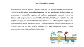
- 1. Notch signaling pathway is highly conserved molecular cell signaling pathway that regulates a vital role in proliferation, stem cell maintenance, cell fate specification, differentiation, and homeostasis of multicellular organism and implicates angiogenesis. Mammals possess four different notch receptors, referred to as NOTCH1, NOTCH2, NOTCH3, and NOTCH4. The notch receptor is a single-pass transmembrane receptor protein. It is a hetero-oligomer composed of a large extracellular portion, which associates in a calcium-dependent, non-covalent interaction with a smaller piece of the notch protein composed of a short extracellular region, a single transmembrane-pass, and a small intracellular region. Notch Signaling Pathway
- 2. Notch signaling is initiated by ligand binding to Notch receptor, which undergoes a two-step proteolytic cleavage by ADAM family proteases and γ-secretase, releasing the Notch intracellular domain (NICD). The NICD translocates to the nucleus where it binds to CSL and converts the complex from a repressor to an activator of Notch target genes. Notch absence: Transcription repressor Notch present: Transcription activator
- 3. Crosstalk signaling pathway Crosstalk refers to instances in which one or more components of one signal transduction pathway affects another. In these signal transduction pathways, there are often shared components that can interact with either pathway. For example, YAP/TAZ and Notch interplay that impacts on the balance between stem cells’ self-renewal versus differentiation, cell fate decisions, inflammation, morphogenesis, and large-scale gene oscillations.
- 4. Figure: Yap/Taz forms a critical positive feedback loop with Notch signaling, to promote liver enlargement and tumorigenesis (Red). Breaking this positive feedback loop leads to reduced hepatomegaly and tumor progression. The inhibitory role of Wnt/β-catenin in the liver tumor caused by the vicious positive feedback, is at least in part through the DP1 (Dimerization Partner of E2F transcription factor)- mediated inhibition of Notch signaling (Blue).
- 5. Notch Inhibitors Notch signaling could be targeted by inhibiting the signaling pathway by two major classes of notch inhibitors that primarily focuses on the clinical development of promising agents that either obstruct Notch receptor cleavages such as γ-secretase inhibitors (GSIs) or interfere with the Notch ligand-receptor interaction by monoclonal antibodies (mAbs). Combining notch inhibitors with current cancer therapies can be effective method for treatment strategies that can give the most promising results.
- 6. γ-secretase is a large protease complex and is composed of catalytic and accessory subunits. The activation of Notch signaling pathway mainly depends on the γ-secretase enzyme activity that helps in the proteolytic cleavage of the receptors that release the active intracellular fragment which is one of the most crucial steps. Thus, GSIs is a promising target for Notch inhibition. GSI were the first class of inhibitors that reached clinical development in oncology. There are more than 100 GSIs synthesized and they can be divided into three classes: • Peptide isosteres • Azepines • Sulfonamides GSI
- 7. Name Target Type of study MK-0752 (Merck and Co, Whitehouse Station, NJ, USA) Metastatic or locally advanced breast cancer Phase I PF03084014 (Pfizer, Groton, CT, USA) Advanced solid tumors Phase I RO-4929097 (Roche, Nutley, NJ, USA) Advanced or metastatic breast cancer or recurrent triple negative breast cancer Phase II Clinical trials employing γ-secretase inhibitors in the treatment of breast cancer:
- 8. Monoclonal antibodies (mAb) These are mAbs tested in clinical trials, an example being MAb604.107, which recognize specific ligand (DLL-4) or receptors (Notch1–3) and they either prevent ligand/ receptor interaction or the conformational change within the extracellular domain which is required to expose the TACE cleavage site. Delta like ligand 4 (DLL-4) is an important component of NOTCH pathway that controls the proper growth, stem cell renewal and development.
- 9. AKT signaling pathway (The survival pathway) AKT is a serine/threonine kinase, also known as protein kinase B (PKB). Akt signaling pathway has roles in- • Cell cycle progression • Regulation of glucose metabolism (for the generation of new biomass and facilitate nutrient signaling) • Protein synthesis • Promoting cell survival by blocking apoptosis
- 10. AKT has three isoforms: • AKT1, AKT2, AKT3 Structure: Consists of three domains: 1. N-terminal domain: PH domain (Pleckstrin homology-domain) consisting of 100 amino acid, which interacts with PIP3 (phosphatidylinositol triphosphate) and PIP2 (phosphatidylinositol bisphosphate). 2. Central domain: kinase domain having a regulatory threonine residue, Thr308. 3. C-terminal domain: consists of 40 amino acids having a regulatory serine residue, Ser473. Figure: Isoforms of AKT
- 11. Activation of AKT signaling Protein synthesis autophagy Glycogen synthesis PIP2
- 12. • AS160: Negative regulator of GLUT 4 translocation • Rheb: Activator of mTORC1 pathway • TSC 1/2: Inhibitor of mTORC1 pathway • FOXO: Inhibitor of cell survival and proliferation • GS (Glycogen synthase): Helps glycogenesis • GSK (Glycogen synthase kinase): Phosphorylates glycogen synthase (GS) to inactivate it In a nutshell, an activated AKT signaling pathway- Increases glucose uptake and utilization by cells Enhances glycogen synthesis Increases fatty acid synthesis Increases protein synthesis Upregulates cell survival and proliferation Downregulates autophagy
- 13. Inactivation of AKT signaling Glycogen synthesis autophagy Protein synthesis
- 14. Crosstalk between signaling pathways
- 15. AKT in Cancer AKT1 gene amplification has been reported in gastric carcinoma, glioblastomas, and gliosarcomas. AKT2 gene amplification has been identified in head and neck squamous cell carcinoma, pancreatic, ovarian, and breast cancers. AKT3 expression in androgen resistant prostate cancer cell, estrogen receptor deficient breast cancer cells and in primary ovarian cells. • Mutations leading to the amplification of genes in the receptor-PI3K pathway, resulting in enhanced PI3K signaling. • PTEN activity can be impaired by various mechanisms like, somatic mutations, homozygous deletion, epigenetic silencing through gene promoter methylation or post transcriptional modifications. • The AKT activated mTOR signaling pathway negatively regulates autophagy.
- 16. Overexpression of AKT is linked to resistance to chemotherapeutic agents such as cisplatin, methotrexate or paclitaxel. • Cisplatin-induced DNA damage causes the phosphorylation of BAD via AKT, suppressing its (BAD) apoptotic effect. • However, MK-2206, an AKT inhibitor, has shown to improve the effectiveness of cisplatin in gastric cancer cell lines and ovarian cancer cell lines.
- 17. AKT Inhibitors 1. ATP competitive agents: GSK690693, afuresertib, uprosertib, AZD5363, ipatasertib etc. Resistance of ATP competitive inhibitors: ATP competitive inhibitor A-443654 induce “paradoxical” AKT hyper- phosphorylation. Targeting AKT kinase to the cell membrane markedly reduce sensitivity of phosphorylated AKT to dephosphorylation by protein phosphatase 2A (PP2A). This effect was amplified by occupancy of the ATP binding pocket by either ATP or ATP-competitive inhibitors. Thus, occupancy of the nucleotide binding pocket of AKT kinases enables intramolecular interactions that restrict phosphatase access and sustain AKT phosphorylation. 2. Allosteric inhibitors: Allosteric modulators offer distinct advantages compared to orthosteric ligands that target to active sites, such as greater specificity, reduced side-effects and lower toxicity. e.g. MK-2206, Triciribine, BAY 1125976 etc.
- 18. Class Description ATP-competitive inhibitors Orthosteric inhibitors targeting the ATP-binding pocket of the protein kinase B (Akt) Isoquinoline-5-sulfonamides H-8, H-89, NL-71-101 Azepane derivatives Structures derived from (−)-balanol Aminofurazans GSK690693 Heterocyclic rings 7-azaindole, 6-phenylpurine derivatives, 3- aminopyrrolidine, AZD5363, ipatasertib, A-674563, A- 443654 Phenylpyrazole derivatives AT7867, AT13148 Thiophenecarboxamide derivatives Afuresertib, uprosertib Allosteric inhibitors Superior to orthosteric inhibitors providing greater specificity, reduced side-effects and less toxicity 2,3-diphenylquinoxaline analogues MK-2206 Alkylphospholipids Edelfosine, ilmofosine, miltefosine, perifosine, erufosine AKT- inhibiting drugs listed into major classes:
- 19. Class (cont.) Description Indole-3-carbinol analogues Indole-3-carbinol, 3-chloroacetylindole, SR13668, OSU-A9 Sulfonamide derivatives PH-316, PHT-427 Thiourea derivatives PIT-1, PIT-2, DM-PIT-1 Purine derivatives Triciribine, triciribine mono-phosphate active analogue (TCN-P), ARQ 092 Other structures, derivatives BAY 1125976, 3-methyl-xanthine, 3α- and 3β- acetoxy-tirucallic acids, acetoxy-tirucallic acid Irreversible inhibitors Natural products, antibiotics Lactoquinomycin, Frenolicin B, kalafungin, medermycin
- 20. MAPK Pathway • The MAPK/ERK pathway (also known as Ras-Raf-MEK-ERK pathway) is a chain of proteins in the cell that communicates a signal from a receptor on the surface of the cell to the DNA in the nucleus of the cell. • The pathway includes many proteins including MAPK (mitogen-activated protein kinases, originally called ERK, extracellular signal-regulated kinases), which communicate by adding phosphate groups to neighboring protein which acts as an “on” or “off” switch.
- 21. Understanding the MAPK pathway as it relates to oncology • MAPK pathway plays a role in the regulation of gene expression, cellular growth and survival. • Abnormal MAPK signaling may lead to increased or uncontrolled cell proliferation and resistance to apoptosis. • Research into the MAPK pathway has shown it to be important in some cancers such as- lung cancer, colorectal cancer, pancreatic cancer, endometrial cancer etc. • The MAPK pathway includes the signaling molecules Ras, Raf, MEK, and ERK. • Normally extracellular growth factors activate the pathway by binding to receptor tyrosine kinases. This mobilizes a cascade of signaling via the MAPK pathway signaling molecules. • Ultimately, activation of the MAPK pathway leads to the transcription of genes that encode proteins involved in the regulation of essential cellular functions such as cell growth, cell proliferation and cell differentiation.
- 22. Understanding the MAPK pathway as it relates to oncology (Cont.)
- 23. MAPK pathway as tumor suppressor P38 pathways regulate Ras oncogene activity by negative feedback (A) and signaling integration (B). Along the P38 pathway, MKK6, P38α, and MK2/PRAK have been shown to be activated by Ras oncogene through the MEK/ERK pathways and in turn, suppress Ras activity by negative feedback (A). The P38γ expression, on the other hand, is transduced by Ras, that is required for Ras transformation. Phosphorylated P38α is shown to downregulate P38γ protein expression by ubiquitin/proteasome pathways. In a given system, Ras transforming activity will be determined by integrated signaling from Ras-suppressor P38α and the Ras-effector P38γ (B).
- 24. Cross talk with other signaling pathways • MAPK pathway and TGF-β: One of the stimulating factors of MAPK pathway is Ras protein which can antagonize TGF-β induced apoptosis and cell cycle arrest, resulting in cancer cell proliferation. The synergistic relation between these two pathways lead to increased cytokines and growth factors and thereby epithelial to mesenchymal transition (EMT).
- 25. Cross talking with other pathways
- 26. Drugs against MAPK Figure: Imatinib, Nilotinib, Dasatinib, and Sunitinib target and inhibit c-KIT. Selumetinib and Trametinib inhibit MEK activity. Temsirolimus and Everolimus inhibit the mTOR protein. Resistance to Vemurafenib arises from MAPK pathway reactivation by (1) a MEK1C121S mutation, (2) NRASQ61R/K mutations, (3) COT1 overexpression, (4) alternatively spliced variants of BRAFV600E or amplification of the mutant BRAF allele, (5) Overexpression or activation of RTKs (PDGFRβ or IGF1R) that bypasses mutant BRAF and activates ERK via CRAF-MEK or through independent ERK mechanisms by activating the PI3K/AKT pathway.
- 27. Transforming growth factor-β (TGF-β) signaling pathway TGF-β cell signaling pathway has major roles in- Cell proliferation Cell survival Apoptosis TGF-β ligand superfamily includes: Activin Nodal TGF-β Bone morphogenetic proteins etc.
- 28. TGFβ-2 signaling pathway 1. TGF-β ligand binds to TGFβ-2 receptors which are serine/ threonine receptor kinases. 2. TGFβ-2 then phosphorylates serine residue of type-1 receptor and forms hetero-tetrameric complex. 3. SARA (SMAD anchor for receptor activation protein) internalizes the complex and permits binding of R- SMADs (SMAD2, SMAD3) to L45 region of the type 1 receptor. 4. SARA orients R-SMADs in such a way that R-SMADs’ C-terminus faces with type 1 receptor. 5. Type 1 receptor phosphorylates serine residue of R-SMADs. 6. Phosphorylation of R-SMADs causes their dissociation from SARA.
- 29. TGFβ-2 signaling pathway (Cont.) 7. SMAD 2/3 form complex with coSMAD (SMAD 4). 8. The complex then enter into nucleus and binds with transcription factor and starts transcription of DNA.
- 31. How TGF-β is associated with cancer ● TGF-β has dual role in cancer. ● It plays both as tumor suppressor and oncogene. Tumor suppressive role is often lost by mutation in TGFβ or SMAD pathways such as in colorectal cancer, breast cancer, gastrointestinal cancer, pancreatic cancer etc. 1. Inactivating mutations in components of SMAD pathway: SMAD4, a tumor suppressor gene, is deleted or mutated in pancreatic carcinoma. Mutant protein cannot form transcriptionally active DNA binding complex. SMAD2 mutation is found in colon, head, neck, lung carcinoma. In breast cancer, inactivating mutation of SMAD4 is rare and SMAD2 is not found. It is still unknown why inactivating mutations in TGFβR-II and SMAD4, respectively, are uncommon in breast (and other) cancers but play a substantial role in considerable percentages of gastrointestinal and pancreatic cancers (Kretzschmar, 2000).
- 32. TGF-β and its association with cancer 2. Activation of Ras protein: TGF-β can activate Ras–Mapk signaling (Arteaga, 2006). Also, oncogenic Ras gene interfere with the phosphorylation of SMAD3 and results in proliferative function of TGF-β. Mutation of Ras protein result in permanently activated Ras protein which cause SMAD3 phosphorylation and cell proliferation (Kretzschmar, 2000). 3. Alteration in expression of SMAD inhibitor: SMAD 6 &7, which are inhibitors of SMAD signaling pathway and inhibit phosphorylation of SMAD2 and SMAD3, cause tumor cell proliferation in pancreatic carcinoma (Kleff et al., 1999; Kretzschmar, 2000). 4. Stimulation of epithelial mesenchymal transmission (EMT) (metastasis): Increased expression of SMAD3 & 4 induce EMT (Deryck & Zhang, 2014).
- 33. How TGF-β induces Epithelial Mesenchymal Transition 1. TGF-β acts as a common and potent inducer of EMT via Smad-dependent and independent activation of the expression of EMT transcription factors Snail, Slug, ZEB1 and 2, and Twist . The Smad3/4 complex directly binds to the regulatory portion of the promoter of Snail, inducing its transcription. Subsequently a Smad3/4/Snail complex is formed that binds to the regulatory promoter sequences of genes encoding for E-cadherin and occludin, leading to repression of their expression. 2. Smad signaling also increases the expression of ZEB transcription factors, which repress miR-200 family expression, further increasing ZEB protein levels and EMT. 3. Moreover, TGF-β can activate EMT transcription factor expression via alternative splicing.
- 34. 4. EMT is also controlled by a group of microRNAs that define changes in cytoskeleton reorganization and epithelial polarity, and it is directly activated in response to TGF-β via the Smad/RhoA pathway. 5. TGF-β also induces EMT through ubiquitylation and sumoylation. Smad3/4 complex regulates the expression of HDM2, increasing the ubiquitylation and degradation of p53, inducing EMT progression. TGF-β signaling downregulates the expression of the SUMO E3 ligase.
- 35. How TGF-β polymorphism is related to resistance to chemotherapy? • Inactivating mutations in components of the TGF-β/SMAD signaling pathway • Reduced expression of TGF-β/SMAD signaling components • Inhibition of the TGF-β/SMAD pathway by Ras/MAP-kinase signaling • Altered expression of TGF-β/SMAD inhibitory molecules • Interference with TGF-β/SMAD responses downstream of SMAD proteins
- 36. ● Anticancer drugs can become resistant to apoptosis due to anti-apoptotic state induced by TGFβ. Such as oxaliplatin. ● TGFβ can also prevent chemotherapy induced apoptosis by inhibiting tumor suppressor gene p53 directly and indirectly. ● In case of paclitaxel, it may become resistant due to increased expression of TGFβ signaling component.
- 37. Cross talk with other pathways TGF-β and MAPK Pathways: One of the stimulating factors of MAPK pathway is, Ras protein can antagonize TGF beta induced apoptosis and cell cycle arrest, resulting in cancer cell proliferation. The synergistic relation between TGF-β and RAS/ MAPK pathways lead to cytokines and growth factors and thereby epithelial to mesenchymal transition. TGF-β and PI3/AKT pathway: PI3/AKT reduce the TGF-β induced cell cycle arrest and apoptosis. SMAD3 is inhibited by PI3/AKT (Guo et al., 2009).
- 38. Deregulation of major components of mTOR pathway leading to chemoresistance Molecular events Resistant to Tumor/cell types Activation of PI3K pathway, either via loss of the tumor suppressor PTEN or through amplification of the PI3K encoding PIK3CA gene Trastuzumab Breast cancer PI3K overexpression and PTEN reduction Cisplatin Ovarian cancer PI3K regulates MDR1 expression Vincristine Leukemia, prostate cancer Activation of PI3K and AKT Endocrine therapy Breast cancer
- 39. Deregulation of major components of mTOR pathway leading to chemoresistance (Cont.) Molecular events Resistance to Tumor/cell types Activation of PI3K and AKT Imatinib Gastrointestinal stromal tumor Constitutive AKT activation Tamoxifen Breast cancer Constitutive AKT activation TRAIL Leukemia, prostate cancer AKT inhibits p53 phosphorylation Cisplatin Ovarian cancer
- 40. Molecular events Resistance to Tumor/cell types Overexpression of mTOR and p70S6K1 TRAIL Glioblastoma Defect in the regulation of 4EBP1 and 4EBP2 Retinoic acid NB4 cell mTOR activation Vincristine FL5.12 cells Raf activation Doxorubicin and paclitaxel Breast cancer MEK/ERK regulates MDR1 expression Doxorubicin and paclitaxel Colorectal cancer Deregulation of major components of mTOR pathway leading to chemoresistance (Cont.)
- 41. Classification of mTOR inhibitors
- 42. The Wnt/β-catenin pathway The name Wnt is a portmanteau of “int” and “Wg” and stands for "Wingless-related integration site". Wnts are secreted factors that regulate cell growth, motility, and differentiation during embryonic development. β-Catenin is an extremely important effector in the Wnt signaling pathway.
- 43. When Wnt proteins are present, the so- called Wnt-on state, the Wnt ligands bind to FZD and LRP5/6 on the cell membrane. The binding of Wnt to FZD exposes an intracellular binding site for DVL in FZD. A destruction complex exists in the cytoplasm, consisting mainly of AXIN, the tumor suppressor gene APC, GSK3β and CK1α. In Wnt- on state, the DVL protein inhibits the destruction complex. Subsequently, GSK3β and CK1α can not phosphorylate β-catenin. This results in functional β-catenin accumulating in the cytoplasm and entering the nucleus with the assistance of relevant molecules. Thereafter, β-catenin regulates the transcription of transcriptional regulators such as TCF/LEF and genes that are ultimately targeted by Wnt. The Wnt-on State
- 44. Phosphorylation of β-catenin exposes a binding site for the E3 ubiquitin ligase β-TrCP, and β-catenin is thus ubiquitinated and degraded, as such, it is unable to enter the nucleus to initiate downstream gene transcription. In the absence of Wnt proteins, the so-called Wnt-off state, AXIN acts as a scaffolding protein that binds β-catenin, and AXIN also binds GSK3β, CK1α, and APC. CK1α and GSK3β can sequentially phosphorylate β- catenin, and APC then ensures that phosphorylated β-catenin is not dephosphorylated by PP2A and later phosphorylation of β-catenin. The Wnt-off State
- 45. Crosstalk involving the Wnt/β-catenin pathway Evidence of crosstalk: The Notch and Wnt signal transduction pathways can regulate each other and hence affect their transcriptional output. Notch and Wnt signaling pathways act together during wing development in Drosophila. In mouse, the Wnt pathway regulate the expression of the Notch ligand: Delta-like ligand1 (DLL1). • The Notch target gene Hes1 is also regulated by β-catenin-mediated Wnt signaling. • There is direct interaction between β-catenin and Notch-1. • Studies have shown Notch/RBP-J/β-catenin interaction at several target genes and synergism of them in angiogenesis.
- 46. Influence of Notch on β-catenin Notch tethers β-catenin Negative regulation • Notch upregulates the mRNA levels of the canonical Wnt-transcription factor TCF1. • Frizzled receptor is activated by Notch signaling in dendritic cell. • The Notch target gene Nrarp acts as both positive & negative regulator on Wnt.
- 47. Influence of β-catenin on NOTCH Wnt/Ca2+ pathway mediated activation of Frizzled Release of Ca2+ Activation of CaMKII Activation of the β- catenin-pathways Phosphorylation of the RBP-J- interacting co- repressor SMRT Increased promoter activity of a Notch receptor