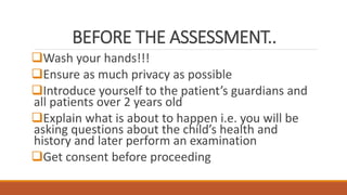
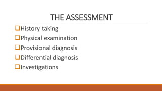
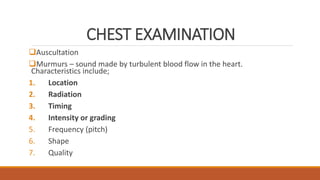
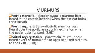
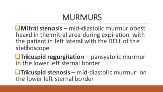
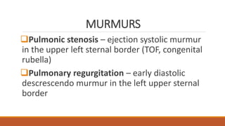
CARDIOVASCULAR
ASSESSMENT
OCTOBER 2021
BEFORE THE ASSESSMENT..
Wash your hands!!!
Ensure as much privacy as possible
Introduce yourself to the patient’s guardians and
all patients over 2 years old
Explain what is about to happen i.e. you will be
asking questions about the child’s health and
history and later perform an examination
Get consent before proceeding
THE ASSESSMENT
History taking
Physical examination
Provisional diagnosis
Differential diagnosis
Investigations
HISTORY TAKING
DEMOGRAPHICS
PRESENTING COMPLAINT
HISTORY OF PRESENTING COMPLAINT
REVIEW OF SYSTEMS
PAST MEDICAL OR SURGICAL HISTORY
HISTORY TAKING
DRUG/ALLERGIES HISTORY
BIRTH HISTORY
NEONATAL HISTORY
DEVELOPMENTAL HISTORY
IMMUNISATION HISTORY
HISTORY TAKING
NUTRITIONAL HISTORY
FAMILY HISTORY
SOCIAL HISTORY
DEMOGRAPHICS
Name of patient
Age of patient
Sex of patient
Address
Informant - adolescents should be encouraged to
provide their own history
Religion
PRESENTING COMPLAINTS
Cardiovascular symptom s??
PRESENTING COMPLAINTS
Cardiovascular symptoms;
1. Cough – productive vs non-productive
2. Shortness of breath or difficulty breathing
(dyspnea) – at rest? On exertion?
3. Orthopnea – difficulty breathing when lying flat
4. Paroxysmal nocturnal dyspnea (PND) – waking
up from sleep to catch a breath
5. Cyanosis – bluish discoloration of the skin
PRESENTING COMPLAINTS
Cardiovascular symptoms;
1. Edema – pedal/sacral/generalized; isolated facial edema
unlikely cardiac, rule out renal or allergic reactions
2. Chest pain – SOCRATES rule
3. Palpitations – mothers may report feeling the child’s heart
beating quickly
4. Intermittent claudication – muscle pain usually in calves
worse on exertion, better with rest ddx PVD
5. Syncope
6. Dizziness
HISTORY OF PRESENTING COMPLAINT
How long has the patient been unwell? Acute
vs chronic?
What was the patients previous state of
health? i.e.. Was healthy then became ill or has
been chronically ill then got worse
HISTORY OF PRESENTING COMPLAINT
Obtain more detail on presenting complaint;
SOB – rest? Exertion? Orthopnea? PND? breastfeeding with
diaphoresis? (infants in CCF), squatting? (TOF)
Edema – generalized? Location? Unilateral vs bilateral pedal
edema?
Cyanosis – at rest? When crying? Breastfeeding? From birth?
Central? Peripheral?
Chest pain – SOCRATES
Cough – productive/non productive, sputum details??
Dizziness – postural or exertional, associated syncope?
CHEST PAIN - DDx
Angina – substernal, crushing, radiates down the left arm or
to the jaw, associated with vomiting, diaphoresis
Aortic dissection – severe central or epigastric pain radiating
to the back, feels like tearing
Pleuritic pain with SOB i.e. lateral chest pain worse with
breathing – pneumonia, pneumothorax, pulmonary
embolism, ACS in SCD
Exertional pain – MI, cardiomyopathy
Pericarditis – sharp retrosternal pain, worse when lying
down, radiates to the left shoulder and associated with fever
HISTORY OF PRESENTING COMPLAINT
Associated symptoms :
Patients presenting with only edema and SOB should
be evaluated for
1. Renal disease i.e. urine frequency, color and quantity
2. Malnutrition – feeding habits
3. Allergic reactions; history of insect bites, food
ingestion, drug use prior to onset of edema
4. Liver disease – yellowing of eyes/skin? Itchy skin?
Pale stools? Easy bruising/bleeding?
HISTORY OF PRESENTING COMPLAINT
Associated symptoms :
Systemic symptoms e.g.
1. Fever – with chest pain in pericarditis or
endocarditis
2. Easy fatiguability - CCF
3. Weight gain – CCF
4. Poor appetite – CCF in infants
REVIEW OF SYSTEMS
*Systemic – fever, fatigue, weight changes
*Respiratory – most overlap with CVS e.g. cough,
SOB, noisy breathing, chest pain
Gastrointestinal –dyspepsia. Nausea, vomiting,
dysphagia, abdominal pain (GERD vs ANGINA/CAD)
Genitourinary – oliguria, polyuria, hematuria (CCF
vs renal failure/nephrotic syndrome), hematuria in
infective endocarditis??
REVIEW OF SYSTEMS
Neurological – visual changes, headaches
(syncope –neuro vs cardiac cause)
Musculoskeletal – chest pain, trauma
history?? Joint pain? (rheumatic fever)
Dermatological – rashes? Ulcers? (Rheumatic
fever, vasculitidies e.g. Kawasaki disease)
PAST MEDICAL/SURGICAL HISTORY
First presentation or has had similar
symptoms before?
First admission or known patient? Records
available?
Other hospital admissions?
Frequent clinic visits?
History of heart disease?
PAST MEDICAL/SURGICAL HISTORY
HIV? TB? –increased risk for pericarditis
Asthma? Wheezing due to cardiac asthma?
Epilepsy? Syncope vs atonic seizure??
Hypertension – renal disease, thyroid
disease(palpitations too) COA
Sickle cell disease – DCM, CCF from severe
anaemia
Previous cardiac surgeries
DRUG HISTORY
Prescribed vs OTC?
Use of herbal medications?
Important drugs to note;
1. Beta blockers- propranolol/atenolol (HTN,
hyperthyroidism)
2. Calcium channel blockers e.g. nifedipine (HTN)
3. ACE inhibitors e.g. enalapril in HTN, CCF
4. Diuretics e.g. furosemide, spirono,actone (CCF)
Food and drug allergies – esp for patients with edema
ANTENATAL HISTORY
Maternal drug use e.g. lithium for mood
disorders (Epstein anomaly), sodium valproate
for epilepsy (TOF, septal defects), alcohol (FAS)
Maternal illnesses – rubella (PDA), diabetes
mellitus (VSD, AVSD, TGA), SLE, coxsackie
Maternal age at conception – Downs
syndrome (AVSD, endocardial cushion defects)
BIRTH HISTORY/NEONATAL HISTORY
Term or preterm delivery – PDA >>> preterm
births
Cyanosis at birth/neonatal period
Shortness of breath in the neonatal period
Difficulties breast feeding, diaphoresis when
feeding
Failure to thrive – child not gaining weight despite
adequate breast feeding
DEVELOPMENTAL HISTORY
Failure to thrive – most children with congenital
heart disease have FTT due to either difficulties
with feeding resulting in low energy intake or high
energy requirements
CHD associated with genetic disorders like Downs
syndrome or Edward syndrome may present with
developmental delay i.e. motor, cognitive or
language delays
NUTRITIONAL HISTORY
Establish the cause of FTT or possible
malnutrition as a cause for edema
Exclusive breastfeeding? Age of weaning?
Duration of breast feeding? Other foods part of
the diet?
Difficulty breast feeding? Poor appetite?
FAMILY HISTORY/SOCIAL HISTORY
Other family members with cardiac disease
E.g. Marfan’s syndrome
History of sudden deaths in the family –
suggestive of arrhythmias e.g. long QT syndrome
Education level of patient
Socio-economic status of parents
PHYSICAL EXAMINATION
Vital signs
Arthropometric measurements
LOOK before you TOUCH!!! – inspection,
percussion, palpation, auscultation
Hands  face  neck  chest - abdomen
 extremities
VITAL SIGNS
Temperature – fever ?? Think endocarditis,
pericarditis, rheumatic fever, hypothermic??
Assess for shock
Pulse rate/heart rate
Blood pressure – hypotensive (shock?? CCF??)
hypertensive (COA, renal artery stenosis)
Respiratory rate – tachypnea in CCF
ANTHROPOMETRIC MEASUREMENTS
Weight – compare with previous weights, FTT??
Weight gain from edema?? SAM??
Height – stunted?? Likely chronic disease e,g,
sickle cell or congenital heart disease
Weight for height
MUAC
Head circumference – microcephalic in torches
like rubella, think PDA??
LOOK!!!
Did the patient walk in unaided or were they carried?
Alert or lethargic??
Is the patient sitting up in bed? Unable to lie flat?
Using multiple pillows?
In respiratory distress? Signs of respiratory distress??
On oxygen? Cyanosed?
Edematous?
Pallor? Remember : Severe anaemia causes CCF
HANDS
Peripheral cyanosis – cyanotic CHD, Eisenmenger syndrome
Finger clubbing – cyanotic CHD, infective endocarditis
Stigmata of infective endocarditis i.e. Janeway lesions,
Osler’s nodes, splinter hemorrhages
Palmar pallor
Warm or cool peripheries – febrile vs shock
Capillary refill time – peripheral vascular disease vs shock
Arachnodactyly – abnormally long and slender fingers in
Marfan syndrome – risk for MVP or aortic dissection
PERIPHERAL CYANOSIS
FINGER CLUBBING
GRADING FINGER CLUBBING
Grade 1 – nail bed fluctuation
Grade 2 – loss of the Lovibond angle with
obliteration of Schamroths window
Grade 3 – increased convexity of the nail fold with
evident clubbing
Grade 4 – drumstick appearance
Grade 5 – hypertrophic osteoarthropathy (shiny
nail with striae)
JANEWAY LESIONS/OSLER’S NODES
SPLINTER HEMORRHAGES
RADIAL PULSE (wrist)
Rate – tachycardic vs bradycardic, pulse deficit
(>10mmHg A-fib??)
Rhythm – regular, irregular? Irregular irregular?
(atrial fibrillation)
Volume – absent, weak/thready, strong, bounding
Character – collapsing pulse( water hammer) in
aortic regurgitation,
Radial-radial synchrony - RR-delay in COA
BLOOD PRESSURE
Standing, sitting, lying down??
Hypotension – CCF, shock
Hypertension – COA, renal artery stenosis, thyroid disease
Narrow pulse pressure (<25 mmHg) – aortic stenosis, CCF,
cardiac tamponade
Wide pulse pressure (>100 mmHg) – aortic regurgitation,
aortic dissection
Difference in right and left arm BP -> 20 mmHg ??arotic
dissection
FACE/NECK
Eyes – Roth spots in infective endocarditis,
conjunctival pallor, Kayser Fleischer rings
(Wilsons disease causes cardiomyopathy)
Central cyanosis
High arched palate – Marfan syndrome
Dental hygiene – high risk for infective
endocarditis
FACE/NECK
Distended neck veins with increased jugular
venous pressure in CCF, tricuspid regurgitation
or constrictive pericarditis (normal 3-4 cm
above sternal angle)
Carotid pulse – dancing carotid (Corrigan’s
sign) in aortic regurgitation, carotid bruit
(carotid stenosi or radiating murmur from aortic
stenosis)
CENTRAL CYANOSIS
JVP ASSESSMENT
CHEST EXAMINATION
INSPECTION
1. Tachypnea,
2. Chest asymmetry – pectus
excavatum/carinatum,
3. Hyperactive precordium
4. Surgical scars
CHEST EXAMINATION
PALPATION
1. Apex beat (point of maximal impulse) – normal
is 4th to 5th intercostal space midclavicular line
(displaced in DCM laterally or downwards)
2. Thrill – palpable murmur in valve disease
3. Parasternal Heave – palpable precordial impulse
in RVH
CHEST EXAMINATION
Auscultation
1. Heart sounds
First heart sound (S1) – closure of the atrioventricular valves i.e.
mitral and tricuspid
Second heart sound (S2) – closure of the semilunar valves i.e.
aortic and pulmonic valves
Third heart sound (S3) – AKA ventricular gallop due to passive
filling of the left ventricle in CCF
Fourth heart sound (S4) – AKA atrial gallop produced during atrial
systole by the flow of blood into a poorly compliant ventricle
AUSCULTATION
CHEST EXAMINATION
Auscultation
Murmurs – sound made by turbulent blood flow in the heart.
Characteristics include;
1. Location
2. Radiation
3. Timing
4. Intensity or grading
5. Frequency (pitch)
6. Shape
7. Quality
MURMURS
Aortic stenosis – ejection systolic murmur best
heard in the carotid arteries when the patient holds
their breath
Aortic regurgitation – diastolic murmur best
heard over the aortic area during expiration when
the patient sits forward (RHD)
Mitral regurgitation – pansystolic murmur best
heard over the mitral area or apex beat and radiates
to the axilla (RHD)
MURMURS
Mitral stenosis – mid-diastolic murmur obest
heard in the mitral area during expiration with
the patient in left lateral with the BELL of the
stethoscope
Tricuspid regurgitation – pansystolic murmur
in the lower left sternal border
Tricuspid stenosis – mid-diastolic murmur on
the lower left sternal border
MURMURS
Pulmonic stenosis – ejection systolic murmur
in the upper left sternal border (TOF, congenital
rubella)
Pulmonary regurgitation – early diastolic
descrescendo murmur in the left upper sternal
border
GRADING OF MURMURS
Grade 1 – faint murmur, only heard by experts
Grade 2 – soft murmur, heard in all positions with NO THRILL
Grade 3 – moderately loud murmur, NO THRILL
Grade 4 – loud murmur with a palpable thrill
Grade 5 – very loud murmur with thrill, can be heard with
the stethoscope partly off the chest
Grade 6 – very loud murmur with a thrill, heard with
stethoscope completely off the chest
CHEST EXAMINATION
Auscultation
1. Pericardial friction rub – grating or scratching
sound produced by friction of the heart
against the pericardium in pericarditis
2. Basal crepitations – pulmonary edema in CCF
ABDOMINAL EXAM
Inspection – distended abdomen with ascites
in CCF
Palpation – fluid thrill, shifting dullness, tender
hepatomegaly in CCF
Hepatojugular reflux in CCF
EXTREMITIES
Inspection – edema, peda? Ankle? Sacral?
Generalized?
Palpation of peripheral pulses – dorsalis pedis,
anterior tibialis, popliteal, femoral, carotid,
brachial and radial – absent pulses in PVD
Radial-femoral delay in coarctation of the
aorta
CARDIOVASCULAR ASSESSMENT.pptx

CARDIOVASCULAR ASSESSMENT.pptx

  • 1.
  • 2.
    BEFORE THE ASSESSMENT.. Washyour hands!!! Ensure as much privacy as possible Introduce yourself to the patient’s guardians and all patients over 2 years old Explain what is about to happen i.e. you will be asking questions about the child’s health and history and later perform an examination Get consent before proceeding
  • 3.
    THE ASSESSMENT History taking Physicalexamination Provisional diagnosis Differential diagnosis Investigations
  • 4.
    HISTORY TAKING DEMOGRAPHICS PRESENTING COMPLAINT HISTORYOF PRESENTING COMPLAINT REVIEW OF SYSTEMS PAST MEDICAL OR SURGICAL HISTORY
  • 5.
    HISTORY TAKING DRUG/ALLERGIES HISTORY BIRTHHISTORY NEONATAL HISTORY DEVELOPMENTAL HISTORY IMMUNISATION HISTORY
  • 6.
  • 7.
    DEMOGRAPHICS Name of patient Ageof patient Sex of patient Address Informant - adolescents should be encouraged to provide their own history Religion
  • 8.
  • 9.
    PRESENTING COMPLAINTS Cardiovascular symptoms; 1.Cough – productive vs non-productive 2. Shortness of breath or difficulty breathing (dyspnea) – at rest? On exertion? 3. Orthopnea – difficulty breathing when lying flat 4. Paroxysmal nocturnal dyspnea (PND) – waking up from sleep to catch a breath 5. Cyanosis – bluish discoloration of the skin
  • 10.
    PRESENTING COMPLAINTS Cardiovascular symptoms; 1.Edema – pedal/sacral/generalized; isolated facial edema unlikely cardiac, rule out renal or allergic reactions 2. Chest pain – SOCRATES rule 3. Palpitations – mothers may report feeling the child’s heart beating quickly 4. Intermittent claudication – muscle pain usually in calves worse on exertion, better with rest ddx PVD 5. Syncope 6. Dizziness
  • 11.
    HISTORY OF PRESENTINGCOMPLAINT How long has the patient been unwell? Acute vs chronic? What was the patients previous state of health? i.e.. Was healthy then became ill or has been chronically ill then got worse
  • 12.
    HISTORY OF PRESENTINGCOMPLAINT Obtain more detail on presenting complaint; SOB – rest? Exertion? Orthopnea? PND? breastfeeding with diaphoresis? (infants in CCF), squatting? (TOF) Edema – generalized? Location? Unilateral vs bilateral pedal edema? Cyanosis – at rest? When crying? Breastfeeding? From birth? Central? Peripheral? Chest pain – SOCRATES Cough – productive/non productive, sputum details?? Dizziness – postural or exertional, associated syncope?
  • 13.
    CHEST PAIN -DDx Angina – substernal, crushing, radiates down the left arm or to the jaw, associated with vomiting, diaphoresis Aortic dissection – severe central or epigastric pain radiating to the back, feels like tearing Pleuritic pain with SOB i.e. lateral chest pain worse with breathing – pneumonia, pneumothorax, pulmonary embolism, ACS in SCD Exertional pain – MI, cardiomyopathy Pericarditis – sharp retrosternal pain, worse when lying down, radiates to the left shoulder and associated with fever
  • 14.
    HISTORY OF PRESENTINGCOMPLAINT Associated symptoms : Patients presenting with only edema and SOB should be evaluated for 1. Renal disease i.e. urine frequency, color and quantity 2. Malnutrition – feeding habits 3. Allergic reactions; history of insect bites, food ingestion, drug use prior to onset of edema 4. Liver disease – yellowing of eyes/skin? Itchy skin? Pale stools? Easy bruising/bleeding?
  • 15.
    HISTORY OF PRESENTINGCOMPLAINT Associated symptoms : Systemic symptoms e.g. 1. Fever – with chest pain in pericarditis or endocarditis 2. Easy fatiguability - CCF 3. Weight gain – CCF 4. Poor appetite – CCF in infants
  • 16.
    REVIEW OF SYSTEMS *Systemic– fever, fatigue, weight changes *Respiratory – most overlap with CVS e.g. cough, SOB, noisy breathing, chest pain Gastrointestinal –dyspepsia. Nausea, vomiting, dysphagia, abdominal pain (GERD vs ANGINA/CAD) Genitourinary – oliguria, polyuria, hematuria (CCF vs renal failure/nephrotic syndrome), hematuria in infective endocarditis??
  • 17.
    REVIEW OF SYSTEMS Neurological– visual changes, headaches (syncope –neuro vs cardiac cause) Musculoskeletal – chest pain, trauma history?? Joint pain? (rheumatic fever) Dermatological – rashes? Ulcers? (Rheumatic fever, vasculitidies e.g. Kawasaki disease)
  • 18.
    PAST MEDICAL/SURGICAL HISTORY Firstpresentation or has had similar symptoms before? First admission or known patient? Records available? Other hospital admissions? Frequent clinic visits? History of heart disease?
  • 19.
    PAST MEDICAL/SURGICAL HISTORY HIV?TB? –increased risk for pericarditis Asthma? Wheezing due to cardiac asthma? Epilepsy? Syncope vs atonic seizure?? Hypertension – renal disease, thyroid disease(palpitations too) COA Sickle cell disease – DCM, CCF from severe anaemia Previous cardiac surgeries
  • 20.
    DRUG HISTORY Prescribed vsOTC? Use of herbal medications? Important drugs to note; 1. Beta blockers- propranolol/atenolol (HTN, hyperthyroidism) 2. Calcium channel blockers e.g. nifedipine (HTN) 3. ACE inhibitors e.g. enalapril in HTN, CCF 4. Diuretics e.g. furosemide, spirono,actone (CCF) Food and drug allergies – esp for patients with edema
  • 21.
    ANTENATAL HISTORY Maternal druguse e.g. lithium for mood disorders (Epstein anomaly), sodium valproate for epilepsy (TOF, septal defects), alcohol (FAS) Maternal illnesses – rubella (PDA), diabetes mellitus (VSD, AVSD, TGA), SLE, coxsackie Maternal age at conception – Downs syndrome (AVSD, endocardial cushion defects)
  • 22.
    BIRTH HISTORY/NEONATAL HISTORY Termor preterm delivery – PDA >>> preterm births Cyanosis at birth/neonatal period Shortness of breath in the neonatal period Difficulties breast feeding, diaphoresis when feeding Failure to thrive – child not gaining weight despite adequate breast feeding
  • 23.
    DEVELOPMENTAL HISTORY Failure tothrive – most children with congenital heart disease have FTT due to either difficulties with feeding resulting in low energy intake or high energy requirements CHD associated with genetic disorders like Downs syndrome or Edward syndrome may present with developmental delay i.e. motor, cognitive or language delays
  • 24.
    NUTRITIONAL HISTORY Establish thecause of FTT or possible malnutrition as a cause for edema Exclusive breastfeeding? Age of weaning? Duration of breast feeding? Other foods part of the diet? Difficulty breast feeding? Poor appetite?
  • 25.
    FAMILY HISTORY/SOCIAL HISTORY Otherfamily members with cardiac disease E.g. Marfan’s syndrome History of sudden deaths in the family – suggestive of arrhythmias e.g. long QT syndrome Education level of patient Socio-economic status of parents
  • 26.
    PHYSICAL EXAMINATION Vital signs Arthropometricmeasurements LOOK before you TOUCH!!! – inspection, percussion, palpation, auscultation Hands  face  neck  chest - abdomen  extremities
  • 27.
    VITAL SIGNS Temperature –fever ?? Think endocarditis, pericarditis, rheumatic fever, hypothermic?? Assess for shock Pulse rate/heart rate Blood pressure – hypotensive (shock?? CCF??) hypertensive (COA, renal artery stenosis) Respiratory rate – tachypnea in CCF
  • 28.
    ANTHROPOMETRIC MEASUREMENTS Weight –compare with previous weights, FTT?? Weight gain from edema?? SAM?? Height – stunted?? Likely chronic disease e,g, sickle cell or congenital heart disease Weight for height MUAC Head circumference – microcephalic in torches like rubella, think PDA??
  • 29.
    LOOK!!! Did the patientwalk in unaided or were they carried? Alert or lethargic?? Is the patient sitting up in bed? Unable to lie flat? Using multiple pillows? In respiratory distress? Signs of respiratory distress?? On oxygen? Cyanosed? Edematous? Pallor? Remember : Severe anaemia causes CCF
  • 30.
    HANDS Peripheral cyanosis –cyanotic CHD, Eisenmenger syndrome Finger clubbing – cyanotic CHD, infective endocarditis Stigmata of infective endocarditis i.e. Janeway lesions, Osler’s nodes, splinter hemorrhages Palmar pallor Warm or cool peripheries – febrile vs shock Capillary refill time – peripheral vascular disease vs shock Arachnodactyly – abnormally long and slender fingers in Marfan syndrome – risk for MVP or aortic dissection
  • 31.
  • 32.
  • 33.
    GRADING FINGER CLUBBING Grade1 – nail bed fluctuation Grade 2 – loss of the Lovibond angle with obliteration of Schamroths window Grade 3 – increased convexity of the nail fold with evident clubbing Grade 4 – drumstick appearance Grade 5 – hypertrophic osteoarthropathy (shiny nail with striae)
  • 34.
  • 35.
  • 36.
    RADIAL PULSE (wrist) Rate– tachycardic vs bradycardic, pulse deficit (>10mmHg A-fib??) Rhythm – regular, irregular? Irregular irregular? (atrial fibrillation) Volume – absent, weak/thready, strong, bounding Character – collapsing pulse( water hammer) in aortic regurgitation, Radial-radial synchrony - RR-delay in COA
  • 37.
    BLOOD PRESSURE Standing, sitting,lying down?? Hypotension – CCF, shock Hypertension – COA, renal artery stenosis, thyroid disease Narrow pulse pressure (<25 mmHg) – aortic stenosis, CCF, cardiac tamponade Wide pulse pressure (>100 mmHg) – aortic regurgitation, aortic dissection Difference in right and left arm BP -> 20 mmHg ??arotic dissection
  • 38.
    FACE/NECK Eyes – Rothspots in infective endocarditis, conjunctival pallor, Kayser Fleischer rings (Wilsons disease causes cardiomyopathy) Central cyanosis High arched palate – Marfan syndrome Dental hygiene – high risk for infective endocarditis
  • 39.
    FACE/NECK Distended neck veinswith increased jugular venous pressure in CCF, tricuspid regurgitation or constrictive pericarditis (normal 3-4 cm above sternal angle) Carotid pulse – dancing carotid (Corrigan’s sign) in aortic regurgitation, carotid bruit (carotid stenosi or radiating murmur from aortic stenosis)
  • 40.
  • 41.
  • 42.
    CHEST EXAMINATION INSPECTION 1. Tachypnea, 2.Chest asymmetry – pectus excavatum/carinatum, 3. Hyperactive precordium 4. Surgical scars
  • 43.
    CHEST EXAMINATION PALPATION 1. Apexbeat (point of maximal impulse) – normal is 4th to 5th intercostal space midclavicular line (displaced in DCM laterally or downwards) 2. Thrill – palpable murmur in valve disease 3. Parasternal Heave – palpable precordial impulse in RVH
  • 44.
    CHEST EXAMINATION Auscultation 1. Heartsounds First heart sound (S1) – closure of the atrioventricular valves i.e. mitral and tricuspid Second heart sound (S2) – closure of the semilunar valves i.e. aortic and pulmonic valves Third heart sound (S3) – AKA ventricular gallop due to passive filling of the left ventricle in CCF Fourth heart sound (S4) – AKA atrial gallop produced during atrial systole by the flow of blood into a poorly compliant ventricle
  • 45.
  • 46.
    CHEST EXAMINATION Auscultation Murmurs –sound made by turbulent blood flow in the heart. Characteristics include; 1. Location 2. Radiation 3. Timing 4. Intensity or grading 5. Frequency (pitch) 6. Shape 7. Quality
  • 47.
    MURMURS Aortic stenosis –ejection systolic murmur best heard in the carotid arteries when the patient holds their breath Aortic regurgitation – diastolic murmur best heard over the aortic area during expiration when the patient sits forward (RHD) Mitral regurgitation – pansystolic murmur best heard over the mitral area or apex beat and radiates to the axilla (RHD)
  • 48.
    MURMURS Mitral stenosis –mid-diastolic murmur obest heard in the mitral area during expiration with the patient in left lateral with the BELL of the stethoscope Tricuspid regurgitation – pansystolic murmur in the lower left sternal border Tricuspid stenosis – mid-diastolic murmur on the lower left sternal border
  • 49.
    MURMURS Pulmonic stenosis –ejection systolic murmur in the upper left sternal border (TOF, congenital rubella) Pulmonary regurgitation – early diastolic descrescendo murmur in the left upper sternal border
  • 50.
    GRADING OF MURMURS Grade1 – faint murmur, only heard by experts Grade 2 – soft murmur, heard in all positions with NO THRILL Grade 3 – moderately loud murmur, NO THRILL Grade 4 – loud murmur with a palpable thrill Grade 5 – very loud murmur with thrill, can be heard with the stethoscope partly off the chest Grade 6 – very loud murmur with a thrill, heard with stethoscope completely off the chest
  • 51.
    CHEST EXAMINATION Auscultation 1. Pericardialfriction rub – grating or scratching sound produced by friction of the heart against the pericardium in pericarditis 2. Basal crepitations – pulmonary edema in CCF
  • 52.
    ABDOMINAL EXAM Inspection –distended abdomen with ascites in CCF Palpation – fluid thrill, shifting dullness, tender hepatomegaly in CCF Hepatojugular reflux in CCF
  • 53.
    EXTREMITIES Inspection – edema,peda? Ankle? Sacral? Generalized? Palpation of peripheral pulses – dorsalis pedis, anterior tibialis, popliteal, femoral, carotid, brachial and radial – absent pulses in PVD Radial-femoral delay in coarctation of the aorta