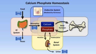
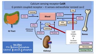
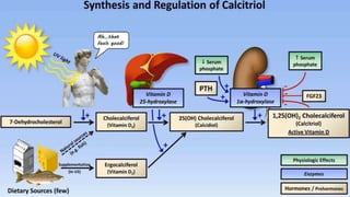
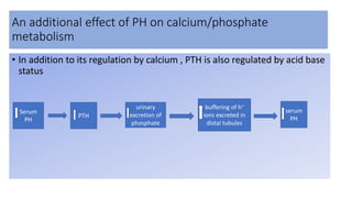
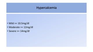
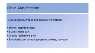
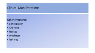
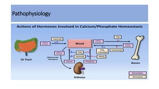
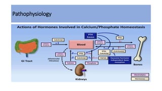
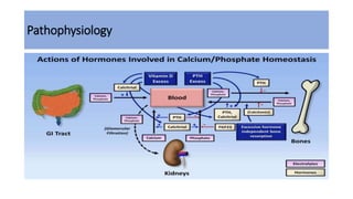
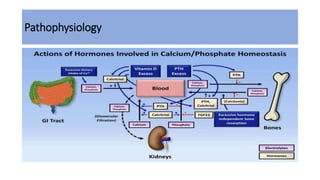
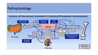
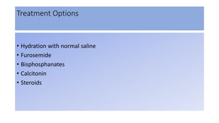
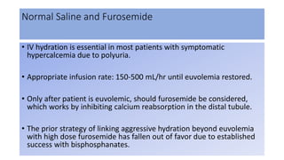
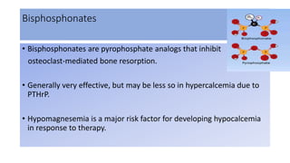
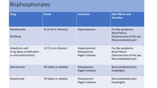
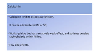
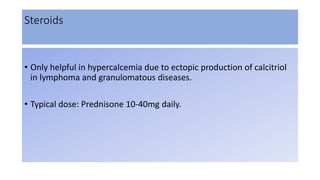
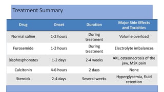
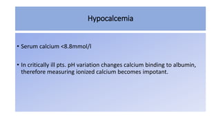
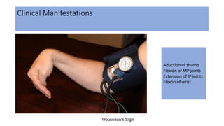
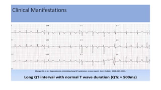
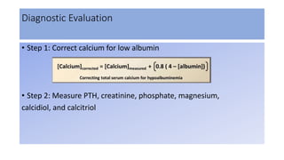
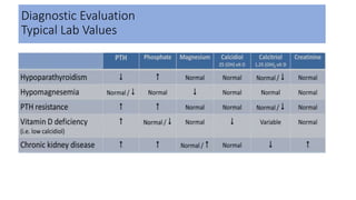
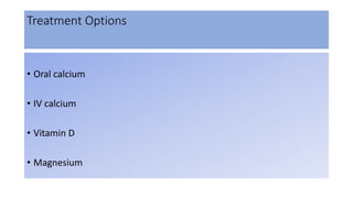
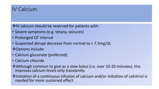
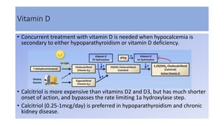
HYPER & HYPOCALCEMIA
IN ICU
PRESENTER
MAYANK SACHAN
Functions
• Skeletal mineralization
• Signal transduction
• Nerve conduction
• Muscle contraction
• Blood coagulation
Normal serum levels
• Total calcium
 2.2– 2.6 mmol/l
4.4 – 5.2 mEq/l
8.8 – 10.4 mg/dl
• Ionised levels (half of total)
1.1 - 1.3 mmol/l
Different Forms Of Calcium
• Most of the calcium in the body is stored in the bones as
hydroxyapatite.
• Calcium in the plasma:
 45% free ionized form physiological active
45% bound to protein (predominantly albumin)
10% complexed with anions( eg. citrate, sulfate, phosphate)
Typically
measured
in routine
blood
tests.
For
hypoalbunimnemia
Ca10(PO4)6(OH)2
1000
mg
800
mg
200
mg
200
mg
Calcium sensing receptor CaSR
G protein coupled receptor – it senses extracellular ionized ca+2
CaSR
Inhibits
PTH
&
Increase
calciuresis
Role of Magnesium
• Acute hypomagnesemia – increases PTH secretion
• But profound hypomagnesemia –
decreases PTH secretion
Increases PTH resistance in bone – leading to hypocalcemia
An additional effect of PH on calcium/phosphate
metabolism
• In addition to its regulation by calcium , PTH is also regulated by acid base
status
Serum
PH
PTH
urinary
excretion of
phosphate
buffering of h+
ions excreted in
distal tubules
serum
PH
Hypercalcemia
• Mild >= 10.5mg/dl
• Moderate >= 12mg/dl
• Severe >= 14mg/dl
Clinical Manifestations
“Stones, bones, groans and psychiatric overtones”
• Stones- Nephrolithiasis
• BONES- Bone pain
• Groans- abdominal pain
• Psychiatric overtones- Depression, anxiety, confusion
Clinical Manifestations
Other symptoms:
• Constipation
• Anorexia
• Nausea
• Weakness
• lethargy
Clinical Manifestations
Pathophysiology
Pathophysiology
Pathophysiology
Pathophysiology
Pathophysiology
Pathophysiology
Etiologies By Mechanism
Diagnostic Evaluation
• Step 1: Correct calcium for low albumin
• Step 2: Perform thorough physical exam and obtain chest X-ray
• Step 3: Measure PTH
Diagnostic Algorithm
Measure PTH
Measure 24 hr
urinary fractional
calcium excretion
1°
Hyperparathyroidism
Familial hypocalciuric
hypercalcemia
Measure PTHrP,
calcidiol, and
calcitriol
Malignancy
Excessive dietary
intake of calcium or
vitamin D
Ectopic production of
calcitriol
Bone mets,
Consider rare
causes
High or high-normal Low or low-normal
High Low
PTHrP elevated Elevated calcidiol
Elevated calcitriol
with normal calicdiol
None of 3 are
elevated
Treatment Options
• Hydration with normal saline
• Furosemide
• Bisphosphanates
• Calcitonin
• Steroids
Normal Saline and Furosemide
• IV hydration is essential in most patients with symptomatic
hypercalcemia due to polyuria.
• Appropriate infusion rate: 150-500 mL/hr until euvolemia restored.
• Only after patient is euvolemic, should furosemide be considered,
which works by inhibiting calcium reabsorption in the distal tubule.
• The prior strategy of linking aggressive hydration beyond euvolemia
with high dose furosemide has fallen out of favor due to established
success with bisphosphanates.
Bisphosphonates
• Bisphosphonates are pyrophosphate analogs that inhibit
osteoclast-mediated bone resorption.
• Generally very effective, but may be less so in hypercalcemia due to
PTHrP.
• Hypomagnesemia is a major risk factor for developing hypocalcemia
in response to therapy.
Bisphosphonates
Drug Route Indication Side Effects and
Toxicities
Pamidronate
60-90mg
IV (2-24 hr infusion) Hypercalcemia Flu-like symptoms
Renal failure
Osteonecrosis of the jaw
Musculoskelatal pain
Zoledronic acid
4 mg (dose modification
in renal dysfunction)
IV (15 min infusion) Hypercalcemia
Osteoporosis
Paget's disease
Flu-like symptoms
Renal failure
Osteonecrosis of the jaw
Musculoskelatal pain
Alendronate PO (daily or weekly) Osteoporosis
Paget's disease
Musculoskelatal pain
Esophagitis
Risedronate PO (daily or weekly) Osteoporosis
Paget's disease
Musculoskelatal pain
Esophagitis
Calcitonin
• Calcitonin inhibits osteoclast function.
• It can be administered IM or SQ.
• Works quickly, but has a relatively weak effect, and patients develop
tachyphylaxis within 48 hrs.
• Few side effects.
Steroids
• Only helpful in hypercalcemia due to ectopic production of calcitriol
in lymphoma and granulomatous diseases.
• Typical dose: Prednisone 10-40mg daily.
Treatment Summary
Hypocalcemia
• Serum calcium <8.8mmol/l
• In critically ill pts. pH variation changes calcium binding to albumin,
therefore measuring ionized calcium becomes impotant.
Clinical Manifestations
• The primary physical manifestations of hypocalcemia are various
forms of tetany.
• Tetany - Repetitive discharge of peripheral nerves after a single
stimulus.
• Chvostek’s sign – facial spasm by tapping on ipsilateral facial nerve
anterior to ear
• Trousseau’s sign – carpopedal spasm by inflation of BP cuff above
systolic BP for 3 min
Clinical Manifestations
Trousseau's Sign
Aduction of thumb
Flexion of MP joints
Extension of IP joints
Flexon of wrist
Clinical Manifestations
Other symptoms include:
• Seizures
• Hypotension
• Emotional lability
• Psychosis
Clinical Manifestations
Vitamin D
Deficiency
PTH
Deficiency
Etiologies By Mechanism
Function of 1a hydroxylase
Chronic kidney disease
Hyperphosphatemia
Vit. D dependent rickets, type 1
Poor Dietary
Intake
Calcitriol
Resistance Vit. D
resistant rickets
Chronic Kidney Disease and Secondary Hyperparathyroidism
Chronic kidney disease-
mineral and bone disorder
(CKD-MBD)
Renal osteodystrophy
Extraosseous calcifications
Diagnostic Evaluation
• Step 1: Correct calcium for low albumin
• Step 2: Measure PTH, creatinine, phosphate, magnesium,
calcidiol, and calcitriol
Diagnostic Evaluation
Typical Lab Values
Treatment Options
• Oral calcium
• IV calcium
• Vitamin D
• Magnesium
IV Calcium
IV calcium should be reserved for patients with:
• Severe symptoms (e.g. tetany, seizures)
• Prolonged QT interval
• Suspected abrupt decrease from normal to < 7.5mg/dL
Options include:
• Calcium gluconate (preferred)
• Calcium chloride
Although common to give as a slow bolus (i.e. over 10-20 minutes), this
improves calcium levels only transiently.
Initiation of a continuous infusion of calcium and/or initiation of calcitriol is
needed for more sustained effect.
Vitamin D
• Concurrent treatment with vitamin D is needed when hypocalcemia is
secondary to either hypoparathyroidism or vitamin D deficiency.
• Calcitriol is more expensive than vitamins D2 and D3, but has much shorter
onset of action, and bypasses the rate limiting 1a hydroxylase step.
• Calcitriol (0.25-1mcg/day) is preferred in hypoparathyroidism and chronic
kidney disease.
Magnesium
• In patients in whom hypomagnesemia is contributing to the
hypocalcemia, a sustained increase in serum calcium will be difficult
without concurrent repletion of magnesium.
• Standard treatment for this is IV magnesium sulfate 1-2gm at a time,
with frequent monitoring of magnesium levels, particularly in patients
with impaired renal function.
CALCIUM REGULATION.pptx

CALCIUM REGULATION.pptx

  • 1.
    HYPER & HYPOCALCEMIA INICU PRESENTER MAYANK SACHAN
  • 2.
    Functions • Skeletal mineralization •Signal transduction • Nerve conduction • Muscle contraction • Blood coagulation
  • 3.
    Normal serum levels •Total calcium  2.2– 2.6 mmol/l 4.4 – 5.2 mEq/l 8.8 – 10.4 mg/dl • Ionised levels (half of total) 1.1 - 1.3 mmol/l
  • 4.
    Different Forms OfCalcium • Most of the calcium in the body is stored in the bones as hydroxyapatite. • Calcium in the plasma:  45% free ionized form physiological active 45% bound to protein (predominantly albumin) 10% complexed with anions( eg. citrate, sulfate, phosphate) Typically measured in routine blood tests. For hypoalbunimnemia Ca10(PO4)6(OH)2
  • 5.
  • 6.
    Calcium sensing receptorCaSR G protein coupled receptor – it senses extracellular ionized ca+2 CaSR Inhibits PTH & Increase calciuresis
  • 8.
    Role of Magnesium •Acute hypomagnesemia – increases PTH secretion • But profound hypomagnesemia – decreases PTH secretion Increases PTH resistance in bone – leading to hypocalcemia
  • 9.
    An additional effectof PH on calcium/phosphate metabolism • In addition to its regulation by calcium , PTH is also regulated by acid base status Serum PH PTH urinary excretion of phosphate buffering of h+ ions excreted in distal tubules serum PH
  • 10.
    Hypercalcemia • Mild >=10.5mg/dl • Moderate >= 12mg/dl • Severe >= 14mg/dl
  • 11.
    Clinical Manifestations “Stones, bones,groans and psychiatric overtones” • Stones- Nephrolithiasis • BONES- Bone pain • Groans- abdominal pain • Psychiatric overtones- Depression, anxiety, confusion
  • 12.
    Clinical Manifestations Other symptoms: •Constipation • Anorexia • Nausea • Weakness • lethargy
  • 13.
  • 14.
  • 15.
  • 16.
  • 17.
  • 18.
  • 19.
  • 20.
  • 21.
  • 23.
    Diagnostic Evaluation • Step1: Correct calcium for low albumin • Step 2: Perform thorough physical exam and obtain chest X-ray • Step 3: Measure PTH
  • 24.
    Diagnostic Algorithm Measure PTH Measure24 hr urinary fractional calcium excretion 1° Hyperparathyroidism Familial hypocalciuric hypercalcemia Measure PTHrP, calcidiol, and calcitriol Malignancy Excessive dietary intake of calcium or vitamin D Ectopic production of calcitriol Bone mets, Consider rare causes High or high-normal Low or low-normal High Low PTHrP elevated Elevated calcidiol Elevated calcitriol with normal calicdiol None of 3 are elevated
  • 25.
    Treatment Options • Hydrationwith normal saline • Furosemide • Bisphosphanates • Calcitonin • Steroids
  • 26.
    Normal Saline andFurosemide • IV hydration is essential in most patients with symptomatic hypercalcemia due to polyuria. • Appropriate infusion rate: 150-500 mL/hr until euvolemia restored. • Only after patient is euvolemic, should furosemide be considered, which works by inhibiting calcium reabsorption in the distal tubule. • The prior strategy of linking aggressive hydration beyond euvolemia with high dose furosemide has fallen out of favor due to established success with bisphosphanates.
  • 27.
    Bisphosphonates • Bisphosphonates arepyrophosphate analogs that inhibit osteoclast-mediated bone resorption. • Generally very effective, but may be less so in hypercalcemia due to PTHrP. • Hypomagnesemia is a major risk factor for developing hypocalcemia in response to therapy.
  • 28.
    Bisphosphonates Drug Route IndicationSide Effects and Toxicities Pamidronate 60-90mg IV (2-24 hr infusion) Hypercalcemia Flu-like symptoms Renal failure Osteonecrosis of the jaw Musculoskelatal pain Zoledronic acid 4 mg (dose modification in renal dysfunction) IV (15 min infusion) Hypercalcemia Osteoporosis Paget's disease Flu-like symptoms Renal failure Osteonecrosis of the jaw Musculoskelatal pain Alendronate PO (daily or weekly) Osteoporosis Paget's disease Musculoskelatal pain Esophagitis Risedronate PO (daily or weekly) Osteoporosis Paget's disease Musculoskelatal pain Esophagitis
  • 29.
    Calcitonin • Calcitonin inhibitsosteoclast function. • It can be administered IM or SQ. • Works quickly, but has a relatively weak effect, and patients develop tachyphylaxis within 48 hrs. • Few side effects.
  • 30.
    Steroids • Only helpfulin hypercalcemia due to ectopic production of calcitriol in lymphoma and granulomatous diseases. • Typical dose: Prednisone 10-40mg daily.
  • 31.
  • 32.
    Hypocalcemia • Serum calcium<8.8mmol/l • In critically ill pts. pH variation changes calcium binding to albumin, therefore measuring ionized calcium becomes impotant.
  • 33.
    Clinical Manifestations • Theprimary physical manifestations of hypocalcemia are various forms of tetany. • Tetany - Repetitive discharge of peripheral nerves after a single stimulus. • Chvostek’s sign – facial spasm by tapping on ipsilateral facial nerve anterior to ear • Trousseau’s sign – carpopedal spasm by inflation of BP cuff above systolic BP for 3 min
  • 34.
    Clinical Manifestations Trousseau's Sign Aductionof thumb Flexion of MP joints Extension of IP joints Flexon of wrist
  • 35.
    Clinical Manifestations Other symptomsinclude: • Seizures • Hypotension • Emotional lability • Psychosis
  • 36.
  • 37.
  • 38.
  • 39.
    Function of 1ahydroxylase Chronic kidney disease Hyperphosphatemia Vit. D dependent rickets, type 1 Poor Dietary Intake Calcitriol Resistance Vit. D resistant rickets
  • 40.
    Chronic Kidney Diseaseand Secondary Hyperparathyroidism Chronic kidney disease- mineral and bone disorder (CKD-MBD) Renal osteodystrophy Extraosseous calcifications
  • 41.
    Diagnostic Evaluation • Step1: Correct calcium for low albumin • Step 2: Measure PTH, creatinine, phosphate, magnesium, calcidiol, and calcitriol
  • 42.
  • 43.
    Treatment Options • Oralcalcium • IV calcium • Vitamin D • Magnesium
  • 44.
    IV Calcium IV calciumshould be reserved for patients with: • Severe symptoms (e.g. tetany, seizures) • Prolonged QT interval • Suspected abrupt decrease from normal to < 7.5mg/dL Options include: • Calcium gluconate (preferred) • Calcium chloride Although common to give as a slow bolus (i.e. over 10-20 minutes), this improves calcium levels only transiently. Initiation of a continuous infusion of calcium and/or initiation of calcitriol is needed for more sustained effect.
  • 45.
    Vitamin D • Concurrenttreatment with vitamin D is needed when hypocalcemia is secondary to either hypoparathyroidism or vitamin D deficiency. • Calcitriol is more expensive than vitamins D2 and D3, but has much shorter onset of action, and bypasses the rate limiting 1a hydroxylase step. • Calcitriol (0.25-1mcg/day) is preferred in hypoparathyroidism and chronic kidney disease.
  • 46.
    Magnesium • In patientsin whom hypomagnesemia is contributing to the hypocalcemia, a sustained increase in serum calcium will be difficult without concurrent repletion of magnesium. • Standard treatment for this is IV magnesium sulfate 1-2gm at a time, with frequent monitoring of magnesium levels, particularly in patients with impaired renal function.