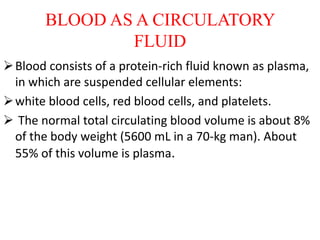
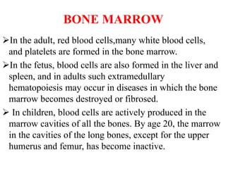
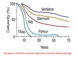
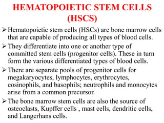
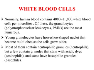
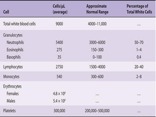
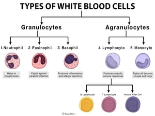
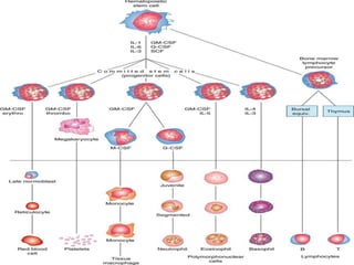
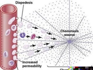
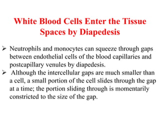
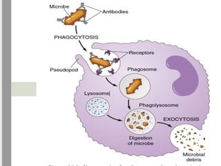
BLOOD
BLOOD AS A CIRCULATORY
FLUID
Blood consists of a protein-rich fluid known as plasma,
in which are suspended cellular elements:
white blood cells, red blood cells, and platelets.
 The normal total circulating blood volume is about 8%
of the body weight (5600 mL in a 70-kg man). About
55% of this volume is plasma.
BONE MARROW
In the adult, red blood cells,many white blood cells,
and platelets are formed in the bone marrow.
In the fetus, blood cells are also formed in the liver and
spleen, and in adults such extramedullary
hematopoiesis may occur in diseases in which the bone
marrow becomes destroyed or fibrosed.
 In children, blood cells are actively produced in the
marrow cavities of all the bones. By age 20, the marrow
in the cavities of the long bones, except for the upper
humerus and femur, has become inactive.
Changes in red bone marrow cellularity in various bones with age.
HEMATOPOIETIC STEM CELLS
(HSCS)
Hematopoietic stem cells (HSCs) are bone marrow cells
that are capable of producing all types of blood cells.
They differentiate into one or another type of
committed stem cells (progenitor cells). These in turn
form the various differentiated types of blood cells.
There are separate pools of progenitor cells for
megakaryocytes, lymphocytes, erythrocytes,
eosinophils, and basophils; neutrophils and monocytes
arise from a common precursor.
The bone marrow stem cells are also the source of
osteoclasts, Kupffer cells , mast cells, dendritic cells,
and Langerhans cells.
The HSCs are derived from uncommitted,
totipotent stem cells that can be stimulated to
form any cell in the body. Adults have a few of
these, but they are more readily obtained from the
blastocysts of embryos.
WHITE BLOOD CELLS
 Normally, human blood contains 4000–11,000 white blood
cells per microliter . Of these, the granulocytes
(polymorphonuclear leukocytes, PMNs) are the most
numerous.
 Young granulocytes have horseshoe-shaped nuclei that
become multilobed as the cells grow older.
 Most of them contain neutrophilic granules (neutrophils),
but a few contain granules that stain with acidic dyes
(eosinophils), and some have basophilic granules
(basophils).
The other two cell types found normally in
peripheral blood are lymphocytes, which have
large round nuclei and scanty cytoplasm, and
monocytes, which have abundant agranular
cytoplasm and kidney-shaped nuclei .
Acting together, these cells provide the body
with the powerful defenses against tumors
and viral, bacterial, and parasitic infections
that were.
The granulocytes and monocytes are formed only
in the bone marrow.
Lymphocytes and plasma cells are produced mainly in
the various lymphogenous tissues— especially the
lymph glands, spleen, thymus, tonsils, and various
pockets of lymphoid tissue elsewhere in the body, such
as in the bone marrow and in Peyer’s patches
underneath the epithelium in the gut wall.
 The WBCs formed in the bone marrow are stored in
the marrow until they are needed in the circulatory
system.
 Normally, about three times as many WBCs are stored
in the marrow as circulate in the entire blood.
 The lymphocytes are mostly stored in the various
lymphoid tissues, except for a small number that are
temporarily being transported in the blood.
 megakaryocytes are also formed in the bone
marrow. These megakaryocytes fragment in the
bone marrow and the small fragments, known as
platelets (or thrombocytes), then pass into the
blood. They are very important in the initiation of
blood clotting.
LIFE SPAN OF WHITE BLOOD
CELLS
 The life of the granulocytes is normally 4 to 8 hours
circulating in the blood and another 4 to 5 days in tissues
where they are needed.
 In times of serious tissue infection, this total life span is
often shortened to only a few hours.
 The monocytes also have a short transit time, 10 to 20 hours
in the blood, before wandering through the capillary
membranes into the tissues.
 Once in the tissues, they swell to much larger sizes to
become tissue macrophages and, in this form, they can live
for months unless destroyed while performing phagocytic
functions.
Lymphocytes enter the circulatory system continually,
along with drainage of lymph from the lymph nodes
and other lymphoid tissue. After a few hours, they pass
out of the blood back into the tissues by diapedesis .
 Then, they re-enter the lymph and return to the blood
again and again; thus, there is continual circulation of
lymphocytes through the body. Lymphocytes have life
spans of weeks or months, depending on the body’s
need for these cells.
The platelets in the blood are replaced about once
every 10 days. In other words, about 30,000 platelets
are formed each day for each microliter of blood.
NEUTROPHILS AND
MACROPHAGES
DEFEND AGAINST INFECTIONS
It is mainly the neutrophils and tissue macrophages
that attack and destroy invading bacteria, viruses,
and other harmful agents.
The neutrophils are mature cells that can attack and
destroy bacteria, even in the circulating blood.
Tissue macrophages begin life as blood
monocytes, which are immature cells while still in
the blood they have little ability to fight
infectious agents at that time.
However, once they enter the tissues, they begin
to swell a size that can barely be seen with the
naked eye.
These cells are now called macrophages, and they
are extremely capable of combating disease
agents in the tissues.
White Blood Cells Enter the Tissue
Spaces by Diapedesis
 Neutrophils and monocytes can squeeze through gaps
between endothelial cells of the blood capillaries and
postcapillary venules by diapedesis.
 Although the intercellular gaps are much smaller than
a cell, a small portion of the cell slides through the gap
at a time; the portion sliding through is momentarily
constricted to the size of the gap.
White Blood Cells Move Through
Tissue Spaces by
Ameboid Motion
 Both neutrophils and macrophages can move
through the tissues by ameboid motion.
 Some cells move at velocities as great as 40 μm/min,
a distance as great as their own length each minute.
White Blood Cells Are Attracted to
Inflamed Tissue
Areas by Chemotaxis
 Many different chemical substances in the tissues cause
both neutrophils and macrophages to move toward the
source of the chemical.
 This phenomenon is known as chemotaxis.
 When a tissue becomes inflamed, at least a dozen different
products that can cause chemotaxis toward the inflamed
area are formed.
They include the following:
1. Some of the bacterial or viral toxins.
2. Degenerative products of the inflamed tissues.
3. Several reaction products of the complement complex
activated in inflamed tissues.
4. Several reaction products caused by plasma clotting in
the inflamed area, as well as other substances.
 Chemotaxis depends on the concentration gradient of
the chemotactic substance.
 The concentration is greatest near the source, which
directs the unidirectional movement of the WBCs.
 Chemotaxis is effective up to 100 micrometers away
from an inflamed tissue.
PHAGOCYTOSIS BY NEUTROPHILS
 On approaching a particle to be phagocytized, the neutrophil
first attaches itself to the particle and then projects
pseudopodia in all directions around the particle.
 The pseudopodia meet one another on the opposite side and
fuse.
 This action creates an enclosed chamber that contains the
phagocytized particle.
 Then, the chamber invaginates to the inside of the
cytoplasmic cavity and breaks away from the outer cell
membrane to form a free-floating phagocytic vesicle (also
called a phagosome) inside the cytoplasm.
 A single neutrophil can usually phagocytize 3 to 20 bacteria
before the neutrophil becomes inactivated and dies.
PHAGOCYTOSIS BY
MACROPHAGES.
Macrophages are the end-stage product of
monocytes that enter the tissues from the blood.
When activated by the immune system, they are
much more powerful phagocytes than neutrophils,
often capable of phagocytizing as many as 100
bacteria.
They also have the ability to engulf much larger
particles, even whole RBCs or, occasionally,
malarial parasites, whereas neutrophils are not
capable of phagocytizing particles much larger
than bacteria.
Neutrophils and Macrophages Can Kill Bacteria. In
addition to the digestion of ingested bacteria in
phagosomes, neutrophils and macrophages contain
bactericidal agents that kill most bacteria, even
when the lysosomal enzymes fail to digest them.
This characteristic is especially important because
some bacteria have protective coats or other factors
that prevent their destruction by digestive enzymes.
 Much of the killing effect results from several
powerful oxidizing agents formed by enzymes in the
membrane of the phagosome or by a special
organelle called the peroxisome.
These oxidizing agents include large quantities of
superoxide (O2−), hydrogen peroxide (H2O2), and
hydroxyl ions (OH−), which are lethal to most
bacteria, even in small quantities.
Also, one of the lysosomal enzymes, myeloperoxidase,
catalyzes the reaction between H2O2 and chloride ions
to form hypochlorite, which is exceedingly
bactericidal.
Some bacteria, notably the tuberculosis bacillus, have
coats that are resistant to lysosomal digestion and also
secrete substances that partially resist the killing
effects of the neutrophils and macrophages.
These bacteria are responsible for many chronic
diseases, such as tuberculosis.
MONOCYTE-MACROPHAGE
CELL SYSTEM
(RETICULOENDOTHELIAL
SYSTEM)
The macrophages mainly as mobile cell capable of
wandering through the tissues.
However, after entering the tissues and becoming
macrophages, another large portion of monocytes
becomes attached to the tissues and remains
attached for months or even years until they are
called on to perform specific local protective
functions.
 They have the same capabilities as the mobile
macrophages to phagocytize large quantities of
bacteria, viruses, necrotic tissue, or other foreign
particles in the tissue.
 In addition, when appropriately stimulated, they
can break away from their attachments and, once
again, become mobile macrophages that respond
to chemotaxis and all the other stimuli related to
the inflammatory process.
 Thus, the body has a widespread monocyte-
macrophage system in virtually all tissue areas.
 The total combination of monocytes, mobile
macrophages, fixed tissue macrophages, and a few
specialized endothelial cells in the bone marrow,
spleen, and lymph nodes is called the
reticuloendothelial system.
 However, all or almost all these cells originate from
monocytic stem cells; therefore, the
reticuloendothelial system is almost synonymous
with the monocyte-macrophage system.
 Because the term reticuloendothelial system is
much better known in medical literature than the
term monocyte-macrophage system.
INFLAMMATION
ROLE OF NEUTROPHILS AND
MACROPHAGES
INFLAMMATION
When tissue injury occurs, whether caused by
bacteria, trauma, chemicals, heat, or any other
phenomenone multiple substances are released
by the injured tissues and cause dramatic
secondary changes in the surrounding uninjured
tissues.
This entire complex of tissue changes is called
inflammation.
Inflammation is characterized by the following:
 1) vasodilation of the local blood vessels, with
consequent increased local blood flow;
2) increased permeability of the capillaries,
allowing leakage of large quantities of fluid into the
interstitial spaces;
3) often, clotting of the fluid in the interstitial
spaces because of increased amounts of fibrinogen
and other proteins leaking from the capillaries;
4) migration of large numbers of granulocytes and
• monocytes into the tissue; and
5) swelling of the tissue cells.
Some of the many tissue products that cause these
reactions are histamine, bradykinin, serotonin,
prostaglandins, several different reaction products
of the complement system reaction products of the
blood clotting system, and multiple substances
called lymphokines that are released by sensitized T
cells.
Several of these substances strongly activate the
macrophage system, and within a few hours, the
macrophages begin to devour the destroyed tissues.
 At times, however, the macrophages may also
further injure the still-living tissue cells.
BLOOD

BLOOD

  • 1.
  • 3.
    BLOOD AS ACIRCULATORY FLUID Blood consists of a protein-rich fluid known as plasma, in which are suspended cellular elements: white blood cells, red blood cells, and platelets.  The normal total circulating blood volume is about 8% of the body weight (5600 mL in a 70-kg man). About 55% of this volume is plasma.
  • 4.
    BONE MARROW In theadult, red blood cells,many white blood cells, and platelets are formed in the bone marrow. In the fetus, blood cells are also formed in the liver and spleen, and in adults such extramedullary hematopoiesis may occur in diseases in which the bone marrow becomes destroyed or fibrosed.  In children, blood cells are actively produced in the marrow cavities of all the bones. By age 20, the marrow in the cavities of the long bones, except for the upper humerus and femur, has become inactive.
  • 5.
    Changes in redbone marrow cellularity in various bones with age.
  • 6.
    HEMATOPOIETIC STEM CELLS (HSCS) Hematopoieticstem cells (HSCs) are bone marrow cells that are capable of producing all types of blood cells. They differentiate into one or another type of committed stem cells (progenitor cells). These in turn form the various differentiated types of blood cells. There are separate pools of progenitor cells for megakaryocytes, lymphocytes, erythrocytes, eosinophils, and basophils; neutrophils and monocytes arise from a common precursor. The bone marrow stem cells are also the source of osteoclasts, Kupffer cells , mast cells, dendritic cells, and Langerhans cells.
  • 7.
    The HSCs arederived from uncommitted, totipotent stem cells that can be stimulated to form any cell in the body. Adults have a few of these, but they are more readily obtained from the blastocysts of embryos.
  • 8.
    WHITE BLOOD CELLS Normally, human blood contains 4000–11,000 white blood cells per microliter . Of these, the granulocytes (polymorphonuclear leukocytes, PMNs) are the most numerous.  Young granulocytes have horseshoe-shaped nuclei that become multilobed as the cells grow older.  Most of them contain neutrophilic granules (neutrophils), but a few contain granules that stain with acidic dyes (eosinophils), and some have basophilic granules (basophils).
  • 9.
    The other twocell types found normally in peripheral blood are lymphocytes, which have large round nuclei and scanty cytoplasm, and monocytes, which have abundant agranular cytoplasm and kidney-shaped nuclei . Acting together, these cells provide the body with the powerful defenses against tumors and viral, bacterial, and parasitic infections that were.
  • 13.
    The granulocytes andmonocytes are formed only in the bone marrow. Lymphocytes and plasma cells are produced mainly in the various lymphogenous tissues— especially the lymph glands, spleen, thymus, tonsils, and various pockets of lymphoid tissue elsewhere in the body, such as in the bone marrow and in Peyer’s patches underneath the epithelium in the gut wall.  The WBCs formed in the bone marrow are stored in the marrow until they are needed in the circulatory system.  Normally, about three times as many WBCs are stored in the marrow as circulate in the entire blood.
  • 14.
     The lymphocytesare mostly stored in the various lymphoid tissues, except for a small number that are temporarily being transported in the blood.  megakaryocytes are also formed in the bone marrow. These megakaryocytes fragment in the bone marrow and the small fragments, known as platelets (or thrombocytes), then pass into the blood. They are very important in the initiation of blood clotting.
  • 15.
    LIFE SPAN OFWHITE BLOOD CELLS  The life of the granulocytes is normally 4 to 8 hours circulating in the blood and another 4 to 5 days in tissues where they are needed.  In times of serious tissue infection, this total life span is often shortened to only a few hours.  The monocytes also have a short transit time, 10 to 20 hours in the blood, before wandering through the capillary membranes into the tissues.  Once in the tissues, they swell to much larger sizes to become tissue macrophages and, in this form, they can live for months unless destroyed while performing phagocytic functions.
  • 16.
    Lymphocytes enter thecirculatory system continually, along with drainage of lymph from the lymph nodes and other lymphoid tissue. After a few hours, they pass out of the blood back into the tissues by diapedesis .  Then, they re-enter the lymph and return to the blood again and again; thus, there is continual circulation of lymphocytes through the body. Lymphocytes have life spans of weeks or months, depending on the body’s need for these cells. The platelets in the blood are replaced about once every 10 days. In other words, about 30,000 platelets are formed each day for each microliter of blood.
  • 17.
    NEUTROPHILS AND MACROPHAGES DEFEND AGAINSTINFECTIONS It is mainly the neutrophils and tissue macrophages that attack and destroy invading bacteria, viruses, and other harmful agents. The neutrophils are mature cells that can attack and destroy bacteria, even in the circulating blood.
  • 18.
    Tissue macrophages beginlife as blood monocytes, which are immature cells while still in the blood they have little ability to fight infectious agents at that time. However, once they enter the tissues, they begin to swell a size that can barely be seen with the naked eye. These cells are now called macrophages, and they are extremely capable of combating disease agents in the tissues.
  • 20.
    White Blood CellsEnter the Tissue Spaces by Diapedesis  Neutrophils and monocytes can squeeze through gaps between endothelial cells of the blood capillaries and postcapillary venules by diapedesis.  Although the intercellular gaps are much smaller than a cell, a small portion of the cell slides through the gap at a time; the portion sliding through is momentarily constricted to the size of the gap.
  • 21.
    White Blood CellsMove Through Tissue Spaces by Ameboid Motion  Both neutrophils and macrophages can move through the tissues by ameboid motion.  Some cells move at velocities as great as 40 μm/min, a distance as great as their own length each minute.
  • 22.
    White Blood CellsAre Attracted to Inflamed Tissue Areas by Chemotaxis  Many different chemical substances in the tissues cause both neutrophils and macrophages to move toward the source of the chemical.  This phenomenon is known as chemotaxis.  When a tissue becomes inflamed, at least a dozen different products that can cause chemotaxis toward the inflamed area are formed.
  • 23.
    They include thefollowing: 1. Some of the bacterial or viral toxins. 2. Degenerative products of the inflamed tissues. 3. Several reaction products of the complement complex activated in inflamed tissues. 4. Several reaction products caused by plasma clotting in the inflamed area, as well as other substances.  Chemotaxis depends on the concentration gradient of the chemotactic substance.  The concentration is greatest near the source, which directs the unidirectional movement of the WBCs.  Chemotaxis is effective up to 100 micrometers away from an inflamed tissue.
  • 24.
    PHAGOCYTOSIS BY NEUTROPHILS On approaching a particle to be phagocytized, the neutrophil first attaches itself to the particle and then projects pseudopodia in all directions around the particle.  The pseudopodia meet one another on the opposite side and fuse.  This action creates an enclosed chamber that contains the phagocytized particle.  Then, the chamber invaginates to the inside of the cytoplasmic cavity and breaks away from the outer cell membrane to form a free-floating phagocytic vesicle (also called a phagosome) inside the cytoplasm.  A single neutrophil can usually phagocytize 3 to 20 bacteria before the neutrophil becomes inactivated and dies.
  • 26.
    PHAGOCYTOSIS BY MACROPHAGES. Macrophages arethe end-stage product of monocytes that enter the tissues from the blood. When activated by the immune system, they are much more powerful phagocytes than neutrophils, often capable of phagocytizing as many as 100 bacteria. They also have the ability to engulf much larger particles, even whole RBCs or, occasionally, malarial parasites, whereas neutrophils are not capable of phagocytizing particles much larger than bacteria.
  • 27.
    Neutrophils and MacrophagesCan Kill Bacteria. In addition to the digestion of ingested bacteria in phagosomes, neutrophils and macrophages contain bactericidal agents that kill most bacteria, even when the lysosomal enzymes fail to digest them. This characteristic is especially important because some bacteria have protective coats or other factors that prevent their destruction by digestive enzymes.  Much of the killing effect results from several powerful oxidizing agents formed by enzymes in the membrane of the phagosome or by a special organelle called the peroxisome.
  • 28.
    These oxidizing agentsinclude large quantities of superoxide (O2−), hydrogen peroxide (H2O2), and hydroxyl ions (OH−), which are lethal to most bacteria, even in small quantities. Also, one of the lysosomal enzymes, myeloperoxidase, catalyzes the reaction between H2O2 and chloride ions to form hypochlorite, which is exceedingly bactericidal. Some bacteria, notably the tuberculosis bacillus, have coats that are resistant to lysosomal digestion and also secrete substances that partially resist the killing effects of the neutrophils and macrophages. These bacteria are responsible for many chronic diseases, such as tuberculosis.
  • 29.
    MONOCYTE-MACROPHAGE CELL SYSTEM (RETICULOENDOTHELIAL SYSTEM) The macrophagesmainly as mobile cell capable of wandering through the tissues. However, after entering the tissues and becoming macrophages, another large portion of monocytes becomes attached to the tissues and remains attached for months or even years until they are called on to perform specific local protective functions.
  • 30.
     They havethe same capabilities as the mobile macrophages to phagocytize large quantities of bacteria, viruses, necrotic tissue, or other foreign particles in the tissue.  In addition, when appropriately stimulated, they can break away from their attachments and, once again, become mobile macrophages that respond to chemotaxis and all the other stimuli related to the inflammatory process.  Thus, the body has a widespread monocyte- macrophage system in virtually all tissue areas.
  • 31.
     The totalcombination of monocytes, mobile macrophages, fixed tissue macrophages, and a few specialized endothelial cells in the bone marrow, spleen, and lymph nodes is called the reticuloendothelial system.  However, all or almost all these cells originate from monocytic stem cells; therefore, the reticuloendothelial system is almost synonymous with the monocyte-macrophage system.  Because the term reticuloendothelial system is much better known in medical literature than the term monocyte-macrophage system.
  • 33.
  • 34.
    INFLAMMATION When tissue injuryoccurs, whether caused by bacteria, trauma, chemicals, heat, or any other phenomenone multiple substances are released by the injured tissues and cause dramatic secondary changes in the surrounding uninjured tissues. This entire complex of tissue changes is called inflammation.
  • 35.
    Inflammation is characterizedby the following:  1) vasodilation of the local blood vessels, with consequent increased local blood flow; 2) increased permeability of the capillaries, allowing leakage of large quantities of fluid into the interstitial spaces; 3) often, clotting of the fluid in the interstitial spaces because of increased amounts of fibrinogen and other proteins leaking from the capillaries; 4) migration of large numbers of granulocytes and • monocytes into the tissue; and
  • 36.
    5) swelling ofthe tissue cells. Some of the many tissue products that cause these reactions are histamine, bradykinin, serotonin, prostaglandins, several different reaction products of the complement system reaction products of the blood clotting system, and multiple substances called lymphokines that are released by sensitized T cells. Several of these substances strongly activate the macrophage system, and within a few hours, the macrophages begin to devour the destroyed tissues.  At times, however, the macrophages may also further injure the still-living tissue cells.