Biomarkers and mediators of cardiovascular risk
•Download as PPT, PDF•
3 likes•387 views

Anti-ApoA-1 auto-antibodies: biomarkers and mediators of cardiovascular risk - Montecucco geneva 30th october 2014 - Fabrizio Montecucco, MD, PhD Division of Laboratory Medicine, HUG, Switzerland, Division of Cardiology, University of Geneva, Switzerland Email: fabrizio.montecucco@unige.ch

Recommended
Recommended
More Related Content
Similar to Biomarkers and mediators of cardiovascular risk
Similar to Biomarkers and mediators of cardiovascular risk (20)
More from Hopitaux Universitaires de Genève
More from Hopitaux Universitaires de Genève (20)
Biomarkers and mediators of cardiovascular risk
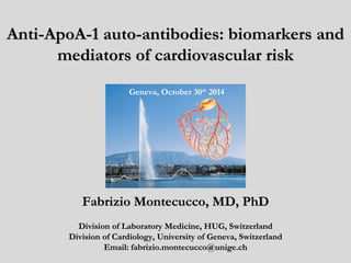
- 1. AAnnttii--AAppooAA--11 aauuttoo--aannttiibbooddiieess:: bbiioommaarrkkeerrss aanndd mmeeddiiaattoorrss ooff ccaarrddiioovvaassccuullaarr rriisskk Geneva, October 30th 2014 FFaabbrriizziioo MMoonntteeccuuccccoo,, MMDD,, PPhhDD DDiivviissiioonn ooff LLaabboorraattoorryy MMeeddiicciinnee,, HHUUGG,, SSwwiittzzeerrllaanndd DDiivviissiioonn ooff CCaarrddiioollooggyy,, UUnniivveerrssiittyy ooff GGeenneevvaa,, SSwwiittzzeerrllaanndd EEmmaaiill:: ffaabbrriizziioo..mmoonntteeccuuccccoo@@uunniiggee..cchh
- 2. 22001144 vviieeww ooff iinnffllaammmmaattiioonn iinn ppllaaqquuee rruuppttuurree aanndd tthhrroommbboossiiss Libby P et al. Circulation Research. 2014;114:1867.
- 3. AAuuttooiimmmmuunniittyy aass aa CCVV rriisskk ccoonnddiittiioonn?? Anti-phospholipid SLE Rheumatoid arthritis syndrome => CV risk increased of 2-3 times Due to which auto-antibodies ? Skaggs BJ ; Nat Rev Reumatol 2012 ; 8:214-23 Edward CJ, et al. Heart 2007; 93:1263-67. Liao Y et al., Int J Cardiol 2006; 21-26
- 4. AAnnttii--AAppooAA--11 IIggGG ppoossiittiivviittyy iiss aassssoocciiaatteedd wwiitthh iinnccrreeaasseedd ccaarrddiioovvaassccuullaarr mmoorrttaalliittyy AMI patients (n=221) RA patients (n=133) Carotid stenosis patients (n=178) Vuilleumier N, et al. Eur Heart J. 2010;31:815. Vuilleumier N, et al. Arthritis Rheum. 2010;62:2640. Vuilleumier N, Montecucco F, et al. Thromb Haemost. 2013;109:706.
- 5. AAnnttii--AAppooAA--11 IIggGG mmiigghhtt aaffffeecctt HHDDLL ffuunnccttiioonn,, bbuutt aallssoo bbiinndd iinnffllaammmmaattoorryy cceellllss vviiaa TToollll--lliikkee rreecceeppttoorr 22 ApoA-1 TLR2 NF-kB nuclear translocation in human macrophages Pagano S, et al. J Intern Med. 2012;272:344. Montecucco F, et al. Unpublished data. ApoE-/- MΦ ApoE-/- TLR2-/- MΦ Anti-ApoA-1 IgG CTL IgG Anti-ApoA-1 IgG CTL IgG
- 6. PPaassssiivvee iimmmmuunniizzaattiioonn wwiitthh aannttii--AAppooAA--11 IIggGG iinn TTLLRR22--//-- AAppooEE--//-- aanndd TTLLRR44--//-- AAppooEE--//-- mmiiccee 0 16 TLR2-/-ApoE-/- TLR4-/-ApoE-/- ApoE -/- mice (aged 11 weeks) Normal diet -Isotype IgG (50 microg every two weeks) -Anti-ApoA1 IgG (50 microg every two weeks) weeks sacrifice I.v. injection Montecucco F, et al. Unpublished data
- 7. PPaassssiivvee iimmmmuunniizzaattiioonn wwiitthh aannttii--AAppooAA--11 IIggGG p=0.04 (LogRank) Follow-up (days) Cumulative survival proportion (%) ApoE -/- mouse survival IgG CTL (n=33) anti-ApoA-1 IgG (n=39) 0 20 40 60 80 100 120 100 80 60 40 20 0 iinnccrreeaassee mmoouussee mmoorrttaalliittyy Montecucco F, et al. Unpublished data
- 8. AAnnttii--AAppooAA--11 IIggGG--iinndduucceedd mmoorrttaalliittyy iiss aabbrrooggaatteedd bbyy TTLLRR22 aanndd TTLLRR44 ddeeffiicciieenncciieess TLR2-/-ApoE-/- (n=19) 0 20 40 60 80 100 120 Follow-up (days) Cumulative survival proportion (%) Survival after treatment with anti-ApoA-1 IgG TLR4-/-ApoE-/- (n=14) ApoE-/-(n=39) 100 80 60 40 20 0 TLR4-/-ApoE-/- vs. ApoE-/- p=0.17 (LogRank) TLR2-/-ApoE-/- vs. ApoE-/- p=0.26 (LogRank) Montecucco F, et al. Unpublished data
- 9. AAnnttii--AAppooAA--11 IIggGG--iinndduucceedd iinnttrraappllaaqquuee eeffffeeccttss aarree aabbrrooggaatteedd bbyy TTLLRR22 aanndd TTLLRR44 ddeeffiicciieenncciieess TLR4-/-ApoE-/- CTL IgG anti-ApoA-1 IgG TLR2-/-ApoE-/- CTL IgG anti-ApoA-1 IgG CTL IgG anti-ApoA-1 IgG CTL IgG anti-ApoA-1 IgG p=0.144 p=0.005 p=0.763 p=0.094 p=0.129 p=0.508 p=0.501 ApoE-/- TLR2-/-ApoE-/- TLR4-/-ApoE-/- ApoE-/- p=0.412 p<0.0010. p=0.001 p=0.043 p=0.001 p=0.581 p=0.329 ApoE-/- TLR2-/-ApoE-/- TLR4-/-ApoE-/- TLR4-/-ApoE-/- CTL IgG anti-ApoA-1 IgG TLR2-/-ApoE-/- CTL IgG anti-ApoA-1 IgG ApoE-/- CTL IgG anti-ApoA-1 IgG
- 10. TTeelleemmeettrryy ddeevviiccee iimmppllaannttaattiioonn iinn AAppooEE--//-- mmiiccee 0 16 ApoE -/- mice (aged 11 weeks) Normal diet -Isotype IgG (50 microg every two weeks) -Anti-ApoA1 IgG (50 microg every two weeks) weeks sacrifice I.v. injection 12 TELEMETRY Montecucco F, et al. Unpublished data
- 11. AAnnttii--AAppooAA--11 IIggGG iinndduuccee SSTT sseeggmmeenntt ddeepprreessssiioonn aanndd iinnccrreeaassee TTrrooppoonniinn II lleevveellss iinn AAppooEE--//-- mmiiccee p<0.001 p<0.001 p<0.001 p<0.001 p<0.001 p<0.001 p<0.001 p<0.001 day night Week 15 p<0.001 day night Week 14 p=0.001 p=0.477 p=0.0.653 day night day night Week 16 p=0.001 p=0.350 p=0.571 Week 13 ApoE-/- Tlr2-/-ApoE-/- Tlr4-/-ApoE-/- Troponin I (ng/ml) ST (mV) CTL IgG anti-apoA-1 IgG Normal EKG at 12.00 am STEMI at 04.00 pm Asystolia at 16.30 pm Montecucco F, et al. Unpublished data
- 12. CCoonncclluussiioonn:: aannttii--AAppooAA--11 IIggGG aarree aaccttiivvee ccaarrddiioovvaassccuullaarr rriisskk ffaaccttoorrss iinn mmiiccee Anti-ApoA-1 IgG + Adapted from Libby P et al. Circulation Research. 2014;114:1867.
- 13. Therapeutic ppeerrssppeeccttiivveess:: hhuummaann aannttii--AAppooAA--11 IIggGG bbiinndd tthhee CC--tteerrmmiinnaall ppoorrttiioonn ooff AAppooAA--11 Patent PCT 059948, 6.10.2013 Teixeira PC et al, JBC 2014, in press
- 14. AA mmooddiiffiieedd ppeeppttiiddee iimmppaaccttss aannttii--AAppooAA--11 IIggGG-- mmeeddiiaatteedd aaccttiivviittiieess iinn vviittrroo Impact on chronotropic response, n=8 Impact on lnflammation, n=9 +F3L1 p=0.004 CTRL Anti-apoA-1 IgG 40ug/ml SP p=0.45 Ig G F3L1 SP Patent PCT 059948, 6.10.2013
- 15. Group: Acknowledgements Nicolas Vuilleumier Sabrina Pagano Priscilla Teixeira Julien Virzi Thiphaine Mannic Nathalie Satta Funding: Fondations: Leenaards Télémaque De Reuters Gustave Simone Prévost Schmidheiny Swiss Heart Foundation Gerbex-Bourget Collaborators: Dick James Cem Gabay Michel Rossier Pascale Roux-Lombard Denis Hochstrasser Oliver Hartley François Mach
Editor's Notes
- Inflammation in plaque rupture and thrombosis. This diagram shows a cross-section of the intima of part of an artery affected by atherosclerosis. Altered hydrodynamics, illustrated in the top left, cause loss of atheroprotective functions of endothelial cells—including vasodilator, anti-inflammatory, profibrinolytic, and anticoagulant properties. Antigens presented on antigen-presenting cells such as dendritic cells (DCs) can activate TH1 lymphocytes to produce interferon-γ (IFN-γ), which activates macrophages (MΦ, yellow). Other subtypes of lymphocytes (shown in blue) include TH2 lymphocytes, which can elaborate the anti-inflammatory cytokine interleukin 10 (IL-10) and regulatory T cells that secrete the anti-inflammatory cytokine transforming growth factor-β (TFG-β). On its surface, the macrophage contains Toll-like receptors (TLRs) 2 and 4, which can bind pathogen-associated molecular patterns and damage-associated molecular patterns (see text). The intracellular TLRs 3, 7, and 9 may also contribute to lipid accumulation and other proatherogenic functions of the macrophage. Macrophages can undergo stress of the endoplasmic reticulum (ER) under atherogenic conditions. Cholesterol crystals found in plaques can activate the NOD-, LRR- and pyrin domain-containing 3 inflammasome (see text) that can generate mature IL-1β from its inactive precursor. The activated macrophage secretes collagenases that can degrade the triple helical interstitial collagen that lends strength to the plaque’s fibrous cap. Activated macrophages also express tissue factor, a potent procoagulant, and elaborate proinflammatory cytokines that amplify and sustain the inflammatory process in the plaque. When the plaque ruptures because of a collagen-poor, weakened fibrous cap, blood in the lumen can contact tissue factor in the lipid core, triggering thrombus formation (red). When the thrombus forms, polymorphonuclear leukocytes (PMNs) can accumulate and elaborate myeloperoxidase (MPO), which in turn elaborates the potent pro-oxidant hypochlorous acid. Dying PMNs extrude DNA that can form neutrophil extracellular traps (NETs), which can entrap leukocytes and propagate thrombosis. Other inflammatory cells modulate atherosclerosis. B1 lymphocytes secrete natural antibody that can inhibit plaque inflammation. On the contrary, B2 lymphocytes, in part via B-cell activating factor (BAFF), can promote inflammation and plaque complication. Mast cells can augment atherogenesis by releasing histamine and the cytokines IFN-γ and IL-6. The consequences of a given plaque rupture depend not only on the solid state of the intimal plaque but also on the fluid phase of blood, as depicted in the top right. Systemic inflammation can give rise to cytokines, culminating in the overproduction of IL-6, the trigger of the hepatic acute phase response. The acute phase reactant fibrinogen (not shown) participates directly in thrombus formation. Another acute phase reactant, plasminogen activator inhibitor-1 (PAI-1), can impair fibrinolysis by inhibiting the endogenous fibrinolytic mediators, urokinase- and tissue-type plasminogen activators (uPA and tPA). (Illustration credit: Ben Smith.)
- Inflammation in plaque rupture and thrombosis. This diagram shows a cross-section of the intima of part of an artery affected by atherosclerosis. Altered hydrodynamics, illustrated in the top left, cause loss of atheroprotective functions of endothelial cells—including vasodilator, anti-inflammatory, profibrinolytic, and anticoagulant properties. Antigens presented on antigen-presenting cells such as dendritic cells (DCs) can activate TH1 lymphocytes to produce interferon-γ (IFN-γ), which activates macrophages (MΦ, yellow). Other subtypes of lymphocytes (shown in blue) include TH2 lymphocytes, which can elaborate the anti-inflammatory cytokine interleukin 10 (IL-10) and regulatory T cells that secrete the anti-inflammatory cytokine transforming growth factor-β (TFG-β). On its surface, the macrophage contains Toll-like receptors (TLRs) 2 and 4, which can bind pathogen-associated molecular patterns and damage-associated molecular patterns (see text). The intracellular TLRs 3, 7, and 9 may also contribute to lipid accumulation and other proatherogenic functions of the macrophage. Macrophages can undergo stress of the endoplasmic reticulum (ER) under atherogenic conditions. Cholesterol crystals found in plaques can activate the NOD-, LRR- and pyrin domain-containing 3 inflammasome (see text) that can generate mature IL-1β from its inactive precursor. The activated macrophage secretes collagenases that can degrade the triple helical interstitial collagen that lends strength to the plaque’s fibrous cap. Activated macrophages also express tissue factor, a potent procoagulant, and elaborate proinflammatory cytokines that amplify and sustain the inflammatory process in the plaque. When the plaque ruptures because of a collagen-poor, weakened fibrous cap, blood in the lumen can contact tissue factor in the lipid core, triggering thrombus formation (red). When the thrombus forms, polymorphonuclear leukocytes (PMNs) can accumulate and elaborate myeloperoxidase (MPO), which in turn elaborates the potent pro-oxidant hypochlorous acid. Dying PMNs extrude DNA that can form neutrophil extracellular traps (NETs), which can entrap leukocytes and propagate thrombosis. Other inflammatory cells modulate atherosclerosis. B1 lymphocytes secrete natural antibody that can inhibit plaque inflammation. On the contrary, B2 lymphocytes, in part via B-cell activating factor (BAFF), can promote inflammation and plaque complication. Mast cells can augment atherogenesis by releasing histamine and the cytokines IFN-γ and IL-6. The consequences of a given plaque rupture depend not only on the solid state of the intimal plaque but also on the fluid phase of blood, as depicted in the top right. Systemic inflammation can give rise to cytokines, culminating in the overproduction of IL-6, the trigger of the hepatic acute phase response. The acute phase reactant fibrinogen (not shown) participates directly in thrombus formation. Another acute phase reactant, plasminogen activator inhibitor-1 (PAI-1), can impair fibrinolysis by inhibiting the endogenous fibrinolytic mediators, urokinase- and tissue-type plasminogen activators (uPA and tPA). (Illustration credit: Ben Smith.)