1. BIOLOGY
ANIMAL PHYSIOLOGY AND MORPHOLOGY
GAMETOGENESIS
Formation of Gametes
Classifications: Spermatogenesis &
Oogenesis
Process: germ cells ---- mitosis & meiosis ---
> gametes
FERTILIZATION & ZYGOTE FORMATION
Fertilization – fusion of gametes leading to a zygote
Zygote – fertilized egg that has the potential to give
rise to all diverse cell types of the complete
individual
Cytoplasm of vertebral zygotes contains yolk
as food of the developing embryo
Yolk varies among different animal groups
Yolk is absent in human zygote
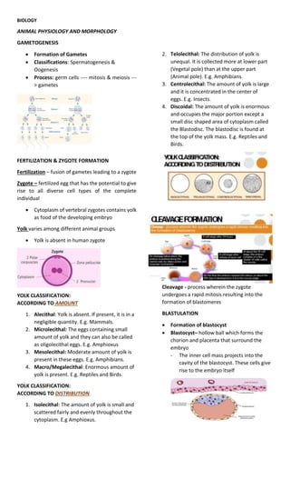
YOLK CLASSIFICATION:
ACCORDING TO AMOUNT
1. Alecithal: Yolk is absent. If present, it is in a
negligible quantity. E.g. Mammals.
2. Microlecithal: The eggs containing small
amount of yolk and they can also be called
as oligolecithal eggs. E.g. Amphioxus
3. Mesolecithal: Moderate amount of yolk is
present in these eggs. E.g. Amphibians.
4. Macro/Megalecithal: Enormous amount of
yolk is present. E.g. Reptiles and Birds.
YOLK CLASSIFICATION:
ACCORDING TO DISTRIBUTION
1. Isolecithal: The amount of yolk is small and
scattered fairly and evenly throughout the
cytoplasm. E.g Amphioxus.
2. Telolecithal: The distribution of yolk is
unequal. It is collected more at lower part
(Vegetal pole) than at the upper part
(Animal pole). E.g. Amphibians.
3. Centrolecithal: The amount of yolk is large
and it is concentrated in the center of
eggs. E.g. Insects.
4. Discoidal: The amount of yolk is enormous
and occupies the major portion except a
small disc shaped area of cytoplasm called
the Blastodisc. The blastodisc is found at
the top of the yolk mass. E.g. Reptiles and
Birds.
Cleavage - process wherein the zygote
undergoes a rapid mitosis resulting into the
formation of blastomeres
BLASTULATION
Formation of blastocyst
Blastocyst– hollow ball which forms the
chorion and placenta that surround the
embryo
- The inner cell mass projects into the
cavity of the blastocyst. These cells give
rise to the embryo itself
2. GASTRULATIOIN
Gastrula – hollow cup-shaped
structure having three layers of cells
Implantation of embryo – happens
in the endometrium or lining of the
uterus which begins on the seventh
day of embryonic development
-Enzymes destroy tiny maternal
capillaries in the endometrium
- Blood from these capillaries
comes in direct contact with
trophoblast of the embryo providing
nutrition
Formation of germ layers – Cells of
the inner cell mass arrange into a
two-layered disk -Completion is
done by ninth day
Formation of germ layers
-Lower-level merges to line the
digestive tract and other structures
-Endoderm is made up by these
cells while the cells that remain to
cover the embryo becomes the
ectoderm
-Mesoderm proliferates between
the two
NEURULATION
Formation of neurula which eventually
leads to the formation of the nervous
system
Neurula – technical term to describe the
embryo during neurulation
After neurulation, organogenesis proceeds
where organs are slowly produced and
made
LESSON 2: ANIMAL NUTRITION
FOOD UPTAKE IN CELLS VIA ENDOCYTOSIS
Phagocytosis – engulfment of organic
fragments or big particles, e.g. pseudopod
formation in Amoeba.
Pinocytosis – uptake of extracellular fluid by a
cell using small vesicles derived from the
plasma membrane.
Receptor-mediated endocytosis – this relies on
membrane receptor recognition of specific
solutes which are then taken up by the cell via
receptor-coated pits.
TYPES OF ANIMALS BASED ON FEEDING
MECHANISMS
1. Substrate-feeders – animals that live in
or on their food source. Examples:
earthworms that feed through the soil
where they live in; caterpillars that eat
through the leaves where they live on
2. Filter-feeders – include many aquatic
animals which draw in water and strain
small organisms and food particles
present in the medium. Examples:
whales and coelenterates
3. Fluid-feeders – suck fluids containing
nutrients from a living host. Examples:
mosquitoes, leeches, head lice, aphids
4. Bulk-feeders – eat relatively large
chunks of food and have adaptations
like jaws, teeth, tentacles, claws,
pincers, etc. that help in securing the
food and tearing it to pieces.
3. DIFFERENT KINDS OF DIGESTIVE COMPARTMENTS
IN ANIMALS
Food vacuoles in unicellular organisms – these
fuse with lysosomes that contain hydrolytic
enzymes.
Example: food vacuole in a protozoa like
Paramecium
Gastrovascular cavity or incomplete digestive
system – composed of a single opening through
which the food is taken in and where wastes are
disposed of. It is a sac-like body cavity.
Examples: cnidarian Hydra and flatworm
Planaria
Complete digestive system – essentially like a tube
with an opening at one end for taking in food
(mouth) and an opening at the outer end where
unabsorbed waste materials are eliminated (anus).
In between the mouth and anus, are specialized
organs that carry out transport, processing, and
absorption of digested nutrients.
ACCESSORY ORGANS IN DIGESTIVE SYSTEM
1. Liver – secretes bile for emulsifying fats.
2. Gallbladder – stores bile produced by the
liver.
3. Pancreas – secretes enzymes that break
down all major food molecules; secretes
buffers against HCl from the stomach;
secretes the hormone insulin for control of
glucose metabolism.
ANIMAL NUTRITION
Calorie is a unit of energy that indicates the
amount of energy contained in food.
The greater the number of Calories in a
quantity of food, the greater energy it
contains.