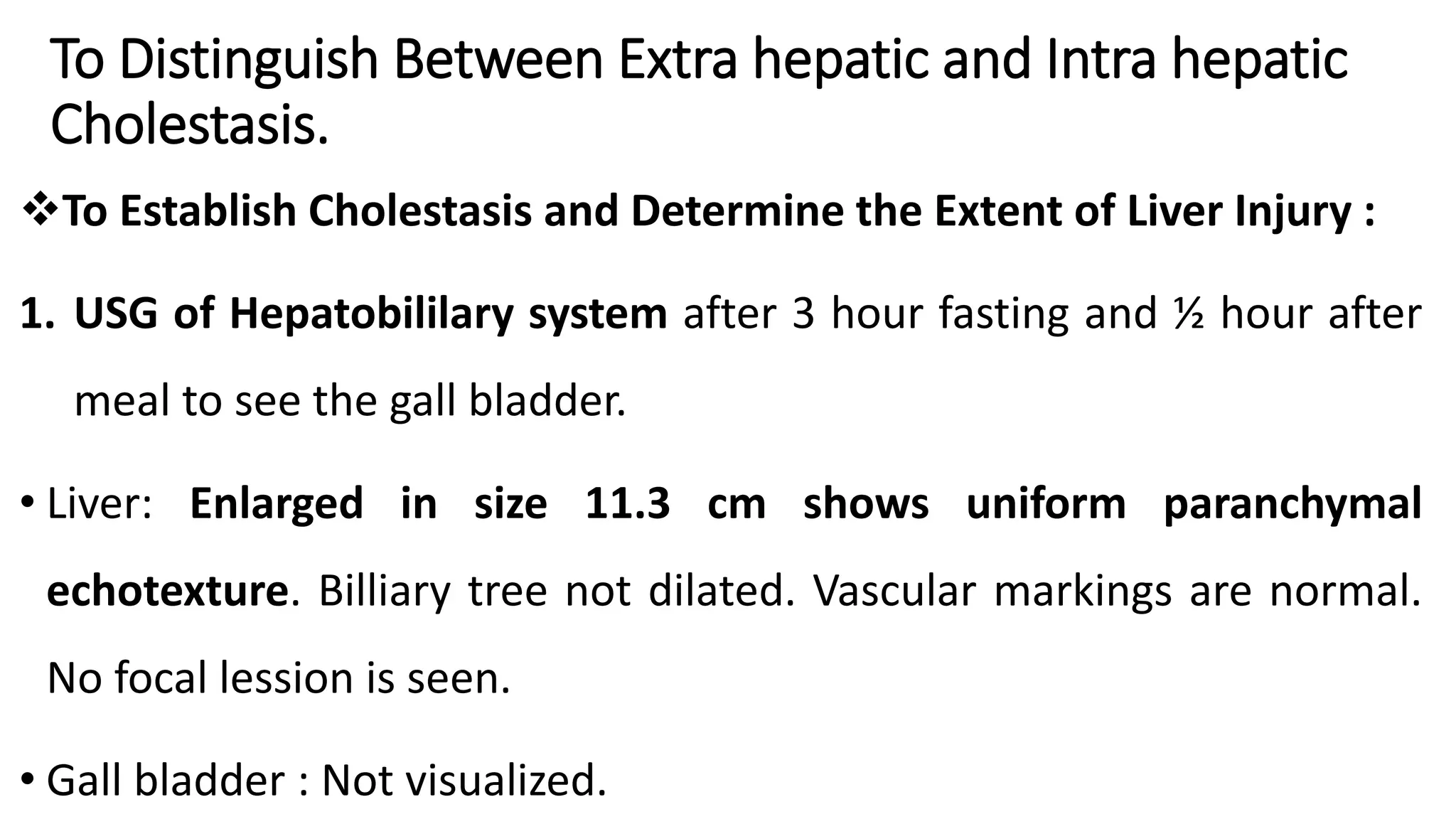
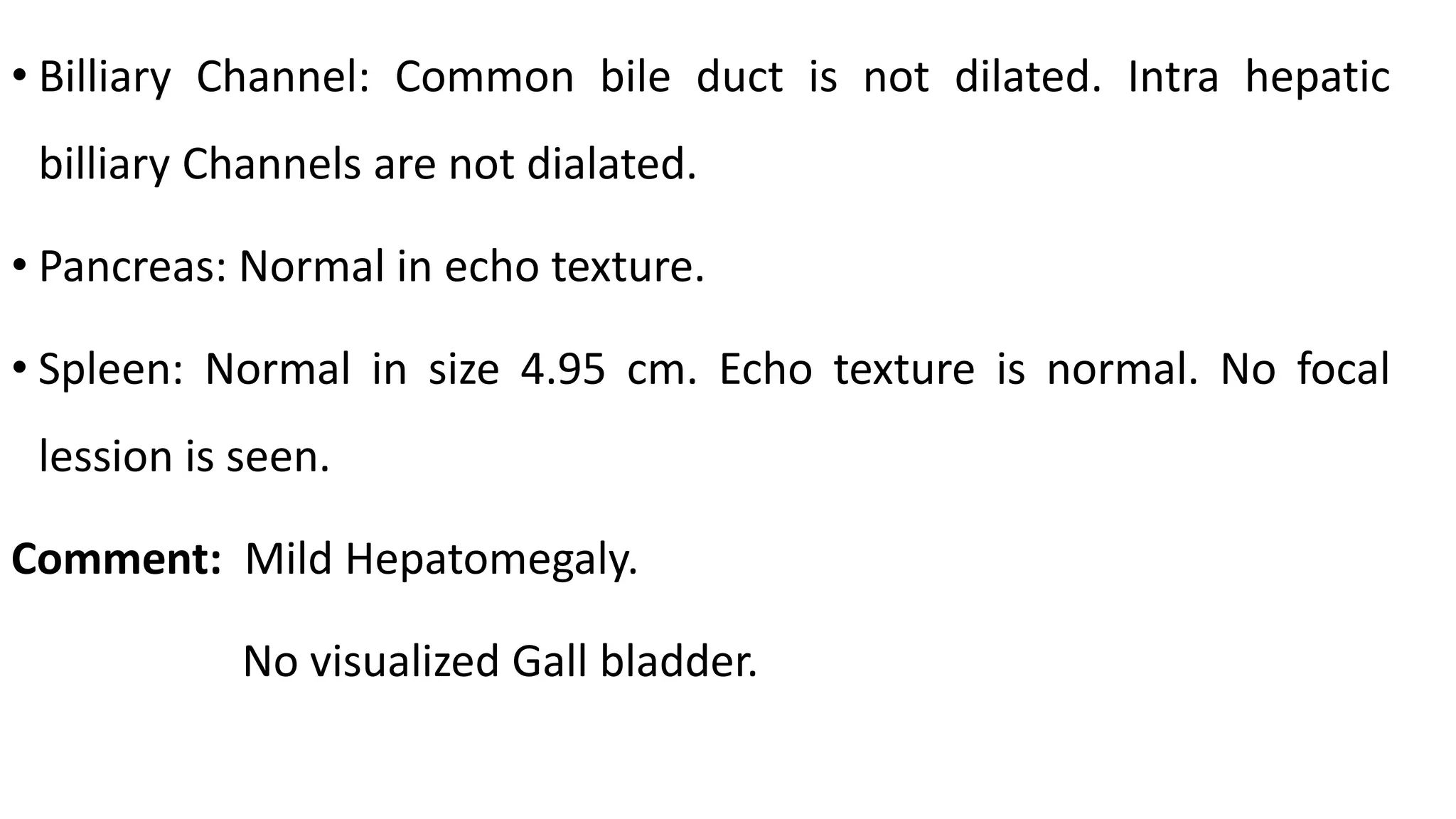
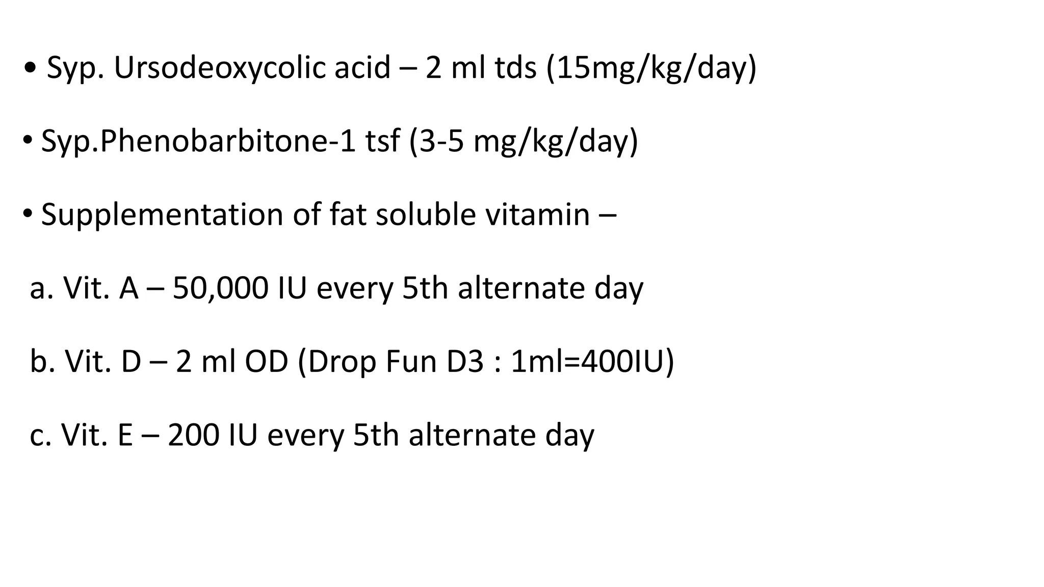
WELCOME
TO
CLINICAL MEETING
DR. ISRAT ZAMAN AKHI
MD Phase A Resident
NIKDU
Particularies of patient
• Name : Ayan
• Age : 3 months
• Sex : Male
• Address : Ulail, Savar
• DOA : 12/9/2023
• DOE : 12/9/2023
• Informant: Mother
Presenting Complaints:
• Yellow discoloration of sclera and skin since 20th day of his
life.
• Passage of pale stool and dark urine for same duration.
History of Present Illness:
According to the statement of informant mother, Ayan was alright
upto 20 days of his life, then he developed yellow discoloration of skin
and sclera which was gradually deepening day by day. Mother also
mentioned that, her baby has been persistently passing pale color
stool and dark urine for same duration. He has no history of delayed
passage of meconium, constipation, poor feeding, lethargy, any rash,
bleeding manifestation, vomiting.
With these above mentioned complaints, he was treated initially by
local physician and then got referred to our hospital as condition didn’t
improve, for further evaluation and better management.
History of Past Illness
There was no other significant past illness.
Birth History
• Antenatal: Mother was on irregular antenatal check up. There is no
history of diabetes, hypertension or fever with rash during entire
pregnancy period.
• Natal: Baby was delivered by NVD at hospital with birth weight
2500 gm.
• Postnatal: Mother noticed yellow discoloration of both eyes of the
baby since 20th day of life .
Feeding History
He was on exclusive breast feeding till date.
Immunization History
Immunization going on as per EPI schedule.
Developmental History
Age appropriate.
 Treatment History
He was treated by some oral medications but couldn’t mention names
or show any documents.
 Family History
He is the only issue of his non consanguineous parents. There is no
history of similar type of illness in the family or any sib death due to
same type of illness.
 Socioeconomic history
He belongs from a middle class socioeconomic background. His father
is a job holder and mother is a homemaker. Their monthly income is
approximately 30000tk.
General examination
• Appearance : Well alert, playful , no facial dysmorphism.
• Anaemia : Mildly pale
• Jaundice : Icteric
• Cyanosis
• Clubbing
• Koilonychia
• Leukonychia
Absent
• Edema Absent
• Dehydration
• Anterior frontanalle is open .
• Skin survey: Normal
• Lymph node : Not palpable
• Eyes : Normal
• Ear nose throat : Normal
Vital signs
• Temperature: 98°F
• Respiratory rate : 34 /min
• Heart rate : 100/min
• BP : 80/50 mmhg ( falls on 50th centile)
Anthropometry
• Length : 60 cm
• Weight : 4.5 kg
• OFC : 38 cm ( falls just below 50th centile)
Anthropometry
Parameter Z score Comment
Weight for length -1.8 SD Normal
Length for age -1.6 SD Normal
Weight for age -1.3 SD Normal
Systemic Examination
Alimentary System
• Mouth and fauces : Normal
Abdomen:
A. Inspection : Abdomen was mildly distended
Flanks was not full
Umbilicus is centrally placed and inverted
B. Palpation : Abdomen soft, non tender
Liver : enlarged, 6 cm from right costal margin along with mid clavicular
line,non tender, firm in consistency, sharp border with smooth surface
and upper border of liver dullness was on right 5th intercostal space.
Spleen: not palpable
Kidney: not ballotable
Urinary bladder: not palpable
Para aortic lymphnodenode: not palpable
cont
C. Percussion : Fluid thrill
Shifting dullness
D. Auscultation: Bowel sound present
• Other systemic examination revealed no significant findings
Salient Feature
Ayan , 3 month old boy, only issue of his non-consanguineous parents
was admitted with the complaints of persistent jaundice along with
passage of dark urine and persistent pale color stool since 20th days
of life till date. There is no H/O delayed passage of meconium,
constipation, any bleeding manifestations, vomiting or family H/O
similar type of illness. Ayan was a term baby with average birth
weight 2500 gm.
• His mother had an uncomplicated pregnancy period. He is well
alert, well thrived , playful , mildly pale and moderately icteric ,
no facial dysmorphism. There was hepatomegaly with no
evidence of ascites.
Provisional Diagnosis
Neonatal cholestasis due to biliary atresia.
Differential Diagnosis
Neonatal cholestasis due to neonatal hepatitis syndrome.
Investigations
To Establish Cholestasis and Determine The Extent Of Liver Injury
• S.Billirubin : Total -10.66 mg/dl
Direct - 7.01 mg/dl
Indirect- 2.05 mg/dl
• S.SGPT : 122 U/L (upto 65 U/L)
• S.ALP : 400 U/L (upto 300 U/L)
• S.GGT : 1124 U/L (30-110 U/L)
cont
• PT : 11.0 sec (Control =12 sec)
• INR : 0.86
• S.Albumin : 35.4 gm/L
• BT : 3 min (1-6 min)
• CT : 6 min (6-10 min)
To Distinguish Between Extra hepatic and Intra hepatic
Cholestasis.
To Establish Cholestasis and Determine the Extent of Liver Injury :
1. USG of Hepatobililary system after 3 hour fasting and ½ hour after
meal to see the gall bladder.
• Liver: Enlarged in size 11.3 cm shows uniform paranchymal
echotexture. Billiary tree not dilated. Vascular markings are normal.
No focal lession is seen.
• Gall bladder : Not visualized.
• Billiary Channel: Common bile duct is not dilated. Intra hepatic
billiary Channels are not dialated.
• Pancreas: Normal in echo texture.
• Spleen: Normal in size 4.95 cm. Echo texture is normal. No focal
lession is seen.
Comment: Mild Hepatomegaly.
No visualized Gall bladder.
2) Liver Biopsy
• Specimen : Liver tissue
• Clinical information
Gross description:
Specimen consists of 1.5 cm long linear piece of tissue. Submitted as
such.
cont
Microscopic examination
Sections show core of liver tissue. It shows moderate cholestasis.
The portal areas show moderate fibrosis and contain increase
number of bile ductules. Moderate infiltration of chronic
inflammatory cell is present. Bridging fibrosis is also present.
Diagnosis: Competible with billiary atresia.
 For Management Purpose
• CBC: Hb- 11.5 gm/dl
TRBC- 2.94×10^12/L
HCT- 27%
• TWBC -18.7×10^9/L
• DLC: N- 30.8%
L- 62.1%
M- 3.4%
E- 3.5%
• TPC -313×10^9/L
• S.TSH- 5.33 mul/ml
• S.T4- 2.05 ng/dl
• TORCH Screening :
CMV :IgM negative
:IgG negative
• Eye examination for
Cataract
Chorioretinitis
Cherry red spot
Posterior embryotoxon
Final Diagnosis
• Neonatal cholestasis due to Billiary atresia.
Treatment
1. Counselling – Nature of disease
Course
Complication
Treatment option
Prognosis
2. Supportive treatment :
• Diet : Breast feeding + MCT ( 4 ml Tds )
• Syp. Ursodeoxycolic acid – 2 ml tds (15mg/kg/day)
• Syp.Phenobarbitone-1 tsf (3-5 mg/kg/day)
• Supplementation of fat soluble vitamin –
a. Vit. A – 50,000 IU every 5th alternate day
b. Vit. D – 2 ml OD (Drop Fun D3 : 1ml=400IU)
c. Vit. E – 200 IU every 5th alternate day
• d. Vit. K – Inj. Vit K 5mg for 5 days once daily
• Supplementation of water soluble vitamin –
a. Vit. B complex – 5 drops OD
• Supplementation of micronutrients -
a. Syp . Calcium – 2 tsf twice daily
b. Syp . Zinc – ½ tsf twice daily
c. Tab. Folic acid – 1/5th tab OD (mixed with
milk )
• Kasai operation
3. Specific treatment –
Liver transplantation
Follow up
• Monthly for 3 month
• 3 monthly for 6 month
• Yearly for 3 years
• Clinical follow up – Jaundice
Pale stool
Signs of CLD
Anthropometry
cont
• Laboratory investigation during follow up –
S. bilirubin
ALT
GGT
ALP
THANK YOU

billiary atresia by Akhi.pptx

  • 1.
    WELCOME TO CLINICAL MEETING DR. ISRATZAMAN AKHI MD Phase A Resident NIKDU
  • 2.
    Particularies of patient •Name : Ayan • Age : 3 months • Sex : Male • Address : Ulail, Savar • DOA : 12/9/2023 • DOE : 12/9/2023 • Informant: Mother
  • 3.
    Presenting Complaints: • Yellowdiscoloration of sclera and skin since 20th day of his life. • Passage of pale stool and dark urine for same duration.
  • 4.
    History of PresentIllness: According to the statement of informant mother, Ayan was alright upto 20 days of his life, then he developed yellow discoloration of skin and sclera which was gradually deepening day by day. Mother also mentioned that, her baby has been persistently passing pale color stool and dark urine for same duration. He has no history of delayed passage of meconium, constipation, poor feeding, lethargy, any rash, bleeding manifestation, vomiting.
  • 5.
    With these abovementioned complaints, he was treated initially by local physician and then got referred to our hospital as condition didn’t improve, for further evaluation and better management.
  • 6.
    History of PastIllness There was no other significant past illness.
  • 7.
    Birth History • Antenatal:Mother was on irregular antenatal check up. There is no history of diabetes, hypertension or fever with rash during entire pregnancy period. • Natal: Baby was delivered by NVD at hospital with birth weight 2500 gm. • Postnatal: Mother noticed yellow discoloration of both eyes of the baby since 20th day of life .
  • 8.
    Feeding History He wason exclusive breast feeding till date. Immunization History Immunization going on as per EPI schedule. Developmental History Age appropriate.
  • 9.
     Treatment History Hewas treated by some oral medications but couldn’t mention names or show any documents.  Family History He is the only issue of his non consanguineous parents. There is no history of similar type of illness in the family or any sib death due to same type of illness.
  • 10.
     Socioeconomic history Hebelongs from a middle class socioeconomic background. His father is a job holder and mother is a homemaker. Their monthly income is approximately 30000tk.
  • 11.
    General examination • Appearance: Well alert, playful , no facial dysmorphism. • Anaemia : Mildly pale • Jaundice : Icteric • Cyanosis • Clubbing • Koilonychia • Leukonychia Absent
  • 12.
    • Edema Absent •Dehydration • Anterior frontanalle is open . • Skin survey: Normal • Lymph node : Not palpable • Eyes : Normal • Ear nose throat : Normal
  • 13.
    Vital signs • Temperature:98°F • Respiratory rate : 34 /min • Heart rate : 100/min • BP : 80/50 mmhg ( falls on 50th centile)
  • 16.
    Anthropometry • Length :60 cm • Weight : 4.5 kg • OFC : 38 cm ( falls just below 50th centile)
  • 17.
    Anthropometry Parameter Z scoreComment Weight for length -1.8 SD Normal Length for age -1.6 SD Normal Weight for age -1.3 SD Normal
  • 18.
    Systemic Examination Alimentary System •Mouth and fauces : Normal Abdomen: A. Inspection : Abdomen was mildly distended Flanks was not full Umbilicus is centrally placed and inverted B. Palpation : Abdomen soft, non tender
  • 19.
    Liver : enlarged,6 cm from right costal margin along with mid clavicular line,non tender, firm in consistency, sharp border with smooth surface and upper border of liver dullness was on right 5th intercostal space. Spleen: not palpable Kidney: not ballotable Urinary bladder: not palpable Para aortic lymphnodenode: not palpable
  • 20.
    cont C. Percussion :Fluid thrill Shifting dullness D. Auscultation: Bowel sound present
  • 21.
    • Other systemicexamination revealed no significant findings
  • 22.
    Salient Feature Ayan ,3 month old boy, only issue of his non-consanguineous parents was admitted with the complaints of persistent jaundice along with passage of dark urine and persistent pale color stool since 20th days of life till date. There is no H/O delayed passage of meconium, constipation, any bleeding manifestations, vomiting or family H/O similar type of illness. Ayan was a term baby with average birth weight 2500 gm.
  • 23.
    • His motherhad an uncomplicated pregnancy period. He is well alert, well thrived , playful , mildly pale and moderately icteric , no facial dysmorphism. There was hepatomegaly with no evidence of ascites.
  • 24.
    Provisional Diagnosis Neonatal cholestasisdue to biliary atresia. Differential Diagnosis Neonatal cholestasis due to neonatal hepatitis syndrome.
  • 25.
    Investigations To Establish Cholestasisand Determine The Extent Of Liver Injury • S.Billirubin : Total -10.66 mg/dl Direct - 7.01 mg/dl Indirect- 2.05 mg/dl • S.SGPT : 122 U/L (upto 65 U/L) • S.ALP : 400 U/L (upto 300 U/L) • S.GGT : 1124 U/L (30-110 U/L)
  • 26.
    cont • PT :11.0 sec (Control =12 sec) • INR : 0.86 • S.Albumin : 35.4 gm/L • BT : 3 min (1-6 min) • CT : 6 min (6-10 min)
  • 27.
    To Distinguish BetweenExtra hepatic and Intra hepatic Cholestasis. To Establish Cholestasis and Determine the Extent of Liver Injury : 1. USG of Hepatobililary system after 3 hour fasting and ½ hour after meal to see the gall bladder. • Liver: Enlarged in size 11.3 cm shows uniform paranchymal echotexture. Billiary tree not dilated. Vascular markings are normal. No focal lession is seen. • Gall bladder : Not visualized.
  • 28.
    • Billiary Channel:Common bile duct is not dilated. Intra hepatic billiary Channels are not dialated. • Pancreas: Normal in echo texture. • Spleen: Normal in size 4.95 cm. Echo texture is normal. No focal lession is seen. Comment: Mild Hepatomegaly. No visualized Gall bladder.
  • 29.
    2) Liver Biopsy •Specimen : Liver tissue • Clinical information Gross description: Specimen consists of 1.5 cm long linear piece of tissue. Submitted as such.
  • 30.
    cont Microscopic examination Sections showcore of liver tissue. It shows moderate cholestasis. The portal areas show moderate fibrosis and contain increase number of bile ductules. Moderate infiltration of chronic inflammatory cell is present. Bridging fibrosis is also present. Diagnosis: Competible with billiary atresia.
  • 31.
     For ManagementPurpose • CBC: Hb- 11.5 gm/dl TRBC- 2.94×10^12/L HCT- 27% • TWBC -18.7×10^9/L • DLC: N- 30.8% L- 62.1% M- 3.4% E- 3.5% • TPC -313×10^9/L
  • 32.
    • S.TSH- 5.33mul/ml • S.T4- 2.05 ng/dl • TORCH Screening : CMV :IgM negative :IgG negative • Eye examination for Cataract Chorioretinitis Cherry red spot Posterior embryotoxon
  • 33.
    Final Diagnosis • Neonatalcholestasis due to Billiary atresia.
  • 34.
    Treatment 1. Counselling –Nature of disease Course Complication Treatment option Prognosis 2. Supportive treatment : • Diet : Breast feeding + MCT ( 4 ml Tds )
  • 35.
    • Syp. Ursodeoxycolicacid – 2 ml tds (15mg/kg/day) • Syp.Phenobarbitone-1 tsf (3-5 mg/kg/day) • Supplementation of fat soluble vitamin – a. Vit. A – 50,000 IU every 5th alternate day b. Vit. D – 2 ml OD (Drop Fun D3 : 1ml=400IU) c. Vit. E – 200 IU every 5th alternate day
  • 36.
    • d. Vit.K – Inj. Vit K 5mg for 5 days once daily • Supplementation of water soluble vitamin – a. Vit. B complex – 5 drops OD • Supplementation of micronutrients - a. Syp . Calcium – 2 tsf twice daily b. Syp . Zinc – ½ tsf twice daily
  • 37.
    c. Tab. Folicacid – 1/5th tab OD (mixed with milk ) • Kasai operation 3. Specific treatment – Liver transplantation
  • 38.
    Follow up • Monthlyfor 3 month • 3 monthly for 6 month • Yearly for 3 years • Clinical follow up – Jaundice Pale stool Signs of CLD Anthropometry
  • 39.
    cont • Laboratory investigationduring follow up – S. bilirubin ALT GGT ALP
  • 40.