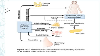
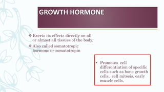
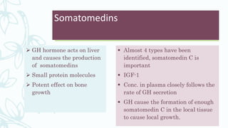
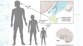
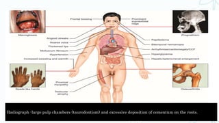
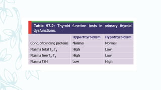
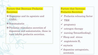
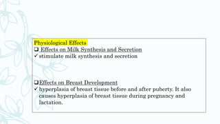
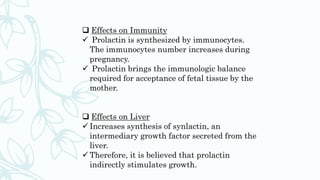
Endocrinology
Contents
Introduction
General principles of endocrinology
Pituitary gland
Anterior pituitary
Posterior pituitary
Growth hormone
Hormonal excess
Growth Hormone excess
Acromegaly
Gigantism
Dwarfism
Thyroid Stimulating Hormone
Thyroid hormones
Adrenocorticotropic hormone
Prolactin
Hyperprolactinemia
Luteinizing Hormone and Follicle
Stimulating Hormone
Anti-diuretic hormone
Diabetes insipidus
Oxytocin
Hypopituitarism and periodontium
Hyperpituitarism and periodontium
Conclusion
references
Introduction
 The multiple activities of the cells, tissues and organs of the body
are coordinated by the interplay of several types of chemical
messenger systems.
• Neurotransmitters
• Endocrine hormones
• Neuroendocrine hormones
• Paracrines
• Autocrines
• Cytokines
General principles of
endocrinology
 Cell function is broadly controlled by:
• Neural control
• Endocrinal control
Neural control Endocrinal control
Due to depolarisation through the nerve Hormone directly into the blood stream
Rapid action due to shorter latent period Much longer latent period
Affects certain group of cells for a
shorter period
Endocrine glands
– Also known as ductless glands
– Secrete hormones directly into the blood stream
1. Hypothalamus
2. Anterior pituitary
3. Posterior pituitary
4. Pancreas
5. Adrenal cortex
6. Adrenal medulla
7. Thyroid
8. Parathyroid
9. Kidney
10. Ovary and
testes
Other organs with endocrine
function and hormones
Organ Hormone
Heart Anti-Natriuretic Peptide( ANP)
GIT Cholecystokinin-pancreozymin
Secretin and vasointestinal
peptide(VIP)
Kidney 1,25-Dihydroxycholecalciferol
Pineal gland Melatonin
Skin Calciferol
Liver Insulin Like Growth Factor( IGF)
Platelets Platelet Derived Growth Factor
Lymphocytes Interleukins
Hormone
 Greek- Hormacin “ I excite or arouse”
 Introduced by Starling in 1905
 Def: secretory products of ductless glands released directly into
the circulation in small amounts to a specific stimulus and on
delivery in circulation produces a response on the target cells
 Interact with target cells via receptors- specific binding sites
 Most hormones are metabolised rapidly after secretion and gets
inactivated mainly in liver and kidney
Classes of Hormones
I)Based on mechanism of action:
Group 1- Bind to intracellular receptors
eg: androgens, calcitriol, estrogens, glucocorticoids, thyroid hormone
Group 2- Bind to cell surface receptors
A)Second messenger cAMP
ACTH, ADH, CRH, FSH, LH
B)cGMP as second messenger
Atrial natriuretic factor
Nitric oxide
C)Calcium/phosphatidyl inositols as second messenger
GnRH, TRH, PDGF, Substance P
D)Second messenger is a kinase/phosphate cascade
Insulin, GH, Prolactin, Insulin like growth factors (IGF-1, IGF-2)
II)Based on nature:
-Steroid hormones
-amine hormones
-peptide hormones
-protein hormones
-glycoprotein hormones
III)Based on nature of action:
-Local hormones eg: Testosterone
-General hormones eg: Insulin, Thyroid hormone
IV)Based on effect:
-Kinetic hormones
-Metabolic hormones
-Morphogenetic hormones
V)Based on stimulation of endocrine glands:
-Tropic hormones
-Non-tropic hormones
PITUITARY GLAND
 Hypophysis
 Connected to hypothalamus –
pituitary stalk
Parts
1. Anterior pituitary-adenohypophysis
2. Posterior pituitary- neurohypophysis
Embryology
• Anterior pituitary- Rathke’s pouch
• Posterior pituitary from neural tissue outgrowth from
hypothalamus
Anterior pituitary hormones
– 6 important hormones and other lesser important
ones
– Plays a major role in control of metabolic functions
throughout the body
Cells of anterior pituitary gland
Posterior pituitary gland
 Neurohypophysis
 Nerve endings are bulbous and contain secretory
granules
 2 hormones:
1. Anti diuretic hormone (ADH) or vasopressin
2. Oxytocin
 ADH -primarily formed in the supraoptic nuclei
( neurophysin II)
 Oxytocin- paraventricular nuclei ( nerophysin I)
 These hormones are released from the endings by
exocytosis.
Hypothalamus controls pituitary
secretion
– Secretion from the posterior pituitary is controlled by
humoral or nerve signals that originate in the
hypothalamus and terminate in the posterior pituitary
– Secretion from the anterior pituitary is controlled by
hormones- hypothalamic releasing and hypothalamic
inhibitory hormones
Hypothalamic releasing and inhibitory hormones are secreted into the
median eminence.
 Special neurons that originate in the hypothalamus, send nerve
fibres to the median eminence and tuber cinereum, an extension of
hypothalamic tissue into the pituitary stalk.
 Secrete releasing and inhibitory hormones into tissue fluids
 Hormones immediately absorbed into hypothalamo-hypophyseal
portal system
 Sinuses in anterior pituitary gland
Hypopituitarism And
Periodontium
 Increased gingival inflammation
 Resorption of cementum in molar furcation area
 Reduced apposition of cementum
 Decreased osteogenesis in interdental area
 Reduced vascularity in pdl
 Degeneration of the ligament with cystic degeneration and
calcification of many epithelial rests
 Osteoporosis
Hyperpituitarism and
periodontium
 Marked overgrowth of alveolar process
 Increase in size of dental arch
 Spacing of the teeth
 Food impaction
 Hypercementosis
GROWTH HORMONE
 Exerts its effects directly on all
or almost all tissues of the body.
 Also called somatotropic
hormone or somatotropin
• Promotes cell
differentiation of specific
cells such as bone growth
cells, cell mitosis, early
muscle cells.
GH promotes protein deposition
in tissues
 Enhancement of Amino Acid transport through the cell
membrane
 Enhancement of RNA translation to cause protein
synthesis by ribosomes
 Decreased catabolism of protein and aminoacids
GH decreases carbohydrate
utilisation
 Decreased glucose uptake in tissues such as skeletal
muscle
 GH stimulates cartilage and bone growth
 Increased deposition of protein by chondrocytic and
osteogenic cells that cause bone growth
 Increased rate of reproduction of these cells
 Specific effect of converting chondrocytes to osteogenic cells ,
thus causing deposition of new bone
 2 principal mechanisms of bone growth:
1. GH long bones grow at the epiphyseal
cartilages
2. Osteoblasts in the bone periosteum and in some bone
cavities deposit new bone on the surface of older bone.
Osteoclasts remove old bone. When the rate of deposition
is higher thickening of bone
GH strongly stimulates osteoblasts
 Effect skeletal muscle
 Increases the growth of skeletal
muscle by increasing protein
synthesis
 Causes hypertrophy of skeletal
muscle cell
 Promotes activity of satellite cells of
skeletal muscle
 Effect on visceral organs
 Growth of liver, kidney, heart
pancreas and intestine
 Due to hypertrophy and
hyperplasia
 Effect on fat metabolism
Lipolysis and increased
plasma FFA
Promotes ketogenesis
Effect on electrolyte and water
metabolism
Increases plasma Ca2
+
Causes Na+ retention
Maintains ECF volume
Increases plasma phosphate level
Somatomedins
 GH hormone acts on liver
and causes the production
of somatomedins
 Small protein molecules
 Potent effect on bone
growth
 Almost 4 types have been
identified, somatomedin C is
important
 IGF-1
 Conc. in plasma closely follows the
rate of GH secretion
 GH cause the formation of enough
somatomedin C in the local tissue
to cause local growth.
GH regulation
 After adolescence secretion decreases slowly with aging,
finally falling to about 25% of adolescent level in old age.
 GH secreted in a pulsatile pattern, increasing and
decreasing
 Normal conc-1.6-3ng/ml adults
 Child or adolescent-6ng/ml
GH secretion and aging
 Aged appearance due to the
1. decreased protein deposition
2. Increased fat deposition
 Increased wrinkling of the skin
 Diminished rates of function of
some organs
 Diminished muscle mass
 Maximal release during the
intense growth periods of late
childhood and adolescence
Age Concn(ng/ml)
5-20 yrs 6
20-40yrs 3
40-70yrs 1.6
Classification of diseases of the pituitary and hypothalamus
primary secondary
Hormone excess
1. Anterior pituitary
Prolactinoma
Acromegaly
Cushing’s syndrome
Disconnection
hyperprolactinemia
2 hypothalamus and posterior
pituitary
Syndrome of inappropriate
ADH (SIADH)
Hormone deficiency
1. Anterior pituitary
Hypopituitarism Eg: GnRH deficiency
(Kallman’s syndrome)
2. Hypothalamus and posterior
pituitary
Cranial diabetes insipidus
Hormone resistance GH resistance ( Laron
dwarfism)
Non- functioning tumors Pituitary adenoma
Craniopharyngioma
Metastatic tumors
Hormonal excess
 Overgrowth –Adenomas
 Pituitary adenomas are classified
as
 Microadenomas (<1cm)
 Macroadenomas (>1cm)
Pituitary apoplexy
Infarction of the pituitary
 Due to sudden haemorrhage
within the tumour
 Symptoms
 Sudden and severe headache
 Diplopia
Extension of the tumour
inferiorly- rare
Abnormalities of GH secretion
 Panhypopituitarism
 Dwarfism
 Gigantism
 Acromegaly
Panhypopituitarism
– Decreased secretion of all anterior
pituitary hormones.
– Congenital or sudden or slowly after birth
due to adenomas
Gigantism
 GH excess occurs before the completion of linear
growth (before closure of epiphyseal plates)
 Mild to moderate obesity
 Tall stature
 Progressive macrocephaly
 Could be related to pituitary adenoma
 Accelerated growth
Radiographically: enlarged sella
Other features:
 Enlargement of soft
tissues
 Enlarged mandible
 Increased size of hand
and feet
 Generalised
macrodontia
Treatment:
 Surgical removal of
adenoma
 Radiation therapy
Oral manifestations: Prognathic mandible, frontal
bossing, dental malocclusion, and interdental spacing
are the other features which may be seen in such
individuals. Intraoral radiograph -hypercementosis of
the roots.
Acromegaly
 GH excess occurs after the growth plates
have fused
 Usually with pituitary adenoma
 Average age of diagnosis -42 years
Features
 Macrognathia, macroglossia, large
diastemas
 Mandibular prognathism
 Apertognathia
 The lips become thick and negroid.
 Intraoral spacing
 buccal tipping of the teeth due to
enlarged tongue
 Hypertrophy of soft palatal tissues
Radiograph -large pulp chambers (taurodontism) and excessive deposition of cementum on the roots.
– Diagnosis :
– serum IGF-1 levels along with oral Glucose
Tolerance Test
Treatment
 Surgical removal of the
mass
 Radiation therapy
 Pharmacotherapy
somatostatin analogues
(octreotide,
lanreotide,vapreotide)
PITUITARY DWARFISM
 Caused by
1. Diminished production of GH
by anterior pituitary
2. Reduced capacity of the
tissues to respond to GH
Clinical features:
 Short stature
 Small face
 Smaller maxilla
and mandible
 Delayed eruption
 Delayed shedding
 Reduced size of
the teeth
Lab diagnosis:
Radioimmunoassay for GH
Treatment:
 Replacement therapy with human GH (
if detected before closure of epiphyseal
growth plates)
 If GH deficiency is caused by
hypothalamic defect- GHRH is
appropriate
 No treatment for GH receptor lacking
patients
Prognosis:
Identified and treated at early age  normal height
Thyroid hormones
The principal hormones synthesized and secreted by thyroid gland
are:
1. T4 (thyroxine)
2. T3 (triiodothyronine).
3. RT3 (reverse-triiodothyronine), secreted in small quantity.
4. Calcitonin
 T4, T3, and RT3 -thyroid follicles
 Calcitonin -parafollicular cells (C cells).
Thyroid Hormone Synthesis
The thyroid hormone synthesis involves following steps:
1. Iodide trapping
2. Conversion of iodide into iodine
3. Thyroglobulin synthesis
4. Proteolysis of thyroglobulin
5. Secretion of thyroid hormones
 In a normal individual, total plasma T4
concentration is about 8 µg/dL, and plasma
T3 is about 0.15 µg/dL.
 Free T4 level in plasma is about 2 ng/dL, and
free T3 is about 0.3 ng/dL
Thyroid hormones bind with three types of plasma
proteins:
1. Thyroxine binding globulin (TBG)
2. Thyroxine binding prealbumin (TBPA)
3. Albumin.
Normally, T4 binds mainly with TBG and TBPA,
and T3 binds with albumin and TBG.
Fasting decreases conversion of T4 to
T3.
RT3 is increased and T4 remains normal
in fasting.
 In starvation (chronic and severe
fasting), RT3 returns to normal, but T3
continues to remain low.
In overfeeding, opposite happens; T3 is
increased and RT3 is decreased.
Clinical correlation
1. Hyperthyroidism
A. Thyrotoxicosis
B. Grave’s disease
C. Thyroid storm
2. Hypothyroidism
Thyroid Stimulating Hormone
 Glycoprotein
 Controls the secretion of T3 and
T4
 2 subunits α and β
 Secreted by thyrotrophs of
anterior pituitary
Mechanism of Action : TSH exerts its effects on thyroid
cells by increasing concentration of intracellular cyclic
AMP.
Functions:
 Facilitates iodide uptake
 Enhances intermolecular
coupling and thyroglobin
synthesis and secretion into
colloid
 Stimulates release of thyroid
hormones and iodotyrosines from
the gland
 Promotes endocytosis of colloid
 Increases blood flow to the
thyroid gland
 Causes hypertrophy and growth
of the thyroid gland
Clinical correlation:
 Decreased secretion of TSH due to
pituitary diseases results in thyroid
atrophy and decreased secretion of
thyroid hormones (secondary
hypothyroidism or hypopituitary
hypothyroidism).
 Chronic increase in TSH secretion
results in hypertrophy of thyroid
gland, called Goitre.
Hyperthyroidism
Hypersecretion of thyroid hormones from thyroid
gland or from extrathyroidal tissues,
Two types
1. primary
2. secondary
 Hyperthyroidism occurs due to pathology of the
thyroid gland- primary hyperthyroidism.
The common causes are:
1. Adenoma of thyroid
2. Multinodular Goiter
3. Metastatic carcinoma of functioning thyroid
gland
4. Graves’ disease
5. Activating mutation of TSH receptors
6. Iodine excess (Jod-Basedow phenomenon)
Secondary Hyperthyroidism
 When hyperthyroidism occurs due to the
pathology outside thyroid gland
 Pituitary causes-Tumor of thyrotrophs of
anterior pituitary (secretes excess of TSH).
 Extrathyroidal Causes- Chronic excess
administration of thyroid hormones
(iatrogenic), tumor of ectopic thyroid tissue
(lingual thyroid), human chorionic gonadotropin
secreting tumors such as choriocarcinoma.
Hypothyroidism
 Hypothyroidism in adult –Myxedema
 Children- Cretinism
 Primary Hypothyroidism- due to the diseases or causes that
primarily affect thyroid gland
 Secondary Hypothyroidism- primarily due to a defect outside the
thyroid gland
( pituitary or hypothalamus)
Symptoms
– Cold intolerance, weakness and
easy tiredness
– dry thick skin, loss of hair
– poor memory and inability to
concentrate, constipation (due to
decreased GI motility), weight
gain in spite of poor appetite
– thick and husky voice
– yellow skin,
– psychosis (myxedema madness)
menorrhagia (in females),
– galactorrhea and infertility
Signs
– Cool extremities with dry coarse
skin, dry hair
– puffy face, with edematous
hands and feet (myxedema)
– diffuse alopecia
– goiter,
– bradycardia, hyper tension
(diastolic), anemia,
– decreased reaction time of
tendon reflexes (especially,
delayed Achilles tendon reflex
relaxation),
– carpal tunnel syndrome and
periorbital edema.
Treatment of Hypothyroidism : Thyroid hormone replacement
T4 is instituted at a dose (usually 10 to 15 µg/kg/ day)
Hashimoto’s Thyroiditis - chronic form of autoimmune
thyroiditis
• antibodies are formed against the thyroglobulin and thyroid
peroxidase.
• thyroid cells are damaged -hypothyroidism develops.
Oral manifestations of thyroid disorders
and its management:
Dental management of hypothyroidism:
Patients who have hypothyroidism are susceptible to
cardiovascular disease from arteriosclerosis and elevated LDL.
-anti coagulant therapy
Antibiotic prophylaxis
Drug actions and interactions - sensitive to central nervous
system depressants and barbiturates
Dental management of hyperthyroidism:
-Susceptibility to infection
-Drug actions and interactions : acetylsalicylic acid is
contraindicated in patient with hyperthyroidism
-Epinephrine- contraindicated
Cretinism
– When hypothyroidism develops from or
before birth, the patients are called
cretins.
– Causes:
 Maternal iodine deficiency during
pregnancy,
 maldevelopment of thyroid gland
during fetal life,
 inborn errors of thyroid hormone
synthesis
 antithyroid antibodies in mother that
crosses placenta and enters fetal
circulation
 hypopituitarism in fetal life
Patients are dwarf and mentally
retarded. Typically, they have
potbelly and protrusion of tongue
Adrenocortiocotropic hormone
(ACTH)
 Controls growth and secretion of adrenal cortex.
 Influences the secretion of cortisol.
 ACTH plays vital role in physiology of stress and pathophysiology
of stress disorders.
 secreted from corticotrophs of anterior pituitary
 synthesized as part of a larger molecule called pro-
opiomelanocortin (POMC), which cleaves to form β-lipotropin, and
ACTH in human beings
 Mechanism of Action :ACTH acts primarily by increasing cAMP in
the target cells.
Circadian Rhythm of ACTH
Secretion
– Normally, ACTH secretion occurs in irregular bursts.
– The pulsatile secretion of ACTH is due to several bursts of CRH
(corticotropin releasing hormone) secretion in 24 hours.
– However, there is a prominent diurnal rhythm for ACTH secretion,
in which secretion is more in the early morning, which constitutes
about 75% of the total 24 hours secretion
- This diurnal variation in ACTH secretion is
due to the natural sleep-wake cycle,
controlled by inherent biological rhythm of
suprachiasmatic nucleus of hypothalamus.
- The pattern of secretion is reversed in
individuals who sleep in the day and remain
awake during night. The pattern of
glucocorticoid secretion closely follows the
ACTH secretion
Regulation of ACTH
 Primary factor controlling the ACTH secretion is CRH secreted from
hypothalamus
 Factors that Increase ACTH Secretion
 Corticotropin releasing hormone, ADH, sleepwake transition, stress
(hypoglycemia, surgery, anesthesia, injury, infection, fever, etc.),
anxiety, depression, α receptor agonist, β receptor antagonist,
serotonin, acetylcholine, interleukins, and GI hormones.
 Factors that Inhibit ACTH Secretion
 Cortisol, somatostatin, GABA, natriuretic peptide and opioids.
Functions
1. ACTH stimulates synthesis and secretion of cortisol and
other steroid hormones from adrenal cortex The impact of
ACTH is more on glucocorticoid secretion than on other
steroids.
2. ACTH stimulates growth and activity of melanocytes. This
results in hyperpigmentation of the skin due to increased
synthesis of melanin.
3. Acts as a local neurotransmitter.
4. ACTH influences immunity by controlling secretion of
cytokines from lymphocytes. Cytokines also stimulate ACTH
secretion.
Prolactin
 Hormone for milk synthesis
 Also influences development of the mammary gland, reproductive
functions and immune responses.
 prolactin is secreted from the lactotrophs
 Lactotroph population increases during pregnancy, lactation, and
estrogen therapy.
 Prolactin is synthesized as preproprolactin that forms proprolactin,
which finally forms prolactin.
 After synthesis, the hormone is stored in the granules of the
lactotrophs and on appropriate stimulation, secreted into
circulation.
Regulation of Secretion
– Prolactin secretion increases steadily during later part of
pregnancy and attains peak at term.
– The increased prolactin secretion correlates with increase
in plasma estrogen concentration during pregnancy:
1. Estrogen causes hyperplasia of lactotrophs though
it does not stimulate prolactin synthesis.
2. Estrogen also increases responsiveness of
lactotrophs to other stimuli that increase prolactin
synthesis and secretion.
Factors that Decrease Prolactin
Secretion
 Dopamine and its agonists and
GABA.
 Somatostatin
 Prolactin stimulates secretion of
dopamine and somatostatin, those in
turn inhibit prolactin secretion.
Factors that Increase
Prolactin Secretion
 Prolactin releasing factor
 TRH
 pregnancy
 estrogen therapy
 nursing (breastfeeding),
 Sleep and stress
 angiotensin II,
 oxytocin
 dopamine antagonists,
serotonin
Physiological Effects
 Effects on Milk Synthesis and Secretion
 stimulate milk synthesis and secretion
Effects on Breast Development
 hyperplasia of breast tissue before and after puberty. It also
causes hyperplasia of breast tissue during pregnancy and
lactation.
 Effects on Immunity
 Prolactin is synthesized by immunocytes.
The immunocytes number increases during
pregnancy.
 Prolactin brings the immunologic balance
required for acceptance of fetal tissue by the
mother.
 Effects on Liver
 Increases synthesis of synlactin, an
intermediary growth factor secreted from the
liver.
 Therefore, it is believed that prolactin
indirectly stimulates growth.
HypeRprolactinemia
Etiology-
1. Hyperplasia of
lactotroph cells
2. Decreased tonic
dopaminergic
inhibition of prolactin
secretion
– Commonly diagnosed in
females
– 20-40years
– Serum prolactin levels-
elevated to very high
– Causes hypogonadism
Treatment:
Dopamine agonists: Cabergoline
or Bromocriptine.
If not responding- transsphenoidal surgery
Gonadotropins
 Regulate growth and development of gonads, pubertal
maturation and secretion of sex steroids.
 The secretion of gonadotropins is pulsatile, periodic,
diurnal, cyclic, and seasonal.
 Secretion of both the hormones is mainly controlled by
gonadotropin releasing hormone (GnRH) secreted from
hypothalamus.
MOA:
Luteinizing hormone and FSH exert their effects by
increasing cyclic AMP concentration in the target cells.
Luteinizing Hormone And
Follicle Stimulating Hormone
ANTI DIURETIC HORMONE
 Synthesized in Hypothalamus
 Kidney predominant site of action
 Reduces diuresis
 Results in overall
retention of water
 Vasoconstriction at
higher concentration
Factors Affecting ADH Secretion
Factors that Increase ADH Secretion:
 Increased plasma osmolality
 decreased ECF volume
 decreased blood pressure
 angiotensin II
Stimuli that Inhibit ADH
Secretion:
 Decreased plasma
osmolality
 increased ECF volume
 decreased temperature
 ethanol
 Cortisol
 thyroxine
 α-adrenergic agonist
ADH Receptors
Two types of ADH receptors:
1. V1
2. V2.
 V2 receptors are located on the
kidney tubules.
 ADH increases permeability of the
tubular cells by acting on the V2
receptors.
Functions of ADH
1. Increase water reabsorption.
2. Acting on blood vessels- causes
vasoconstriction. Thus, in higher
concentration it increases blood
pressure.
3. It causes contraction of smooth
muscles of spermatic cord.
5. Increases CRH release and
therefore increases ACTH secretion
6. Decrease cardiac output.
7. ADH acts as a neurotransmitter
in some areas of brain and
spinal cord
DIABETES INSIPIDUS
– Decreased ADH
– Inability to hold water by kidneys
– Inadequate pituitary production of
ADH or resistance to the action of
ADH on kidneys
Symptoms :
 Polyuria
 Polydypsia
 Hypernatremia
 Compensatory polydipsia
 Daily urine output- 10-15L-severe
dehydration
 Diagnosis- water deprivation test
Nephrogenic DI
Etiology :
 In nephrogenic DI, ADH secretion is normal, but kidney is
unresponsive to it due to receptor deficiency or abnormality.
 It may be acquired or genetic:
1. Acquired causes are usually due to
 drugs such as demeclocycline, rifampicin, aminoglycoside,
lithium, cisplantin and amphotericin
 ischemia resulting in acute tubular necrosis, metabolic disorders
such as hypercalcemia and hypokalemia
2. Genetic disorders :
X-linked recessive defect in which V2
receptor gene is deficient and autosomal
defect in which aquaporin gene is deficient.
Treatment: Chlorpropamide is used for
treatment of the disease as it increases the
renal response to ADH.
Neurogenic DI
Etiology:
 Diseases of CNS in which hypothalamus, hypothalamo-hypophyseal tract
or posterior pituitary are affected.
 Central , neurohypophyseal, pituitary type
 It occurs in
 Head injury
 Tumors such as craniopharyngeoma and suprasellar pituitary tumors
 infections such as meningitis and encephalitis,
 vascular lesions such as Sheehan’s syndrome and aneurysm of internal
carotid artery, and congenital or genetic defects.
ADH secretion is deficient in these conditions.
Treatment
– Injection of vasopressin.
– Clofibrate therapy.
SIADH- Syndrome of
Inappropriate ADH Secretion
– Excessive secretion of ADH
– ADH secretion is inappropriately high relative to serum osmolality.
– SIADH is seen in:
1. Head injury
2. Ectopic production of ADH by some malignant tumors such as
carcinoma of lungs, pancreas, ovary and bladder.
3. Neurologic diseases like multiple sclerosis, Guillain- Barré syndrome,
brain abscess, meningitis, encephalitis, etc.
4. Drugs such as desmopressin, chlorpropamide, high dose of oxytocin,
vincrisitine, phenothiazine, carbamazepine, etc.
In SIADH,
 dilutional hyponatremia
 natriuresis
 If SIADH is due to brain diseases, the condition is
called cerebral salt wasting, and if due to lung
diseases the condition is called pulmonary salt
wasting.
OXYTOCIN
 Oxytocin is mainly synthesized in the paraventricular
cells of hypothalamus though synthesis also occurs in
supraoptic nucleus
 After synthesis, oxytocin is secreted into the posterior
pituitary where it is stored.
Regulation of Secretion
Oxytocin secretion occurs in response to
two important physiological stimuli:
1. Suckling at the time of breastfeeding
2. Cervical dilatation at the time of
parturition.
 Stressful stimuli facilitate oxytocin
release.
 Oxytocin secretion is inhibited by
alcohol.
Functions
Oxytocin mediates two physiological reflexes:
1. milk ejection reflex
2. parturition reflex.
Clinical correlation
Oxytocics (preparations of oxytocin) are used routinely for:
1. Induction of labor.
2. Oxytocic infusion is given to facilitate the progress of labor.
3. It is routinely injected immediately following delivery, to
prevent excessive postpartum hemorrhage, in which uterus
contracts severely in response to oxytocic and bleeding vessels
are compressed in the contracted uterus that prevents
bleeding.
Conclusion
Hormonal disturbances may affect the periodontal tissues
directly as periodontal manifestations of the endocrine
diseases, and this may modify the tissue response in gingival
and periodontal disease. So a thorough knowledge of
different hormones and its effect on periodontal tissues is
important in managing such conditions.
References:
 Comprehensive textbook of medical physiology- GK Pal Vol 1- 1st edition
 Davidsons’s principles and practice of medicine- edited by Brian R Walker, Nicki
R colledge, Stuart H, Ian D- 22nd edition
 Oral and maxillofacial pathology- Brad W Neville, Douglas D Damm, Carl M
Allen, Jerry E Bouquot- 2nd edition
 Glickman's Clinical Periodontology - Irving Glickman, Fermin A. Carranza- 7th
edition
 Textbook of physiology- AK Jain
 Burket’s Textbook of oral medicine. Michael Glick: 12th edition

Basic concepts of endocrine system explained

  • 1.
  • 2.
    Contents Introduction General principles ofendocrinology Pituitary gland Anterior pituitary Posterior pituitary Growth hormone Hormonal excess Growth Hormone excess Acromegaly Gigantism
  • 3.
    Dwarfism Thyroid Stimulating Hormone Thyroidhormones Adrenocorticotropic hormone Prolactin Hyperprolactinemia Luteinizing Hormone and Follicle Stimulating Hormone Anti-diuretic hormone Diabetes insipidus Oxytocin Hypopituitarism and periodontium Hyperpituitarism and periodontium Conclusion references
  • 4.
    Introduction  The multipleactivities of the cells, tissues and organs of the body are coordinated by the interplay of several types of chemical messenger systems. • Neurotransmitters • Endocrine hormones • Neuroendocrine hormones • Paracrines • Autocrines • Cytokines
  • 5.
    General principles of endocrinology Cell function is broadly controlled by: • Neural control • Endocrinal control Neural control Endocrinal control Due to depolarisation through the nerve Hormone directly into the blood stream Rapid action due to shorter latent period Much longer latent period Affects certain group of cells for a shorter period
  • 6.
    Endocrine glands – Alsoknown as ductless glands – Secrete hormones directly into the blood stream 1. Hypothalamus 2. Anterior pituitary 3. Posterior pituitary 4. Pancreas 5. Adrenal cortex 6. Adrenal medulla 7. Thyroid 8. Parathyroid 9. Kidney 10. Ovary and testes
  • 7.
    Other organs withendocrine function and hormones Organ Hormone Heart Anti-Natriuretic Peptide( ANP) GIT Cholecystokinin-pancreozymin Secretin and vasointestinal peptide(VIP) Kidney 1,25-Dihydroxycholecalciferol Pineal gland Melatonin Skin Calciferol Liver Insulin Like Growth Factor( IGF) Platelets Platelet Derived Growth Factor Lymphocytes Interleukins
  • 8.
    Hormone  Greek- Hormacin“ I excite or arouse”  Introduced by Starling in 1905  Def: secretory products of ductless glands released directly into the circulation in small amounts to a specific stimulus and on delivery in circulation produces a response on the target cells  Interact with target cells via receptors- specific binding sites  Most hormones are metabolised rapidly after secretion and gets inactivated mainly in liver and kidney
  • 9.
    Classes of Hormones I)Basedon mechanism of action: Group 1- Bind to intracellular receptors eg: androgens, calcitriol, estrogens, glucocorticoids, thyroid hormone Group 2- Bind to cell surface receptors A)Second messenger cAMP ACTH, ADH, CRH, FSH, LH B)cGMP as second messenger Atrial natriuretic factor Nitric oxide
  • 10.
    C)Calcium/phosphatidyl inositols assecond messenger GnRH, TRH, PDGF, Substance P D)Second messenger is a kinase/phosphate cascade Insulin, GH, Prolactin, Insulin like growth factors (IGF-1, IGF-2) II)Based on nature: -Steroid hormones -amine hormones -peptide hormones -protein hormones -glycoprotein hormones
  • 11.
    III)Based on natureof action: -Local hormones eg: Testosterone -General hormones eg: Insulin, Thyroid hormone IV)Based on effect: -Kinetic hormones -Metabolic hormones -Morphogenetic hormones V)Based on stimulation of endocrine glands: -Tropic hormones -Non-tropic hormones
  • 12.
    PITUITARY GLAND  Hypophysis Connected to hypothalamus – pituitary stalk Parts 1. Anterior pituitary-adenohypophysis 2. Posterior pituitary- neurohypophysis
  • 13.
    Embryology • Anterior pituitary-Rathke’s pouch • Posterior pituitary from neural tissue outgrowth from hypothalamus
  • 15.
    Anterior pituitary hormones –6 important hormones and other lesser important ones – Plays a major role in control of metabolic functions throughout the body
  • 16.
    Cells of anteriorpituitary gland
  • 17.
  • 18.
     Nerve endingsare bulbous and contain secretory granules  2 hormones: 1. Anti diuretic hormone (ADH) or vasopressin 2. Oxytocin  ADH -primarily formed in the supraoptic nuclei ( neurophysin II)  Oxytocin- paraventricular nuclei ( nerophysin I)  These hormones are released from the endings by exocytosis.
  • 19.
    Hypothalamus controls pituitary secretion –Secretion from the posterior pituitary is controlled by humoral or nerve signals that originate in the hypothalamus and terminate in the posterior pituitary – Secretion from the anterior pituitary is controlled by hormones- hypothalamic releasing and hypothalamic inhibitory hormones
  • 20.
    Hypothalamic releasing andinhibitory hormones are secreted into the median eminence.  Special neurons that originate in the hypothalamus, send nerve fibres to the median eminence and tuber cinereum, an extension of hypothalamic tissue into the pituitary stalk.  Secrete releasing and inhibitory hormones into tissue fluids  Hormones immediately absorbed into hypothalamo-hypophyseal portal system  Sinuses in anterior pituitary gland
  • 22.
    Hypopituitarism And Periodontium  Increasedgingival inflammation  Resorption of cementum in molar furcation area  Reduced apposition of cementum  Decreased osteogenesis in interdental area  Reduced vascularity in pdl  Degeneration of the ligament with cystic degeneration and calcification of many epithelial rests  Osteoporosis
  • 23.
    Hyperpituitarism and periodontium  Markedovergrowth of alveolar process  Increase in size of dental arch  Spacing of the teeth  Food impaction  Hypercementosis
  • 24.
    GROWTH HORMONE  Exertsits effects directly on all or almost all tissues of the body.  Also called somatotropic hormone or somatotropin • Promotes cell differentiation of specific cells such as bone growth cells, cell mitosis, early muscle cells.
  • 26.
    GH promotes proteindeposition in tissues  Enhancement of Amino Acid transport through the cell membrane  Enhancement of RNA translation to cause protein synthesis by ribosomes  Decreased catabolism of protein and aminoacids
  • 27.
    GH decreases carbohydrate utilisation Decreased glucose uptake in tissues such as skeletal muscle
  • 28.
     GH stimulatescartilage and bone growth  Increased deposition of protein by chondrocytic and osteogenic cells that cause bone growth  Increased rate of reproduction of these cells  Specific effect of converting chondrocytes to osteogenic cells , thus causing deposition of new bone  2 principal mechanisms of bone growth: 1. GH long bones grow at the epiphyseal cartilages 2. Osteoblasts in the bone periosteum and in some bone cavities deposit new bone on the surface of older bone. Osteoclasts remove old bone. When the rate of deposition is higher thickening of bone GH strongly stimulates osteoblasts
  • 29.
     Effect skeletalmuscle  Increases the growth of skeletal muscle by increasing protein synthesis  Causes hypertrophy of skeletal muscle cell  Promotes activity of satellite cells of skeletal muscle  Effect on visceral organs  Growth of liver, kidney, heart pancreas and intestine  Due to hypertrophy and hyperplasia  Effect on fat metabolism Lipolysis and increased plasma FFA Promotes ketogenesis
  • 30.
    Effect on electrolyteand water metabolism Increases plasma Ca2 + Causes Na+ retention Maintains ECF volume Increases plasma phosphate level
  • 31.
    Somatomedins  GH hormoneacts on liver and causes the production of somatomedins  Small protein molecules  Potent effect on bone growth  Almost 4 types have been identified, somatomedin C is important  IGF-1  Conc. in plasma closely follows the rate of GH secretion  GH cause the formation of enough somatomedin C in the local tissue to cause local growth.
  • 32.
    GH regulation  Afteradolescence secretion decreases slowly with aging, finally falling to about 25% of adolescent level in old age.  GH secreted in a pulsatile pattern, increasing and decreasing  Normal conc-1.6-3ng/ml adults  Child or adolescent-6ng/ml
  • 34.
    GH secretion andaging  Aged appearance due to the 1. decreased protein deposition 2. Increased fat deposition  Increased wrinkling of the skin  Diminished rates of function of some organs  Diminished muscle mass  Maximal release during the intense growth periods of late childhood and adolescence Age Concn(ng/ml) 5-20 yrs 6 20-40yrs 3 40-70yrs 1.6
  • 35.
    Classification of diseasesof the pituitary and hypothalamus primary secondary Hormone excess 1. Anterior pituitary Prolactinoma Acromegaly Cushing’s syndrome Disconnection hyperprolactinemia 2 hypothalamus and posterior pituitary Syndrome of inappropriate ADH (SIADH) Hormone deficiency 1. Anterior pituitary Hypopituitarism Eg: GnRH deficiency (Kallman’s syndrome) 2. Hypothalamus and posterior pituitary Cranial diabetes insipidus Hormone resistance GH resistance ( Laron dwarfism) Non- functioning tumors Pituitary adenoma Craniopharyngioma Metastatic tumors
  • 36.
    Hormonal excess  Overgrowth–Adenomas  Pituitary adenomas are classified as  Microadenomas (<1cm)  Macroadenomas (>1cm)
  • 37.
    Pituitary apoplexy Infarction ofthe pituitary  Due to sudden haemorrhage within the tumour  Symptoms  Sudden and severe headache  Diplopia Extension of the tumour inferiorly- rare
  • 38.
    Abnormalities of GHsecretion  Panhypopituitarism  Dwarfism  Gigantism  Acromegaly
  • 39.
    Panhypopituitarism – Decreased secretionof all anterior pituitary hormones. – Congenital or sudden or slowly after birth due to adenomas
  • 40.
    Gigantism  GH excessoccurs before the completion of linear growth (before closure of epiphyseal plates)  Mild to moderate obesity  Tall stature  Progressive macrocephaly  Could be related to pituitary adenoma  Accelerated growth
  • 41.
    Radiographically: enlarged sella Otherfeatures:  Enlargement of soft tissues  Enlarged mandible  Increased size of hand and feet  Generalised macrodontia Treatment:  Surgical removal of adenoma  Radiation therapy Oral manifestations: Prognathic mandible, frontal bossing, dental malocclusion, and interdental spacing are the other features which may be seen in such individuals. Intraoral radiograph -hypercementosis of the roots.
  • 42.
    Acromegaly  GH excessoccurs after the growth plates have fused  Usually with pituitary adenoma  Average age of diagnosis -42 years
  • 43.
    Features  Macrognathia, macroglossia,large diastemas  Mandibular prognathism  Apertognathia  The lips become thick and negroid.  Intraoral spacing  buccal tipping of the teeth due to enlarged tongue  Hypertrophy of soft palatal tissues
  • 44.
    Radiograph -large pulpchambers (taurodontism) and excessive deposition of cementum on the roots.
  • 45.
    – Diagnosis : –serum IGF-1 levels along with oral Glucose Tolerance Test
  • 46.
    Treatment  Surgical removalof the mass  Radiation therapy  Pharmacotherapy somatostatin analogues (octreotide, lanreotide,vapreotide)
  • 47.
    PITUITARY DWARFISM  Causedby 1. Diminished production of GH by anterior pituitary 2. Reduced capacity of the tissues to respond to GH Clinical features:  Short stature  Small face  Smaller maxilla and mandible  Delayed eruption  Delayed shedding  Reduced size of the teeth
  • 48.
    Lab diagnosis: Radioimmunoassay forGH Treatment:  Replacement therapy with human GH ( if detected before closure of epiphyseal growth plates)  If GH deficiency is caused by hypothalamic defect- GHRH is appropriate  No treatment for GH receptor lacking patients Prognosis: Identified and treated at early age  normal height
  • 49.
    Thyroid hormones The principalhormones synthesized and secreted by thyroid gland are: 1. T4 (thyroxine) 2. T3 (triiodothyronine). 3. RT3 (reverse-triiodothyronine), secreted in small quantity. 4. Calcitonin  T4, T3, and RT3 -thyroid follicles  Calcitonin -parafollicular cells (C cells).
  • 50.
    Thyroid Hormone Synthesis Thethyroid hormone synthesis involves following steps: 1. Iodide trapping 2. Conversion of iodide into iodine 3. Thyroglobulin synthesis 4. Proteolysis of thyroglobulin 5. Secretion of thyroid hormones
  • 51.
     In anormal individual, total plasma T4 concentration is about 8 µg/dL, and plasma T3 is about 0.15 µg/dL.  Free T4 level in plasma is about 2 ng/dL, and free T3 is about 0.3 ng/dL Thyroid hormones bind with three types of plasma proteins: 1. Thyroxine binding globulin (TBG) 2. Thyroxine binding prealbumin (TBPA) 3. Albumin. Normally, T4 binds mainly with TBG and TBPA, and T3 binds with albumin and TBG.
  • 52.
    Fasting decreases conversionof T4 to T3. RT3 is increased and T4 remains normal in fasting.  In starvation (chronic and severe fasting), RT3 returns to normal, but T3 continues to remain low. In overfeeding, opposite happens; T3 is increased and RT3 is decreased.
  • 53.
    Clinical correlation 1. Hyperthyroidism A.Thyrotoxicosis B. Grave’s disease C. Thyroid storm 2. Hypothyroidism
  • 54.
    Thyroid Stimulating Hormone Glycoprotein  Controls the secretion of T3 and T4  2 subunits α and β  Secreted by thyrotrophs of anterior pituitary Mechanism of Action : TSH exerts its effects on thyroid cells by increasing concentration of intracellular cyclic AMP.
  • 56.
    Functions:  Facilitates iodideuptake  Enhances intermolecular coupling and thyroglobin synthesis and secretion into colloid  Stimulates release of thyroid hormones and iodotyrosines from the gland  Promotes endocytosis of colloid  Increases blood flow to the thyroid gland  Causes hypertrophy and growth of the thyroid gland
  • 57.
    Clinical correlation:  Decreasedsecretion of TSH due to pituitary diseases results in thyroid atrophy and decreased secretion of thyroid hormones (secondary hypothyroidism or hypopituitary hypothyroidism).  Chronic increase in TSH secretion results in hypertrophy of thyroid gland, called Goitre.
  • 58.
    Hyperthyroidism Hypersecretion of thyroidhormones from thyroid gland or from extrathyroidal tissues, Two types 1. primary 2. secondary  Hyperthyroidism occurs due to pathology of the thyroid gland- primary hyperthyroidism.
  • 59.
    The common causesare: 1. Adenoma of thyroid 2. Multinodular Goiter 3. Metastatic carcinoma of functioning thyroid gland 4. Graves’ disease 5. Activating mutation of TSH receptors 6. Iodine excess (Jod-Basedow phenomenon)
  • 60.
    Secondary Hyperthyroidism  Whenhyperthyroidism occurs due to the pathology outside thyroid gland  Pituitary causes-Tumor of thyrotrophs of anterior pituitary (secretes excess of TSH).  Extrathyroidal Causes- Chronic excess administration of thyroid hormones (iatrogenic), tumor of ectopic thyroid tissue (lingual thyroid), human chorionic gonadotropin secreting tumors such as choriocarcinoma.
  • 62.
    Hypothyroidism  Hypothyroidism inadult –Myxedema  Children- Cretinism  Primary Hypothyroidism- due to the diseases or causes that primarily affect thyroid gland  Secondary Hypothyroidism- primarily due to a defect outside the thyroid gland ( pituitary or hypothalamus)
  • 63.
    Symptoms – Cold intolerance,weakness and easy tiredness – dry thick skin, loss of hair – poor memory and inability to concentrate, constipation (due to decreased GI motility), weight gain in spite of poor appetite – thick and husky voice – yellow skin, – psychosis (myxedema madness) menorrhagia (in females), – galactorrhea and infertility Signs – Cool extremities with dry coarse skin, dry hair – puffy face, with edematous hands and feet (myxedema) – diffuse alopecia – goiter, – bradycardia, hyper tension (diastolic), anemia, – decreased reaction time of tendon reflexes (especially, delayed Achilles tendon reflex relaxation), – carpal tunnel syndrome and periorbital edema.
  • 64.
    Treatment of Hypothyroidism: Thyroid hormone replacement T4 is instituted at a dose (usually 10 to 15 µg/kg/ day) Hashimoto’s Thyroiditis - chronic form of autoimmune thyroiditis • antibodies are formed against the thyroglobulin and thyroid peroxidase. • thyroid cells are damaged -hypothyroidism develops.
  • 65.
    Oral manifestations ofthyroid disorders and its management: Dental management of hypothyroidism: Patients who have hypothyroidism are susceptible to cardiovascular disease from arteriosclerosis and elevated LDL. -anti coagulant therapy Antibiotic prophylaxis Drug actions and interactions - sensitive to central nervous system depressants and barbiturates Dental management of hyperthyroidism: -Susceptibility to infection -Drug actions and interactions : acetylsalicylic acid is contraindicated in patient with hyperthyroidism -Epinephrine- contraindicated
  • 66.
    Cretinism – When hypothyroidismdevelops from or before birth, the patients are called cretins. – Causes:  Maternal iodine deficiency during pregnancy,  maldevelopment of thyroid gland during fetal life,  inborn errors of thyroid hormone synthesis  antithyroid antibodies in mother that crosses placenta and enters fetal circulation  hypopituitarism in fetal life Patients are dwarf and mentally retarded. Typically, they have potbelly and protrusion of tongue
  • 68.
    Adrenocortiocotropic hormone (ACTH)  Controlsgrowth and secretion of adrenal cortex.  Influences the secretion of cortisol.  ACTH plays vital role in physiology of stress and pathophysiology of stress disorders.  secreted from corticotrophs of anterior pituitary  synthesized as part of a larger molecule called pro- opiomelanocortin (POMC), which cleaves to form β-lipotropin, and ACTH in human beings  Mechanism of Action :ACTH acts primarily by increasing cAMP in the target cells.
  • 69.
    Circadian Rhythm ofACTH Secretion – Normally, ACTH secretion occurs in irregular bursts. – The pulsatile secretion of ACTH is due to several bursts of CRH (corticotropin releasing hormone) secretion in 24 hours. – However, there is a prominent diurnal rhythm for ACTH secretion, in which secretion is more in the early morning, which constitutes about 75% of the total 24 hours secretion
  • 70.
    - This diurnalvariation in ACTH secretion is due to the natural sleep-wake cycle, controlled by inherent biological rhythm of suprachiasmatic nucleus of hypothalamus. - The pattern of secretion is reversed in individuals who sleep in the day and remain awake during night. The pattern of glucocorticoid secretion closely follows the ACTH secretion
  • 71.
    Regulation of ACTH Primary factor controlling the ACTH secretion is CRH secreted from hypothalamus  Factors that Increase ACTH Secretion  Corticotropin releasing hormone, ADH, sleepwake transition, stress (hypoglycemia, surgery, anesthesia, injury, infection, fever, etc.), anxiety, depression, α receptor agonist, β receptor antagonist, serotonin, acetylcholine, interleukins, and GI hormones.  Factors that Inhibit ACTH Secretion  Cortisol, somatostatin, GABA, natriuretic peptide and opioids.
  • 73.
    Functions 1. ACTH stimulatessynthesis and secretion of cortisol and other steroid hormones from adrenal cortex The impact of ACTH is more on glucocorticoid secretion than on other steroids. 2. ACTH stimulates growth and activity of melanocytes. This results in hyperpigmentation of the skin due to increased synthesis of melanin. 3. Acts as a local neurotransmitter. 4. ACTH influences immunity by controlling secretion of cytokines from lymphocytes. Cytokines also stimulate ACTH secretion.
  • 74.
    Prolactin  Hormone formilk synthesis  Also influences development of the mammary gland, reproductive functions and immune responses.  prolactin is secreted from the lactotrophs  Lactotroph population increases during pregnancy, lactation, and estrogen therapy.  Prolactin is synthesized as preproprolactin that forms proprolactin, which finally forms prolactin.  After synthesis, the hormone is stored in the granules of the lactotrophs and on appropriate stimulation, secreted into circulation.
  • 75.
    Regulation of Secretion –Prolactin secretion increases steadily during later part of pregnancy and attains peak at term. – The increased prolactin secretion correlates with increase in plasma estrogen concentration during pregnancy: 1. Estrogen causes hyperplasia of lactotrophs though it does not stimulate prolactin synthesis. 2. Estrogen also increases responsiveness of lactotrophs to other stimuli that increase prolactin synthesis and secretion.
  • 76.
    Factors that DecreaseProlactin Secretion  Dopamine and its agonists and GABA.  Somatostatin  Prolactin stimulates secretion of dopamine and somatostatin, those in turn inhibit prolactin secretion. Factors that Increase Prolactin Secretion  Prolactin releasing factor  TRH  pregnancy  estrogen therapy  nursing (breastfeeding),  Sleep and stress  angiotensin II,  oxytocin  dopamine antagonists, serotonin
  • 77.
    Physiological Effects  Effectson Milk Synthesis and Secretion  stimulate milk synthesis and secretion Effects on Breast Development  hyperplasia of breast tissue before and after puberty. It also causes hyperplasia of breast tissue during pregnancy and lactation.
  • 78.
     Effects onImmunity  Prolactin is synthesized by immunocytes. The immunocytes number increases during pregnancy.  Prolactin brings the immunologic balance required for acceptance of fetal tissue by the mother.  Effects on Liver  Increases synthesis of synlactin, an intermediary growth factor secreted from the liver.  Therefore, it is believed that prolactin indirectly stimulates growth.
  • 79.
    HypeRprolactinemia Etiology- 1. Hyperplasia of lactotrophcells 2. Decreased tonic dopaminergic inhibition of prolactin secretion – Commonly diagnosed in females – 20-40years – Serum prolactin levels- elevated to very high – Causes hypogonadism
  • 80.
    Treatment: Dopamine agonists: Cabergoline orBromocriptine. If not responding- transsphenoidal surgery
  • 81.
    Gonadotropins  Regulate growthand development of gonads, pubertal maturation and secretion of sex steroids.  The secretion of gonadotropins is pulsatile, periodic, diurnal, cyclic, and seasonal.  Secretion of both the hormones is mainly controlled by gonadotropin releasing hormone (GnRH) secreted from hypothalamus. MOA: Luteinizing hormone and FSH exert their effects by increasing cyclic AMP concentration in the target cells. Luteinizing Hormone And Follicle Stimulating Hormone
  • 83.
    ANTI DIURETIC HORMONE Synthesized in Hypothalamus  Kidney predominant site of action  Reduces diuresis  Results in overall retention of water  Vasoconstriction at higher concentration
  • 85.
    Factors Affecting ADHSecretion Factors that Increase ADH Secretion:  Increased plasma osmolality  decreased ECF volume  decreased blood pressure  angiotensin II Stimuli that Inhibit ADH Secretion:  Decreased plasma osmolality  increased ECF volume  decreased temperature  ethanol  Cortisol  thyroxine  α-adrenergic agonist
  • 86.
    ADH Receptors Two typesof ADH receptors: 1. V1 2. V2.  V2 receptors are located on the kidney tubules.  ADH increases permeability of the tubular cells by acting on the V2 receptors.
  • 88.
    Functions of ADH 1.Increase water reabsorption. 2. Acting on blood vessels- causes vasoconstriction. Thus, in higher concentration it increases blood pressure. 3. It causes contraction of smooth muscles of spermatic cord.
  • 89.
    5. Increases CRHrelease and therefore increases ACTH secretion 6. Decrease cardiac output. 7. ADH acts as a neurotransmitter in some areas of brain and spinal cord
  • 90.
    DIABETES INSIPIDUS – DecreasedADH – Inability to hold water by kidneys – Inadequate pituitary production of ADH or resistance to the action of ADH on kidneys
  • 91.
    Symptoms :  Polyuria Polydypsia  Hypernatremia  Compensatory polydipsia  Daily urine output- 10-15L-severe dehydration  Diagnosis- water deprivation test
  • 92.
    Nephrogenic DI Etiology : In nephrogenic DI, ADH secretion is normal, but kidney is unresponsive to it due to receptor deficiency or abnormality.  It may be acquired or genetic: 1. Acquired causes are usually due to  drugs such as demeclocycline, rifampicin, aminoglycoside, lithium, cisplantin and amphotericin  ischemia resulting in acute tubular necrosis, metabolic disorders such as hypercalcemia and hypokalemia
  • 93.
    2. Genetic disorders: X-linked recessive defect in which V2 receptor gene is deficient and autosomal defect in which aquaporin gene is deficient. Treatment: Chlorpropamide is used for treatment of the disease as it increases the renal response to ADH.
  • 94.
    Neurogenic DI Etiology:  Diseasesof CNS in which hypothalamus, hypothalamo-hypophyseal tract or posterior pituitary are affected.  Central , neurohypophyseal, pituitary type  It occurs in  Head injury  Tumors such as craniopharyngeoma and suprasellar pituitary tumors  infections such as meningitis and encephalitis,  vascular lesions such as Sheehan’s syndrome and aneurysm of internal carotid artery, and congenital or genetic defects. ADH secretion is deficient in these conditions.
  • 95.
    Treatment – Injection ofvasopressin. – Clofibrate therapy.
  • 96.
    SIADH- Syndrome of InappropriateADH Secretion – Excessive secretion of ADH – ADH secretion is inappropriately high relative to serum osmolality. – SIADH is seen in: 1. Head injury 2. Ectopic production of ADH by some malignant tumors such as carcinoma of lungs, pancreas, ovary and bladder. 3. Neurologic diseases like multiple sclerosis, Guillain- Barré syndrome, brain abscess, meningitis, encephalitis, etc. 4. Drugs such as desmopressin, chlorpropamide, high dose of oxytocin, vincrisitine, phenothiazine, carbamazepine, etc.
  • 97.
    In SIADH,  dilutionalhyponatremia  natriuresis  If SIADH is due to brain diseases, the condition is called cerebral salt wasting, and if due to lung diseases the condition is called pulmonary salt wasting.
  • 98.
    OXYTOCIN  Oxytocin ismainly synthesized in the paraventricular cells of hypothalamus though synthesis also occurs in supraoptic nucleus  After synthesis, oxytocin is secreted into the posterior pituitary where it is stored.
  • 99.
    Regulation of Secretion Oxytocinsecretion occurs in response to two important physiological stimuli: 1. Suckling at the time of breastfeeding 2. Cervical dilatation at the time of parturition.  Stressful stimuli facilitate oxytocin release.  Oxytocin secretion is inhibited by alcohol.
  • 100.
    Functions Oxytocin mediates twophysiological reflexes: 1. milk ejection reflex 2. parturition reflex.
  • 101.
    Clinical correlation Oxytocics (preparationsof oxytocin) are used routinely for: 1. Induction of labor. 2. Oxytocic infusion is given to facilitate the progress of labor. 3. It is routinely injected immediately following delivery, to prevent excessive postpartum hemorrhage, in which uterus contracts severely in response to oxytocic and bleeding vessels are compressed in the contracted uterus that prevents bleeding.
  • 102.
    Conclusion Hormonal disturbances mayaffect the periodontal tissues directly as periodontal manifestations of the endocrine diseases, and this may modify the tissue response in gingival and periodontal disease. So a thorough knowledge of different hormones and its effect on periodontal tissues is important in managing such conditions.
  • 103.
    References:  Comprehensive textbookof medical physiology- GK Pal Vol 1- 1st edition  Davidsons’s principles and practice of medicine- edited by Brian R Walker, Nicki R colledge, Stuart H, Ian D- 22nd edition  Oral and maxillofacial pathology- Brad W Neville, Douglas D Damm, Carl M Allen, Jerry E Bouquot- 2nd edition  Glickman's Clinical Periodontology - Irving Glickman, Fermin A. Carranza- 7th edition  Textbook of physiology- AK Jain  Burket’s Textbook of oral medicine. Michael Glick: 12th edition