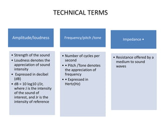
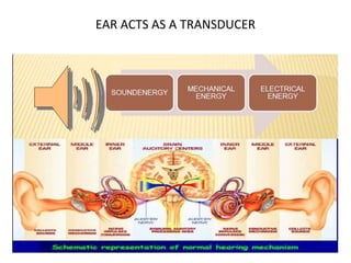
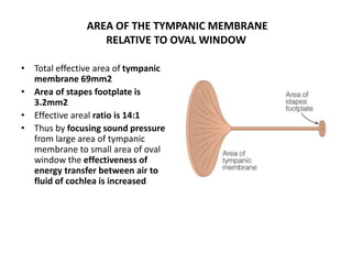
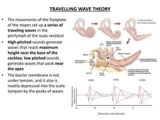
The document discusses the physiology of hearing. It describes the requirements for normal hearing including adequate stimulus, conduction to the sensory organ, transduction at the organ, neural transmission, and central processing. It then discusses the anatomy and functions of the external ear, middle ear, and inner ear. The external ear collects and conducts sound waves. The middle ear acts as an impedance matcher and lever system to efficiently transmit vibrations from the eardrum to the inner ear.

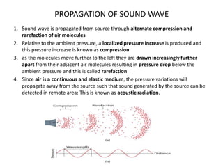

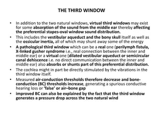
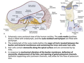
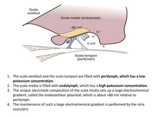
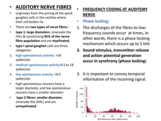
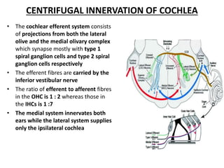
![• At a frequency with a wavelength four times the length of the canal, the canal and
the pinna together will resonate and vibrate with the incoming signal to augment
or magnify the incident acoustic signal given by the equation:
f = c/4l.
[where f is the frequency, c is the speed of sound and I is the length of the canal]
• TOTAL GAIN:The resonant frequency which enhances the incident sound by 10–20
dB is generally 2.7 kHz with a range of 2–7 kHz
• Signals below 1 kHz are generally resonated by the head and the torso whereas
frequencies above 3 kHz are augmented by the pinna](https://image.slidesharecdn.com/ayansir-240204042116-464b5819/85/ayan-sir-pptx-16-320.jpg)


![CTEV [ clubfoot] DR ARUN LAL ,DR MOHAMED ASHRAF travancore medical college k...](https://cdn.slidesharecdn.com/ss_thumbnails/ctevclubfootdrarunlaldrmohamedashraftravancoremedicalcollegekollamkeralaindia-260208063247-18fc466c-thumbnail.jpg?width=640&height=640&fit=bounds)


