
Recommended
More Related Content
Similar to atherosclerosis.pptx
Similar to atherosclerosis.pptx (20)
Recently uploaded
Recently uploaded (20)
atherosclerosis.pptx
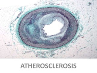
- 2. ASVD • Definition • Atherosclerosis also called arteriosclerotic vascular disease or ASVD) 1-is a condition in which an artery wall thickens as a result of the accumulation of fatty materials cholesterol and triglyceride. 2-Also is a syndrome affecting arterial blood vessels, • a chronic complex inflammatory response in the walls of arteries, • caused largely by the accumulation of macrophages (a type white blood cell that ingests blood cell foreign material, it is key player in the immune response to foreign invaders of the body) white blood cells promoted by low-density lipoproteins (LDL, in the Intima plaque of mid and large arteries.
- 3. Regarding the plaque ASVD divides in 2 groups . A-Stable atherosclerosis • B-unstable (also called vulnerable). • stable atherosclerotic plaques, which tend to be asymptomatic, are rich in extracellular matrix and smooth muscle cells, • while, unstable plaques are rich in macrophages and foam cells and the extracellular matrix separating the lesion from the arterial lumen (also known as the fibrous cap) is usually weak and prone to rupture
- 4. . Ruptures of the fibrous cap expose thrombogenic material, such as collagen to the circulation and eventually induce thrombus formation in the lumen. • Upon formation, intraluminal thrombi can occlude arteries outright (e.g. coronary occlusion), • but more often they detach, move into the circulation • eventually occluding smaller downstream branches causing thrombo embolism.
- 5. • The Atheroma ("lump of gruel",(cereal) which is the nodular accumulation of a soft, flaky, yellowish material at the center of large plaques, Underlying areas of cholesterol crystals Calcification at the outer base of older • composed of macrophages nearest the lumen of the artery Atherosclerotic lesions,
- 6. • the risk may be higher while in the blood stream. • However, LDL particles have a half-life of only a couple of days and their content (LDL particles carry cholesterol, cholesteryl esters(disease) (is a rare genetic disorder characterized by sub total defect of an enzyme known as lisosomal acid lipase (LIPA or LAL)
- 7. • Once inside the vessel wall, LDL particles get stuck and their content becomes more prone to oxidation. • Than The damage caused by the oxidized LDL molecules triggers a cascade of immune responses which over time can produce an atheroma.
- 8. • The body's immune system responds to the damage to the artery wall caused by oxidized LDL by sending specialized white blood cells (macrophages and T- lymphocytes) to absorb the oxidized-LDL forming specialized foam cells. • These white blood cells are not able to process the oxidized-LDL, and ultimately grow then rupture, depositing a greater amount of oxidized cholesterol into the artery wall. • This triggers more white blood cells, continuing the cycle.
- 9. • Ther 2 type of plaque • The fibro-lipid (fibro-fatty) plaque is characterized by an accumulation of lipid- laden cells underneath the intima of the arteries, • The fibrous plaque • is also localized under the intima, within the wall of the artery resulting in thickening and expansion of the wall and, sometimes, spotty localized narrowing of the lumen with some atrophy of the muscular layer.
- 10. Lipid fraction and the risk of CAD • Cholesterol is caried primarily 3 defferent lipoprotien • The VLDL,LDL,and HDL molecules • 1- Total cholesterole= HDL+VLDL+LDL • 2-VLDL cholesterole= Triglyceride/5 • 3-LDL = Total chol-HDL chol-Trigly /5 = mg/dl SI unit • 4-LDL= Total chol-HDL chol-Trigly/2 = mmol/l • mmol/L -mmol/L- Trigly/2.2= mmol/l
- 11. • Interestingly, chronically expanding lesions are often asymptomatic until lumen stenos is is so severe (usually over 80%) that blood supply to downstream tissue(s) is insufficient, resulting in ischemia.
- 12. • These complications of advanced atherosclerosis are chronic, slowly progressive and cumulative. • Most commonly, soft plaque suddenly ruptures , causing the formation of a thrombus that will rapidly slow or stop blood flow, • leading to death of the tissues fed by the artery in approximately 5 minutes. • This catastrophic event is called an infarction.
- 13. • One of the most common recognized scenarios is called coronary thrombosis of a coronary artery emboli , • causing myocardial infarction (a heart attack). • The same process in an artery to the brain is commonly called stroke. • Another common scenario in very advanced disease is claudicating from insufficient blood supply to the legs, typically caused by a combination of both stenosis and aneurysm segments narrowed with clots called thrombophilibitis .
- 14. Atherosclerosis affects • the entire artery tree, but mostly larger, high- pressure vessels such as the • coronary, • renal, • femoral, • cerebral, • and carotid arteries. • These are termed "clinically silent" because the person having the infarction does not notice the problem and does not seek medical help, • or when they do, physicians do not recognize what has happened.
- 15. Signs and symptom • Clinically, atherosclerosis is typically associated with men over the age of 45y. • Sub-clinically, the disease begins to appear at early childhood, and perhaps even at birth. • Noticeable signs can begin developing at puberty. • Though symptoms are rarely exhibited in children, • Early screening of children for cardiovascular diseases could be beneficial to both the child and his/her relatives. • ATheroma in arm, or more often in leg arteries, which produces decreased blood flow is called peripheral artery occlusive disease (PAOD).
- 16. • While coronary artery disease is more prevalent in men than women, atherosclerosis of the cerebral arteries and strokes equally affect both sexes. • According to United States data for the year 20013, • for about 66% of men and 47% of women, the first symptom of atherosclerotic cardiovascular disease is heart attack or sudden cardiac death (death within one hour of onset of the symptom).
- 17. • Most artery flow disrupting events occur at locations with less than 50% lumen narrowing • Diagnosis • Cardiac stress testing, • traditionally the most commonly performed non- invasive testing method for blood flow limitations, in general, detects only lumen narrowing of ~75% or greater, • Nuclear STRESS TEST • Although some physicians claim that nuclear stress methods can detect as little as 50%.
- 18. • A famous case study involved autopsies of American soldiers killed in World War II and the Korean War. • Although these were mostly young, healthy men in their 20s, many already had evidence of developing atherosclerosis. • Other studies done on soldiers in the Second Indochina War showed similar results, although often worse than the ones from the earlier wars. • Theories include high rates of tobacco use and (in the case of the Vietnam soldiers), the advent of processed foods after WWII.
- 19. Herpes virus infection of arterial smooth muscle cells has been shown to cause cholesteryl ester (CE) accumulation. Cholesteryl ester accumulation is associated with atherosclerosis. Also, cytomegalovirus (CMV) infection is associated with cardiovascular diseases.
- 20. • Risk factors • Various anatomic and physiological risk factors for atherosclerosis are known. • These can be divided into various categories: • A-Congenital • B- Acquired, • C-Modifiable , •
- 21. • Risks multiply, with two factors increasing the risk of atherosclerosis fourfold. • Hyperlipidemia, hypertension and cigarette smoking together increases the risk seven times. • Modifiable Diabetesor Impaired glucose tolerance (IGT)
- 22. • Lesser or uncertainThe following factors are of relatively lesser importance, are uncertain or unquantified: • Obesity - (in particular central obesity, also referred to as abdominal or male-type obesity) + • Postmenopausal syndrom estrogen deficiency • High intake of saturated fat (may raise total and LDL cholesterol • Intake of trans fat (may raise total and LDL cholesterol while lowering HDL cholesterol)(is the worst type of fat also called unsaturated which has double carbon means (E-esomer) and fatty acid (s)
- 23. • High carbohydrate intake[ • Elevated serum levels of triglycerides+ • Elevated serum levels of homocysteine(is a naturally occuring amino acid found in plasma . • Elevated serum levels of uric acid (also responsible for gout) • Elevated serum fibrinogen concentrations(is a protien produce by the liver –help stop bleeding –helping blood to create clot • Elevated serum lipoprotein(a) concentrations[ (contain both protien and lipids allows fat to move through the water inside and outside the cell
- 24. • Chronic systemic inflammation as reflected by upper normal WBC concentrations, elevated hs-CRP and many other blood chemistry markers, most only research level at present, not clinically done. • Hyperthyroidism (an over-active thyroid) • Elevated serum insulin levels + • Short sleep duration • Chlamydia pneumoniae infection
- 25. • Stenoses can be slowly progressive, whereas plaque ulceration is a sudden event that occurs specifically in atheromas with thinner/weaker fibrous caps that have become "unstable".
- 26. • Examples of anatomical detection methods include • (1) coronary calcium scoring by CT, • (2) carotid IMT (intimal media thickness) measurement by ultrasound, and • (3) intravascular ultrasound (IVUS). Examples of physiologic measurement methods include (1) lipoprotein subclass analysis, • (2) HbA1c, • (3) hs-CRP, and • (4) homocysteine.
- 27. • Screeing of patients –called framingham 10 years CAD risk projection are the standard • Depend of age of patients gaving special score. • Treatment plan • 1- reduction of LDL and other risk factors Smoking .hypertension ,dibetis ,alcohole use ,sedentary life style ,non of exercise ,deitary life style
- 28. Pharmacologic therapy • KEEP LDL less than 100mg/L (2.6 mmol/L) with asprin 81mg reduce the risk to 10-20% • 1-Niacin (nicotinic acid ) 3-4,5mg/daily reduce chance of CHD 15-20% -but cause flushing (hot flushes ) which reduced the use • 2-Cholestyramin (Bile Acid-Binding resins ) reduce the risk 20%- unfortunately increased triglyceride to be careful
- 29. 3-Statin- Hydroxymethylglutaryl A (HMG-COA)reductase inhibitors Include atorvastatin –fluvastatin-lovastain-pitavastatin- pravstatin –rosuvastatin- and simivastatin they reduce the enzymes which creating cholesterole. Reduce MI-STROKE, Atrovastatin 10-40mg/d Rosuvastatin5-40mg/d Fluvastatin 20-40mg/d Pravastatin 10-40mg/d Simvastatin 5-40mg/d Side effect muscle aches-GI effect-livetr effect myositis and rhabdomyolysis
- 30. D-Fibric Acid derivative • Reduce LDL –and plasma triglycerid 10 to 15% • Include gemfebrozil-600mg once or twice daily • Ciprofibrate and bezafibrate • Side effect are include chlelithithisis,hepatitis,myosistis reduce the use now E- Ezitimibe Reduce the absorbtion of dietary cholesterole in intestine -10-15mg/d have les side effect
- 31. • Best with low risk medication are • 1-HMG-COA statines better to use with low dose • 2-Niacine can be used with combination must be carefull • 3-Resins (cholestyramin ) is safe during pregnancy • 4-combination of those medication would creating side effects on liver –GI and musles