2. Overview
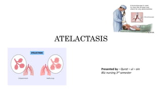
Atelectasis is defined as the collapse or closure of the lung
resulting in reduced or absent gas exchange.
It may affect part or all of one lung
It occurs when the tiny air sacs (alveoli) within the lung
become deflated or possibly filled with alveolar fluid.
3. Risk factors
Factors that make you more likely to develop atelectasis include:
Older age
Any condition that makes it difficult to swallow
Confinement to bed with infrequent changes of position
Lung disease, such as asthma, COPD, bronchiectasis or cystic fibrosis
Recent abdominal or chest surgery
Recent general anesthesia
Weak breathing (respiratory) muscles due to muscular dystrophy, spinal cord
injury or another neuromuscular condition
Medications that may cause shallow breathing
Pain or injury that may make it painful to cough or cause shallow breathing,
including stomach pain or rib fracture
Smoking
4. Causes
Atelectasis occurs from a blocked airway
(obstructive) or pressure from outside
the lung (nonobstructive).
General anesthesia is a common cause of
atelectasis.
It changes your regular pattern of
breathing and affects the exchange of
lung gases, which can cause the air sacs
(alveoli) to deflate.
Nearly everyone who has major surgery
develops some amount of atelectasis. It
often occurs after heart bypass surgery.
5. • Mucus plug. A mucus plug is a buildup of mucus in your airways.
• Foreign body. Atelectasis is common in children who have inhaled an object,
such as a peanut or small toy part, into their lungs.
• Tumor inside the airway. An abnormal growth can narrow the airway.
6. • Injury. Chest trauma — from a fall or car accident, for example — can cause you to avoid taking deep
breaths (due to the pain), which can result in compression of your lungs.
• Pleural effusion. This condition involves the buildup of fluid between the tissues (pleura) that line the lungs
and the inside of the chest wall.
• Pneumonia. Various types of pneumonia, a lung infection, can cause atelectasis.
• Pneumothorax. Air leaks into the space between your lungs and chest wall, indirectly causing some or all of
a lung to collapse.
• Scarring of lung tissue. Scarring could be caused by injury, lung disease or surgery.
• Tumor. A large tumor can press against and deflate the lung, as opposed to blocking the air passages.
Possible causes of nonobstructive atelectasis include:
7. Pathophysiology
• Reduced alveolar ventilation or any type of blockage
• Impedes the passage of air
• The trapped alveolar air becomes absorbed into the bloodstream, but outside
air cannot replace the absorbed air because of the blockage
• Isolated portion of lung becomes airless and the alveoli collapse.
• Excessive pressure on the lung tissue.
• Restricts normal lung expansion on inspiration.
• Becomes airless for prolong period.
• Alveolar collapse.
8.
9. Symptoms
There may be no obvious signs or symptoms of atelectasis. If you
do have signs and symptoms, they may include:
• Difficulty breathing
• Rapid, shallow breathing
• Wheezing
• Cough
10. Assessment and diagnostic findings
• Chest x-ray : patchy infiltrates or consolidated areas.
• Pulse oximetry : (spO2) (less than 90%) or a (PaO2)
• Physical examination : decreased breath sounds and crackles are heard over
the affected area.
11. Complications
A small area of atelectasis, especially in an adult, usually is treatable. The
following complications may result from atelectasis:
• Low blood oxygen (hypoxemia). Atelectasis makes it more difficult for your lungs
to get oxygen to the air sacs (alveoli).
• Pneumonia. Your risk for pneumonia continues until the atelectasis goes away.
Mucus in a collapsed lung may lead to infection.
• Respiratory failure. Loss of a lobe or a whole lung, particularly in an infant or in
someone with lung disease, can be life-threatening.
12. Prevention
Atelectasis in children is often caused by a blockage in the airway.
To decrease atelectasis risk, keep small objects out of reach of
children.
In adults, atelectasis most commonly occurs after major surgery. If
you're scheduled for surgery, talk with your doctor about
strategies to reduce your risk. Some research suggests that certain
breathing exercises and muscle training may lower the risk of
atelectasis after certain surgeries.
13.
14. Nursing interventions for atelectasis
When we’re talking about post-op atelectasis or atelectasis from someone not
taking deep breaths or getting out of bed and moving around as they should, the
remedy is usually pretty simple.
• Cough and deep breathe: If you assess your patient and you notice they are taking
shallow breaths, even with or without a lower-than-expected O2 saturation, you
want to have them cough and take some deep breaths. If they say their pain level is
too high to do so, you’ll need to explore pain management options. This could be
medication, heat, ice, positioning.
• Ambulate: Getting patients up and moving around is going to increase their
respiratory drive and help them keep their lungs inflated.
• Incentive spirometer: It’s a little contraption that patients use to practice taking full
deep breaths