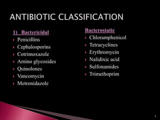
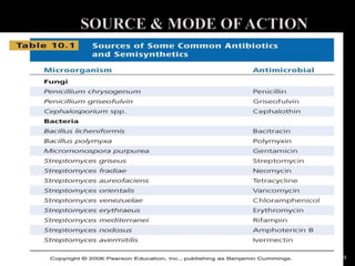
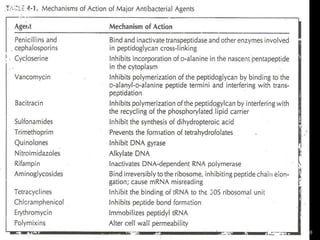
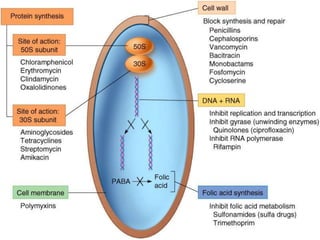
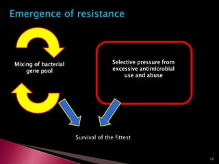
Shilpa.k
Microbiology Tutor
AIMSRC
1
•A substance produced by or derived from certain fungi, bacteria, and
other organisms, that can destroy or inhibit the growth of other
microorganisms.
Antibiotics are widely used in the prevention and treatment of infectiou
diseases.
•Antibiotics = “against life”
•Antibiotics can be either natural products or man-made synthetic
chemicals
2
IDEAL QUALITIES OF ANTIBIOTIC:
1. kill or inhibit the growth of pathogens
2. cause no damage to the host
3. cause no allergic reaction to the host
4. stable when stored in solid or liquid form
5. remain in specific tissues in the body long enough to be
effective
6. kill the pathogens before they mutate and become
resistant to it
3
1) Bactericidal
 Penicillins
 Cephalosporins
 Cotrimoxazole
 Amino glycosides
 Quinolones
 Vancomycin
 Metronidazole
Bacterostatic
 Chloramphenicol
 Tetracyclines
 Erythromycin
 Nalidixic acid
 Sulfonamides
 Trimethoprim
4
2) According to bacterial spectrum of activity
1. Narrow spectrum
2. Broad spectrum
3) According to absorbability from the site of administration
to attain significant concentration for the treatment of
systemic infection
1. Locally acting
2. Systemic
5
 A. Inhibitors of cell wall synthesis
 1. Penicillins
 2. Cephalosporins
 3. Other antibacterial agents that act on cell walls
 B. Disrupters of cell membranes
 1. Polymyxins
 2. Tyrocidins
 C. Inhibitors of protein synthesis
 1. Aminoglycosides
 2. Tetracyclines
 3. Chloramphenicol
 4. Other antibacterial agents that affect protein synthesis
 a. Macrolides
 b. Lincosamides
 D. Inhibitors of nucleic acid synthesis
 1. Rifampin
 2. Quinolones
 E. Antimetabolites and other antibacterial agents
 1. Sulfonamides
 2. Isoniazid
 3. Ethambutol
 4. Nitrofurans 6
First Generation
First generation cephalosporins are effective in fighting gram-positive
bacteria such as strains of staphylococcus and streptococcus. Among
first generation cephalosporin antibiotics are Ancef and Kefzol
(cefazolin)and Keflex (cephalexin).
Second Generation
In addition to the gram-positive effects of the first generation drugs,
second generation cephalosporins expand coverage to gram-negative
bacteria such as enterobacter, pseudomonas and salmonella. Second
generation drugs vary in coverage, and testing is often required to
determine the appropriate agent to prescribe. Among second generation
drugs are Cefzil (cefprozil), Ceclor (cefaclor) and Lorabid (loracarbef).
The only second generation cephalosporin that crosses the blood brain
barrier is cefuroxime; therefore, it is used to treat meningitis
7
Third Generation
Third generation cephalosporins extend coverage of gram-negative
organisms. These drugs have the ability to penetrate the central
nervous system to treat infections that affect the neurological
system. These third generation drugs also require testing for
determination of the appropriate agent. Among third generation
cephalosporins are Omnicef (cefdinir), Rocephin (ceftriaxone) and
Suprax (cefixime).
Fourth Generation
As of 2010, a fourth generation of cephalosporins is in
development that will be used to treat Methicillin-resistant
Staphylococcus aureus (MRSA).
8
9
10
11
Emergence of resistance
Mixing of bacterial
gene pool
Selective pressure from
excessive antimicrobial
use and abuse
Survival of the fittest
12
 Resistance is concered because of the emerging of resistant
micro-organisms causing serious infections with out
responding to the antibiotic treatment.
 They are causing resistance not only in hospital patients even
in community also and increasing microbial population and
decreasing drug potency.
13
 Cidal + cidal – Synergistic
 Static + cidal – Antagonistic
 Static + static – Additive
 Often impossible to predict the effect of a particular
combination against a particular isolate
14
ACQUISITION OF BACTERIAL RESISTANCE
INTRINSIC RESISTANCE
• Stable genetic property encoded in the chromosome and
shared by all strains of the species
• Usually related to structural features (e.g. permeability of
the cell wall)  e.g. Pseudomonas limits penetration of
antibiotics cellwall
15
ACQUIRED RESISTANCE
• Species develop ability to resist an antimicrobial drug to
which it is as a whole naturally susceptible
• Two mechanisms:
1. Mutational – chromosomal
2. Genetic exchange – transformation, transduction,
conjugation
Ex: Horizontal transformation by Plasmids
Mec A genes they code for PBP with low affinity for ß-
Lactam antibiotics
16
ORIGIN OF DRUG RESISTANCE
NON-GENETIC
Certain bacteria normally sensitive to pencillins become
resistant when they become protoplasts(loss of cell
wall)
1. Loss of specific target structure for a drug for several
generations
2. Organism infects host at sites where antimicrobials are
excluded or are not active – vancomycin over gram
negative bacilli.
3. Non replicating bacteria in their resting stage are less
sensitive to the action of cell wall inhibitors such as
pencillins and cephalosporins.
17
•Non replicating bacteria in their resting stage are less sensitive to
the action of cell wall inhibitors such as pencillins and
cephalosporins
Ex: Mycobacterium tuberculosis remains in resting stage in tissues
for many years during the period they are resistant when they begin
to multiply they become suspectable to antibiotics.
18
 Chromosomal mediated resistance
 Plasmid mediated resistance
 Transposons mediated resistance
19
 Occurs at a frequency of 10-12 to 10-7
 It occurs less than Plasmid mediated drug resistance
 This is due to mutation in the gene that codes for:
a) drug target
b) transport system in the membrane that
controls drug uptake
 Multi drug treatment is used in case of tuberculosis this
is because tubercle bacilli develops resistance through
chromosomal mediated resistance when are given
simultaneously.
20
 This occurs by transfer of plasmid and genetic materials.
 It is mediated by transfer of resistance Plasmid.
Resistance Plasmids are :
 Circular
 Double stranded DNA molecules that carry genes responsible
for resistance against antibiotics.
21
 Transposons are small pieces of DNA that move from one site
of bacterial chromosome to another.
 They also move from chromosome to plasmid DNA.
 They offer resistance to multiple drugs.
Plasmid & transposons mediated resistance
plays an important role of genetic mediated
resistance:
•High rate of transfer of Plasmids by conjugation.
•Resistance to multiple antibiotics
•It occurs mostly in gram negative bacilli 22
MOSAIC GENES:
 Some bacterial species absorb naked DNA molecules from
the environment under appropriate circumstances .
 These foreign pieces of DNA enters the bacterial
chromosomes by recombining across regions of homology.
 Some causes functional genes result from the recombination.
If the Acquired gene encodes for a protein that is susceptible
to inhibition than native protein ,a reduction in susceptibility may
occurs.
Ex: Mosaic genes confer resistance is penicillin and cephalosporins
in streptococcus Pneumonia
23
1. Production of enzymes that inactivate the drug
a. -lactamase
 S. aureus, Enterobacteriaceae, Pseudomonas, H. influenzae
b. Chloramphenicol acetyltransferase
 S. aureus, Enterobacteriaceae
c. Adenylating, phosphorylating or acetylating enzymes
(aminoglycosides)
 S. aureus, Strep, Enterobacteriaceae, Pseudomonas
24
2. Altered permeability to the drug  result to decreased effective
intracellular concentration
Tetracycline, Penicillin, Polymixins, Aminoglycosides,
Sulfonamides.
3. Synthesis of altered structural targets for the drug
a. Streptomycin resistance – mutant protein in 30S ribosomal
subunit  delete binding site  Enterobacteriaceae
b. Erythromycin resistance – altered receptor on 50S subunit
due to methylation of a 23S rRNA  S. aureus
25
4. Altered metabolic pathway that bypasses the reaction inhibited
by the drug
Sulfonamide resistance – utilize preformed folic acid instead of
extracellular PABA  S. aureus, Enterobacteriaceae
5. Multi-drug resistance pump
Bacteria actively export substances including drugs in exchange
for protons
Quinolone resistance
26
27
 Preferred drugs are used for the specific species of
microorganism this is because of mode of action and effective
treatment.
 The drugs used routinely are grouped under first line and the
reserved group are second line drugs.
 Azetreonam which is a monobactem should not be tested
against gram positive because it binds majorly to PBP of
GNB. •.
o Alkaline pH is favoured for the good action of
tetracycline and novabiocin but it interfere with
nitrofurantoin.
28
 Vancomycin should not used against GNB .This is because the
pore size of them is big and can not penetrate the cell wall.
 Cephalosporins should not be used against enterococcus .This
is because lack of PBP’s that effectively bind and inhibited by
these ß-lactams.
 Teracyclines against proteus species this is because lack of
uptake resulting inabillity of antibiotics.
 Aminoglycosides should not be used for anaerobic bacteria, as
their transport requires oxygen.
 Nalidixic acid and Nitrofurantoin are used only for UTI
because, they are active at acidic pH.
29
 First generation cephelosporins should not be used against
Haemophilus because they have modest activity.
 To avoid cross resistance only one representative need to be
tested from any group of closely releated drugs.
 Drugs that combine ß-lactamase inhibitors must be tested
individually.
 Ex: Piperacillin Tozobactem
Amoxycillin / clavulanic acid.
 The Presence of calcium and magnesium at low concentration
increases the activity of drugs,where as presence of high
concentration decreases the activity of drugs.
30
 ANTIMICROBIAL ASSAYS ARE USED TO
DETERMINE;
1)Extent of organisms acquired resistance
2 ) Organisms whose susceptibility profile is
unpredicted
31
 The primary goal of antibiotic sensitivity is to determine
whether bacteriology etiology of concern capable of
expressing resistance to the antimicrobial agents that are
potentially choosen
 Intrinsic resistance is usually well known for most of
microorganisms
32
 Inoculum size
 Growth medium
 pH
 Cation concentration
 Blood &serum supplements
 Thymidine content
 Incubation atmosphere
 Incubation temperature
• Incubation duration
• Antimicrobial concentrate tested
• In some cases however antimicrobial activity of bactericidal
activity is required to yield a high percentage of treatment
success than bacteriostatic
33
ANTIBIOTIC DISCS:
 Commercially prepared 6mm in diameter discs are used.
 Discs and disc dispensers should be stored at -20°c or 8°c/less
than 8°c
 To prevent hydrolysis of the antibiotic before use they should
be brought to room temperature.
34
MEDIUM:
• The most commonly used medium for antibiotic
susceptibility testing is MULLER-HINTON AGAR.
• In muller-Hinton agar starch is present it acts as
detoxifying agent for bacteria
 Calcium & magnesium ions should be maintained at
minimum levels
• Wilkins-chalgren agar is recommended for testing of
anaeorobic bacteria.
35
 Susceptibility testing is to predict the outcome of treatment
with the antimicrobial agent tested
 They are generally reported as;
a) Susceptible
b) Resistant
c) Intermediate
36
 The implication of susceptable is that there is a high propability
that the patient will respond to treatment with appropriate
dosage.
 The implication of” resistant “ implies that the treatment with
that antimicrobial agent is likely to fail and no use with the drug
of choice.
• The implication of “intermediate ”catogory include that the
higher dosage is required for the efficacy of treatment.
• This testing should not be done on normal microbial flora.
37
INOCULUM PREPARATION:
 Inoculum preparation should be limited by preventing
excessive growth of micro-organism which may affect
suspectibility testing.
 This is done by comparision of inoculum with Mc Farland
standards.
PREPARATION OF Mc FARLAND:
STANDARD:
•This is prepared by mixing solution –A and Solution-B
•Solution-A:
Add barium chloride to 100ml of dist.water.
•Solution-B:
Add 1ml of sulphuric acid to 100ml of distilled water.
•Then 0.5 ml of solution –A is mixed with 99.5ml of solution-B
and is disturbed in test tubes .
38
 Then 0.5 ml of solution –A is mixed with 99.5ml of solution-B
and is disturbed in test tubes .
 Stored in dark places.
 Mixture is agitated and used before using it.
39
40
 It is defined as the lowest concentration that inhibit the growth
of test organism over a defined interval of 18-24hrs.
 MIC measurements are influenced by:
a) Composition of the medium
b) Size of inoculum.
c) Duration of incubation.
d) Presence of resistant sub population.
41
 MIC are used in the determination of antibiotic sensitivities of
organism with series infection.
 This is used to distinguish strains for which MIC are
abnormally high (Resistant) and for which MIC are
normal(sensitive).
42
MINIMUM BACTERICIDAL CONCENTRATION:
 It is defined as the amount of antibiotic that will prevent the
growth of microorganism after subculture to antibiotic free
medium .
 This is useful in testing ths bactericidal concentrationof the
drug .
 This can be checked with the MIC ,the inoculum with MIC is
cultured on a antibiotic free medium & after incubation
observed for growth.
43
BREAKPONTS:
 It refers to the sensitivity testing at specific concentration.
 After sensitivity testing it is essential to determine whether the
causative organism is sensitive to the concentration of the
antibiotic at the site of infection.
 The test is set in exactly the same manner of MIC in media
preparation,concentration of antibiotic,inoculum etc.,
 1-2µl of sample delivered to agar plate.
 After incubation the plates are read for the growth &
inhibition.
44
CHOICE OF BREAKPOINT:
 This may also vary for the same antibiotic in relation to same
pathogen at same site of infection
 Most developed countries have devised their own breakpoints.
 The choice of breakpoints become particularly difficult when drug
combinations are used.
Ex:
 Co-trimaxazole & trimethoprim are administered in 1:5
combination.
 It is unlikely that 50% of the pathogens are resistant to
sulphamethaxazole but clinical microbiologists recommend use of
co-trimaxazole.
 Then this combination should be used if the organism is completely
sensitive to trimethoprim.
 If the organism is sensitive then only trimethoprim be the drug of
choice.
 Same in case of co-amoxyclav.
45
 1.Dilution Testing:
A)Agar dilution Method
B)Broth dilution Method
a)Broth macro dilution
b) Broth micro dilution
C)Disc diffusion Method
D)E-Test
46
Media :
 Muller Hinton Agar is the recommended medium for testing
more commonly encountered aerobic and facultatively
anaerobes.
 Before sterilization the agar is usually distributed into screw
cap tubes in exact aliquots sufficient to dilute the desired the
anti microbial concentration.
47
 Once the agar has equilibrated appropriate volume of
antimicrobial agent is added.
 Tubes of Agar of desired concentration are sterilized by
autoclaving at 121°c for 15 min.
 The Tubes contents are mixed by gentle inversion and are
poured into sterile petri plates, and allowed to solidify.
 The plate should be filled to a depth of 3-4mm and are stored
at 4-8°c.
48
49
 For Streptococci 5% Sheep or horse blood can be added
blood may antagonize sulphonamide and trimethoprim.
 Blood may also effect novabiocin
 So blood should be added in necessary conditions of growth
only
 Agar can be supplemented with 2% Nacl for testing Oxacillin
against Staphylococcus.
INOCULATION PROCEDURE:
 The recommended final inoculum for agar dilution is 104 cfu
per spot.
 4 or 5 colonies from overnight growth cultures are inoculated
into 4-5ml of suitable broth.
 Broths are incubated at 35°c until visible turbidity.
50
 The Turbidity of the broths are compared with 0.5Mc Farland
standard, normal saline or sterile broth is used to make 1:10
dilution of suspension.
 Bacterial inoculum is adjusted by using a pipette 1-2 µlof the
suspension is delivered to agar surface.
 The surface of the agar plate should be dried before
inoculation and should begin with lowest concentration.
 INCUBATION:
 Plates are incubated at 35°c for 16-20hrs
 Incubation should not be carried in increase Co2
51
 Results are best determined by placing plates on a dark
background and examine for lowest concentration that inhibit
visible growth(MIC).
 Presence of faint colonies before inoculation should not be
considered as growth.
 ADVANTAGES AND DIS-ADVANTAGES;
 This method is well standardized , reliable suspectibility
testing technique.
 It is time consuming and labor intensive task .
52
 In the broth macrodilution method the broth volume for each
antimicrobial agent is >1.0ml or =2ml.
 MEDIUM:
Cation Adjusted muller-hinton broth or muller-hinton
with adjusted divalent cations is used.
Calcium &magnesium levels should be 20-
25mg/lit&10-12.5mg/lit respectively.
Stock solutions are prepared according to the CLSI as
per agar dilution method.
53
 The recommended final inoculum of macrodilution is 5x10⁵
cfu/ml
 Isolates adjusted to Mc-Farland standards
 When 1ml of this dilution is added to each tube containing
1ml of drug diluted in CAMHB a final inoculum of 5x10⁵
CFU/ml is achieved.
 INCUBATION:
Incubated at 37c for 16-20 hrs.
increased Co2 should not be present.
54
INTERPRETATION:
 Before MIC’s for test strain are read &recorded the growth controls
should be examined for viability &inoculum.
 Growth is indicated by turbidity or sediment at the bottom of the
tube.
 The tube without antibiotic drug is considered as growth control.
 Growth or lack of growth in the antimicrobial agent containing tube
is best determined by comparing with growth control.
 The consideration of SKIPPED TUBE PHENOMENON
phenomenon is important.
55
 Skipped tube phenomenon is that growth is not observed at
one concentration but observed at higher &lower
concentration of the drug.
 Skipped tubes should be ignored ,the concentration that finally
inhibits growth should be recorded as MIC.
 ADVANTAGES &DISADVANTAGES:
 Best used for research purposes testing one drug with bacterial
isolate at various concentrations.
 It is more laborious
 Cannot be used for routine susceptibility testing.
56
57
 Broth microdilution method is internationally accepted
susceptibility testing method.
 Here,the broth volume for each microbial agent is 0.05-0.1ml
 They are freeze dried
 This contains disposable plastic trays containing panel of
several antibiotics to be tested simultaneously.
MEDIA:
CAMHB is used as in macro dilution method.
 The media are routinely prepared as macrodilution &agar
dilution.
 The main future is after the antimicrobial agents are dispensed
they are sealed with empty trays above.
 They are immediately frozen to -20c
58
 Care must be taken in storing without the loss of potency of
the drug.
 The tubes contain 10-200ml of broth containing each
antimicrobial agent of 0.05-0.1ml which are simultaneously
dispensed.
 One well without antimicrobial agent is used as control.
INCUBATION:
 The final desired inoculum for microdilution is 5x10⁵ CFU/ml.
 Compared with Mc Farland standards.
INCUBATION
 Incubated at 35c for 16-20hrs.
59
INTERPRETATION:
 Various viewing are available to facilitate microdilution.
 Growth is best determimed by comparing with growth control
well &generally indicated by turbidity.
ADVANTAGES &DISADVANTAGES:
 The broth microdilutions method represents a most
standardized susceptibility testing.
 More convenient method for many laboratories.
 It is the best method to determine MIC
 The wells should be carefully stored without the antimicrobial
agent getting dried.
60
61
62
Disk diffusion Method:
KIRBY-BAUER METHOD :
• Surface of agar plate seeded with lawn of test organism
• Inoculum: swab from 0.5 McFarland
• Disks containing known conc. of agent placed
on surface of plate
• Measure diameter of zone of inhibition
• Zones are measured with vernier callipers.
• The minimum number of drugs used in this method are 6per
90mm petridish
63
64
65
66
STOKES METHOD:
 Stokes method is another example of disc diffusion method.
 This method makes use of inbuilt controls against many
variables &therefore provide dependable results.
 In this method antibiotic discs are applied between the standard
& test inoculum so that zones of inhibition are formed around
each disc are composed of standard &test bacteria.
 In this only 4 discs are placed in 90mm plate with 2cms between
two discs.
 Rotary plating method can also be used ,control strains in outer
periphery &test strains in inner periphery.
 6 discs can be used in method.
67
68
INTERPRETATION:
 In this method zone size is measured from the edge of the disc
to zone edge.
69
 Epsilometer-test
 Tear drop shape inhibition is seen with MICon agar.
 Plastic strip containing a gradient of ANTIBIOTIC on
one side & interpretive scale one the other.
 Very useful & can be adapted to any agar medium & most
micro organisms.
 The zone edge intersects the gradient strip at he minimum
inhibitory concentration of the antibiotic.
70
71
72
TESTS USED TO DETECT β-LACTAMASES & ESBL:
β-LACTAMASES:
 Some sensitivity testings of laboratory may fail to indicate
paticularly can the drugs be used or not.
 The main factor for this is production of β-lactamase.
 The expression &demonstration of β-lactamas is difficult
because:
a) The bacterial concentration should be high.
b)insufficient β-lactamase production from the test
organism before an inhibitory concentration of antibiotic as
diffused from disc.
 So the sensitivity testing may fail to recognize a patient
resistance mechanism.
 The determination of β-lactamase is measured by production
of pencillonic acid when benzyl pencillin is used.
73
 The detection of ß-lactamase is measured by pencillonic acid.
This acid production has been detected by:
a)measuring change in pH with an indicator
dye.(acidometric)
b)the ability of pencillonic acid to reduce iodine
&formation of blue colour.(complex of starch iodometric
method).
ACIDOMETRIC METHOD:
 Filter paper impregnated with pencillin & pH indicator dye
bacterial growth from over night applied on it.
 If the strain is β-lactamase it lowers the pH & changes the
colour of filter paper to red.
74
IODOMETRIC METHOD:
•The ability of pencillonic acid to reduce iodine &formation of
blue colour.(complex of starch iodometric method).
•The colour development of blue indicates the strain is β-
lactamase.
CHROMOGENIC CEPHELOSPORIN:
•Nitrocephin is the chromogenic cephelosporin usually yellow in
colour.
•When β-lactamas is brokendown it is converted to red colour
•EX: 50ml suspension of over night culture+10ml of nitrocephin
development of red colour without incubation.
75
76
TESTS OF INDUCIBLE β-LACTAMASES:
 The chromosomally encoded β-lactamases are serine based ambler
class C enzymes whose expression is repressed.This shows only
sufficient amount of β-lactamase is produced without waste of
energy.
 When the inducer β-lactam antibiotic is withdrawn the enzyme
becomes normal.
 A simple technique for screening potentially inducible enzymes is to
test sensitivity to cefoxitin.
 This is tested with two β-lactum antibotics.
 In this strong inducer cefoxitin and a weak inducer cefotaxime are
placed at a distance of twice the radius of zone of inhibition.
 After overnight incubation,if induction takes place,the radius of
inhibition of cefotaxime disc is flattened one side to cefoxitin.
77
 EXTENDED SPECTRUM β-LACTAMSES:
 Extended spectrum β-lactaases are the result result of mutations in
β-lactamases.
 The common ESBL all remain sensitive to inhibition by clavulanic
acid.
 If the strain is ESBL & if it shows resistance to ceftezidime or
cefotaxime becomes sensitive in the presence of clavulanic acid
after overnigth incubation the zone of inhibition is extended.
 EX: the drugs are tested as distance seperated twice the radius of the
zone of inhibition.
 cefotaxime--------amoxyclav+clavulanic acid(10µg)
If the zone of clavulanic acid is >5mm it is ESBL & clavulanic acid is
acting on .
78
79
ASSAY OF ONE ANTIBIOTIC IN THE PRESENCE
OF OTHER:
Two drugs can be used in efficient treatment because:
 a)to broaden spectrum of activity.
 b)to treat mixed infections where one do not work.
 c)to prevent emergence of mutants.
 d)to neutralize β -lactamases.
 e)synergical action of antibiotic.
 F)reduce drug related toxicity.
 DRUG INTERACTIONS:
1) BACTERIOSTATIC
2) BACTERIOCIDAL
80
BACTERIOSTATIC INTERACTIONS:
 As easier method to show whether the two drugs have a
positive interaction with one another is DOUBLE DISC TEST.
 This is most often done with trimethoprim&
sulphamethaxazole.
 The two drugs are placed at a distance of 25mm apart.
 Trimethoprim &sulphamethoxazole of concentration1. 25µg
&25µg are placed respectively.
 Plates are examined next day this shows the presence of BOW
outwards another.
 The action may SYNERGY but it is less clear whether they
treat common infections.
81
82
 BACTERICIDAL INTERACTION:
 This shows the two drugs act cooperatively or one antagonize
the action of other.
83
DETECTION OF INDUCIBLE CLINDAMYCIN
RESISTANCE:
 If 15µg of erythromycin to a 2µg of clindamycin are placed
adjacently after overnight incubation they look like flattering
of clindamycin zone like letter of D(disc detection of inducible
clindamycin).
 They should be 15mm apart .
 This should generally reported as clindamycin resistant.
 This is because of inducible erm genes & is known as
MACROLIDE-LINCOSAMIDE-STREPTOGRAMIN
RESISTANCE.
84
85
86
87
CEFOXITIN RESISTANCE FOR STAPHYLOCOCCUS:
 Cefoxitin is the preferred drug for detection of mec-A
mediated oxacillin in staphylococcus aureus.
 Cefoxitin is the third generation cephelosporin.
88
POST ANTIBIOTIC EFFECT:
 This is defined as long term effect of antibiotic particularly
after antibiotic is excreted.
 Some of the drugs such as quinolenes may persist even after
the excretion or dilution in the laboratory testing.
 This shows whether the antibiotic is bactericidal or
bacteriostatic.
89
90

Antibioticsusceptibilitytesting

  • 1.
  • 2.
    •A substance producedby or derived from certain fungi, bacteria, and other organisms, that can destroy or inhibit the growth of other microorganisms. Antibiotics are widely used in the prevention and treatment of infectiou diseases. •Antibiotics = “against life” •Antibiotics can be either natural products or man-made synthetic chemicals 2
  • 3.
    IDEAL QUALITIES OFANTIBIOTIC: 1. kill or inhibit the growth of pathogens 2. cause no damage to the host 3. cause no allergic reaction to the host 4. stable when stored in solid or liquid form 5. remain in specific tissues in the body long enough to be effective 6. kill the pathogens before they mutate and become resistant to it 3
  • 4.
    1) Bactericidal  Penicillins Cephalosporins  Cotrimoxazole  Amino glycosides  Quinolones  Vancomycin  Metronidazole Bacterostatic  Chloramphenicol  Tetracyclines  Erythromycin  Nalidixic acid  Sulfonamides  Trimethoprim 4
  • 5.
    2) According tobacterial spectrum of activity 1. Narrow spectrum 2. Broad spectrum 3) According to absorbability from the site of administration to attain significant concentration for the treatment of systemic infection 1. Locally acting 2. Systemic 5
  • 6.
     A. Inhibitorsof cell wall synthesis  1. Penicillins  2. Cephalosporins  3. Other antibacterial agents that act on cell walls  B. Disrupters of cell membranes  1. Polymyxins  2. Tyrocidins  C. Inhibitors of protein synthesis  1. Aminoglycosides  2. Tetracyclines  3. Chloramphenicol  4. Other antibacterial agents that affect protein synthesis  a. Macrolides  b. Lincosamides  D. Inhibitors of nucleic acid synthesis  1. Rifampin  2. Quinolones  E. Antimetabolites and other antibacterial agents  1. Sulfonamides  2. Isoniazid  3. Ethambutol  4. Nitrofurans 6
  • 7.
    First Generation First generationcephalosporins are effective in fighting gram-positive bacteria such as strains of staphylococcus and streptococcus. Among first generation cephalosporin antibiotics are Ancef and Kefzol (cefazolin)and Keflex (cephalexin). Second Generation In addition to the gram-positive effects of the first generation drugs, second generation cephalosporins expand coverage to gram-negative bacteria such as enterobacter, pseudomonas and salmonella. Second generation drugs vary in coverage, and testing is often required to determine the appropriate agent to prescribe. Among second generation drugs are Cefzil (cefprozil), Ceclor (cefaclor) and Lorabid (loracarbef). The only second generation cephalosporin that crosses the blood brain barrier is cefuroxime; therefore, it is used to treat meningitis 7
  • 8.
    Third Generation Third generationcephalosporins extend coverage of gram-negative organisms. These drugs have the ability to penetrate the central nervous system to treat infections that affect the neurological system. These third generation drugs also require testing for determination of the appropriate agent. Among third generation cephalosporins are Omnicef (cefdinir), Rocephin (ceftriaxone) and Suprax (cefixime). Fourth Generation As of 2010, a fourth generation of cephalosporins is in development that will be used to treat Methicillin-resistant Staphylococcus aureus (MRSA). 8
  • 9.
  • 10.
  • 11.
  • 12.
    Emergence of resistance Mixingof bacterial gene pool Selective pressure from excessive antimicrobial use and abuse Survival of the fittest 12
  • 13.
     Resistance isconcered because of the emerging of resistant micro-organisms causing serious infections with out responding to the antibiotic treatment.  They are causing resistance not only in hospital patients even in community also and increasing microbial population and decreasing drug potency. 13
  • 14.
     Cidal +cidal – Synergistic  Static + cidal – Antagonistic  Static + static – Additive  Often impossible to predict the effect of a particular combination against a particular isolate 14
  • 15.
    ACQUISITION OF BACTERIALRESISTANCE INTRINSIC RESISTANCE • Stable genetic property encoded in the chromosome and shared by all strains of the species • Usually related to structural features (e.g. permeability of the cell wall)  e.g. Pseudomonas limits penetration of antibiotics cellwall 15
  • 16.
    ACQUIRED RESISTANCE • Speciesdevelop ability to resist an antimicrobial drug to which it is as a whole naturally susceptible • Two mechanisms: 1. Mutational – chromosomal 2. Genetic exchange – transformation, transduction, conjugation Ex: Horizontal transformation by Plasmids Mec A genes they code for PBP with low affinity for ß- Lactam antibiotics 16
  • 17.
    ORIGIN OF DRUGRESISTANCE NON-GENETIC Certain bacteria normally sensitive to pencillins become resistant when they become protoplasts(loss of cell wall) 1. Loss of specific target structure for a drug for several generations 2. Organism infects host at sites where antimicrobials are excluded or are not active – vancomycin over gram negative bacilli. 3. Non replicating bacteria in their resting stage are less sensitive to the action of cell wall inhibitors such as pencillins and cephalosporins. 17
  • 18.
    •Non replicating bacteriain their resting stage are less sensitive to the action of cell wall inhibitors such as pencillins and cephalosporins Ex: Mycobacterium tuberculosis remains in resting stage in tissues for many years during the period they are resistant when they begin to multiply they become suspectable to antibiotics. 18
  • 19.
     Chromosomal mediatedresistance  Plasmid mediated resistance  Transposons mediated resistance 19
  • 20.
     Occurs ata frequency of 10-12 to 10-7  It occurs less than Plasmid mediated drug resistance  This is due to mutation in the gene that codes for: a) drug target b) transport system in the membrane that controls drug uptake  Multi drug treatment is used in case of tuberculosis this is because tubercle bacilli develops resistance through chromosomal mediated resistance when are given simultaneously. 20
  • 21.
     This occursby transfer of plasmid and genetic materials.  It is mediated by transfer of resistance Plasmid. Resistance Plasmids are :  Circular  Double stranded DNA molecules that carry genes responsible for resistance against antibiotics. 21
  • 22.
     Transposons aresmall pieces of DNA that move from one site of bacterial chromosome to another.  They also move from chromosome to plasmid DNA.  They offer resistance to multiple drugs. Plasmid & transposons mediated resistance plays an important role of genetic mediated resistance: •High rate of transfer of Plasmids by conjugation. •Resistance to multiple antibiotics •It occurs mostly in gram negative bacilli 22
  • 23.
    MOSAIC GENES:  Somebacterial species absorb naked DNA molecules from the environment under appropriate circumstances .  These foreign pieces of DNA enters the bacterial chromosomes by recombining across regions of homology.  Some causes functional genes result from the recombination. If the Acquired gene encodes for a protein that is susceptible to inhibition than native protein ,a reduction in susceptibility may occurs. Ex: Mosaic genes confer resistance is penicillin and cephalosporins in streptococcus Pneumonia 23
  • 24.
    1. Production ofenzymes that inactivate the drug a. -lactamase  S. aureus, Enterobacteriaceae, Pseudomonas, H. influenzae b. Chloramphenicol acetyltransferase  S. aureus, Enterobacteriaceae c. Adenylating, phosphorylating or acetylating enzymes (aminoglycosides)  S. aureus, Strep, Enterobacteriaceae, Pseudomonas 24
  • 25.
    2. Altered permeabilityto the drug  result to decreased effective intracellular concentration Tetracycline, Penicillin, Polymixins, Aminoglycosides, Sulfonamides. 3. Synthesis of altered structural targets for the drug a. Streptomycin resistance – mutant protein in 30S ribosomal subunit  delete binding site  Enterobacteriaceae b. Erythromycin resistance – altered receptor on 50S subunit due to methylation of a 23S rRNA  S. aureus 25
  • 26.
    4. Altered metabolicpathway that bypasses the reaction inhibited by the drug Sulfonamide resistance – utilize preformed folic acid instead of extracellular PABA  S. aureus, Enterobacteriaceae 5. Multi-drug resistance pump Bacteria actively export substances including drugs in exchange for protons Quinolone resistance 26
  • 27.
  • 28.
     Preferred drugsare used for the specific species of microorganism this is because of mode of action and effective treatment.  The drugs used routinely are grouped under first line and the reserved group are second line drugs.  Azetreonam which is a monobactem should not be tested against gram positive because it binds majorly to PBP of GNB. •. o Alkaline pH is favoured for the good action of tetracycline and novabiocin but it interfere with nitrofurantoin. 28
  • 29.
     Vancomycin shouldnot used against GNB .This is because the pore size of them is big and can not penetrate the cell wall.  Cephalosporins should not be used against enterococcus .This is because lack of PBP’s that effectively bind and inhibited by these ß-lactams.  Teracyclines against proteus species this is because lack of uptake resulting inabillity of antibiotics.  Aminoglycosides should not be used for anaerobic bacteria, as their transport requires oxygen.  Nalidixic acid and Nitrofurantoin are used only for UTI because, they are active at acidic pH. 29
  • 30.
     First generationcephelosporins should not be used against Haemophilus because they have modest activity.  To avoid cross resistance only one representative need to be tested from any group of closely releated drugs.  Drugs that combine ß-lactamase inhibitors must be tested individually.  Ex: Piperacillin Tozobactem Amoxycillin / clavulanic acid.  The Presence of calcium and magnesium at low concentration increases the activity of drugs,where as presence of high concentration decreases the activity of drugs. 30
  • 31.
     ANTIMICROBIAL ASSAYSARE USED TO DETERMINE; 1)Extent of organisms acquired resistance 2 ) Organisms whose susceptibility profile is unpredicted 31
  • 32.
     The primarygoal of antibiotic sensitivity is to determine whether bacteriology etiology of concern capable of expressing resistance to the antimicrobial agents that are potentially choosen  Intrinsic resistance is usually well known for most of microorganisms 32
  • 33.
     Inoculum size Growth medium  pH  Cation concentration  Blood &serum supplements  Thymidine content  Incubation atmosphere  Incubation temperature • Incubation duration • Antimicrobial concentrate tested • In some cases however antimicrobial activity of bactericidal activity is required to yield a high percentage of treatment success than bacteriostatic 33
  • 34.
    ANTIBIOTIC DISCS:  Commerciallyprepared 6mm in diameter discs are used.  Discs and disc dispensers should be stored at -20°c or 8°c/less than 8°c  To prevent hydrolysis of the antibiotic before use they should be brought to room temperature. 34
  • 35.
    MEDIUM: • The mostcommonly used medium for antibiotic susceptibility testing is MULLER-HINTON AGAR. • In muller-Hinton agar starch is present it acts as detoxifying agent for bacteria  Calcium & magnesium ions should be maintained at minimum levels • Wilkins-chalgren agar is recommended for testing of anaeorobic bacteria. 35
  • 36.
     Susceptibility testingis to predict the outcome of treatment with the antimicrobial agent tested  They are generally reported as; a) Susceptible b) Resistant c) Intermediate 36
  • 37.
     The implicationof susceptable is that there is a high propability that the patient will respond to treatment with appropriate dosage.  The implication of” resistant “ implies that the treatment with that antimicrobial agent is likely to fail and no use with the drug of choice. • The implication of “intermediate ”catogory include that the higher dosage is required for the efficacy of treatment. • This testing should not be done on normal microbial flora. 37
  • 38.
    INOCULUM PREPARATION:  Inoculumpreparation should be limited by preventing excessive growth of micro-organism which may affect suspectibility testing.  This is done by comparision of inoculum with Mc Farland standards. PREPARATION OF Mc FARLAND: STANDARD: •This is prepared by mixing solution –A and Solution-B •Solution-A: Add barium chloride to 100ml of dist.water. •Solution-B: Add 1ml of sulphuric acid to 100ml of distilled water. •Then 0.5 ml of solution –A is mixed with 99.5ml of solution-B and is disturbed in test tubes . 38
  • 39.
     Then 0.5ml of solution –A is mixed with 99.5ml of solution-B and is disturbed in test tubes .  Stored in dark places.  Mixture is agitated and used before using it. 39
  • 40.
  • 41.
     It isdefined as the lowest concentration that inhibit the growth of test organism over a defined interval of 18-24hrs.  MIC measurements are influenced by: a) Composition of the medium b) Size of inoculum. c) Duration of incubation. d) Presence of resistant sub population. 41
  • 42.
     MIC areused in the determination of antibiotic sensitivities of organism with series infection.  This is used to distinguish strains for which MIC are abnormally high (Resistant) and for which MIC are normal(sensitive). 42
  • 43.
    MINIMUM BACTERICIDAL CONCENTRATION: It is defined as the amount of antibiotic that will prevent the growth of microorganism after subculture to antibiotic free medium .  This is useful in testing ths bactericidal concentrationof the drug .  This can be checked with the MIC ,the inoculum with MIC is cultured on a antibiotic free medium & after incubation observed for growth. 43
  • 44.
    BREAKPONTS:  It refersto the sensitivity testing at specific concentration.  After sensitivity testing it is essential to determine whether the causative organism is sensitive to the concentration of the antibiotic at the site of infection.  The test is set in exactly the same manner of MIC in media preparation,concentration of antibiotic,inoculum etc.,  1-2µl of sample delivered to agar plate.  After incubation the plates are read for the growth & inhibition. 44
  • 45.
    CHOICE OF BREAKPOINT: This may also vary for the same antibiotic in relation to same pathogen at same site of infection  Most developed countries have devised their own breakpoints.  The choice of breakpoints become particularly difficult when drug combinations are used. Ex:  Co-trimaxazole & trimethoprim are administered in 1:5 combination.  It is unlikely that 50% of the pathogens are resistant to sulphamethaxazole but clinical microbiologists recommend use of co-trimaxazole.  Then this combination should be used if the organism is completely sensitive to trimethoprim.  If the organism is sensitive then only trimethoprim be the drug of choice.  Same in case of co-amoxyclav. 45
  • 46.
     1.Dilution Testing: A)Agardilution Method B)Broth dilution Method a)Broth macro dilution b) Broth micro dilution C)Disc diffusion Method D)E-Test 46
  • 47.
    Media :  MullerHinton Agar is the recommended medium for testing more commonly encountered aerobic and facultatively anaerobes.  Before sterilization the agar is usually distributed into screw cap tubes in exact aliquots sufficient to dilute the desired the anti microbial concentration. 47
  • 48.
     Once theagar has equilibrated appropriate volume of antimicrobial agent is added.  Tubes of Agar of desired concentration are sterilized by autoclaving at 121°c for 15 min.  The Tubes contents are mixed by gentle inversion and are poured into sterile petri plates, and allowed to solidify.  The plate should be filled to a depth of 3-4mm and are stored at 4-8°c. 48
  • 49.
  • 50.
     For Streptococci5% Sheep or horse blood can be added blood may antagonize sulphonamide and trimethoprim.  Blood may also effect novabiocin  So blood should be added in necessary conditions of growth only  Agar can be supplemented with 2% Nacl for testing Oxacillin against Staphylococcus. INOCULATION PROCEDURE:  The recommended final inoculum for agar dilution is 104 cfu per spot.  4 or 5 colonies from overnight growth cultures are inoculated into 4-5ml of suitable broth.  Broths are incubated at 35°c until visible turbidity. 50
  • 51.
     The Turbidityof the broths are compared with 0.5Mc Farland standard, normal saline or sterile broth is used to make 1:10 dilution of suspension.  Bacterial inoculum is adjusted by using a pipette 1-2 µlof the suspension is delivered to agar surface.  The surface of the agar plate should be dried before inoculation and should begin with lowest concentration.  INCUBATION:  Plates are incubated at 35°c for 16-20hrs  Incubation should not be carried in increase Co2 51
  • 52.
     Results arebest determined by placing plates on a dark background and examine for lowest concentration that inhibit visible growth(MIC).  Presence of faint colonies before inoculation should not be considered as growth.  ADVANTAGES AND DIS-ADVANTAGES;  This method is well standardized , reliable suspectibility testing technique.  It is time consuming and labor intensive task . 52
  • 53.
     In thebroth macrodilution method the broth volume for each antimicrobial agent is >1.0ml or =2ml.  MEDIUM: Cation Adjusted muller-hinton broth or muller-hinton with adjusted divalent cations is used. Calcium &magnesium levels should be 20- 25mg/lit&10-12.5mg/lit respectively. Stock solutions are prepared according to the CLSI as per agar dilution method. 53
  • 54.
     The recommendedfinal inoculum of macrodilution is 5x10⁵ cfu/ml  Isolates adjusted to Mc-Farland standards  When 1ml of this dilution is added to each tube containing 1ml of drug diluted in CAMHB a final inoculum of 5x10⁵ CFU/ml is achieved.  INCUBATION: Incubated at 37c for 16-20 hrs. increased Co2 should not be present. 54
  • 55.
    INTERPRETATION:  Before MIC’sfor test strain are read &recorded the growth controls should be examined for viability &inoculum.  Growth is indicated by turbidity or sediment at the bottom of the tube.  The tube without antibiotic drug is considered as growth control.  Growth or lack of growth in the antimicrobial agent containing tube is best determined by comparing with growth control.  The consideration of SKIPPED TUBE PHENOMENON phenomenon is important. 55
  • 56.
     Skipped tubephenomenon is that growth is not observed at one concentration but observed at higher &lower concentration of the drug.  Skipped tubes should be ignored ,the concentration that finally inhibits growth should be recorded as MIC.  ADVANTAGES &DISADVANTAGES:  Best used for research purposes testing one drug with bacterial isolate at various concentrations.  It is more laborious  Cannot be used for routine susceptibility testing. 56
  • 57.
  • 58.
     Broth microdilutionmethod is internationally accepted susceptibility testing method.  Here,the broth volume for each microbial agent is 0.05-0.1ml  They are freeze dried  This contains disposable plastic trays containing panel of several antibiotics to be tested simultaneously. MEDIA: CAMHB is used as in macro dilution method.  The media are routinely prepared as macrodilution &agar dilution.  The main future is after the antimicrobial agents are dispensed they are sealed with empty trays above.  They are immediately frozen to -20c 58
  • 59.
     Care mustbe taken in storing without the loss of potency of the drug.  The tubes contain 10-200ml of broth containing each antimicrobial agent of 0.05-0.1ml which are simultaneously dispensed.  One well without antimicrobial agent is used as control. INCUBATION:  The final desired inoculum for microdilution is 5x10⁵ CFU/ml.  Compared with Mc Farland standards. INCUBATION  Incubated at 35c for 16-20hrs. 59
  • 60.
    INTERPRETATION:  Various viewingare available to facilitate microdilution.  Growth is best determimed by comparing with growth control well &generally indicated by turbidity. ADVANTAGES &DISADVANTAGES:  The broth microdilutions method represents a most standardized susceptibility testing.  More convenient method for many laboratories.  It is the best method to determine MIC  The wells should be carefully stored without the antimicrobial agent getting dried. 60
  • 61.
  • 62.
  • 63.
    Disk diffusion Method: KIRBY-BAUERMETHOD : • Surface of agar plate seeded with lawn of test organism • Inoculum: swab from 0.5 McFarland • Disks containing known conc. of agent placed on surface of plate • Measure diameter of zone of inhibition • Zones are measured with vernier callipers. • The minimum number of drugs used in this method are 6per 90mm petridish 63
  • 64.
  • 65.
  • 66.
  • 67.
    STOKES METHOD:  Stokesmethod is another example of disc diffusion method.  This method makes use of inbuilt controls against many variables &therefore provide dependable results.  In this method antibiotic discs are applied between the standard & test inoculum so that zones of inhibition are formed around each disc are composed of standard &test bacteria.  In this only 4 discs are placed in 90mm plate with 2cms between two discs.  Rotary plating method can also be used ,control strains in outer periphery &test strains in inner periphery.  6 discs can be used in method. 67
  • 68.
  • 69.
    INTERPRETATION:  In thismethod zone size is measured from the edge of the disc to zone edge. 69
  • 70.
     Epsilometer-test  Teardrop shape inhibition is seen with MICon agar.  Plastic strip containing a gradient of ANTIBIOTIC on one side & interpretive scale one the other.  Very useful & can be adapted to any agar medium & most micro organisms.  The zone edge intersects the gradient strip at he minimum inhibitory concentration of the antibiotic. 70
  • 71.
  • 72.
  • 73.
    TESTS USED TODETECT β-LACTAMASES & ESBL: β-LACTAMASES:  Some sensitivity testings of laboratory may fail to indicate paticularly can the drugs be used or not.  The main factor for this is production of β-lactamase.  The expression &demonstration of β-lactamas is difficult because: a) The bacterial concentration should be high. b)insufficient β-lactamase production from the test organism before an inhibitory concentration of antibiotic as diffused from disc.  So the sensitivity testing may fail to recognize a patient resistance mechanism.  The determination of β-lactamase is measured by production of pencillonic acid when benzyl pencillin is used. 73
  • 74.
     The detectionof ß-lactamase is measured by pencillonic acid. This acid production has been detected by: a)measuring change in pH with an indicator dye.(acidometric) b)the ability of pencillonic acid to reduce iodine &formation of blue colour.(complex of starch iodometric method). ACIDOMETRIC METHOD:  Filter paper impregnated with pencillin & pH indicator dye bacterial growth from over night applied on it.  If the strain is β-lactamase it lowers the pH & changes the colour of filter paper to red. 74
  • 75.
    IODOMETRIC METHOD: •The abilityof pencillonic acid to reduce iodine &formation of blue colour.(complex of starch iodometric method). •The colour development of blue indicates the strain is β- lactamase. CHROMOGENIC CEPHELOSPORIN: •Nitrocephin is the chromogenic cephelosporin usually yellow in colour. •When β-lactamas is brokendown it is converted to red colour •EX: 50ml suspension of over night culture+10ml of nitrocephin development of red colour without incubation. 75
  • 76.
  • 77.
    TESTS OF INDUCIBLEβ-LACTAMASES:  The chromosomally encoded β-lactamases are serine based ambler class C enzymes whose expression is repressed.This shows only sufficient amount of β-lactamase is produced without waste of energy.  When the inducer β-lactam antibiotic is withdrawn the enzyme becomes normal.  A simple technique for screening potentially inducible enzymes is to test sensitivity to cefoxitin.  This is tested with two β-lactum antibotics.  In this strong inducer cefoxitin and a weak inducer cefotaxime are placed at a distance of twice the radius of zone of inhibition.  After overnight incubation,if induction takes place,the radius of inhibition of cefotaxime disc is flattened one side to cefoxitin. 77
  • 78.
     EXTENDED SPECTRUMβ-LACTAMSES:  Extended spectrum β-lactaases are the result result of mutations in β-lactamases.  The common ESBL all remain sensitive to inhibition by clavulanic acid.  If the strain is ESBL & if it shows resistance to ceftezidime or cefotaxime becomes sensitive in the presence of clavulanic acid after overnigth incubation the zone of inhibition is extended.  EX: the drugs are tested as distance seperated twice the radius of the zone of inhibition.  cefotaxime--------amoxyclav+clavulanic acid(10µg) If the zone of clavulanic acid is >5mm it is ESBL & clavulanic acid is acting on . 78
  • 79.
  • 80.
    ASSAY OF ONEANTIBIOTIC IN THE PRESENCE OF OTHER: Two drugs can be used in efficient treatment because:  a)to broaden spectrum of activity.  b)to treat mixed infections where one do not work.  c)to prevent emergence of mutants.  d)to neutralize β -lactamases.  e)synergical action of antibiotic.  F)reduce drug related toxicity.  DRUG INTERACTIONS: 1) BACTERIOSTATIC 2) BACTERIOCIDAL 80
  • 81.
    BACTERIOSTATIC INTERACTIONS:  Aseasier method to show whether the two drugs have a positive interaction with one another is DOUBLE DISC TEST.  This is most often done with trimethoprim& sulphamethaxazole.  The two drugs are placed at a distance of 25mm apart.  Trimethoprim &sulphamethoxazole of concentration1. 25µg &25µg are placed respectively.  Plates are examined next day this shows the presence of BOW outwards another.  The action may SYNERGY but it is less clear whether they treat common infections. 81
  • 82.
  • 83.
     BACTERICIDAL INTERACTION: This shows the two drugs act cooperatively or one antagonize the action of other. 83
  • 84.
    DETECTION OF INDUCIBLECLINDAMYCIN RESISTANCE:  If 15µg of erythromycin to a 2µg of clindamycin are placed adjacently after overnight incubation they look like flattering of clindamycin zone like letter of D(disc detection of inducible clindamycin).  They should be 15mm apart .  This should generally reported as clindamycin resistant.  This is because of inducible erm genes & is known as MACROLIDE-LINCOSAMIDE-STREPTOGRAMIN RESISTANCE. 84
  • 85.
  • 86.
  • 87.
  • 88.
    CEFOXITIN RESISTANCE FORSTAPHYLOCOCCUS:  Cefoxitin is the preferred drug for detection of mec-A mediated oxacillin in staphylococcus aureus.  Cefoxitin is the third generation cephelosporin. 88
  • 89.
    POST ANTIBIOTIC EFFECT: This is defined as long term effect of antibiotic particularly after antibiotic is excreted.  Some of the drugs such as quinolenes may persist even after the excretion or dilution in the laboratory testing.  This shows whether the antibiotic is bactericidal or bacteriostatic. 89
  • 90.