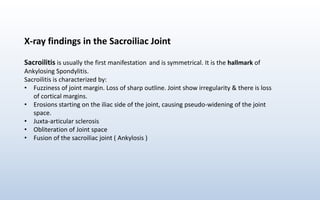
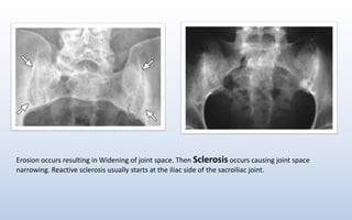
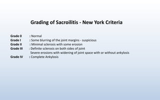
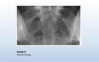
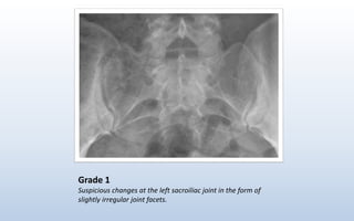
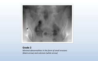
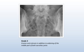
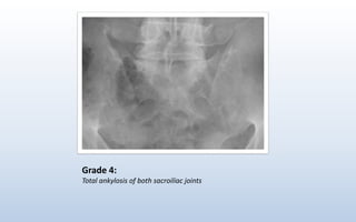
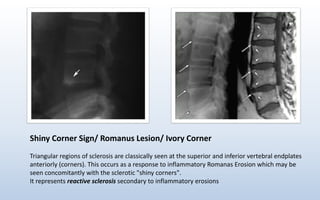
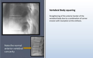
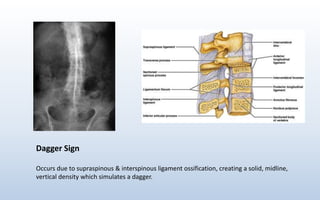
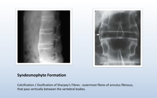
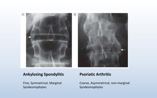
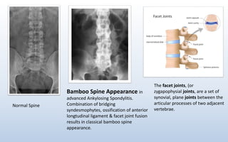
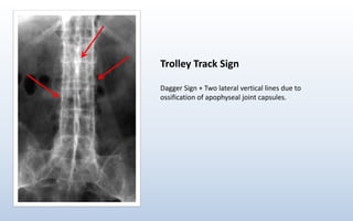
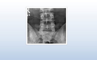
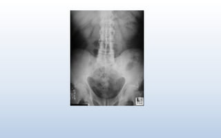
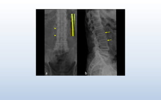
X-Ray findings of
Ankylosing Spondylitis
Dr. Faisal Abdullah
MBBS
https://faisalization.wordpress.com
https://youtube.com/faisalization
X-ray findings in the Sacroiliac Joint
Sacroilitis is usually the first manifestation and is symmetrical. It is the hallmark of
Ankylosing Spondylitis.
Sacroilitis is characterized by:
• Fuzziness of joint margin. Loss of sharp outline. Joint show irregularity & there is loss
of cortical margins.
• Erosions starting on the iliac side of the joint, causing pseudo-widening of the joint
space.
• Juxta-articular sclerosis
• Obliteration of Joint space
• Fusion of the sacroiliac joint ( Ankylosis )
Irregular Fuzzy Outline of Sacroiliac Joints
Erosion occurs resulting in Widening of joint space. Then Sclerosis occurs causing joint space
narrowing. Reactive sclerosis usually starts at the iliac side of the sacroiliac joint.
Normal Sacroiliac joint Fusion of the sacroiliac joint ( Ankylosis )
Grading of Sacroilitis - New York Criteria
Grade 0 : Normal
Grade I : Some blurring of the joint margins - suspicious
Grade II : Minimal sclerosis with some erosion
Grade III : Definite sclerosis on both sides of joint
Severe erosions with widening of joint space with or without ankylosis
Grade IV : Complete Ankylosis
Grade 0
Normal Findings
Grade 1
Suspicious changes at the left sacroiliac joint in the form of
slightly irregular joint facets.
Grade 2
Minimal abnormalities in the form of small erosions
(black arrow) and sclerosis (white arrow)
Grade 3
Erosion and sclerosis in addition to widening of the
middle part of both sacroiliac joints
Grade 4:
Total ankylosis of both sacroiliac joints
X- Ray findings in the Spine
• Shiny Corner Sign/ Ivory Corner/ Romanus Lesion
• Vertebral Body Squaring
• Dagger Spine Appearance
• Trolley track Sign
• Bamboo Spine
Shiny Corner Sign/ Romanus Lesion/ Ivory Corner
Triangular regions of sclerosis are classically seen at the superior and inferior vertebral endplates
anteriorly (corners). This occurs as a response to inflammatory Romanas Erosion which may be
seen concomitantly with the sclerotic "shiny corners".
It represents reactive sclerosis secondary to inflammatory erosions
Vertebral Body squaring
Straightening of the anterior border of the
vertebral body due to a combination of corner
erosion with resorption at the enthesis.
Dagger Sign
Occurs due to supraspinous & interspinous ligament ossification, creating a solid, midline,
vertical density which simulates a dagger.
Syndesmophyte Formation
Calcification / Ossification of Sharpey’s Fibres : outermost fibres of annulus fibrosus,
that pass vertically between the vertebral bodies.
Psoriatic Arthritis
Coarse, Asymmetrical, non-marginal
Syndesmophytes
Ankylosing Spondylitis
Fine, Symmetrical, Marginal
Syndesmophytes
Bamboo Spine Appearance in
advanced Ankylosing Spondylitis.
Combination of bridging
syndesmophytes, ossification of anterior
longtudinal ligament & facet joint fusion
results in classical bamboo spine
appearance.
The facet joints, (or
zygapophysial joints, are a set of
synovial, plane joints between the
articular processes of two adjacent
vertebrae.
Normal Spine
Trolley Track Sign
Dagger Sign + Two lateral vertical lines due to
ossification of apophyseal joint capsules.
• Davidson’s Principles & Practice of Medicine: 23rd Edition
• Diagnostic Imaging : 7th Edition
• https://radiopaedia.org
• https://slideshare.net
• http://www.spa-imaging.org/
Special Thanks to:
• Dr. Ashiqur Rahman Sir
Registrar, NIDCH
• Dr. Sanjoy Kumar Roy Sir
Registrar, Medicine
Khulna Medical College Hospital
REFERENCE

Ankylosing Spondylitis

  • 1.
    X-Ray findings of AnkylosingSpondylitis Dr. Faisal Abdullah MBBS https://faisalization.wordpress.com https://youtube.com/faisalization
  • 2.
    X-ray findings inthe Sacroiliac Joint Sacroilitis is usually the first manifestation and is symmetrical. It is the hallmark of Ankylosing Spondylitis. Sacroilitis is characterized by: • Fuzziness of joint margin. Loss of sharp outline. Joint show irregularity & there is loss of cortical margins. • Erosions starting on the iliac side of the joint, causing pseudo-widening of the joint space. • Juxta-articular sclerosis • Obliteration of Joint space • Fusion of the sacroiliac joint ( Ankylosis )
  • 3.
    Irregular Fuzzy Outlineof Sacroiliac Joints
  • 4.
    Erosion occurs resultingin Widening of joint space. Then Sclerosis occurs causing joint space narrowing. Reactive sclerosis usually starts at the iliac side of the sacroiliac joint.
  • 5.
    Normal Sacroiliac jointFusion of the sacroiliac joint ( Ankylosis )
  • 6.
    Grading of Sacroilitis- New York Criteria Grade 0 : Normal Grade I : Some blurring of the joint margins - suspicious Grade II : Minimal sclerosis with some erosion Grade III : Definite sclerosis on both sides of joint Severe erosions with widening of joint space with or without ankylosis Grade IV : Complete Ankylosis
  • 7.
  • 8.
    Grade 1 Suspicious changesat the left sacroiliac joint in the form of slightly irregular joint facets.
  • 9.
    Grade 2 Minimal abnormalitiesin the form of small erosions (black arrow) and sclerosis (white arrow)
  • 10.
    Grade 3 Erosion andsclerosis in addition to widening of the middle part of both sacroiliac joints
  • 11.
    Grade 4: Total ankylosisof both sacroiliac joints
  • 12.
    X- Ray findingsin the Spine • Shiny Corner Sign/ Ivory Corner/ Romanus Lesion • Vertebral Body Squaring • Dagger Spine Appearance • Trolley track Sign • Bamboo Spine
  • 13.
    Shiny Corner Sign/Romanus Lesion/ Ivory Corner Triangular regions of sclerosis are classically seen at the superior and inferior vertebral endplates anteriorly (corners). This occurs as a response to inflammatory Romanas Erosion which may be seen concomitantly with the sclerotic "shiny corners". It represents reactive sclerosis secondary to inflammatory erosions
  • 14.
    Vertebral Body squaring Straighteningof the anterior border of the vertebral body due to a combination of corner erosion with resorption at the enthesis.
  • 15.
    Dagger Sign Occurs dueto supraspinous & interspinous ligament ossification, creating a solid, midline, vertical density which simulates a dagger.
  • 16.
    Syndesmophyte Formation Calcification /Ossification of Sharpey’s Fibres : outermost fibres of annulus fibrosus, that pass vertically between the vertebral bodies.
  • 17.
    Psoriatic Arthritis Coarse, Asymmetrical,non-marginal Syndesmophytes Ankylosing Spondylitis Fine, Symmetrical, Marginal Syndesmophytes
  • 18.
    Bamboo Spine Appearancein advanced Ankylosing Spondylitis. Combination of bridging syndesmophytes, ossification of anterior longtudinal ligament & facet joint fusion results in classical bamboo spine appearance. The facet joints, (or zygapophysial joints, are a set of synovial, plane joints between the articular processes of two adjacent vertebrae. Normal Spine
  • 19.
    Trolley Track Sign DaggerSign + Two lateral vertical lines due to ossification of apophyseal joint capsules.
  • 23.
    • Davidson’s Principles& Practice of Medicine: 23rd Edition • Diagnostic Imaging : 7th Edition • https://radiopaedia.org • https://slideshare.net • http://www.spa-imaging.org/ Special Thanks to: • Dr. Ashiqur Rahman Sir Registrar, NIDCH • Dr. Sanjoy Kumar Roy Sir Registrar, Medicine Khulna Medical College Hospital REFERENCE