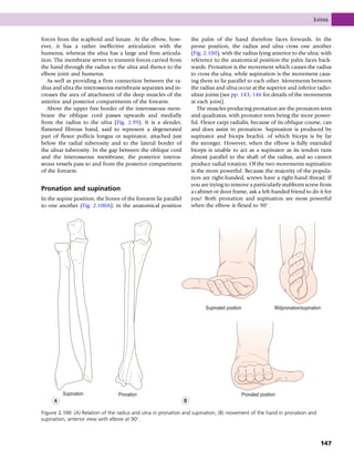
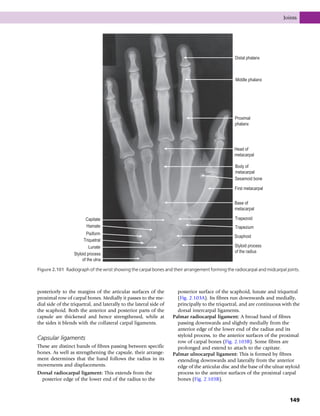
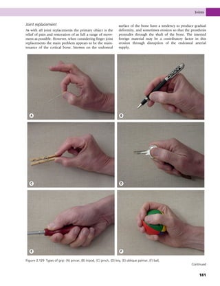
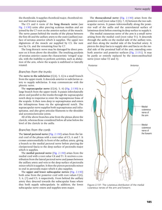
Anatomy and Human Movement
Commissioning Editor: Rita Demetriou-Swanwick
Development Editor: Sally Davies
Project Manager: Elouise Ball
Designer/Design Direction: Charles Gray
Illustration Manager: Gillian Richards
Original Illustration: Keyword
New and Updated Artwork: Cactus, Jennifer Rose
Illustration Colouring: Cactus
Anatomy and
Human Movement
Structure and Function
SIXTH EDITION
Nigel Palastanga MA BA FCSP DMS DipTP
Pro-Vice-Chancellor, University of Wales; formerly Director of Physiotherapy Education,
Cardiff University, Cardiff, UK
Roger Soames BSc PhD
Centre for Anatomy and Human Identification, College of Life Sciences,
University of Dundee, Dundee, UK
Edinburgh London New York Oxford Philadelphia St Louis Sydney Toronto 2012
# 2012 Elsevier Ltd. All rights reserved.
No part of this publication may be reproduced or transmitted in any form or by any means, electronic or
mechanical, including photocopying, recording, or any information storage and retrieval system, without
permission in writing from the publisher. Details on how to seek permission, further information about the
Publisher’s permissions policies and our arrangements with organizations such as the Copyright Clearance
Center and the Copyright Licensing Agency, can be found at our website: www.elsevier.com/permissions.
This book and the individual contributions contained in it are protected under copyright by the Publisher
(other than as may be noted herein).
First edition 1989
First published as a paperback edition 1990
Second edition 1994
Third edition 1998
Fourth edition 2002
Fifth edition 2006
Sixth edition 2012
ISBN 978 0 7020 3553 1
International ISBN 978 0 7020 4053 5
British Library Cataloguing in Publication Data
A catalogue record for this book is available from the British Library
Library of Congress Cataloging in Publication Data
A catalog record for this book is available from the Library of Congress
Notices
Knowledge and best practice in this field are constantly changing. As new research and experience
broaden our understanding, changes in research methods, professional practices, or medical treatment
may become necessary.
Practitioners and researchers must always rely on their own experience and knowledge in
evaluating and using any information, methods, compounds, or experiments described herein.
In using such information or methods they should be mindful of their own safety and the safety of
others, including parties for whom they have a professional responsibility.
With respect to any drug or pharmaceutical products identified, readers are advised to check the
most current information provided (i) on procedures featured or (ii) by the manufacturer of each
product to be administered, to verify the recommended dose or formula, the method and duration
of administration, and contraindications. It is the responsibility of practitioners, relying on their own
experience and knowledge of their patients, to make diagnoses, to determine dosages and the best
treatment for each individual patient, and to take all appropriate safety precautions.
To the fullest extent of the law, neither the Publisher nor the authors, contributors, or editors, assume
any liability for any injury and/or damage to persons or property as a matter of products liability,
negligence or otherwise, or from any use or operation of any methods, products, instructions, or ideas
contained in the material herein.
Printed in China
Contents
Preface to the first edition ....................................vii
Preface to the sixth edition.................................... ix
Acknowledgements.................................................. xi
About the authors..................................................xiii
1. Introduction................................................... 1
Terminology .........................................................1
Terms used in describing movement...............2
Nervous system....................................................3
Components of the musculoskeletal system...8
Skin and its appendages.................................. 22
Early embryology.............................................. 29
2. The upper limb............................................35
Introduction ...................................................... 36
Development of the musculoskeletal
system ................................................................. 36
Bones................................................................... 40
Muscles ............................................................... 52
Joints .................................................................101
Nerve supply....................................................183
Blood supply ...................................................195
Lymphatics.......................................................198
3. The lower limb ..........................................201
Introduction ....................................................202
Development of the musculoskeletal
system ...............................................................203
Bones.................................................................205
Muscles .............................................................223
Joints .................................................................275
Nerve supply....................................................384
The sacral plexus.............................................395
Blood supply ...................................................396
Lymphatics.......................................................402
4. The trunk and neck ..................................405
Introduction ....................................................406
Bones.................................................................412
Muscles .............................................................423
Joints .................................................................445
Nerve supply....................................................486
The cardiovascular system ............................507
The respiratory system...................................514
The digestive system.......................................521
The urogenital system....................................530
The endocrine system ....................................540
5. Head and brain .........................................543
Bones.................................................................543
Muscles .............................................................550
Joints .................................................................559
The ear, eye and brain ...................................565
Index ...................................................................... 593
v
This page intentionally left blank
Preface
to the first edition
We have designed and written this book for the student of anatomy who is concerned with the study
of the living body and who wishes to use this knowledge functionally for a greater understanding of
the mechanisms which allow movement to take place. Traditional anatomy texts are written as an
adjunct to the study of the human body in the dissecting room, but only the surgeon has the advan-
tage of directly viewing living musculoskeletal structures. The vast majority of students interested in
musculoskeletal anatomy as well as those involved with human movement and its disorders are con-
fronted by an intact skin and therefore must visualize the structures involved by palpation and anal-
ysis of movement. Anatomy and Human Movement presents the musculoskeletal structures as a living
dynamic system – an approach lacking in many existing textbooks. The applied anatomy of the mus-
culoskeletal system occupies the greater part of the book and is built up from a study of the bones and
muscles (which are grouped according to their major functions, rather than as seen in the dissecting
room) to a consideration of joints and their biomechanics. Anatomical descriptions of each joint are
given with a detailed explanation of how it functions, the forces generated across it and how it might
fail. We have placed great emphasis on the joints, as these are of major concern to those interested in
active movement and passive manipulation, and we give examples of common traumatic or patho-
logical problems affecting the structures described. Where possible, we describe palpation and ana-
lyse movement with respect to the joints and muscles involved, as well as any accessory movements.
The course and distribution of the major peripheral nerves and blood vessels, together with the lym-
phatic drainage of the region, are given at the end of each relevant section. There are separate chapters
on embryology and the skin and its appendages, and we have included, in the introduction, a section
on the terminology used in the book. There is also an account of the structure and function of the
nervous system written by Nikolai Bogduk whose contribution has been extremely valuable.
The format of the book matches a page of text to a page of illustrations, whenever possible, and we
hope that this will allow the reader to confirm his or her understanding of the text with the visual
information provided. The book is extensively illustrated with large, clear, fully labelled diagrams,
all of which have been specially prepared. In the sections covering the joints and biomechanics,
the illustrations have been drawn by Roger Soames, and these are particularly detailed as they pull
together the anatomy from the previous parts of that chapter.
We hope that this new approach to the teaching of anatomy will serve to fill the gap which has
always existed for those who have to learn their anatomy on a living subject and eventually have
to determine their diagnoses and apply their treatments through an intact skin.
Nigel Palastanga
Derek Field
Roger Soames
1989
vii
This page intentionally left blank
Preface
to the sixth edition
In this sixth edition of Anatomy and Human Movement, we have made a number of small but signif-
icant changes, mainly to the text, which are designed to further improve the usefulness of this already
successful book. In this edition, there have been further improvements to the illustrations and
we have added some new illustrations. Where possible, structures such as ligaments or cartilage have
been coloured the same throughout the book, in an attempt to further enhance the clarity of
illustrations. The book has progressed a long way since the first edition in 1989 when only black
and white was used throughout the text.
As regards the content, we have continued to engage with the community of users of the book,
namely, students, teachers, practitioners, and have responded to yet more suggestions. As a result
of this very positive feedback, there has been some minor reordering within the text and expansion
of some sections, namely those on joint replacement.
The use of Summary Boxes at the end of various sections has been retained: they are intended to be
used as a quick aid to revision when the majority of the subject area has been learned, but feedback
suggests that some will use them to get a general overview before starting to tackle the detailed text.
Helping individuals to understand, remember and apply anatomy has always been the prime purpose
of this book.
In response to the advances in the range of electronic aids to learning, the sixth edition also gives
automatic access (unlocked by the unique PIN found on the inside front cover) to the e-book via
Pageburst, allowing users to quickly search the entire book, make notes, add highlights and study
more efficiently. In addition to this, the authors have helped create a separate five-hour modular
e-learning course in functional human anatomy. At present, this is only available for purchase by
universities and other institutions. This is presented in short lesson chunks following the body’s re-
gional structure – ideal for the student to study at their own pace and time and the lecturer to assign
accordingly. Learning is delivered via outcome measures, animation, video, quizzes, activity analyses
and exams.
Whether the book is being used by undergraduates, postgraduates, lecturers or practitioners, the
changes and additions made to this edition are designed to make it even more useful to your learning,
teaching or practice. Changes to the way education is managed and delivered require students and
educators to be flexible in their learning. It is recognized that most learning takes place outside
the classroom, and this requires the provision of high-quality material, both electronic and hard copy,
to support learning wherever it is taking place. This edition of Anatomy and Human Movement and its
accompanying electronic resources are designed to facilitate study in this new environment of varied
ix
learning spaces. The range of health professional groups using Anatomy and Human Movement has
grown extensively over the past 22 years, with its use spreading to other groups interested in human
movement, such as sports scientists.
Anatomy and Human Movement has been written by authors experienced in teaching living anatomy
and human movement. This remains the prime reason for producing the book, and whilst the
authors visit dissecting rooms regularly, the appreciation of anatomy through the intact skin of a
living person is fundamental to the practice of a number of professions. We hope that this sixth
edition will provide the reader with stimulation that will aid their understanding and learning of
anatomy and their ability to apply this knowledge to human movement.
Nigel Palastanga
Roger Soames
2012
x
Preface to the sixth edition
Acknowledgements
Nigel Palastanga and Roger Soames would like to acknowledge the tremendous contribution to the
first four editions made by Derek Field.
In addition, we are very grateful to Dot Palastanga who is a lecturer teaching Occupational Therapy
students at Cardiff University. She has been very much involved in the proofreading of every edition,
including this the sixth: her attention to detail, as well as gentle reminders, has kept things on sched-
ule and allowed us to meet deadlines. This is a very belated acknowledgement for her contribution
over the past 20 years.
xi
This page intentionally left blank
About the authors
Nigel Palastanga has been involved in higher education for nearly 40 years and for most of those has
taught undergraduate and postgraduate physiotherapists. He has been the head of physiotherapy ed-
ucation at Cardiff University and was a pro-vice chancellor both there and in the University of Wales
College of Medicine. He was awarded a Fellowship of the Chartered Society of Physiotherapy in 2001
and is still an external examiner for a number of physiotherapy programmes. He is currently pro-vice
chancellor of the University of Wales where his role is concerned with the quality and standards of
their awards.
Roger Soames moved from James Cook University in Townsville, Queensland, Australia in 2007 to
become Principal Anatomist at the University of Dundee. In 2009 he was awarded a personal chair in
Functional and Applied Anatomy and later the same year was appointed to the Cox Chair of Anatomy
at the University of Dundee. He has spent the whole of his career within the discipline of anatomy,
teaching and examining on a wide range of undergraduate and postgraduate degree programmes and
courses. He has research interests in the musculoskeletal system and is also involved in developing
surgical and clinical training opportunities utilizing Thiel preserved cadaveric material.
xiii
This page intentionally left blank
Part | 1 |
Introduction
CONTENTS
Terminology 1
Terms used in describing movement 2
Nervous system 3
Introduction 3
Cellular structure 4
Interneural connections 4
Myelination 5
Structure of a peripheral nerve 6
The peripheral nervous system 7
Components of the musculoskeletal system 8
Connective tissue 8
Skeletal tissue 9
Muscular tissue 12
Joints 17
Spin, roll and slide 20
Levers 21
Skin and its appendages 22
Introduction 22
Structure 23
Appendages of the skin 26
Glands 27
Blood supply and lymphatic
drainage of the skin 28
Nerves of the skin 28
Application 28
Early embryology 29
Stages in development 29
TERMINOLOGY
It is essential for students beginning their study of anatomy
to become familiar with an internationally accepted
vocabulary, allowing communication and understanding
between all members of the medical and paramedical pro-
fessions throughout the world. Perhaps the single, most
important descriptive feature of this vocabulary is the
adoption of an unequivocal position of the human body.
This is known as the anatomical position. It is described as
follows: the body is standing erect and facing forwards;
the legs are together with the feet parallel so that the toes
point forwards; the arms hang loosely by the sides with
the palm of the hand facing forwards so that the thumb
is lateral (Fig. 1.1). All positional terminology is used with
reference to this position, irrespective of the actual position
of the body when performing an activity.
The following is a list of more commonly used terms
which describe the position of anatomical structures:
Anterior (ventral) To the front or in front, e.g. the patella
lies anterior to the knee joint.
Posterior (dorsal) To the rear or behind, e.g. gluteus
maximus lies posterior to the hip joint. (Ventral and
dorsal are used more commonly in quadrupeds.)
Superior (cephalic) Above, e.g. the head is superior to
the trunk.
Inferior (caudal) Below, e.g. the knee is inferior to the hip.
Cephalic (the head) Caudal (the tail) May be used in
relation to the trunk.
Lateral Away from the median plane or midline, e.g. the
thumb lies lateral to the index finger.
Medial Towards the median plane or midline, e.g. the
index finger lies medial to the thumb.
© 2012 Elsevier Ltd. All rights reserved.
1
Distal Further away from the trunk or root of the limb,
e.g. the foot is distal to the knee.
Proximal Closer to the trunk or root of the limb, e.g. the
elbow is proximal to the hand.
Superficial Closer to the surface of the body or skin, e.g.
the ulnar nerve passes superficial to the flexor
retinaculum of the wrist.
Deep Further away from the body surface or skin, e.g. the
tendon of tibialis posterior passes deep to the flexor
retinaculum at the ankle.
To facilitate the understanding of the relation of structures
one to another and the movement of one segment with
respect to another, imaginary reference planes pass through
the body in such a way that they are mutually perpendicu-
lar to each other (Fig. 1.1). Passing through the body from
front to back and dividing it into two symmetrical right and
left halves is the sagittal (median) plane. Any plane parallel
to this is also known as a parasagittal (paramedian) plane.
A plane passing through the body from top to bottom and
lying at right angles to the sagittal plane is the coronal (frontal)
plane. This divides the body into anterior and posterior parts.
All planes that divide the body in this way are known as cor-
onal planes. Finally, a plane passing through the body at right
angles to both the sagittal and coronal planes dividing it into
upper and lower parts is known as a transverse (horizontal)
plane. A whole family of parallel transverse planes exist; it is
therefore usual when presenting a particular transverse sec-
tion to specify the level at which it is taken. This may be done
by specifying the vertebral level or the position within the
limb, e.g. C6 or mid-shaft of humerus, respectively.
Within each plane a single axis can be identified, usually
in association with a particular joint, about which move-
ment takes place. An anteroposteriorly directed axis in
the sagittal (or a paramedian) plane allows movement in
a coronal plane. Similarly, a vertical axis in a coronal plane
allows movement in a transverse plane. Lastly a transverse
(right to left) axis in a coronal plane provides movement in
a paramedian plane.
By arranging that these various axes intersect at the centre
of joints, the movements possible at the joint can be bro-
ken down into simple components. It also becomes easier
to understand how specific muscle groups produce partic-
ular movements, as well as to determine the resultant
movement of combined muscle actions.
Section summary
Terminology
• Specific terms are used to describe the relationship of
one body part/segment/region to another and are
considered in relation to the anatomical position of the
body.
• The anatomical position is: standing erect facing
forwards, legs together toes pointing forwards, arms
at the side and palms facing forwards.
TERMS USED IN DESCRIBING
MOVEMENT
Rarely do movements of one body segment with respect to
another take place in a single plane. They almost invariably
occur in two or three planes simultaneously, producing a
complex pattern of movement. However, it is convenient
to consider movements about each of the three defined
axes separately. Movement about a transverse axis occur-
ring in the paramedian plane is referred to as flexion and
extension; that about an anteroposterior axis in a coronal
Sagittal
(median)
plane
Coronal
(frontal)
plane
Posterior
Transverse
(horizontal)
plane
Inferior
Dorsum
of hand
Palmar
surface
of hand
Proximal
Distal
Plantar
surface
of foot
Dorsum
of foot
Medial
Lateral
Anterior
Superior
Figure 1.1 Anatomical position showing the cardinal planes
and directional terminology.
Part | 1 | Introduction
2
plane is termed abduction and adduction; and finally, that
about a vertical axis in a transverse plane is termed medial
and lateral rotation.
All movements are described, unless otherwise stated,
with respect to the anatomical position, this being the posi-
tion of reference. In this position joints are often referred to
as being in a ‘neutral position’.
Flexion The bending of adjacent body segments in a
paramedian plane so that their two anterior/posterior
surfaces are brought together, e.g. bending the elbow so
that the anterior surfaces of the forearm and arm move
towards each other. (For flexion of the knee joint the
posterior surfaces of the leg and thigh move towards each
other.)
Extension The moving apart of two opposing surfaces in
a paramedian plane, e.g. the straightening of the flexed
knee or elbow. Extension also refers to movement beyond
the neutral position in a direction opposite to flexion, e.g.
extension at the wrist occurs when the posterior surfaces of
the hand and forearm move towards each other.
Flexion and extension of the foot at the ankle joint may be
referred to as plantarflexion and dorsiflexion respectively.
Plantarflexion Moving the top (dorsum) of the foot away
from the anterior surface of the leg.
Dorsiflexion Bringing the dorsum of the foot towards the
anterior surface of the leg.
Abduction The movement of a body segment in a coronal
plane such that it moves away from the midline of the
body, e.g. movement of the upper limb away from the
side of the trunk.
Adduction The movement of a body segment in a coronal
plane such that it moves towards the midline of the
body, e.g. movement of the upper limb back towards the
side of the trunk.
Lateral flexion (bending) A term used to denote bending
of the trunk (vertebral column) to one side, e.g. lateral
bending of the trunk to the left. The movement occurs in
the coronal plane.
Medial rotation Rotation of a limb segment about its
longitudinal axis such that the anterior surface comes to
face towards the midline of the body, e.g. turning the
lower limb inwards so that the toes point towards the
midline.
Lateral rotation Rotation of a limb segment about its
longitudinal axis so that its anterior surface faces away
from the midline plane, e.g. turning the lower limb so
that the toes point away from the midline.
Supination and pronation are terms used in conjunction with
the movements of the forearm and foot.
Supination Movement of the forearm so that the palm of
the hand faces forwards. In the foot it is the movement
whereby the forefoot is turned so that the sole faces
medially; it is always accompanied by adduction of the
forefoot.
Pronation Movement of the forearm that makes the palm
of the hand face backwards. In the foot it is a movement
of the forefoot which causes the sole to face laterally; it is
always accompanied by abduction of the forefoot.
Inversion and eversion are terms used to describe composite
movements of the foot.
Inversion Movement of the whole foot to make the sole
face medially. It consists of supination and adduction of
the forefoot.
Eversion Movement of the whole foot so that the sole
comes to face laterally. It consists of pronation and
abduction of the forefoot.
Section summary
Movements
• Specific terms refer to different types of movement
between body parts/segments and/or regions.
• Flexion/extension occur about a transverse axis in a
paramedian plane; abduction/adduction occur about an
anteroposterior axis in a coronal plane; medial/lateral
rotation occur about a vertical axis in a transverse plane.
• More specific terms are used for movements associated
with some segments/regions: plantarflexion/dorsiflexion
of the foot at the ankle joint; supination/pronation of
the forearm; supination/pronation/inversion/eversion
within the foot; lateral flexion of the vertebral column.
NERVOUS SYSTEM
Introduction
The nervous system consists of highly specialized cells
designed to transmit information rapidly between various
parts of the body. Topographically it can be divided into
two major parts: the central nervous system (CNS) and the
peripheral nervous system (PNS). The brain and spinal cord
constitute the CNS, which lies within the skull and vertebral
canal (see p. 572 and p. 489), while nerves in the PNS
connect the CNS with all other parts of the body (see p. 7).
The CNS is a massive collection of nerve cells connected
in an intricate and complex fashion to subserve the higher
order functions of the nervous system, such as: thought,
language, emotion, control of movement and the analysis
of sensation. It is isolated from the rest of the body; its car-
dinal characteristic is that it is located wholly within the
skull and vertebral canal.
The PNS consists of cells that connect the CNS with the
other tissues of the body. These cells are aggregated into a
large number of cable-like structures called nerves, which
are threaded like wires throughout the tissues of the body.
Nervous system
3
Cellular structure
The basic cellular unit of the nervous system is the nerve cell
or neuron, which differ in size and shape according to their
function and location within the nervous system. All neu-
rons have three characteristic components: a cell body, an
axon, and dendrites (Fig. 1.2A).
The cell body is an expanded part of the cell containing the
nucleus and apparatus necessary to sustain the metabolic
activities of the cell. The axon is a longitudinal, tubular
extension (process) of the cell membrane and cytoplasm
which transmits information away from the cell body.
The cell membrane surrounding the axon is referred to as
the axolemma. Dendrites are extensions of the cell mem-
brane that radiate from the cell body in various directions,
and are responsible for receiving information and transmit-
ting it to the cell body.
Structurally, dendrites differ from axons as they typically
undergo extensive branching close to the cell body,
whereas axons remain singular for most of their course only
branching at their terminal ends. A neuron has only one
axon, but may have several dendrites; the lengths and cali-
bre of axons and dendrites vary, depending on the particu-
lar function of the neuron.
Interneural connections
Individual neurons convey information by conducting
electrical action potentials along their cell membrane, with
communication between separate neurons occurring
chemically at a specialized structure called a synapse.
Synapses are formed by the close approximation of a
small discrete area of the cell membrane of one neuron
to a reciprocal area of the membrane of a second neuron
(Fig. 1.2B). The apposed membranes are specially modified
and are separated by a narrow gap about 0.02 mm in width,
called the synaptic cleft. Across the synapse one cell commu-
nicates with the next, with communication being uni-
directional. The membrane of the cell transmitting the
information is the presynaptic membrane, and that of the
cell receiving the information the postsynaptic membrane.
Near the presynaptic membrane, the cytoplasm of the
transmitting neuron contains numerous small vesicles
filled with chemicals known as neurotransmitters. The
nature of the neurotransmitters varies according to the
function of the neuron, but in all cases, when an electrical
signal arrives at the terminal end of an axon, the neuro-
transmitter is released from the vesicles through the presyn-
aptic membrane into the synaptic cleft. There it flows across
to the postsynaptic membrane where it exerts its effect. This
effect can be excitatory or inhibitory depending on the
nature of the neurotransmitter and the nature of the recep-
tors on the postsynaptic membrane with which it interacts.
Excitatory substances generate an action potential in the
postsynaptic neuron which then propagates this potential
to its other end. Inhibitory substances alter the electrical
potential of the postsynaptic neuron temporarily reducing
its capacity to be stimulated by other neurons.
Dendrites
Nucleus
Cell Body
Axon
ad
as
aa
Axon terminal
Mitochondrion
Vesicle
Synaptic cleft
Axon terminal
Dendrite
Presynaptic
membrane
Postsynaptic
membrane
Axo-somatic
synapse
Axo-axonic
synapse
Axo-dendritic
synapse
A B
Figure 1.2 (A) Microscopic appearance of a neuron and its component parts, (B) neuron with multiple axo-somatic (as), axo-dendritic
(ad) and axo-axonic (aa) synapses. The insets show the detailed structure of the synapse.
Part | 1 | Introduction
4
Synapses typically occur between the axon of one neuron
and the dendrite of another. However, they may also occur
between axons and cell bodies, axons and axons, and even
between dendrites and dendrites. Some neurons receive
only a few synapses while others may receive thousands.
The purpose of synapses is not to relieve one neuron of
information and simply pass it on to the next, but to allow
the interaction of information from perhaps several diverse
sources on a single neuron. The activity of a given neuron
may be influenced by many others and, conversely, by hav-
ing several terminal branches to its axon, a single neuron
may influence many other neurons.
By being connected to one another in diverse ways,
groups of neurons are organized to subserve different func-
tions of the nervous system. The patterns of connections, or
circuits, vary in complexity and, in general, the more
sophisticated the function being subserved, the more com-
plex the circuitry.
Myelination
Myelination is a process whereby individual axons are
wrapped in a sheath of lipid called myelin. The myelin
sheath serves as a protective and insulating coating for
the axon, and enhances the speed of conduction of electri-
cal impulses along it.
In the PNS, myelination is by cells called Schwann cells: it is
achieved by a Schwann cell curling an extension of its cell
membrane around the shaft of the axon in a spiral manner
(Fig. 1.3A). As the Schwann cell extension wraps around
Axon
Neurolemma Axolemma Myelin sheath
Schwann cell
Membrane
Nucleus
Cytoplasm
Axon
Axolemma
Stages in myelination
Cytoplasm
Nucleus
Axons
Myelin sheath
Schwann
cell nucleus
Schwann cell
cytoplasm
Node of
Ranvier
Schwann
cell nucleus
Node of
Ranvier
Schwann cell
membrane
A
B
Figure 1.3 (A) Myelinated axon in the peripheral nervous system, (B) relationship between unmyelinated axons and Schwann cells.
Nervous system
5
the axon, the cytoplasm in it is squeezed out until only
a double layer of the cell membrane of the Schwann cell
remains spiralling around the axon (Fig. 1.3A). In this way
the myelin sheath is formed by the lipid and protein of
the cell membrane of the Schwann cell that surrounds the
axon.Peripheraltothemyelinsheath,theaxonissurrounded
bythe cytoplasm of the Schwann cell, with the outermost cell
membrane of the Schwann cell acting as a second membrane
to the axon; this forms the neurolemma (Fig. 1.3A).
Along its length, a given axon is ensheathed in series by a
large number of Schwann cells, but each Schwann cell is
related only to a single axon (Fig. 1.3A). Junction points
where the myelin sheath of one Schwann cell ends and
the sheath of the next begins are known as Nodes of
Ranvier. An axon together with the myelin sheath and the
Schwann cells that surround it is referred to as a nerve fibre.
While many axons in the PNS are myelinated a large
number remain unmyelinated. Instead of being enveloped
by a tightly spiralling sheath of myelin, they run embedded
in invaginations of Schwann cell membranes, so that a sin-
gle Schwann cell may envelop several axons (Fig. 1.3B).
Unmyelinated axons are afforded less physical protection
than myelinated ones, but the foremost difference between
them is that unmyelinated axons conduct impulses at
much slower velocities.
In the CNS, myelination is performed by specialized cells
called oligodendrocytes. The process is basically similar to
that in the PNS, except that a given oligodendrocyte is
usually involved in the simultaneous myelination of sev-
eral separate axons (Fig. 1.4). Unmyelinated axons in the
CNS run embedded in cytoplasmic extensions, or pro-
cesses, of oligodendrocytes.
Several diseases may affect myelinating cells, including
toxic and metabolic diseases, and most notably multiple
sclerosis. In these diseases, neurons are not necessarily
directly affected, but the loss of their myelin covering affects
the way they conduct action potentials resulting in disor-
dered neural function.
Structure of a peripheral nerve
A peripheral nerve is formed by the parallel aggregation of a
variable number of myelinated and unmyelinated axons:
the greater the number of axons, the larger the size of the
nerve. Microscopically, within a peripheral nerve, myelin-
ated axons are surrounded by their individual Schwann cell
sheaths and unmyelinated axons run embedded in invagi-
nations of the Schwann cell membrane. The axons are held
together by sheaths of fibrous tissue which constitute addi-
tional coatings that protect them from external mechanical
and chemical insults.
Individual myelinated axons are surrounded by a tubular
sheath of fibrous tissue, the endoneurium, whereas clusters
of axons are held together by a larger fibrous sheath, the
perineurium (Fig. 1.5). Unmyelinated axons are not
enclosed by endoneurium but run in isolated bundles
parallel to myelinated axons and are enclosed with them
in the perineurial sheath.
A bundle of axons enclosed within a single perineurial
sheath is referred to as a nerve fascicle (Fig. 1.5). Axons
within a fascicle largely remain within that same fascicle
Oligodendrocyte
Axon
Non-myelinated
axons
Node of
Ranvier
Process of
oligodendrocyte
Myelin
sheath
Figure 1.4 Relationship between an oligodendrocyte and the
several myelinated and unmyelinated axons it ensheathes.
Epineurium
Endoneurium
Fascicle and
perineurium
Figure 1.5 Composition of a peripheral nerve.
Part | 1 | Introduction
6
throughout the course of the nerve until their peripheral
distribution. However, along the course of a peripheral
nerve axons may at times leave one fascicle to enter and
continue within an adjacent fascicle.
The fascicles within a peripheral nerve are bound
together by an external sheath of fibrous tissue, the epineu-
rium (Fig. 1.5), which forms the external surface of the
macroscopic nerve. As a peripheral nerve passes through
the tissues of the body, it gives off branches composed of
one or more fascicles of the parent nerve, which leave it
to reach their particular destination. Along the course of
a nerve, this process is repeated until all the fascicles and
axons in the nerve have been distributed to their target
tissues.
The peripheral nervous system
Nerves that supply the structural tissues, such as bone, mus-
cle and skin, are referred to as somatic nerves. Groups of
somatic nerves innervate specific areas or regions and are
covered in detail in the respective region.
The nerves that supply the viscera, such as the heart,
lungs and digestive tract, are referred to as visceral nerves.
However, because the nervous functions concerning viscera
are largely automatic and subconscious, that part of the
nervous system that innervates viscera is referred to as
the autonomic nervous system (ANS). It has components in
both the central and PNSs: it is described in detail on
page 500.
Constituents of peripheral nerves
Peripheral nerves are composed of different types of axons
that are classified according to their size, function or phys-
iological characteristics. The broadest classification of
axons recognizes afferent (or sensory) fibres and efferent (or
motor) fibres. The terms ‘afferent’ and ‘efferent’ refer to the
direction in which axons conduct information: afferent
fibres conduct towards, while efferent fibres conduct away
from the CNS. The term ‘sensory’ refers to axons that
convey information to the CNS about events that occur
in the periphery; ‘motor’ fibres cause events in the periph-
ery, usually in the form of contraction of voluntary or
smooth muscle.
Axons may also be classified according to their conduc-
tion velocities, which in turn are proportional to their sizes
(Fig. 1.6). If a peripheral nerve is stimulated electrically and
recordings of the evoked activity are taken some distance
along the nerve, a wave of electrical activity can be
recorded. The wave is generated by the summation of the
electrical activity in each of the axons in the nerve, its size
being proportional to the number of axons in the nerve,
while its shape reflects the type of axons present.
The waveform obtained from a nerve containing every
known type of axon is represented in Fig. 1.6. It has three
principal peaks, named the A, B and C waves. The A wave
occurs very soon after the triggering stimulus, as it is pro-
duced by rapidly conducting axons with velocities in the
range 12–120 ms l
: these are classified as A fibres. The A
wave is broken down into several secondary peaks called
Aa, Ab, Ag and Ad waves, each being produced by a partic-
ular subgroup of rapidly conducting axons.
The B wave is produced by more slowly conducting, but
nevertheless fast, fibres classified as B fibres. The C wave
arrives at the recording electrode much later as it is pro-
duced by slowly conducting axons classified as C fibres with
conduction velocities in the range 0.5–2 ms l
.
Not all peripheral nerves contain every type of fibre,
therefore not all nerves will exhibit A, B and C waves. For
example, B fibres are not regularly present in all nerves;
B waves are therefore not always recordable.
Myelinated axons conduct impulses faster than unmy-
elinated ones, and among myelinated axons those with
larger diameters conduct impulses faster than those with
smaller diameters. The conduction velocity of a myelinated
axon is directly proportional to the total diameter of the
axon and its myelin sheath. A and B fibres are myelinated
axons of various diameters and fast conduction velocities,
while C fibres are unmyelinated axons with small dia-
meters and slow conduction velocities.
V I II III IV
A
B C
Time after stimulus
120 70
Stimulus
Diameter
Group classification by number
(sensory fibres only)
Type classification
by letter (all fibres)
Conduction
velocity
Myelinated
nerve fibres
Unmyelinated
nerve fibres
α
β
γ
δ
30 15 12
2
2
1 0.5 μm
0.5 m s–1
5
12
20
Figure 1.6 Neurogram of an idealized peripheral nerve. Activity
in different types of nerve fibres is reflected in depolarizations
that occur at different times after the delivery of an electrical
stimulus to the nerve.
Nervous system
7
Functionally, Aa and Ag fibres represent motor fibres
connected to voluntary muscles and also include certain
sensory fibres that transmit position sensation from
skeletal muscles. Ab fibres mediate the sensations of touch,
vibration and pressure from skin; Ad fibres are sensory
fibres that mediate pressure, pain and temperature sensa-
tions from skin, pain and pressure from muscle, and pain,
pressure and position sensation from ligaments and joints.
B fibres are preganglionic sympathetic efferent fibres. C
fibres are largely unmyelinated sensory fibres that arise in
virtually all tissues of the body and transmit pain, temper-
ature and pressure sensations, however, some are postgan-
glionic sympathetic neurons. Because Ab fibres have large
diameters they are sometimes referred to as large diameter
afferent fibres, while Ad and C fibres are referred to collec-
tively as small diameter afferent fibres.
Another system of axon classification relates specifically
to sensory fibres. In this system, sensory fibres are classified
according to their conduction velocities into groups I, II, III
and IV. These groups have the same conduction velocities
as type Aa, Ab, Ad and C fibres respectively, as shown in
Fig. 1.6, but do not include the Aa and Ab motor fibres.
The fibres of groups III and IV largely mediate pain and
temperature sensations. Group II constitutes fibres that
mediate pressure and touch, and fibres that form spray end-
ings in muscle spindles (p. 15). Group I is divided into
groups Ia and Ib. Group Ia fibres are slightly larger and
innervate muscle spindles, while group Ib fibres innervate
Golgi tendon organs (p. 15).
Nerve endings
The terminals of axons in peripheral nerves have unique
structures depending on their function. Motor axons have
terminals designed to deliver a stimulus to muscle cells (see
p. 16), while sensory axons have terminals designed to
detect particular types of stimuli. These terminals are called
receptors, and a diversity of morphological types can be
found (see p. 26).
Section summary
Nervous system
• Consists of specialized cells (neurons) concerned
with the transmission of information throughout the
body.
• Neurons have a cell body (expanded part of the cell), an
axon (single long process arising from the cell body) and
dendrites (multi-branching radiating processes arising
from the cell body).
• Two parts: CNS comprises brain, spinal cord and spinal
nerves; PNS comprises nerves (peripheral and cranial)
and sensory receptors.
• Connections (synapses) between neurons allow
integration of information from several sources.
Resultant effect may be excitatory or inhibitory
depending on the type of neurotransmitter and type
of receptors on the postsynaptic membrane.
• Axons may be myelinated or unmyelinated. In CNS
myelination is by oligodendrocytes, in PNS by Schwann
cells. Myelinated axons have faster conduction
velocities.
Peripheral nerves
• Parallel aggregations of individual axons (fibres).
• Afferent (sensory) fibres transmit information towards
and efferent (motor) away from the CNS.
• The larger the fibre diameter the faster the nerve
conduction velocity.
COMPONENTS OF THE
MUSCULOSKELETAL SYSTEM
As this book is concerned essentially with the musculoskel-
etal system a brief account of the major tissues of the sys-
tem, i.e. connective, skeletal and muscular tissue, and of
the type of joints which enable varying degrees of move-
ment to occur, is given as it will aid in understanding the
mobility and inherent stability of various segments. The
initiation and coordination of movement is the responsi-
bility of the nervous system.
Connective tissue
Connective tissue is of mesodermal origin and in the adult
has many forms; the character of the tissue depends on the
organization of its constituent cells and fibres.
Fat
Fat is a packing and insulating material; however, in some
circumstances it can act as a shock absorber, an important
function as far as the musculoskeletal system is concerned.
Under the heel, in the buttock and palm of the hand, the fat
is divided into lobules by fibrous tissue septa, thereby stiff-
ening it for the demands placed upon it.
Fibrous tissue
Fibrous tissue is of two types. In white fibrous tissue there is
an abundance of collagen bundles, whereas in yellow fibrous
tissue there is a preponderance of elastic fibres.
White fibrous tissue is dense, providing considera-
ble strength without being rigid or elastic. It forms:
(1) ligaments, which pass from one bone to another in
Part | 1 | Introduction
8
the region of joints, uniting the bones and limiting
joint movement; (2) tendons for attaching muscles
to bones; and (3) protective membranes around muscle
(perimysium), bone (periosteum) and many other structures.
Yellow fibrous tissue, on the other hand, is highly
specialized, being capable of considerable deformation
yet returning to its original shape. It is found in the liga-
menta flava associated with the vertebral column as well
as in the walls of arteries.
Skeletal tissue
Skeletal tissues are modified connective tissues, in which
the cells and fibres have a particular organization and
become condensed so that the tissue is rigid.
Cartilage
Cartilage is supplementary to bone, forming wherever
strength, rigidity and some elasticity are required. In fetal
development, cartilage is often a temporary tissue, being
later replaced by bone. However, in many places cartilage
persists throughout life. Although a rigid tissue, it is not
as hard or strong as bone. It is also relatively non-vascular,
being nourished by tissue fluids. A vascular invasion of car-
tilage often results in the death of the cells during the pro-
cess of ossification of the cartilage and its eventual
replacement by bone. Except for the articular cartilage of
synovial joints, cartilage possesses a fibrous covering layer,
the perichondrium.
There are three main types of cartilage: hyaline cartilage,
white fibrocartilage and yellow fibrocartilage.
Hyaline cartilage
Hyaline cartilage forms the temporary skeleton of the fetus
from which many bones develop. Its remnants can be seen
as the articular cartilages of synovial joints, the epiphyseal
growth plates between parts of an ossifying bone during
growth, and the costal cartilages of the ribs. At joint surfaces
it provides a limited degree of elasticity to offset and absorb
shocks, as well as providing a relatively smooth surface
permitting free movement to occur. With increasing age,
hyaline cartilage tends to become calcified and sometimes
ossified.
White fibrocartilage
White fibrocartilage contains bundles of white fibrous tis-
sue which give it great tensile strength combined with some
elasticity so that it is able to resist considerable pressure.
It is found at many sites within the musculoskeletal system:
(1) within the intervertebral discs between adjacent verteb-
rae; (2) in the menisci of the knee joint; (3) in the labrum
surrounding and deepening the glenoid fossa of the
shoulder joint and the acetabulum of the hip joint;
(4) in the articular discs of the radiocarpal (wrist), sterno-
clavicular, acromioclavicular and temporomandibular
joints; and (5) as the articular covering of bones which
ossify in membrane, e.g. the clavicle and mandible.
White fibrocartilage may calcify and ossify.
Yellow fibrocartilage
Yellow fibrocartilage contains bundles of elastic fibres
with little or no white fibrous tissue. It does not calcify or
ossify and is not found within the musculoskeletal system.
Bone
Bone is extremely hard with a certain amount of resilience.
It is essentially an organic matrix of fibrous connective tis-
sue impregnated with mineral salts. The connective tissue
gives the bone its toughness and elasticity, while the min-
eral salts provide hardness and rigidity, the two being skil-
fully blended together. The mineral component provides a
ready store of calcium, which is continuously exchanged
with that in body fluids, with the rate of exchange and over-
all balance of these mineral ions being influenced by sev-
eral factors including hormones.
Each bone is enclosed in a dense layer of fibrous tissue,
the periosteum, with its form and structure adapted to the
functions of support and resistance of mechanical stresses.
Being a living tissue, bone is continually remodelled to
meet these demands; this is particularly so during growth.
The structure of any bone cannot be satisfactorily consid-
ered in isolation; it is dependent upon its relationship to
adjacent bones and the type of articulation between them,
as well as the attachment of muscles, tendons and liga-
ments to it.
The internal architecture of bone reveals a system of struts
and plates (trabeculae) running in many directions (Fig. 1.7),
which are organized to resist compressive, tensile and shear-
ing stresses. Surrounding these trabecular systems, which
tend to be found at the ends of long bones, is a thin layer
of condensed or compact bone (Fig. 1.7). The network of
the trabeculae, because of its appearance, is known as cancel-
lous or spongy bone. In the shaft of a long bone there is an
outer, relatively thick ring of compact bone surrounding a cav-
ity, which in life contains bone marrow.
Red and white blood cells are formed in red bone mar-
row, which after birth is the only source of red blood cells
and the main source of white blood cells. In infants, the
cavities of all bones contain red marrow. However, this
gradually becomes replaced by yellow fat marrow, so that
at puberty red marrow is only found in cavities associated
with cancellous bone. With increasing age many of these
red marrow containing regions are replaced by yellow mar-
row. Nevertheless, red marrow tends to persist throughout
life in the vertebrae, ribs and sternum, and the proximal
ends of the femur and humerus.
Components of the musculoskeletal system
9
For descriptive purposes bones can be classified accord-
ing to their shape:
1. Long bones are found within the limbs: each consists
of a shaft (diaphysis) and two expanded ends
(epiphyses).
2. Short bones are the bones of the wrist and part of the
foot, the carpal and tarsal bones respectively.
3. Flat bones are thin and tend to be curved in spite of their
classification; they include the bones of the skull vault
and ribs. Structurally, they consist of two layers of
compact bone enclosing cancellous bone.
4. Irregular bones are those which fit none of the above
categories, and include the vertebrae and many of the
bones of the skull and face.
Both irregular and short bones consist of a thin layer of
compact bone surrounding cancellous bone.
Bone development
Bone develops either directly in the mesoderm by the depo-
sition of mineral salts, or in a previously formed cartilage
model. When the process of calcification and then ossifica-
tion takes place without an intervening cartilage model, it is
known as intramembranous ossification, with the bone being
referred to as membrane bone. However, if there is an inter-
vening cartilage model, the process is known as endo-
chondral ossification, with the bone being referred to as
cartilage bone. This latter process is by far the most common.
Intramembranous ossification
The site of bone formation is initially indicated by a con-
densation of cells and collagen fibres accompanied by
the laying down of organic bone matrix, which becomes
impregnated with mineral salts. The formation of new
bone continues in a manner similar to bone developed
in cartilage (p. 11). Intramembranous ossification occurs
in certain bones of the skull, the mandible and clavicle.
Endochondral ossification
Again the first step in the process is the accumulation of
mesodermal cells in the region where the bone is to
develop. A cartilage model of the future bone develops
from these mesodermal cells (Fig. 1.8A). In long bones
the cartilage model grows principally at its ends, so that
the oldest part of the model is near the middle. As time pro-
gresses, the cartilage matrix in this older region is impreg-
nated with mineral salts so that it becomes calcified.
Consequently the cartilage cells, being cut off from their
nutrient supply, die. The greater part of the calcified carti-
lage is subsequently removed and bone is formed around
its few remaining spicules (Fig. 1.8A). Ultimately, the con-
tinual process of excavation of calcified cartilage and the
deposition of bone leads to the complete removal of the
calcified cartilage (Fig. 1.8A).
The cartilage at the ends of the bone continues to grow
due to multiplication of its cells. However, the deeper layers
gradually become calcified and replaced by bone. There-
fore, the increase in length of a long bone is due to active
cartilage at its ends, while an increase in width is by depo-
sition of new bone on that already existing.
When first laid down, bone is cancellous in appearance,
having no particular organization: it is referred to as woven
bone. In the repair of fractures, the newly formed bone also
has this woven appearance. However, in response to stresses
applied to the bone by muscles, tendons, ligaments as well as
the forces transmitted across joints, the woven bone gradu-
ally assumes a specific pattern in response to these stresses.
Growth and remodelling of bone
During growth there is an obvious change in the shape of a
bone. However, it should be remembered that even in the
adult, bone is being continuously remodelled, principally
under the direct control of hormones to stabilize blood
A
B
Figure 1.7 Trabecular arrangement within bone: (A) coronal
section through the lower end of the femur, (B) sagittal section
through the calcaneus.
Part | 1 | Introduction
10
calcium levels, but also in response to long-term changes in
the pattern of forces applied to the bone.
Both growth and remodelling depend on the balanced
activity of two cell types, one of which removes bone
tissue (osteoclasts) and one which lays down new bone
(osteoblasts). In a growing bone, for example, new bone is
laid down around the circumference of the shaft in order
to increase its diameter. At the same time the deepest layers
of bone are being removed, thereby maintaining a reason-
able thickness of cortical bone and enlarging the marrow
cavity (Fig. 1.8B). Should the combined process of deposi-
tion and removal fail to match, then either a very thick or a
very thin shaft results.
Ossification centres
The regions where bone begins to be laid down are known
as ossification centres. It is from these centres that the
process of ossification spreads. The earliest, and usually
the principal, centre of ossification in a bone is referred
to as the primary ossification centre. Primary centres of ossi-
fication appear at different times in different bones, but are
relatively constant between individuals: they also appear in
an orderly sequence. The majority of such centres appear
between the seventh and twelfth week of intrauterine life:
virtually all are present before birth. In long bones, the pri-
mary ossification centre appears in the shaft of the bone
(Fig. 1.8C).
Secondary ossification centres appear much later than pri-
mary centres, usually after birth, being formed in parts of
the cartilage model into which ossification from the pri-
mary centre has not spread (Fig. 1.8C). All of the long bones
in the body, and many others, have secondary centres of
ossification: the bone formed is almost entirely cancellous.
The part of a long bone which ossifies from the primary
centre is the diaphysis, while that from the secondary centres
Cartilage
Cartilage
model Calcification
Ossification
Adult bone
Calcified
cartilage
Marrow
cavity
Bone
Articular
cartilage
of joint
surface
Periosteal
surface
Osteoblast
activity
(laying down
new bone)
Osteoclast
activity
removes
bone
Endosteal
surface
Marrow
cavity
Direction
of growth
EPIPHYSIS
EPIPHYSIS
DIAPHYSIS
(shaft)
Compact
bone
Secondary
centre
Primary
centre
Epiphyseal
growth plate
Cancellous
bone
A
B
C
Figure 1.8 (A) Stages in calcification and ossification of bone from a cartilage model, (B) schematic representation of osteoblast and
osteoclast activity, (C) sites of ossification centres in long bones, and the parts of the bone in each form.
Components of the musculoskeletal system
11
is an epiphysis. The plate of cartilage between these two
regions is where the diaphysis continues to grow in length.
Consequently, it is referred to as the epiphyseal growth
plate (Fig. 1.8C). When this growth plate disappears the
diaphysis and epiphysis become fused and growth of the
bone ceases.
Section summary
Skeletal tissues
Cartilage
• Supplementary to bone providing strength, rigidity and
some elasticity.
• Hyaline cartilage forms the temporary skeleton of bone
and the articular surfaces of synovial joints.
• White fibrocartilage has great tensile strength and is
able to resist compressive stresses.
• Yellow fibrocartilage contains elastic fibres.
Bone
• An organic matrix of cells and fibres impregnated with
mineral salts surrounded by periosteum.
• Compact bone is found in the shaft (diaphysis) of long
bones and as a thin shell covering cancellous bone.
• Cancellous bone is found in the ends of long bones
(epiphyses), short and flat bones.
• Bone develops by either intramembranous or
endochondral ossification initiated from a series of
primary and secondary ossification centres.
Muscular tissue
Within the body there are three varieties of muscle:
(1) smooth muscle, also referred to as involuntary or non-
striated muscle; (2) cardiac muscle, and (3) skeletal muscle,
also known as voluntary or striated muscle. Smooth muscle
forms the muscular layer in the walls of blood vessels and
of hollow organs such as the stomach. It is not under
voluntary control, contracting more slowly and less power-
fully than skeletal muscle. However, it is able to maintain
its contraction longer. Cardiac muscle is also not under
voluntary control; although it exhibits striations, it is con-
sidered to be different from skeletal muscle.
Skeletal muscle
Skeletal muscle constitutes over one-third of the total
human body mass. It consists of non-branching striated
muscle fibres, bound together by loose areolar tissue.
Muscles have various forms; some are flat and sheet-like,
some short and thick, while others are long and slender.
The length of a muscle, exclusive of tendons, is closely
related to the distance through which it is required to
contract. Experiments have shown that muscle fibres
have the ability to shorten to almost half their resting
length. Consequently, the arrangement of fibres within a
muscle determines how much it can shorten when it
contracts. However, irrespective of muscle fibre arrange-
ment, all movement is brought about by muscle contrac-
tion (shortening), with the consequent action of pulling
across joints changing the relative positions of the bones
involved.
Muscle forms
The arrangement of the individual fibres within a muscle
can be in one of two ways only; either parallel or oblique
to the line of pull of the whole muscle. When the fibres
are parallel to the line of pull they are arranged as a discrete
bundle giving a fusiform muscle (Fig. 1.9A) (e.g. biceps
brachii) or spread out as a broad, thin sheet (Fig. 1.9B)
(e.g. external oblique of the abdomen). When contraction
occurs it does so through the maximal distance allowed by
the length of the muscle fibres. However, the muscle has
limited power.
Muscles whose fibres are oblique to the line of pull can-
not shorten to the same extent, but because of the increased
number of fibres packed into the same unit area they are
much more powerful. Such arrangements of fibres are
known as pennate, of which there are three main patterns
(Fig. 1.9C). In unipennate muscles the fibres attach to
one side of the tendon only (e.g. flexor pollicis longus).
Bipennate muscles have a central septum with the muscle
fibres attaching to both sides and to its continuous central
tendon (e.g. rectus femoris). Finally, some muscles possess
several intermediate septa, each of which has associated
with it a bipennate arrangement of fibres: the whole is
known as a multipennate muscle (e.g. deltoid (Fig. 1.9C)).
Muscle structure
Muscle consists of many individual fibres, each being a
long, cylindrical, multinucleated cell of varying length
and width. Each fibre has a delicate connective tissue cov-
ering, the endomysium, separating it from its neighbours, yet
connecting them together. Bundles of parallel fibres
(fasciculi) are bound together by a more dense connective
tissue covering, the perimysium. Groups of fasciculi are
bound together to form whole muscles (Fig. 1.10) and
are enclosed in a fibrous covering, the epimysium, which
may be thick and strong or thin and relatively weak.
Muscle attachments
The attachment of muscle to bone or some other tissue is
always via its connective tissue elements. Sometimes the
perimysium and epimysium unite directly with the perios-
teum of bone or with the joint capsule. Where this connec-
tive tissue element cannot readily be seen, the muscle has a
fleshy attachment and leaves no mark on the bone,
although the area is often flattened or depressed. In many
instances the connective tissue elements of the muscle fuse
Part | 1 | Introduction
12
together to form a tendon, consisting of bundles of colla-
gen fibres. There is, however, no direct continuity between
the fibres of the muscle and those of the tendon. Tendons
can take various forms, all of which are generally strong.
They can be round cords, flattened bands or thin sheets,
the latter being known as an aponeurosis. Attachments of
tendon to bone nearly always leave a smooth mark; it is
only when the attachment is by a mixture of fleshy and ten-
dinous fibres, or when the attachment is via a long aponeu-
rosis, that the bone surface is roughened.
Where a muscle or tendon passes over or around the edge
of a bone it is usually separated from it by a bursa, which
serves to reduce friction during movement. Bursae are
sac-like dilatations which may communicate directly with
an adjacent joint cavity or exist independently: they contain
a fluid similar to synovial fluid.
When a tendon is subjected to friction it may develop a
sesamoid bone. Once formed these have the general effect of
increasing the lever arm of the muscle, and act as a pulley
enablinga slight change in the direction ofpull of the muscle,
e.g. the patella and the quadriceps tendon (Fig. 1.11A).
Because each end of a muscle attaches to different bones,
observation of its principal action led to the designation of
one end being the origin and the other the insertion: the
insertion being to the bone that showed the freest move-
ment. Such a designation is, however, misleading, since
Fusiform
muscle
belly
Tendon
Tendon
Tendon
Tendon
Aponeurosis
Sheet of
muscle
Bipennate
Multipennate
Unipennate
A
C
B
Figure 1.9 Arrangement of fibres within muscles: (A) fusiform, (B) sheet, (C) pennate.
Components of the musculoskeletal system
13
the muscle can cause either of the two attachments to move
relatively freely. The term attachment is therefore preferred.
Muscle action
When stimulated, a muscle contracts bringing its two
ends closer together. If this is allowed to happen the
length of the muscle obviously changes, although the
tension generated remains more or less constant; such
contractions are termed isotonic. If, however, the length
of the muscle remains unaltered (due, for example, to
an externally applied force) then the tension it develops
usually increases in an attempt to overcome the resistance;
such contractions are termed isometric.
Isotonic contractions can be of two types: concentric, in
which the muscle shortens, or eccentric, in which the
muscle lengthens. Eccentric contraction occurs when the
muscle is being used to control the movement of a body
segment against an applied force.
When a muscle, or group of muscles, contracts to pro-
duce a specific movement, it is termed a prime mover. Mus-
cles which directly oppose this action are called antagonists.
Muscles which prevent unwanted movements associated
with the action of the prime movers are known as synergists.
In all actions, part (often the larger part) of the muscle
activity is directed across the joint, thereby stabilizing it
by pulling the two articular surfaces together (Fig. 1.11B).
When testing the action of a muscle to determine
whether it is weakened or paralysed, the subject is usually
asked to perform the principal action of the muscle against
resistance. This may be insufficient to confirm the integrity
of the muscle. The only infallible guide is to palpate the
muscle belly or its tendons to determine whether it is con-
tracting during the manoeuvre.
Sensory receptors in muscle
Muscles contain free nerve endings and two types of
specialized receptors: Golgi tendon organs and muscle spin-
dles. The free nerve endings are responsible for mediating
pain; they are sparsely scattered throughout the muscle belly
occurring more densely at the myotendinous junction.
Axon of motor neuron
Muscle fibre
Sarcolemma
Blood capillary
Blood vessel
EPIMYSIUM
PERIMYSIUM
PERIMYSIUM
ENDOMYSIUM
Blood vessels
FASCICULUS
Figure 1.10 Organization of individual muscle fibres into whole
muscles, together with their investing connective layers.
Quadriceps
femoris
Patella
(sesamoid bone)
Ligamentum
patellae
Stability
component of
muscular contraction
Movement of
distal bone
Direction of
muscle pull
Movement component
of muscular contraction
A B
Figure 1.11 (A) Arrangement of sesamoid bone within a tendon, (B) the components of muscle action across a joint.
Part | 1 | Introduction
14
Golgi tendon organs are formed by multiple, terminal
branches of an axon (nerve fibre) weaving between the col-
lagen fibres of the tendon, with the region encompassed
by these branches surrounded by a fibrous capsule
(Fig. 1.12A). When a muscle contracts, the collagen fibres
in its tendon are stretched and are brought closer together
compressing the nerve terminals and triggering the stimu-
lus. In this way, the Golgi tendon organ monitors the extent
of muscle contraction and the force exerted by the muscle.
Muscle spindles are highly elaborate structures essentially
consisting of two types of modified muscle fibres
(Fig. 1.12B). Muscle spindles occur throughout the muscle
belly and are surrounded by a fibrous capsule.
The muscle fibres within a spindle monitor changes in
muscle length. Because of this difference in function, and
because they are located within the fusiform capsule of
the spindle, these muscle fibres are referred to as intrafusal
muscle fibres. The muscle fibres that produce movement
upon muscle contraction are known as the extrafusal fibres.
There are two types of intrafusal muscle fibres: one type
(nuclear bag fibres) has its nuclei grouped into an
expanded region in the middle of the fibre, while the other
type (nuclear chain fibres) has its nuclei spread along its
length. At their middle, both nuclear bag and nuclear chain
fibres are surrounded, in a spiral fashion, by branches of a
group Ia sensory neuron. Group II neurons form similar
spiral endings around nuclear chain fibres, but spray-like
endings on nuclear bag fibres (Fig. 1.12B).
As a muscle lengthens or shortens, the degree of stretch-
ing or relaxation of the intrafusal muscle fibres alters the
activity in the Ia and II fibres that innervate them. This
activity is relayed to the CNS where it is used to determine
the length of the muscle and its rate of lengthening or
shortening. Indirectly, and in combination with other
receptors, joint position is thereby determined. Various
subgroups of bag and chain fibres have been identified,
and each is responsible for detecting a different component
of the change in muscle length, or the rate at which it
occurs.
Muscle spindles also receive a motor innervation; this is
from g-motor neurons. Terminals of g-motor neurons end
on intrafusal fibres either side of the central regions sur-
rounded by the sensory neurons. Activity in the g-motor
neurons causes the peripheral ends of the intrafusal fibres
to contract, thereby stretching the central sensory region.
This stretch alters the sensitivity or setting of the central
Intrafusal fibres
Tendon
Tendon
Myelin sheath
Tendon
Nerve terminals
weaving between
collagen fibres
Fibrous capsule
of Golgi tendon
organ
Group lb
afferent fibre
Muscle
belly
Annulospiral
endings
Extrafusal
fibre
Connective
sheath of
spindle
Nuclear
chain fibres
Nuclear
bag fibre
Flower spray
ending
Extrafusal
fibre
Nerve bundle
to spindle
Group la
Group ll
Sensory
fibres
γ-motor
neurons
α-motor nerves to
extrafusal fibres
Nerve to
muscle
A
B
Figure 1.12 Location and microscopic appearance of (A) Golgi organs and (B) muscle spindles.
Components of the musculoskeletal system
15
region and allows the sensory function of the spindle to
keep in phase with the overall lengthening or shortening
of the muscle as a whole as it relaxes or contracts.
Without g activity, muscle spindles would respond to
stretch of the muscle only when it was fully extended.
Shortening the muscle would relieve the stretch, with the
muscle spindle ceasing to signal muscle length. By contract-
ing, under the control of g-motor neurons, intrafusal fibres
keep the central sensory region of the spindle taut at all
times, allowing it to keep monitoring muscle length
throughout the total range of movement.
Motor nerve endings
a-motor neurons form endings designed to deliver stimuli
to extrafusal muscle fibres; similar but smaller endings are
formed by g-motor neurons on intrafusal fibres. The junc-
tion between a motor neuron and a muscle cell involves
elaboration of both the neuron and muscle cell and of
certain surrounding tissues, the entire complex being
known as a motor end plate (Fig. 1.13).
Structurally and functionally, neuromuscular junctions
resemble synapses (Fig. 1.2B). As a motor axon approaches
its target muscle cell it loses its myelin sheath and forms a
Motor neuron
Muscle fibre
Myofibrils Muscle cell nucleus Axon Schwann cell Sarcoplasm Myelin sheath Axon
Axon
Mitochondrion
Sarcoplasm
Collagen fibres
Schwann cell
Mitochondria
Primary
synaptic cleft
Secondary
synaptic cleft
Postjunctional
membrane
Prejunctional
membrane
Figure 1.13 Microscopic appearance of motor end plates.
Part | 1 | Introduction
16
flattened expansion applied to the surface of the muscle
membrane: the expansion is covered by a Schwann cell
sheath insulating the neuromuscular junction from the
external environment. That portion of the muscle cell to
which the nerve is apposed is modified by forming a flat-
tened bump on its surface, formed by the focal accumula-
tion of cytoplasm (sarcoplasm) and organelles within the
muscle cell that raises its membrane underneath the nerve
terminal. That part of the muscle cell membrane applied to
the nerve cell is known as the postjunctional membrane
(Fig. 1.13). Reciprocally, that part of the nerve cell mem-
brane applied to the muscle cell is known as the prejunc-
tional membrane.
The postjunctional membrane forms troughs which
lodge reciprocal folds of the prejunctional membrane.
Within the troughs, the membranes of the nerve cell and
muscle cell are separated by a synaptic cleft measuring
20–50 nm which is filled with an amorphous ground sub-
stance. In the floor of the trough, the postjunctional mem-
brane is thrown into further folds, known as secondary
synaptic clefts, which increases the surface area of the recep-
tive muscle membrane (Fig. 1.13).
The terminal expansion of the motor neuron contains
vesicles filled with the transmitter substance acetylcholine.
When an action potential arrives at the nerve terminal, the
vesicles release the acetylcholine through the presynaptic
membrane into the synaptic cleft. The transmitter sub-
stance then flows across the cleft to react with molecular
receptors on the postjunctional membrane, which gener-
ates an action potential in the muscle membrane. This
potential is then propagated into the muscle cell causing
contraction of the muscle fibres.
Section summary
Muscular tissue
• There are three varieties of muscle: smooth, associated
with viscera and blood vessels; cardiac, associated
with the heart; and skeletal.
• Muscle fibres are either aligned with (fusiform) or
oblique to (pennate) the direction of pull.
• Fusiform muscles can shorten quickly over a large
distance, but have limited power; pennate muscles
are more powerful, but cannot shorten to the same
extent.
• Muscles attach either directly to bones or via a
tendon.
• Isotonic muscle contraction involves a change in muscle
length often under constant force generation: in
concentric contraction the muscle shortens, while in
eccentric contraction it lengthens.
• Isometric contraction involves no change in muscle
length but is usually associated with a change in the
tension generated.
• Prime movers are responsible for producing a
specific movement, antagonists oppose a
movement and synergists help prevent unwanted
movements.
• Muscles contain free nerve endings (which mediate
pain), Golgi tendon organs (which monitor the extent
and force of contraction) and muscle spindles (which
monitor changes in length).
• Muscle spindles (intrafusal fibres) are of two types
(nuclear bag and nuclear chain fibres).
• Innervation of muscle spindles by g-motor neurons
maintains their length in relation to the overall muscle
length, keeping them taut throughout the full range of
movement.
• Motor end plates are specialized endings between the
nerve and muscle.
• Transmission of the neural impulse to the muscle occurs
at the motor end plate.
Joints
The bones of the body come together to form joints: it is
through these articulations that movement occurs. How-
ever, the type and extent of the movement possible
depend on the structure and function of the joint; these
latter can, and do, vary considerably. Nevertheless, the
variation that exists in the form and function of the many
joints of the body allows them to be grouped into well-
defined classes: fibrous, cartilaginous and synovial, with
the degree of mobility gradually increasing from fibrous
to synovial.
Fibrous joints
Fibrous joints are of three types: suture, gomphosis and
syndesmosis.
Suture
A form of fibrous joint that exists between the bones of the
skull. It permits no movement as the edges of the articulat-
ing bones are often highly serrated, as well as being united
by an intermediate layer of fibrous tissue (Fig. 1.14A).
Either side of this fibrous tissue the inner and outer perios-
teal layers of the bones are continuous, and in fact consti-
tute the main bond between them.
Sutures are not permanent joints, as they usually become
partially obliterated after 30 years of age.
Gomphosis
In this type of fibrous joint a peg fits into a socket, being
held in place by a fibrous ligament or band; the roots of
the teeth held within their sockets in the maxilla and man-
dible are such examples (Fig. 1.14B): the fibrous band
Components of the musculoskeletal system
17
connecting tooth and bone being the periodontal ligament
(membrane).
Syndesmosis
In a syndesmosis the uniting fibrous tissue is greater in
amount than in a suture, constituting a ligament or an
interosseous membrane (Fig. 1.14C). In adults, examples
are the inferior tibiofibular joint where the two bones are
held together by an interosseous ligament, and the inteross-
eous membrane between the radius and ulna. Flexibility of
the membrane or twisting and stretching of the ligament
permit movement at the joint; however, the movement
allowed is restricted and controlled.
Cartilaginous joints
In cartilaginous joints the two bones are united by a contin-
uous pad of cartilage. There are two types of cartilaginous
joint, primary and secondary (synchondrosis and symphysis
respectively).
Primary cartilaginous
Between the ends of the bones involved is a continuous
layer of hyaline cartilage (Fig. 1.15A). These joints occur
at the epiphyseal growth plates of growing and developing
bone; these obviously become obliterated with fusion of
the two parts (diaphysis and epiphysis). Because the plate
of hyaline cartilage is relatively rigid, such joints exhibit no
movement. However, there is one such joint in the adult,
which is slightly modified because, by virtue of its structure,
it enables slight movement to occur. This is the first sterno-
costal joint.
Secondary cartilaginous
These occur in the midline of the body and are slightly
more specialized. Furthermore, their structure enables a
small amount of controlled movement to take place. Hya-
line cartilage covers the articular surfaces of the bones
involved, but interposed between the hyaline cartilage cov-
erings is a pad of fibrocartilage. Examples are the joints
between the bodies of adjacent vertebrae (Fig. 1.15B),
where the fibrocartilaginous pad is in fact the intervertebral
disc, and the joint between the bodies of the pubic bones
(pubic symphysis).
Synovial joints
Synovial joints are a class of freely mobile joints, with
movement being limited by associated joint capsules,
ligaments and muscles crossing the joint. The majority
of the joints of the limbs are synovial. In synovial joints
the articular surfaces of the bones involved are covered
Sutural ligament (fibrous tissue)
BONE
BONE
Periosteum
Periodontal
membrane
(fibrous
tissue)
Interosseous
membrane
(fibrous
tissue)
Tooth
A
B C
Figure 1.14 Fibrous joints: (A) suture, (B) gomphosis,
(C) syndesmosis.
Bone Bone
Hyaline cartilage
Ligament
Vertebral
body
Intervertebral
disc
A
B
Figure 1.15 Cartilaginous joints: (A) primary cartilaginous joint,
(B) secondary cartilaginous joint.
Part | 1 | Introduction
18
with articular (hyaline) cartilage, which because of their
hardness and smoothness enable the bones to move
against each other with minimum friction. Surrounding
the joint, attaching either at or away from the articular
margins, is a fibrous articular capsule, which may be
strengthened by ligaments or the deeper parts of muscles
crossing the joint. Lining the deep surface of the capsule is
the synovial membrane, which covers all non-articular sur-
faces within the capsule (Fig. 1.16A). The synovial mem-
brane secretes synovial fluid into the joint space (cavity)
enclosed by the capsule, and serves to lubricate and nour-
ish the articular cartilage as well as the opposing joint
surfaces. During movement the joint surfaces either glide
or roll past each other.
If the bones involved in the articulation originally ossi-
fied in membrane (p. 10), then the articular cartilage has
a large fibrous element. In addition, an intra-articular disc,
which may not be complete separates the two articular sur-
faces enclosed by the capsule (Fig. 1.16B).
Bursae are often associated with synovial joints, some-
times communicating directly with the joint space.
Because of the large number of synovial joints within the
body and their differing forms they can be subdivided
according to the shape of their articular surfaces and the
movements possible at the joint.
Plane joint
The joint surfaces are flat, or at least relatively flat, and of
approximately equal extent. The movement possible is
either a single gliding or twisting of one bone against the
other, usually within narrow limits. An example is the acro-
mioclavicular joint.
Saddle joint
The two surfaces are reciprocally concavoconvex, as a rider
sitting on a saddle. The principal movements possible at
the joint occur about two mutually perpendicular axes.
However, because of the nature of the joint surfaces there
is usually a small amount of movement about a third axis.
The best example in the body is the carpometacarpal joint
of the thumb.
Hinge joint
The surfaces are so arranged as to allow movement about
one axis only. Consequently, the ‘fit’ of the two articular
surfaces is usually good; in addition the joint is supported
by strong collateral ligaments. The elbow is a typical hinge
joint. The knee joint is considered to be a modified hinge
joint, as it permits some movement about a second axis. In
this case the movement is possible because of the poor fit of
the articular surfaces.
Pivot joint
Again movement occurs about a single axis, with the artic-
ular surfaces arranged so that one bone rotates within a
fibro-osseous ring. The atlantoaxial joint is a good example
of a pivot joint.
Ball and socket joint
As the name suggests the ‘ball’ of one bone fits into the
‘socket’ of the other. This type of joint allows movement
about three principal mutually perpendicular axes. The
hip joint is a ball and socket joint.
Condyloid joint
This is a modified form of a ball and socket joint, which
only allows active movement to occur about two perpen-
dicular axes. However, passive movement may occur about
the third axis. The metacarpophalangeal joints are exam-
ples of such joints.
Articular
cartilage
Articular
cartilage
Intracapsular
ligament
Fat pad
HIP
BONE
Fibrous
joint capsule
Fibrous
joint capsule
Joint cavity
Synovial
membrane
Synovial
membrane
FEMUR
Joint
space
Intra-articular
disc
HEAD OF
MANDIBLE
MANDIBULAR FOSSA
A
B
Figure 1.16 Structure of the synovial joints: (A) without the
presence of an intra-articular disc, (B) with the presence of an
intra-articular disc.
Components of the musculoskeletal system
19
Ellipsoid joint
This is another form of a ball and socket joint, although in
this case the surfaces are ellipsoid in nature. Consequently,
movement is only possible about two perpendicular axes.
The radiocarpal joint is an ellipsoid joint.
Receptors in joints and ligaments
Joints and ligaments typically have three types of receptors:
free nerve endings believed to be responsible for mediating
pain, nerve endings that resemble the Ruffini corpuscles
and Pacinian corpuscles found in skin. The latter are not
as well developed as the Pacinian corpuscles in the skin,
and are referred to as paciniform endings. Ruffini-type
and paciniform endings are responsible for detecting
stretch and pressure applied to joint capsules and/or liga-
ments and so are involved in position sense.
Section summary
Joints
The articulation between two or more bones: there are
three classes – fibrous, cartilaginous and synovial.
Fibrous joints
• Generally permit little or no movement: there are three
types – sutures, gomphoses and syndesmoses.
Cartilaginous joints
• Permit some limited and controlled movement: there
are two types – primary and secondary cartilaginous.
• In primary cartilaginous joints a layer of hyaline cartilage
separates the bones.
• In secondary cartilaginous joints a fibrocartilage pad
separates the hyaline cartilage layers.
Synovial joints
• No direct connection between the articular surfaces,
therefore freely mobile; movement is limited by the joint
capsule, ligaments and muscles crossing the joint.
• Surrounded by fibrous capsule lined with synovial
membrane, which secretes synovial fluid to lubricate
and nourish the articulating surfaces.
• Different surface shapes permit different types of
movement.
• Subdivisions are: plane, saddle, hinge, pivot, ball and
socket, condyloid and ellipsoid.
Spin, roll and slide
The movements actually occurring between articular sur-
faces can be complex; the terms spin, roll and slide are used
to help explain them. Spin, in which one surface spins rela-
tivetothe other,occursabout afixed central axis(Fig.1.17A).
Roll is where one surface rolls across the other so that new
Roll Slide
Spin
Spin
axis
Slide only Roll only Roll then slide
(i) (ii) (iii) (iv)
A
B
Figure 1.17 Diagrammatic representation of (A) spin, roll and slide between articular surfaces, (B) the effect of pure sliding (i) or
rolling (ii) and a combination of rolling then sliding at the knee (iii), together with the spin that accompanies full extension (iv).
Part | 1 | Introduction
20
parts of both surfaces are continually coming into contact
with each other, as in a wheel rolling along the ground
(Fig. 1.17A). Slide occurs when one surface slides over the
other so that new points on one surface continually make
contact with the same point on the other surface, as in a
wheel sliding across an icy surface (Fig. 1.17A).
Normally, spin, roll and slide do not occur separately as
they complement one another to facilitate the complex
movements available at joints. Combinations of spin, roll
and slide are, therefore the basic components underlying
movement at all joints. This concept can best be illustrated
at the knee joint, a modified hinge joint, because of the type
of movement available: a true hinge joint would permit
slide only as one surface moves past the other about a fixed
axis. For example, if only sliding movements were possible
at the knee, movement would soon be restricted because of
contact of the popliteal surface of the femur with the pos-
terior part of the tibial condyle (Fig. 1.17B(i)). Similarly, if
the femoral condyles only rolled over the tibial plateaux a
situation would soon be reached where the femur would
hypothetically roll off the tibia, because the profile of the
femoral articular condyle is much longer than that of the
tibia (Fig. 1.17B(ii)). The actual movement at the knee
joint is a combination of both rolling and sliding between
the two articular surfaces, under the control of ligaments
associated with the joint, allowing a greater range of move-
ment to be achieved (Fig. 1.17B(iii)). Spin also occurs at
the knee joint as full extension is approached, as the femur
spins on the tibia about its longitudinal axis so that the
medial femoral condyle moves backwards (Fig. 1.17B
(iv)). The resultant effect is to place the knee into its
close-packed position of maximum congruity between
the joint surfaces. As with the combination of rolling and
sliding, the ligaments of the joint are primarily responsible
for bringing about spin at the knee. See also page 323,
where movements of the knee are described in more detail.
Levers
An understanding of the action and principle of levers is
important when considering the application of the forces
applied to bones. The following is a simplified description
of the mechanics of levers and how they are applied in the
human body.
A lever may be considered to be a simple rigid bar, with
no account taken of its shape or structure. Most long bones
appear as rigid bars but although many bones, such as
those of the skull, are far from the usual concept of a lever
they can, nevertheless, still act in this way.
The fulcrum is the point around which the lever rotates.
That part of the lever between the fulcrum and point of
application of the force is known as the force arm, and that
between the fulcrum and the point of application of the
load is known as the load arm. This concept is easy to under-
stand when applied to a child’s see-saw (Fig. 1.18A). Differ-
ent arrangements of the fulcrum, load and force arms
produce different classes of lever. There are three possible
arrangements: a first class lever has the fulcrum between
the load and force arms; a second class lever has the fulcrum
at one end and the applied force at the other, with the load
situated between them; a third class lever again has the ful-
crum at one end but the load at the other with the applied
force between (Fig. 1.18B).
All three classes of lever are found within the human
body: the fulcra are usually situated at the joints; the load
may be body weight or some external resistance; the force is
usually produced by muscular effort. It is the complex
Load
Load arm Force arm
Force
First
class
lever
Second
class
lever
Third
class
lever
Fulcrum
Load Force
Fulcrum
Fulcrum
Load arm
Load arm
Load
Force arm
Force
arm
Force
A
B
Figure 1.18 Levers: (A) a child’s see-saw, (B) classes of lever.
Components of the musculoskeletal system
21
arrangement of all three classes of lever within the human
body that produces movement.
A first class lever is used in balancing weight and/or chang-
ing the direction of pull. There is usually no gain in mechan-
ical advantage, e.g. when standing on the right lower limb
the fulcrum is the right hip joint, the load being body weight
to the left of the hip, while the force is provided by the con-
traction of the right gluteus medius and minimus muscles.
Asecondclasslever(theprincipleonwhichweightisliftedin
a wheelbarrow) gains mechanical advantage, thereby allow-
ing large loads to be moved, but with a loss of speed. Rising
upontothe toesisagoodexampleofsuchasystem;the meta-
tarsal heads act as the fulcrum, the weight of the body acting
downthrough the tibia isthe loadwhile the calf musclescon-
tract to produce the required force. The load arm is thus
the distance from the tibia to the metatarsal heads, while
the force arm is the distance between the attachment of the
calf muscles to the calcaneus and the metatarsal heads.
A third class lever is the most commonly found within
the body. It works at a mechanical disadvantage moving less
weight but often at great speed. Biceps brachii acting across
the elbow joint is a good example of this class of lever. The
elbow is the fulcrum, the weight is the forearm and hand
being supported, with the force being provided by the
contraction of biceps. In this example the load arm is the
distance between the elbow and centre of mass of the fore-
arm and hand, whereas the force arm is the distance
between the elbow joint and the attachment of biceps.
All movements of the human body are dependent on the
interaction of these three classes of lever. It is as well to
remember when studying the structure of the human body
the relationship between the joints, the attachment of rele-
vant muscles and the load to be moved. This will lead to an
understanding of functional anatomy and with it, human
movement.
Section summary
Levers
• Three classes of lever are found within the body, with
the third class being the most common.
• First class levers have the load and muscle action on
opposite sides of the joint (fulcrum); second class levers
have the load between the joint and the muscle
attachment; third class levers have the muscle
attachment between the load and the joint.
SKIN AND ITS APPENDAGES
Introduction
The skin is a tough, pliable waterproof covering of the
body, blending with the more delicate lining membranes
of the body at the mouth, nose, eyelids, urogenital and anal
openings. It is the largest single organ in the body. Not only
does it provide a surface covering, it is also a sensory organ
endowed with a host of nerve endings which provide sen-
sitivity to touch and pressure, changes in temperature and
painful stimuli. As far as general sensations are concerned,
the skin is their principal source. The waterproofing func-
tion of the skin is essentially concerned with the prevention
of fluid loss from the body. To this end, fatty secretions
from sebaceous glands help to maintain this waterproof-
ing, as well as being acted upon to produce vitamin D.
However, the efficient waterproofing mechanism does
not prevent the skin having an absorptive function when
certain drugs, vitamins and hormones are applied to it in
a suitable form. Nor does it prevent the excretion of certain
crystalloids through sweating; if sweating is copious as
much as 1 g of non-protein nitrogen may be eliminated
in an hour. Because human beings are warm-blooded,
body temperature must be kept within relatively narrow
limits despite enormous variations in environmental tem-
perature. Reduction of body temperature is a special func-
tion of the skin; because of the variability in its blood
supply and the presence of sweat glands heat is lost through
radiation, convection and evaporation. Together with the
lungs, the skin accounts for over 90% of total heat loss from
the body. As well as the ability of the blood vessels to ‘open
up’ to promote heat loss, they can also be ‘closed down’ in
an attempt to conserve body heat in cool environments.
The metabolic functions of the skin require a large sur-
face area for effective functioning. In adults, the area is
approximately 1.8 m2
, being seven times greater than at
birth. Skin thickness also varies, not only with age but also
from region to region. It is thinnest over the eyelids (0.5
mm) and thickest over the back of the neck and upper
trunk, the palm of the hand and the sole of the foot. It gen-
erally tends to be thicker over posterior and extensor sur-
faces than over anterior and flexor surfaces, usually being
between 1 and 2 mm thick.
The total thickness of the skin depends on the thickness
of both the epidermis and the dermis. On the palms of the
hands and soles of the feet the epidermis is responsible for
the thickness of the skin, the dermis being relatively thin.
This arrangement provides protection for the underlying
dermis, as the palms and soles are regions of great wear
and tear. The character of flexor and extensor skin differs
in more respects than just thickness. The extensor skin of
the limbs tends to be more hairy than the flexor skin, while
the flexor skin is usually far more sensitive as it has a rich
nerve supply.
The skin is loosely applied to underlying tissues so that it
is easily displaced. However, in some regions it may be
firmly attached to the underlying structures, e.g. the carti-
lage of the ear and nose, the subcutaneous periosteal
surface of the tibia, and the deep fascia surrounding joints.
In response to continued friction, skin reacts by increasing
the thickness of its superficial layers. When wounded it
responds by increased growth and repair.
Part | 1 | Introduction
22
The skin of young individuals is extremely elastic, rapidly
returning to its original shape and position. However, this
elasticity is increasingly lost with increasing age so that
unless it is firmly attached to the underlying tissues it
stretches. The stretching tends to occur in one direction
because of the orientation of the collagen fibres in the dee-
per layers, which runs predominantly at right angles to the
direction of stretch, therefore being parallel to the commu-
nicating grooves present on the skin surface. In some places
the skin is bound down to the underlying deep fascia to
allow freedom of movement without interference from
subcutaneous fat and otherwise highly mobile skin. For
example, at flexion creases of joints the skin is bound down
to the underlying fibrous tissue. Where the skin has to be
pulled around a joint when it is flexed, it is bound down
in loose folds which are taken up in flexion: the joints of
the fingers clearly show this arrangement.
In adjusting to allow movement, the skin follows the
contours of the body. Although this is enabled by its intrin-
sic elasticity the skin is nevertheless subjected to internal
stresses, which vary from region to region. These stress lines
are often referred to as Langer’s lines or cleavage lines
(Fig. 1.19). They are important because incisions along
these lines heal with a minimum of scarring, whereas in
wounds across them the scar may become thicker, with
the possible risk of scar contraction, because the wound
edges are being pulled apart by the internal stresses
of the skin. Langer’s lines do not always correspond with
the stress lines of life; they merely reflect the stresses
within the skin at rest.
Skin colour depends on the presence of pigment (mela-
nin) and the vascularity of the dermis. When hot the skin
appears reddened due to the reflection of large quantities of
blood through the epidermis. Similarly, when cold the skin
appears paler due to the reduction of blood flow to it. The
degree of oxygenation of the blood also influences skin col-
our; anaemic individuals generally appear pale. Individual
and racial variations in skin colour are dependent upon the
presence of melanin in the deepest layers of the epidermis.
In darker skinned races, the melanin is distributed through-
out the layers of the epidermis. In response to sunlight and
heat the skin increases its degree of pigmentation, thereby
making it appear darker. This physiological increase in pig-
ment formation, i.e. tanning from exposure to sunlight, is
widely sought after in white populations. Some areas of the
body show a constant deeper pigmentation, these being the
external genital and perianal regions, the axilla and the are-
ola of the breast.
On the pads of the fingers and toes, and extending over
the palm of the hand and sole of the foot, are a series of
alternating ridges and depressions. The arrangement of
these ridges and depressions is highly individual, so much
so that even identical twins have different patterns. It is this
patterning which forms the basis for identification through
finger prints. They are due to the specific arrangement of
the large dermal papillae under the epidermis and act to
improve the grip and prevent slippage. Along the summits
of these ridges sweat glands open; sebaceous glands and
hair are absent on these surfaces.
The skin and subcutaneous tissues camouflage the dee-
per structures of the body. Nevertheless, it is often necessary
to identify and manipulate these deeper structures through
the skin, and also to test their function, effectiveness and
efficiency. To do this the examiner relies heavily on sensory
information provided by their own skin, particularly that of
the digits and hands. Indeed, the skin of this region is so
richly endowed with sensory nerve endings that it allows
objects to be identified by touch alone, culminating in
the ability of the blind to read with their fingers.
As well as the functions of the skin referred to above,
nails, hair, sebaceous and sweat glands are all derived from
the epidermis. The delicate creases extending in all direc-
tions across the skin form irregular diamond-shaped
regions. It is at the intersections of these creases that hairs
typically emerge.
Structure
The skin consists of a superficial layer of ectodermal origin
known as the epidermis, and a deeper mesodermal-derived
layer known as the dermis (Fig. 1.20A).
Epidermis
The epidermis is a layer of stratified squamous epithelium
of varying thickness (0.3–1.0 mm), being composed of
many layers of cells. The deeper cells are living and actively
Figure 1.19 Cleavage line orientation.
Skin and its appendages
23
Duct of sweat gland
Stratum corneum
Stratum lucidum
Stratum granulosum
Stratum spinosum
Stratum basale
Dermis
Capillary loop
in dermal ridge
Dermal
papilla
Meissner's
corpuscle
EPIDERMIS
DERMIS
Tactile corpuscle
Subcutaneous
papilla
Sweat gland
SUBCUTANEOUS
CONNECTIVE
TISSUE
Free nerve
endings
Dermal papilla
Hair
Sebaceous gland
Blood vessel
Blood vessel
Arrector pili
muscle
Hair follicle
Hair papilla
Pacinian corpuscle
Nerve bundle
A
B
Figure 1.20 (A) Arrangement of the epidermal and dermal layers of the skin, (B) three-dimensional representation of the skin and
subcutaneous connective tissue layer showing the arrangement of hair, glands and blood vessels.
Part | 1 | Introduction
24
proliferating, with the cells produced gradually passing
toward the surface; as they do so they become cornified
(keratinized). They are ultimately shed as the skin rubs
against the clothing and other surfaces. The epidermis is
avascular but is penetrated by sensory nerve endings. Its
deep surface is firmly locked to the underlying dermis by
projections into it known as epidermal pegs, with the recip-
rocal projections from the dermis being known as dermal
papillae (Fig. 1.20A).
It is convenient to consider the epidermis as being
divided into a number of layers, particularly in the so-called
thick skin of the palm or sole of the foot. These layers are
from within outwards the stratum basale, stratum spinosum,
stratum granulosum, stratum lucidum and finally the stratum
corneum (Fig. 1.20A).
The stratum basale consists of a single layer of cells adja-
cent to the dermis. It is in this layer, as well as in the stratum
spinosum that new cells are produced to replace those lost
from the surface. The stratum spinosum itself consists of
several layers of irregularly shaped cells, which become flat-
tened as they approach the stratum granulosum. The stra-
tum basale and stratum spinosum together are often
referred to as the germinal zone, because of their role in
new cell production.
Collectively, the remaining layers of the epidermis (gran-
ulosum, lucidum and corneum) are often referred to as the
horny layer. In the stratum granulosum the cells become
increasingly flattened and the process of keratinization
begins: the cells in this layer are in the process of dying.
A relatively thin transparent layer (the stratum lucidum)
lies between the granulosum and the superficial stratum
corneum. It is from the stratum corneum that the cells
are shed: it is also mainly responsible for the thickness of
the skin.
The epidermal melanocytes, which are responsible for the
pigmentation of the skin, lie within the deepest layers of the
epidermis.
Dermis
The dermis is the deeper interlacing feltwork of collagen
and elastic fibres, which generally comprises the greater
part of total skin thickness. It can be divided into a superfi-
cial finely-textured papillary layer, which, although clearly
separated from it, interdigitates with the epidermis, and a
deeper coarser reticular layer, which gradually blends into
the underlying subcutaneous connective tissue.
The projecting dermal papillae usually contain capillary
networks which bring the blood into close association with
the epidermis (Fig. 1.20A). The ability to open up or close
down these networks is responsible for the regulation of
heat loss through the skin, as well as causing individuals
to blush in moments of embarrassment. Some papillae
contain tactile receptors, which are obviously more numer-
ous in regions of high tactile sensitivity (e.g. fingers, lips)
and less so in other regions (e.g. back).
The reticular layer of dermis consists of a dense mass of
interweaving collagen and elastic connective tissue fibres. It
is this layer which gives the skin its toughness and strength.
The fibres run in all directions, but are generally tangential
to the surface. However, there is a predominant orientation
of fibre bundles, with respect to the skin surface, which var-
ies in different regions of the body. It is this orientation
which gives rise to the cleavage lines of the skin (Fig. 1.19).
The dermis contains numerous blood vessels and lym-
phatic channels, nerves and sensory nerve endings as well
as a small amount of fat. In addition, it also contains hair
follicles, sweat and sebaceous glands, and smooth muscle
(arrector pili). The deep surface of the dermis is invaginated
by projections of subcutaneous connective tissue, which
serve partly for the entrance of the nerves and blood vessels
into the skin (Fig. 1.20B).
Subcutaneous connective tissue
This is a layer of loosely arranged connective tissue contain-
ing fat and some elastic fibres. The amount of subcutane-
ous fat varies in different parts of the body, being
completely absent in only a few regions (eyelid, scrotum,
penis, nipple and areola). The distribution of subcutaneous
fat also differs between men and women, constituting a sec-
ondary sexual characteristic in women, e.g. the breast as
well as the rounded contour of the hips. The subcutaneous
connective tissue contains blood and lymph vessels, the
roots of hair follicles, the secretory parts of sweat glands,
cutaneous nerves, and sensory endings (particularly Paci-
nian (pressure) corpuscles) (Figs. 1.20B and 1.21).
In the subcutaneous tissue overlying joints, subcutane-
ous bursae exist, which contain a small amount of fluid
thereby facilitating movement of the skin in these regions.
Cutaneous sensory receptors
The nerve endings in skin vary in structure from simple to
complex formations. Simple nerve endings are those in
which the axon terminates without undergoing any
branching or other elaboration. Complex endings are those
in which the axons undergo any of a variety of changes,
such as forming expansions, assuming a tangled appear-
ance, or becoming surrounded by additional specialized
tissue. When an axon terminates by losing its myelin sheath
ending as a naked axon, it is known as a free nerve ending.
The types of nerve endings in skin are shown in Fig. 1.21.
Simple, free nerve endings occur within the epidermis and
the dermis. In the epidermis they run as naked axons
between the epidermal cells and may undergo branching;
they are generally orientated perpendicular to the skin sur-
face. In contrast, in the dermis free nerve endings run par-
allel to the skin surface. Free nerve endings are thought to
detect stimuli that generate the sensations of pain and tem-
perature, but many are also capable of responding to
mechanical stimuli like touch and pressure which deform
Skin and its appendages
25
the skin. Free nerve endings are also found surrounding the
roots of hairs. These endings are stimulated by deformation
of the hair, and are involved in detecting coarse stimuli
causing the hairs on the skin to bend.
Merkel’s discs are complex endings formed when a free
nerve ending terminates as a disc-like expansion under a
specialized cell in the epidermis called a Merkel cell or a tac-
tile cell. They are found in hairless skin, particularly in the
fingertips, and are thought to mediate the sensation of
touch.
Meissner’s corpuscles are complex encapsulated receptors
formed by a spiralling axon surrounded by flattened
Schwann cells in turn surrounded by fibrous tissue contin-
uous with the endoneurium of the axon. Meissner’s corpus-
cles are believed to be involved in the sensation of touch.
Krause’s end bulbs consist of an axon that forms a cluster
of multiple short branches surrounded by a fibrous cap-
sule. Simpler varieties occur in which the axon does not
undergo branching, forming only a bulbous ending sur-
rounded by a poorly developed capsule. The function of
these nerve endings is unclear, but they occur in the dermis
and are believed to respond to mechanical stimulation of
the skin.
Ruffini corpuscles consist of an axon that forms a flattened
tangle of branches embedded in a bundle of collagen fibres.
These nerve endings occur in the dermis and respond to
stretching of the collagen fibres when the skin is deformed
by pressure. Pacinian corpuscles are the most complex of sen-
sory nerve endings and consist of a single axon surrounded
by several concentric laminae of modified Schwann cells all
enclosed in a fibrous capsule. These structures are located
in the dermis and are designed to respond to pressure
stimuli.
Appendages of the skin
These are nails, hairs, sebaceous, sweat and mammary
glands, and are all derived from the epidermis.
Nails
A nail consists of an approximately rectangular plate of
horny tissue found on the dorsum of the terminal phalanx
of the fingers, thumb and the toes (Fig. 1.22). They are a
special modification of the two most superficial layers of
the epidermis, particularly the stratum lucidum. Its trans-
parency allows the pinkness of the underlying highly vascu-
lar nail bed to show through. The nail is partly surrounded
by a fold of skin, the nail wall, which is firmly adherent to
the underlying nail bed with some fibres ending in the peri-
osteum of the distal phalanx. It is this firm attachment
Free nerve endings Hair follicle afferents Merkel's discs Meissner corpuscle
Krause end bulb Ruffini corpuscle Pacinian corpuscle
Figure 1.21 Types of receptors found in skin.
Distal phalanx (bone)
Eponychium
Nail
bed
Nail body
Free edge
Proximal
nail fold
Nail
root Matrix
Figure 1.22 Relationship of the nail to the skin.
Part | 1 | Introduction
26
which enables the nails to be used for scratching and as
instruments for prizing open various objects.
The distal end of the nail is free, while the proximal cov-
ered part constitutes the nail root. There is an abundant sup-
ply of sensory nerve endings and blood vessels to the nail
bed. The nails grow at approximately 1 mm per week, being
faster in summer than in winter.
Hairs
Hairs are widely distributed over the body surface, notable
exceptions being the palm of the hand and the sole of the
foot. Hairs vary in thickness and length; most are extremely
fine so that the skin may appear hairless. There is a marked
sexual difference in the distribution of coarse hair, particu-
larly on the face and trunk, and in its loss from the scalp.
This coarse hair tends to become more prominent after
puberty, particularly in the axilla, over the pubes, and on
the face in males.
Except for the eyelashes, all hairs emerge obliquely from
the skin surface, with the hairs in any one region doing so
in the same direction. The part of the hair which projects
from the skin surface is the shaft and that under the skin is
the root, which is ensheathed in a sleeve of epidermis known
as the follicle extending into the subcutaneous tissue
(Fig. 1.20B). The shaft appears circular in cross-section.
Throughout most of its length the hair consists of the kerati-
nized remains of cells. Hair colour is due to pigment in the
hair cells (melanin and a subtle red pigment), and to air
withintheshaftofthehair.Thehairoftheheadhasalifespan
of between 2 and 4 years, while that of the eyelashes is only
3–5 months. All hairs are intermittently shed and replaced.
In the growing hair, the deepest part of the hair follicle
expands to form a cap, known as the bulb of the hair, which
almost completely surrounds some loose, vascular connec-
tive tissue, known as the papilla. The cells of the follicle
around the papilla proliferate to form the various layers of
thehair.Intherestinghairfolliclethebulbandpapillashrink,
with the deepest part of the follicle being irregular in shape.
Associated with each hair are one or more sebaceous
glands, which lie in the angle between the slanting hair fol-
licle and the skin surface with their ducts opening into the
neck of the follicle. Bundles of smooth muscle fibres (the
arrector pili) attach to the sheath of the hair follicle, deep
to the sebaceous gland, and pass to the papillary layers
of the dermis on the side towards which the hair slopes
(Fig. 1.20B). Contraction of the muscle fibres causes the
hair to stand away from the skin, elevating the skin around
the opening of the hair follicles, producing ‘goose flesh’.
This action also compresses the sebaceous glands causing
them to empty their secretions onto the skin surface. Eleva-
tion of the hairs traps a layer of air against the skin surface
in an attempt to produce an insulating layer to reduce heat
loss, while the sebaceous secretions are important in ‘water-
proofing’ the skin surface and in aiding the absorption of
fat-soluble substances through the skin.
Glands
Sebaceous glands
These are associated with all hairs and hair follicles, there
being between one and four associated with each hair. They
may also exist where there is no hair, such as the corner of
the mouth and adjacent mucosa, the lips, the areola and
the nipple, opening directly onto the skin surface. How-
ever, they are absent from the skin of the palm and sole
and the dorsum of the distal segments of the digits. The
glands vary in size between 0.2 and 2.0 mm in diameter.
The cells of the glands are continuously destroyed in the
production of the oily secretions, known as sebum. This
mode of secretion production is known as holocrine
secretion.
Inflammation and accumulation of secretion within the
sebaceous glands give rise to acne. If plugging of the outlet
is permanent, a sebaceous cyst may be formed in the duct
and follicles. These may become so enlarged that they
require surgical removal. Sebaceous glands do not appear
to be under nervous control.
Sweat glands
These have a wide distribution throughout the body
(Fig. 1.23), being more numerous on its exposed parts,
especially on the palms, soles and flexor surfaces of the
Eccrine Apocrine
Axillae
Groin
Palms and soles
Eyelids
External acoustic meatus
Axillae
Areolae
Groin
Figure 1.23 Distribution of eccrine and apocrine sweat glands.
Skin and its appendages
27
digits. Here the ducts open onto the summits of the epider-
mal ridges. Each gland has a long tube extending into the
subcutaneous tissue, where it becomes coiled, forming
the secretory body of the gland (Fig. 1.20B). The glands
produce sweat, which is a clear fluid without any cellular
elements, for secretion (eccrine secretion). The production
of sweat is important in temperature regulation, as its evap-
oration from the skin surface promotes heat loss. These
eccrine sweat glands are innervated by sympathetic nerves.
Consequently, any disturbance in the sympathetic nervous
system will result in a dry warm skin (anhydrosis) either
locally or extensively.
In the axilla, groin and around the anus there are large
modified sweat glands between 3 and 5 mm in diameter
lying deeply in the subcutaneous layer. Their ducts may
be associated with a hair follicle or they may open directly
onto the skin surface. The secretions of the glands include
some disintegration products of the gland cells (apocrine
secretions). The odour associated with these glands is not
from the secretion itself, but is due to bacterial invasion
and contamination from the skin. Pigment granules asso-
ciated with axillary glands produce a slight coloration of
the secretion. The apocrine glands vary with sexual devel-
opment, enlarging at puberty. In females they show cyclical
changes associated with the menstrual cycle.
The glands which open at the margins of the eyelid
(ciliary glands) are modified, uncoiled sweat glands, as are
the glands of the external auditory meatus (ceruminous
glands). The cells of the ceruminous glands contain a yel-
lowish pigment which colours the wax secretion (cerumen).
Mammary gland (breast)
The mammary glands are modified sweat glands, being
accessory to the reproductive function in females, secreting
milk (lactation) for the nourishment of the infant. In chil-
dren prior to puberty and in adult males, the glands are
rudimentary and functionless.
Blood supply and lymphatic
drainage of the skin
The arterial supply of the skin is derived from vessels in the
subcutaneous connective tissue layer, which form a net-
work at the boundary between the dermis and subcutane-
ous tissue (Fig. 1.20B). Branches from the network supply
the fat, sweat glands and deep parts of the hair follicles.
Branches within the dermis form a subpapillary plexus.
The epidermis is avascular. Abundant arteriovenous ana-
stomoses occur within the skin. Lymphatics of the skin
begin in the dermal papillae as networks or blind out-
growths which form a dense mesh of lymphatic capillaries
in the papillary layer. Larger lymphatic vessels pass deeply
to the boundary between the dermis and subcutaneous tis-
sue to accompany the arteries as they pass centrally.
Nerves of the skin
The nerves of the skin are of two types, afferent somatic
fibres mediating pain, touch, pressure, heat and cold (gen-
eral sensations), and efferent autonomic (sympathetic)
fibres supplying blood vessels, arrector pili and sweat
glands. The sensory (afferent) endings have several forms.
Free nerve endings extend between cells of the basal layer of
the epidermis, terminating around and adjacent to hair fol-
licles. They are receptive to general tactile sensation as well
as painful stimuli. Enclosed tactile corpuscles lie in the der-
mal papillae, being sensitive to touch. Pacinian corpuscles
(Figs. 1.20B and 1.21) exist in the subcutaneous tissue,
being particularly plentiful along the sides of the digits,
and act as pressure receptors. Specific endings for heat
and cold have been described, although general agreement
as to their identity has not been reached. Details of all of the
above receptors are given on pages 25–26.
Application
The majority of physiotherapy techniques are applied
either directly or indirectly via the patient’s skin. Manual
manipulations, such as massage, and thermal treatments
both have an effect on the skin. The skin provides an
extremely important barrier as it restricts the penetration
of damaging electromagnetic radiations in the ultraviolet
spectrum. All but the very longest ultraviolet wavelengths
are absorbed by the skin, and if sufficiently high levels of
ultraviolet have been absorbed then the characteristic
effects of erythema, thickening of the epidermis, increased
pigmentation and finally peeling will all be seen.
The general dryness and natural greasiness of the skin
surface mean that it has a high electrical resistance. Conse-
quently, if electrical currents are to be applied directly to
body tissues this resistance must be reduced. This is usually
successfully achieved by the application of moist pads or
conducting gels to the skin below the site of electrode
attachment.
Section summary
Skin
• A tough pliable waterproof covering of the body
endowed with sensory receptors for pain, pressure,
touch and temperature.
• On average it is between 1 and 2 mm thick, being
thicker over posterior and extensor surfaces.
• Consists of a superficial epidermis (0.3–1.0 mm thick)
and an intimately related deeper dermis overlying a
variable amount of subcutaneous connective tissue.
• Nails are modifications of the two superficial layers of
the epidermis.
Part | 1 | Introduction
28
• Hairs are widely distributed over the skin, emerging at
an angle from the surface.
• Sebaceous glands release oily secretions onto the skin
surface; sweat glands are important in temperature
regulation; the mammary gland (breast) is a modified
sweat gland.
EARLY EMBRYOLOGY
Stages in development
The process of development begins with the penetration of
the zona pellucida of the ovum (egg) by the head of the
sperm – this is fertilization. It is this event which activates
the ovum biochemically and leads, some 40 weeks later, to
the birth of an infant. Between fertilization and birth, how-
ever, a series of complicated changes, involving both differ-
entiation and reorganization, occur which gradually build
up different tissues, organs and organ systems to create a
viable individual. This account is intended as an introduc-
tion designed to help in the understanding of early human
development. Unfortunately many new, and sometimes
confusing, terms are used particularly when describing
early development; these new terms, wherever possible,
are kept to a minimum.
Fertilization and cleavage
Fertilization usually occurs within the Fallopian (uterine)
tubes, and it is not until some 6 days later that the resulting
cell mass implants itself in the uterine wall. However,
during this time considerable differentiation and organiza-
tion has already taken place. Within 12 h of fertilization
the male and female pronuclei have met and fused near
the centre of the ovum, forming a zygote. Within another
12 h cleavage occurs; this consists of repeated mitotic divi-
sions of the zygote (still within the zona pellucida) with the
consequent formation of an increasing number of cells
(now called blastomeres) without an increase in total cyto-
plasmic mass, which obviously becomes partitioned
among the blastomeres. By the time there are 16 cells,
i.e. after four divisions, the mass of cells is known as a
morula (Fig. 1.24A).
The morula
Evidence suggests that the nuclei of individual cells come to
lie in quantitatively and qualitatively different cytoplasmic
environments; the initial circumstances of cellular differ-
entiation have therefore been created. Furthermore, the
blastomeres of the future inner cell mass are able to move
with respect to one another. Consequently, the basis for
cell movement during gastrulation and the possibility of
inductive cellular interaction, resulting from the acquisi-
tion of a new microenvironment by an individual cell,
are established.
At the 16-cell stage, the morula enters the uterus and the
process of compaction occurs, as a result of which individual
blastomeres become less distinct. Cells on the outside of the
morula adhere to each other and a topographical difference
is established between these surface cells and those inside.
The outer cells, under the influence of the inner cell mass,
form the trophoectoderm, which eventually forms the fetal
membranes. The inner cell mass forms the embryonic cells,
and may also contribute to the extra-embryonic membranes.
The blastocyst
Some 4–6 days after fertilization the morula takes in uter-
ine fluid, which passes through the zona pellucida, eventu-
ally forming a blastocyst cavity (Fig. 1.24B). This separates
the inner cell mass from the trophoblast except in the
region of the polar trophoblast, which overlies the inner cell
mass (Fig. 1.24B). During blastocyst formation the zona
pellucida thins and is eventually shed, exposing the cells.
The polar trophoblast adheres to the uterine wall and the
process of implantation begins.
Implantation
During the early stages of implantation (6–8 days) the inner
cell mass begins to differentiate and will eventually form the
complete embryo. First, those cells facing the blastocyst cav-
ity form a single layer of primary embryonic endoderm. The
remaining cells form another layer which constitutes the
precursor of the embryonic ectoderm, also giving rise later
to the embryonic mesoderm (Fig. 1.24C). The amniotic cavity
appears between these cells and an overlying layer of cells
which form the amniotic ectoderm, derived from the deep
aspect of the polar trophoblast (Fig. 1.24C). The tropho-
blast has formed the cytotrophoblast surrounding the blasto-
cyst cavity, and the syncytiotrophoblast which penetrates the
endometrial lining of the uterus (Fig. 1.24C). Thus the
inner cell mass, from which the cells of the future embryo
arise, forms no more than a bilaminar disc while the remain-
der of the blastocyst forms the fetal membranes.
Development of the yolk sac
While the amniotic cavity is being formed, the blastocyst
cavity becomes lined by cells of extra-embryonic endoderm.
The blastocyst wall now consists of three layers: an outer
trophoblast (the extra-embryonic ectoderm) consisting of
cytotrophoblast and syncytiotrophoblast, a loose reticular
layer of extra-embryonic mesoderm and an inner cell layer
of extra-embryonic endoderm. The space remaining is
the yolk sac (Fig. 1.24D).
Early embryology
29
Embryonic pole
Polar trophoblast
Trophoblast
Inner cell mass
BLASTOCYST
CAVITY
BLASTOCYST
CAVITY
ENDOMETRIUM
ENDOMETRIUM
Bilaminar
disc
Amniotic
cavity
Amniotic
cavity
Amnion
Syncytiotrophoblast
penetrating
endometrium
Syncytiotrophoblast
Amniotic ectoderm
Embryonic endoderm
Embryonic ectoderm
Embryonic endoderm
Embryonic ectoderm
Cytotrophoblast
Cytotrophoblast
Endometrial epithelium
Embryonic trophoblast
Extra-embryonic
mesoderm
Extra-embryonic
endoderm
Yolk sac cavity
A
B
C
D
Figure 1.24 (A) The 16-cell stage following fertilization – the morula, (B) the blastocyst, (C) implantation, (D) the appearance of the
yolk sac.
Part | 1 | Introduction
30
Prochordal plate
In a localized area in the roof of the yolk sac the prochordal
plate is formed. It occurs at the cranial end of the future
embryo and gives the bilaminar disc bilateral symmetry.
At the same time the extra-embryonic mesoderm begins
to develop fluid-filled spaces, which join together forming
a large cavity surrounding the whole of the yolk sac and
the amnion, except for the mesodermal connecting stalk
(Fig. 1.25A). This space is the extra-embryonic coelom, which
splits the extra-embryonic mesoderm into visceral and pari-
etal layers and separates the amniotic and yolk cavities
from the outer wall of the conceptus (Fig. 1.25A).
The primary germ layers
The primary germ layers as well as the supporting mem-
branes have now been established. From the embryonic
ectoderm the outer covering of the embryo is formed. This
includes the outer layers of the skin and its derivatives
(hair, nails); the mucous membrane of the cranial and
caudal ends of the alimentary canal; the central and peri-
pheral nervous systems, including the retina; and part of
the iris of the eye. In general terms, the embryonic endoderm
forms epithelial tissues in the adult, these being the epithelial
lining of the alimentary canal, the parenchyma of its asso-
ciated glands (liver, pancreas), the lining of the respiratory
Amnion
(part of extra-
embryonic
somatopleure)
Extra-
embryonic
coelom
Prochordal
plate
Yolk sac
cavity
Connecting
stalk
Extra-
embryonic
splanchnopleure
Extra-
embryonic
mesoderm
Ectoderm
(cranial)
Ectoderm
Ectoderm
Endoderm
Endoderm
Prochordal
plate
(Caudal)
Primitive streak
Primitive
streak
Endoderm
AMNIOTIC CAVITY
Intra-embryonic
mesoderm
Neural
groove
YOLK SAC
Neural
crest
Paraxial mesoderm
beginning to form
somite
Notochord
AMNIOTIC
CAVITY
YOLK SAC
A
B
C
D
Figure 1.25 (A) Site of development of the prochordal plate, (B) further development of the prochordal plate and appearance of the
primitive streak, (C) the trilaminar disc, (D) formation of the neural groove.
Early embryology
31
system, and most of the epithelium of the bladder and
urethra.
About 15 days after fertilization, the trilaminar disc begins
to form as a heaping up of cells in the upper layer of the bila-
minar disc towards the posterior part of the midline, forming
the primitive streak (Fig. 1.25B). The heaping up is mainly due
to a medial and backward migration of actively proliferating
ectodermal cells which spread laterally and forward between
the ectoderm and endoderm layers as intra-embryonic meso-
derm (Fig. 1.25C). At the lateral extremes of this migration,
the mesodermal cells become continuous with the extra-
embryonic mesoderm (splanchnopleuric) covering the yolk
sac and that covering the amnion (somatopleuric). Ante-
riorly, the embryonic mesodermal cells become continuous
across the midline in front of the prochordal plate.
Soon after the appearance of the primitive streak, which
forms as a line on the surface, a further heaping of cells
occurs at the anterior end. This is the primitive knot from
which the notochordal process extends forwards to the pos-
terior edge of the prochordal plate. The ectoderm overlying
the notochordal process, as well as that immediately ante-
rior, becomes thickened to form the neural plate, from
which the neural tube, and eventually the brain and spinal
cord, develop.
On either side of the notochord the mesoderm forms
two longitudinal strips known as the paraxial mesoderm
(Fig. 1.25D). Each of these becomes segmented, forming
approximately 44 blocks of mesoderm – somites, none of
which are formed anterior to the notochord. Lateral to
the paraxial mesoderm is a thinner layer, the lateral plate
mesoderm, which is continuous at its edges with the extra-
embryonic mesoderm. Connecting the edge of the paraxial
mesoderm to the lateral plate mesoderm is a longitudinal
tract, the intermediate mesoderm, from which arises the
nephrogenic cord.
Withinthelateralplatemesodermsmall,fluid-filledspaces
appear which join together to form the intra-embryonic
mesoderm, continuous across the midline (Fig. 1.26A).
The intra-embryonic mesoderm forms the pericardial
(heart), pleural (lung) and peritoneal (abdominal) cavities.
During the fourth week following fertilization the trila-
minar disc bulges further into the amniotic cavity. Under
the cranial and caudal parts of the disc a head and tail fold
appear; at the same time marked folding occurs along the
lateral margins of the embryo (Fig. 1.26B).
Further development is considered in the appropriate
section as necessary. Development of the ear and eye
are outlined on pages 565 and 568 respectively, the
musculoskeletal system and limbs on pages 36 (upper
limb) and 203 (lower limb), the cardiovascular system
on page 507, the respiratory system on page 515, the diges-
tive system on page 522, the urinary system on page 532,
the genital system on page 535, and the nervous system
on page 490.
AMNIOTIC
CAVITY
Somatopleure
Somatopleure
Intermediate
mesoderm
Junction of
intra- and
extra-embryonic
coelom
Endoderm
Splanchnopleure
Splanchnopleure
Midgut
YOLK SAC
YOLK SAC
Neural
tube
Neural tube
Notochord
Notochord
Somite
Dorsal
aorta
Dorsal
aorta
Direction of movement of
dermomyotome cells to form myotome
Embryonic
ectoderm
Intermediate
mesoderm
Amniotic
cavity
Intra-
embryonic
coelom Extra-embryonic coelom
Dermatome
Myotome
Sclerotome
MID
GUT
A
B
Figure 1.26 (A) Completion of neural tube formation, appearance of paraxial mesoderm (somite) and intermediate mesoderm,
(B) differentiation and migration of the somite mesoderm.
Part | 1 | Introduction
32
Section summary
Stages in development
Time from ovulation Event
Within 24 h Fertilization
72 h Passage of conceptus through isthmus of Fallopian tubes
80 h Entry of conceptus into uterus
4–6 days Blastocyst formation
7 days Implantation
9–13 days Bilaminar embryonic disc
14–15 days Beginning of primitive streak; appearance of extra-embryonic coelom
16 days Beginning of notochord process
17–18 days Neural plate appears
19–20 days Intra-embryonic coelom begins to appear
20–30 days Formation of somites
24 days Head, tail and lateral body folds have established basic embryonic shape
24–26 days Limb buds appear
5 weeks Hands and feet begin to develop
8 weeks Primary ossification centres appear in long bones
12 weeks Formation of definitive body wall complete
3 months Nails appear
4 months Hair (lanugo) appears
5 months Whitish slippery coating (vernix caseosa) begins to develop – probably makes skin more
waterproof
7 months Scalp and eyebrow hair develops
Birth Vertebrae in three parts (centrum and two neural arches); nails have grown to end of digits;
shafts of long bones completely ossified; secondary ossification beginning to appear (distal
end femur; proximal end tibia)
Early embryology
33
This page intentionally left blank
Part | 2 |
The upper limb
CONTENTS
Introduction 36
Development of the musculoskeletal system 36
Mesodermal somites 36
Development of the limbs 37
Bones 40
The scapula 40
The clavicle 41
The humerus 42
The radius 45
The ulna 45
The carpus 48
The metacarpus 51
The phalanges 51
Muscles 52
Movements of the pectoral (shoulder) girdle 52
Muscles retracting the pectoral (shoulder)
girdle 52
Muscles protracting the pectoral (shoulder)
girdle 55
Muscles elevating the pectoral (shoulder)
girdle 56
Muscles laterally rotating the pectoral
(shoulder) girdle 58
Muscles medially rotating the pectoral
(shoulder) girdle 58
Muscles stabilizing the clavicle 58
Movements of the shoulder joint 59
Muscles abducting the arm at the shoulder
joint 59
Muscles flexing the arm at the shoulder
joint 61
Muscles extending the arm at the shoulder
joint 62
Muscles adducting the arm at the shoulder
joint 64
Muscles medially rotating the arm at the
shoulder joint 65
Muscles laterally rotating the arm at the
shoulder joint 65
Muscles flexing the elbow joint 68
Muscles extending the elbow joint 70
Muscles supinating the forearm 74
Muscles pronating the forearm 75
Muscles flexing the wrist 76
Muscles extending the wrist 78
Muscles flexing the fingers 81
Muscles flexing the thumb 84
Muscles extending the fingers 85
Muscles extending the thumb 88
Muscles abducting/adducting/opposing
the thumb 89
Muscles abducting/adducting/opposing
the fingers 92
Fasciae of the upper limb 94
Simple activities of the upper limb 97
Joints 101
The pectoral girdle 101
The sternoclavicular joint 101
The acromioclavicular joint 107
The shoulder joint 112
© 2012 Elsevier Ltd. All rights reserved.
35
The elbow joint 127
Radioulnar articulations 138
The superior radioulnar joint 141
The Inferior radioulnar joint 144
The wrist 148
The intercarpal joints 151
The midcarpal joint 153
Articulations within the hand 161
The intermetacarpal joints 164
The joints of the thumb 164
The metacarpophalangeal joint 168
The interphalangeal joint 170
Joints of the fingers 171
The interphalangeal joints 174
Nerve supply 183
The brachial plexus 183
Dermatomes of the upper limb 194
Blood supply 195
The arteries and pulses 195
The veins 197
Lymphatics 198
Superficial nodes and lymph vessels 199
Deep nodes and lymph vessels 200
Application 200
INTRODUCTION
The human upper limb has almost no locomotor function,
instead it is an organ for grasping and manipulating. With
the evolutionary adaptation of bipedalism the upper limb
acquired a great degree of freedom of movement. However,
the upper limb has still retained its ability to act as a loco-
motor prop, as when grasping an immobile object and
pulling the body towards the hand. Alternatively, it may
be used in conjunction with a walking aid to support the
body during gait. Nevertheless, the bones of the upper limb
are not as robust as their counterparts in the lower limb.
The upper limb is attached to the trunk by the pectoral
girdle, which consists of the scapula and the clavicle, the
only point of articulation with the axial skeleton is at the
sternoclavicular joint. The scapula rides in a sea of muscles
attaching it to the head, neck and thorax, while the clavicle
acts as a strut holding the upper limb away from the trunk.
Between the trunk and the hand are a series of highly mo-
bile joints and a system of levers, which enable the hand to
be brought to any point in space and to hold it there
steadily and securely while it performs its task. However,
it is the development of the hand as a sensitive instrument
of precision, power and delicacy which is the acme of
human evolution. The importance of the opposability of
the thumb in providing effective grasping and manipulat-
ing skills makes the hand the most efficient tool in the an-
imal kingdom. In grasping, the thumb is equal in value to
the other four digits; loss of the thumb is as disabling as the
loss of all four fingers. For these skills the hand has a rich
motor and sensory nerve supply. It is no coincidence that
the hand has large representations in both the motor and
sensory regions of the cerebral cortex. The adoption of a bi-
pedal gait during human evolution freed the upper limbs
for functions other than locomotion; this is one reason
why the brain developed and enlarged to its present form.
Not to be overlooked in the functional effectiveness of the
hand is the important contribution made by the extensive
vascular network in supporting its metabolic requirements.
As the upper limb is also used for carrying loads and sup-
porting the body, the question arises as to how these forces
are transmitted to the axial skeleton. The usual means is by
tension developed in the muscles and ligaments crossing
the various joints. In addition, because the upper limb itself
isheavy,everymovementthatitmakeshastobeaccompanied
by postural contractions of the muscles of the trunk and lower
limb to compensate for shifts in the body’s centre of gravity.
DEVELOPMENT OF THE
MUSCULOSKELETAL SYSTEM
Mesodermal somites
By the end of the third week after fertilization the paraxial
mesoderm begins to divide into mesodermal somites which
are easily recognizable during the fourth and fifth weeks
(Fig. 2.1A). Eventually some 44 pairs of somites develop,
although not all are present at the same time; however, the
paraxial mesoderm at the cranial end of the embryo remains
unsegmented. There are 4 occipital somites, followed by
8 cervical, 12 thoracic, 5 lumbar, 5 sacral and 8–10 coccygeal
somites. The growth and migration of these somitic cells are
responsible for the thickening of the body wall, as well as the
development of bone and muscle. The deeper layers of the
skin are also of somitic origin. Somite-derived tissue spreads
medially to form the vertebrae, dorsally to form the muscu-
lature of the back, and ventrally into the body wall to form
the ribs, and the intercostal and abdominal muscles.
Soon after its formation each somite becomes differenti-
ated into three parts. The ventromedial part forms the scler-
otome, which migrates medially towards the notochord and
neural tube to take part in the formation of the vertebrae
and ribs (Fig. 2.1A). The remainder of the somite is known
as the dermomyotome. The cells of the dorsal and ventral
edges proliferate and move medially to form the myotome,
whose cells migrate widely and become differentiated into
myoblasts (primitive muscle cells). The remaining thin
layer of cells forms the dermatome, which spreads out to
form the dermis of the skin.
Part | 2 | The upper limb
36
The myotome of each somite receives a single spinal
nerve which innervates all the muscle derived from that
myotome, no matter how far it eventually migrates. The
dorsal aortae lie adjacent to the somites and give off a series
of intersegmental arteries which lie between them.
Development of the limbs
The limbs initially appear as flipper-like projections
(the limb buds), with the forelimbs appearing first, between
24 and 26 days, eachbud consisting of a mass of mesenchyme
covered by ectoderm with a thickened ectodermal ridge at
the tip; the ectodermal ridge controls normal development
of the limb. Consequently, damage to it will result in some
trauma to the limb. At the beginning of the second month
the elbow and knee prominences project laterally and back-
wards. At about the same time, the hand and foot plates
appear as flattened expansions at the end of the limb bud.
Between 36 and 38 days, five radiating thickenings forming
the fingers and toes can be distinguished, the webs between
the thickenings disappear freeing the digits. Appropriate
spinal nerves grow into the limbs in association with migra-
tion of the myotomes: C5, 6, 7, 8 and T1 for the upper limb,
and L4, 5, S1, 2 and 3 for the lower limb. The limb
bones differentiate from the mesenchyme of the bud. The
limbs grow in such a way that they rotate in opposite direc-
tions, the upper limb laterally and lower limb medially
(Fig.2.1B).Consequently,thethumbbecomesthelateraldigit
of the hand, while the great toe is the medial digit of the foot.
During development the upper limb bud appears as a
swelling from the body wall (Fig. 2.2(i)) at the level of
the lower cervical and first thoracic segments. At first the
limb buds project at right angles to the surface of the body,
having ventral and dorsal surfaces and cephalic (preaxial)
and caudal (postaxial) borders (Fig. 2.2(ii)). As the limb in-
creases in length it becomes differentiated, during which
time it is folded ventrally so that the ventral surface be-
comes medial (Fig. 2.2(iii)), with the convexity of the el-
bow directed laterally (Fig. 2.2(iv)). At a later stage the
upper and lower limbs rotate in opposite directions so that
the convexity of the elbow is directed towards the caudal
end of the body (Fig. 2.2(v)). As the limb bud develops,
the primitive muscle mass becomes compartmentalized,
foreshadowing the adult pattern. Intermuscular septa,
extending outwards from the periosteum of the humerus,
divide the arm into anterior and posterior compartments.
As in the lower limb, some of the anterior compartment
musculature has become separated into an adductor group.
However, in the upper limb this muscle mass has degener-
ated phylogenetically so that all that remains is coracobra-
chialis. Adduction of the upper limb is a powerful action in
humans, being served by great sheets of muscle that have
migrated into it; these are latissimus dorsi posteriorly
and pectoralis major anteriorly. In the forearm, the radius
(preaxial bone) and the ulna (postaxial bone) are con-
nected by an interosseous membrane, and to the investing
fascia by intermuscular septa. The compartments so formed
enclose muscles of similar or related functions.
With the upper limb in the anatomical position the an-
terior preaxial compartments are in a continuous plane
with the muscles being supplied by branches from the
MID
GUT
Somatopleure
Splanchnopleure
YOLK SAC
Neural tube
Notochord
Dorsal
aorta
Embryonic
ectoderm
Intermediate
mesoderm
Extra-embryonic coelom
Dermatome
Myotome
Sclerotome
Direction of movement of
dermomyotome cells to form myotome
Amniotic
cavity
Mesodermal
somites
Abdomen
Preaxial
border of limb
Postaxial
border
of limb
Umbilical
cord
Pericardial
cavity
Intra-embryonic
coelom
A B
Figure 2.1 (A) Somites in the developing embryo, (B) the development of the upper limb showing limb buds and the direction they
will rotate.
Development of the musculoskeletal system
37
lateral and medial cords of the brachial plexus, which are
all derived from the anterior divisions of the nerve trunks.
Similarly, the posterior postaxial compartment muscles are
all supplied by branches of the posterior cord, derived from
the posterior divisions of the nerve trunks.
The median, musculocutaneous and ulnar nerves are re-
sponsible for preaxial innervation, whereas the radial nerve
supplies all of the postaxial musculature of the upper limb
below the shoulder. Within the pectoral girdle the clavicle
is the anterior preaxial bone and the scapula, with the ex-
ception of the coracoid process (which is also an anterior
bone), is the posterior postaxial bone. The distinction with
respect to the coracoid process is that phylogenetically it is a
separate bone; its fusion with the scapula is secondary.
Consequently, muscles arising from the clavicle or coracoid
process belong to the preaxial group, and are therefore sup-
plied by preaxial branches of the brachial plexus. Similarly,
muscles arising from the remainder of the scapula are part
of the postaxial group and are innervated by postaxial
branches of the plexus.
Furthermore, there is a serial arrangement of the nerves
in the brachial plexus with respect to both their motor
innervation and their sensory supply. The order is retained
from the primitive serial morphology of the embryo.
Remembering that the skin has essentially been stretched
over the developing limb, the fifth cervical nerve in the
adult is sensory to the cranial part of the limb, and the first
thoracic to its caudal part, with the seventh cervical nerve
lying in the middle of the limb. The pattern of motor inner-
vation, in simple terms, progresses from C5 for shoulder
movements to T1 for intrinsic hand movements, with the
elbow being served by C5 and 6, the forearm by C6, the
wrist by C6 and 7, and the fingers and thumb by C7 and 8.
As in the lower limb, many muscles cross two or more
joints, and can therefore act on all of them. Consequently,
a complex system of synergists and fixators is required in
order to prevent or restrict unwanted movements. Proce-
dures for testing for loss of muscle action, as in paralysis,
can thus be quite complicated.
The upperlimitofthe upperlimbisnotsoeasilydefined as
in the lower limb. In spite of muscular attachments to the
head, neck and thorax, the upper limits can be conveniently
considered as the superior surface of the clavicle anteriorly
and the superior border of the scapula posteriorly. The free
upper limb is divided into the arm between the shoulder
and elbow, the forearm between the elbow and the wrist,
and the hand beyond the wrist. The hand has an anterior
or palmar surface, and a posterior or dorsal surface (Fig. 2.3).
Ventral
Dorsal
(i) (ii) (iii)
(v)
(iv)
Figure 2.2 Rotation of the limbs during development.
Part | 2 | The upper limb
38
JOINTS
Sternoclavicular
Acromioclavicular
Shoulder
Elbow
Superior radioulnar
Inferior radioulnar
Radiocarpal
Midcarpal
Metacarpophalangeal
Interphalangeal
Carpometacarpal
Clavicle
BONES
Scapula
Humerus
Radius
Ulna
Carpal
bones
Metacarpal
Phalanges
Palmar
surface
Dorsal
surface
Hand
Forearm
Arm
Pectoral
REGIONS
Figure 2.3 The regions, bones and joints of the upper limb.
Development of the musculoskeletal system
39
The bones of the upper limb are the clavicle and scapula of
the pectoral girdle, the humerus in the arm, the lateral radius
and medial ulna in the forearm, the eight carpal bones of the
wrist, the five metacarpals of the hand and the phalanges of the
digits – two in the thumb and three in each finger (Fig. 2.3).
BONES
The scapula
This is a large, flat, triangular plate of bone on the postero-
lateral aspect of the thorax, overlying the second to the sev-
enth ribs. Suspended in muscles, the scapula is held in
position by the strut-like clavicle, but retains great mobility
relative to the thorax. Being a triangular bone it has three
angles, three borders and two surfaces which support three
bony processes (Figs. 2.4 and 2.5A).
The costal surface which faces the ribs is slightly hollowed
and ridged with a smooth, narrow strip along its entire
medial border. It is also known as the subscapular fossa.
The dorsal surface faces posterolaterally and is divided by
the spine of the scapula into a smaller supraspinous fossa
above and a larger infraspinous fossa below. The supraspi-
nous and infraspinous fossae communicate via the spinogle-
noid notch between the lateral end of the spine and the neck
of the scapula. The spine of the scapula has upper and lower
free borders which diverge laterally enclosing the acromion.
The thin medial border lies between the inferior and supe-
rior angles, being slightly angled at the medial end of the
spine. The lateral border is thicker, being deeply invested in
muscles, and runs down from the infraglenoid tubercle below
the glenoidfossatomeetthe medialborderatthe inferiorangle.
The superior border, which is thin and sharp, is the shortest,
and has the suprascapular notch at the junction with the root
of the coracoid process.
Spine
Infraspinous
fossa
Medial
border
Inferior angle
Supraspinous fossa
Coracoid
process
Acromion
Acromial angle
Glenoid fossa
Infraglenoid tubercle
Spinoglenoid notch
Superior border Superior angle
Subscapular
fossa
Inferior angle
Lateral
border
Suprascapular
notch
Clavicular
facet
A
B
Figure 2.4 The right scapula: (A) posterior view, (B) anterior view.
Part | 2 | The upper limb
40
Inferiorly the thick inferior angle lies over the seventh rib
and is easily palpated. The superior angle lies at the junction
of the medial and superior borders, whilst the lateral angle is
truncated and broadened to support the head and glenoid
fossa of the scapula.
The head of the scapula is an expanded part of the bone
joined to a flat blade by a short inconspicuous neck. The gle-
noid fossa (or cavity) is found on the head as a shallow,
pear-shaped concavity, facing anterolaterally. The glenoid
fossa is broader below and articulates with the head of the
humerus forming the shoulder (glenohumeral) joint. Imme-
diately above the glenoid fossa is the supraglenoid tubercle.
The acromion, which is the expanded lateral end of the
spine, is large and quadrilateral, projecting forwards at right
angles to the spine. The lower border of the crest of the spine
continues as the lateral border of the acromion; the junction
ofthesetwobordersformsthepalpableacromialangle.Theup-
per border of the crest becomes continuous with the medial
border of the acromion and presents an oval facet for articula-
tion with the clavicle at the acromioclavicular joint. The supe-
rior surface of the acromion is flattened and subcutaneous.
The coracoid process is a hook-like projection with a broad
base directed upwards and forwards from the upper part of
the head, and a narrow more horizontal part which passes
anterolaterally from the upper edge of the base. The tip lies
below the junction of the middle and lateral thirds of the
clavicle.
Palpation
Starting at the lowest point, the inferior angle can readily be
gripped between thumb and index finger and, if the subject
relaxes sufficiently, can be lifted away from the thorax. The
medial border can be followed along its whole length from
inferior to superior angles. The spine of the scapula can be
palpated as a small triangular area medially increasing in
size as the fingers move laterally along it. The flat crest with
its upper and lower borders can be identified. Continuing
along the lower border of the crest to its most lateral point,
thesharp 90
acromial angle can be felt; this continuesas the
palpable lateral border of the acromion. Running onto this
lateral border, the flat upper surface of the acromion can be
felt above the shoulder joint. The coracoid process can
be palpated as an anterior projection below the junction
of the middle and lateral thirds of the clavicle, and therefore
is a useful reference point for surface marking the shoulder
joint as it lies just medial to the joint line.
Ossification
The scapula ossifies from a number of centres. The primary
ossification centre appears in the region of the neck by the
eighth week in utero, so that at birth the coracoid process,
acromion, glenoid cavity, medial border and inferior angle
are still cartilaginous. Secondary centres appear in each of
these regions, except the coracoid, between the ages of 12
and 14 years, fusing with the body between 20 and 25
years. The secondary centre for the coracoid process, how-
ever, appears during the first year and fuses with the body
between 12 and 14 years.
The clavicle
This is a subcutaneous bone running horizontally from the
sternum to the acromion (Fig. 2.5B,C). It acts as a strut
holding the scapula laterally, thus enabling the arm to be
clear of the trunk – an essential feature in primates. The
scapula and clavicle together form the pectoral (shoulder)
girdle, transmitting the weight of the upper limb to the
Acromion
Acromial
angle
Infraglenoid
tubercle
Lateral
border
Inferior angle
Subscapular
fossa
Glenoid fossa
Supraglenoid
tubercle
Coracoid
process
Deltoid tubercle
Acromial facet
Articular facet for sternum
and costal cartilage
Costoclavicular
roughened area
Subclavian
groove
Conoid
tubercle
Trapezoid
line
A
B
C
Figure 2.5 (A) Left scapula, lateral view, (B) right clavicle,
superior view, (C) right clavicle, inferior view.
Bones
41
axial skeleton and facilitating a wide range of movement
of the upper limb.
The medial two-thirds of the clavicle is convex forwards
and is roughly triangular in cross-section. The lateral third
is concave forwards and flattened from above downwards.
The medial convexity conforms to the curvature of the su-
perior thoracic aperture, the lateral concavity to the shape
of the shoulder.
The lateral (acromial) end of the clavicle is the most
flattened part and has a small deltoid tubercle on its anterior
border. Inferiorly the rounded conoid tubercle is present
at the posterior edge, with the rough trapezoid line
(Fig. 2.5C) running forwards and laterally away from it.
The conoid tubercle and trapezoid line give attachment
to the conoid and trapezoid parts of the coracoclavicular
ligament binding the clavicle and scapula together. Later-
ally is a small oval facet for the acromion: it faces obliquely
downwards and laterally.
The medial (sternal) end of the clavicle is enlarged and
faces downwards and medially. The lower three-quarters
is bevelled and articulates with the clavicular notch of the
manubrium and the costal cartilage of the first rib, forming
the sternoclavicular joint. The cylindrical clavicle projects
above the shallow notch on the sternum; this can be con-
firmed by palpation. The superior quarter of the sternal
end is roughened for the attachment of the intra-articular
disc and ligaments. Between the lateral and medial ends,
the superior surface is smooth, while the inferior surface
is marked by a rough subclavian groove centrally and a large
oval roughened area for the costoclavicular ligament medi-
ally. The anterior and posterior borders are roughened by
muscle attachments.
The clavicle is often fractured by the direct violence of a
blow, or by indirect forces transmitted up the limb follow-
ing a fall on the outstretched arm. The fracture usually oc-
curs at the junction of the two curvatures, and the resultant
fracture appearance is caused by the weight of the arm pull-
ing the shoulder downwards and medially so that the me-
dial fragment of the clavicle overrides the lateral at the
fracture site.
Palpation
In a slender subject the whole length of the clavicle can of-
ten be seen directly beneath the skin. Initially, the enlarged
medial end of the clavicle can be palpated with the fingers,
with the line of the sternoclavicular joint also being iden-
tified. Moving laterally, almost the whole length of the
shaft of the clavicle can be gripped between finger and
thumb. At the lateral end, the bulk of deltoid may require
deeper pressure; nevertheless the line of the acromioclavi-
cular joint should be palpable, particularly from above.
Ossification
The clavicle ossifies in membrane, being the first bone in
the body to begin ossification. Two primary centres appear
during the fifth week in utero which unite, with ossification
spreading towards the ends of the bone. A secondary centre
appears in the medial end between 14 and 18 years, fusing
with the main part of the bone as early as 18–20 years in
females and 23–25 years in males. An additional centre
may appear in the lateral end at puberty; however, it soon
fuses with the main bone.
Section summary
Bones of the pectoral girdle
Scapula
• Triangular bone located on the posterolateral aspect of
the thorax.
• Has superior, lateral and inferior angles; medial, lateral
and superior borders; posterior spine; laterally
projecting acromion; anteriorly projecting coracoid
process.
• Articulates with head of humerus at glenoid fossa
forming the shoulder (glenohumeral) joint; clavicle at
acromion forming acromioclavicular joint.
Clavicle
• Curved bone – medially convex anteriorly, laterally
concave anteriorly.
• Has smooth superior surface; roughened inferior
surface; expanded medial and flattened lateral ends.
• Articulates with acromion of scapula laterally forming
acromioclavicular joint; sternum medially forming
sternoclavicular joint.
The humerus
Largest bone in the upper limb (Fig. 2.6), being a typical
long bone with a shaft (body) and two extremities (ends).
Proximally, it articulates with the glenoid fossa of the scap-
ula forming the shoulder joint, and distally with the radius
and ulna forming the elbow joint.
Proximally, the major feature is the almost hemispheri-
cal head of the humerus with its smooth, rounded articular
surface facing upwards, medially and backwards; it is con-
siderably larger than the socket formed by the glenoid
fossa. The head is joined to the upper end of the shaft by
the anatomical neck, a slightly constricted region encircling
the bone at the articular margin, separating it from the
tubercles.
The greater tubercle is a prominence on the upper lateral
part of the bone, next to the head. It merges with the shaft
below and is marked by three distinct impressions for mus-
cular attachment. The greater tubercle projects laterally past
the margin of the acromion and is the most lateral bony
point at the shoulder.
The smaller lesser tubercle is a distinct prominence on
the anterior aspect below the anatomical neck. It has a
Part | 2 | The upper limb
42
well-marked impression on its medial side for muscular
attachment. Between these two tubercles, and passing onto
the shaft of the humerus, is the deep intertubercular groove
(sulcus). The crests of the greater and lesser tubercles con-
tinue down from the anterior borders of the tubercles to
form the lateral and medial lips of the groove. Between
the two lips is the floor of the groove.
Below where the head and tubercles join the shaft there is
a definite constriction. This region is termed the surgical
neck because fractures often occur here, particularly in the
elderly.
The shaft of the humerus is almost cylindrical above, be-
coming triangular in its lower part with distinct medial and
lateral borders. It presents three borders (anterior, medial,
lateral), although they are frequently rounded and indis-
tinct, between which are the three surfaces (anteromedial,
anterolateral, posterior) of the shaft. The intertubercular
groove is continuous with the anteromedial surface, the
medial border beginning as the crest of the lesser tubercle
and ending by curving towards the medial epicondyle. The
smooth anterolateral surface is marked about its middle by
the deltoid tuberosity. The posterior surface is crossed
obliquely from superomedial to inferolateral by the spiral
(radial) groove, which reaches the lateral border below
the deltoid tuberosity, but is often poorly marked.
The lower end of the humerus is expanded laterally, flat-
tened anteroposteriorly, and curves slightly forwards. It
presents two articular surfaces separated by a ridge. The
Head
Anatomical neck
Greater tubercle
Surgical neck
Spiral groove
(for radial nerve)
Deltoid tuberosity
Intertubercular
groove
Lateral supracondylar ridge
Radial fossa
Lateral epicondyle
Capitulum
Medial
supracondylar ridge Medial
supracondylar ridge
Olecranon fossa
Medial epicondyle
Medial epicondyle
Trochlea
Trochlea
Ulnar groove
Coronoid fossa
Lesser
tubercle
A B
Figure 2.6 Right humerus: (A) posterior view, (B) anterior view.
Bones
43
lateral articular surface, the capitulum, is situated anteroin-
feriorly and is a rounded, convex surface, being less than a
hemisphere in size. The capitulum articulates with the ra-
dius, making its greatest contact with the radius when the
elbow is fully flexed.
Medial to the capitulum is the trochlea, the articular sur-
face for the ulna. The trochlea is a grooved surface rather
like a pulley, the medial edge projecting further distally
and anteriorly than the lateral. This causes the ulna also
to project laterally and results in a carrying angle between
the humerus and ulna (see Fig. 2.79B).
On the medial side of the trochlea is the large medial epi-
condyle; its posterior surface is smooth with a shallow
groove for the ulnar nerve. The sharp medial supracondylar
ridge, comprising the lower third of the medial border, runs
upwards onto the shaft. On the lateral side of the capitulum
is the lateral epicondyle with the lateral supracondylar ridge,
comprising the lower third of the lateral border, also run-
ning upwards onto the shaft.
Just above the articular surfaces, the lower end of the hu-
merus presents three fossae for the bony processes of the
radius and ulna. Posteriorly is the deep olecranon fossa,
which on full extension of the elbow receives the olecranon
process of the ulna. Anteriorly, there are two fossae, the lat-
eral radial and medial coronoid fossae, which on full flexion
of the elbow receive the head of the radius and coronoid
process of the ulna respectively. Many of the bony features
previously described can be seen in Figs. 2.7 and 2.9.
Palpation
At the upper end of the humerus the most lateral bony
point at the shoulder is the greater tubercle, whose quadri-
lateral superior, anterior and posterior surfaces can be felt.
Further differentiation can be made by palpating the lateral
margin of the acromion (p. 41) and then running the fin-
gers off its edge onto the greater tubercle. The rounded
lesser tubercle can be felt through the deltoid, and is just
lateral to the tip of the coracoid process. To the lateral side
of the lesser tubercle the impression of the intertubercular
sulcus can usually be felt. The shaft of the humerus is cov-
ered with thick muscle, but can be palpated on its medial
and lateral sides. At the lower end, the prominent medial
epicondyle is the most obvious bony landmark. The ulnar
nerve can be rolled in the groove behind it (the ‘funny
bone’). Running upwards from the medial epicondyle
the sharp medial supracondylar ridge can be palpated.
The lateral epicondyle can be palpated at the base of a dim-
ple on the lateral aspect of the elbow, as can the lateral
supracondylar ridge running upwards from it. Posteriorly,
the olecranon fossa can be felt through the triceps tendon,
if the relaxed elbow is flexed.
Ossification
A primary ossification centre appears in the shaft in the
eighth week in utero and spreads until, at birth, only
the ends are cartilaginous. Secondary centres appear in
Acromion
Coracoid
process
Greater
tubercle
Lesser
tubercle
Head of
humerus
Glenoid
fossa
Spine of
scapula
Medial border
of scapula
Lateral border
of scapula
Inferior
angle
Figure 2.7 Radiograph of left shoulder, anteroposterior view with the arm laterally rotated and slightly abducted.
Part | 2 | The upper limb
44
the head early in the first year, in the greater tubercle at
about 3 years and in the lesser tubercle at about 5 years.
These fuse to form a single cap of bone between the ages
of 6 and 8 years, finally fusing with the shaft between 18
and 20 years in females and 20 and 22 years in males.
At the lower end of the humerus, secondary centres
appear for the capitulum during the second year, for the
trochlea between 9 and 10 years, and for the lateral epicon-
dyle between 12 and 14 years. These join together at about
14 years, fusing with the shaft at 15 years in females and
18 years in males. A separate centre for the medial epicon-
dyle appears between 6 and 8 years and fuses between 15
and 18 years with a spicule of bone projecting down from
the shaft medial to the trochlea. This latter ossification
centre lies entirely outside the joint capsule. Most of the
growth in length of the humerus occurs at its upper end.
The bones of the forearm are the radius laterally and
the ulna medially, which articulate proximally with the
humerus at the elbow joint and contribute to the wrist
joint distally. They are connected by a strong interosseous
membrane between their shafts, and synovial pivot joints
at each end. Both are long bones, the ulna being expanded
proximally and the radius distally (Fig. 2.8A,B). The shaft
of the radius is convex laterally, allowing it to move
around the ulna carrying the hand with it in pronation
of the forearm.
The radius
The radius lies lateral to the ulna and is the shorter of the
two bones. It articulates proximally with the capitulum
of the humerus, distally with the scaphoid and lunate
bones of the proximal row of the carpus, and at each end
with the ulna. It has a shaft and two ends, the inferior being
the larger.
The head is a thick disc with a concave superior surface
for articulation with the capitulum. The outer, articular
surface of the head is flattened, articulating with a fibro-
osseous ring formed by the radial notch of the ulna and
the annular ligament. Below the head is the constricted neck,
which slopes medially as it approaches the shaft. Where the
shaft joins the neck it is round, but it becomes triangular
lower down. Together with the neck, the shaft has a slight
medial convexity in its upper quarter, with a lateral convexity
in its remaining lower part. The radial tuberosity lies antero-
medially on the upper part of the shaft at the maximum con-
vexity of the medial curve. The majority of the shaft presents
three borders (anterior, posterior, interosseous) and three
surfaces (lateral, anterior, posterior).
The sharp interosseous border, to which the interosseous
membrane attaches, faces medially. It extends from just be-
low the radial tuberosity to the medial side of the lower end
of the radius, splitting into two ridges which become contin-
uous with the anterior and posterior margins of the ulnar
notch. The anterior and posterior borders pass obliquely
downwards and laterally from either side of the radial
tuberosity to the roughened area for pronator teres lower
down. The anterior border becomes distinct lower down,
while the posterior border becomes more rounded. These
borders enclose the lateral, anterior and flatter posterior
surfaces.
The inferior end of the radius is expanded having five dis-
tinct surfaces. The lateral surface, which extends down to
the styloid process, has a shallow groove anteriorly for the
tendons of the abductor pollicis longus and extensor polli-
cis brevis. The medial surface forms the concave ulnar
notch for articulation with the head of the ulna: it has a
roughened triangular area superiorly. The posterior surface
is convex and grooved by tendons. The prominent ridge in
the middle of this surface is the dorsal (Lister’s) tubercle. The
lateral half of this surface continues down onto the styloid
process. The anterior surface is smooth and curves forward
to a distinct anterior margin. The distal articular surface is
concave and extends onto the styloid process; it is divided
by a ridge into two areas, a lateral triangular area for artic-
ulation with the scaphoid and a medial quadrilateral area
for the lunate (Fig. 2.8C).
Palpation
The head of the radius can be palpated in a ‘dimple’ on the
posterolateral aspect of the elbow, particularly when the el-
bow joint is extended as it overhangs the capitulum; it can
be felt rotating during pronation and supination. The shaft
of the radius can be palpated on the lateral side in the lower
half of the forearm. Distally, on the posterior aspect, the
dorsal tubercle can be identified above the wrist, as can
the styloid process laterally between the extensor tendons
of the thumb within the ‘anatomical snuffbox’.
Ossification
A primary ossification centre appears in the shaft during the
eighth week in utero, so that at birth only the head, inferior
end and radial tuberosity are cartilaginous. The first second-
ary centre appears in the inferior end during the first year of
life, fusing with the shaft between the ages of 20 and
22 years. The secondary centre for the head appears at about
6 years and fuses with the shaft between 15 and 17 years.
A secondary centre usually appears in the radial tuberosity
between 14 and 15 years, but soon fuses with the shaft.
The ulna
The ulna lies medial to the radius and is the longer of the
two bones. It has a shaft and two ends, of which the supe-
rior is the larger presenting as a hook-like projection for ar-
ticulation with the trochlea of the humerus. The smaller
rounded distal end is the head of the ulna (Fig. 2.8B): it
does not articulate directly with the carpus. The ulna artic-
ulates laterally at each end with the radius.
The large upper end of the ulna has two projecting
processes, enclosing a concavity. The olecranon process is
Bones
45
the larger of the two processes and forms the proximal part
of the bone. It is beak-shaped and is directed forwards, be-
ing continuous inferiorly with the shaft. Posteriorly, it is
smooth and subcutaneous while anteriorly it is concave,
forming the upper part of the articular surface of the
trochlear notch. The borders of the olecranon are thickened
and rough.
The coronoid process projects from the front of the shaft
and has an upper articular surface which completes the
trochlear notch (Figs. 2.8B and 2.9). These two surfaces
Articular surface
for lunate
Ulnar styloid
process
Ulnar styloid
process
Head
Articular area on ulna
for intra-articular disc
Dorsal tubercle
Radial
styloid
process
Articular surface
for scaphoid
Dorsal tubercle
Ulnar
styloid
process
Neck
Radial styloid process
Radial tuberosity
Brachialis
impression
Coronoid
process
ULNA
ULNA
Olecranon
Trochlear notch
RADIUS
RADIUS
Head
Neck
Radial
tuberosity
Supinator
crest
A
C
B
Figure 2.8 Right radius and ulna: (A) posterior view, (B) anterior view, (C) inferior view.
Part | 2 | The upper limb
46
Medial epicondyle
Superimposed olecranon
and coronoid fossae
Joint space between
coronoid and trochlea
Coronoid process
Lateral epicondyle
Head of radius
Tuberosity of radius
Supracondylar ridges
Capitulum
Olecranon
Radial head
Coronoid process
B
A
Figure 2.9 Radiographs of elbow region of the right arm: (A) posterior view in full extension, (B) lateral view in flexion.
Bones
47
are often separated by a roughened non-articular area run-
ning horizontally across the notch. The trochlear notch is
divided by a vertical ridge into a larger medial part and a
smaller lateral part, the latter being continuous over its
outer edge with the articular surface of the radial notch
on the lateral side of the coronoid process. There is a small
tubercle where the medial and anterior edges of the articu-
lar surface of the coronoid process meet; this gives attach-
ment to the anterior part of the ulnar collateral ligament.
The irregular, anterior surface of the coronoid ends inferi-
orly as the rough tuberosity of the ulna. Both this surface
and the tuberosity give attachment to brachialis. At the up-
per medial part of the coronoid is the small sublime tuber-
cle from which the pronator ridge runs downwards and
laterally.
On the lateral side of the coronoid process, the concave
radial notch receives the head of the radius. Below this and
extending onto the shaft is the triangular supinator fossa
bound posteriorly by the distinct supinator crest. The medial
border of this area forms a prominent ridge which has a
small tubercle at its upper end.
The prominent interosseous border, to which the inter-
osseous membrane attaches, runs down from the apex of
the supinator fossa. The anterior border runs down from
the medial margin of the coronoid process but is indistinct.
The sinuous, subcutaneous posterior border, prominent in
its upper part, is continuous with the subcutaneous region
of the olecranon and upper part of the shaft. Between these
borders are three surfaces, the anterior and medial being
continuous at the rounded anterior border. The lower quar-
ter of the anterior surface is marked by an oblique ridge
running downwards and medially. On the posterior sur-
face, an oblique ridge runs downwards and backwards
from the radial notch to the posterior border. The remain-
ing posterior surface has faint ridges laterally and is smooth
medially.
The lower end of the ulna has a narrowed neck which ex-
pands into a small, rounded head. From the posteromedial
part of the head the conical styloid process projects down-
wards. The head has a smooth articular surface for the ra-
dius on its anterior and lateral aspects. The distal surface
of the head is smooth and almost flat, and articulates with
an articular disc which intervenes between it and the trique-
tral (one of the carpal bones).
Palpation
At the upper and posterior aspect of the elbow the outline
of the olecranon can be identified; it forms the ‘point’ of
the elbow seen in flexion. Running downwards from this
point the posterior border can be palpated throughout its
length. At the lower end, the neck, head and styloid pro-
cess can all be palpated, with the styloid process being
the most posterior. When the forearm is fully pronated
the rounded head of the ulna is prominent on the back
of the wrist.
Ossification
A primary ossification centre appears in the shaft during the
eighth week in utero. The body, coronoid process and major
part of the olecranon ossify from this primary centre. A sec-
ondary centre appears in the head during the fifth year and
fuses with the shaft between 20 and 22 years. The secondary
centre for the remainder of the olecranon appears at about
11 years, with fusion occurring between 16 and 19 years.
There may be several secondary centres for the olecranon.
Section summary
Bones of the arm and forearm
Humerus
• Long bone of arm having a proximal head and greater
and lesser tubercles; rounded shaft; flattened distal end
with medial and lateral epicondyles, capitulum and
trochlea.
• Head articulates with glenoid fossa of scapula forming
shoulder (glenohumeral) joint.
• Capitulum articulates with head of radius; trochlea with
trochlear notch of ulna forming elbow joint.
Radius
• Lateral bone of forearm having proximal disc-like head;
shaft with radial tuberosity; expanded distal end with
radial styloid process laterally and ulnar notch medially.
• Head articulates with capitulum forming lateral part of
elbow joint and with radial notch of ulna forming
superior radioulnar joint.
• Distally articulates with scaphoid and lunate as part of
radiocarpal joint of wrist and with head of ulna with
ulnar notch of radius forming inferior radioulnar joint.
Ulna
• Medial bone of forearm having olecranon (expanded
hook-like projection) posterior and coronoid process
anterior; shaft with sharp lateral interosseous border;
distal head with ulnar styloid process medially.
• Trochlear notch articulates with trochlea of humerus
forming medial part of elbow joint.
• Radial notch articulates with head of radius forming
superior radioulnar joint.
• Head articulates with ulnar notch of radius forming
inferior radioulnar joint and with intra-articular disc
proximal to radiocarpal joint.
The carpus
The carpus consists of eight separate bones arranged
around the capitate, but commonly described as being in
two rows each of four bones. Three of the bones in the prox-
imal row articulate proximally with the radius or articular
disc at the radiocarpal joint, whilst distally they articulate
Part | 2 | The upper limb
48
with the distal row of bones forming the midcarpal joint.
The four carpal bones of the distal row articulate with
the bases of the five metacarpal bones via the carpometa-
carpal joints. There are intercarpal joints between the adja-
cent carpal bones in each of the rows.
The bones are bound together by ligaments forming a
compact mass, which is curved giving a posterior convexity
and a pronounced anterior concavity (the carpal sulcus).
The sulcus is converted into a canal (carpal tunnel) by
the flexor retinaculum.
The individual carpal bones are clinically important be-
cause they are often injured, especially the scaphoid and lu-
nate, and because they provide recognizable bony
landmarks in the wrist region.
From lateral to medial the proximal and distal rows are
arranged as follows (Figs. 2.10 and 2.11):
Proximal: scaphoid, lunate, triquetral, pisiform
Distal: trapezium, trapezoid, capitate, hamate.
The three lateral bones of the proximal row form a convex
articular surface facing proximally to fit into the concavity
formed by the radius and the articular disc. Individually,
each bone has a characteristic shape and its own set of ar-
ticular surfaces.
Scaphoid
Marked anteriorly by a prominent palpable tubercle and
a narrowed waist around its centre. Articular surfaces are pre-
sent proximally for the radius, medially for the lunate and
more distally for the head of the capitate, and lateral to the
tubercle for the trapezium and trapezoid. The small, non-
articular surface of the tubercle is the only region available
for the entry of blood vessels. It is a common site of fracture.
Lunate
Has a smooth convex palmar surface which is larger than its
dorsal surface. On its medial side is a square articular sur-
face for the triquetral, and laterally a crescent-shaped area
for the scaphoid. Distally, there is a deep concavity for
the head of the capitate, while proximally the bone is con-
vex where it articulates with the radius and articular disc.
Triquetral
Lies in the angle between the lunate and hamate, with which
it articulates via a sinuous surface. The square lateral articular
surface is for the lunate. The triquetral is distinguished by a
circular articular surface for the pisiform. The proximal part
enters the radiocarpal joint during adduction of the hand.
Pisiform
A small round sesamoid bone found in the tendon of flexor
carpi ulnaris. It articulates with the palmar surface of the
triquetral. The anterior surface projects distally and laterally
forming the medial part of the carpal tunnel.
The distal row of carpal bones presents a more complex
proximal articular surface, being flat laterally and convex me-
dially. Individually, the bones all have a characteristic shape.
Trapezium
The most irregular of the carpal bones, with a palpable
tubercle and groove medially on its anterior surface. It
has articular surfaces proximally for the scaphoid and trap-
ezoid, which are set at an angle to each other. Its main fea-
ture is the articular surface for the base of the first
metacarpal, which is saddle-shaped and faces distally, later-
ally and slightly forwards; it contributes greatly to the mo-
bility of the carpometacarpal joint of the thumb.
Trapezoid
A small irregular bone, which articulates with the second
metacarpal. It lies in the space bounded by the metacarpal,
scaphoid, capitate and trapezium, articulating with each.
Capitate
The largest of the carpal bones being centrally placed with
a rounded head articulating with the concavities of the
lunate and scaphoid. Medially and laterally are flatter arti-
cular surfaces for the hamate and trapezoid respectively. The
dorsalsurfaceisflat,butthepalmaraspectisroughenedbylig-
amentous attachments. The distal surface articulates mainly
Middle
Index
Thumb
Ring
Little
Phalanges
Metacarpal
Hamate
Hook of
hamate
Triquetral
Pisiform
Lunate Scaphoid
Capitate
Trapezium
Trapezoid
Figure 2.10 Right hand, anterior view.
Bones
49
with the base of the third metacarpal, but also by narrow
surfaces with the bases of the second and fourth metacarpals.
Hamate
Wedge-shaped with a large curved palpable hook project-
ing from its palmar surface near the base of the fifth meta-
carpal. The hook is concave medially forming part of the
carpal tunnel. The distal base of the wedge articulates with
the bases of the fourth and fifth metacarpals. The wedge
passes up between the capitate and triquetral to reach the
lunate. The articular surface for the capitate is flat while that
for the triquetral is sinuous.
Overall the carpus presents a deep transverse concavity
on the palmar surface of the wrist. The flexor retinaculum
bridges the concavity, attaching to the tubercles of the
scaphoid and trapezium laterally, and the pisiform and
hook of hamate medially, forming the roof of the carpal
tunnel.
Palpation
Starting on the medial side of the palmar aspect of the wrist
at the proximal part of the hypothenar eminence, the pisi-
form can be easily distinguished with the tendon of flexor
Capitate
Hamate
Pisiform
Triquetral
Lunate
Styloid process
of the ulna
Distal phalanx
Middle phalanx
Proximal phalanx
Head of
metacarpal
Body of
metacarpal
Sesamoid bone
First metacarpal
Base of
metacarpal
Trapezoid
Trapezium
Scaphoid
Styloid process
of the radius
Figure 2.11 Radiograph of the right hand and wrist, anterior view.
Part | 2 | The upper limb
50
carpi ulnaris running proximally from it. Immediately dis-
tal and slightly lateral to the pisiform, the hook of the
hamate can be palpated if sufficient pressure is applied
through the hypothenar muscles.
On the lateral side of the carpus just proximal to the
distal wrist crease, the prominent tubercle of the scaphoid
can be palpated, immediately beyond which is the tubercle
of the trapezium. The scaphoid can be ‘pinched’ between
the palpating thumb and index finger if these are placed
on the tubercle and in the ‘anatomical snuffbox’ at the base
of the thumb on its dorsal surface.
Ossification
Each carpal bone ossifies from a single centre, all of which
appear after birth. During the first year of life the centres for
the capitate and hamate appear. These are followed by cen-
tres for the triquetral between 2 and 4 years, the lunate be-
tween 3 and 5 years, the scaphoid, trapezium and trapezoid
all between 4 and 6 years, and finally the pisiform between
9 and 14 years. Ossification is not complete until between
20 and 25 years. The hook of hamate may remain separate.
Small additional nodules may also be present. The shape of
the individual carpal bones, and not their size, can be used
to determine the age of an individual.
The metacarpus
The metacarpus consists of five bones, the metacarpals, one
corresponding to each digit and numbered in sequence
from the lateral side. Each is a long bone with a proximal
quadrilateral base, a shaft (body) and a distal rounded head
(Figs. 2.10 and 2.11). Variations in the shape of the bases
provide a means by which each can be distinguished. The
base of the first metacarpal has a saddle-shaped articular
surface which fits a corresponding surface on the trape-
zium. The base of the second metacarpal articulates with
the trapezium, trapezoid and capitate. The base of the third
has a single articulation with the capitate. The bases of the
fourth and fifth metacarpals articulate with the hamate.
The bases of the second to fifth also articulate with the
adjacent metacarpals, having articular facets in appropriate
positions.
The heads of the metacarpals are smooth and rounded,
extending further onto the palmar surface. The palmar ar-
ticular margin is notched in the midline. The head of the
first metacarpal is wider than the others having two sesa-
moid bones, usually found in the short tendons crossing
the joint, which articulate with the palmar part of the joint
surface, occasionally grooving it. The heads fit into a con-
cavity on the base of the proximal phalanx at the metacar-
pophalangeal joints. The shaft of the metacarpals is slightly
curved with a longitudinal palmar concavity. The shaft of
the first metacarpal is nearly as wide as the base and has
a rounded dorsal surface. The palmar surface is divided
by a blunt ridge into a larger lateral part and a smaller
medial part.
Palpation
If the fingers are flexed to form a fist, the heads of the meta-
carpals can easily be palpated as the knuckles. Running
proximally on the dorsal surface of the hand the shafts
can also be distinguished. At the proximal end of the shaft
the gap between the base of the metacarpal and the carpus
can be palpated as the line of the carpometacarpal joint.
Ossification
Primary ossification centres appear in the shaft in the
ninth week in utero, so that the bones are well ossified
at birth. Secondary centres appear in the heads of the
second to fifth metacarpals between 2 and 3 years. The sec-
ondary centre for the base of the first metacarpal appears
slightly later. Fusion of the epiphysis with the shaft occurs
between 17 and 19 years for all metacarpals. Occasionally,
a secondary centre may appear in the head of the first
metacarpal.
The phalanges
There are 14 phalanges in each hand, three for each finger
(2nd to 5th digits) and two for the thumb (1st digit). As
they are long bones, each phalanx has a shaft, a large prox-
imal end and a smaller distal end, the head (Figs. 2.10 and
2.11). The phalanges of the thumb are shorter and broader
than those of the fingers.
The proximal phalanx has a concave oval facet on its base
for articulation with the head of the metacarpal. The
rounded head, which extends further onto the palmar sur-
face, has a wide, pulley-shaped articular surface for the base
of the next phalanx. The shaft is curved along its length,
being convex dorsally. It is convex from side to side on
its dorsal surface and flat on the palmar surface. The middle
and distal phalanges are similar to the proximal phalanx.
However, the base of the distal phalanx is large, and the
head is expanded to support the pulp pad of the digits.
By convention, the digits are described by name rather
than by number, and are from lateral to medial, the thumb,
index, middle, ring and little fingers.
Palpation
By flexing the fingers into a fist, the heads of the proximal
and middle phalanges can be palpated. The shafts of the
phalanges are also easily followed throughout their length,
especially on their dorsal surface.
Ossification
Primary ossification centres appear in the shafts of the pha-
langes between the eighth and twelfth week in utero, with
the distal phalanges ossifying first. Secondary centres ap-
pear in the bases of the phalanges during the second and
Bones
51
third year, fusing with the shaft between 17 and 19 years.
Occasionally, a secondary centre may appear in the head
as well as in the base.
Section summary
Bones of the wrist and hand
Carpus
• Eight small bones arranged as two rows between the
forearm and hand. Proximal row (lateral to medial):
scaphoid, lunate, triquetral, pisiform (sesamoid bone).
Distal row (lateral to medial) trapezium, trapezoid,
capitate, hamate.
• Proximal row articulates with distal end of the radius and
intra-articular disc forming the radiocarpal joint.
• Distal row articulates with proximal row (except
pisiform) forming the midcarpal joint and with bases of
metacarpals forming the carpometacarpal joints.
Metacarpus
• Five long bones (metacarpals) in hand each with
quadrilateral base proximally; shaft, rounded head
distally.
• Bases articulate with distal row of carpal bones forming
the carpometacarpal joints and with adjacent
metacarpals forming intercarpal joints.
• Heads articulate with base of corresponding proximal
phalanx forming the metacarpophalangeal joints.
Phalanges
• Fourteen individual bones with two in thumb (proximal,
distal) and three in each finger (proximal, middle, distal)
each having base; shaft; head (flattened pad in distal
phalanges).
• Bases of proximal phalanges articulate with heads of
metacarpals forming the metacarpophalangeal joints.
• Interphalangeal joints formed between heads and bases
of adjacent phalanges.
MUSCLES
Movements of the pectoral
(shoulder) girdle
The pectoral girdle consists of the scapula and clavicle, ar-
ticulating with each other at the acromioclavicular joint. It
provides the link between the upper limb and the axial skel-
eton via the shoulder (glenohumeral) and sternoclavicular
joints respectively. The flattened triangular scapula pro-
vides attachment for many muscles, some of which anchor
the pectoral girdle to the thorax while others control the
position of the upper limb. The clavicle, however, acts pri-
marily as a strut holding the upper limb away from the
trunk. Because of the connections (both muscular and lig-
amentous) between the scapula and clavicle, movements of
the pectoral girdle, either independently or in association
with the upper limb, mean that both bones are always in-
volved. The position and movement of the scapula are de-
termined by the activity of the muscles attached to it. An
individual muscle when acting in concert with various
combinations of other muscles may be involved in produc-
ing several different movements of the pectoral girdle. Con-
sequently, in the following account individual muscles will
be described in detail according to their major action.
Movements between the scapula and thorax are allowed be-
cause the fascia covering adjacent layers of muscles facili-
tates gliding and sliding movements.
The movements of the shoulder girdle are described as
taking place from the anatomical position where the scap-
ula lies obliquely over the second to seventh ribs on the
posterior wall of the thorax with the coracoid process
pointing anteriorly. The movements described are:
Retraction – movement of the scapula, whilst maintaining
its vertical position, such that its medial border
approaches the vertebral column, as in bracing the
shoulder. The glenoid fossa thus comes to face more
directly lateral.
Protraction – movement of the scapula forwards around the
chest wall, as in rounding the shoulders. There may be
some associated lateral rotation in this movement. The
glenoid fossa comes to face more directly forwards.
Elevation – where the pectoral girdle is lifted upwards as in
shrugging the shoulders.
Depression – where the pectoral girdle is pulled downwards.
Lateral (forward) rotation of the pectoral girdle is a complex
movement whereby the inferior angle of the scapula
moves laterally around the chest wall, while the strut-like
action of the clavicle results in a concomitant upward
movement of the scapula, thus causing the glenoid fossa
to be turned increasingly upwards.
Medial (backward) rotation of the pectoral girdle returns the
scapula to its resting position from lateral rotation.
Muscles retracting the pectoral
(shoulder) girdle
Rhomboid minor
Rhomboid major
Trapezius
Rhomboid minor
A small quadrilateral muscle (Fig. 2.12) whose fibres run
obliquely downwards and laterally from the spinous pro-
cesses of C7 and T1 and the supraspinous ligament between
them and the lower part of the ligamentum nuchae, to attach
to the medial border of the smooth triangular area at the base
of the spine of the scapula.
Part | 2 | The upper limb
52
Rhomboid major
Although larger than rhomboid minor, rhomboid major
may be continuous with it (Fig. 2.12). It arises by tendinous
slips from the spinous processes of T2 to T5 inclusive and the
intervening supraspinous ligament. The muscle fibres run
obliquely downwards and laterally to attach to the medial
border of the scapula between the base of the spine and
the inferior angle.
Both rhomboids lie superficial to the long back muscles,
being themselves covered by trapezius, except for the lower
border of rhomboid major which forms the floor of the ‘tri-
angle of auscultation’.
Nerve supply
Both rhomboids are supplied by the dorsal scapular nerve
(root value C5).
Action
They act principally to retract the scapula but are also active
in medial rotation of the pectoral girdle. In addition, they
also act as important stabilizers of the scapula when other
muscle groups are active.
Palpation
With the subject’s hand placed in the small of the back (to
relax trapezius), the rhomboids can be palpated through tra-
pezius when the hand is moved backwards. Contraction of
the rhomboids can be felt (and occasionally seen) between
the medial border of the scapula and the vertebral column.
Trapezius
A large, flat triangular sheet of muscle extending from the
skull and spine medially to the pectoral girdle laterally
C1
C2
C3
C4
C5
C6
C7
T1
T2
T3
T4
T5
T6
T7
Levator scapulae
Rhomboid minor
Rhomboid
major
Figure 2.12 The attachments of levator scapulae, rhomboid minor and rhomboid major shown on the posterior aspect of the thorax.
Muscles
53
(Fig. 2.13). It is the most superficial muscle in the upper
back and with its fellow of the opposite side it forms a
trapezium, hence its name.
The medial attachment runs from the medial third of the
superior nuchal line and external occipital protuberance of the
occipital bone, the ligamentum nuchae, the spinous processes of
C7 to T12 inclusive and the intervening supraspinous liga-
ment. The majority of this attachment is by direct muscular
slips; however, a triangular aponeurosis exists in the trape-
zius between C6 and T3 which corresponds to a hollow
seen in the living subject.
From this extensive medial attachment there is a continu-
ous line of attachment to the clavicle and scapula; the upper
fibres run downwards and laterally, the middle fibres almost
horizontally, and the lower fibres upwards and laterally. The
upper fibres descend to the posterior border of the lateral third
of the clavicle, while the middle fibres pass to the medial bor-
der of the acromion and upper border of the crest of the spine of
the scapula, being separated from the smooth area on the me-
dial part of the spine by a small bursa. The lowermost fibres
converge to a tendon which attaches to the tubercle on the
inferior edge at the medial end of the spine of the scapula.
The upper free edge of trapezius forms the posterior bor-
der of the posterior triangle of the neck, while the lower free
border forms the medial boundary of the triangle of auscul-
tation. This latter triangle is an area of the chest wall free of
bony obstruction by the scapula and thinly covered by
muscle. Its other boundaries are inferiorly the upper border
of latissimus dorsi and laterally the medial border of the
scapula.
Nerve supply
The motor supply is via the spinal part of the accessory nerve
(XI) which enters it from the posterior triangle. It also re-
ceives sensory fibres from the ventral rami of C3 and 4
via the cervical plexus. The skin over trapezius is supplied
by the dorsal rami of C3 to T12.
Action
Trapezius has an important function in stabilizing the scap-
ula during movements of the upper limb. The middle hori-
zontal fibres pull the scapula towards the midline, that is
retraction, and may be aided by the upper and lower fibres
contracting together to produce a ‘resolved’ force towards
the midline. The upper fibres of trapezius elevate the pectoral
girdle and maintain the level of the shoulders against the
effect of gravity, or when a weight is being carried in the
hand. When both left and right muscles contract they can
extend the neck, but when acting singularly the upper fibres
produce lateral (side) flexion of the neck. The lower fibres
pull down the medial part of the scapula and thus lower
the shoulder, especially against resistance, for example when
using the arms to get out of a chair. The upper and lower
fibres working together produce lateral rotation of the scap-
ula about a point towards the base of the spine. Trapezius is
thus important inthe overall function ofthe upper limbasits
action increases the possible range of movement.
Paralysis of trapezius, particularly its upper part, results in
the scapula moving forwards around the chest wall with the
inferior angle moving medially. The usually smooth curve of
its upper border between the occiput and the acromion may
become markedly angulated.
Palpation
To demonstrate and palpate all three parts of trapezius, the
subject should abduct both arms to 90
, flex the elbows to
90
and then rotate them laterally so that the fingers are
pointing upwards.
In this position the three sets of fibres can be readily pal-
pated; in a lean subject contraction of the various parts of
the muscle can be seen. For the lower fibres the contraction
T12
T1
C7
External occipital protuberance
Superior nuchal
line
Ligamentum
nuchae
Clavicle
Figure 2.13 The attachments of trapezius shown on the
posterior aspect of the thorax.
Part | 2 | The upper limb
54
can be further enhanced by asking the subject to clasp his or
her hands together above the head and pull hard.
Soft tissue techniques are often applied to the upper
muscular fibres of trapezius in the presence of muscle
spasm secondary to neck pain, with the aim of inducing re-
laxation. Deep transverse frictions can also be applied to
the tendinous attachment of trapezius on the superior nu-
chal line when this is the site of a lesion causing pain in the
neck or occipital region.
Muscles protracting the pectoral
(shoulder) girdle
Serratus anterior
Pectoralis minor
Serratus anterior
A large flat muscular sheet covering the side of the thorax
sandwiched between the ribs and the scapula (Fig. 2.14).
Loose fascia exists between the deep surface of the muscle
and the ribs or intercostal fascia, and also between its
superficial surface and subscapularis in order to facilitate
free movement of the scapula. Serratus anterior forms
the medial wall of the axilla, and is partly covered by
the breast inferolaterally. The upper digitations are be-
hind the clavicle while latissimus dorsi crosses its lower
border.
It attaches by fleshy digitations just anterior to the mid-
axillary line to the outer surfaces of the upper eight or nine ribs
and the intervening intercostal fascia. The upper digitation
arises from ribs one and two, whereas each of the remain-
ing digitations arises from a single rib. The lower four digi-
tations interdigitate with the costal attachment of external
oblique of the abdomen.
From this extensive attachment the muscle fibres run
backwards to insert into the costal surface of the medial bor-
der of the scapula between the superior and inferior angles.
The digitations are not, however, evenly distributed in their
attachment to the scapula. The first passes almost horizon-
tally to the superior angle, while the lower four condense to
Subscapular
fossa
Figure 2.14 The attachments of serratus anterior shown on the lateral thoracic wall with the scapula lifted posteriorly.
Muscles
55
attach to the inferior angle, with the intervening ones
spread along the medial border.
Nerve supply
By the long thoracic nerve (root value C5, 6 and 7), with the
first two digitations supplied by C5, the next two by C6 and
the remaining four by C7. The nerve enters the muscle on
its superficial aspect. The skin over the accessible parts of
the muscle is supplied by nerves with root values T3 to T7.
Action
Serratus anterior is a major protractor of the pectoral girdle
and as such is involved in all thrusting, pushing and punch-
ing movements where the scapula is driven forwards carry-
ing the upper limb with it. Note the massive development
of this muscle in boxers.
It plays a vital role in stabilizing the scapula during
movements of the upper limb and contracts strongly to
hold the medial border of the scapula against the chest wall
when the arm is flexed or when a weight is carried in front
of the body. Failure to perform this action, as when paral-
ysed, results in ‘winging’ of the scapula in which the medial
border stands away from the chest wall, thus severely affect-
ing the function and mobility of the upper limb.
The lower digitations of the muscle work together with
trapezius to rotate the scapula laterally, so that the glenoid
fossa looks upwards and forwards. When paralysed, loss
of the rotating action of serratus anterior means that the
upper limb cannot be abducted by more than approxi-
mately 90
, thereby seriously limiting the functional capac-
ity of the upper limb. There is some controversy as to
whether serratus anterior acts as an accessory muscle of
inspiration during respiratory distress. The line of action
of the muscle fibres, except perhaps for the first two digita-
tions and maybe the last, are not directed to cause elevation
of the ribs. Indeed, they are more likely to cause depression
of the ribs.
Palpation
In a muscular subject the digitations of serratus anterior can
be felt and often seen running forwards in the region of the
midaxillary line, especially when performing ‘press-ups’.
Pectoralis minor
A thin, flat, triangular muscle situated on the anterior chest
wall deep to pectoralis major (Fig. 2.15). Inferiorly it at-
taches to the outer surfaces of the third, fourth and fifth ribs
close to their costal cartilages and the intervening intercostal
fascia. There may be additional attachments to the second
or sixth rib, or more rarely, to both. The fibres converge to a
short, flat tendon as they pass superolaterally to attach to
the upper surface and medial border of the coracoid process
of the scapula.
Nerve supply
By the medial pectoral nerve which pierces it. However,
within the axilla the medial and lateral pectoral nerves
communicate, thereby ensuring that pectoralis minor is
supplied by both nerves. The segmental supply is by C6,
7 and 8.
Action
By exerting a strong pull on the coracoid process, the scap-
ula can be pulled forwards and downwards during pushing
and punching movements. When leaning on the hands it
helps to transfer the weight of the trunk to the upper limb.
Its attachment to the coracoid process allows pectoralis mi-
nor to help produce medial rotation of the scapula against
resistance (Fig. 2.16B). With the scapula and upper limb
fixed, pectoralis minor may be used as an accessory muscle
of inspiration during respiratory distress.
Palpation
Contraction of the pectoralis minor, lying deep to the great
bulk of the pectoralis major, is difficult to palpate.
Muscles elevating the pectoral
(shoulder) girdle
Trapezius (upper fibres) (p. 53)
Levator scapulae
Pectoralis minor Subclavius Coracoid process
Figure 2.15 The attachments of subclavius and pectoralis
minor shown on the anterior aspect of the thorax.
Part | 2 | The upper limb
56
T1
T1
Rhomboids
P
e
c
t
o
r
a
l
i
s
m
i
n
o
r
L
e
v
a
t
o
r
s
c
a
p
u
l
a
e
Serratus anterior
Upper fibres
trapezius
L
o
w
e
r
f
i
b
r
e
s
t
r
a
p
e
z
i
u
s
A
B
Figure 2.16 Movement of the scapula into (A) lateral (forward) rotation and (B) medial (backward) rotation, represented in
diagrammatic form indicating the direction of pull of the principal muscles involved.
Muscles
57
Levator scapulae
Situated in the posterior part of the neck, the upper part of
levator scapulae is under cover of sternomastoid and the
lower part deep to trapezius (Fig. 2.12). Its middle portion
forms part of the floor of the posterior triangle. It lies super-
ficial to the extensor muscles of the neck and attaches by
tendinous slips to the transverse processes of the upper three
or four cervical vertebrae (attaching to the posterior tubercles
of the lower two) behind the attachment of scalenus med-
ius. From here the fibres run downwards and laterally to at-
tach to the medial margin of the scapula between the
superior angle and base of the spine.
Nerve supply
Partly from the dorsal scapular nerve (C5) and directly from
the ventral rami of C3 and 4.
Action
Working with trapezius, levator scapulae can produce ele-
vation and retraction of the pectoral girdle or resist its
downward movement, as when carrying a load in the hand.
Again when working with trapezius, contraction of both
sides produces extension of the neck, while one side pro-
duces lateral flexion of the neck. Levator scapulae also helps
to stabilize the scapula and is active in producing medial
rotation of the scapula.
Palpation
Levator scapulae can be palpated when trapezius is
not contracting (as for the rhomboids), with the subject
upright and the hands in the small of the back. Levator
scapulae can be felt anterior to trapezius in the posterolat-
eral part of the neck when the subject’s hand is moved back-
wards with the elbow flexed.
Muscles laterally rotating the
pectoral (shoulder) girdle
Trapezius
Serratus anterior
The detailed attachments of both muscles have already
been described (pp. 53–56).
As can be seen in Figs. 2.13 and 2.14, both muscles are
well positioned to cause the inferior angle of the scapula
to move laterally around the thoracic wall. The clavicle act-
ing as a strut restricts movement at the acromion, so that
the overall effect of their action is to elevate the acromion
and move the inferior angle laterally, thereby enabling the
glenoid fossa to face more directly upwards. This move-
ment of the pectoral girdle is extremely important for
increasing the range of movement possible, particularly
in terms of abduction and flexion of the upper limb at
the shoulder (p. 124).
Trapezius contributes to the rotation by contraction of its
upper fibres which lift the lateral end of the clavicle and
acromion upwards while at the same time its lower fibres
pull downwards on the medial end of the spine of the
scapula.
Serratus anterior, the more important of the two muscles
in this movement, pulls strongly on the inferior angle of the
scapula, where the majority of its muscle fibres insert, to
pull it laterally around the chest wall. The notional axis
about which this rotation takes place is just below the spine
of the scapula towards the base. The resultant movements
are shown in Fig. 2.16.
Muscles medially rotating the
pectoral (shoulder) girdle
Rhomboid major
Rhomboid minor
Pectoralis minor
Levator scapulae
The detailed attachments of the above muscles have already
been described on pages 52–58, as all are active in other
movements of the pectoral girdle.
Movement of the inferior angle of the scapula towards the
vertebral column is frequently produced by the action of
gravity, being controlledby the eccentric activityof the lateral
rotators trapezius and serratus anterior. However, the above
muscles contract strongly if the pectoral girdle is to be medi-
ally rotated against resistance, as when moving the weight of
the body from a position of hanging from a beam to full
chin-up. The notional axis of rotation is just below the spine
of the scapula, towards the base. Pectoralis minor exerts a
downward pull on the lateral side of this axis via its attach-
ment to the coracoid process, while the rhomboids and
levator scapulae pull upwards on the medial side. The resul-
tant movements are shown in Fig. 2.16. Details of the
movements of the joints of the pectoral girdle can be found
in the section on joints (pp. 101–112).
Muscles stabilizing the clavicle
Subclavius
Subclavius
The subclavius (Fig. 2.15) lies entirely beneath the clavicle
under cover of pectoralis major. The small, fleshy belly
attaches to the floor of the subclavian groove on the undersur-
face of the clavicle. The fibres converge and pass medially,
becoming tendinous, to attach to the first rib near its
junction with the costal cartilage.
Nerve supply
By the nerve to the subclavius (root value C5 and 6), which
arises from the upper trunk of the brachial plexus.
Part | 2 | The upper limb
58
Action
The principal action of subclavius is to steady the clavicle,
by pulling it towards the disc of the sternoclavicular joint,
and the sternum during movements of the pectoral girdle.
This action tends to depress the lateral end of the clavicle.
Paralysis of subclavius has no demonstrable effect.
Section summary
Movements at pectoral (shoulder) girdle
The pectoral (shoulder) girdle consists of the clavicle and
scapula. It is capable of a range of independent movements
produced by the following muscles:
Movement Muscles
Retraction Rhomboid major and minor
Trapezius
Protraction Serratus anterior
Pectoralis minor
Elevation Trapezius (upper fibres)
Levator scapulae
Depression Pectoralis minor
Trapezius (lower fibres)
Lateral rotation Trapezius
Serratus anterior
Medial rotation Rhomboid major and minor
Levator scapulae
Pectoralis minor
• It is important to consider the effect of gravity or
resistance on these movements and muscle work – for
example, the elevators may work concentrically against
gravity or resistance to raise the pectoral girdle but
eccentrically to lower it back down.
• The muscles, especially serratus anterior, act to hold the
scapula against the thorax.
• Movements of the pectoral girdle increase the range of
movement of the upper limb by combining with that
available at the shoulder joint during functional
activities.
Movements of the shoulder joint
The movements of the shoulder joint will be considered as
follows: abduction and adduction in the coronal plane,
flexion and extension in the sagittal plane, and medial
and lateral rotation about the long axis of the arm.
Reference will be made to functional movements which
combine some of the above.
Muscles abducting the arm
at the shoulder joint
Supraspinatus
Deltoid
Supraspinatus
Supraspinatus (Fig. 2.17) arises from the medial two-thirds
of the supraspinous fossa and the deep surface of the dense
fascia which covers the muscle. The muscle and the tendon
which form within it pass laterally below trapezius, the
acromion and the coracoacromial ligament to cross over
the top of the shoulder joint. The tendon of supraspinatus
blends on its deep surface with the capsule of the shoulder
joint prior to inserting into the upper facet on the greater
tubercle of the humerus.
Nerve supply
By the suprascapular nerve (root value C5 and 6), a branch
from the upper trunk of the brachial plexus. The skin over
the muscle is supplied from roots C4 and T2.
Action
The supraspinatus initiates abduction at the shoulder joint,
being more important during the early part of the move-
ment than later when deltoid takes over. Its role is probably
twofold during this movement; it braces the head of the
humerus firmly against the glenoid fossa to prevent an up-
ward shearing of the humeral head (this has been likened
to a ‘foot on the ladder’ where a small force applied at one
end will produce a rotatory rather than a shearing move-
ment) while at the same time producing abduction. After
the initial 20
of abduction, when the stronger deltoid takes
over, supraspinatus acts to hold the humeral head against
the glenoid fossa.
Functional activity
Supraspinatus is one of the four muscles which form a mus-
culotendinous cuff (or rotator cuff) around the head of the
humerus. They function to keep the head of the humerus in
the glenoid fossa during movements of the shoulder joint.
Palpation
Contraction of supraspinatus can be felt through the trape-
zius if the examiner’s fingers are pressed into the medial
part of the supraspinous fossa when the subject initiates ab-
duction at the shoulder joint. In the anatomical position,
the tendon of supraspinatus is covered by the acromion
but it can be palpated if the subject medially rotates the
shoulder with the hand resting passively in the small of
the back. During this manoeuvre, the greater tubercle
moves anteriorly so that the tendon can now be rolled
against the bone by a medial to lateral pressure of the
examiner’s finger against the tubercle. The tendon of
supraspinatus is the most frequently damaged soft tissue
in the shoulder region and techniques such as transverse
frictions, injection and ultrasound are often applied to this
exact location. In severe cases the tendon may be suffi-
ciently eroded to cause its rupture, which then affects the
Muscles
59
ease with which abduction can occur. In such cases, or
when supraspinatus is paralysed, the patient can still initi-
ate abduction by leaning slightly to the side, so using grav-
ity. Alternatively, the patient may use the opposite arm to
push the affected limb away from the side, or jerk the hips
to ‘kick’ the elbow out. By each of these actions a small yet
sufficient degree of abduction occurs to enable the power-
ful deltoid to take over.
Deltoid
A coarse, thick, triangular muscle (Fig. 2.18) giving the
shoulder its rounded contour. Functionally it can be di-
vided into three parts, anterior, posterior and middle, of
which only the middle portion is multipennate. It has an
extensive attachment to the pectoral girdle. Anteriorly,
the fibres attach to the anterior border of the lateral third
of the clavicle, whilst posteriorly they attach to the lower
lip of the crest of the spine of the scapula. The most anterior
and posterior fibres both run obliquely, in an uninter-
rupted manner, to the deltoid tuberosity on the lateral surface
of the shaft of the humerus.
The middle muscle fibres are more complex because of
their multipennate arrangement (Fig. 2.18). These shorter
oblique fibres run from four tendinous slips attached to
the lateral margin of the acromion to join three intersecting
tendinous slips which ultimately run to the deltoid tuberosity
of the humerus. Consequently, these shorter, more numer-
ous middle fibres, working under considerable mechanical
disadvantage when active, give this part of the muscle great
strength.
Deltoid is separated from the coracoacromial arch and
the upper and lateral aspects of the shoulder joint (and
the tendons lying on it) by the subacromial bursa.
Nerve supply
By the axillary nerve (root value C5 and 6) from the poste-
rior cord of the brachial plexus. The skin covering deltoid is
supplied by roots C4 and 5.
Action
Deltoid is the principal abductor of the arm at the shoul-
der joint, the movement being produced by its middle,
multipennate fibres. However, deltoid can only produce
this movement efficiently after it has been initiated by
supraspinatus.
The true plane of abduction is in line with the blade
of the scapula and for this the anterior and posterior
fibres are active in order to maintain the plane of abduc-
tion by acting as ‘guy ropes’. The tendency for deltoid
to produce an upward shearing of the head of the hu-
merus is resisted by the rotator cuff muscles, that is by
Greater
tubercle
Clavicle
Scapula
A B
Figure 2.17 The attachments of supraspinatus: (A) posterior view, the broken lines of the muscle indicating its passage
below the coracoacromial arch, (B) superior view.
Part | 2 | The upper limb
60
subscapularis anteriorly, teres minor and infraspinatus
posteriorly, and supraspinatus superiorly.
The anterior part of deltoid is a strong flexor and medial
rotator of the humerus, while the posterior part is a strong
extensor and lateral rotator, and can help in the transfer of
the strain of heavy weights carried in the hand to the pec-
toral girdle. The posterior part of deltoid is also active dur-
ing adduction of the arm, to counteract the medial rotation
produced by pectoralis major and latissimus dorsi.
Functional activity
Deltoid is active in abduction when the middle fibres con-
tract concentrically, but the massive development and mul-
tipennate nature of the muscle are probably due to the fact
that many activities of the upper limb require that it is
maintained or ‘held’ in this position for long periods of
time. Consequently, the middle fibres contract statically
when performing activities with the arms in front of the
trunk; they then lower the arm back to the side by working
eccentrically.
Palpation
If the seated subject is asked to raise the arm to 60
of
abduction in the plane of the scapula, the triangular bulk
of deltoid can be felt and seen. Palpating the upper surface
of the acromion and moving the fingers laterally from its
edge, the depressions in the muscle caused by the tendi-
nous intersections can be felt if anteroposterior pressure
of the fingers is applied.
The anterior and posterior fibres can be made to stand
out more clearly if, in the same position as above, the sub-
ject is asked to maintain the position against resistance first
anteriorly and then posteriorly.
Paralysis of deltoid severely affects the functioning of the
shoulder joint and therefore of the upper limb.
Muscles flexing the arm
at the shoulder joint
Pectoralis major
Deltoid (anterior fibres) (p. 60)
Biceps brachii – long head (p. 68)
Coracobrachialis (p. 64)
Pectoralis major
A thick trianglar muscle located on the upper half of
the anterior surface of the thoracic wall (Fig. 2.19). It
has clavicular and sternocostal parts, which may be
separated by a groove, although they are usually conti-
nuous with each other. As the fibres pass towards the
humerus they twist forming the rounded anterior fold of
the axilla.
The smaller, clavicular attachment is from the medial half
of the anterior surface of the clavicle; the larger, sternocostal
attachment comes from the anterior surface of the manu-
brium and body of the sternum, the anterior aspects of the
upper six costal cartilages, the anterior part of the sixth rib
as well as the aponeurosis of the external oblique muscle of
the abdomen.
From this large medial attachment, the muscle narrows
and inserts via a laminated tendon into the lateral lip of
the intertubercular groove of the humerus. The anterior lam-
ina, which is the clavicular part of the muscle, runs to the
lower part of the humeral attachment. The sternocostal part
forms the posterior lamina, which passes upwards behind
the anterior lamina to the upper part of the attachment to
the humerus. In this way the tendon comes to resemble a U
in cross-section. The posterior part blends with the shoul-
der joint capsule, while the anterior, clavicular fibres blend
with the attachment of deltoid.
As the most superficial muscle of the anterior thoracic
wall, pectoralis major lies on top of pectoralis minor, the
ribs and serratus anterior. In females, the muscle is covered
by the breast; indeed the fibrous septa of the breast are
attached to the deep fascia overlying pectoralis major.
Clavicle
Scapula
Posterior fibres
Humerus
Middle
fibres
Anterior
fibres
Figure 2.18 Deltoid, viewed from above, with its attachments.
Muscles
61
Pectoralis major is separated from the deltoid by the delto-
pectoral groove (the infraclavicular fossa) in which lie the
cephalic vein and branches from the thoracoacromial
artery.
Nerve supply
By the medial (C8, T1) and the lateral (C5, 6, 7) pectoral
nerves; the clavicular part by roots C5 and 6, and the
sternocostal part by C7, 8 and T1. The skin over pectoralis
major is supplied by roots T2 to T6.
Action
Pectoralis major as a whole is a powerful adductor and me-
dial rotator of the humerus at the shoulder joint. In addi-
tion, the clavicular part can flex the humerus to the
horizontal, while the sternocostal fibres, because of their
direction, can extend the flexed humerus, particularly
against resistance in the anatomical position. With the
humerus fixed, as in gripping a bed, table or chair back,
pectoralis major pulls on the upper ribs to assist inspiration
during respiratory distress.
Functional activity
Pectoralis major is one of the major climbing muscles, so
that if the arms are fixed above the head, the power of
the muscle can be used to pull the trunk upwards. It is
assisted in this activity by latissimus dorsi. In pushing,
punching and throwing movements, pectoralis major acts
to move the humerus forcefully forwards, while serratus an-
terior and pectoralis minor simultaneously protract the
pectoral girdle.
In exercises, such as the ‘press-up’, pectoralis major con-
tracts concentrically on the upward movement raising the
body and eccentrically on the downward movement when
lowering the body.
Palpation
The clavicular part of pectoralis major can be readily pal-
pated if the arm is flexed to 60
and held against downward
pressure. The sternocostal part is best palpated if this same
position is maintained against an upward pressure. The in-
tegrity of the muscle can be tested by adduction of the arm
against resistance.
Muscles extending the arm
at the shoulder joint
Latissimus dorsi
Teres major
Pectoralis major (p. 61)
Deltoid (posterior fibres) (p. 60)
Triceps (long head) (p. 71)
Figure 2.19 The attachments of pectoralis major, anterior view.
Part | 2 | The upper limb
62
Latissimus dorsi
A large flat triangular sheet of muscle running between
the trunk and the humerus (Fig. 2.20); consequently, it
acts on the shoulder joint. The superior border forms
the lower border of the triangle of auscultation, while its
lateral border forms the medial border of the lumbar
triangle.
Latissimus dorsi arises from the posterior layer of the
thoracolumbar fascia, which attaches to the spinous processes
of the lower six thoracic and all of the lumbar and sacral ver-
tebrae, as well as to the intervening supraspinous and interspi-
nous ligaments.
That part arising from the lower six thoracic vertebrae is
covered by trapezius. In addition to this vertebral attach-
ment, latissimus dorsi arises from the posterior part of
the outer lip of the iliac crest, most laterally by direct muscu-
lar slips. As the muscle fibres sweep upwards and laterally
across the lower part of the thorax, they attach to the outer
surfaces of the lower three or four ribs and via fascia to the in-
ferior angle of the scapula. From this widespread origin the
fibres converge as they pass to the humerus forming a thin
flattened tendon, which winds around the lower border of
teres major, and inserts into the floor of the inter tubercular
groove of the humerus anterior to the tendon of teres major,
being separated from it by a bursa. The effect of twisting
of the muscle through 180
means that the anterior surface
of the tendon is continuous with the posterior surface of
the rest of the muscle. Consequently, the fibres with the
lowest origin on the trunk gain the highest attachment
on the humerus.
Nerve supply
By the thoracodorsal nerve (root value C6, 7 and 8) from
the posterior cord of the brachial plexus, which enters
the muscle on its deep surface. The skin covering the muscle
is supplied by roots T4 to T12 inclusive, by both ventral and
dorsal rami, and by the dorsal rami of L1 to L3.
Action
The latissimus dorsi is a strong extensor of the flexed arm;
however, if the humerus is fixed relative to the scapula it
retracts the pectoral girdle. It is also a strong adductor
and medial rotator of the humerus at the shoulder joint.
Functional activity
Functionally, latissimus dorsi is a climbing muscle, and
with the arms fixed above the head it can raise the trunk up-
wards, in conjunction with pectoralis major. Latissimus
dorsi has an important function in rowing and during the
downstroke in swimming. Attachment of the muscle to
the ribs means that it is active in violent expiration, and
can be felt pressing forcibly inwards during a cough or
sneeze, as it acts to compress the thorax and abdomen.
The attachment to the inferior angle of the scapula allows
latissimus dorsi to assist in holding it against the thorax
during movements of the upper limb.
If the humerus becomes the fixed point when standing,
as for example when using crutches, latissimus dorsi is
able to pull the trunk forwards relative to the arms; asso-
ciated with this is a lifting of the pelvis. In patients with
paralysis of the lower half of the body, the fact that latis-
simus dorsi attaches to the pelvis and is still innervated
allows it to be used to produce movement of the pelvis
and trunk. Consequently, patients wearing calipers and
using crutches can produce a modified gait by fixing the
arms and hitching the hips by the alternate contraction
of each latissimus dorsi.
Palpation
In a lean subject, latissimus dorsi can be made to stand out
relative to the thorax by asking the subject to raise the arm
to 90
flexion and to hold it steady against an upwardly di-
rected pressure. The muscle can be felt contracting if the
posterior axillary fold is held between the finger and thumb
Twelfth rib
Figure 2.20 The attachments of latissimus dorsi, posterior
view.
Muscles
63
while the subject coughs. Adduction of the abducted arm
against resistance also enables latissimus dorsi to be seen
and felt.
Teres major
In the posterior part of the axilla, teres major (Fig. 2.21)
forms the lower boundary of both the upper triangular
and quadrangular spaces. A thick, chunky muscle, with
latissimus dorsi, it forms the posterior fold of the axilla.
It arises from an oval area on the dorsal surface of the scapula
near the inferior angle, and from the fascia between it and
adjacent muscles. The muscle fibres adjacent to latissimus
dorsi run upwards and laterally forming a broad, flat ten-
don which attaches along the medial lip of the intertubercular
groove of the humerus. The tendon is separated from that of
latissimus dorsi by a bursa, with the latter muscle virtually
covering the whole of teres major.
Nerve supply
By the lower subscapular nerve (root value C6 and 7) from
the posterior cord of the brachial plexus.
Action
Teres major adducts and medially rotates the humerus at
the shoulder joint. In addition it can help to extend the
flexed arm.
Functional activity
Teres major, like latissimus dorsi, is a climbing muscle and
works with the latter and pectoralis major to pull the trunk
upwards when the arms are fixed. In conjunction with the
latissimus dorsi and pectoralis major, teres major is impor-
tant in stabilizing the shoulder joint.
Palpation
Teres major is covered by latissimus dorsi, and as these two
muscles have similar actions, considerable care must be
exercised when it is tested. The inferior angle of the scapula
must first be found, the fingers are then moved upwards
and laterally into the posterior wall of the axilla. The sub-
ject should abduct the arm to 90
and then adduct against
an upwardly directed resistance. The rounded contour of
teres major should now be palpable. During this same ma-
noeuvre the flattened tendon of latissimus dorsi, as it twists
around the teres major, may also be felt.
Muscles adducting the arm
at the shoulder joint
Coracobrachialis
Pectoralis major (p. 61)
Latissimus dorsi (p. 63)
Teres major
Coracobrachialis
The only true representative in the arm of the adductor
group of muscles found on the leg, coracobrachialis
(Fig. 2.22) arises via a rounded tendon, in conjunction with
the short head of biceps brachii, from the apex of the cora-
coid process of the scapula and attaches by a flat tendon to
the medial side of the shaft of the humerus at its midpoint,
between triceps and brachialis. Some fibres may continue
into the medial intermuscular septum of the arm.
Nerve supply
By the musculocutaneous nerve (root value C6 and 7) from
the lateral cord of the brachial plexus. However, as it pierces
the muscle, the nerve to coracobrachialis may arise directly
from the lateral cord of the brachial plexus. The skin over
the muscle is supplied by roots T1 and T2.
Action
Coracobrachialis is an adductor and weak flexor of the arm
at the shoulder joint.
Palpation
Coracobrachialis can be seen and felt as a rounded muscu-
lar ridge on the medial side of the arm when it is fully
abducted and then adducted against resistance.
Medial lip of
intertubercular
groove
Figure 2.21 The attachments of teres major, posterior view.
Part | 2 | The upper limb
64
Muscles medially rotating the arm
at the shoulder joint
Subscapularis
Teres major (p. 64)
Latissimus dorsi (p. 63)
Pectoralis major (p. 61)
Deltoid (anterior fibres) (p. 60)
Subscapularis
Forming the greater part of the posterior wall of the axilla,
subscapularis (Fig. 2.23) lies close to teres major and latis-
simus dorsi. The anterior surface of the muscle lies on
serratus anterior. When seen from the front it forms the
upper boundary of the triangular and quadrangular spaces
(see Fig. 2.71).
Subscapularis is a multipennate muscle which arises
from the medial two-thirds of the subscapular fossa and
from the tendinous septa, which reinforce the muscle,
attached to bony ridges in the fossa. There is also an
attachment to the fascia covering the muscle. The muscle
fibres narrow and form a broad, thick tendon which at-
taches to the lesser tubercle of the humerus, the capsule
of the shoulder joint and the front of the humerus below
the tuberosity. A bursa, which communicates directly
with the shoulder joint, separates the tendon from the
neck of the scapula.
Nerve supply
By the upper and lower subscapular nerves (root value C5,
6 and 7) from the posterior cord of the brachial plexus.
Action
Subscapularis is a strong medial rotator of the arm at the
shoulder joint; it may also assist in adduction of the arm.
Functional activity
As part of the ‘rotator cuff’, subscapularis plays an impor-
tant role in maintaining the integrity of the shoulder joint
during movement, by keeping the head of the humerus
within the glenoid fossa. It also resists upward displace-
ment of the humeral head when deltoid, biceps brachii
and the long head of triceps are active.
Palpation
The muscle belly cannot be palpated as it lies deep to the
scapula. However, careful deep palpation may allow the
tendon to be felt just before its insertion onto the lesser
tuberosity.
Muscles laterally rotating the arm
at the shoulder joint
Teres minor
Infraspinatus
Deltoid (posterior fibres) (p. 60)
Teres minor
When seen from the back, teres minor (Fig. 2.24) forms the
upper boundary of both the triangular and quadrangular
spaces (see Fig. 2.71). It is a thin muscle which arises by
two heads, separated by a groove for the circumflex scapu-
lar artery, from the upper two-thirds of the lateral border of the
scapula, and the fascia between it and teres major (below)
and infraspinatus (above). The fibres run upwards and
laterally forming a narrow tendon which attaches to
the inferior facet on the greater tubercle of the humerus and
to the bone immediately below. The tendon reinforces
Coracobrachialis
Figure 2.22 The attachments of coracobrachialis,
anterior view.
Muscles
65
and blends with the lower posterior part of the shoulder
joint capsule.
Nerve supply
By the axillary nerve (root value C5 and 6) from the poste-
rior cord of the brachial plexus. The skin over the muscle is
supplied by roots T1, 2 and 3.
Action
In the anatomical position teres minor is a lateral rotator,
but when the arm is abducted it laterally rotates and
adducts.
Palpation
Teres minor can be felt contracting if the examiner’s fingers
are placed halfway up the lateral border of the scapula
and the arm is then actively laterally rotated. The tendon
is found just below that of infraspinatus as determined
above.
Infraspinatus
A thick, triangular muscle, infraspinatus (Fig. 2.24) arises
from the medial two-thirds of the infraspinous fossa of the
scapula, tendinous intersections attached to ridges in this
fossa, and the thick fascia covering the muscle. The fibres
converge to a narrow tendon which inserts onto the mid-
dle facet on the greater tubercle of the humerus, and into the
posterior part of the shoulder joint capsule. A bursa,
which occasionally communicates with the shoulder
joint, separates the muscle from the neck of the scapula.
The upper part of the muscle lies deep to trapezius,
deltoid and the acromion process; however, the lower
part is superficial.
Nerve supply
By the suprascapular nerve (root value C5 and 6) from the
upper trunk of the brachial plexus. The skin over the muscle
is supplied by the dorsal rami of T1 to T6.
Figure 2.23 The attachments of subscapularis, anterior view.
Part | 2 | The upper limb
66
Action
Infraspinatus is a lateral rotator of the arm at the shoulder
joint.
Palpation
When the arm is laterally rotated, contraction of infraspi-
natus can be felt in the medial part of the infraspinous
fossa. Its tendon can be palpated if the greater tubercle
is moved from below the acromion. To accomplish this,
the subject lies prone supporting themselves on the
elbows and forearms. The arm is then laterally rotated
some 25
and slightly adducted. The tendon can now
be palpated just below the acromial angle. It is at this
point that soft tissue techniques, such as transverse fric-
tions or electrical treatments, are applied if the tendon
becomes inflamed.
Functional activity
Infraspinatus and teres minor are of importance during the
sequence of movements which occur when the arm is fully
abducted. During the latter part of this movement the hu-
merus is laterally rotated so that the greater tubercle moves
clear of the coracoacromial arch, thereby enabling the
remaining part of the humeral head to come into contact
with the glenoid fossa, and full abduction to occur (p. 124).
Teres minor, infraspinatus, supraspinatus and subscapu-
laris, the musculotendinous ‘rotator cuff’ of extensible lig-
aments around the shoulder joint, are all concerned with its
stability; the proximity of their tendons to the joint en-
hances their effect. During movements of the head of the
humerus on the glenoid fossa, interplay between these
muscles reduces the sliding and shearing movements
which tend to occur. When carrying a weight in the hand
these same four muscles brace the head of the humerus
against the glenoid fossa.
Teres minor
Infraspinatus
Figure 2.24 The attachments of infraspinatus and teres minor, posterior view.
Muscles
67
Section summary
Movements at shoulder joint
The shoulder joint consists of the head of humerus and
glenoid fossa of the scapula. It is capable of a wide range of
independent movements produced by the following
muscles:
Movement Muscles
Abduction Supraspinatus
Deltoid
Adduction Coracobrachialis
Pectoralis major
Latissimus dorsi
Teres major
Flexion Pectoralis major
Deltoid (anterior fibres)
Coracobrachialis
Biceps (long head)
Extension Latissimus dorsi
Teres major
Pectoralis major (to midline)
Deltoid (posterior fibres)
Triceps (long head)
Medial rotation Subscapularis
Teres major
Latissimus dorsi
Pectoralis major
Deltoid (anterior fibres)
Lateral rotation Teres minor
Infraspinatus
Deltoid (posterior fibres)
• All the muscles listed contribute to the stability of the
shoulder joint.
• It is important to consider the effects of gravity or
resistance on the above movements and muscle work –
for example, adduction against resistance is produced
by the muscles listed, whereas when lowering the arm
(adduction) with gravity producing the movement, the
abductors will work eccentrically to control the rate of
descent.
Muscles flexing the elbow joint
Biceps brachii
Brachialis
Brachioradialis
Pronator teres (p. 75)
Biceps brachii
A prominent fusiform muscle on the anterior aspect of
the arm (Fig. 2.25). It arises by two tendinous heads (short
and long) at its upper end, and attaches by one tendinous
insertion and one aponeurotic insertion at its lower end.
The upper end is covered by deltoid and pectoralis major,
but the main part of the muscle is only covered by skin and
subcutaneous fat.
The short head arises by a flat tendon, shared with cora-
cobrachialis, from the apex of the coracoid process of the
scapula. The long head arises from the supraglenoid tubercle
of the scapula and adjacent glenoid labrum of the shoulder
joint. The tendon of the long head runs within the shoulder
joint enclosed within a synovial sleeve and leaves to enter
Short head
Long head
Bicipital
aponeurosis
Figure 2.25 The attachments of biceps brachii, anterior view.
Part | 2 | The upper limb
68
the intertubercular groove by passing deep to the transverse
humeral ligament (see Fig. 2.66). The two fleshy bellies
continue towards the elbow fusing to form a single muscle
just below the middle of the arm. At the elbow, a single
flattened tendon is formed which twists through 90
before
attaching to the posterior part of the radial tuberosity. A bursa
separates the tendon from the remainder of the radial
tuberosity. The prominent bicipital aponeurosis, a strong
membranous band arising from the lateral side of the main
tendon, runs downwards and medially across the cubital
fossa, in front of the brachial artery and median nerve, to
attach to the deep fascia on the ulnar side of the forearm
(Fig. 2.25).
Nerve supply
By the musculocutaneous nerve (root value C5 and 6) from
the lateral cord of the brachial plexus. The skin over biceps
is supplied by the roots C5, 6, T2 and 3.
Action
Biceps brachii is not only an important flexor of the elbow
joint, but also a powerful supinator of the forearm. Often
these two actions are performed together with any
unwanted actions being cancelled by antagonists. Maxi-
mum power is achieved for both flexion and supination
with the elbow at 90
. When the elbow is fully extended
the supinating action of biceps is lost. Biceps is also a
flexor of the shoulder joint, and the fact that the long
head crosses the superior part of the joint means that it
has an important stabilizing role.
Functional activity
Biceps may use its supinatory and flexing actions sequen-
tially in an activity, as, for example, in inserting a corkscrew
and pulling out the cork. During this activity the head of
the ulna may move medially due to the force of the biceps
contraction transmitted to its posterior border via the bicip-
ital aponeurosis.
When deltoid is paralysed, the long head of biceps can
be re-educated to abduct the shoulder. This is achieved
by laterally rotating the humerus at the shoulder joint
in order to put the long head into a more appropriate
position.
Palpation
With the elbow flexed to 90
and the forearm pronated, the
muscle can be felt contracting in the middle of the arm
when supination against resistance is attempted.
The lower part of the muscle is easily palpated through
the skin. Proximally, each tendon may be palpated but with
some degree of difficulty. The tendon of the long head lies
between the greater and lesser tubercles of the humerus.
Having determined these, firm deep pressure between them
is needed to locate the tendon. This is the point at which
deep transverse frictions or electrical treatments are applied
when the tendon becomes inflamed.
The short head can be found by first palpating the apex
of the coracoid process, and then placing the fingers just
below it. As the elbow is flexed, the tendon can be felt to
stand out.
At the elbow, the tendon of insertion is best palpated with
the elbow flexed to 20
. In this position it can be easily
gripped between the index finger and thumb. If, in this
same position, the subject is asked to resist a strong down-
ward pressure on the forearm, the upper border of the bicip-
ital aponeurosis can be seen and felt as a crescentic border
running downwards and medially from the main tendon.
The tendon of biceps is the point at which the biceps
reflex is tested, often by the examiner placing their thumb
over the tendon and then tapping it with a patella hammer.
The resultant reflex contraction can be felt below the thumb
and biceps may be seen contracting if the reflex is brisk
enough.
Brachialis
Brachialis (Fig. 2.26) lies under cover of biceps brachii in
the lower half of the anterior aspect of the arm. It arises
from the distal two-thirds of the anterior surface of the shaft
of the humerus extending outwards onto the medial and lat-
eral intermuscular septa. The muscle fibres are separated
from the lower part of the lateral intermuscular septum
by brachioradialis, with which it may be partly fused,
and extensor carpi radialis longus. The fibres converge to
a thick tendon which forms the floor of the cubital fossa
and attaches to the rough triangular brachialis impression
on the inferior part of the coronoid process and tuberosity of
the ulna. Some deeper fibres of brachialis insert into the
capsule of the elbow joint serving to pull it away from
the moving bones during flexion preventing it from becom-
ing trapped.
Nerve supply
Mainly mainly by the musculocutaneous nerve (root value C5
and 6) from the lateral cord of the brachial plexus, but also
by a branch from the radial nerve from the posterior cord of
the brachial plexus (root value C5 and 6), as it runs along
the lateral border of the muscle. However, this latter branch
is thought to be almost entirely sensory.
Action
Brachialis is the main flexor of the elbow joint.
Functional activity
Although brachialis flexes the elbow, it is important in
controlling the extension produced by gravity. In this
situation, the flexors of the elbow control the movement
by an eccentric contraction.
Muscles
69
Palpation
When biceps brachii has been identified (p. 69), brachia-
lis can be felt extending either side of its belly with the
elbow flexed. The tendon can be palpated by applying
deep pressure just above the coronoid process of the
ulna.
Brachioradialis
Brachioradialis is a superficial muscle on the lateral side of
the forearm extending almost as far as the wrist (Fig. 2.27).
Brachioradialis forms the lateral border of the cubital fossa,
and is covered in its upper part by brachialis, with which it
may be partly fused. Its proximal attachment is to the upper
two-thirds of the front of the lateral supracondylar ridge of the
humerus and the adjacent part of the lateral intermuscular
septum. From here, the fibres run downwards forming a
long, narrow, flat tendon in the middle of the lateral side
of the forearm. The tendon is crossed by those of abductor
pollicis longus and extensor pollicis brevis before it
attaches to the lateral surface of the radius just above the
styloid process.
Nerve supply
By a branch from the radial nerve (root value C5 and 6)
from the posterior cord of the brachial plexus, which enters
its medial side above the elbow. The skin over the muscle is
also supplied by roots C5 and 6.
Action
Brachioradialis flexes the elbow joint, particularly when the
forearm is midway between pronation and supination. It
also helps to return the forearm to this mid position from
the extremes of either pronation or supination; this can be
confirmed by palpation.
Functional activity
The brachioradialis acts primarily to maintain the integrity
of the elbow joint since its fibres run more or less parallel to
the radius. It also works eccentrically as an extensor of the
elbow joint in activities such as hammering.
Palpation
With the elbow flexed to 90
and the forearm in a midpro-
nated position, brachioradialis can be felt along the top of
the forearm when the position is maintained against resis-
tance. Using firm pressure, the tendon can be palpated
proximal to the radial styloid process. The brachioradialis
reflex can be elicited by firmly tapping its tendon just above
the wrist.
Muscles extending the elbow joint
Triceps brachii
Anconeus
Figure 2.26 The attachments of brachialis, anterior view.
Part | 2 | The upper limb
70
Triceps brachii
Situated on the posterior aspect of the arm, triceps
(Fig. 2.28), as its name suggests, arises by three heads.
Two of the heads arise from the humerus, being separated
by the spiral groove, and the third from the scapula; the
three heads are referred to as the long head, lateral head
and medial head. The muscle attaches via a tendon to
the olecranon of the ulna.
Figure 2.27 The attachments of brachioradialis,
anterior view.
Long
head
Lateral
head
Spiral
groove
Medial
head
Figure 2.28 The attachments of triceps, posterior view.
Muscles
71
Long head The tendinous long head comes from the
infraglenoid tubercle of the scapula and the adjacent glenoid
labrum, where it blends with the lower part of the
shoulder joint capsule. It is the most medial of the
three heads, the fibres running downwards superficial to
the medial head before joining the tendon of insertion.
As the long head descends from the infraglenoid
tubercle, it passes anterior to teres minor and posterior to
teres major. In its course it forms the medial border of the
quadrangular space and triangular interval, and the
lateral border of the triangular space (see Fig. 2.71).
Lateral head The fleshy lateral head arises above and lateral to
the spiral groove on the posterior surface of the humerus
between the attachments of the teres minor and deltoid.
As the fibres pass to join with those of the medial head,
they cover the spiral groove.
Medial head The large, fleshy medial head lies deep to the
other two, and arises from the posterior surface of the
humerus, below and medial to the spiral groove as far
distally as the olecranon fossa. It has an additional
attachment to the posterior aspect of the medial and
lateral intermuscular septa.
The three heads of triceps come together to form a broad,
laminated tendon, the superficial part of which covers
the posterior aspect of the lower third of the muscle, while
the deeper part arises from within the substance of the
muscle. This arrangement provides a larger surface area
for the attachment of the muscle fibres. Both laminae blend
to form a single tendon which attaches to the posterior part
of the proximal surface of the olecranon of the ulna, and to
the deep fascia of the forearm on either side. Some muscle
fibres from the medial head attach to the posterior part
of the capsule of the elbow joint serving to pull it clear
of the moving bones preventing it becoming trapped
during extension of the joint.
Nerve supply
All three parts of the muscle are supplied separately by
branches from the radial nerve from the posterior cord of
the brachial plexus. The branch to the lateral head is de-
rived from C6, 7 and 8, while those to the long and medial
heads come from C7 and 8. Of these, the medial head re-
ceives two branches, one of which accompanies the ulnar
nerve for some distance before entering the distal part of
the muscle. The other branch enters more proximally, con-
tinuing through the muscle to end in, and supply, anco-
neus. The skin over the muscle is supplied by roots C5,
7, T1 and 2.
Action
Triceps brachii is the extensor of the elbow joint. The long
head can also adduct the arm and extend it from a flexed
position.
Functional activity
Once the elbow has been flexed, gravity often provides the
necessary force for extension, with the elbow flexors work-
ing eccentrically to control the movement. Triceps only
becomes active in this form of extension when the speed
of the movement becomes important as in executing a
karate chop. Triceps works strongly in pushing and punch-
ing activities, and when performing ‘press-ups’. In the latter
it is working concentrically in the upward movement and
eccentrically in the downward movement. It works in a
similar manner when using the arms to get out of or to
lower oneself into a chair with arms, or when using
crutches or parallel bars to relieve body-weight from the
legs during walking. When using a wheelchair, triceps
brachii works strongly to push the wheel around and so
propel the chair forwards.
Triceps brachii is also an important extensile ‘ligament’
on the undersurface of the shoulder joint capsule during
abduction of the arm.
Palpation
The bulk of the triceps is easy to see and feel on the poste-
rior aspect of the arm (Fig. 2.29). All three heads can be felt
contracting if the subject flexes the elbow to 90
with the
hand resting on a table, and then alternately presses down-
wards and relaxes. The long head can be felt high up on the
back of the arm almost at the axilla; careful palpation en-
ables it to be traced almost to its insertion on the scapula.
The lateral head can be felt on the upper lateral part of the
arm, extending as far round as the biceps brachii, while the
medial head, covered by the other two heads, can be felt
contracting just above the olecranon.
The thick tendon of the triceps can be easily gripped be-
tween the thumb and index finger of the examiner’s hand,
just above the olecranon of the ulna. The triceps reflex is
elicited by tapping the tendon just above its insertion, with
the elbow slightly flexed.
Anconeus
A small triangular muscle situated immediately behind
the elbow joint, anconeus (Fig. 2.30) appears almost to
be part of triceps brachii. It arises from the posterior surface
of the lateral epicondyle of the humerus and the adjacent part
of the elbow joint capsule. The fibres pass medially and
distally to attach to the lateral surface of the olecranon and
upper quarter of the posterior surface of the ulna and to the
fascia which covers it.
Nerve supply
By a branch of the radial nerve (root value C7 and 8) to
the medial head of triceps. The skin over anconeus is
supplied by root T1.
Part | 2 | The upper limb
72
Action
Anconeus assists in extension of the elbow joint.
Functional activity
By virtue of its long attachment on the ulna, it is thought
that anconeus produces lateral movement (abduction of
the ulna) and extension of the bone at its distal end. These
movements occur during pronation and are essential if a
tool, such as a screwdriver, is being used. The movement
of the ulna with respect to the radius allows the axis
of pronation and supination to be altered so that the fore-
arm rotates about a single axis and so does not describe
an arc. This can be seen on an articulated skeleton. This
action allows the rotatory movement of the forearm to
be transmitted along the screwdriver into the head of
the screw.
Palpation
Anconeus can be palpated between the lateral epicondyle
of the humerus and the upper part of the ulna during
pronation and supination, particularly if the axis of rota-
tion is maintained through the extended index finger. As
a practical exercise, it can be demonstrated that anconeus
alters the axis of pronation and supination by making each
of the fingers, in turn, the axis of rotation.
Deltoid
Teres
major
Latissimus
dorsi
Triceps
Long head
Lateral head
Tendon
Trapezius
Deltoid
Coracobrachialis
Biceps
Brachialis
Bicipital
aponeurosis
Latissimus
dorsi
Pectoralis
major
Biceps
Figure 2.29 Muscles of the shoulder region and arm. (Lower figures, reproduced with permission from Keogh B, Ebbs S (1984) Normal Surface
Anatomy, William Heinemann Medical Books Ltd.)
Muscles
73
Section summary
Movements at elbow joint
The elbow joint consists of the distal end of the humerus and
upper ends of the radius and ulna. It is capable of only two
major movements produced by the following muscles:
Movement Muscles
Flexion Biceps brachii
Brachialis
Brachioradialis
Pronator teres
Superficial forearm flexors:
Flexor digitorum superficialis
Flexors carpi radialis and ulnaris
Palmaris longus
Extension Triceps
Anconeus
Superficial forearm extensors:
Extensors carpi radialis longus and brevis
Extensor carpi ulnaris
Extensors digitorum and digiti minimi
• The muscles shown in italics have their primary action at
the wrist or in the digits. Once this has been achieved
they can also act to aid movement at the elbow joint.
• During many functional activities extension of the elbow
is produced by gravity and is controlled by eccentric
work of the flexors.
• Anconeus can move the ulna into slight abduction to
alter the axis for pronation/supination during some fine
movements.
Muscles supinating the forearm
Supinator
Biceps brachii (p. 68)
Brachioradialis (p. 70)
Supinator
Deep in the upper part of the forearm, supinator (Fig. 2.31)
lies concealed by the superficial muscles as it surrounds the
upper end of the radius. Its two heads arise in a continuous
manner from the inferior aspect of the lateral epicondyle of the
humerus, the radial collateral ligament, the annular ligament,
the supinator crest and fossa of the ulna. It is often conve-
nient, however, to think of supinator as arising by two
heads, humeral and ulnar, between which passes the pos-
terior interosseous nerve to gain access to the extensor
compartment of the forearm. From this extensive origin,
the muscle fibres pass downwards and laterally to wrap
around the upper third of the radius. They insert into the
posterior, lateral and anterior aspects of the radius, as far
forwards as the anterior margin between the neck and
the attachment of pronator teres.
Lateral
epicondyle
Figure 2.30 The attachments of anconeus, posterior view.
Supinator crest
Ulna
Radius
Lateral
epicondyle
Figure 2.31 The attachments of supinator of the left
arm, lateral view.
Part | 2 | The upper limb
74
Nerve supply
By the posterior interosseous branch of the radial nerve (root
value C5 and 6). However, the skin overlying supinator
is supplied by roots C5, 6 and T1.
Action
As its name suggests, supinator supinates the forearm, in
which there is an anterolateral movement of the distal end
of the radius around the ulna causing the two bones to lie
parallel to each other. Unless a particularly powerful supina-
tory action is required, supinator is probably the prime
mover. However, if a powerful movement is required,
biceps brachii is also recruited. It must be remembered, how-
ever, that biceps brachii cannot function as a supinator
with the elbow fully extended, and consequently powerful
supinatory movements are performed with the elbow flexed
to about 120
.
Palpation
With the arm fully extended at the elbow, and the forearm
in a midpronated position, supinator can be felt contract-
ing over the posterior part of the upper third of the radius
when the arm is supinated against resistance.
Muscles pronating the forearm
Pronator teres
Pronator quadratus
Brachioradialis (p. 70)
Pronator teres
Forming the medial border of the cubital fossa at the elbow,
pronator teres (Fig. 2.32) is the most lateral of the
superficial muscles in the flexor compartment of the fore-
arm. It arises by two heads – the humeral head and the
ulnar head. The humeral head arises from the lower part
of the medial supracondylar ridge and adjacent intermuscular
septum, as well as from the common flexor origin on the
medial epicondyle of the humerus and the covering fascia.
The ulnar head arises from the pronator ridge on the ulna,
which runs downwards from the medial part of the coronoid
process, joining the humeral head on its deep surface.
Between these two heads passes the median nerve. The
muscle fibres pass downwards and laterally to attach via
a flattened tendon into a roughened oval area on the middle
of the lateral surface of the radius.
Nerve supply
By the median nerve (root value C6 and 7) from the medial
and lateral cords of the brachial plexus. The overlying skin
is supplied by roots C6 and T1.
Action
Pronator teres pronates the forearm by producing an ante-
romedial movement of the lower end of the radius across
the ulna, carrying the hand with it. Pronator teres is also
a weak flexor of the elbow.
Palpation
The muscle can be palpated running along the medial bor-
der of the cubital fossa between the medial epicondyle of
the humerus and the middle of the radius. Pronator teres
can be most easily felt, and occasionally seen, when resist-
ing pronation of the forearm.
Pronator quadratus
Pronator quadratus is a fleshy, quadrangular muscle lying
within the flexor compartment of the forearm (Fig. 2.32),
passing transversely from the lower quarter of the anterior
surface of the ulna to the lower quarter of the anterior surface
of the radius. Some of its deeper fibres attach to the trian-
gular area above the ulnar notch of the radius.
Medial
epicondyle
Pronator
teres
Pronator
quadratus
Figure 2.32 The attachments of pronator teres and pronator
quadratus of the left arm, anterior view.
Muscles
75
Nerve supply
By the anterior interosseous branch of the median nerve (root
value C8 and T1). The skin overlying the muscle is supplied
by roots C6, 7 and 8.
Action
Pronator quadratus initiates pronation of the forearm. The
transverse nature of its fibres allows the lower ends of the
radius and ulna to be held together when upward pressure
is applied, for example when the hand is weight-bearing. It
therefore protects the inferior radioulnar joint.
Palpation
Pronator quadratus is difficult to palpate because of its deep
position, but if firm pressure is applied between the long
flexor tendons in the lower part of the forearm, contraction
of the muscle may be felt when it acts against resistance.
Section summary
Movements at radioulnar joints
The radius and ulna articulate at their proximal and distal
ends at the superior and inferior radioulnar joints. The lower
end of the radius is moved around the ulna by the following
muscles:
Movement Muscles
Supination Supinator
Biceps brachii
Brachioradialis
Pronation Pronators teres and quadratus
Brachioradialis
• Movement of the radius around the ulna carries the
hand with it and is therefore an important component
of hand function.
• The proximal end of the radius remains lateral to the
ulna whilst the lower end crosses to the medial side
during pronation and returns during supination.
Muscles flexing the wrist
Flexor carpi ulnaris
Flexor carpi radialis
Palmaris longus
Flexor digitorum superficialis (p. 81)
Flexor digitorum profundus (p. 82)
Flexor pollicis longus (p. 84)
Flexor carpi ulnaris
Lying along the medial border of the forearm flexor carpi
ulnaris (Fig. 2.33A) is the most medial of the superficial
flexor group of muscles. It arises from the humerus and
ulna. The humeral head arises from the common flexor origin
on the medial epicondyle of the humerus and the adjacent fas-
cia. The ulnar head arises from the medial border of the olec-
ranon and, by an aponeurotic attachment, from the upper
two-thirds of the posterior border of the ulna. Between these
two heads passes the ulnar nerve to gain the medial side
of the flexor compartment of the forearm.
The muscle forms a long tendon about halfway down the
forearm, which attaches to and invests the pisiform. The ten-
don is prolonged to reach the hook of the hamate and base of
the fifth metacarpal by the pisohamate and pisometacarpal
ligaments respectively. Occasionally, some fibres may be
prolonged into abductor digiti minimi. Lateral to the ten-
don are the ulnar nerve and blood vessels.
Nerve supply
By several branches from the ulnar nerve (root value C7
and 8) from the medial cord of the brachial plexus.
Medial
epicondyle
Flexor
retinaculum
A B
Figure 2.33 The attachments of (A) flexor carpi ulnaris and
(B) flexor carpi radialis, anterior view.
Part | 2 | The upper limb
76
The skin overlying the muscle is supplied by roots C8
and T1.
Action
In conjunction with flexor carpi radialis, and to some ex-
tent palmaris longus, flexor carpi ulnaris flexes the hand
at the wrist. However, when working with extensor carpi
ulnaris, it produces adduction or ulnar deviation of the
hand at the wrist. It also plays an important role in
stabilizing the pisiform during abduction of the little fin-
ger, so that abductor digiti minimi has a firm base from
which to work. As with flexor carpi radialis, flexor carpi
ulnaris is an important synergist in extension of the fingers,
preventing unwanted extension of the wrist.
Palpation
The tendon of flexor carpi ulnaris can easily be identified
running proximally from the pisiform where it can be
pinched between the thumb and index finger. In the upper
medial part of the forearm, the muscle belly can be palpated
when flexion of the wrist is performed against resistance.
Flexor carpi radialis
A fusiform muscle, flexor carpi radialis (Fig. 2.33B) is the
most lateral of the superficial flexor muscles in the lower
half of the forearm. It arises from the medial epicondyle of
the humerus via the common flexor tendon and the adjacent
fascia. Halfway down the forearm, the muscle fibres
condense to form a long tendon which passes beneath
the flexor retinaculum, where it lies in its own lateral
compartment within the carpal tunnel. Here the tendon
is surrounded by its own synovial sheath as it grooves the
trapezium. Distally the tendon inserts into the palmar
surface of the bases of the second and third metacarpals. Its
course in the forearm is oblique, running from medial to
lateral and from above downwards. At the wrist, the tendon
lies between the radial vessels laterally and median nerve
medially.
Nerve supply
By the median nerve (root value C6 and 7) from the medial
and lateral cords of the brachial plexus. The skin over the
muscle is supplied by roots C6 and T1.
Action
Working with palmaris longus and flexor carpi ulnaris,
flexor carpi radialis acts to flex the wrist. Abduction or
radial deviation of the wrist is produced by the combined
action of flexor carpi radialis and extensors carpi radialis
longus and brevis. Because of its oblique course in the
forearm, flexor carpi radialis may aid pronation. It can also
help to flex the elbow. It works in a similar way to flexor
carpi ulnaris in preventing unwanted extension of the
wrist when extending the fingers.
Palpation
When the wrist joint is flexed and abducted, the tendon
of flexor carpi radialis can be palpated as the most lateral
of the tendons on the anterior aspect of the wrist, at the
level of the radial styloid process.
Palmaris longus
A small vestigial muscle palmaris longus (Figs. 2.34 and
2.35) is absent in about 10% of the population. Lying
centrally among the superficial flexor muscles of the fore-
arm, palmaris longus arises from the front of the medial
epicondyle of the humerus via the common flexor origin. The
short muscle fibres soon form a long and slender tendon
which passes distally to attach to the superficial surface
of the flexor retinaculum and inserts into the apex of the
palmar aponeurosis. At the wrist, the tendon lies on top of
the median nerve.
Flexor
retinaculum
Figure 2.34 The attachments of palmaris longus, anterior view.
Muscles
77
Nerve supply
By the median nerve (root value C8) from the medial and
lateral cords of the brachial plexus. The skin over the mus-
cle is supplied by roots C7 and T1.
Action
Palmaris longus is a weak flexor of the wrist. However, be-
cause of its attachment to the palmar aponeurosis, it may
have some slight action in flexing the metacarpophalangeal
joints as it tightens the palmar fascia.
Palpation
The tendon of palmaris longus can be identified just
proximal to the wrist, where it is the most central structure
when flexion of the wrist is resisted. The tendon lies on the
medial side of that of flexor carpi radialis.
Muscles extending the wrist
Extensor carpi radialis longus
Extensor carpi radialis brevis
Extensor carpi ulnaris
Extensor digitorum (p. 85)
Extensor indicis (p. 87)
Extensor digiti minimi (p. 87)
Extensor pollicis longus (p. 88)
Extensor pollicis brevis (p. 89)
Extensor carpi radialis longus
Lying on the lateral side of the posterior compartment of
the forearm, extensor carpi radialis longus (Fig. 2.36A) is
partly covered by brachioradialis. It arises from the anterior
part of the lower third of the lateral supracondylar ridge of the
humerus and adjacent intermuscular septum. Occasionally,
Pronator teres
Flexor carpi radialis
Tendon of brachioradialis
Flexor digitorum superficialis
Flexor carpi ulnaris
Palmaris longus
Figure 2.35 Flexor surface of the left forearm. (Lower part, reproduced with permission from Keogh B, Ebbs S (1984) Normal Surface Anatomy,
William Heinemann Medical Books Ltd.)
Part | 2 | The upper limb
78
there may be an attachment to the lateral epicondyle by
the common extensor tendon. Approximately in the middle
of the forearm, the muscle forms a flattened tendon
which runs distally over the lateral surface of the radius.
In the lower third of the forearm, the tendon, together with
that of extensor carpi radialis brevis, is crossed by the
tendons of abductor pollicis longus and extensor pollicis
brevis. The tendons of extensor carpi radialis longus and
brevis pass deep to the extensor retinaculum in a common
synovial sheath (see Fig. 2.109B). Together they groove
the posterior surface of the styloid process of the radius.
The tendon of extensor carpi radialis longus attaches to
the posterior surface of the base of the second metacarpal.
Nerve supply
By the radial nerve (root value C6 and 7) from the posterior
cord of the brachial plexus, which enters the muscle above
the elbow. The skin over the muscle is supplied by roots
C5 and 6.
Action and palpation
These are considered with extensor carpi radialis brevis.
Extensor carpi radialis brevis
Lying adjacent to and partly covered by extensor carpi
radialis longus, to which it may be partly fused, is extensor
carpi radialis brevis (Fig. 2.36B). It arises from the lateral
epicondyle of the humerus via the common extensor tendon,
the lateral ligament of the elbow and the adjacent fascia.
The tendon of extensor carpi radialis brevis forms halfway
down the forearm and runs with that of extensor carpi
radialis longus deep to abductor pollicis longus and exten-
sor pollicis brevis. It passes below the extensor retinaculum,
in a common synovial sheath, to attach to the posterior
surface of the base of the third metacarpal.
Nerve supply
By the posterior interosseous branch of the radial nerve (root
value C6 and 7). The skin over the muscle is supplied by
roots C5, 6 and 7.
Action
Working with extensor carpi ulnaris, extensors carpi radialis
longus and brevis produce extension of the wrist. Working
with flexor carpi radialis, however, they will produce abduc-
tion (radial deviation) of the wrist. In addition, extensor
carpi radialis longus may help to flex the elbow joint.
Functional activity
Functionally, the wrist extensors work strongly in the
action of gripping, where they have a synergistic role. The
synergy of extensors carpi radialis longus and brevis and
carpi ulnaris is a vital factor in the gripping action. By main-
taining the wrist in an extended position, flexion of the
wrist under the action of flexors digitorum superficialis
and profundus is prevented, with the result that these mus-
cles act on the fingers. If the wrist is then allowed to flex the
flexor tendons cannot shorten sufficiently to produce effec-
tive movement at the interphalangeal joints. This therefore
becomes a state of active insufficiency.
If the radial nerve is damaged the patient is unable to
produce an effective grip because of paralysis of the wrist
extensors. However, with the wrist splinted in extension,
the tendons of flexors digitorum superficialis and profun-
dus act on the fingers and a functional grip can be obtained.
(see also p. 191)
Palpation
When the wrist is extended and abducted against resistance,
both extensors carpi radialis longus and brevis can be
palpated in the upper lateral aspect of the posterior part
of the forearm. The tendons, particularly longus, can be
palpated in the floor of the ‘anatomical snuffbox’ if the
same movement of extension and abduction is carried out.
Lateral
epicondyle
Extensor
retinaculum
A B
Figure 2.36 The attachments of (A) extensor carpi radialis
longus and (B) extensor carpi radialis brevis, posterior view.
Muscles
79
Extensor carpi ulnaris
Extensor carpi ulnaris (Figs. 2.37 and 2.38) arises from the
lateral epicondyle of the humerus via the common extensor ten-
don and the adjacent fascia. There is also a strong attachment,
via a common aponeurosis shared with flexor digitorum pro-
fundus and flexor carpi ulnaris, from the posterior border of
the ulna. The muscle forms a tendon near the wrist which
passes below the extensor retinaculum in its own synovial
sheath and compartment (see Fig. 2.109B) in a groove next
to the ulnar styloid process. The tendon attaches to a tubercle
on the medial side of the base of the fifth metacarpal.
Nerve supply
By the posterior interosseous branch of the radial nerve (root
value C7 and 8). The skin over the muscles is supplied by
roots C6, 7 and 8.
Action
Working with extensors carpi radialis longus and brevis ex-
tensor carpi ulnaris produces extension of the wrist. The
functional significance of this has been described in the ac-
tions of extensor carpi radialis muscles. Working with
flexor carpi ulnaris, extensor carpi ulnaris produces adduc-
tion (ulnar deviation) at the wrist.
Palpation
The tendon of extensor carpi ulnaris can be identified on
the dorsum of the wrist, when the wrist is extended and
adducted against resistance. It lies on the lateral side of
the ulnar styloid process.
Section summary
Movements at wrist joint
The wrist joint consists of the distal ends of the radius and
ulna (via an intra-articular disc) and the proximal row of
carpal bones. In addition to the prime movers working on
the joint, other muscles crossing the wrist (to the fingers and
thumb) also contribute to the movements.
Movement Muscles
Flexion Flexors carpi ulnaris and radialis
Palmaris longus
Flexors digitorum superficialis and
profundus
Flexor pollicis longus
Extension Extensors carpi ulnaris and radialis
longus and brevis
Extensors digitorum, indicis and
digiti minimi
Extensors pollicis longus and brevis
Abduction (radial
deviation)
Flexor carpi radialis
Extensors carpi radialis longus and
brevis
Adduction (ulnar
deviation)
Flexor carpi ulnaris
Extensor carpi ulnaris
• The muscles shown in italics have a primary function
of flexing or extending the digits. Once this action has
been achieved they act to move the wrist in continued
action.
• The extensors of the wrist have an important functional
action during gripping. Acting as synergists, they serve
to prevent unwanted continued action of the finger
flexors at the wrist. Holding the wrist in extension during
gripping also prevents active insufficiency of the finger
flexors.
Extensor
retinaculum
Figure 2.37 The attachments of the extensor carpi ulnaris,
posterior view.
Part | 2 | The upper limb
80
Muscles flexing the fingers
Flexor digitorum superficialis
Flexor digitorum profundus
Lumbricals
Flexor digiti minimi brevis
Flexor digitorum superficialis
A large muscle (Fig. 2.39A) lying in the anterior compart-
ment of the forearm deep to pronator teres, palmaris
longus and flexors carpi radialis and ulnaris and superficial
to flexor digitorum profundus and flexor pollicis longus. It
has a long, linear origin but may be considered to arise by
two heads. The medial or humeroulnar head arises from
the medial epicondyle of the humerus via the common flexor
tendon, the anterior part of the ulnar collateral ligament
and the sublime tubercle at the upper medial part of the
coronoid process of the ulna. The lateral (radial) head arises
from the upper two-thirds of the anterior border of the radius,
which runs downwards and laterally from the radial
tuberosity.
About halfway down the forearm, the muscle narrows to
form four separate tendons which pass deep to the flexor
retinaculum where they are arranged in two pairs to enter
the hand. The superficial pair pass to the middle and ring
fingers, while the deep pair pass to the index and little
fingers. Within the carpal tunnel the tendons of flexor digi-
torum superficialis are superficial to those of flexor
digitorum profundus, with which they share a common
synovial sheath (see Figs. 2.51A and 2.109B). In the palm,
the tendons separate and pass towards their respective fin-
gers, still lying superficial to the profundus tendons. At the
level of the metacarpophalangeal joint, each superficialis
tendon splits longitudinally into two parts. The two parts
Distal head of radius
Abductor pollicis longus
Extensor pollicis brevis
Extensor digitorum
Extensor carpi radialis brevis
Extensor carpi radialis longus
Brachioradialis
Lateral epicondyle
Olecranon
Anconeus
Extensor digiti minimi
Extensor carpi ulnaris
Flexor carpi ulnaris
Subcutaneous posterior border of ulna
Styloid process of ulna
Head of ulna
Figure 2.38 Extensor surface of the right forearm and hand. (Lower part, reproduced with permission from Keogh B, Ebbs S (1984) Normal
Surface Anatomy, William Heinemann Medical Books Ltd.)
Muscles
81
pass around the profundus tendon, twisting so that their
outer surfaces unite to form a groove along which the
tendon of flexor digitorum profundus passes. Prior to
attaching to either side of the palmar surface of the base of
the middle phalanx, the tendon splits again.
This peculiar arrangement of the superficialis tendons
provides a tunnel which allows the profundus tendon to
become superficial (Fig. 2.39B). The effect, as far as the
tendon of flexor digitorum superficialis is concerned, is
to increase its lever arm at the proximal interphalangeal
joint, thereby enabling a powerful grip of the fingers to
be exerted. As well as their main attachment to the middle
phalanx, tendons of flexor digitorum superficialis also
provide attachments for the vincula tendinum which
convey blood vessels to the tendon.
Nerve supply
By the median nerve (root value C7, 8 and T1) from the
medial and lateral cords of the brachial plexus. The skin
overlying the muscle and its tendons are supplied by roots
C6, 7, 8 and T1.
Action
Flexor digitorum superficialis is primarily a flexor of the
metacarpophalangeal and proximal interphalangeal joints.
Because it crosses the wrist joint, it also aids flexion of the
wrist if its action is continued.
Palpation
Contraction of flexor digitorum superficialis can be felt by
applyingdeeppressurethroughthe superficial flexormuscles
in the upper part of the forearm whilst the fingers are flexed.
The tendons can bepalpated in a similar manner proximal to
the flexor retinaculum. The muscle can be tested specifically
by asking the subject to flex the proximal interphalangeal
joint without flexing the distal interphalangeal joint.
Flexor digitorum profundus
The flexor digitorum profundus (Fig. 2.40A) lies deep to
flexor digitorum superficialis on the medial side of the fore-
arm. It arises from the medial side of the coronoid process of
Flexor
retinaculum
Tendon of flexor
digitorum profundus
Tendon of flexor
digitorum superficialis
A B
Figure 2.39 (A) The position and attachments of flexor digitorum superficialis, (B) the relationships and insertions of the long flexor
tendons.
Part | 2 | The upper limb
82
the ulna, the upper three-quarters of the anterior and medial
surfaces of the ulna, and from the medial, middle-third of
the anterior surface of the adjacent interosseous membrane.
It also arises from the aponeurosis attaching flexor carpi
ulnaris to the posterior border of the ulna.
The part of the muscle arising from the interosseous
membrane forms a separate tendon about halfway down
the forearm which passes to the index finger. The remain-
ing tendons are not usually formed until just proximal to
the flexor retinaculum. The separate tendons, however,
pass below the flexor retinaculum where they lie side by
side, deep to those of flexor digitorum superficialis, but
within the same synovial sheath (see Figs. 2.51A and
2.109B). In the palm, the four tendons pass to their respec-
tive fingers. At first, they travel deep to superficialis, but
then pass superficially as they go through the tunnel
formed by the superficialis tendon at the level of the meta-
carpophalangeal joint (Fig. 2.39B).
The tendon of flexor digitorum profundus eventually
inserts into the base of the palmar surface of the distal
phalanx having passed through a fibro-osseous tunnel
(see Fig. 2.126). Like the tendons of flexor digitorum
superficialis, the profundus tendons are provided with
vincula tendinum.
Nerve supply
Flexor digitorum profundus has a dual nerve supply. The
lateral part of the muscle, which gives tendons to the index
and middle fingers, is supplied by the anterior interosseous
branch of the median nerve (root value C7, 8 and T1). The
medial part of the muscle, which gives tendons to the ring
and little fingers, is supplied by the ulnar nerve (root value
C8 and T1) from the medial cord of the brachial plexus.
The skin over the muscle is supplied by roots C7, 8 and T1.
Action
The primary action of flexor digitorum profundus is flexion
of the distal interphalangeal joint. However, because it
crosses several other joints during its course, it also aids
in flexion of the proximal interphalangeal, metacarpopha-
langeal and wrist joints.
A B
Medial
epicondyle
Flexor
digitorum
profundus
tendons
Lumbrical
muscles
Flexor
retinaculum
Figure 2.40 (A) The position and attachments of flexor digitorum profundus, anterior view, (B) the four lumbrical muscles,
anterior view.
Muscles
83
Palpation
The muscular part of flexor digitorum profundus can be
palpated immediately medial to the posterior border of
the ulna, where its contraction can be felt as the fingers
are fully flexed from a position of extension.
Lumbricals
Four small, round muscles found in the palm in association
with the tendons of flexor digitorum profundus. The lateral
two lumbricals are frequently unipennate arising from the
lateral side of the flexor digitorum profundus tendons, while the
medial two muscles are bipennate arising from the adjacent
sides of the tendons of the middle and ring fingers
(Fig. 2.40B).
Fromthisproximalattachment the muscles passdistallyto
their respective fingers, anterior to the deep transverse meta-
carpal ligament and then obliquely on the lateral side of the
metacarpophalangeal joint. They then attach to the lateral
edge of the dorsal digital expansion at the side of the proximal
phalanx (see Fig. 2.123C). A few fibres make their way to the
boneof the middlephalanx,but the majoritycan betracedto
the base of the distal phalanx via the extensor expansion.
Nerve supply
The nerve supply varies, but the most common arrangement
is for the lateral two lumbricals to be supplied by the median
nerve (root value T1) from the medial and lateral cords of the
brachial plexus, and the medial two by the ulnar nerve (root
value T1) from the medial cord of the brachial plexus.
Action
The lumbricals in both hands and feet are unique muscles
as they pass between the flexor and extensor tendons. Their
anatomical position means that in the hand they flex the
metacarpophalangeal joint, and extend both interphalan-
geal joints of the corresponding finger, the ‘lumbrical ac-
tion’. In theory the lumbricals should be able to rotate
their respective fingers at the metacarpophalangeal joint,
however this appears to be well marked only in the index
finger so that the pad faces medially. The first dorsal inter-
osseous muscle is also involved in producing this move-
ment. Functionally, the lumbricals are involved in the
coordination of complex activities of the fingers which in-
volve movements of both flexion and extension, for exam-
ple as in writing. As a group they have major functional
significance in the dexterity of the hand, which is further
enhanced by their rich sensory innervation.
Flexor digiti minimi brevis
Flexor digiti minimi brevis is not always present, but when it
is it lies on the lateral side of abductor digiti minimi, arising
from the hook of the hamate and the adjacent flexor retinacu-
lum. It inserts, together with abductor digiti minimi, into
the base of the proximal phalanx of the little finger on its ulnar
side (see Fig. 2.42).
Nerve supply
By the deep branch of the ulnar nerve (root value T1) from
the medial cord of the brachial plexus.
Action
Flexor digiti minimi brevis flexes the metacarpophalangeal
joint of the little finger.
Muscles flexing the thumb
Flexor pollicis longus
Flexor pollicis brevis
Flexor pollicis longus
Flexor pollicis longus (Fig. 2.41) lies on the lateral side of
flexor digitorum profundus. It arises from the anterior sur-
face of the radius between the radial tuberosity above and
Flexor
retinaculum
Figure 2.41 The attachments of flexor pollicis longus.
Part | 2 | The upper limb
84
pronator quadratus below, and the adjacent anterior surface
of the interosseous membrane. Occasionally, it also arises by a
small slip from the medial border of the coronoid process
of the ulna. The fibres pass almost to the wrist before a
single tendon is formed.
The tendon of flexor pollicis longus passes below the
flexor retinaculum, in its own synovial sheath (see
Fig. 2.109B), to insert into the palmar surface of the base
of the distal phalanx of the thumb.
Nerve supply
By the anterior interosseous branch of the median nerve (root
value C8 and T1).
Action
The flexor pollicis longus is the only flexor of the interpha-
langeal joint of the thumb, and is thus vital for all gripping
activities of the hand. It also flexes the metacarpophalan-
geal joint of the thumb and the wrist joint.
Palpation
When only the thumb is flexed contraction of flexor pollicis
longus can be felt in the lower third of the forearm, imme-
diately lateral to the superficial flexor tendons.
Flexor pollicis brevis
The most medial of the three thenar muscles, flexor pollicis
brevis (Fig. 2.42) is usually partly fused with opponens pol-
licis. It arises from the distal border of the flexor retinaculum
and tubercle of the trapezium (the superficial part), with a
deeper attachment to the capitate and trapezoid. Both parts
form a single tendon containing a sesamoid bone which
inserts into the radial side of the base of the proximal phalanx
of the thumb.
Nerve supply
By the median nerve (root value T1) from the medial and
lateral cords of the brachial plexus, but frequently the deep
part is supplied by the ulnar nerve (root value T1) from the
medial cord of the brachial plexus. The skin over the muscle
is supplied by root C6.
Action
Flexor pollicis brevis produces flexion of the thumb at the
metacarpophalangeal and carpometacarpal joints. Its con-
tinued action tends to produce medial rotation of the
thumb, since flexion of the carpometacarpal joint automat-
ically involves this movement.
Palpation
Flexor pollicis brevis can be palpated in the medial part of
the thenar eminence if flexion of the thumb is resisted.
Muscles extending the fingers
Extensor digitorum
Extensor digiti minimi
Extensor indicis
Interossei (p. 92)
Lumbricals (p. 84)
Extensor digitorum
Extensor digitorum (Fig. 2.43A) is centrally placed within
the posterior compartment of the forearm. It arises from
the lateral epicondyle of the humerus via the common extensor
tendon, the covering fascia and the intermuscular septa at its
sides. In the lower part of the forearm it forms four tendons
which pass deep to the extensor retinaculum in a synovial
sheath shared with the tendon of extensor indicis (see
Fig. 2.109B). On the dorsum of the hand the tendons di-
verge towards the medial four digits, being interconnected
by obliquely placed fibrous bands. The arrangement of
these bands is variable; however, the tendons for the ring
and little fingers are attached usually until just proximal
to the metacarpophalangeal joints. The tendon to the index
finger may not be attached to that of the middle finger.
The distal attachment of extensor digitorum is complex
in that each tendon helps to form an aponeurosis over
the dorsum of the fingers known as the dorsal digital expan-
sion or extensor hood. In its simplest form it is best thought of
as a movable triangular hood, the base of which lies prox-
imally over the metacarpophalangeal joint. From here the
sides of the hood wrap around the phalanx with the apex
pointing distally. The extensor hood forms the dorsal part
Flexor
pollicis brevis
Flexor digiti
minimi brevis
Figure 2.42 Flexor pollicis brevis and flexor digiti minimi brevis,
anterior view.
Muscles
85
of the capsule of the metacarpophalangeal joint and ex-
tends forwards either side of the metacarpal head to fuse
with the deep transverse metacarpal ligament. As the exten-
sor hood approaches the proximal interphalangeal joint, it
narrows and is reinforced on either side by the interosseous
and lumbrical muscles associated with that finger. At the
distal end of the proximal phalanx, the extensor hood di-
vides into three parts (Fig. 2.43B). The central part, which
is directly continuous with the extensor tendon, inserts into
the base of the middle phalanx on its dorsal aspect. The two
collateral parts, which are continuous with the tendons
of the interossei and lumbricals, reunite to insert into the
base of the distal phalanx on its dorsal aspect. There may be
an attachment to the base of the proximal phalanx but this
is unusual.
The way in which the extensor hood wraps around the
phalanges towards the palm facilitates the attachment of
the lumbricals and interossei. These enable the complex co-
ordinated movements of the fingers to take place, involving
flexion of some joints and extension of others, as for exam-
ple in writing. The overall arrangement of this complex
structure is shown in Fig. 2.43B. Detachment or rupture
of the dorsal digital expansion from the distal phalanx gives
rise to ‘mallet finger’.
Nerve supply
By the posterior interosseous branch of the radial nerve (root
value C7 and 8). The skin over the muscle is supplied by
roots C6, 7 and 8.
Action
The primary action of extensor digitorum is to produce ex-
tension of the metacarpophalangeal joint. It also helps to
Dorsal view
Lateral view
Lateral
epicondyle
A B
Figure 2.43 The attachments of (A) extensor digitorum, posterior view, (B) the dorsal digital expansion (extensor hood).
Part | 2 | The upper limb
86
extend both interphalangeal joints: however, the main
extensors of these joints are the interossei and lumbricals,
which also help to prevent hyperextension of the meta-
carpophalangeal joint. When these small muscles are
paralysed, extensor digitorum hyperextends the metacarpo-
phalangeal joint and is then unable to extend the interpha-
langeal joints. Extensor digitorum also extends the wrist.
Palpation
The tendons of extensor digitorum can easily be palpated
on the dorsum of the hand when the fingers are
extended. It may also be possible to identify the fibrous
interconnections between some of these tendons. Contrac-
tion of the muscle belly can be palpated in the posterior
central part of the upper forearm during the same
movement.
Extensor digiti minimi
A small muscle lying on the medial side of the extensor
digitorum (Fig. 2.44A). It arises from the lateral epicondyle
of the humerus via the common extensor tendon and the sur-
rounding fascia. In the lower part of the forearm, extensor
digiti minimi forms a single tendon which passes deep to
the extensor retinaculum in a separate compartment in
its own synovial sheath (see Fig. 2.109B). Below the exten-
sor retinaculum it lies immediately behind the inferior
radioulnar joint. On the dorsum of the hand, the tendon
splits into two with both parts inserting into the dorsal dig-
ital expansion of the little finger. The double tendon of exten-
sor digiti minimi lies medial to that of the extensor
digitorum.
Nerve supply
By the posterior interosseous branch of the radial nerve (root
value C7 and 8). The skin over the muscle is supplied by
roots C8 and T1.
Action
Extensor digiti minimi assists extensor digitorum in extend-
ing the metacarpophalangeal joint of the little finger, and
via the extensor hood, the interphalangeal joints. It can also
cause ulnar deviation of the little finger. The muscle also
aids extension of the wrist.
Palpation
The tendon of extensor digiti minimi can be palpated just
distal to the inferior radioulnar joint when the little finger
is extended. The muscle belly can be palpated medial to
that of extensor digitorum when the same movement is
performed.
Extensor indicis
Lying deep to extensor digitorum, extensor indicis
(Fig. 2.44B) arises from the lower part of the posterior surface
of the ulna and the adjacent interosseous membrane. It forms a
single tendon which passes deep to the extensor retinacu-
lum in the same synovial sheath as extensor digitorum.
On the dorsum of the hand the tendon lies on the medial
side of the extensor digitorum tendon. It eventually inserts
into the dorsal digital expansion on the back of the proximal
phalanx of the index finger.
Extensor
retinaculum
A B
Figure 2.44 The attachments of (A) extensor digiti minimi and
(B) extensor indicis, posterior view.
Muscles
87
Nerve supply
By the posterior interosseous branch of the radial nerve (root
value C7 and 8). The skin over the muscle is supplied by
roots C6 and 7.
Action
Extensor indicis assists extensor digitorum in its actions
with respect to the joints of the index finger. It does, how-
ever, enable the index finger to be used independently, and
also aids extension of the wrist.
Palpation
The tendon of extensor indicis, lying medial to that of
extensor digitorum, can be palpated on the dorsum
of the hand when the index finger is extended. The muscle
belly can be palpated by deep pressure over the lower part
of the ulna during the same movement.
Muscles extending the thumb
Extensor pollicis longus
Extensor pollicis brevis
Extensor pollicis longus
The extensor pollicis longus (Fig. 2.45A) lies deep to exten-
sor digitorum in the posterior compartment of the forearm.
It arises from the lateral part of the middle third of the poste-
rior surface of the ulna and the adjacent interosseous mem-
brane above extensor indicis. Above the wrist it forms a
single tendon which passes in its own synovial sheath deep
to the extensor retinaculum (see Fig. 2.109B), lying in a
groove on the back of the radius medial to the dorsal
tubercle. Winding around the dorsal tubercle, the tendon
changes direction and crosses the tendons of extensor carpi
radialis longus and brevis (see Fig. 2.110A) and the radial
artery. As the tendon crosses the metacarpophalangeal joint
it forms the dorsal part of the joint capsule, where it is
joined by slips from abductor pollicis brevis laterally and
adductor pollicis medially. The whole arrangement forms
a triangular expansion, not unlike that of extensor digi-
torum. The expansion may also be joined by extensor pol-
licis brevis. The tendon of extensor pollicis longus attaches
to the dorsal surface of the base of the distal phalanx of the
thumb. As it passes across the back of the hand, the tendon
forms the medial boundary of a region known as the ‘ana-
tomical snuffbox’.
Nerve supply
By the posterior interosseous branch of the radial nerve (root
value C7 and 8). The skin over the muscle is supplied by
roots C6 and 7.
Action
Extensor pollicis longus extends all joints of the thumb,
and assists in extension and abduction of the wrist. Once
this has been achieved, the obliquity of the tendon pulls
the first metacarpal into a laterally rotated and abducted
position. In full abduction or extension of the thumb, ex-
tensor pollicis longus can act as an adductor. In addition,
it may contribute slightly to supination, due to its oblique
course across the lower part of the forearm.
Extensor
retinaculum
A B
Figure 2.45 The attachments of (A) extensor pollicis longus and
(B) extensor pollicis brevis, posterior view.
Part | 2 | The upper limb
88
Palpation
The tendon of extensor pollicis longus is easily identified
with the thumb extended as the medial border of the
‘anatomical snuffbox’. The tendon frequently ruptures in
conditions such as rheumatoid arthritis or post Colles’
fracture. As a result, the patient is incapable of extending
the interphalangeal joint of the thumb.
Extensor pollicis brevis
Extensor pollicis brevis (Fig. 2.45B) lies on the lateral side
of and is adjacent to extensor pollicis longus, and distal to
abductor pollicis longus to which it closely adheres. It
arises from the middle part of the posterior surface of the
radius and adjacent interosseous membrane. The tendon is
formed above the wrist, and runs with that of abductor
pollicis longus deep to the extensor retinaculum in a com-
mon synovial sheath (see Fig. 2.109B), in a groove on the
lateral surface of the radial styloid process. Below this
point, the two tendons form the lateral boundary of the
‘anatomical snuffbox’. The tendon of extensor pollicis
brevis partially replaces the dorsal part of the capsule of
the metacarpophalangeal joint of the thumb and inserts
into the dorsal surface of the base of the proximal phalanx.
Occasionally the tendon may be prolonged, so that it runs
with and attaches to the tendon of extensor pollicis longus.
Nerve supply
By the posterior interosseous branch of the radial nerve (root
value C7 and 8). The skin over the muscle is supplied by
roots C6 and 7.
Action
Extensor pollicis brevis extends both the carpometacarpal
and metacarpophalangeal joints of the thumb. It may also
help in extending and abducting the wrist, particularly
against resistance.
Palpation
The tendons of extensor pollicis brevis and abductor polli-
cis longus run together from the lower lateral aspect of the
radius where they form the lateral boundary of the ‘ana-
tomical snuffbox’. Both tendons can be palpated when
the thumb is extended, that of abductor pollicis longus
being the most anteriorly placed.
Application
The synovial sheath of extensor pollicis brevis and abductor
pollicis longus frequently becomes inflamed in the region
of the radial styloid process (de Quervain’s syndrome).
Techniques such as transverse frictions, ultrasound or
injection can be usefully applied to this point.
Muscles abducting/adducting/
opposing the thumb
Abductor pollicis longus
Abductor pollicis brevis
Opponens pollicis
Adductor pollicis
Palmaris brevis
Abductor pollicis longus
Abductor pollicis longus (Fig. 2.46) lies deep to extensor
digitorum in the posterior compartment of the forearm.
It arises from the upper part of the posterior surface of the ulna
below anconeus, the middle third of the posterior surface of
the radius below supinator and intervening interosseous
membrane.
As it passes distally, abductor pollicis longus emerges
from its deep position to lie superficially in the lower part
of the forearm. The tendon forms above the wrist and
passes, with that of extensor pollicis brevis, in the same
synovial sheath below the extensor retinaculum (see
Fig. 2.109B), where it lies in a groove on the lateral surface
of the radial styloid process. The tendon inserts primarily
into the radial side of the base of the first metacarpal, with
a slip which passes to the trapezium, and another which
passes to the abductor pollicis brevis and the fascia over
the thenar eminence.
Nerve supply
By the posterior interosseous branch of the radial nerve (root
value C7 and 8). The skin over the muscle is supplied by
roots C6 and 7.
Action
By itself, the muscle puts the thumb into a mid-extended
and abducted position. Working with the extensors, abduc-
tor pollicis longus helps to extend the thumb at the carpo-
metacarpal joint, while with abductor pollicis brevis it
abducts the thumb.
Palpation
Palpation of this muscle is described with that of extensor
pollicis brevis on page 89.
Abductor pollicis brevis
Abductor pollicis brevis (Fig. 2.47) is the most lateral and
superficial of the three muscles forming the thenar emi-
nence. It takes its origin mainly from the front of the flexor
retinaculum, extending onto the tubercles of the scaphoid and
trapezium with an occasional contribution from the tendon
of abductor pollicis longus. The muscle forms a short ten-
don which attaches to the radial side of the base of the
Muscles
89
proximal phalanx of the thumb, with some fibres reaching the
expansion of the extensor pollicis longus tendon.
Nerve supply
By the median nerve (root value T1) from the medial and
lateral cords of the brachial plexus. The skin over the
muscle is supplied by root C6.
Action
Abductor pollicis brevis abducts the thumb at both the
carpometacarpal and metacarpophalangeal joints, causing
it to move anteriorly at right angles to the palm. In order to
achieve this position there must be some medial rotation at
the carpometacarpal joint, the remainder occurring at the
metacarpophalangeal joint. This movement is of great sig-
nificance in terms of the function of the hand as the thumb
can be moved towards the fingertips (opposition) where
it can carry out precision tasks that require a pincer grip.
Because of its partial insertion into the long extensor
tendon of the thumb, abductor pollicis brevis can aid in
flexion of the metacarpophalangeal joint and extension
of the interphalangeal joint.
Opponens pollicis
Opponens pollicis (Fig. 2.48A) is covered by abductor pol-
licis brevis. Arising from the flexor retinaculum and tubercle
of the trapezium, it inserts into the whole length of the lat-
eral half of the anterior surface of the first metacarpal.
Nerve supply
By the median nerve (root value T1) from the medial and
lateral cords of the brachial plexus. Occasionally, it may
be supplied by the ulnar nerve. The skin over the muscle
is supplied by root C6.
Extensor
retinaculum
Figure 2.46 The attachments of abductor pollicis longus,
posterior view.
Abductor
digiti minimi
Abductor
pollicis brevis
Figure 2.47 The attachments of abductor pollicis brevis
and abductor digiti minimi, anterior view.
Part | 2 | The upper limb
90
Action
Opponens pollicis produces the complex movements of
the thumb called opposition where the first metacarpal is
drawn forwards and medially in an arc towards the fingers.
The movement involves, in order of action, abduction, me-
dial rotation, and finally flexion and adduction at the car-
pometacarpal joint of the thumb. The importance of this
action is that the tip of the thumb can be brought into con-
tact with the tip of any finger, thus allowing for very precise
action of the hand.
Palpation
The three muscles forming the thenar eminence of the
thumb lie closely together and are covered with tough fas-
cia which makes identification of individual muscles diffi-
cult. However, if the thenar eminence is carefully palpated
during resisted movements, all but opponens pollicis can
be identified. If abduction of the thumb is resisted (that
is movement away from the palm in a plane at 90
to
the palm) abductor pollicis brevis can be identified in
the lateral part of the thenar eminence.
Adductor pollicis
Adductor pollicis (Fig. 2.48B) is found in the web space of
the thumb on its palmar aspect, and has oblique and trans-
verse heads. The oblique head arises from the sheath of the
tendon of the flexor carpi radialis, the bases of the second, third
and fourth metacarpals, and the trapezoid and capitate. The
transverse head arises from the longitudinal ridge on the an-
terior surface of the shaft of the third metacarpal. The radial
artery passes between the two heads to gain access to the
palmar aspect of the hand. Both heads are inserted into
the medial side of the base of the proximal phalanx of the
thumb by a tendon containing a sesamoid bone.
Nerve supply
By the deep branch of the ulnar nerve (root value C8
and T1). The skin over the muscle is supplied by roots
C6 and 7.
Action
Adductor pollicis is a strong muscle that brings the thumb
back to the palm from a position of abduction. It is also
active in the later stages of opposition. Functionally, the
strength of this muscle can be demonstrated when the tip
of the index finger and thumb are held together in a pincer
grip and an attempt is made to pull them apart. Conse-
quently, adductor pollicis is an important muscle in main-
taining the precision grip of the hand.
Palpation
Adductor pollicis is found in the web between the thumb
and index finger, and can be felt on the palmar aspect when
the movement of adduction is resisted.
Opponens
pollicis
Opponens
digiti minimi
Adductor
pollicis
Transverse
head
Oblique
head
A B
Figure 2.48 The attachments of (A) opponens pollicis and opponens digiti minimi, (B) adductor pollicis, anterior view.
Muscles
91
Palmaris brevis
A superficial muscle found covering the hypothenar emi-
nence of the little finger (see Fig. 2.49). It arises from the
medial border of the palmar aponeurosis and the front of
the flexor retinaculum and inserts into the skin of the medial
border of the hand.
Nerve supply
By the superficial branch of the ulnar nerve (root value T1).
The skin over the muscle is supplied by root C8.
Action
Contraction of palmaris brevis wrinkles the skin on the
ulnar side of the hand. It is included in this section as
its main function is to assist the thumb in producing a
good grip.
Muscles abducting/adducting/
opposing the fingers
Palmar interossei
Dorsal interossei
Abductor digiti minimi
Opponens digiti minimi
Palmar interossei
Situated between the metacarpals are the four palmar inter-
ossei (Fig. 2.50A), with one running to the thumb, index,
ring and little fingers. Each muscle arises from the shaft
of the metacarpal of the digit on which it acts, and inserts
into the dorsal digital expansion and the base of the proximal
phalanx of the same digit.
The first palmar interosseous lies on the medial side of
the thumb and passes between the first dorsal interosseous
and the oblique head of adductor pollicis to insert into the
medial side of the base of its proximal phalanx with adduc-
tor pollicis. Of the remaining palmar interossei, the second
lies on the medial side of the index finger, while the third
and fourth lie on the lateral side of the ring finger and little
finger respectively. The tendons of all but the first palmar
interosseous pass posterior to the deep transverse metacar-
pal ligament on their way to the extensor expansion.
Nerve supply
All are supplied by the deep branch of the ulnar nerve (root
value T1).
Action
The palmar interossei adduct the thumb, index, ring and
little finger towards the middle finger (the midline of the
hand). The first palmar interosseous also assists flexor
pollicis brevis in flexing the thumb at the metacarpopha-
langeal joint. The three remaining interossei, by their
attachment to the dorsal digital expansion, assist the
lumbricals in flexing the metacarpophalangeal joint and
extending both interphalangeal joints.
Dorsal interossei
Lying superficially in the spaces between the metacarpals
on the dorsum of the hand are the four dorsal interossei
(Fig. 2.50B). Two attach to the middle finger, and one each
to the index and ring fingers. Each is a bipennate muscle
arising from the sides of adjacent metacarpals. The first and
second dorsal interossei lie on the lateral side of the index
and middle finger respectively, while the third and fourth
lie on the medial side of the middle and ring fingers respec-
tively. All of the dorsal interossei insert into the proximal
phalanx and dorsal digital expansion of the appropriate fin-
ger, having passed posterior to the deep transverse metacar-
pal ligament.
Nerve supply
All are supplied by the deep branch of the ulnar nerve (root
value T1). The skin overlying the muscles is supplied by
roots C6, 7 and 8.
Action
The dorsal interossei are abductors of the index, middle
and ring fingers. In addition, the first dorsal interosseous
Flexor
retinaculum
Palmaris
longus
tendon
Palmar
aponeurosis
Palmaris
brevis
Fibro-osseous
canal
Figure 2.49 The palmar aponeurosis.
Part | 2 | The upper limb
92
can rotate the index finger at the metacarpophalangeal
joint, and may assist adductor pollicis in adduction
of the thumb. Their attachment to the dorsal digital
expansion means that, like the palmar interossei, they
assist the lumbricals in producing flexion and extension
of the metacarpophalangeal and interphalangeal joints
respectively.
Palpation
During resisted abduction of the index, middle or ring fin-
gers the dorsal interossei can be palpated on the dorsum of
the hand between the metacarpals. The first dorsal inteross-
eous may be seen contracting against resistance in the
thumb web. The palmar interossei and lumbricals are too
deep to palpate but their actions can be demonstrated by
accurate electrical stimulation.
Abductor digiti minimi
Abductor digiti minimi (Fig. 2.47) is the most superficial of
the hypothenar muscles, lying in series with the dorsal
interossei. It arises from the pisiform, the pisohamate and
pisometacarpal ligaments, and the tendon of flexor carpi
ulnaris, to insert by a tendon into the ulnar side of the
proximal phalanx of the little finger and its dorsal digital
expansion.
Nerve supply
By the deep branch of the ulnar nerve (root value T1).
Action
Abductor digiti minimi pulls the little finger away from the
ring finger into a position of abduction. It also helps to flex
the metacarpophalangeal joint. By its attachment to the
dorsal digital expansion, the muscle may help in extending
the interphalangeal joints. Abductor digiti minimi is a
powerful muscle playing an important role in grasping
a large object with outspread fingers.
Opponens digiti minimi
Lying deep to abductor digiti minimi, opponens digiti
minimi (Fig. 2.48A) arises from the hook of the hamate
and adjacent flexor retinaculum, to insert into the medial half
of the palmar surface of the fifth metacarpal.
Nerve supply
By the deep branch of the ulnar nerve (root value T1).
Action
Opponens digiti minimi pulls the little finger forwards
towards the palm and rotates it laterally at the
A B
Figure 2.50 The attachments of the (A) palmar interossei, anterior view, (B) dorsal interossei, posterior view.
Muscles
93
carpometacarpal joint. This movement deepens the
hollow of the hand, and is a necessary part of opposition
of the little finger.
Palpation
The hypothenar muscles are closely related to one another
making individual palpation difficult. Resistance to abduc-
tion of the little finger enables abductor digiti minimi to be
palpated on the ulnar border of the hand. For the move-
ments of flexion and opposition of the little finger it is dif-
ficult to localize the action of the remaining hypothenar
muscles, so accurate palpation is hard to achieve.
Section summary
Movements at joints of fingers and thumb
Each finger comprises three joints, the distal (DIP) and
proximal (PIP) interphalangeal joints and the
metacarpophalangeal (MCP) joint. The thumb has one
interphalangeal (IP) joint, a metacarpophalangeal (MCP)
joint and a carpometacarpal (CMJ) joint. The joints that the
muscles work on are given in brackets.
Movement
(fingers)
Muscles
Flexion Flexor digitorum profundus (DIP, PIP, MCP)
Flexor digitorum superficialis (PIP, MCP)
Lumbricals and interossei (MCP)
Extension Extensor digitorum (DIP, PIP, MCP)
Extensors indicis and digiti minimi (DIP, PIP,
MCP for named digit)
Movement
(fingers)
Muscles
Abduction Dorsal interossei (MCP) Abductor digiti
minimi (MCP)
Adduction Palmar interossei (MCP)
Opposition Opponens digiti minimi (CMJ) (little finger
only)
Movement
(thumb)
Muscles
Flexion Flexor pollicis longus (IP, MCP) Flexor
pollicis brevis (MCP)
Extension Extensor pollicis longus (IP, MCP)
Extensor pollicis brevis (MCP)
Abduction Abductors pollicis longus and brevis (CMJ)
Adduction Adductor pollicis (CMJ)
Opposition Opponens pollicis (CMJ)
Many of the above muscles cross several joints and are
involved in complex combined movements; therefore
considering individual muscles is somewhat artificial.
Interaction of the extrinsic and intrinsic muscles of the
fingers and thumb allows the wide range of complex grips
and functions possible in the human hand.
Fasciae of the upper limb
The superficial fascia
The superficial fascia of the upper limb shows regional dif-
ferences between, for example, the shoulder and the hand.
In the shoulder region and arm, it contains a variable
amount of fat. In females there is deposition of fat in this
region (a secondary sexual characteristic), the amount of
which tends to increase after middle age. At the elbow, a
subcutaneous bursa is present between the skin and the
olecranon process. This may become enlarged in individ-
uals who often tend to lean on their elbows, giving rise
to a condition known as ‘student’s elbow’.
There is nothing particularly noteworthy about the su-
perficial fascia in the forearm. However, in the hand there
are several specializations, most of which enhance the
hand’s tactile or prehensile capabilities. On the dorsum
of the hand the fascia is loose and thin, and can be readily
lifted away from the underlying tissue. It is in the palm of
the hand, as well as the palmar surface of the digits, where
specializations of the fascia can be seen. In the centre of the
palm, strong bands of connective tissue connect the skin to
the palmar aponeurosis, a thickening of the deep fascia.
Overlying the thenar and hypothenar regions fixation of
the skin to the deep fascia is less marked, but here the su-
perficial fascia is thicker and less fibrous to facilitate the
gripping action of the hand. This is because it can adapt
to the contours of the object being held. Palmaris brevis lies
in the superficial fascia over the hypothenar eminence; by
wrinkling the skin, it improves the grip. Similar less fibrous
pads of tissue are also found opposite the metacarpopha-
langeal joints, where the superficial transverse metacarpal
ligament (a band of transverse fibres) connects to the pal-
mar surfaces of the fibrous flexor sheaths of the fingers.
The pads on the palmar surfaces of the distal phalanges
are highly specialized regions which house numerous tac-
tile nerve endings. Here the skin is firmly attached to the
distal two-thirds of the distal phalanx. The blood supply
to the distal phalanx passes through this highly specialized
pad; if the pad becomes infected there may be compression
of the artery with death of this part of the bone. On the dor-
sum of the distal phalanx is the nail; there is no superficial
fascia deep to it.
The deep fascia
The deep fascia of the upper limb is continuous with that of
the upper back, and consequently can be traced superiorly
to the superior nuchal line on the occipital bone, to the
ligamentum nuchae in the midline in the cervical region,
and to the supraspinous and interspinous ligaments in
the thoracic region. In the shoulder region, the deep fascia
is extremely strong over infraspinatus and teres minor,
being firmly attached to the medial and lateral borders of
the scapula. Superiorly, a sheath is formed for deltoid,
Part | 2 | The upper limb
94
which attaches to the clavicle, acromion and spine of the
scapula.
The deep fascia covering pectoralis major attaches above
to the clavicle, and may be traced, via the clavicle, to the
neck. Inferiorly it is continuous with the fascia of the ante-
rior abdominal wall. Medially, the fascia is firmly attached
to the sternum, whereas laterally it becomes thickened
as the axillary fascia, which forms the floor of the axilla.
Further laterally it becomes continuous with the deep fascia
of the arm.
Deep to pectoralis major is the clavipectoral fascia. Me-
dially, it attaches to the first costal cartilage and passes to
the coracoid process and coracoclavicular ligament later-
ally. The clavipectoral fascia splits to surround subclavius
superiorly to attach to the undersurface of the clavicle. It
also splits to enclose pectoralis minor inferiorly. An exten-
sion of the fascia from the lateral border of pectoralis minor
passes into the axilla and attaches to the axillary floor. This
is often referred to as the suspensory ligament of the axilla.
The deep surface of the clavipectoral fascia is connected to
the axillary sheath surrounding the axillary vessels and the
brachial plexus.
In the arm, the deep fascia forms an investing layer
around the muscles. At the elbow it attaches to the medial
and lateral epicondyles of the humerus and the olecranon
process, becoming continuous with the deep fascia of the
forearm. Two intermuscular septa arise from the deep sur-
face of this investing layer and pass to attach to the supra-
condylar ridges of the humerus. Both the medial and lateral
intermuscular septa are found only in the lower half of the
arm. Besides separating the arm into flexor and extensor
compartments the septa also give attachment to muscles
in each compartment. Of the two, the medial intermuscular
septum is said to be the stronger.
In the forearm, the deep fascia of the elbow is very strong
because many of the muscles arising from either the com-
mon flexor or extensor origins also arise from the overlying
fascia. The bicipital aponeurosis helps to strengthen the fas-
cia anteriorly, while the triceps insertion does so posteriorly.
The deep fascia is also strong and thick where it attaches to
the posterior border of the ulna, because it gives attachment
to flexor digitorum profundus and flexor and extensor
carpi ulnaris. However, the fascia becomes thinner as it
approaches the wrist, although at the wrist there are two
thickenings of the transverse fibres forming the flexor and
extensor retinacula, which serve to hold the tendons enter-
ing the hand in place preventing ‘bowstringing’.
The flexor retinaculum
Found anterior to the carpus the flexor retinaculum
(Fig. 2.51A) acts as a strong band for retention of the long
A
B
C
D
Flexor
retinaculum
Digital fibrous sheath
Digital synovial sheath
Palmar digital
artery and nerve
Proximal phalanx
Extensor expansion
Flexor
digitorum
superficialis
Flexor
digitorum
profundus
Dorsal digital
artery and
nerve
(i)
(ii)
Flexor digitorum profundus
Distal phalanx
Extensor expansion
A B
Figure 2.51 (A) Diagrammatic representation of the synovial sheaths of: A, flexors digitorum superficialis and profundus; B, flexor
tendons in the fingers; C, flexor pollicis longus; D, flexor carpi radialis. (B) Cross-section at the level of the proximal (i) and distal (ii)
phalanx to show the arrangement of the tendons and synovial sheath.
Muscles
95
flexor tendons, converting the carpal sulcus into a tunnel.
It attaches to the tubercle of the scaphoid and to both lips
of the groove on the trapezium laterally, and to the pisi-
form and the hook of the hamate medially. Flexor carpi
radialis passes below the flexor retinaculum in its own
lateral compartment surrounded by its synovial sheath.
Medially the single tendon of flexor pollicis longus lies
within its synovial sheath. In a common synovial sheath
all eight tendons of flexors digitorum superficialis and
profundus pass deep to the retinaculum. The median nerve
also enters the hand by passing below the flexor retinacu-
lum, where it lies in front of the superficialis tendons. It
is here that it may become compressed if the synovial
sheaths become inflamed, giving rise to the ‘carpal tunnel
syndrome’.
There are two layers of fascia in the palm of the hand. The
deeper layer covers the interossei and encloses adductor
pollicis, while the more superficial layer is strong in its
central part, forming the palmar aponeurosis. On each side
of the palmar aponeurosis the fascia thins out to cover the
thenar and hypothenar muscles. The palmar aponeurosis
strengthens the hand for gripping, yet also protects the un-
derlying vessels and nerves. It is a dense, thick triangular
structure bound to the overlying superficial fascia. The apex
is at the wrist and its base at the webs of the fingers. From
the base four slips pass into the fingers to become contin-
uous with the digital sheaths of the flexor tendons. Each
slip further divides and has attachments to the deep trans-
verse metacarpal ligament, the capsule of the metacarpo-
phalangeal joint and the sides of the proximal phalanx.
The slips cross in front of the lumbricals, and the digital ves-
sels and nerves.
In some individuals, often the elderly, the medial part
of the palmar aponeurosis and the fibrous flexor sheaths
of the little and ring fingers may become shortened. There
is a progressive flexion of the fingers at the metacarpo-
phalangeal and proximal interphalangeal joints pulling
the fingers towards the palm. The distal phalanx is not
flexed because the tip of the finger is pressed against
the palm. Indeed, in severe cases, the distal interphalan-
geal joint may become hyperextended. There is an inabil-
ity to extend the fingers even passively. Treatment usually
involves the removal of the offending fibrous tissue; how-
ever, there is a tendency for it to reform and contract
again. The condition is known as Dupuytren’s contracture
(Fig. 2.52).
Fibro-osseous canals, retinacula and synovial
sheaths of the flexors of the wrist and fingers
The tendons of the digital flexors are held close to the pha-
langes by fibrous sheaths (Fig. 2.51B), which act to prevent
‘bowstringing’ of the tendons ensuring that their pull pro-
duces immediate movement at the interphalangeal joints.
The canals are formed by a shallow groove on the anterior
surface of the phalanges and by a fibrous sheath attaching
to the raised lateral and medial edges of the palmar surfaces
of the proximal and middle phalanges and the palmar sur-
face of the distal phalanx. The canal is closed distally by
attaching to the distal phalanx, but is open proximally deep
to the palmar aponeurosis. Most of the fibres of this sheath
are arranged transversely, but at the interphalangeal joints
they have a criss-cross arrangement to allow flexion to oc-
cur (see Fig. 2.126). All five fibro-osseous canals are lined
with a synovial sheath which surrounds the enclosed ten-
dons. In the fingers, the synovial sheath surrounds the
tendons of flexors digitorum superficialis and profundus
and is connected to them by the vincula. The sheath of
the thumb contains only the tendon of flexor pollicis
longus within a synovial covering.
In the condition of ‘trigger finger’, a swelling of the flexor
tendons proximal to the fibro-osseous canals means that
when the finger (or thumb) is flexed, the swelling moves
into the canal and difficulty is thus experienced on attempt-
ing extension. In Dupuytren’s contracture, the medial
portion of the palmar aponeurosis and the fibrous sheaths
of the ring and little fingers may become shortened, in
which case these fingers are pulled towards the palm. In
severe cases, the tips of these fingers may become pressed
into the palm. The lateral two fibrous sheaths are less
commonly affected.
The extensor retinaculum and synovial
sheaths
On the back of the forearm and wrist is a thickening of the
deep fascia, the extensor retinaculum. It attaches laterally to
the distal part of the anterior surface of the radius, and me-
dially to the distal end of the ulna, pisiform, triquetral and
ulnar collateral ligament of the wrist. Septa run from the
deep surface of the retinaculum to the distal ends of the ra-
dius and ulna, converting the grooves on the dorsum of the
wrist into six separate tunnels, each of which is lined by a
synovial sheath. From medial to lateral, the compartments
transmit extensor carpi ulnaris; extensor digiti minimi;
Figure 2.52 Dupuytren’s contracture.
Part | 2 | The upper limb
96
extensor digitorum and extensor indicis; extensor pollicis
longus; extensor carpi radialis longus and brevis, and
finally abductor pollicis longus and extensor pollicis brevis.
The synovial sheaths extend just proximal to the extensor
retinaculum. All except that surrounding extensor pollicis
longus, which extends almost as far as the metacarpopha-
langeal joint of the thumb, end at the middle of the dorsum
of the hand (see Fig. 2.110B).
The retinaculum functions to retain the extensor tendons
in their position preventing ‘bowstringing’.
Palmar regions, compartments and spaces
From the medial and lateral borders of the palmar aponeu-
rosis, septa pass down to fuse with the fascia covering the
interossei, dividing the palm into three compartments.
Of these, the medial and lateral compartments contain
the hypothenar and thenar muscles respectively. The inter-
mediate compartment contains the long flexor tendons
and the lumbricals, surrounded by loose connective tissue.
A further septum, although often incomplete, runs from
the deep aspect of this connective tissue to the shaft of
the third metacarpal, further dividing this compartment
into two potential spaces – the lateral and medial midpal-
mar spaces. Proximally they communicate, behind the
flexor tendons, with a similar potential space in front of
pronator quadratus. Distally, they are prolonged around
the lumbrical muscles as the lumbrical canals which pass
into the web of the fingers. The web space in turn is in free
communication with the sub-aponeurotic space on the
back of the hand deep to the extensor tendons, and with
the dorsal subcutaneous space. Accumulations of fluid in
the midpalmar spaces, following injury or infection of
the hand, can therefore track back in the deep aspect of
the flexor compartment, or via the webs of the fingers up
the dorsum of the forearm in the loose superficial fascia.
The common synovial sheath for the flexor digitorum
superficialis and profundus tendons extends from 2 cm
above the flexor retinaculum and downwards through
the carpal tunnel to end in the midpalmar region. Only
the synovial sheath of the little finger is continuous with
the common synovial sheath in the palm; the synovial
sheaths lining the fibro-osseous canals of the other three
fingers are separated from it (Fig. 2.51A). The single
synovial sheath of flexor pollicis longus has the same up-
ward limit but continues downwards through the carpal
tunnel and along the fibro-osseous canal of the thumb
(Fig. 2.51A).
Simple activities of the upper limb
Introduction
The purpose of this section is to provide a simple analysis of
some common activities to show how different muscle
groups cooperate in producing a desired movement. The
analysis describes the starting position, omitting muscle
work, and then goes on to describe the sequence of move-
ments involved, giving the joints and the muscles respon-
sible, and ends with a description of how the muscles work
with respect to joint movement. These general principles
can then be applied to any movement.
Abduction of the arm through
to full elevation
Starting position
This movement begins with the individual standing in the
anatomical position.
Sequence of movements
Abduction of the humerus Abduction is initiated by
supraspinatus, which raises the upper limb by
approximately 10
, thereby putting it into a position that
allows deltoid to produce a rotatory force (torque) to
continue the movement. Upward shearing of the head
of the humerus against the glenoid fossa is prevented by
the rotator cuff muscles, which all act isometrically as
stabilizers with the exception of supraspinatus, which
shortens concentrically. Deltoid also works
concentrically, the principal force being exerted by its
middle multipennate fibres, with the anterior and
posterior fibres acting as guides to control the plane of
abduction.
Lateral rotation of the scapula On reaching 30
of
abduction lateral rotation of the scapula begins, turning
the glenoid fossa to face upwards increasing the total
range of movement. For every 15
of abduction from
this point onwards, approximately 10
occurs at
the shoulder joint and 5
by lateral rotation of the
scapula. Lateral rotation of the scapula is produced by
concentric contraction of the upper and lower fibres of
trapezius, and the lower half of serratus anterior.
Other muscles assist by holding the scapula against
the chest wall.
Lateral rotation of the humerus As shoulder joint movement
approaches 90
(i.e. 120
abduction), there is lateral
rotation of the humerus to prevent it: (1) coming into
contact with the coracoacromial arch and (2) stopping
any further movement – a locked position. This lateral
rotation brings the lower part of the articular surface
of the humeral head into contact with the glenoid fossa
and is brought about by concentric contraction of teres
minor and infraspinatus.
As the clavicle is firmly anchored to the acromion, lateral
scapular rotation elevates the lateral end of the clavicle,
producing a downward movement of the clavicle at the
sternoclavicular joint. There is also some rotation of
the clavicle about its long axis, which takes place at both
the acromioclavicular and sternoclavicular joints.
Muscles
97
Throwing a ball overarm
Techniques for throwing a ball overarm vary enormously
depending on the distance, height and direction of the
throw. The type, weight and size of the ball will influence
the grip used, while the surface of the ball may affect its tra-
jectory and resultant movement as it hits the ground.
For this particular analysis a cricket ball is being thrown
with some force from the boundary of a cricket field to-
wards the wicket keeper.
Starting position
Drawing the arm backwards prior to the throw is an impor-
tant factor in determining the velocity of the ball. The start-
ing position of the activity is with the ball in the right hand
and the arm at the side of the body. As this position has lit-
tle to do with the actual throw, the muscle work and joint
position are not described. However, it is from this position
that the activity begins.
Sequence of movements
Stretch phase: To achieve maximum power in the throw
the muscles involved in the activity must be stretched
as much as possible. The pectoral girdle is retracted
and depressed by the middle and lower fibres of
trapezius, thus stretching the protractors and elevators.
The shoulder joint is extended by the posterior fibres
of deltoid, latissimus dorsi, teres major and possibly also
the long head of triceps stretching the flexors. The
shoulder joint is also abducted, by the middle fibres
of deltoid, and laterally rotated by the posterior fibres
of deltoid and infraspinatus. The elbow is extended
by triceps and anconeus. The wrist is extended by
extensors carpi radialis longus and brevis, extensor
carpi ulnaris, and the long digital extensors, and
adducted by extensor and flexor carpi ulnaris. All the
muscles producing these movements are working
concentrically.
The ball is usually held in the ulnar side of the hand in
a type of power grip, with the fingers flexed around it
by the long digital flexors. The distal and proximal inter-
phalangeal and metacarpophalangeal joints are flexed
by flexor digitorum profundus, flexor digitorum superficialis
and the lumbricals respectively. To obtain a better grip
the little finger is also often opposed to the ball by
opponens digiti minimi. The thumb is flexed at the inter-
phalangeal and at the metacarpophalangeal and carpome-
tacapral joints by flexor pollicis longus and flexor pollicis
brevis. The thumb is also abducted and opposed; abduc-
tion is due to the shape of the ball with the adductors
working to grip the ball, while opposition is brought
about by opponens pollicis. These muscles of the hand
are all working isometrically.
These movements of the upper right limb are accom-
panied by rotation of the trunk to the right with right
lateral flexion and extension, brought about by the con-
centric contraction of the right internal oblique and left
external oblique and right quadratus lumborum, and the
postvertebral muscles respectively. Movements of the lower
limbs will not be covered in this analysis but are
considerable.
The trajectory phase: All muscles producing movement
in this phase of the activity are working concentrically
unless otherwise stated. The pectoral girdle is rapidly
protracted by serratus anterior and pectoralis minor and
elevated by the upper fibres of trapezius and levator
scapulae. The shoulder joint is flexed and elevated
by the anterior fibres of deltoid and pectoralis major
(clavicular head), and medially rotated by the anterior
fibres of deltoid and pectoralis major, aided perhaps,
by latissimus dorsi and teres major acting as a ‘drag’
on the medial side of the arm. During this movement
the shoulder passes through abduction, produced
by deltoid, initially the posterior and middle fibres
then the middle fibres, and finally by the middle and
anterior fibres.
The elbow moves from full extension into a variable degree
of flexion according to technique, brought about by biceps
brachii and brachialis. At the point when the upper limb
moves in front of the trunk the elbow is fully extended
by triceps and anconeus.
The precise trajectory of the movement of the upper limb
is determined by the required elevation the ball. At the
top of the trajectory the ball is released by extension of
the fingers and extension by the action of extensor
digitorum aided by the lumbricals and interossei, and by
extension and abduction of the thumb by the action of
extensors pollicis longus and brevis and abductors longus and
brevis.
At the same time as the trajectory phase of the upper
limb, the trunk powerfully flexes with lateral flexion and
rotation to the left. Flexion is brought about by the oblique
muscles and rectus abdominis of the left side. Left rotation is
brought about by the left internal oblique and right external
oblique, lateral flexion by the left quadratus lumborum. All
of these movements are accompanied by powerful actions
of the lower limbs, with body weight being transferred
from the back of the right foot to the front of the left foot.
There is also considerable movement of the neck to main-
tain the eyes in the direction in which the ball is being
thrown.
Pushing or punching
Starting position
The shoulder joint is abducted 90
and fully extended and
the elbow joint fully flexed. The hand is either open, for
pushing, or closed, for punching. For this activity the mus-
cle work and joint positions of the hand will not be
considered.
Part | 2 | The upper limb
98
Sequence of movements
Abduction and flexion of the shoulder The shoulder is
maintained in abduction by isometric contraction of
the middle fibres of deltoid, and is then rapidly flexed by
the anterior fibres of deltoid and pectoralis major, assisted
by coracobrachialis and the long head of biceps, all of which
work concentrically.
Protraction of the scapula and extension of the forearm To
increase the force and range of the movement, the
scapula is protracted by a strong concentric contraction
of serratus anterior and pectoralis minor. At the same
time there is a forceful contraction of triceps to
produce rapid extension of the forearm at the elbow
joint.
The major difference between pushing and punching is the
velocity of the movement, with the latter being a more
rapid and explosive movement.
Press-ups
Starting position
The subject lies prone with the palmar surfaces of the hands
on the ground below the shoulders, which are abducted
90
and in slight extension; the elbows are fully flexed. It
will be assumed that static muscle work in the legs and
trunk will maintain their neutral position throughout.
Sequence of movements
Upward movement: There is simultaneous elbow
extension by triceps and anconeus, and shoulder flexion
by pectoralis major and the anterior fibres of deltoid, assisted
by coracobrachialis and the long head of biceps. The
pectoral girdle is strongly protracted by serratus anterior
and pectoralis minor, the arms are straight and the
shoulders are protracted. All the muscles work
concentrically.
From this position, the downward movement can now be
considered.
Downward movement: In the downward phase there is
simultaneous movement at both the elbow and shoulder
joints under the influence of gravity. The elbows are
flexed under the eccentric contraction of triceps and
anconeus working to control the rate of descent. At the
shoulder joint, extension is controlled by eccentric
contraction of pectoralis major and the anterior fibres of
deltoid, with the long head of biceps and coracobrachialis
assisting these muscles. The pectoral girdle retracts
under the eccentric control of serratus anterior and
pectoralis minor.
It is worth remembering that the same muscles are involved
in producing both movements, upwards by shortening and
downwards by lengthening.
Pulling
Starting position
The subject stands with the shoulder flexed to 90
, the el-
bow in full extension and the hand firmly gripping the ob-
ject to be pulled.
Sequence of movements
The elbow is flexed by concentric contraction of biceps
and brachialis, while the shoulder is extended by the strong
concentric action of teres major, latissimus dorsi and the
posterior fibres of deltoid. The pectoral girdle is retracted by
concentric contraction of trapezius and rhomboids major
and minor.
Rowing
Rowing a boat is one of life’s most pleasurable activities
whether it takes place on a lake, river or the sea. The type
of rowing, however, varies considerably according to its
purpose. Rowing machines are also frequently used in
homes and sports centres as a means of exercising.
This brief outline of the joint movement and muscle
work involved in rowing is based on an amateur oarsman
in an ordinary rowing boat which has a sliding seat and
rowlocks to extend the fulcrum of the oars. As styles vary
considerably no attempt is made to recommend any partic-
ular technique.
The description begins in the fully forward position with
the arms straight, the grip tightening on the oar, the back
fully flexed, the neck slightly flexed and the head slightly
extended on the neck. The moveable seat is fully forward
with the ankles dorsiflexed and the knees and hips fully
flexed. Description of activities in the lower limb are con-
sidered in the section on the lower limb (p. 275).
Starting position
The blades of the oars are in the water at right angles to the
surface. The hands are placed on the ends of the handles of
the oars with the fingers and palms over the top and the
thumbs below the oars. The interphalangeal and metacar-
pophalangeal joints of all fingers are in a semiflexed posi-
tion around the oars, being held by the static muscle work
of flexor digitorum profundus, and all other joints by flexors
digitorum superficialis and profundus.
The thumb grips the under side of the oars due to the
static action of adductors pollicis longus and brevis. Semiflex-
ion of the interphalangeal joints is due to the static muscle
work of flexor pollicis longus, and semiflexion of the metacar-
pophalangeal joint by the action of flexors pollicis longus and
brevis. There is also abduction of the carpometacarpal joint
due to the thickness of the oar.
The wrists are in a neutral position with both the flexors
and extensors working statically. The elbows are in full
extension with triceps working statically; the shoulders are
Muscles
99
flexed and in almost full elevation due to the static muscle
work of the anterior and middle fibres of deltoid and the
clavicular head of pectoralis major. The trunk is held in full
flexion by the abdominal muscles particularly rectus
abdominis, aided by the abdominal obliques.
Sequence of movements
Stroke phase: At the beginning of this phase the grip is
tightened on the oars as they are pulled backwards by
flexion of the elbows, brought about by powerful
concentric action of biceps brachii, brachialis and
brachioradialis. The shoulders are extended through
abduction by the concentric action of latissimus dorsi
and teres major aided by the middle and posterior fibres
of the deltoid. The pectoral girdle is retracted by the
concentric activity of the rhomboid major and minor and
the middle fibres of the trapezius and depressed by the
lower fibres of the trapezius.
The trunk is almost fully extended by the erectores spinae
(iliocostalis, longissimus and spinalis) aided by quadratus
lumborum and latissimus dorsi all working concentrically.
The neck is extended to the neutral position by concentric
contraction of the upper fibres of trapezius, splenius cervicis
and semispinalis, while the head is stabilized in a neutral
position.
This powerful stroke is arrested by the oar coming
into contact with the upper abdomen. The oars are then
released from the water by a strong extension of the
wrists brought about by the concentric action of extensors
carpi radialis longus and brevis and extensor carpi ulnaris.
During the stroke phase the lower limbs are powerfully
extended (p. 275).
Recovery phase: The grip on the oars is maintained, but
relaxed a little. Extension of the wrists is maintained until
the end of the recovery phase and is brought about by
static muscle work of the wrist extensors.
The elbows are extended fully by the concentric contrac-
tion of triceps. The shoulders are flexed into almost full
elevation by the anterior fibres of deltoid, the clavicular head
of pectoralis major, coracobrachialis and the long head of
biceps brachii. Both pectoral girdles are protracted by the
concentric action of pectoralis minor and serratus anterior
and partially elevated by the upper fibres of trapezius and
levator scapulae.
The trunk is flexed by the powerful concentric action
of the abdominal muscles, particularly rectus abdominis.
The neck is partially flexed by concentric contraction of
sternomastoid and prevertebral muscles. The head is slightly
extended by the posterior suboccipital muscles (rectus
capitis posterior major and minor) and the upper fibres of
trapezius.
During this recovery phase the lower limbs are flexing
and drawing the seat forward on the slide ready for the next
stroke (p. 275).
Finally, when the individual has come as far forward as
possible without leaving the seat, the wrists are flexed to the
neutral position and the blade of the oar put cleanly into
the water ready for the next stroke phase.
Gripping
A number of different types of grip can be described
depending upon the activity being undertaken.
Power grip
A power grip (see p. 180) is the position adopted by the fin-
gers, thumb and wrist when a strong grip on an object, such
as a hammer, is required. There is little movement involved
and so the joint position and muscle work only will be
described.
The distal and proximal interphalangeal joints of the
fingers are flexed by flexor digitorum profundus and flexor
digitorum superficialis respectively. The metacarpophalan-
geal joints are flexed by the lumbricals as well as by the long
flexors. The thumb is pressed strongly against the flexed
fingers by flexor pollicis longus and the intrinsic thumb
muscles. The wrist is held in extension by the synergistic
action of extensors carpi radialis longus and brevis and
extensor carpi ulnaris. This wrist position prevents the
continued contraction of the long flexors pulling the wrist
into flexion, and thus prevents a loss of power in the
gripping fingers by ‘active insufficiency’ of the flexor
muscles.
Once in the gripping position, all of the muscles named
are working isometrically, with the joint positions deter-
mined by the size of the object being held.
Hook grip
In this grip, as in that described earlier, the wrist is main-
tained in a similar position of extension. The fingers are
hooked through some sort of handle to allow carrying,
as when holding a suitcase (see p. 180). The distal and
proximal interphalangeal joints are flexed by flexor digi-
torum profundus and flexor digitorum superficialis respec-
tively, which are working isometrically to maintain the
grip. The metacarpophalangeal joints are held in a neutral
position.
Precision grip
The position of the wrist and the muscle work involved are
the same as for the power grip. Depending upon the activity
being undertaken there will be varying degrees of flexion
and extension in the joints of the fingers and thumb. In
writing, for example, the pen is held between the thumb,
index and middle fingers. The muscle work involved is sim-
ilar to that for the power grip, but the strength of contrac-
tion is much less. In addition to the long flexors of the
fingers and thumb holding the pen, the lumbricals and inter-
ossei produce and control the very fine movements of the
Part | 2 | The upper limb
100
fingers, which, together with those produced in the thumb,
allow the pen to be moved in order to write. See also
page 180.
Hand movements are extremely precise and the ability
to manipulate small objects and carry out very intricate
tasks is one of the characteristics of human development.
JOINTS
The pectoral girdle
Introduction
The upper limb has become highly specialized in its func-
tions of prehension and manipulation. Evolution has pro-
duced a limb which is extremely mobile without losing
the stability required to give this acquired mobility, force
and precision. The result of this evolutionary develop-
ment is an upper limb which has no locomotor function,
except in infants and in those who, of necessity, use walk-
ing aids.
All vertebrates possess four limbs of one form or another,
these being connected to the axial skeleton by the pectoral
and pelvic girdles. In humans the pelvic girdle is firmly an-
chored to the vertebral column, thereby providing the sta-
bility necessary for bipedal locomotion. The pectoral
girdle, however, does not articulate with the vertebral col-
umn; it articulates only with the thoracic cage (Fig. 2.53),
which while providing a mechanism whereby forces gener-
ated in the upper limb can be partially transferred to the
axial skeleton, does not unduly restrict movement of the
pectoral girdle as a whole.
The shoulder blade (scapula) and clavicle are the bones
of the pectoral girdle. The shoulder blade is usually referred
to as the scapula in descriptive anatomy; morphologically
the term has a slightly more restricted meaning (see later).
Although movements of the shoulder joint accompany
nearly all movements of the joints of the pectoral girdle,
it is not part of the pectoral girdle. The shoulder blade is
slung in muscle from the thoracic cage, while the clavicle
interposes itself between shoulder blade and thorax provid-
ing a strut which steadies and braces the pectoral girdle dur-
ing movements, particularly adduction. The clavicle
articulates with the thorax by the sternoclavicular joint
(Fig. 2.53), and with the shoulder blade by the acromiocla-
vicular joint (Fig. 2.53). The humerus of the upper limb
joins the shoulder blade at the glenohumeral (shoulder)
joint. It is the summation of the mobility of these three in-
dividual, yet mutually interdependent, joints which gives
the upper limb its freedom of movement; consequently,
the ultimate range of movement of the shoulder complex
is much greater than that of the hip. The clavicle moves
with respect to the sternum, the shoulder blade with respect
to the clavicle and the humerus with respect to the shoulder
blade. In addition, the shoulder blade moves with respect
to the thoracic wall. This arrangement favours mobility of
the shoulder–arm complex; however, it makes stabilization
of the upper limb against the axial skeleton much more dif-
ficult. Stability is achieved by the powerful musculature
which attaches the pectoral girdle to the thorax, vertebral
column, head and neck. Furthermore, this musculature acts
as a shock absorber when body-weight is received by the
upper limbs.
Just as the innominate bone consists of three parts which
meet and fuse at the acetabulum, so there are three parts of
the shoulder blade (Fig. 2.54). The broad, flat, dorsal part
of the shoulder blade is the scapula; the ventral part is the
coracoid process, the two joining together in the upper part
of the glenoid fossa. (These are the counterparts of the il-
ium and ischium respectively.) A small piece of bone, the
precoracoid, ossifying separately at the tip of the coracoid
process, is the counterpart of the pubis.
The clavicle has no counterpart in the pelvic girdle.
Forces from the upper limb are transmitted by trapezius
to the cervical spine and by the clavicle to the axial skeleton
by the coracoclavicular and costoclavicular ligaments, so
that normally neither end of the clavicle transmits much
force.
The sternoclavicular joint
The synovial sternoclavicular joint provides the only point
of bony connection between the pectoral girdle and upper
limb, and the trunk. Although the joint is functionally a
ball and socket joint, it does not have the form of such
a joint.
Sternoclavicular
joint
Clavicle
Acromioclavicular
joint
Anterior
Posterior
Scapula
Figure 2.53 Relation of the pectoral girdle to the thorax.
Joints
101
Articular surfaces
The medial end of the clavicle articulates with the clavicular
notch at the superolateral angle of the sternum and the ad-
jacent upper medial surface of the first costal cartilage. The
clavicular articular surface tends to be larger than that on
the sternum; consequently the medial end of the clavicle
projects above the upper margin of the manubrium sterni.
The articular surfaces are reciprocally concavoconvex
(Fig. 2.55A), although they do not usually have similar
radii of curvature. The joint, therefore, is not particularly
congruent; congruence is partly provided by an intra-
articular fibrocartilaginous disc. The articular surface on
the manubrium sterni is set at approximately 45
to the ver-
tical. It is markedly concave from above downwards, and
convex from behind forwards, being covered with hyaline
cartilage. The clavicular articular surface is convex vertically
and flattened or slightly concave horizontally (Fig. 2.55A),
with the concavity being continued over the inferior surface
of the shaft for articulation with the costal cartilage of the
first rib. The greater horizontal articular surface of the
clavicle overlaps the sternocostal surface anteriorly and
particularly posteriorly, the whole being covered with fibro-
cartilage rather than hyaline cartilage.
Joint capsule and synovial membrane
A fibrous capsule surrounds the whole joint like a sleeve
attaching to the articular margins of both the clavicle and
the sternum, with its inferior part passing between the clav-
icle and the upper surface of the first costal cartilage
(Fig. 2.55B). Except for this inferior part, which is weak,
the joint capsule is relatively strong, being strengthened
anteriorly, posteriorly and superiorly by capsular thicken-
ings known as the anterior and posterior sternoclavicular
and interclavicular ligaments respectively.
Because there are two separate cavities associated with
the joint (see below), there are two synovial membranes.
A relatively loose lateral membrane lines the capsule, being
reflected from the articular margin of the medial end of the
clavicle to the margins of the articular disc. Similarly, the
medial membrane attaches to the articular margins on
the sternum and to the margins of the disc.
Intra-articular structures
A complete, intra-articular, fibrocartilaginous disc divides
the joint into two separate synovial cavities (Fig. 2.55B).
The disc is flat and round, being thinner centrally, where
it may occasionally be perforated and permit communica-
tion between the two cavities, than around the periphery. It
is attached at its circumference to the joint capsule, partic-
ularly in front and behind. More importantly, however, the
disc is firmly attached superiorly and posteriorly to the up-
per border of the medial end of the clavicle, and inferiorly
to the first costal cartilage near its sternal end (Fig. 2.55B).
Consequently, as well as providing some cushioning be-
tween the articular surfaces from forces transmitted from
the upper limb and compensating for incongruity of the
joint surfaces, the disc also has an important ligamentous
action. Although mainly fibrocartilaginous, it is fibrous
or ligamentous at its circumference, and holds the medial
end of the clavicle against the sternum. It prevents the clav-
icle moving upwards and medially along the sloping ster-
nochondral surface under the influence of strong,
thrusting forces transmitted from the limb, or when the lat-
eral clavicle is depressed as by a heavy weight carried in the
hand.
Ligaments
The joint capsule is strengthened anteriorly, posteriorly and
superiorly by the anterior and posterior sternoclavicular
and interclavicular ligaments respectively. In addition,
an accessory ligament, the costoclavicular ligament,
binds the clavicle to the first costal cartilage just lateral
to the joint.
Anterior sternoclavicular ligament
This is a strong, broad band of fibres attaching above to
the superior and anterior parts of the medial end of the
clavicle, passing obliquely downwards and medially to
the front of the upper part of the manubrium sterni
(Fig. 2.56). It is reinforced by the tendinous origin of
sternomastoid.
Posterior sternoclavicular ligament
Although not as strong as the anterior ligament the poste-
rior sternoclavicular ligament (Fig. 2.56B) is also a broad
Ventral
coracoid bone
Precoracoid
Scapula
Figure 2.54 The parts of the scapula blade equivalent to the
innominate bone.
Part | 2 | The upper limb
102
band running obliquely downwards and medially. Later-
ally it attaches to the superior and posterior parts of the me-
dial end of the clavicle, while medially it is attached to the
back of the upper part of the manubrium sterni. The sternal
attachment of sternohyoid extends across, and reinforces
part of, the posterior ligament.
Interclavicular ligament
This strengthens the capsule superiorly (Fig. 2.56): it is
formed by fibres attaching to the upper aspect of the sternal
end of one clavicle passing across the jugular notch to join
similar fibres from the opposite side. Some of these fibres
attach to the floor of the jugular notch.
Costoclavicular ligament
This is an extremely strong, extracapsular, short, dense band
of fibres (Fig. 2.56). It is attached to the upper surface of the
first costal cartilage near its lateral end, and to a roughened
area on the posterior aspect of the inferior surface of the me-
dial end of the clavicle. The ligament has two laminae, usu-
ally separated by a bursa, which are attached to the anterior
and posterior lips of the clavicular rhomboid impression.
The anterior fibres run upwards and laterally, while those
of the posterior lamina run upwards and medially; thus
the fibres have a cruciate arrangement. The direction of fi-
bres in the two laminae is the same as those in the external
and internal intercostal muscles respectively.
Articular
surfaces
MANUBRIUM
MANUBRIUM
CLAVICLE
Joint capsule Intra-articular
disc
Articular
surfaces
First rib Costal
cartilage
A
B
Figure 2.55 The sternoclavicular joint: (A) articular surfaces, (B) section through joint capsule and intra-articular disc.
Joints
103
The costoclavicular ligament essentially limits elevation
of the clavicle; however, it is also active in preventing exces-
sive anterior or posterior movements of the medial end
of the clavicle. Its position and strength compensate for
the weakness of the adjacent inferior part of the joint
capsule.
Blood and nerve supply
The arterial supply of the sternoclavicular joint is from
branches of the internal thoracic artery, the superior tho-
racic branch of the axillary artery, the clavicular branch
of the thoracoacromial trunk, and the suprascapular artery.
Venous drainage is to the axillary and external jugular
veins. Lymphatics from the joint pass to the lower deep cer-
vical group of nodes, sometimes called the supraclavicular
nodes, and thence to the jugular trunk. A few lymphatics
may pass to the apical group of axillary nodes.
The nerve supply of the joint is by twigs from the medial
supraclavicular nerve (C3 and 4) and the nerve to subcla-
vius (C5 and 6).
Relations
Overlying the joint anteriorly is the tendinous attach-
ment of the sternal head of sternomastoid (Fig. 2.57A).
Interclavicular
ligament
Clavicle
Costoclavicular
ligament
1st rib  costal
cartilage
2nd rib  costal
cartilage
Manubrium
Manubriosternal
joint
Interclavicular
ligament
Anterior sternoclavicular
ligament
Clavicle
Costoclavicular
ligament
1st rib
Posterior
sternoclavicular
ligament
Anterior
sternoclavicular
ligament
Joint
capsule
Anterior
sternoclavicular
ligament
A
B
Figure 2.56 Ligaments associated with the sternoclavicular joint (A) seen from the front and (B) with the joint opened out and
capsule removed.
Part | 2 | The upper limb
104
Posteriorly, the sternoclavicular joint is separated from
the brachiocephalic vein and common carotid artery on
the left and the brachiocephalic trunk on the right, by
sternohyoid and sternothyroid (Fig. 2.57B). The superior
vena cava is formed on the right hand side, by the
union of the two brachiocephalic veins, just below the
joint at the lower border of the first costal cartilage
(Fig. 2.57C).
On the right hand side, both the phrenic and vagus nerves
lie lateral to the sternoclavicular joint as they enter the
thorax from the neck. However, on the left hand side the
vagus may pass behind the joint as it descends between
the common carotid and subclavian arteries.
Stability
The shape of the articular surfaces and the surrounding
musculature provide only a limited amount of security
for the joint. Joint stability is primarily dependent on the
strength and integrity of its ligaments, particularly the cost-
oclavicular ligament. Unfortunately, when dislocation of
the joint takes place it is liable to recur.
Movements
Although the articular surfaces do not conform to those of a
ball and socket joint, the sternoclavicular joint nevertheless
has three degrees of freedom of movement; that is elevation
Sternomastoid Sternothyroid
Clavicle
Oesophagus
Sternohyoid
Sternum
Thoracic duct
Left internal jugular vein
Left subclavian vein
Left brachiocephalic vein
Right subclavian artery
Superior vena cava
Scalenus
Anterior
Medius
Posterior
Right phrenic
nerve
A B
C
Figure 2.57 Relations of the sternoclavicular joint: (A) anterior muscles, (B) posterior muscles, (C) vessels.
Joints
105
and depression, protraction and retraction, and axial rota-
tion. The fulcrum of these movements, except axial rotation,
is not at the joint centre but through the costoclavicular
ligament. Consequently, the movements of elevation and
depression, and protraction and retraction involve a gliding
(i) between the clavicle and the intra-articular disc, and
(ii) between the disc and the sternum.
Elevation and depression
The axis of rotation for elevation and depression runs hor-
izontally and slightly obliquely anterolaterally through the
costoclavicular ligament (Fig. 2.58A). Some authorities
have suggested that two axes of rotation can be identified
for elevation and depression, one for gliding of the clavicle
with respect to the disc, and the other for gliding of the disc
against the sternum. Nevertheless, functionally the com-
bined axis of movement runs through the costoclavicular
ligament.
Because the axis of movement is somewhat removed
from the joint centre, as the lateral end of the clavicle moves
in one direction its medial end moves in the opposite direc-
tion (Fig. 2.58A). Consequently, elevation of the lateral end
of the clavicle causes the medial end to move downwards
and laterally. The range of movement of the lateral end
of the clavicle is approximately 10 cm of elevation and
3 cm of depression, giving a total angular range of move-
ment of some 60
. Elevation is limited by tension in the
Axis of movement
Costoclavicular
ligament
Axis of movement
(through costoclavicular
ligament)
Axial rotation
Protraction
Axis of
movement
Costoclavicular
ligament
Retraction
Elevation
Depression
A
B
C
Figure 2.58 Movements of the clavicle at the sternoclavicular joint: (A) elevation and depression, (B) protraction and retraction
(viewed from above), (C) axial rotation.
Part | 2 | The upper limb
106
costoclavicular ligament and by tone in subclavius. Depres-
sion of the clavicle, in which the medial end moves up-
wards and medially, is limited by tension in the
interclavicular ligament as well as by the intra-articular
disc. If these two mechanisms fail, then movement is even-
tually limited by contact between the clavicle and upper
surface of the first rib.
Protraction and retraction
The axis of movement for protraction and retraction lies
in a vertical plane running obliquely inferolaterally
through the middle part of the costoclavicular ligament
(Fig. 2.58B). Again the two ends of the clavicle move in
opposite directions because of the position of the fulcrum
about which movement takes place (Fig. 2.58B), so that in
protraction of the lateral end, the medial end moves pos-
teriorly and vice versa. In these movements, the medial
end of the clavicle and the intra-articular disc tend to
move as one unit against the sternum. The range of move-
ment of the lateral end of the clavicle is approximately
5 cm of protraction (anterior movement) and 2 cm of
retraction (posterior movement), giving a total angular
range of movement of about 35
. Movement anteriorly
is limited by tension in the anterior sternoclavicular and
costoclavicular ligaments, while posterior movement is
limited by the posterior sternoclavicular and costoclavicu-
lar ligaments.
Axial rotation
Whereas elevation, depression, protraction and retraction
of the clavicle are active movements brought about by
direct muscle action, axial rotation (Fig. 2.58C) is entirely
passive, being produced by rotation of the scapula trans-
mitted to the clavicle by the coracoclavicular ligament. Pure
axial rotation of the clavicle is not possible in the living
subject; it always accompanies movements in other planes.
The axis about which rotation occurs passes through the
centre of the articular surfaces of the sternoclavicular and
acromioclavicular joints.
The range of movement is small when the clavicle is in
the frontal plane, but increases considerably when the lat-
eral end of the clavicle is carried backwards. The degree of
axial rotation possible is between 20
and 40
depending
on the position of the clavicle.
That there should be any axial rotation possible at the
sternoclavicular joint is due to (i) the relative incongruity
of the articular surfaces, (ii) the presence of an intra-
articular disc and (iii) the relative laxness of the capsular
thickenings.
Accessory movements
With the subject lying supine, a downward pressure by the
thumb on the medial end of the clavicle produces a poste-
rior gliding of the clavicle against the sternum.
Palpation
The line of the sternoclavicular joint can be easily identified
through the skin and subcutaneous tissues at the medial
end of the clavicle. The projection of the medial end of
the clavicle above the sternum can also be palpated.
Section summary
Sternoclavicular joint
Type Saddle-shaped synovial, but functionally a
ball and socket joint
Articular
surfaces
Medial end of clavicle, clavicular notch of
sternum and first costal cartilage. A
complete intra-articular disc divides the
joint into two separate compartments
Capsule Complete fibrous capsule
Ligaments Anterior and posterior sternoclavicular:
interclavicular: costoclavicular
Stability Mainly provided by costoclavicular ligament
Movements Active elevation and depression, and
protraction and retraction; passive axial
rotation
The acromioclavicular joint
The plane synovial acromioclavicular joint connects the
clavicle with the shoulder blade. The role it plays in move-
ments of the pectoral girdle is considered by some to be
greater than that of the sternoclavicular joint, particularly
for movements in or close to the sagittal plane.
Articular surfaces
The articulation is between an oval flat or slightly convex
facet on the lateral end of the clavicle, and a similarly
shaped flat or slightly concave facet on the anteromedial
border of the acromion (Fig. 2.59A). Both joint surfaces
are covered with fibrocartilage. The major axis of both
facets runs from anterolateral to posteromedial, so that
the clavicular facet faces laterally and posteriorly, with that
on the acromion facing medially and anteriorly. Conse-
quently, the lateral end of the clavicle tends to over-ride
the acromion, which together with the slope of their artic-
ulating surfaces favours displacement of the acromion
downwards and under the clavicle in dislocations.
Joint capsule and synovial membrane
A relatively loose fibrous capsule surrounds the joint
attaching to the articular margins. Its strong coarse fibres
run in parallel fasciculi from one bone to the other. The
capsule is thickest and strongest above where it is rein-
forced by the fibres of trapezius. Some authorities contend
that the joint capsule is reinforced by two strong ligaments,
the superior and inferior acromioclavicular ligaments,
Joints
107
passing between the adjacent surfaces of the two bones
(Fig. 2.59B). In reality these are no more than capsular
thickenings, which will show varying degrees of thickening
in different individuals.
The synovial membrane lines the inner surface of the
capsule attaching to the margins of the articular surfaces.
Intra-articular structures
A wedge-shaped, fibrocartilaginous articular disc partially
divides the cavity in most joints (Fig. 2.59B). When present,
the disc is attached to the upper inner part of the capsule
extending down between the two articulating surfaces.
Only rarely does the disc form a complete partition within
the joint. The presence of the articular disc partially com-
pensates for the small degree of incongruity between the
two joint surfaces.
Ligaments
Apart from the capsular thickening referred to above, the
strength of the acromioclavicular joint is provided by
an extracapsular accessory ligament, the coracoclavicular
ligament.
Coracoclavicular ligament
An extremely powerful ligament situated medial to the
acromioclavicular joint anchoring the lateral end of the
clavicle to the coracoid process (Fig. 2.60A) and thus stabi-
lizing the clavicle with respect to the acromion. It is in two
parts named according to their shapes, these being the pos-
teromedial conoid and anterolateral trapezoid ligaments
(Fig. 2.60B). The two parts tend to be continuous with each
other posteriorly but are separated anteriorly by a small gap
in which is found a synovial bursa.
The apex of the fan-shaped conoid ligament is attached
posteromedially to the ‘elbow’ of the coracoid process
(Fig. 2.60B). From here the ligament widens as it passes up-
wards, more or less in the frontal plane, to attach to the co-
noid tubercle on the undersurface of the clavicle.
The stronger and more powerful trapezoid ligament is a
flat quadrilateral band attached inferiorly to a roughened
ridge on the upper surface of the coracoid process
(Fig. 2.60B). The wider superior surface of the ligament is
attached to the trapezoid line on the undersurface of the
clavicle, which runs anterolaterally from the conoid tuber-
cle. Although the two surfaces of the trapezoid ligament are
set obliquely, the ligament lies more or less in the sagittal
plane, being more nearly horizontal than vertical.
Because the conoid and trapezoid ligaments lie in differ-
ent planes, which are more or less at right angles to each
other, and because the posterior edge of the trapezoid lig-
ament is usually in contact with the lateral edge of the co-
noid ligament, a solid angle facing anteromedially is
formed between them.
The two parts of the coracoclavicular ligament are set so
as to restrain opposite movements of the scapula with re-
spect to the clavicle. The conoid ligament limits forward
movement of the scapula, while the trapezoid limits
backward movement. The importance of these limiting
movements is discussed more fully in the section on move-
ments of the pectoral girdle (p. 110). Both ligaments, but
especially the trapezoid ligament, prevent the acromion be-
ing carried medially under the lateral end of the clavicle
when laterally directed forces are applied to the shoulder.
Blood and nerve supply
The arterial supply to the joint is by branches from the
suprascapular branch of the subclavian artery and the acro-
mial branch of the thoracoacromial trunk. Venous drainage
is to the external jugular and axillary veins. Lymphatic
drainage is to the apical group of axillary nodes.
The nerve supply to the joint is by twigs from the lateral
supraclavicular, lateral pectoral, suprascapular and axillary
nerves, from roots C4, 5 and 6.
Relations
The attachments of trapezius and deltoid cover the
posterosuperior and anterosuperior aspects of the joint
Posteromedial
Axis of joint
surfaces
Joint
surfaces
Anterolateral
Joint capsule and superior
acromioclavicular ligament
Intra-articular
disc
Clavicle
Coracoclavicular
ligament
Coracoid
process
Joint capsule
and inferior
acromioclavicular
ligament
Acromion
A
B
Figure 2.59 (A) Acromioclavicular joint open showing surfaces
and (B) acromioclavicular joint frontal section, showing intra-
articular disc.
Part | 2 | The upper limb
108
respectively. Medial to the coracoclavicular ligament the
transverse superior scapular ligament converts the scapula
notch into a foramen, through which passes the suprascap-
ular nerve; the suprascapular vessels pass above the trans-
verse ligament. The lateral supraclavicular nerve crosses
the clavicle medial to the acromioclavicular joint.
Although not directly associated with the joint, the cor-
acoacromial ligament, as its name suggests, is attached to
both the coracoid and acromion processes. Full details
are given on page 117.
Stability
The stability of the joint is essentially provided by the
coracoclavicular ligament. Trapezius and deltoid because
they cross the joint will also provide some stability during
movements of the joint.
Movement
The movements of the joint are entirely passive as there are
no muscles connecting the bones which could cause one to
move with respect to the other. Muscles which move the
shoulder blade cause it to move on the clavicle. Indeed,
all movements of the shoulder blade involve movement
at both the acromioclavicular and sternoclavicular joints.
All movements at the acromioclavicular joint, except axial
rotation, are gliding movements with the coracoclavicular
ligament acting to limit the movements.
The acromioclavicular joint has three degrees of freedom
of motion about three axes. These movements are probably
best described in terms of their relation to the shoulder
blade rather than with respect to the cardinal axes of the
body, since the joint constantly changes its relation to
the trunk. The most important function of the joint is that
it provides an additional range of movement for the pecto-
ral girdle after the range of movement at the sternoclavicu-
lar joint has been exhausted.
Movement about a vertical axis
This is associated with protraction and retraction of the
shoulder blade. The axis of movement passes vertically
through the lateral end of the clavicle midway between
the joint and the coracoclavicular ligament (Fig. 2.61A).
As the acromion glides backwards with respect to the clav-
icle, the angle between the clavicle and shoulder blade in-
creases; similarly as the acromion glides forwards this angle
decreases (Fig. 2.61A). Backward movement of the acro-
mion is checked by the anterior joint capsule and is actively
limited by the trapezoid ligament as it becomes stretched.
Forward movement is checked by the posterior joint cap-
sule and limited by the conoid ligament. Towards the
end of forward movement of the acromion, the trapezoid
ligament may also be put under tension and therefore help
to limit the movement. Compensatory movements of the
clavicle at the sternoclavicular joint accompany those at
the acromioclavicular joint.
Trapezoid part
Conoid part
Coracoclavicular
ligament
Clavicle
Joint
capsule
Coracoacromial
ligament
Acromion
Coracoid
process
A
Inferior
surface
of clavicle
Conoid part
Coracoclavicular
ligament
Coracoid
process
Trapezoid
part
B
Figure 2.60 The coracoclavicular ligament (A) from the front and (B) sectioned to show its attachments to the coracoid process
and clavicle.
Joints
109
Movement about a sagittal axis
Movement about a sagittal axis (Fig. 2.59B) occurs when
the shoulder blade is elevated or depressed, with the total
range of movement being no more than 15
. Elevation is
limited by tension developed in both parts of the coracocla-
vicular ligament, with the conoid ligament coming into
play first; depression is checked by the coracoid process
coming into contact with the undersurface of the clavicle.
Axial rotation
This is associated with medial and lateral rotation of the
shoulder blade; that is when the glenoid fossa faces inferi-
orly or superiorly respectively. The range of rotation of
the shoulder blade with respect to the clavicle is of the
order of 30
, and occurs about an axis that passes through
the conoid ligament and the acromioclavicular joint
(Fig. 2.61C). It allows the flexed arm to be fully elevated.
Restraints to rotation are provided by both parts of the
coracoclavicular ligament.
Accessory movements
With the subject lying supine, downward pressure applied
with the thumb on the lateral end of the clavicle causes it to
glide backward against the acromion.
Palpation
The line of the acromioclavicular joint can be palpated
from above by applying a downward pressure to the lateral
end of the clavicle.
Section summary
Acromioclavicular joint
Type Synovial plane joint
Articular
surfaces
Lateral end of clavicle and acromion of
scapula. An incomplete intra-articular
disc is placed between the two bones
Capsule Fibrous capsule surrounds articular margin
Ligaments Coracoclavicular (conoid and trapezoid
parts)
Stability Mainly from coracoclavicular ligament
Movements Passive gliding during movements of
pectoral girdle
Movements of the pectoral girdle as a whole
Movements of the pectoral girdle serve to increase the range
of movement of the shoulder joint, principally by changing
the relative position of the glenoid fossa with respect to the
chest wall. In all of these movements the clavicle, acting as a
Trapezoid
ligament
Conoid ligament
Joint
capsule
Original position
of bones
Axis of
movement
Trapezoid
ligament
Conoid
ligament
Axis of movement
A
B
C
Figure 2.61 Movements at the acromioclavicular joint: (A)
about a vertical axis associated with protraction/retraction of
scapula, (B) about a sagittal axis associated with elevation/
depression of scapula, (C) axial rotation of scapula.
Part | 2 | The upper limb
110
strut, holds the shoulder away from the trunk thereby se-
curing greater freedom of movement of the upper limb.
It should be remembered that movements of the pectoral
girdle accompany virtually all movements of the shoulder
joint (pp. 123–125).
In movements of the pectoral girdle, the glenoid fossa
travels in an arc of a circle whose radius is the clavicle, while
the medial border of the scapula, held against the chest
wall, travels in a curve of shorter radius. Consequently,
the relative positions of the clavicle and shoulder blade
must be capable of changing. This occurs at the acromiocla-
vicular joint. It now becomes obvious that a rigid union be-
tween the clavicle and shoulder blade would severely limit
the mobility of the upper limb.
Lateral movement of the shoulder blade around the chest
wall brings it to lie more in a sagittal plane so that the gle-
noid fossa faces more directly forwards (Fig. 2.62A,B). Me-
dial movement towards the vertebral column brings the
shoulder blade to lie more in the frontal plane, with the gle-
noid fossa facing more directly laterally. These two extreme
positions of the scapula form a solid angle of 40
–45
(Fig. 2.62A). Furthermore, the angle between the clavicle
and shoulder blade decreases to approximately 60
on
full lateral movement of the shoulder blade, and in-
creases to approximately 70
on full medial movement
(Fig. 2.62A). The total range of linear translation of the
shoulder blade around the chest wall is about 15 cm.
Elevation and depression of the shoulder blade has a lin-
ear range of some 10–12 cm (Fig. 2.62C). However, it is
usually accompanied by some degree of rotation, so that
in elevation the glenoid fossa comes to face increasingly up-
wards. In depression, the fossa points increasingly down-
wards. The rotation of the shoulder blade that occurs
with respect to the chest wall does so about an axis perpen-
dicular to the plane of the scapula, and is situated a little
below the spine close to the superomedial angle. The total
range of angular rotation of the scapula amounts to 60
(Fig. 2.62D). This involves a displacement of the inferior
angle of the scapula of 10–12 cm, and that of the supero-
lateral angle of 5–6 cm.
Protraction
Protraction
60°
70°
40–45°
Retraction
Retraction
Elevation
Depression
Axis of
rotation
60°
Lateral rotation
Medial
rotation
A B
C D
Figure 2.62 Movements of the pectoral girdle. Protraction/retraction seen from (A) above and (B) behind, (C) elevation/depression,
(D) axial rotation.
Joints
111
During abduction or flexion of the arm, the clavicle rotates
about its long axis so that its superior surface is increasingly
directed posteriorly. Towards the end of the range of rotation
of the scapula against the clavicle, the coracoclavicular
ligament becomes taut and transmits the rotating force to
the clavicle, whose rotation then accounts for the scapular
rotation on the chest wall. Any impairment to clavicular
rotation at either the sternoclavicular or acromioclavicular
joints will interfere with the free movement of the shoulder
blade and upper limb as a whole.
Although movements of medial and lateral translation
(retraction and protraction) of the scapula, elevation and
depression, and its rotation with respect to the chest wall
have been discussed, it is important to remember that they
do not occur as pure movements. All movements of the
pectoral girdle will involve some degree of each of the
above pure movements.
Biomechanics
Stresses on the clavicle
The clavicle is subjected to both compression and tension
stresses, which under normal conditions are absorbed
within it. They only become apparent when the integrity
of the pectoral girdle is compromised, as by fracture, dislo-
cation or muscular imbalance. Unlike the pelvic girdle, be-
cause the pectoral girdle is not a complete bony ring, the
intrinsic stresses require the cooperation of muscles at-
tached to it for there to be equilibrium.
A compressive stress along the length of the clavicle di-
rected towards the sternoclavicular joint is produced by
the action of trapezius and pectoralis minor as they pull
the clavicle towards the sternum. Forces transmitted medi-
ally from the upper limb to the glenoid fossa are transmitted
from the scapula to the clavicle by the trapezoid ligament,
and from the clavicle to the first rib by the costoclavicular
ligament. Consequently, falling on an outstretched hand
or elbow puts no strain on the joints at either end of
the clavicle. If the clavicle fractures as a result, it does so
between these two ligaments. In such fractures, the two
fragments tend to over-ride one another. These compressive
stresses are increased by lying on one shoulder.
Tension stresses within the clavicle are produced, under
the action of the deltoid, when the upper limb is abducted.
Hanging and swinging forwards by the arms increases these
tension stresses. If sufficiently large they may lead to some
discontinuity of the pectoral girdle; however, a joint dislo-
cation is much more likely to occur than a fracture.
A rotational force transmitted through the scapula to the
clavicle tends to damage the acromioclavicular and/or ster-
noclavicular joints and their associated ligaments rather
than causing a fracture of the bone. Downward forces ap-
plied to the lateral end of the clavicle create bending stres-
ses within it, which, if sufficiently large or if the clavicle
comes into contact with the coracoid process, may lead
to fracture of the bone.
The shoulder joint
Introduction
The shoulder (glenohumeral) joint is the articulation be-
tween the head of the humerus and the glenoid fossa of
the scapula (Fig. 2.63). The intracapsular presence of the
epiphyseal line between the ventral coracoid and dorsal
scapula in the upper part of the glenoid fossa facilitates ad-
justments of the joint surfaces during the growth of the
bone. Similar arrangements are also to be found in the
hip and elbow joints.
It is a synovial joint of the ball and socket variety, with
the head of the humerus forming the ball and the glenoid
fossa the socket, in which freedom of movement has been
developed at the expense of stability. The mobility of the
upper limb is partly due to changes in the shoulder joint
which have occurred with the freeing of the upper limbs
from locomotor activity, and partly due to mobility of
the pectoral girdle linking the upper limb to the trunk.
Comparison of the shoulder and hip joints, equivalent
joints in the upper and lower limbs, together with their
mode of attachment to the axial skeleton, reveals signifi-
cant and important differences between them even though
their basic features are similar.
In the frontal plane, the axis of the head and neck of the
humerus forms an angle of 135
to 140
with the long axis
of the shaft; this is the angle of inclination (Fig. 2.64A). Be-
cause of this angulation, the centre of the humeral head lies
about 1 cm medial to the long axis of the humerus. Although
the anatomical and mechanical axes of the humerus do not
exactly coincide, unlike the femur they both lie inside the
bone (Fig. 2.64A). Consequently, the action of muscle
groupsproducing movementat the shoulder joint, especially
medial and lateral rotation, is more easily understood.
As well as being set at an angle to the shaft of the hu-
merus, the axis of the head and neck is rotated backwards
against the shaft 30
to 40
; this is the angle of retroversion
(Fig. 2.64B). The magnitude of the angle of retroversion is
said to vary both with age and race. It is thought that this
angle has increased with the attainment of bipedalism, in
which there has also been flattening of the thoracic cage
anteroposteriorly, and a backward displacement of the
scapula, the result being that as the glenoid fossa came to
be directed more laterally, the head and neck of the hu-
merus became more twisted in an attempt to maintain
maximum joint contact between the two articulating sur-
faces. Structurally this adaptation was not entirely success-
ful, which to some extent was fortuitous for in humans the
use of the upper limb gradually changed from one of sup-
port to one of manipulation.
Articular surfaces
The articular surfaces of the joint are the rounded head
of the humerus and the rather shallow glenoid fossa. The
adaptation of these two surfaces contributes very little,
Part | 2 | The upper limb
112
if at all, to the stability and security of the joint. As in the
majority of synovial joints, the two articular surfaces are
covered by hyaline cartilage.
Glenoid fossa
Situated at the superolateral angle of the scapula it faces
laterally, anteriorly and slightly superiorly. It is pear-shaped
in outline, with the narrower region superiorly, and
concave both vertically and transversely (Fig. 2.65A). How-
ever, the concavity of the joint is irregular and less deep
than the convexity of the head of the humerus. In the plane
of the axis of the head and neck of the humerus it has
been estimated that the curvature of the glenoid fossa, with
its larger radius, subtends an angle of some 75
. The artic-
ular surface of the fossa is little more than one-third of that
of the humeral head. It is deepened to some extent by the
presence of the glenoid labrum (p. 117).
Acromioclavicular
joint
Acromion
Head of
humerus
Rim of glenoid
fossa
Coracoid
process
Lesser
tubercle
Figure 2.63 Radiograph of the shoulder in the anatomical position, oblique view.
Axis of head and neck
Angle of
inclination
135°
45°
Mechanical axis
Axis through the
epicondyles Anterior
30°– 40°
Angle of
retroversion
Anatomical
axis
A B
Figure 2.64 The angle of (A) inclination and (B) retroversion of the humerus.
Joints
113
Head of the humerus
This represents two-fifths of a sphere and faces superiorly,
medially and posteriorly (Fig. 2.65B,C). With its smaller
radius of curvature, the articular surface subtends an angle
of approximately 150
in the plane of the axis of the head
and neck. Regardless of the position of the joint, only one-
third of the humeral head is in contact with the glenoid
fossa at any time. It is essentially the mismatch in coaptation
of the articular surfaces which gives the joint its mobility.
Joint capsule and synovial membrane
The fibrous joint capsule forms a loose cylindrical sleeve
between the two bones (Fig. 2.65D). The majority of the
capsular fibres pass horizontally between scapula and
humerus, but some oblique and transverse fibres are also
present. Although it is thick and strong in parts, particularly
anteriorly, because of its laxness the capsule conveys little
stability to the joint. On the scapula, the capsule attaches
just outside the glenoid labrum anteriorly and inferiorly,
and to the labrum superiorly and posteriorly. Recesses
formed between the anterior capsular attachment and the
glenoid labrum may have pathological significance in
shoulder joint trauma.
On the humerus, the capsule attaches to the anatomical
neck, around the articular margins of the head, medial to
the greater and lesser tubercles, except inferiorly where it
joins the medial surface of the shaft about 1 cm below
the articular margin (Fig. 2.65D). The downward extension
of the capsular attachment medially causes the medial end
Acromion
Coracoid
process
Glenoid fossa
Intertubercular
groove
Coracoid
process
Coracoacromial
ligament
Acromion
Coracohumeral
ligament
Long head
of biceps
Joint
capsule
Opening of
subscapular
bursa
Head of humerus Head of humerus
Lesser
tubercle
Greater
tubercle
A B C
D
Figure 2.65 Articular surfaces of the shoulder joint: (A) glenoid fossa, (B) head of the humerus viewed anteriorly, (C) head of the
humerus viewed posteriorly, (D) anterior aspect of the lax joint capsule.
Part | 2 | The upper limb
114
of the upper epiphyseal line of the humerus to become
intracapsular.
The anterior part of the capsule is thickened and strength-
ened by the presence of the three glenohumeral ligaments,
which can only be seen on its inner aspect. The superopos-
terior part is strengthened near its humeral attachment by
the coracohumeral ligament. The tendons of the ‘rotator
cuff’ (p. 67) muscles spread out over the capsule, blending
with it near their humeral attachments. These short scapular
muscles act as extensible ligaments, and are extremely im-
portant in maintaining the integrity of the joint.
With the arm hanging in the anatomical position, the
lower part of the joint capsule is lax and forms a redundant
fold. When the arm is abducted this part of the capsule be-
comes increasingly taut.
There are two openings in the fibrous capsule; occasion-
ally a third may be present. One is at the upper end of
the intertubercular groove to allow the long head of
biceps to pass into the arm (Fig. 2.65D). This part of the
capsule is thickened, forming the transverse humeral
ligament, arching over the tendon as it emerges from the
capsule. The second opening is in front of the capsule,
between the superior and middle glenohumeral ligaments,
and communicates with the subscapular bursa deep to the
tendon of subscapularis (Fig. 2.65D). A third opening is
often present posteriorly, allowing a communication of
the joint cavity with the infraspinatus bursa.
Capsular ligaments
The anterior part of the joint capsule is reinforced by three
longitudinal bands of fibres called the glenohumeral
ligaments (Fig. 2.66). They are seldom prominent and
when present radiate from the anterior glenoid margin,
extending downwards from the supraglenoid tubercle.
Superior glenohumeral ligament: A slender ligament
arising from the upper part of the glenoid margin and
adjacent labrum immediately anterior to the attachment
of the tendon of the long head of biceps. It runs laterally
parallel to the biceps tendon to the upper surface of the
lesser tubercle.
Middle glenohumeral ligament: This arises below the
superior ligament and attaches to the humerus on the
front of the lesser tubercle below the insertion of
subscapularis.
Inferior glenohumeral ligament: This is usually the
most well developed of the three ligaments, although
it is occasionally absent. It arises from the glenoid
margin below the notch in its anterior border and
the adjacent anterior border of the glenoid labrum.
It descends slightly obliquely to the humerus to
attach to the anteroinferior part of the anatomical
neck. As it passes from scapula to humerus, the
upper part of the inferior ligament may merge
with the lower part of the middle glenohumeral
ligament.
Although they have no real stabilizing function, certain
movements of the shoulder joint will tend to increase the
tension in some or all of the glenohumeral ligaments. Lat-
eral rotation of the humerus will put all three ligaments un-
der tension, whereas medial rotation relaxes them. In
abduction, only the middle and inferior ligaments become
taut, while the superior becomes relaxed.
Coracoclavicular
ligament Coracoacromial
ligament
Acromioclavicular
joint
Long head
of biceps
Glenohumeral
ligaments
Superior
Middle
Inferior Joint
capsule
A
Coracoclavicular
ligament
Coracoacromial
ligament
Coracohumeral
ligament
Joint capsule
Transverse
humeral
ligament
B
Figure 2.66 Shoulder joint capsule: (A) lateral view of glenoid fossa with the head of humerus removed to show the glenohumeral
ligaments, (B) transverse humeral ligament.
Joints
115
Transverse humeral ligament: At the upper end of the
intertubercular groove, the transverse humeral ligament
bridges the gap between the greater and lesser tubercles
(Fig. 2.66B). It is formed by some of the transverse fibres
of the capsule and serves to hold the biceps tendon in the
intertubercular groove as it leaves the joint.
Synovial membrane
Lines the capsule and therefore also extends downwards as
a pouch when the arm is hanging by the side (Fig. 2.67A). It
attaches to the articular margins of both bones, and is
therefore reflected upwards on the medial side of the hu-
meral shaft to its attachment. Consequently, although
the medial part of the epiphyseal line is intracapsular, it
is extrasynovial (Fig. 2.67A). The membrane extends
through the anterior opening of the capsule forming the
subscapularis bursa, which may be limited in its extent to
the posterior surface of the tendon of subscapularis. How-
ever, it may be sufficiently large to extend above the upper
border of the tendon and so come to lie below the coracoid
process. This upward extension may be replaced by a
Synovial
membrane
Deltoid
Joint
capsule
Synovial
sheath
for tendon
of biceps
Subacromial
bursa
Subacromial
bursa
Subacromial
bursa
Subacromial
bursa
Joint space
Long head of biceps
Glenoid labrum
Articular surfaces
Epiphyseal line
Long head of biceps
and its synovial sheath
Dependent part
of joint capsule
Acromion
SS
IS
TM
Glenoid
labrum
Glenoid
fossa
Glenohumeral
thickenings
TM
IS
SS
Head of
humerus
Long head
of biceps
A
B
C
Figure 2.67 (A) Coronal section of the shoulder joint showing the reflection of the synovial membrane around the long head of
biceps, (B) withdrawal of the subacromial bursa, and protrusion of the biceps synovial sheath from the joint capsule when the arm is
abducted, (C) the joint opened out, also showing the blending of some of the rotator cuff muscles to the capsule. IS, infraspinatus;
SS, supraspinatus; TM, teres minor.
Part | 2 | The upper limb
116
separate subcoracoid bursa. The posterior extension of the
membrane through the joint capsule forms the infraspina-
tus bursa.
The intracapsular part of the long head of biceps is
enclosed within a double-layered tubular sheath of syno-
vial membrane continuous with that of the joint at its
glenoid attachment (Fig. 2.65A). This sheath surrounds
the biceps tendon as it passes beneath the transverse
humeral ligament into the intertubercular groove, and
extends some 2 cm into the arm (Fig. 2.67B).
An important, but non-communicating, bursa associated
with the shoulder joint is the subacromial bursa (Fig. 2.67).
It lies between, and separates, the coracoacromial arch and
deltoid from the superolateral part of the shoulder joint.
That part of the bursa which extends laterally under the del-
toid is usually referred to as the subdeltoid bursa.
Some of these bursae are of clinical significance as adhe-
sions may form, preventing free gliding movements. This is
particularly true for the bicipital sheath and the subdeltoid
bursa. Indeed, the subdeltoid bursa may become inflamed
(bursitis) and affect the underlying tendon of supraspina-
tus, leading to its rupture in a small number of cases.
Intra-articular structures
Glenoid labrum
The glenoid fossa is deepened by the presence of a fibrocar-
tilaginous rim, the glenoid labrum (Fig. 2.67C), which is
triangular in cross-section; it has a thin free edge and is
about 4 mm deep. The base of the labrum attaches to the
margin of the glenoid fossa; the outer surface gives attach-
ment to the joint capsule posteriorly and superiorly, while
the inner (joint) surface is in contact with the head of the
humerus and is lined by cartilage continuous with that of
the glenoid fossa. The upper part of the labrum may not
be completely fixed to the bone so that its inner edge
may project into the joint like a meniscus.
The outer margin of the labrum gives attachment to
the tendon of the long head of biceps superiorly, while
inferiorly the tendon of the long head of triceps partly
arises from it.
Long head of the biceps
The tendon of the long head of biceps runs intracapsularly
from its attachment to the supraglenoid tubercle and
adjacent superior margin of the glenoid labrum until it
emerges from the joint deep to the transverse humeral
ligament (Fig. 2.66). During its intracapsular course, and
for some 2 cm beyond, the tendon is ensheathed in a
synovial sleeve.
Accessory ligaments
Apart from the capsular ligaments, there are two further lig-
aments associated with the shoulder joint. Both are con-
sidered to be accessory ligaments, although one, the
coracohumeral ligament, blends with the joint capsule.
The other, the coracoacromial ligament, completes a
fibro-osseous arch above the joint.
Coracohumeral ligament
A fairly strong, broad band arising from the lateral border
of the coracoid process near its root. It becomes flattened
as it passes laterally, with its two margins diverging above
the intertubercular groove to attach to the upper part of
the anatomical neck in the region of the greater and
lesser tubercles and to the intervening transverse humeral
ligament (Fig. 2.68).
The anterior border of the medial part of the ligament is
free, but as it passes laterally it fuses with the tendon of
subscapularis as it blends with the capsule prior to its inser-
tion on the lesser tubercle. The posterior part of the
ligament blends with the tendon of supraspinatus as it
attaches to the superior facet on the greater tubercle of
the humerus.
Coracoacromial ligament
This is not directly associated with the joint but forms, to-
gether with the coracoid and acromion processes, a fibro-
osseous arch above the head of the humerus. It is a
strong, triangular ligament, with the anterior and posterior
borders tending to be thicker than the intermediate part.
Coracoacromial
ligament
Coracoclavicular
ligament
Coracohumeral
ligament
Transverse
humeral
ligament
Joint
capsule
Figure 2.68 The coracohumeral and coracoacromial ligaments.
Joints
117
Occasionally, the tendon of pectoralis minor is prolonged
and pierces the base of the ligament to become continuous
with the coracohumeral ligament. The coracoacromial lig-
ament is attached by a broad base to the lateral border of
the horizontal part of the coracoid process, with the blunt
apex attached to the apex of the acromion in front of the
acromioclavicular joint (Fig. 2.68). Superiorly are the
clavicle and deltoid, while inferiorly it is separated from
the tendon of supraspinatus and the shoulder joint by
the subacromial bursa.
The arch formed by the ligament and bony processes
serves to increase the surface upon which the head of the
humerus is supported when force is transmitted upwards
along the humerus.
Blood and nerve supply
The arterial supply is from numerous sources as there is an
important anastomosis around the scapula involving vessels
from the subclavian and axillary arteries and the descending
aorta (Fig. 2.69). The supply to the shoulder joint is by
branchesfromthe suprascapular branch ofthe subclavian ar-
tery, the acromial branch of the thoracoacromial artery, and
branches fromthe anterior and posterior circumflex humeral
arteries. The latter three are all branches of the axillary artery.
The venous drainage is by similarly named veins which drain
into the external jugular and axillary veins.
Lymphatic drainage of the joint is to lymph nodes within
the axilla, eventually passing by the apical group of nodes
into the subclavian lymph trunk.
The nerve supply to the shoulder joint is by twigs from
the suprascapular, axillary, subscapular, lateral pectoral
and musculocutaneous nerves, and have a root value of
C5, 6 and 7.
Relations
The shoulder joint is almost completely surrounded by
muscles passing between the pectoral girdle and the hu-
merus (Fig. 2.70). These serve to protect the joint by help-
ing to suspend the upper limb from the pectoral girdle and
so convey some degree of stability to the joint. Some mus-
cles are more important in this respect than others. The an-
terior, superior and posterior parts of the joint are directly
related to the tendons of subscapularis, supraspinatus, and
infraspinatus and teres minor, respectively. These tendons
all blend with the humeral part of the joint capsule, and
because of their action on the shoulder joint (see pp. 59,
65–68), have become known as the rotator cuff. Covering
the superolateral part of the joint is deltoid, giving the
shoulder its rounded appearance (Fig. 2.70). The greater
and lesser tubercles of the humerus can be palpated
through deltoid, as can the long head of biceps as it
emerges from within the capsule. The tendon of the long
head of biceps passes directly above the joint within the
capsule. Superiorly, and separated from the joint by the
tendon of supraspinatus, is the coracoacromial arch. Infe-
riorly, arising from the infraglenoid tubercle, is the long
head of triceps as it passes into the arm almost parallel
to the humerus.
Immediately below the shoulder joint is the quadrangu-
lar space, bounded superiorly by teres minor, inferiorly
by teres major, medially by the long head of triceps and
laterally by the shaft of the humerus (Fig. 2.71). Passing
Levator scapulae
Rhomboid minor
Posterior intercostal arteries
Rhomboid major
Circumflex scapular artery
Subscapular artery
Teres minor
Teres minor
Teres major
Triceps
(long head)
Deltoid
Posterior
circumflex
humeral artery
Infraspinatus
Acromion
Quadrilateral space Suprascapular artery
Figure 2.69 Vessels involved in supplying the shoulder region, together with those involved in the scapular collateral anastomosis.
Part | 2 | The upper limb
118
Trapezius
Trapezius
(cut)
Deltoid
Deltoid
Triceps
Triceps
Biceps
Latissimus
dorsi
Latissimus
dorsi
Serratus
anterior
Lateral
Infraspinatus
Teres major
Teres
minor
SUPERFICIAL
Posterior
DEEP
Pectoralis
major
Sternomastoid
Supraspinatus
Levator
scapulae
Rhomboid
minor
Rhomboid
major
Figure 2.70 Muscles of the shoulder region.
Subscapularis
Teres minor
Circumflex
subscapular vessels
Nerve to teres minor
Teres major
Long head
Lateral head
Medial head
Triceps
Q
Axillary nerve
Deltoid (cut)
Humerus
Posterior
circumflex
humeral artery
T
Radial nerve
Profunda
brachii artery
Figure 2.71 The quadrilateral (Q) and the triangular (T) spaces and their contents, posterior view.
Joints
119
through this space from anterior to posterior are the axillary
nerve and posterior circumflex humeral artery. Downward
dislocation of the head of the humerus or prolonged
upwardly applied pressure, for example as when falling
asleep with the arm hanging over the back of a chair,
may cause temporary or permanent damage to the nerve
and consequent loss of function of deltoid and teres
minor. Immediately medial to the quadrangular space is
the triangular space, bounded by teres minor and major
and the long head of triceps, through which pass the cir-
cumflex scapular vessels (Fig. 2.71). The triangular interval
lies below the quadrilateral space and has the long head
of triceps as its medial border, the shaft of the humerus
laterally and the lower border of teres major as its base
(Fig. 2.71). It is an important region as the radial nerve
and the profunda brachii vessels pass through it to the pos-
terior compartment of the arm. Fractures of the shaft of the
humerus, or pressure from the axillary pad of an incorrectly
used crutch, may involve the radial nerve, resulting in radial
nerve palsy, i.e. wrist drop, which will affect the functional
use of the hand.
The axilla
Inferomedial to the shoulder joint is the pyramid-shaped
axilla – the space between the arm and the thorax which
enables vessels and nerves to pass between the neck and
the upper limb (Fig. 2.72A). The apex of the axilla is formed
by the clavicle anteriorly, scapula posteriorly and outer
border of the first rib medially. Its concave base (floor) is
formed by deep fascia extending from that over serratus
anterior to the deep fascia of the arm, attached to the
margins of the axillary folds anteriorly and posteriorly
and supported by the suspensory ligament of the axilla,
itself a downward extension of the clavipectoral fascia
below pectoralis minor (Fig. 2.72B). The anterior axillary
wall is formed by pectoralis major, the lower rounded
fold formed by twisting of the muscle fibres as they pass
from the chest wall to the humerus (p. 61). The posterior
wall, which extends lower than the anterior, is formed
by subscapularis and teres major, with the tendon of
latissimus dorsi twisting around teres major (Fig. 2.72B).
The medial wall is formed by serratus anterior, and the
Supraspinatus
Infraspinatus
Subscapularis
Teres minor
Teres major
Latissimus dorsi
Central lymph nodes
Axillary
fascia
Suspensory
ligament of axilla
Pectoralis
minor
Pectoralis
major
Thoracoacromial
trunk
Medial pectoral nerve
Cephalic vein
Lateral pectoral nerve
Clavipectoral fascia
Axillary artery
Subclavius
Axillary
vein
Trapezius
Axillary vein
Axillary artery
Central lymph nodes
Subscapular lymph nodes
Serratus anterior
Subscapularis
Axillary
sheath
Biceps
brachii
Coracobrachialis
Lateral lymph
nodes
Pectoralis
minor
Pectoralis
major
LC
PC
MC
LC
PC
MC
Pectoral
lymph nodes
A
B
C
Figure 2.72 (A) The position of the axilla, (B) longitudinal section through the axilla showing its boundaries and contents,
(C) transverse section. LC, lateral cord; MC, medial cord; PC, posterior cord of the brachial plexus.
Part | 2 | The upper limb
120
lateral wall by the floor of the intertubercular groove
(Fig. 2.72C). Both the anterior and posterior axillary folds
can be easily palpated. A vertical line midway between them,
running down the thoracic wall, is the midaxillary line.
When the arm is fully abducted the axillary folds virtually
disappear as the muscles forming them run almost parallel
to the humerus. The axillary hollow may be replaced by a
bulge.
The principal contents of the axilla are the blood vessels
and nerves passing between the neck and upper limb
(Fig. 2.72B,C). These are the axillary artery and its branches,
the axillary vein and its tributaries, and the brachial plexus
and its terminal branches. Together, with the various
groups of lymph nodes, these structures are surrounded
by fat and loose areolar tissue. The tendon of the long head
of biceps runs in the intertubercular groove, and so lies just
within the axilla. Also within the axilla are the short head of
biceps and coracobrachialis. The major vessels and nerve
trunks are enclosed within the axillary sheath, a fascial
extension of the prevertebral layer of cervical fascia. The
axillary sheath is adherent to the clavipectoral fascia behind
pectoralis minor, while just beyond the second part of
the axillary artery it blends with the tunica adventitia
of the vessels.
The axillary artery runs through the axilla, being poste-
rior and superior to the vein; it can be indicated on the
surface of the abducted arm by a straight line running from
the middle of the clavicle to the medial prominence of
coracobrachialis. With the arm hanging by the side, the
artery describes a gentle curve with the concavity facing
inferomedially.
The axillary artery is crossed anteriorly by the tendon of
pectoralis minor, dividing it into three parts. Above the first
part of the artery are the lateral and posterior cords of the
brachial plexus and behind is the medial cord – it is crossed
by a communicating loop between the medial and lateral
pectoral nerves. The second part of the axillary artery,
behind pectoralis minor, has the various cords of the
brachial plexus in their named positions. The third part
of the artery, lying laterally against coracobrachialis, has
the musculocutaneous nerve laterally, the median nerve
anteriorly, the ulnar and medial cutaneous nerves of the
arm and forearm medially, and the axillary and radial
nerves posteriorly.
The axillary lymph nodes are widely distributed within
the axillary fat, but may be conveniently divided into five
groups (Fig. 2.72B,C). The lateral nodes lie along and
above the axillary vein and receive the majority of the lym-
phatic drainage of the upper limb. The subscapular (poste-
rior) nodes lie along the subscapular artery and receive
lymph from the scapular region and back above the level
of the umbilicus. The pectoral (anterior) nodes, alongside
the lateral thoracic artery, receive lymph from the anterior
chest wall including the breast. These groups of nodes drain
to a central group, which lie above the axillary floor. From
here efferents pass to the apical group (the only group lying
above the tendon of the pectoralis minor) and then to the
subclavian lymph trunk.
Because of the involvement of the axillary nodes in the
lymphatic drainage of the breast, they may be subjected
to radiotherapy treatment in an attempt to limit the sec-
ondary spread of cancer from the breast. It is important
to remember that the lateral group of nodes lie above the
axillary vein so that they can be excluded from treatment
programmes; otherwise severe problems with the lym-
phatic drainage of the upper limb may result.
Stability
The incongruity of the articular surfaces, together with the
laxness of the joint capsule, suggests that the shoulder joint
is not very stable. Although dislocation of the shoulder is
common it is by no means an everyday occurrence. What
factors are responsible for conferring stability on the joint?
The glenoid labrum, as well as deepening the fossa, also
improves joint congruency, and thus becomes a significant
stabilizing factor. Fracture of the glenoid or tearing of the
labrum often results in dislocation.
The most important factor, however, is the tone in the
short scapular (rotator cuff) muscles (supraspinatus, infra-
spinatus, teres minor and subscapularis). Not only do they
attach close to the joint, but they fuse with the lateral part of
the capsule (Fig. 2.73). In this way they act as ligaments of
variable length and tension, and also prevent the lax cap-
sule and its synovial lining from becoming trapped be-
tween the articulating surfaces. The inferior part of the
capsule is the weakest, being relatively unsupported by
muscles. However, as the arm is gradually abducted, the
long head of triceps and teres major become increasingly
applied to this aspect of the joint.
In addition to the rotator cuff muscles, all the muscles
passing between the pectoral girdle and the humerus assist
in maintaining the stability of the joint (Fig. 2.74). Partic-
ularly important are the long heads of biceps and triceps.
The tendon of the long head of biceps, being partly intra-
capsular, acts as a strong support over the superior part
of the joint. The long head of triceps gives support below
the joint when the arm is abducted.
An upward displacement of the head of the humerus is
resisted by the coracoacromial arch. Although not part of
the joint, this arch, separated from the joint by the subacro-
mial bursa, functions mechanically as an articular surface.
The arch is so strong that an upward thrust on the humerus
will fracture either the clavicle or the humerus first before
compromising the arch.
Dislocation of the shoulder is more common than for
many joints, being a consequence of the need to have
the joint as mobile as possible. In addition, the long hu-
merus has great leverage in dislocating forces. In anterior
dislocation, which is the more common, the head of the
humerus comes to lie under the coracoid process, produc-
ing a bulge in the region of the clavipectoral groove. At the
Joints
121
same time the roundness of the shoulder is lost. In such
dislocations, the humeral head usually comes through
the joint capsule between the long head of triceps and
the inferior glenohumeral ligament.
Because the glenoid fossa faces anterolaterally, it is better
situated to resist posteriorly directed forces. The presence of
infraspinatus and teres minor reinforces the capsule posteri-
orly. Posterior dislocation may resultwhena large force isap-
plied to the long axis of the humerus when the arm is
medially rotated and abducted. The joint capsule tears in
the region of teres minor with the head of the humerus
coming to lie below the spinous process of the scapula.
Supraspinatus
Teres
minor
Infraspinatus
Supraspinatus
Subscapularis
Long head
of biceps
Subscapularis
Supraspinatus
Infraspinatus
Long head
of biceps
Figure 2.73 The action of the rotator cuff muscles in stabilizing the shoulder joint.
Deltoid (retracted)
Coracoid process
Clavicle
Pectoralis minor
Pectoralis major
Coracobrachialis
Short head
Long head
Biceps brachii
Deltoid (retracted)
Lateral head
of triceps
Long head of triceps
Teres major
Teres minor
Greater tubercle
of humerus
Infraspinatus
Supraspinatus
Figure 2.74 Lateral view of muscles involved in stabilizing the shoulder joint.
Part | 2 | The upper limb
122
Movements
The architecture of the shoulder joint gives it a greater range
of movement than at any other joint within the body. Its
ball and socket shape means that movement can take place
around an infinite number of axes intersecting at the centre
of the head of the humerus. For descriptive purposes, the
movements of which the shoulder joint is capable are flex-
ion and extension, abduction and adduction, and medial
and lateral rotation. However, the axes about which they
occur have to be carefully defined as the plane of the gle-
noid fossa does not coincide with any of the cardinal planes
of the body, being inclined approximately 45
to both the
frontal and sagittal planes. It is therefore possible to define
two sets of axes about which movements occur, one with
respect to the cardinal planes of the body (Fig. 2.75A)
and another with respect to the plane of the glenoid fossa
(Fig. 2.75B). If the cardinal planes are used, flexion and
extension occur about a transverse axis, abduction and ad-
duction occur about an anteroposterior axis, and medial
and lateral rotation occur about the longitudinal axis of
the humerus, passing between the centre of the head and
the centre of the capitulum (Fig. 2.75A). If, however, move-
ments are considered with respect to the plane of the scap-
ula, then flexion and extension take place about an axis
perpendicular to the plane of the glenoid fossa, abduction
and adduction about an axis parallel to the plane of the gle-
noid fossa, with medial and lateral rotation occurring
about the longitudinal axis of the humerus (Fig. 2.75B).
While the presentation of these two sets of axes may appear
to be initially confusing, the importance of those with
respect to the plane of the glenoid fossa is that in the treat-
ment of some injuries of the shoulder, the position of the
joint which will not create asymmetric tension on the joint
capsule is when the joint is abducted to 90
in the plane of
the glenoid fossa.
Irrespective of the orientation of these axes, the incon-
gruity of the joint surfaces means that all movements, ex-
cept axial rotation, are a combination of gliding and
rolling of the articular surfaces against each other. How-
ever, unlike the knee, it is not possible to define the extent
of each type of motion in each of the various movements.
Although the range of movement at the shoulder joint is
relatively large, the mobility of the upper limb against
the trunk is increased by movements of the pectoral girdle.
Indeed, flexion and extension, and abduction and adduc-
tion, may be considered to be always accompanied by
scapular and clavicular movements, except perhaps for
the initial stages. Shoulder joint movement is more
concerned with bringing the arm to the horizontal posi-
tion, while pectoral girdle movements, principally of the
scapula, are more concerned in bringing the arm into a
vertical position.
The association of shoulder and pectoral girdle move-
ment also increases the power of the movement. The rota-
tor cuff muscles, being attached close to the axes of
movement, have a poor mechanical advantage compared
with muscles acting on the scapula, which are generally
more powerful as well as having considerable leverage.
POSTERIOR
Axis of abduction and adduction
Lateral rotation
Lateral rotation
Medial rotation Medial rotation
Extension
Scapula Scapula
Axis of flexion
and extension
Axis of flexion
and extension
MEDIAL
Flexion
Flexion
Abduction
Abduction
Adduction
Adduction
Axis of abduction
and adduction
Extension
Axis of medial
and lateral rotation
Axis of medial
and lateral rotation
ANTERIOR
LATERAL
A B
Figure 2.75 Axes of movement at the shoulder joint: (A) with respect to the cardinal planes of the body, (B) with respect to the
plane of the glenoid fossa.
Joints
123
In patients with fused or fixed shoulder joints a large degree
of upper limb mobility with respect to the trunk is still
possible because of pectoral girdle movements.
In the following account shoulder joint movements are
considered initially with respect to the plane of the glenoid
fossa. How these are related to movements of the upper
limb in the cardinal planes is discussed at the end of this
section.
Flexion and extension
These occur about an axis perpendicular to the plane of the
glenoid fossa, so that in flexion the arm moves forwards
and medially at an angle of approximately 45
to the sag-
ittal plane (Fig. 2.76A). In extension it is carried backwards
and laterally (Fig. 2.74A). The range of flexion is approxi-
mately 110
and that of extension 70
. Both of these ranges
may be extended by movements of the pectoral girdle so
that flexion of the upper limb with respect to the trunk
reaches 180
and extension just exceeds 90
. Extension is
limited by the greater tubercle of the humerus coming into
contact with the coracoacromial arch.
Flexion is produced by the anterior fibres of deltoid,
clavicular head of pectoralis major, coracobrachialis and
biceps. Passive extension from the flexed position is essen-
tially due to the eccentric contraction of these same mus-
cles. Beyond the neutral position, however, extension is
produced by the posterior fibres of deltoid, teres major
and latissimus dorsi; to these may be added the long head
of triceps and the sternal fibres of pectoralis major when
active extension is performed from a flexed position.
Abduction and adduction
These occur about an oblique horizontal axis in the same
plane as the glenoid fossa. In abduction the arm moves
anterolaterally away from the trunk (Fig. 2.76B). The total
range of movement at the shoulder joint is 120
; however,
only the first 25
occurs without concomitant rotation of
the scapula, so that between 30
and 180
scapula rotation
augments shoulder abduction in the ratio of 1:2. The termi-
nal part of shoulder joint abduction is accompanied by lat-
eral rotation of the humerus. This occurs not to prevent
bony contact between the greater tubercle and the acro-
mion, but to provide further articular surface on the head
of the humerus for the glenoid fossa. Abduction of the
medially rotated humerus is limited by tension in the pos-
terior capsule and the lateral rotators. Adduction beyond
the neutral position of the joint is not possible because
of the presence of the trunk.
Abduction is initiated by supraspinatus which, although
nowhere near as strong as deltoid, is better placed to
act on the humerus. With the arm hanging at the side,
the fibres of deltoid, especially the middle fibres, run al-
most parallel to the humerus, so that on contraction
they pull the humerus upwards. Once the arm has been
pulled away from the side then deltoid takes over and
continues the movement. Supraspinatus is required for
the first 20
of abduction. If deltoid is paralysed, supraspi-
natus is not strong enough to abduct the upper limb fully.
If, however, supraspinatus is paralysed, deltoid cannot
initiate abduction. A passive abduction of some 20
, or
leaning to the affected side so that the limb hangs away
Flexion
Extension
Abduction
Adduction
Lateral
rotation
Medial rotation
A
B
C
Figure 2.76 Movements at the shoulder joint with respect to
the plane of the glenoid fossa, (A) flexion and extension,
(B) adduction and abduction, (C) medial and lateral rotation.
Part | 2 | The upper limb
124
from the body, will enable deltoid to continue the move-
ment. In some circumstances biceps may be re-educated
to take over the initiating role of a paralysed supraspi-
natus. As abduction proceeds, teres minor and major
hold the head of the humerus down against the pull
of deltoid, while together with subscapularis and infras-
pinatus, they stabilize the humeral head against the
glenoid fossa.
Lateral rotation of the scapula accompanying abduction
of the humerus is produced by the force-couple of the lower
part of serratus anterior, acting on the inferior angle of the
scapula, and the upper fibres of trapezius pulling on
acromion.
Adduction from an abducted position of the upper
limb is produced by the eccentric contraction of serratus
anterior, trapezius, deltoid and supraspinatus, under the
action of gravity. If adduction is resisted then a forceful
movement is produced by pectoralis major, teres major,
latissimus dorsi and coracobrachialis.
Medial and lateral rotation
Rotation takes place about the longitudinal axis of the
humerus described earlier. In lateral rotation the anterior
surface of the humerus is turned laterally (Fig. 2.76C). It
is produced by infraspinatus, teres minor and the posterior
fibres of deltoid, and has a maximum range of 80
. In
medial rotation the anterior surface of the humerus is
turned medially (Fig. 2.76C). The maximum range of me-
dial rotation is in excess of 90
; however, to reach this value
with the elbow flexed the forearm has to be pulled behind
the trunk, otherwise contact between the trunk and forearm
limits the movements. Medial rotation is produced by
subscapularis, pectoralis major, latissimus dorsi, teres
major and the anterior fibres of deltoid. The combined
range of rotation varies with the position of the arm, being
greatest when the arm is by the side, decreasing to 90
with
the arm horizontal, and being negligible as the arm
approaches the vertical.
Rotation is limited by the extent of the articular surfaces,
and tension in the appropriate part of the joint capsule and
opposing musculature. Furthermore, it is the movement
most commonly affected by pathology or injury to the
shoulder joint. When assessing the range of rotation possi-
ble at the joint, the elbow must be flexed so as to exclude
the possibility of any pronatory or supinatory action of
the forearm.
Movements of the shoulder joint with respect
to the cardinal planes of the body
Although movements of the shoulder joint have been con-
sidered with respect to the plane of the glenoid fossa, it is
often more convenient to test the range of movement pos-
sible with respect to the cardinal planes of the body.
Movements of the arm about a transverse axis through
the humeral head produce what are termed ‘flexion’ and
‘extension’. Strictly speaking these movements are a combi-
nation of flexion and abduction, and extension and adduc-
tion – the degree of each component depending on the
position of the scapula on the chest wall. The forward ‘flex-
ion’ movement has a range of 180
, while the backward
‘extension’ movement is limited to 50
.
In relation to an anteroposterior axis, the movements
produced are called ‘abduction’ and ‘adduction’. ‘Abduc-
tion’ is a combination of abduction and extension, and
has a range of 180
with scapula rotation. ‘Adduction’ is
the combined movement of adduction and flexion. Again
the movement is limited by the trunk so that adduction
beyond the neutral position of the joint is not possible.
However, with protraction of the pectoral girdle some
30
of ‘adduction’ is possible as the arm is brought across
the front of the chest. Similarly, retraction of the pectoral
girdle allows a minimal amount of ‘adduction’ to occur
behind the back.
Although the terminology used to describe these various
movements of the arm at the shoulder joint is of little prac-
tical significance, it is important to understand the context
in which it is being used. It is also important to be fully
aware of which movements are being tested when asking
individuals to perform certain actions. Two simple activi-
ties that demonstrate the mobility of the shoulder joint
and pectoral girdle are (i) combing the hair and (ii) putting
on a coat or jacket.
With respect to the cardinal planes when the arm is
flexed at 45
and abducted 60
and neither medially nor
laterally rotated, it is said to be in the position of function
of the shoulder. This corresponds to the position of equi-
librium of the short scapular muscles; hence its use when
immobilizing fractures of the humeral shaft.
Accessory movements
When the subject is lying supine the muscles around the
shoulder are relatively relaxed. In this position the relative
laxity of the ligaments and joint capsule allows an apprecia-
ble range of accessory movements. By placing the hand
high up in the axilla and applying a lateral force to the up-
per medial aspect of the arm, the head of the humerus can
be lifted away from the glenoid fossa by as much as 1 cm.
Proximal and distal gliding movements of the head of
the humerus against the glenoid fossa can be produced
by forces applied along the shaft of the humerus. Similarly,
anterior and posterior gliding movements can be produced
by applying pressure in an appropriate direction, to the re-
gion of the surgical neck.
Palpation
The line of the shoulder joint cannot be directly palpated
due to the mass of muscles surrounding it. However, the
surface projection of the joint line can be estimated first
by identifying the surface projection of the midpoint of
Joints
125
the joint, which is approximately 1 cm lateral to the apex of
the coracoid process. A vertical line, slightly concave later-
ally, through this point gives an indication of the joint line.
Biomechanics
The rotator cuff muscles are active during abduction and
lateral rotation, providing stability at the shoulder joint.
However, they are probably also involved in the pathogen-
esis of dislocation of the shoulder. In any equilibrium anal-
ysis of the joint certain assumptions have to be made. The
following is based on an account given by Morrey and
Chao (1981).1
The assumptions made were that:
1. each muscle contributing to the equilibrium acts with a
force proportional to its cross-sectional area, this being
6.2 kg/cm2
2. each muscle is equally active
3. the active muscle contracts along a straight line
connecting the centres of its two areas of attachment.
While none of these assumptions is necessarily true, they
do provide a framework within which to work. When the
unloaded arm is laterally rotated and abducted to 90
there
is a compressive force of approximately 70 kg between the
articular surfaces, and anterior and inferior shear forces of
12 and 14 kg respectively. These forces are produced by
muscles actively resisting the weight of the arm; the resul-
tant force is directed 12
anteriorly with a magnitude of
72 kg.
If, as well as being abducted and laterally rotated, the arm
is also extended by 30
and loaded so that the muscles are
contracting maximally, then the magnitude of the various
forces across the joint increases dramatically. The compres-
sive force across the joint is now of the order of 210 kg,
while the anterior and inferior shear forces have increased
to 42 and 58 kg respectively. The resultant force is now di-
rected 36
anteriorly and has a magnitude of 222 kg. To
prevent anterior dislocation the shearing forces must be
balanced by the joint capsule and its associated ligaments,
because the glenoid fossa is too shallow to provide much
constraint. As the tensile strength of the capsule and liga-
ments is of the order of 50 kg, an imbalance of forces
may occur leading to dislocation at the joint. Once the an-
terior part of the capsule has been torn then less force is re-
quired for subsequent dislocations to occur.
The above force analysis is comparable to the situation
when an individual slips when walking on ice and puts
out their hand and arm to break a backward fall.
Velocity of movement
With the shoulder joint being extremely mobile, some of its
movements are performed at fairly high velocities. In many
instances, for example when studying natural or artificial
joints and their lubrication, knowledge of the sliding veloc-
ities at the articulating surfaces is of importance.
Using cine film techniques and by suitable trigonometric
relationships the maximum sliding velocities at the
shoulder joint in various common activities have been
determined. These are for hanging clothes 100 mm/s,
sweeping 34 mm/s, bedmaking 40 mm/s, arm swing dur-
ing walking 30 mm/s, eating 13 mm/s and dressing
25 mm/s. Obviously in activities requiring fast and forceful
movement at the shoulder, such as the tennis serve, then
the sliding velocities will be much greater. The demands
made upon the lubricating fluid and articular surfaces in
such situations are high. It is not surprising therefore that
sometimes the system breaks down and some form of joint
trauma results.
Shoulder joint replacement
Pathologies of the shoulder joint do not all require surgical
intervention; arthroplasty is only considered as a last resort
for those who have exhausted conservative treatments and
have demonstrated unsatisfactory improvement in pain
relief. Replacement of a damaged or arthritic humeral head
may offer the immediate relief of pain, however, an intact
rotator cuff and a normal glenoid fossa are often prerequi-
sites for hemi-arthroplasty. The replacement, when used
following a severe fracture of the humeral head, should
be done as soon as possible, and certainly not later than
4 weeks following the injury, because of the extensive
development of scar tissue and the subsequent limitation
of motion.
Total shoulder arthroplasty replaces both articular sur-
faces with prosthetic components which mimic normal
shoulder anatomy, except for reverse shoulder arthroplasty
in which the glenoid fossa is replaced with a glenosphere
and the head of the humerus is changed into a concave
humerosocket (Fig. 2.77). Constrained prostheses do not
rely on the surrounding tissues to provide support as the
two components are physically linked, however there is
limited joint movement. They are rarely used except as a
last resort. Semi-constrained prostheses, which have a
hooded glenoid component to restrict upward migration
of the humeral head, are also rarely used. Unconstrained
total arthroplasty is used extensively in the treatment of pri-
mary avascular necrosis, fractures of the humeral head and
especially glenohumeral osteoarthritis; however, the pri-
mary cause of failure in such prostheses is glenoid compo-
nent loosening. Interpositional arthroplasty, although not
a new idea, has been developed during the last few years. It
involves resurfacing the glenoid fossa with meniscus, ten-
don or fascia lata allograft, and can be peformed with
hemi-arthroplasty of the humeral head or independently
via arthroscopy to improve joint congruence.
All forms of total shoulder arthroplasty have proved to
be inefficient at restoring function in patients with rotator
1
Morrey BF, Chao EYS (1981) Recurrent anterior dislocation of the
shoulder. In: Black J, Dumbleton JH (eds) Clinical Biomechanics: a case
history approach, pp. 24–46. Edinburgh: Churchill Livingstone.
Part | 2 | The upper limb
126
cuff tears or arthropathy. However, interpositional artho-
plasty appears to be more promising, with some reports
stating that it exceeds the functional results of conventional
hemi-arthroplasty. Nevertheless, superior migration and
subluxation of the humeral head remain a problem.
Section summary
Shoulder joint
Type Synovial ball and socket joint
Articular
surfaces
Head of humerus, glenoid fossa of scapula.
Glenoid fossa is deepened by the glenoid
labrum
Capsule Loose and attaches to articular margins of
both bones but on humerus attaches a
short way down onto shaft
Ligaments Superior, middle and inferior glenohumeral
(reinforce capsule)
Transverse humeral (across intertubercular
groove)
Coracohumeral; coracoacromial (forms an
arch above the joint)
Stability Effectively provided by rotator cuff muscles
(supraspinatus, infraspinatus,
subscapularis and teres minor)
Movements Can be described with respect to the plane
of the scapula or cardinal planes of the
body:
flexion and extension, abduction and
adduction, medial and lateral rotation
• These movements are usually accompanied by
movements of the pectoral girdle.
The elbow joint
Introduction
The elbow joint is the intermediate joint of the upper limb,
being between the arm and the forearm. It can be consid-
ered to be subservient to the hand in the sense that it
enables the hand and fingers to be properly placed in space.
The elbow joint is responsible for shortening and lengthen-
ing the upper limb; the ability to carry food to the mouth is
due to flexion at the elbow. If situations arise in which the
hand and forearm are not able to move, then the arm and
trunk can move towards the hand.
A synovial joint of the hinge variety, the elbow joint
shares its joint capsule with the superior radioulnar joint.
The superior radioulnar joint is considered in detail on
page 141, but it is important to remember that it plays
no part and has no function at the elbow. The elbow joint
shows the fundamental characteristics of all hinge joints.
The articular surfaces are reciprocally shaped (Fig. 2.78);
it has strong collateral ligaments with the forearm muscles
grouped at the sides of the joint where they do not interfere
with movement.
When viewed laterally, the distal end of the humerus
projects anteriorly and inferiorly at an angle of 45
so that
the trochlea lies anterior to the axis of the shaft (Fig. 2.79B).
In a similar way the trochlear notch of the ulna projects an-
teriorly and superiorly at an angle of 45
, and so lies ante-
rior to the axis of the shaft of the ulna (Fig. 2.79A). It is
these projections of articular surfaces that facilitate and
promote a large range of flexion at the elbow. It delays con-
tact between the humerus and ulna, in addition to which
it provides a space between them to accommodate the
musculature until the bones are almost parallel. Without
A
C
B
D
A
A
C
D
B
B
Figure 2.77 (A) Bipolar hemi-arthroplasty shoulder prosthesis: A, humeral stem; B, humeral head; C, polyethylene cup liner; D, humeral
head cup. (B) Reverse shoulder prosthesis: A, humeral stem; B, the humeral socket; C, the glenoid sphere; D, the glenoid base plate.
Joints
127
these two features, particularly the first, flexion beyond 90
would be severely limited.
In spite of the anterior projections of the humerus and
ulna, the long axes of the two bones coincide when viewed
laterally. However, when seen from the front, the ulnar axis
deviates laterally from that of the humerus (Fig. 2.79B).
This deviation is referred to as the carrying angle, and is said
to be approximately 10
–15
in men and 20
–25
in
women. Normally, the transverse axis of the elbow joint
bisects this angle so that when the elbow is fully flexed
the forearm overlies the arm and the hand covers the shoul-
der joint. If, however, the bisected parts of the carrying
angle (A and B, Fig. 2.79B) are not equal then the hand will
be lateral (A  B) or medial (A  B) to the shoulder on
full flexion of the joint.
The transverse axis of the elbow joint runs from inferior
posteromedially to superior anterolaterally, passing ap-
proximately through the middle of the trochlea. Because
of this slight obliquity there has been some debate as to
whether the joint exhibits a pure hinge movement, espe-
cially as this axis also oscillates slightly during flexion
and extension. However, for practical purposes it can be
considered as a pure hinge joint.
Articular surfaces
Three bones are involved in the articulation at the elbow
joint; the distal end of the humerus and the proximal ends
of the radius and ulna. The distal end of the humerus shows
two joined articular regions, the grooved trochlea medially
and rounded capitulum laterally, separated by a groove of
variable depth (Fig. 2.80A). The whole of this composite
surface is covered by a continuous layer of hyaline cartilage.
The trochlea articulates with the trochlear notch of the ulna
and the capitulum with the cupped head of the radius. Both
of the latter surfaces are also covered with hyaline cartilage.
Trochlea of the humerus
The pulley-shaped trochlea with its groove presents a con-
cave surface in the frontal plane and is convex sagittally. It
almost forms a complete circle, being separated by a thin
wall of bone, which itself may be perforated, so that
320
–330
of the surface is cartilage covered (Fig. 2.80A,B).
The medial free border of the trochlea is not circular but
describes part of a helix with a slant directed radially. The
groove of the trochlea is limited by a sharp and prominent
ridge medially and a lower and blunter ridge laterally
Posterior view Lateral view
Figure 2.78 Radiographs of the elbow joint.
Part | 2 | The upper limb
128
which blends with the articular surface of the capitulum
(Fig. 2.80A). The tilt on the trochlea partly accounts for
the carrying angle of the elbow.
Although the groove of the trochlea appears to lie in the
sagittal plane it does in fact run obliquely. This obliquity
shows individual variation; however, the most common
form is with the anterior part of the groove being vertical
and the posterior part running obliquely distally and later-
ally. As a whole the groove runs in a spiral around the axis
of the bone (Fig. 2.80C(i)). Occasionally, the groove runs
obliquely proximally and laterally at the front and distally
and laterally at the back, and so as a whole forms a true spi-
ral around the axis of the bone (Fig. 2.80C(ii)). Rarely, the
groove may run obliquely proximally and medially anteri-
orly, and distally and laterally posteriorly, so that as a
whole it forms a circle (Fig. 2.80C(iii)). The functional sig-
nificance of these variations in the angulation of the troch-
lea is minimal. The only observable differences are in the
degree of the carrying angle and the relative positions of
the arm and forearm in full flexion at the elbow.
Immediately above the trochlea anteriorly is the concave
coronoid fossa (Fig. 2.80A), which receives the coronoid
process of the ulna during flexion. Posteriorly, in a similar
position, is the olecranon fossa which receives the olecra-
non process during extension (Fig. 2.80B). If these two
fossae are particularly deep the intervening thin plate
of bone may be perforated, allowing them to communicate
with each other.
Capitulum
This is not a complete sphere but a hemisphere on the an-
terior and inferior surface of the humerus (Fig. 2.80A): it
does not extend posteriorly like the trochlea. Although de-
scribed as hemispherical, its radius of curvature is not con-
stant, increasing slightly from proximally to distally. The
cartilage covering the capitulum is thickest in its central re-
gion, where it may be as much as 5 mm thick. The medial
border of the capitulum is truncated, forming the capitulo-
trochlear groove. Above the capitulum anteriorly is the ra-
dial fossa, which receives the rim of the head of the radius
during flexion (Fig. 2.80A).
Trochlear notch of the ulna
The proximal end of the ulna has the deep trochlear notch
(Fig. 2.81A,B) which articulates with the trochlea of the hu-
merus. It has a rounded, curved longitudinal ridge extend-
ing from the tip of the olecranon process superiorly to the
tip of the coronoid process inferiorly. The ridge snugly fits
the groove of the trochlea, on either side of which is a con-
cave surface for the lips of the trochlea. The cartilage of
the trochlear notch is interrupted by a transverse line across
its deepest part, providing two separate surfaces, one on the
olecranon and the other on the coronoid process.
The obliquity of the shaft of the ulna to the ridge ac-
counts for the majority of the carrying angle.
Head of the radius
The superior surface of the head of the radius is concave for
articulation with the capitulum, with the raised margin ar-
ticulating with the capitulotrochlear groove (Fig. 2.81C,D).
The cartilage of this surface is continuous with that around
the sides of the head; it is thickest in the middle of the
concavity.
Because of the articulations between the radius and ulna,
their proximal surfaces may be considered as constituting a
single articular surface. However, because of the move-
ments between these two bones, they do not maintain
the same relative positions with respect to the humerus,
nor does the radius always maintain contact with the hu-
merus (p. 137).
As stated earlier, the articular surface of the trochlea has
an angular value of 330
, while that of the capitulum is
180
(Fig. 2.80A,B). The angular values of the articular sur-
faces of the ulna and radius are much smaller, leaving a
large part of the humeral surfaces exposed at all positions
of the joint. The angular value of the trochlear notch is
190
, while that of the head of the radius is only 40
.
The difference in angular values between corresponding
parts of the elbow is therefore 140
, a value very close to
the range of flexion–extension possible at the joint.
Long axis
of humerus
Axis of
elbow
joint
Carrying
angle
Long axis
of ulna
45º
45º
A
B
A B
Figure 2.79 (A) Relation of the axes of the elbow joint, (B) the
carrying angle.
Joints
129
Radial
fossa
Capitulum
Trochlea
i ii iii
Coronoid
fossa
Olecranon
fossa
Trochlea
A
C
B
Figure 2.80 Articular surfaces of the humerus participating in the elbow joint: (A) anterior view, (B) posterior view, (C) the nature
of the trochlear groove, anterior view.
Radial
notch
Olecranon
Trochlear
notch
Coronoid
process
A
B C D
Figure 2.81 Articular surfaces of the ulna: (A) lateral view, (B) from above. Articular surfaces of the radius: (C) anterior view,
(D) from above.
Part | 2 | The upper limb
130
Joint capsule and synovial membrane
A single fibrous capsule completely encloses the elbow and
superior radioulnar joints. It has no openings in it, but
slight pouching of the synovial membrane may occur be-
neath the edge of the capsule in one or two areas.
Anteriorly the capsule arises from the medial epicondyle
away from the articular surface of the trochlea. It arches up-
wards and laterally, attaching to the margins of the coro-
noid and radial fossae, and to the articular margin of the
capitulum as it reaches the lateral epicondyle. Posteriorly
the capsule follows the lateral margins of the capitulum
and arches upwards around the olecranon fossa, returning
to the medial epicondyle some distance from the edge of
the trochlear surface (Fig. 2.82).
Distally the capsule attaches to the margins of the troch-
lear notch around the olecranon and coronoid processes.
As it reaches the region of the radial notch the capsule
passes onto and attaches to the annular ligament of the ra-
dius. Medially and laterally it blends with the collateral lig-
aments of the joint (Fig. 2.82). The joint capsule has no
direct attachment to the radius. If this were the case, then
the movements possible between the radius and ulna
would be severely limited.
Because it blends with the collateral ligaments at the
sides, the capsule is strengthened in these regions; however,
it is relatively weak in front and behind. Anteriorly the cap-
sule consists mainly of longitudinal fibres running from
above the coronoid and radial fossae on the humerus to
the anterior border of the coronoid process and in front
of the annular ligament (Fig. 2.82A). Some bundles among
these longitudinal fibres run obliquely and transversely
(Fig. 2.82A). Consequently, this part of the capsule is
thicker in its middle region than at the sides, a feature
which has led to it being referred to as the capsular liga-
ment. Some of the deep fibres of brachialis insert into
the front of the capsule as the muscle passes anteriorly
across the joint. This serves to pull the capsule and under-
lying synovial membrane upwards when the joint is flexed,
preventing them becoming trapped between the two
moving bones.
Radial
collateral
ligament
Annular
ligament
Synovial
membrane
outpouching
Synovial
membrane
outpouching Olecranon
Radial
collateral
ligament
Annular
ligament
Ulnar
collateral
ligament
Joint
capsule
A B
Figure 2.82 The elbow joint capsule: (A) anterior view, (B) posterior view.
Joints
131
The posterior part of the capsule is thin and membra-
nous, being composed mainly of transverse fibres extend-
ing loosely between the margins of the olecranon and
the edges of the olecranon fossa. A few fibres stretch across
the fossa as a transverse band with a free upper border,
which does not reach as high as the upper margin of the
fossa, without attaching to the olecranon (Fig. 2.82B). Pos-
teriorly the capsule also passes laterally from the lateral epi-
condyle to the posterior border of the radial notch and
posterior part of the annular ligament. The weakest part
of the capsule posteriorly is in the midline of the joint.
However, here it is attached to the tendon of triceps which
supports it, and performs a similar function to the deep
part of the brachialis during extension of the joint.
Synovial membrane
The synovial membrane of the joint is extensive, attaching
to the articular margins of the humerus and ulna. It lines
the joint capsule and is reflected onto the humerus to cover
the coronoid and radial fossae anteriorly and the olecranon
fossa posteriorly. Distally it is prolonged onto the upper
part of the deep surface of the annular ligament. The mem-
brane continues into the superior radioulnar articulation
covering the lower part of the annular ligament, and is then
reflected onto the neck of the radius. Below the lower bor-
der of the annular ligament, the membrane emerges as a
redundant fold to give freedom of movement to the head
of the radius (Fig. 2.82A). This downward reflection is
supported by a few loose fibres which pass from the lower
border of the annular ligament to the neck of the radius.
The quadrate ligament (p. 142) supports the synovial
membrane as it passes from the medial side of the neck
of the radius to the lower border of the radial notch, so pre-
venting its herniation between the anterior and posterior
free edges of the annular ligament.
Various synovial folds project into the recesses of the
joint between the edges of the articular surfaces. An espe-
cially constant fold is one which forms an almost complete
ring overlying the periphery of the head of the radius, pro-
jecting into the crevice between it and the capitulum. Slight
pouching of the synovial membrane may occur below
the lower borders of the annular ligament and the trans-
verse band of the ulnar collateral ligament, as well as above
the transverse capsular fibres across the upper part of the
olecranon fossa (Fig. 2.82B).
Well-marked extrasynovial fat pads lie adjacent to the ar-
ticular fossae. In extension they fill the radial and coronoid
fossae and in flexion the olecranon fossa. They are dis-
placed when the appropriate parts of the ulna or radius
occupy the fossae.
Ligaments
The collateral ligaments associated with the elbow joint are
strong triangular bands which blend with the sides of the
joint capsule. Their location is such that they lie across
the axis of movement in all positions of the joint. Conse-
quently, they are relatively tense in all positions of flexion
and extension, and impose strict limitations on abduction
and adduction and axial rotation.
Ulnar collateral ligament
This fans out from the medial epicondyle and has thick an-
terior and posterior bands united by a thinner intermediate
portion (Fig. 2.83A). The anterior band passes from the
front of the medial epicondyle to the medial edge of the
coronoid process. It is intimately associated with the com-
mon tendon of the superficial forearm flexors, giving rise to
some of the fibres of flexor digitorum superficialis. The
posterior band runs from the back of the medial epicondyle
to the medial edge of the olecranon. The apex of the thinner
intermediate part of the ligament is attached to the under-
surface of the medial epicondyle, while its base is attached
to the transverse band passing between the attachments of
Joint
capsule
Joint
capsule
Protrusion of
synovial membrane
Transverse band
Annular ligament
Annular
ligament
Anterior band
Intermediate band
Posterior band
Ulnar
collateral
ligament
Radial
collateral
ligament
A
B
Figure 2.83 (A) The ulnar collateral ligament of the elbow joint
and (B) the radial collateral ligament of the elbow joint.
Part | 2 | The upper limb
132
the anterior and posterior bands to the coronoid process
and olecranon (Fig. 2.83A). The synovial membrane pro-
trudes below the free edge of the transverse ligament during
movement at the joint. The intermediate grooved part of
the ligament is crossed by the ulnar nerve as it passes be-
hind the medial epicondyle to enter the forearm.
Radial collateral ligament
This is a strong, triangular band attaching above to a
depression on the anteroinferior aspect of the lateral epi-
condyle deep to the overlying common extensor tendon
(Fig. 2.83B). Below, it blends with the annular ligament
of the radius, the slightly thicker anterior and posterior
margins passing forwards and backwards to attach to the
margins of the radial notch on the ulna (Fig. 2.83B). The
ligament is less distinct than the ulnar collateral ligament.
Blood and nerve supply
The arterial supply to the joint is from an extensive anasto-
mosis around the elbow involving the brachial artery and
its terminal branches. Descending from above are the supe-
rior and inferior ulnar collateral branches of the brachial
artery, and the radial and middle collateral branches of
the profunda brachii artery. These anastomose on the
surface of the joint capsule with one another, and with
the anterior and posterior recurrent branches of the ulnar
artery, the radial recurrent branch of the radial artery and
the interosseous recurrent branch of the common inteross-
eous artery (Fig. 2.84).
Venous drainage, by vessels accompanying the above ar-
teries, is into the radial, ulnar and brachial veins. Lym-
phatic drainage of the elbow joint is predominantly to
the deep cubital nodes at the bifurcation of the brachial ar-
tery, the efferents of which pass to the lateral group of
nodes in the axilla. Some of the lymphatics from the joint
may pass to small nodes situated along the interosseous, ul-
nar, radial or brachial arteries and then to the lateral axil-
lary group.
The joint is innervated by twigs derived anteriorly from
the musculocutaneous, median and radial nerves, and pos-
teriorly from the ulnar nerve and radial nerve by its branch
to anconeus. The root value of these nerves is C5, 6, 7 and 8.
Palpation
Palpation of the joint line anteriorly is not possible because
of the muscles crossing the joint and its deepness within the
cubital fossa. Nevertheless, it can be approximated by
drawing a line joining the points 1 cm below the lateral epi-
condyle and 2 cm below the medial epicondyle. Posteriorly
the gap between the head of the radius and the capitulum
can be palpated in the large dimple present at the back of
the extended elbow.
Posteriorly the olecranon is subcutaneous and can be
readily palpated, either side of which the medial and lateral
epicondyles form easily recognizable bony landmarks.
Relations
Brachialis lies anteriorly forming the majority of the floor
of the cubital fossa, a hollow in front of the elbow through
which pass many of the vessels and nerves entering or
leaving the forearm.
The cubital fossa is a triangular space bounded above by
an imaginary line between the medial and lateral epicon-
dyles, and at the sides by the converging medial borders
of brachioradialis laterally and pronator teres medially
(Fig. 2.85A). The floor of the fossa is formed mainly by
brachialis with supinator inferolaterally (Fig. 2.85A). It is
roofed over by the deep fascia of the forearm, reinforced
medially by the bicipital aponeurosis passing from the ten-
don of biceps downwards and medially to the deep fascia
of the forearm (Fig. 2.85B). The deep fascia separates the
Superior
ulnar
collateral
artery
Inferior ulnar
collateral
artery
Ulnar
recurrent
arteries
Ulnar artery
Common
Posterior
Anterior
Interosseous
arteries
Interosseous
recurrent
artery
Brachial
artery
Profunda
brachii
Radial
collateral
artery
Middle
collateral
artery
Radial
recurrent
artery
Radial
artery
Figure 2.84 The arterial anastomosis around the elbow joint,
anterior view.
Joints
133
superficial veins and nerves from the deeper more impor-
tant structures (Fig. 2.85B).
This region is of considerable importance because the
large superficial veins are frequently used for venepuncture,
while the deeper brachial artery is used for determining
blood pressure. The main superficial veins are the cephalic
laterally, the basilic medially and the median cubital pass-
ing obliquely upwards and medially between them
(Fig. 2.85B). It is not unusual for the median cubital vein
to lie towards the lateral side of the fossa, or to be joined
by the median vein of the forearm. Occasionally, the me-
dian cubital vein is absent and the median vein of the fore-
arm divides into lateral and medial branches to join with
the cephalic and basilic veins respectively. Lateral to the ce-
phalic vein runs the lateral cutaneous nerve of the forearm,
the terminal branch of the musculocutaneous nerve. Cross-
ing the median cubital vein and running with the basilic
vein and its tributaries are branches of the medial cutane-
ous nerve of the forearm (Fig. 2.85B).
Lying within the cubital fossa deep to the deep fascia are
several structures passing into the forearm. The most me-
dial is the median nerve as it passes downwards through
the fossa to emerge between the two heads of pronator
teres to enter the forearm (Figs. 2.85A and 2.138). While
in the fossa it gives off a branch to pronator teres, and
the anterior interosseous branch as it passes through prona-
tor teres. Lateral to the median nerve is the brachial artery,
which bifurcates at the neck of the radius in the lower part
of the fossa into the ulnar and radial arteries (Fig. 2.85A;
see also Fig. 2.84). The ulnar artery passes inferomedially
deep to pronator teres, giving off recurrent branches to
the elbow joint and the common interosseous artery. The
radial artery passes inferolaterally over the tendon of biceps
brachii deep to brachioradialis, giving off its recurrent
branch to the elbow joint. Running through the central re-
gion of the fossa, lateral to both the brachial artery and the
median nerve, is the tendon of biceps brachii towards its
insertion on the radial tuberosity (Fig. 2.85). As it passes
through the fossa the tendon twists on itself so that its an-
terior surface faces laterally. The most lateral structure pass-
ing through the fossa is the radial nerve. In the upper part of
the fossa it lies between brachialis and brachioradialis, sup-
plying both muscles, and then divides into its terminal
branches, the superficial radial and posterior interosseous
(deep radial) nerves. The superficial branch continues
downwards into the forearm under cover of brachioradia-
lis. The posterior interosseous nerve passes posteriorly
around the lateral side of the radius to enter the forearm
between the two heads of supinator.
The ulnar nerve passes behind the medial epicondyle of
the humerus on the intermediate part of the ulnar collateral
ligament posteromedial to the elbow joint. It does not pass
through the cubital fossa.
Stability
The shape of the articular surfaces of the trochlea and capit-
ulum of the humerus, and of the trochlear notch of the ulna
and head of the radius confer some stability on the elbow
joint. Nevertheless, without strong collateral ligaments and
Cephalic vein
Deep fascia
Biceps
Brachialis
Radial nerve
Brachioradialis
Posterior
interosseous
nerve
Superficial
radial nerve
Deep fascia
Basilic
vein
Brachial
artery
Median
nerve
Bicipital
aponeurosis
Biceps
tendon
Ulnar
artery
Pronator
teres
Median
nerve
A
Biceps
Cephalic
vein
Median
cubital vein
Lateral
cutaneous
nerve of
forearm
Cephalic
vein
Median vein
of forearm
Deep fascia
of forearm
Bicipital
aponeurosis
Basilic
vein
Medial
cutaneous
nerve of
forearm
Basilic
vein
B
Figure 2.85 The cubital fossa: (A) deep structures, (B) superficial structures.
Part | 2 | The upper limb
134
the muscular cuff of triceps, biceps, brachialis, brachiora-
dialis, and the common tendons of the superficial flexors
and extensors arising from the medial and lateral epicon-
dyles of the humerus, the elbow joint cannot be considered
as an inherently stable joint. The bony surfaces are in clos-
est contact with the forearm flexed to 90
in a position of
mid pronation–supination. This, therefore, is the position
of greatest stability of the joint, and is the position naturally
assumed when fine manipulation of the hand and fingers is
required, as for example when writing.
In spite of ligaments and muscles crossing the joint, dis-
locations of the elbow can and do occur. In young children,
because the head of the radius is small relative to the annu-
lar ligament, it is commonly dislocated by traction forces
applied to the forearm and hand. In older individuals, a fall
on the hand with the forearm extended may tear the annu-
lar ligament with a consequent anterior displacement of
the head of the radius (Fig. 2.86). The head of the radius
may also be dislocated by tearing the annular ligament in
extreme pronation. In either case it can be palpated in
the cubital fossa.
The majority of elbow dislocations involve backward
movement of the ulna through the relatively weak posterior
capsule, often associated with a fracture of the coronoid
process (Fig. 2.86B). Both the radius and ulna may be dis-
placed together due to their connections at the superior
radioulnar joint. This backward displacement can lead to
pressure on the brachial artery, which may go into spasm
and reduce the blood supply to the forearm and hand. Pres-
sure on the brachial artery can also arise in supracondylar
fractures as the lower fragment moves forwards. Either of
these events can also lead to injury to the median nerve
with a consequent loss of pronation and reduced function
of the hand. Both dislocations and supracondylar fractures
result in considerable swelling in the region of the elbow.
The alignment of the humeral epicondyles and the olecra-
non can be used to determine the nature of the trauma in
an individual with a swollen elbow. The alignment remains
unchanged in supracondylar fractures (Fig. 2.86C), while
in dislocations it is changed (Fig. 2.86D). When an appar-
ently dislocated joint cannot be reduced, fracture of the
olecranon must be considered, particularly if the joint is ex-
tremely unstable.
A forceful abduction applied to the forearm may be suf-
ficient to rupture the ulnar collateral ligament, or more
commonly result in avulsion of the medial epicondyle.
The ulnar nerve is especially liable to damage at the time
of the injury. If the fracture does not unite or the ligament
heal, the forearm tends to become more and more
abducted with a consequent stretching of the ulnar nerve,
leading to sensory disturbances and muscle weakness or
paralysis.
Movements
Flexion and extension are possible at the elbow joint,
which take place about a transverse axis through the hu-
meral epicondyles (Fig. 2.87). The axis is not at right angles
to the long axis of the humerus or of the forearm, as it bi-
sects the carrying angle at the elbow (Fig. 2.79B). Conse-
quently, its medial end is slightly lower than the lateral.
Except at the extremes of flexion and extension, movement
between the humerus and the radius and ulna is one of slid-
ing. It is only at the extremes of movement, when the axis
Olecranon
Lateral
epicondyle
Medial
epicondyle
Lateral
epicondyle
A B C D
Figure 2.86 (A) Dislocation of the head of the radius into the cubital fossa, (B) backward dislocation of the ulna with fracture
of coronoid process, (C) normal alignment of the olecranon and epicondyles of the humerus, (D) alignment when ulna is dislocated.
Joints
135
changes slightly, that the sliding motion changes to one of
rolling between the articular surfaces. The collateral liga-
ments are tense in all positions of the joint.
Flexion and extension
Movement of the forearm anteriorly is flexion (Fig. 2.87A)
and continues until contact between the forearm and arm
prevents further movement. The active range of flexion is
145
; passively, 160
of flexion can be attained. Because
of the obliquity of the axis about which flexion occurs,
the hand moves medially to come to lie over the shoulder.
Flexion is brought about by brachialis, biceps brachii and
brachioradialis. In slow gentle movements, brachialis is
the main muscle used, whereas rapid and forceful move-
ments will use all three muscles as well as the superficial
forearm flexors arising from the medial epicondyle. Flexion
is primarily limited by apposition of the anterior muscles of
the arm and forearm, with tension in the posterior part of
the capsule and triceps; the impact of bony surfaces is
insignificant.
Extension is movement of the forearm posteriorly, and is
best defined as returning the forearm to the anatomical po-
sition (Fig. 2.87B). Strictly speaking the range of extension
possible at the joint is zero since full extension corresponds
to the anatomical position. However, relative extension is
always possible from any position of the joint. Active exten-
sion is brought about by triceps and anconeus, while
passive extension is due to gravity controlled by the eccen-
tric contraction of the elbow flexors, particularly when a
weight is being lowered. Extension is usually limited by ten-
sion in the anterior joint capsule and flexor muscles, and to
some extent in the anterior parts of the collateral ligaments.
Limitation of movement, either flexion or extension, is
rarely due to bony contact, although the presence of small
cartilage-covered facets at the bottom of the coronoid fossa
and at the sides of the olecranon fossa in some individuals
suggests that bony contact occurs during life.
Because the axis of flexion–extension bisects the carrying
angle it is only to be expected that there is a linear change in
the carrying angle during flexion of the elbow. This occurs
irrespective of whether the forearm is fully pronated or su-
pinated or is in some position between (Fig. 2.87C). The
small changes in the position of the axis of movement at
the extremes of the range result in a small degree of axial
rotation, which is due to the configuration of the humer-
oulnar articulation and ligamentous constraints. During
initial flexion, the forearm may medially rotate up to 5
,
and may laterally rotate up to 5
during terminal flexion.
Although not much, the movement nevertheless occurs.
Abduction and adduction
Being a hinge joint, the only movements expected at the el-
bow have already been described. However, during prona-
tion and supination of the forearm there is a small degree of
abduction and adduction respectively, between the troch-
lear notch of the ulna and the trochlea of the humerus. Full
details of these movements are given on page 146.
Accessory movements
With the elbow almost fully extended a small degree of ab-
duction and adduction at the joint is possible. This can best
be demonstrated with the subject lying supine. Holding the
lower part of the arm steady and applying alternate medial
and lateral pressure to the distal end of the forearm will
produce these accessory movements.
Flexion
Extension
Supinated/
pronated
Neutral
position
of arm
Flexion (°) 120
10
–10
0
+10
Carrying
angle
(∞)
A B
C
Figure 2.87 (A, B) Movements at the elbow joint, (C) changes
in the carrying angle with flexion at the joint. (Adapted from
Chao EY, Morrey BF (1978) Three-dimensional rotation of the elbow.
Journal of Biomechanics, 11, 57–74).
Part | 2 | The upper limb
136
Biomechanics
Contact areas
Because of the rounded ridge extending from the tip of the
olecranon to the tip of the coronoid process, and the trans-
verse ridge observable on the articular cartilage, the troch-
lear notch can be conveniently divided into four quadrants
(Fig. 2.88A). Direct observation reveals that the contact
area on the humerus changes during flexion and extension.
In general, the humeroulnar contact area increases from
full extension to full flexion, and the head of the radius es-
tablishes more and more contact with the capitulum. The
increasing area of contact supports the view that stability
at the joint increases with flexion. As far as the radius is con-
cerned, it means that it must move proximally during
flexion.
In full extension the contact areas of the elbow are in the
lower part of the trochlear notch, with concentrations on
the medial aspects (Fig. 2.88A); there is no contact between
the radius and capitulum. At 90
flexion, the contact area is
a diagonal strip running from the lower medial to the up-
per lateral compartment. Small parts of the superior surface
of the coronoid process and the inferior surface of the olec-
ranon also show contact. There is some contact between the
capitulum and the head of the radius (Fig. 2.88B). It is only
when the elbow is fully flexed that a definite area of contact
between the radius and capitulum can be identified
(Fig. 2.88B). Trochlear notch contact areas, although of a
similar diagonal orientation, are larger and extend into
the upper medial quadrant (Fig. 2.88A).
This general increase in contact areas across the joint
serves to reduce the pressure applied to the cartilage, and
so help protect it, particularly when loading is being sup-
ported with the elbow flexed, or when fine manipulative
movements are being performed in which many muscles
crossing the joint may be active.
Muscular action and its efficiency
As a group, the flexor muscles are more powerful than the
extensors. Consequently, with the arm hanging loosely by
the side, the elbow tends to be slightly flexed. Not only does
the power of the muscle groups vary with the position
of the shoulder, because of the attachment of biceps
Full extension
Full flexion
90° Flexion
Full extension Full flexion
90° Flexion
Medial
Lateral
A
B
Figure 2.88 Contact areas at different elbow positions: (A) of the trochlear notch, (B) of head of the radius.
Joints
137
and triceps to the supra- and infraglenoid tubercles respec-
tively, but for the flexors the degree of rotation of the fore-
arm is also an important factor. As far as rotation is
concerned, the power of the flexors is greater when the
forearm is pronated, because biceps is stretched, thereby in-
creasing the efficiency of its action. The flexor efficiency
ratio of biceps for pronation and supination is of the
order of 5:3.
Biceps works most efficiently between 80
and 90
,
brachialis between 90
and 100
, and brachioradialis
between 100
and 110
. Consequently, as a whole, the
flexor muscles work at their best advantage when the elbow
is flexed at 90
. In this position the muscles are at their
optimal lengths and their direction of pull (line of action),
particularly for biceps and brachialis, is almost at 90
to the
forearm. Most of the force of contraction is thus directed
towards moving the forearm and not to the maintenance
of the integrity of the elbow joint. The reverse is true when
the elbow is extended, because in this situation the direc-
tion of pull is nearly parallel to the forearm, rather than
perpendicular to it. Because the attachment of the flexor
muscles, between the fulcrum (elbow joint) and the
resistance (weight of the forearm and hand together with
any applied loads), conforms to that of a third class lever,
it follows that the flexors favour range and speed of
movement at the expense of power.
Triceps is most efficient when the elbow is flexed to be-
tween 20
and 30
. As flexion increases, its tendon becomes
wound around the superior surface of the olecranon, which
thus acts as a pulley. At the same time the muscle fibres
become passively stretched. Both of these features help to
compensate for the loss of efficiency of the muscle in
flexion. Triceps is more powerful when the shoulder is
flexed, and also when both the elbow and shoulder are
being extended simultaneously from a flexed position,
for example when executing a karate chop. On the other
hand triceps is weakest when the elbow is extended at
the same time as the shoulder is being flexed.
There are, therefore, preferential positions of the upper
limb in which the muscle groups achieve maximum effi-
ciency. For extension this is with the arm and forearm hang-
ing by the side with an angle of 20
–30
between them. For
flexion it is with the arm and forearm stretched above the
shoulder. Thus the muscles of the upper limb have retained
their adaptation for climbing, developed in the dawn of hu-
man evolution.
Elbow joint replacement
Total elbow arthroplasty is now a well established procedure
for any condition that leads to elbow dysfunction with pain,
stiffness and instability following traumatic or arthritic
conditions being the main indications. A common pathol-
ogy leading to replacement is rheumatoid arthritis; however
osteoarthritis and fracture of the distal humerus may also
lead to total joint replacement. Initial designs were one of
two types, linked and unlinked; however, a new generation
of modular implants are being introduced (Fig. 2.89). The
advantage of linked prostheses is their inherent stability
due to the linkage between the components; consequently
they are generally used where there is traumatic soft tissue
pathology. Linked prostheses are also used in revision proce-
dures because osseous and ligamentous integrity is often
compromised. In unlinked prostheses there is no physical
connection between the components; however they require
precise insertion to ensure the correct alignment and tracking
of the components. Semiconstrained implants consist of
two components which are closely congruent although not
physically linked, often replicating the normal anatomy.
Modular implant systems offer the advantage of inter-
changeability between linked and unlinked designs; they
allow a more precise restoration of the anatomical flexion-
extension axis and a more careful soft tissuebalance enabling
the reproduction of normal elbow kinematics that is likely
to decrease mechanical failure.
Section summary
Elbow joint
Type Synovial hinge joint
Articular
surfaces
Capitulum and trochlea of humerus,
trochlear notch of ulna and head of radius
Capsule Complete fibrous capsule surroundsthe joint
including the superior radioulnar joint,
radial, olecranon and coronoid fossae
Ligaments Ulnar collateral
Radial collateral
Stability Provided by shape of articular surfaces,
collateral ligaments and muscles crossing
joint
Movements Flexion and extension
Radioulnar articulations
Introduction
As well as articulating independently with the humerus at
the elbow joint, the radius and ulna also articulate with
each other at their proximal and distal ends by synovial
joints of the pivot type (Fig. 2.90), and by an interosseous
membrane along their shafts (Fig. 2.91) in the manner of a
syndesmosis. The predominant movement between the
two bones is rotation of the radius around the ulna, so that
the two bones cross in space, producing pronation; the re-
verse movement brings the bones into parallel alignment
producing supination. In pronation and supination the
hand is carried with the rotation of the forearm, thereby
giving a further axis of movement of the hand at the wrist.
In functional terms the combination of pronation and su-
pination of the forearm and the movements possible at the
Part | 2 | The upper limb
138
A
B
C
D
Figure 2.89 (A) Linked elbow prosthesis, (B) examples of different size unlinked capitellocondylar implants, (C) modular
elbow prosthesis, (D) distal humeral hemi-arthroplasty implant.
Joints
139
Medial
epicondyle
Olecranon
process
Head of
radius
Inferior radioulnar joint
Radiocarpal joint
Midcarpal joint
1st Carpometacarpal joint
Posterior view
Anterior view in adduction
A
B
Figure 2.90 Radiographs of (A) the superior and (B) the inferior radioulnar joints.
Part | 2 | The upper limb
140
wrist means that the hand is united to the arm by a univer-
sal joint. The fact that the various axes about which move-
ment occurs do not all pass through a common point, and
that several joints are involved, gives stability to the hand
when performing delicate tasks.
Without freely movable joints between the radius and
ulna, perhaps the evolutionary development of the hand
as a manipulative tool would not have been so successful.
Nor perhaps would there have been so much development
and enlargement of the brain, particularly of the cerebral
cortex.
The superior radioulnar joint
Articular surfaces
The articulation is between the head of the radius rotating
within the fibro-osseous ring formed by the radial notch of
the ulna and the annular ligament.
Head of the radius
The bevelled circumference of the head of the radius is cov-
ered by hyaline cartilage continuous with that on its upper
concave surface, forming a smooth surface for articulation
with the ulna and annular ligament (Fig. 2.92). The ante-
rior, medial and posterior parts of the circumference tend
to be wider than the lateral part, for direct articulation with
the ulna. The head of the radius is slightly oval, with the
major axis lying obliquely anteroposteriorly. The major
and minor axes have a length ratio of approximately 7:6.
Radial notch
The hyaline-covered radial notch is continuous with the
trochlear notch of the ulna on its lateral side, being sepa-
rated from it by a blunt ridge (Fig. 2.81A,C). It forms ap-
proximately one-fifth of the articular fibro-osseous ring;
it is concave anteroposteriorly but almost flat vertically.
Annular ligament
A strong flexible well defined band attached to the ante-
rior and posterior margins of the radial notch of the ulna
completing the remaining four-fifths of the articular sur-
face which encircles the head and neck of the radius
(Fig. 2.92). Its flexibility enables the oval head of the
radius to rotate freely during pronation and supination.
Posteriorly the ligament widens where it attaches to adja-
cent areas of the ulna above and below the posterior mar-
gin of the radial notch. The diameter between its lower
borders is narrower than that above (Fig. 2.92C), so it
cups under the head of the radius and acts as a restraining
ligament preventing downward displacement of the head
through the ring.
Superiorly the annular ligament is supported by fusion
of the radial collateral ligament and blending of the lateral
part of the fibrous capsule of the elbow joint in front and
behind (Fig. 2.83B). Inferiorly a few loose fibres attach the
ligament to the neck of the radius beyond the epiphyseal
line. These fibres are too loose to interfere with movements
at the joint, but give some support to a dependent fold of
synovial membrane. The upper part of the ligament is lined
with fibrocartilage continuous with the hyaline cartilage of
the radial notch. The lower part of the ligament is lined by
the synovial membrane.
Joint capsule and synovial membrane
The superior radioulnar joint is continuous with the elbow
joint and consequently shares the same joint capsule
(Fig. 2.93). For details of the capsular attachments see
page 131. The synovial membrane associated with the
elbow part of the joint space attaches to the upper margin
of the fibrocartilage lining of the annular ligament. From
the lower border of the fibrocartilage, and lining the lower
part of the annular ligament, the synovial membrane ex-
tends below the lower border of the ligament hanging as
a redundant fold which has a loose attachment to the neck
of the radius (Fig. 2.93). The membrane lies on the upper
surface of the quadrate ligament, which limits and supports
it, and passes medially from the radius to attach to the
lower border of the radial notch of the ulna. The redun-
dancy of the synovial membrane below the annular
Oblique cord
Interosseous
membrane
Inferior radioulnar
joint capsule
Articular disc
Annular
ligament
Figure 2.91 The articulations between the radius and the ulna.
Joints
141
ligament accommodates twisting of the membrane that
accompanies rotation of the radius.
Ligaments
Although the annular ligament provides an important sup-
port for the head of the radius, it is not sufficient by itself to
provide the only support to the superior radioulnar joint,
because of its need to change shape with rotation of the ra-
dius. Indeed, this constant need to accommodate to the
changing orientation of the head of the radius may lead
to stretching of the ligament. Consequently, additional
structures provide support to the joint, the quadrate liga-
ment and the interosseous membrane. (For details of the
interosseous membrane see p. 146.)
Quadrate ligament
This stretches from the lower border of the radial notch of
the ulna to the adjacent medial surface of the neck of the
radius proximal to the radial tuberosity (Fig. 2.94). Its
fibres run in a criss-cross manner between the radius and
ulna, so that, irrespective of their relationship, some fibres
Head of
radius
Radial
notch
Annular
ligament
Annular
ligament
A B
C
Figure 2.92 Articular surfaces of the superior radioulnar joint: (A) exposed, (B) from the front, (C) from above.
Humerus
Radial collateral ligament
Capsule of elbow joint
Joint space
Annular ligament
Synovial membrane
outpouchings
Quadrate ligament
Radius
Ulna
Articular
cartilage
Ulnar collateral
ligament
Figure 2.93 Coronal section through the elbow and superior radioulnar joints.
Part | 2 | The upper limb
142
are always under tension. The overall tension within the lig-
ament therefore remains relatively constant in all positions
of pronation and supination. Its two borders are strength-
ened by fibres from the lower border of the annular
ligament.
Blood and nerve supply
The arterial supply to the superior radioulnar joint is by
branches from vessels supplying the lateral part of the el-
bow joint, namely the middle and radial collateral
branches of the profunda brachii, and the radial and inter-
osseous recurrent branches from the radial and common
interosseous arteries respectively. Venous drainage is by
similarly named vessels eventually draining to the brachial
vein. Lymphatic drainage is by vessels travelling with the
arteries to small nodes associated with the main arteries
and then to the lateral group of axillary nodes.
The nerve supply to the joint is by twigs from the posterior
interosseous branch of the radial nerve, the musculocuta-
neous and median nerves, with a root value of C5, 6 and 7.
Surface marking and palpation
The line of the superior radioulnar joint can be palpated
posteriorly. Having identified the head of the radius in
the depression on the posterolateral aspect of the elbow,
a vertical groove between the radius and ulna can be felt
medially. This is the position of the joint line. During pro-
nation and supination, the head of the radius can be felt
rotating against the ulna.
Relations
Anteriorly the joint is crossed by the tendon of biceps
passing to its attachment on the radial tuberosity; posteri-
orly is the fleshy belly of anconeus. Medial to the tendon of
biceps is the brachial and then the radial artery from above
downwards.
Stability
The joint has a reasonable degree of inherent stability.
However, in children, the head of the radius may be pulled
from the confines of the annular ligament in traction dislo-
cation. Tears of the annular ligament will also result in
dislocation at the joint. For further details see page 134.
Movement
The main movement that occurs at the superior radioulnar
joint is rotation of the head of the radius within the fibro-
osseous ring of the annular ligament and radial notch of
A
Olecranon
Quadrate
ligament
Annular
ligament
Trochlear
notch
Radial
notch
Head of
radius
B
Figure 2.94 The quadrate ligament: (A) attachment to the ulna, (B) seen from above.
Joints
143
the ulna (Fig. 2.95A). The movement is probably limited by
tension developed in the quadrate ligament.
In addition to this principal movement, there are four
other related movements, these are:
1. rotation of the superior concave surface of the radial
head in relation to the capitulum of the humerus
2. gliding of the bevelled ridge of the radial head against
the capitulotrochlear groove of the humerus
3. lateral displacement of the head of the radius as its
major axis comes to lie transversely (Fig. 2.95A)
4. lateral and inferior tilting of the plane of the radial head
during pronation due to the radius moving obliquely
around the ulna (Fig. 2.95B).
Accessory movements
With the head of the radius gripped between the thumb
and index finger, it can be moved anteroposteriorly with re-
spect to both the ulna and the capitulum.
The Inferior radioulnar joint
Articular surfaces
The articulation is between the head of the ulna and the
ulnar notch on the lower end of the radius. The joint is
closed inferiorly by an articular disc between the radius
and ulna, thereby separating the inferior radioulnar joint
from the radiocarpal joint of the wrist.
Head of the ulna
This is the slightly expanded distal end of the ulna
(Fig. 2.96). The crescent-shaped articular surface is situated
on its anterior and lateral aspects and is covered with hya-
line cartilage continuous with that on the distal end of the
ulna over a rounded border. The distal end of the head of
the ulna articulates with an intra-articular disc.
Ulnar notch of the radius
Situated between the two edges of its interosseous border
the ulnar notch of the radius faces medially (Fig. 2.96). It
is concave anteroposteriorly and plane or slightly concave
vertically. The notch is lined by hyaline cartilage.
Articular disc
A triangular, fibrocartilaginous articular disc is the princi-
pal structure uniting the radius and ulna. It attaches by
its apex to the lateral side of the root of the ulna styloid pro-
cess, and by its base to the sharp inferior edge of the ulnar
notch between the ulnar and carpal surfaces of the radius
(Fig. 2.96B). The disc is thicker peripherally than centrally;
it is rarely perforated.
It is an essential part of the total bearing surface of the
inferior radioulnar joint by its articulation with the distal
surface of the head of the ulna. Inferiorly it participates
in the radiocarpal joint. Perforation of its central part
would therefore lead to a communication between the
inferior radioulnar and radiocarpal joints.
Joint capsule and synovial membrane
The relatively weak and loose fibrous capsule is formed by
transverse bands of fibres attaching to the anterior and pos-
terior margins of the ulnar notch of the radius and
Radial notch
Major axis
Annular
ligament
Annular
ligament
Minor axis
Head of
radius
A B
Figure 2.95 Superior radioulnar joint showing axes of movement during pronation and supination (A) from above and (B) anteriorly.
Part | 2 | The upper limb
144
corresponding regions on the head of the ulna (Fig. 2.97).
The inferior margins of these bands blend with the anterior
and posterior edges of the articular disc; however, superi-
orly they remain separated.
The synovial membrane is large in relation to the size of
the joint, extending upwards above the margins of the joint
capsule between the radius and ulna in front of the inter-
osseous membrane forming the recessus sacciformis
(Fig. 2.97).
Blood and nerve supply
The arterial supply to the joint is by branches from the an-
terior and posterior interosseous arteries, and the dorsal
and palmar carpal networks, which receive branches from
the radial and ulnar arteries. Venous drainage is by simi-
larly named vessels into the deep system of veins. The
lymphatic drainage of the joint is by vessels accompanying
the deeper blood vessels, some of which pass to nodes in
the cubital fossa, but most go directly to the lateral group
of axillary nodes.
The nerve supply to the joint is by twigs from the anterior
and posterior interosseous nerves, with a root value of C7
and 8.
Relations
Passing directly behind the inferior radioulnar joint is the
tendon of extensor digiti minimi, enclosed within its syno-
vial sheath, on its way to the little finger (see Fig. 2.110).
Anteriorly lies the lateral part of flexor digitorum pro-
fundus enclosed within the common flexor sheath. Proxi-
mal to the joint, pronator quadratus passes between
the radius and ulna holding them together, thereby
protecting the joint.
Stability
Although the joint capsule is loose permitting movement
between the radius and ulna, the inferior radioulnar joint
is extremely stable and is rarely dislocated. Joint stability
is due primarily to the articular disc, but also to the inter-
osseous membrane and pronator quadratus.
A fall on the outstretched hand with the wrist extended
frequently results in a transverse fracture in the lower 2
or 3 cm of the radius (Colles’ fracture), with the fragment
being displaced posteriorly. The ulna is usually not
Radius
Ulnar notch
Head of
ulna
Articular disc
A
B
Figure 2.96 Articular surfaces of the inferior radioulnar joint:
(A) without the articular disc, (B) with the articular disc.
Interosseous
membrane
Recessus
sacciformis
(synovial
membrane)
Joint capsule
Articular disc
Figure 2.97 Anterior view of the inferior radioulnar joint.
Joints
145
involved except that its styloid process may be torn off.
In a Colles’ fracture the hand is displaced laterally and
dorsally. Alternatively, the fall may result in dislocation
at the radiocarpal joint, but not at the inferior radioulnar
joint.
Movements
The main movement at the inferior radioulnar joint is a
rotation of the distal end of the radius around the head
of the ulna during pronation and supination. However,
because during everyday activities the axis of pronation
and supination coincides with the axis of the hand along
the middle finger, radial rotation is accompanied by
movement of the head of the ulna. As the radius rotates
about the ulna, the ulna is also displaced with respect to
the radius (Fig. 2.98). The ulna displacement observed
during rotation is the result of two elementary move-
ments: slight extension and medial displacement of the
ulna at the elbow. The slight side-to-side movement pos-
sible between the trochlea of the humerus and the troch-
lear notch of the ulna is mechanically amplified at the
lower end of the ulna to become a movement of apprecia-
ble magnitude. Both the extension and lateral displace-
ment of the ulna are brought about by the action of
anconeus, and therefore occur simultaneously during
pronation. The arc of the movement described by the
head of the ulna does not involve rotation; it remains
parallel to itself throughout, that is, the ulnar styloid
process remains posteromedial (Fig. 2.98).
Accessory movements
With the distal ends of the radius and ulna gripped firmly,
the head of the ulna can be moved anteroposteriorly with
respect to the radius.
Palpation
The line of the inferior radioulnar joint can be palpated
on the posterior aspect of the wrist, running vertically
between the two bones.
Interosseous membrane
A strong, fibrous sheet stretching between the interosseous
borders of the radius and ulna; the fibres pass predomi-
nantly obliquely downward and medially (Fig. 2.99). Defi-
cient superiorly, the free oblique border is attached 2–3 cm
below the radial tuberosity passing to a slightly lower level
on the ulna. Inferiorly the membrane is continuous with
the fascia on the posterior surface of pronator quadratus,
attaching to the posterior of the two lines into which the
radial interosseous border divides. An opening in the lower
part of the membrane enables the anterior interosseous ves-
sels to pass into the posterior compartment of the forearm.
On the posterior part of the membrane there are a small
number of fibrous bands which pass obliquely downwards
and laterally (Fig. 2.99). During pronation and supination,
tension in the membrane varies, being greatest in the
midprone position.
The oblique direction of its fibres serves to transmit
forces from the radius to the ulna. Through its articulation
at the radiocarpal joint the radius receives impacts and
Pronation
Radius
Ulna
Extension
of ulna
Abduction
of ulna
Lateral
Medial
Figure 2.98 Movements of the radius and ulna at the inferior
radioulnar joint.
Oblique cord
Interosseous
membrane
Opening
for vessels
Opening
for vessels
Figure 2.99 The interosseous membrane.
Part | 2 | The upper limb
146
forces from the scaphoid and lunate. At the elbow, how-
ever, it has a rather ineffective articulation with the
humerus, whereas the ulna has a large and firm articula-
tion. The membrane serves to transmit forces carried from
the hand through the radius to the ulna and thence to the
elbow joint and humerus.
As well as providing a firm connection between the ra-
dius and ulna the interosseous membrane separates and in-
creases the area of attachment of the deep muscles of the
anterior and posterior compartments of the forearm.
Above the upper free border of the interosseous mem-
brane the oblique cord passes upwards and medially
from the radius to the ulna (Fig. 2.99). It is a slender,
flattened fibrous band, said to represent a degenerated
part of flexor pollicis longus or supinator, attached just
below the radial tuberosity and to the lateral border of
the ulnar tuberosity. In the gap between the oblique cord
and the interosseous membrane, the posterior interos-
seous vessels pass to and from the posterior compartment
of the forearm.
Pronation and supination
In the supine position, the bones of the forearm lie parallel
to one another (Fig. 2.100A); in the anatomical position
the palm of the hand therefore faces forwards. In the
prone position, the radius and ulna cross one another
(Fig. 2.100), with the radius lying anterior to the ulna; with
reference to the anatomical position the palm faces back-
wards. Pronation is the movement which causes the radius
to cross the ulna, while supination is the movement caus-
ing them to lie parallel to each other. Movements between
the radius and ulna occur at the superior and inferior radio-
ulnar joints (see pp. 143, 146 for details of the movements
at each joint).
The muscles producing pronation are the pronators teres
and quadratus, with pronator teres being the more power-
ful. Flexor carpi radialis, because of its oblique course, can
and does assist in pronation. Supination is produced by
supinator and biceps brachii, of which biceps is by far
the stronger. However, when the elbow is fully extended
biceps is unable to act as a supinator as its tendon runs
almost parallel to the shaft of the radius, and so cannot
produce radial rotation. Of the two movements supination
is the more powerful. Because the majority of the popula-
tion are right-handed, screws have a right-hand thread. If
you are trying to remove a particularly stubborn screw from
a cabinet or door frame, ask a left-handed friend to do it for
you! Both pronation and supination are most powerful
when the elbow is flexed to 90
.
Supination Pronation
Supinated position Midpronation/supination
Pronated position
A B
Figure 2.100 (A) Relation of the radius and ulna in pronation and supination, (B) movement of the hand in pronation and
supination, anterior view with elbow at 90
.
Joints
147
The axis of pronation and supination varies depending on
which finger the movement is occurring about. It always
passes through the centre of the head of the radius, but at
the level of the wrist it can pass through any point between
the ulnar and radial styloid processes. Nevertheless, it will
tend to lie in the medial half of this region in most instances.
Therefore, to state that the axis runs between the centre of the
radial head and the base of the ulnar styloid process is not
strictly correct. When rotation occurs about a more laterally
placed centre at the wrist, ulnar movement at the trochlea is
insufficient. Consequently, with the elbow flexed the move-
ment is supplemented by rotation of the humerus.
The forearm can be pronated through almost 180
, with-
out medial rotation of the humerus (Fig. 2.100B). The con-
straint to further movement comes predominantly from
passive resistance of the opposing muscles, and not from
ligamentous ties. However, if the humerus is allowed to ro-
tate then it becomes possible to turn the hand through al-
most 360
.
Pronation and supination are frequently used move-
ments in many activities. Consequently loss of the ability
to pronate and supinate can be a marked disability. When
these movements are lost it is less disabling if the forearm is
fixed in a mid position, so that the palm faces medially.
Section summary
Radioulnar articulations
Superior radioulnar joint
Type Synovial pivot joint
Articular
surfaces
Rounded head of radius, fibro-osseous ring
formed by radial notch of ulna and
annular ligament
Capsule Continuous with that of elbow joint
Ligaments Annular and quadrate
Stability Mainly due to ligaments
Inferior radioulnar joint
Type Synovial pivot joint
Articular
surfaces
Head of ulna and ulnar notch of radius and
intra-articular disc
Capsule Weak and loose
Stability Very stable due to intra-articular disc,
interosseous membrane and pronator
quadratus
Movements Pronation and supination, which occur at
the superior and inferior radioulnar joints
The wrist
Introduction
The wrist joint is not a single joint but comprises the
articulations between the carpal bones (intercarpal and
midcarpal joints) and the articulation with the forearm
(radiocarpal joint) (Fig. 2.101). Functionally, however,
the eight carpal bones are arranged and move as two
rows of bones: a proximal row, comprising from lateral
to medial, scaphoid, lunate, triquetral and pisiform; and
a distal row, again from lateral to medial, formed by the
trapezium, trapezoid, capitate and hamate. The two
rows articulate with each other at the midcarpal joint, a
sinuous articular area convex laterally and concave
medially (Fig. 2.101). The distal surface of the distal
row of carpal bones articulates with the bases of the
metacarpals.
Because of the functional interdependence of the wrist
and hand, all movements of the hand are accompanied
by movements at the radiocarpal and intercarpal joints.
The wrist complex is capable of movement in two direc-
tions. However, when combined with pronation and supi-
nation of the forearm the hand appears to be connected to
the forearm by a ball and socket joint, having great intrinsic
stability because of the separation of the three axes about
which movement occurs.
The radiocarpal joint
Formed between the distal surfaces of the radius and artic-
ular disc, and the scaphoid, lunate and triquetral of the
proximal row of carpal bones. It is a synovial joint of the
ellipsoid type allowing movement in two planes.
Articular surfaces
Distal surface of the radius and articular disc
The radius and articular disc form a continuous, concave
ellipsoid surface, shallower about its transverse long axis
than about its shorter anteroposterior axis (Fig. 2.102).
The articular cartilage on the radius is divided by a low
ridge into a lateral triangular and medial quadrangular
area.
Proximal carpal row
The proximal row of carpal bones presents an almost con-
tinuous convex articular surface (Fig. 2.102B). The three
carpal bones are closely united by interosseous ligaments
continuous with the cartilage on the proximal surfaces of
the bones. In the anatomical position, the scaphoid lies op-
posite the lateral area on the radius, the lunate opposite the
medial radial area and the articular disc, while the triquetral
is in contact with the medial part of the joint capsule
(Fig. 2.102B).
Joint capsule and synovial membrane
A fibrous capsule completely encloses the joint
(Fig. 2.103). It is attached to the distal edges of the radius
and ulna anteriorly and posteriorly, and to the radial and
ulnar styloid processes laterally and medially, respectively.
Distally the capsule is firmly attached anteriorly and
Part | 2 | The upper limb
148
posteriorly to the margins of the articular surfaces of the
proximal row of carpal bones. Medially it passes to the me-
dial side of the triquetral, and laterally to the lateral side of
the scaphoid. Both the anterior and posterior parts of the
capsule are thickened and hence strengthened, while at
the sides it blends with the collateral carpal ligaments.
Capsular ligaments
These are distinct bands of fibres passing between specific
bones. As well as strengthening the capsule, their arrange-
ment determines that the hand follows the radius in its
movements and displacements.
Dorsal radiocarpal ligament: This extends from the
posterior edge of the lower end of the radius to the
posterior surface of the scaphoid, lunate and triquetral
(Fig. 2.103A). Its fibres run downwards and medially,
principally to the triquetral, and are continuous with the
dorsal intercarpal ligaments.
Palmar radiocarpal ligament: A broad band of fibres
passing downwards and slightly medially from the
anterior edge of the lower end of the radius and its
styloid process, to the anterior surfaces of the proximal
row of carpal bones (Fig. 2.103B). Some fibres are
prolonged and extend to attach to the capitate.
Palmar ulnocarpal ligament: This is formed by fibres
extending downwards and laterally from the anterior
edge of the articular disc and the base of the ulnar styloid
process to the anterior surfaces of the proximal carpal
bones (Fig. 2.103B).
Distal phalanx
Middle phalanx
Proximal
phalanx
Head of
metacarpal
Body of
metacarpal
Sesamoid bone
First metacarpal
Base of
metacarpal
Trapezoid
Trapezium
Scaphoid
Styloid process
of the radius
Capitate
Hamate
Pisiform
Triquetral
Lunate
Styloid process
of the ulna
Figure 2.101 Radiograph of the wrist showing the carpal bones and their arrangement forming the radiocarpal and midcarpal joints.
Joints
149
The anterior and posterior capsular ligaments become taut
in extension and flexion, respectively, of the radiocarpal
joint.
Synovial membrane
A relatively lax synovial membrane lines the deep surface of
the joint capsule attaching to the margins of all articular
surfaces. It presents numerous folds, particularly posteri-
orly. Because of the presence of the articular disc of the in-
ferior radioulnar joint and the completeness of the
interosseous ligaments uniting the proximal surfaces of
the proximal carpal row, the synovial cavity is limited to
the radiocarpal space. Only occasionally does it communi-
cate with the inferior radioulnar joint via a perforation in
the articular disc, or with the intercarpal joint when one
of the interosseous ligaments is incomplete.
Ligaments
At the sides of the radiocarpal joint, collateral ligaments re-
inforce and strengthen the joint capsule. They are active in
limiting abduction and adduction at the joint. In adduc-
tion, the radial ligament becomes taut while the ulnar re-
laxes; in abduction the reverse occurs.
Radial collateral carpal ligament
This passes from the tip of the radial styloid process to the
lateral side of the scaphoid, immediately adjacent to its
proximal articular surface and lateral side of the trapezium
(Fig. 2.103).
Ulnar collateral carpal ligament
A rounded cord attached to the ulnar styloid process above
and at the base of the pisiform and medial and posterior
non-articular surfaces of the triquetral below (Fig. 2.103).
By its attachment to the pisiform it also blends with the me-
dial part of the flexor retinaculum.
Blood and nerve supply
The arterial supply to the joint is by branches from the dor-
sal and palmar carpal arches, with venous drainage going to
the deep veins of the forearm. Lymphatic drainage of the
joint follows the deep vessels.
The nerve supply to the joint is by twigs from the anterior
interosseous branch of the median nerve, the posterior
interosseous branch of the radial nerve, and the dorsal
and deep branches of the ulnar nerve, with root value C7
and 8.
Surface marking
The position of the joint is indicated by a line, slightly con-
vex proximally, between the radial styloid process and the
head of the ulna, so that the concavity of the radius and ar-
ticular disc face distally, medially and slightly anteriorly.
Movements
Flexion and extension, and adduction and abduction are
possible at the radiocarpal joint. However, each is also con-
tributed to by movements between the proximal and distal
row of carpal bones at the midcarpal joint.
Flexion and extension
Flexion and extension occur about a transverse axis more or
less in the sagittal plane so that the hand moves towards the
front and back of the forearm, respectively. Flexion is freer
than extension and has a maximum range of 50
, whereas
extension has a maximum range of 35
(Fig. 2.104). The
movements are checked by the margins of the radius; as
Articular
disc
Surfaces for
lunate scaphoid
Ulnar styloid
process
A
Radiocarpal
joint
Scaphoid
Lunate
Triquetral
Pisiform
Articular
disc
B
Figure 2.102 The articular surfaces of (A) the radius and articular disc and (B) the proximal row of carpal bones.
Part | 2 | The upper limb
150
the posterior margin extends further distally than the ante-
rior, extension is checked earlier than flexion.
During flexion the scaphoid and lunate move within the
concave distal surface of the radius so their proximal sur-
faces face posterosuperiorly. In addition, the scaphoid
twists about its long axis so that its tubercle becomes less
prominent in full flexion. During extension the twisting
of the scaphoid about its long axis makes the tubercle more
prominent in full extension.
Abduction and adduction
Also referred to as radial and ulnar deviation, abduction
and adduction are lateral or medial movements respec-
tively of the proximal row of carpal bones in relation to
the distal end of the radius (Fig. 2.104). The radial styloid
process extends further distally than the ulnar styloid pro-
cess; consequently abduction is more limited at the radio-
carpal joint having a range of only 7
, whereas adduction
has a range of 30
. In adduction the scaphoid rotates so that
its tubercle moves away from the radial styloid process, en-
abling the lunate to move laterally so that it comes to lie
entirely distal to the radius. The triquetral lies distal to
the articular disc. In abduction the triquetral moves medi-
ally and distally to be clear of the radius; the lunate follows
so that its centre lies distal to the inferior radioulnar joint.
The movement is limited by the impact of the scaphoid tu-
bercle against the radial styloid process.
Accessory movements
An anteroposterior gliding of the proximal row of carpal
bones against the radius and articular disc can be produced
by firmly gripping the distal end of the radius and ulna with
one hand, and the proximal row of carpal bones with the
other. Alternate anterior and posterior pressure elicits a pal-
pable gliding movement at the radiocarpal joint. With the
same grip, a longitudinally applied force along the line of
the forearm pulls the carpal bones away from the radius
and articular disc.
The intercarpal joints
The carpal bones are arranged as two transverse rows be-
tween which is the important midcarpal joint (Fig. 2.105).
For the majority, the joints between the adjacent individual
carpal bones are of the plane synovial type, permitting
only slight movement between the bones involved. The
only bone which moves appreciably is the capitate.
Joints of the proximal row
Plane synovial joints exist between the distal parts of the
adjacent surfaces of the scaphoid, lunate and triquetral
(Fig. 2.105). However, because the bones are bound to-
gether by interosseous, dorsal and palmar intercarpal liga-
ments there is minimal movement between them.
The interosseous intercarpal ligaments are short bands
attaching to the margins of the joint surfaces involved in
the radiocarpal articulation (Fig. 2.105), uniting the bones
along their whole anteroposterior length. The palmar and
dorsal intercarpal ligaments are transverse bands passing
from scaphoid to lunate and from lunate to triquetral on
the anterior and posterior aspects of the bones respectively.
The pisiform rests on the palmar surface of the triquetral
and has a separate synovial joint with it, which is
completely enclosed by a thin but strong fibrous capsule.
Dorsal
radiocarpal
ligament
Radial
collateral
carpal
ligament
Scaphoid
Lunate
Triquetral
Ulnar
collateral
carpal
ligament
Joint
capsule
Palmar
ulnocarpal
ligament
Ulnar
collateral
carpal
ligament
Pisiform
Triquetral
Lunate
Scaphoid
Radial
collateral
carpal
ligament
Palmar
radiocarpal
ligament
Joint
capsule
A
B
Figure 2.103 The capsular and collateral ligaments of the
radiocarpal joint: (A) posterior, (B) anterior.
Joints
151
The pisiform is also anchored to the hook of the hamate
by the pisohamate ligament, as well as to the base of the
fifth metacarpal by the pisometacarpal ligament. These
two ligaments resist the pull of flexor carpi ulnaris, and
transfer its action to the hamate and base of the fifth
metacarpal. In this way the ligaments form extensions of
the muscle.
Joints of the distal row
As in the proximal row, the four bones of the distal row are
united by interosseous, palmar and dorsal intercarpal liga-
ments, with the joints between the individual bones being
of the plane synovial type. Because of these ligaments,
movement between adjacent bones is minimal.
The interosseous ligaments are not as extensive as in the
proximal row, leaving clefts between the bones which com-
municate with the midcarpal joint proximally, and with the
common carpometacarpal joint distally (Fig. 2.105). Occa-
sionally, the midcarpal and common carpometacarpal
joints communicate with each other between the bones
of the distal row. This occurs when one of the interosseous
ligaments is incomplete with communication being
around the borders of the ligament, or when one ligament
is absent (most commonly that between the trapezium and
trapezoid). Dorsal and palmar ligaments generally run
Radius
Lunate
Capitate
Lateral view
Flexion
Extension
35°
50°
30°
7°
Abduction Anterior view Adduction
Figure 2.104 Movements at the radiocarpal joint.
Part | 2 | The upper limb
152
transversely across the appropriate surfaces of the bones,
uniting trapezium to trapezoid, trapezoid to capitate, and
capitate to hamate.
The midcarpal joint
The articulation between the proximal and distal rows of
carpal bones, each considered to act as a single functional
unit (Fig. 2.106). The lateral part of the joint consists of two
plane surfaces arranged to form a slight convexity directed
distally. The larger medial part of the joint is concave dis-
tally in all directions.
Articular surfaces
Laterally, plane joint surfaces on the trapezium and trape-
zoid articulate with the slightly rounded distal surface of
the scaphoid. The head of the capitate articulates with
the scaphoid and lunate in the central part of the joint.
The apex of the hamate also articulates with the lunate,
while its ulnar surface articulates with the triquetral
(Figs. 2.105 and 2.106).
Joint capsule
The midcarpal joint is surrounded by a fibrous capsule,
composed primarily of irregular bands of fibres running be-
tween the two rows of bones. Anteriorly and posteriorly
these bands constitute the palmar and dorsal intercarpal
ligaments. At the sides of the midcarpal joint the capsule
is strengthened by collateral ligaments.
Intercarpal synovial cavity
The joint cavity is large and complex (Fig. 2.105) extending
from side to side between the two rows of carpal bones.
However, this may be partially or completely interrupted
by an interosseous ligament between the scaphoid and cap-
itate. Extensions of the cavity pass proximally between the
scaphoid, lunate and triquetral as far as the interosseous
Inferior
radioulnar
joint
Articular disc
Articular
cartilage
Radiocarpal joint
Intercarpal joints
Midcarpal joint
Intercarpal
ligaments
Common
carpometacarpal
joint
V
IV
III
II
I
TRAPEZOID
METACARPALS
TRAPEZIUM
CAPITATE
HAMATE
LUNATE
ULNA
TRIQUETRAL
SCAPHOID
RADIUS
Interosseous
ligament
First
carpometacarpal
joint
Figure 2.105 Coronal section through the wrist showing the relationship between the radiocarpal, midcarpal, intercarpal
and the first and common carpometacarpal joints.
Joints
153
ligaments between them (Fig. 2.105). Rarely is there com-
munication with the radiocarpal joint cavity. Further exten-
sions of the cavity pass distally between the trapezium,
trapezoid, capitate and hamate. If the interosseous liga-
ments connecting these bones do not extend the full depth
of the articulation, or one is missing (usually that between
trapezium and trapezoid) then the intercarpal joint cavity
communicates with the carpometacarpal joint and is pro-
longed between the bases of the medial four metacarpals
(Fig. 2.105). The intercarpal cavity does not, however, com-
municate with the first carpometacarpal or the pisiform–
triquetral joint spaces.
The synovial membrane lines the capsule and all non-
articular surfaces, attaching to the margins of all joint
surfaces.
Ligaments
Palmar intercarpal ligament
From the bones of the proximal row predominantly to the
head of the capitate (Fig. 2.107). It is sometimes referred
to as the radiate capitate ligament.
Scaphoid
Lunate
Pisiform
Triquetral
Hamate
Capitate
Trapezoid
Trapezium
Figure 2.106 Articular surfaces of the midcarpal joint.
Joint capsule
Tendon of flexor
carpi ulnaris
Palmar
intercarpal
ligament
Ulnar
collateral
ligament
Intercarpal
ligament
Radial
collateral
ligament
Intercarpal
ligaments
I
II
III IV
V
Figure 2.107 Ligaments associated with the midcarpal joint, palmar aspect.
Part | 2 | The upper limb
154
Dorsal intercarpal ligament
Passes from the bones of one row to those of the other.
Radial collateral ligament
A strong distinct band passing from the scaphoid to the
trapezium (Fig. 2.107). It is a continuation of the radial
collateral carpal ligament of the radiocarpal joint.
Ulnar collateral ligament
Connects the triquetral and hamate, and is a continuation
of the ulnar collateral carpal ligament of the radiocarpal
joint (Fig. 2.107).
Interosseous ligament
An occasional slender interosseous ligament passes from
the lateral side of the capitate to the scaphoid near its trap-
ezoid articular surface (Fig. 2.107).
Blood and nerve supply
The arterial supply to all the intercarpal joints is by
branches from the palmar and dorsal carpal arches.
The nerve supply to the joints is by twigs from the ante-
rior and posterior interosseous nerves, and the deep and
dorsal branches of the ulnar nerve; root value C7 and 8.
Movements
Movements at the intercarpal joints, except the midcarpal
joint, are small, accompanying and facilitating movements
at the radiocarpal and midcarpal joints. Movements possi-
ble at the midcarpal joint are flexion and extension, and
abduction and adduction, which occur about transverse
and anteroposterior axes passing through the head of the
capitate.
Flexion and extension
In flexion the hand moves towards the front of the forearm,
while in extension it moves towards the back of the forearm
(Fig. 2.108). Extension is freer than flexion, having a range
of 50
; flexion has a range of only 35
. In these movements
the head of the capitate rotates within the concavity formed
by the scaphoid and lunate, while the hamate rotates
against the triquetral. Accompanying these movements is
a compensatory swing of the scaphoid on the lunate in or-
der to receive the head of the capitate.
Abduction and adduction
During adduction the capitate rotates so that its distal part
moves medially; the hamate approaches the lunate and
separates from the triquetral (Fig. 2.108). In abduction
the capitate comes close to the triquetral, separating the ha-
mate from the lunate. Accompanying abduction and ad-
duction is a complex movement of torsion between the
two rows of carpal bones. During abduction the distal
row undergoes a ‘rotation’ in the direction of supination
and extension, while the proximal row ‘rotates’ in the direc-
tion of pronation and flexion. The twisting of the scaphoid
delays its impact on the radial styloid process by bringing
its tubercle forwards; it also makes the tubercle more easily
palpable. In adduction a reverse twisting motion occurs so
that the proximal row ‘rotates’ in the direction of supina-
tion and extension, while the distal row moves in the direc-
tion of pronation and flexion. It must be emphasized that
these movements are of extremely small magnitude. It is de-
batable whether they contribute much to the normal func-
tioning of the wrist.
The range of abduction and adduction are 8
and 15
re-
spectively. The principal limit to abduction is a closing of
the lateral part of the joint space between the scaphoid
and the trapezium.
Accessory movements
Anteroposterior gliding movements of any two adjacent
carpal bones can be produced if one is stabilized while
the other is moved. This can be achieved by gripping each
bone between the thumb and index finger.
Anteroposterior movement at the midcarpal joint can be
elicited using a similar technique to that described for the
radiocarpal joint (p. 151). A firm circular grip is applied
around each carpal row. While the proximal row is stabi-
lized, the distal row can be moved anteroposteriorly. Ap-
plying the same grip, a longitudinally applied force
separates the two joint surfaces.
Relations
All of the structures enteringor leaving the handhaveto cross
the region of the wrist. Some of these lie directly against the
carpal bones; others are separated by intervening soft tissues.
The nature of the arrangement of the carpal bones, in which
they form part of a fibro-osseous canal, makes the anterior
aspect of the wrist an extremely important region.
The carpal bones of each row form a transverse arch with a
palmar concavity (Fig. 2.109A). The principal structure
maintaining the bones in this position is the flexor retinac-
ulum. Consequently, it is considered by some to be an
accessory ligament to the intercarpal joints. The flexor reti-
naculum attaches medially to the pisiform and hook of
the hamate, and laterally to the scaphoid tubercle and to
both lips of the groove on the trapezium, so forming a small
lateral compartment separate from the rest of the canal. Pass-
ing through this lateral compartment is the tendon of flexor
carpi radialis enclosed within its own synovial sheath
(Fig. 2.109B). Through the larger main part of the canal pass:
1. the tendon of flexor pollicis longus most laterally, deep
in the concavity of the carpal bones
2. the four tendons of flexor digitorum profundus lying
side by side directly over the capitate
Joints
155
3. the four tendons of flexor digitorum superficialis
overlying those of profundus, with the tendons to the
third and fourth digits anterior to those of the second
and fifth: the tendons of flexors digitorum superficialis
and profundus are enclosed within the same synovial
sheath
4. the median nerve lying lateral to the superficialis
tendons (Fig. 2.109B).
Inflammation of the synovial sheaths within the so-called
carpal tunnel may lead to compression of the median nerve
giving rise to carpal tunnel (or median nerve) syndrome. This
leads to paraesthesia and diminution of sensory acuity in
the region of the median nerve’s sensory distribution, loss
of power and limitation of some thumb movements, to-
gether with some wasting of the thenar eminence. Passing
anterior to and blending with the flexor retinaculum is the
tendon of palmaris longus. Also passing superficial to the
flexor retinaculum medially is the ulnar nerve with the ul-
nar artery lateral to it. In addition, the palmar cutaneous
branches of the median and ulnar nerves, and the superfi-
cial palmar branch of the radial artery enter the hand by
crossing the retinaculum.
35°
Flexion
Lateral view
Extension
50°
15°
Adduction
Anterior view
Abduction
8°
Figure 2.108 Movements at the midcarpal joint.
Part | 2 | The upper limb
156
On the posterior aspect of the carpal bones, the extensor
tendons pass into the hand. They are separated by the
fibrous septa passing from the deep surface of the extensor
retinaculum to ridges on the radius, ulna and capsular tis-
sues of the joint (Fig. 2.109B). The six longitudinal compart-
ments formed transmit the tendons of the nine muscles of
the extensor compartment. Most laterally, over the lateral
surface of the radial styloid process and continuing over
the scaphoid and trapezium, pass the tendons of abductor
pollicis longus and extensor pollicis brevis within the same
synovial sheath (Fig. 2.110A) In the adjacent compartment
over the radius lateral to the dorsal tubercle, and then over
the scaphoid and the most medial part of trapezium and the
trapezoid, run the tendons of the extensors carpi radialis
longus and brevis (Fig. 2.110A). In the third compartment,
in a groove on the medial side of the dorsal tubercle, is the
tendon of extensor pollicis longus (Fig. 2.110A). However,
because this tendon uses the dorsal tubercle as a pulley, it
deviates laterally towards the thumb and so crosses the
scaphoid and trapezium between the tendons of the two pre-
vious compartments. Running over the most medial part of
the dorsum of the radius, and then over the adjacent parts
of the scaphoid and lunate and the capitate are the four ten-
dons of extensor digitorum, with the tendon of extensor
indicis deep to them (Fig. 2.110B). All five tendons are
enclosed with a common synovial sheath. Crossing the
posterior surface of the inferior radioulnar joint, the lunate
and the adjacent surfaces of the capitate and hamate is the
tendon of extensor digiti minimi (Fig. 2.110B). Finally,
the tendon of extensor carpi ulnaris passes in a groove on
the back of the ulna and onto the triquetral before attaching
to the base of the fifth metacarpal (Fig. 2.110B). Crossing the
extensor retinaculum to enter the dorsum of the hand on its
medial and lateral sides are the dorsal branch of the ulnar
nerve and the terminal branches of the superficial radial
nerve, respectively. The other major structure to enter the
Flexor carpi radialis
Thenar muscles
Flexor pollicis longus
Abductor pollicis longus
Extensor pollicis brevis
Superficial radial nerve
Cephalic vein
Radial artery
Extensor carpi radialis longus
Extensor carpi radialis brevis
Synovial sheaths
Extensor pollicis longus
Extensor
indicis
Extensor
digitorum
Intercarpal ligament
Extensor digiti
minimi
Basilic vein
Dorsal cutaneous
branch of ulnar nerve
Extensor carpi ulnaris
Extensor retinaculum
Flexor digitorum
profundus
Flexor
digitorum superficialis
Hypothenar
muscles
Ulnar nerve
and artery
Palmar cutaneous branch
of ulnar and median nerves
Palmaris
longus
Median nerve
Flexor retinaculum
Lunate
Scaphoid
Flexor retinaculum
Pisiform
Triquetral
Trapezium
Trapezoid
Capitate
Hamate
Flexor
retinaculum
CAPITATE
HAMATE
TRAPEZOID
TRAPEZIUM
A
B
Figure 2.109 (A) Arrangement of the carpal bones and attachment of the flexor retinaculum, (B) transverse section through
the wrist region showing the relationships of the various structures which pass into the hand.
Joints
157
hand is the radial artery, and it does so by a convoluted route
(Fig. 2.110A). In the forearm proximal to the flexor retinac-
ulum, the radial artery can be palpated lateral to the tendon
of flexor carpi radialis. It then turns laterally to cross the
radial collateral carpal ligament, the scaphoid and trape-
zium, being crossed by the tendons of abductor pollicis
longus and extensors pollicis brevis and longus before pass-
ing into the palm of the hand between the two heads of the
first dorsal interosseous. The hollowed region between the
tendons of abductor pollicis longus and extensor pollicis
brevis laterally, and extensor pollicis longus medially when
the thumb is extended is known as the ‘anatomical snuffbox’
(Fig. 2.110A). The radial artery crosses the floor of this hol-
low, formed from proximal to distal by the radial styloid
process, scaphoid, trapezium and base of the first metacar-
pal; its pulsations can be felt readily by applying firm
pressure between the tendons.
Stability
Because of the attachment of the flexor retinaculum and the
many tendons crossing the joint both anteriorly and poste-
riorly, the wrist is a relatively stable region (Figs. 2.109 and
2.110). Nevertheless, abnormal stresses applied to this re-
gion may result in dislocation or fracture.
A fall on the outstretched hand may result in a disloca-
tion at the radiocarpal and/or midcarpal joints, involving
anterior dislocation of the lunate. Usually this can be re-
duced by manipulation. Care must be taken, however, in
not confusing a dislocation with a Colles’ fracture. A fall
on the hand is more likely to result in the force being trans-
mitted through the trapezium and trapezoid to the scaph-
oid, which tends to fracture across its waist. Persistent pain
on applying pressure in the anatomical snuffbox is charac-
teristic of scaphoid fracture. Care should be taken when
setting the fracture that the two fragments are aligned
and in contact, otherwise non-union and/or avascular
necrosis may result if viable blood vessels reach only one
of the fragments. (The blood supply to the scaphoid in
the majority of individuals is from distal to proximal.)
Movements
The movements which occur at the radiocarpal and midcar-
pal joints take place at the same time. The total range of
flexion and extension is therefore 85
in each direction
(Fig. 2.111). Flexion is limited by tension in the extensor
tendons and is greatly reduced if the fingers are fully flexed.
The main muscles producing flexion are flexors carpi
radialis and ulnaris. The main muscles producing extension
are extensors carpi ulnaris and radialis longus and brevis.
Radiographic film shows that flexion and extension move-
ments at the wrist occur about a single transverse axis
through the head of the capitate (Fig. 2.111).
The total range of abduction and adduction possible at
the wrist is also the sum of the ranges possible at the radio-
carpal and midcarpal joints. Consequently, abduction has
a range of 15
and adduction a range of 45
(Fig. 2.111).
Brachioradialis
Abductor pollicis
longus
Extensor pollicis
brevis
Extensor retinaculum
Extensor carpi
radialis longus
Extensor pollicis
longus
Extensor carpi radialis
brevis
Extensor digitorum
Radial
artery
Flexor carpi
radialis
Flexor pollicis
longus
Joint
capsule
Region of the
anatomical
snuffbox
Extensor carpi ulnaris
Extensor digiti minimi
Extensor digitorum
Extensor digitorum
Extensor retinaculum
Abductor digiti minimi
Extensor
indicis
Radial
artery
Extensor
pollicis
longus
Extensor
pollicis
brevis
Abductor
pollicis
longus
A B
Figure 2.110 Principal relations of the wrist region: (A) lateral, (B) posterior.
Part | 2 | The upper limb
158
The movements occur about a single anteroposterior axis
passing through the head of the capitate slightly more distal
to the axis for flexion and extension (Fig. 2.111). Abduction
is more limited than adduction primarily because the radial
styloid process projects further distally than the ulnar
styloid process. Abduction is produced by the flexor carpi
radialis and extensors carpi radialis longus and brevis,
while adduction is produced by flexor and extensor carpi
ulnaris.
Biomechanics
The lines of action of the muscles of the wrist are
always oblique with respect to the axes of movement
(Fig. 2.112). By using only one muscle the movement
produced is not a pure movement. For example, contrac-
tion of flexor carpi radialis produces flexion and abduction
at the wrist. To produce pure flexion the unwanted abduc-
tion has to be cancelled; this is achieved by contracting
flexor carpi ulnaris. Thus by combining various forces act-
ing in different directions any desired movement can be
produced within the complete range of motion of the joint.
The carpal flexors and extensors fix the wrist during ex-
tension or flexion of the fingers, thereby preventing the dig-
ital muscles from losing power and efficiency, which would
occur if they also acted on the radiocarpal and midcarpal
joints. When powerful movements of the fingers are re-
quired, both the flexors and extensors of the wrist contract
simultaneously. The importance of such actions is obvious
when attempts are made to grip strongly with the finger
flexors when the wrist is already flexed. Extending the wrist
stretches these muscles so that they can now exert consid-
erable power. Slight extension of the wrist is the position
naturally adopted when the hand is used for gripping: look
at your own wrist when writing or picking up a mug. If the
wrist is likely to become fixed through disease, it should be
secured in a position of slight extension so that a powerful
and precise grip can still be achieved.
15°
Abduction
85°
Flexion Extension
85°
Axis of
movement
Axis of
movement
Adduction
40°–45°
Figure 2.111 Movements at the wrist region.
Joints
159
The extrinsic finger flexors are the major force-producing
muscles during exertions of the hand. Deviation of the
wrist causes these tendons to move against the adjacent
walls of the carpal tunnel. When the wrist is flexed the ten-
dons are supported by the flexor retinaculum, and when ex-
tended by the carpal bones. The force between the tendons
and the retinaculum may compress the median nerve and
be an important factor in carpal tunnel syndrome; such
compression has been confirmed by direct pressure mea-
surement. As well as the median nerve being compressed,
the synovial sheaths surrounding the flexor tendons are
also compressed, both in flexion and extension. This may
lead to their inflammation and subsequent swelling, lead-
ing to further compression of the median nerve. Taking into
account wrist size, the loading of the flexor retinaculum in
flexion is 14% greater in females than in males. This may be
one of the reasons why carpal tunnel syndrome is between
2 and 10 times more prevalent in women than men.
In some cases of limitation or absence of movement at
the wrist, often associated with persistent pain, total wrist
arthroplasty can relieve pain and improve mobility.
Section summary
The wrist joints
Radiocarpal joint
Type Synovial ellipsoid joint
Articular
surfaces
Distal surface of radius and articular disc,
proximal surfaces of scaphoid, lunate
and triquetral of the proximal row of
carpal bones
Capsule Complete fibrous capsule reinforced by
ligaments
Ligaments Dorsal radiocarpal
Palmar radiocarpal
Palmar ulnocarpal
Radial collateral carpal
Ulnar collateral carpal
Movements Flexion and extension
Abduction (radial deviation) and adduction
(ulnar deviation)
Midcarpal joint
Type Complex synovial joint
Articular
surfaces
Distal surfaces of scaphoid, lunate and
triquetral with proximal surfaces of
trapezium, trapezoid, capitate and
hamate
Capsule: Complete fibrous capsule reinforced by
ligaments
Ligaments: Palmar intercarpal
Dorsal intercarpal
Radial collateral
Ulnar collateral
Movements: Flexion and extension
Abduction (radial deviation) and adduction
(ulnar deviation)
• Movements at the radiocarpal and midcarpal joints
combine to give a greater range of movement at the
wrist.
• Stability at the wrist is mainly due to ligaments and
tendons crossing the joints.
POSTERIOR
ANTERIOR
LATERAL MEDIAL
Axis of abduction/adduction
Extensor
carpi ulnaris
Axis of flexion/extension
Flexor carpi
ulnaris
Palmaris
longus
Flexor carpi
radialis
Brevis
Longus
Extensor
carpi
radialis
Figure 2.112 Relationship of the tendons of muscles producing movement at the wrist to the axes of flexion/extension and
abduction/adduction.
Part | 2 | The upper limb
160
Articulations within the hand
Introduction
Just as the foot has evolved as an organ of support and lo-
comotion so the hand has developed into an instrument of
manipulation endowed with fine sensory discrimination. It
is often hard to accept that the hand and wrist and the foot
and ankle have similar building blocks in terms of bony
and muscular constituents, patterns of innervation and
blood supply. Undoubtedly, the refinements that have oc-
curred in the hand followed its release from the burden of
supporting and propelling the body. The extent to which
the hand is used indicates its importance in everyday life.
We use hands to grip and manipulate; they enable us to
dress, eat, play instruments and games. The hand has to
be capable of applying large gripping forces between the
fingers and thumb while also performing precision move-
ments. However, its sensory functions must not be over-
looked; it relays information regarding texture and
surface contour, warns against extremes of hot and cold,
and prevents collisions, especially when sight cannot be
used. All of these motor and sensory functions require con-
siderable representation in the motor and sensory cortices
of the brain. As the hand developed, so the cerebral cortex
enlarged to obtain maximum benefit for the new freely mo-
bile, sensitive structure.
Much of the motor functioning of the hand is due to its
ability to grip objects. Yet prehension can be observed in
many animals from the pincers of the crab to the hand
of the great ape. It is the concomitant development of hand
and brain forming an interacting functional pair that has
led to human dominance in the animal kingdom. Because
of the uses to which the hand is put, it is particularly dis-
abling when part or all of it is injured. It is especially vul-
nerable because it is usually unprotected. Lesions of the
peripheral and central nervous systems and infections, ac-
cidental amputations, burns, lacerations and penetrating
wounds, as well as diseases of the joints, all serve to disable
the hand.
In many respects the arrangement of the bones and their
intervening joints are simpler in the hand than the foot,
principally because the carpus is limited to the wrist
(Fig. 2.113), whereas the tarsus forms the hindfoot. The
metacarpals articulate with the wrist region via the carpo-
metacarpal joints, of which the first is separate and different
from the remainder, and with each other via the intermeta-
carpal joints. The head of each metacarpal articulates with
the base of the proximal phalanx at the metacarpophalan-
geal joint (Fig. 2.113A). Adjacent phalanges articulate via
interphalangeal joints (Fig. 2.113A). However, because
the thumb has only two phalanges it has only one interpha-
langeal joint, whereas the fingers have three phalanges and
two interphalangeal joints.
Care has to be taken when using the terms fingers and digits
as confusion can often arise. There are four fingers and a
thumb, or five digits. If finger and thumb is the preferred
terminology then, to avoid confusion, use of an appropriate
prefix is advised, that is index, middle, ring and little.
The axis of the hand runs along the middle finger (third
digit), and is in line with the long axis of the forearm
(Fig. 2.113B). Certain movements of the digits are made
with reference to this axis. In describing movements of
the thumb, remember that it is rotated through 90
with
respect to the remaining digits.
However fine the movements produced in the hand, they
must be controlled from a stable base, so that the origins of
intrinsic muscles of the hand remain fixed by the muscula-
ture of the forearm which is brought into play. The origins
of the muscles in the forearm in turn require fixation at the
elbow by muscles of the arm, and these in turn require fix-
ation at their origin at the shoulder and pectoral girdle.
Even writing therefore involves use of the shoulder muscles
as well as those of the fingers and thumb.
The common carpometacarpal joint
The carpometacarpal joints are the sites of articulation be-
tween the carpal and metacarpal bones. The bases of the
medial four metacarpals and the medial three carpal bones
of the distal row form the common carpometacarpal joint,
which has an irregular joint line. The joints are, on the
whole, plane synovial joints, the only exception being
the slightly bevelled joint surfaces between the hamate
and the base of the fifth metacarpal.
Articular surfaces
The base of the second metacarpal fits into a recess formed
by the medial side of the trapezium, the distal surface of the
trapezoid and the anterolateral corner of the capitate. The
third metacarpal base articulates only with the distal sur-
face of the capitate. The base of the fourth metacarpal artic-
ulates mainly with the anterolateral distal surface of the
hamate, but also just catches the anteromedial corner of
the capitate. Finally, the base of the fifth metacarpal abuts
against the anteromedial part of the distal surface of the ha-
mate (Figs. 2.114 and 2.116).
Joint capsule and synovial membrane
A fibrous capsule surrounds the common carpometacarpal
joint; various capsular thickenings can be identified. Syno-
vial membrane lines the capsule and all non-articular sur-
faces, attaching to the articular margins. The joint cavity
extends proximally between the carpal bones, and usually
communicates with the midcarpal joint (p. 153). Distally
the joint space extends between the bones of the medial
four metacarpals.
Ligaments
The dorsal and palmar carpometacarpal ligaments are little
more than thickenings of the joint capsule.
Joints
161
Dorsal carpometacarpal ligaments
These present as a series of bands of fibres which pass from
the distal row of carpal bones to the bases of the metacar-
pals; each metacarpal generally receives two bands. Those
to the second metacarpal come from the trapezium and
trapezoid; those to the third from the trapezoid and capi-
tate, and those to the fourth from the capitate and hamate.
The base of the fifth metacarpal receives only a single band
arising from the hamate.
Palmar carpometacarpal ligaments
The arrangement of the fibrous bands constituting the pal-
mar carpometacarpal ligaments is similar to those for the
dorsal ligaments, except that the base of the third
metacarpal receives three bands arising from the trapezoid,
capitate and hamate (Fig. 2.115).
Interosseous ligament
A short interosseous ligament is usually present passing
from the adjacent inferior angles of the capitate and hamate
to the base of the third or fourth metacarpal or both
(Fig. 2.116). Occasionally, the ligament divides the joint
space into medial and lateral compartments.
Blood and nerve supply
The arterial supply to the joint is from the palmar and dor-
sal carpal networks, while the nerve supply is by twigs from
Phalanges
Interphalangeal
joint
Metacarpophalangeal
joint
Metacarpals
Carpometacarpal
joint
Sesamoid
bone
Anterior view
A
Figure 2.113 (A) Radiograph of the hand,
Continued
Part | 2 | The upper limb
162
Life line
Wrist
creases
Distal
Proximal
Long axis of
forearm
Fate line
Fortune line
Head line
Heart line
Axis of the hand
B
Figure 2.113, Cont’d (B) relation of bony elements to surface features.
II
III IV V
For II
For II
For IV
For IV
For V
For
hamate
Hamate
For
capitate
Capitate
Capitate
For
trapezoid
Trapezoid
Trapezoid
Trapezium
Proximal
surfaces
Distal
surfaces
Joint line
Hamate
Palmar
surfaces
II
I
III
IV
V
A B
Figure 2.114 (A) Relationship of the metacarpals (I–V) with the distal row of carpal bones, (B) articular surfaces of the common
carpometacarpal joint.
the anterior and posterior interosseous nerves and the deep
and dorsal branches of the ulnar nerve, root value C7 and 8.
Relations
The carpometacarpal joints of the fingers lie deep to the
tendons of flexors digitorum superficialis and profundus.
Most laterally on the palmar surface, the tendon of flexor
carpi radialis crosses the joint to insert into the base of
the second metacarpal, while the tendon of flexor carpi
ulnaris passes most medially. Also overlying the joint
medially are the muscles of the hypothenar eminence.
On the posterior aspect of the joint are the tendons of the
extensor muscles as they pass into the hand. From lateral to
medial these are: extensors carpi radialis longus and brevis,
extensor pollicis longus, extensor indicis, extensors digi-
torum and digiti minimi, and extensor carpi ulnaris.
Stability
The joint is extremely stable, providing a firm base between
the joints of the wrist and the hand.
Movements
There is little movement at the carpometacarpal joints of
the fingers. The second and third metacarpals are essen-
tially immobile, while a slight gliding may occur between
the fourth metacarpal and the hamate. Only the fifth meta-
carpal shows any appreciable movement as it glides on the
hamate; this is because of the bevelled joint surfaces. The
movement that occurs is flexion, and is seen during a tight
grasp and in opposition of the thumb to the little finger. In
addition, there is also a slight rotation during opposition
due to the action of opponens digiti minimi.
Accessory movements
A slight degree of anteroposterior gliding can be produced
between the base of the metacarpal and the adjacent carpal
bone if the appropriate pressure is applied.
The intermetacarpal joints
These are plane synovial joints between the adjacent sides
of the bases of the second and third, third and fourth, and
fourth and fifth metacarpals. The joints are closed anteri-
orly, posteriorly and distally by palmar and dorsal metacar-
pal ligaments, and interosseous ligaments respectively, that
pass transversely between the adjacent bones (Fig. 2.116).
The joint spaces are continuous with the common carpo-
metacarpal joint distally. The blood and nerve supply to
these joints is similar to that for the common carpometa-
carpal joint. Movements at these joints accompany move-
ments of the metacarpals against the distal row of carpal
bones. In accessory movements, a small amount of antero-
posterior gliding can be produced between any two meta-
carpal bases by appropriately applied pressure.
The joints of the thumb
The thumb is an extremely mobile and specialized digit,
both of which are important prerequisites for the move-
ment of opposition, and for the normal prehensile func-
tioning of the hand.
The carpometacarpal joint
Although extremely mobile, the carpometacarpal joint of
the thumb nevertheless provides a stable base from which
it can work effectively and efficiently. It plays a vital role in
movements of the thumb allowing movement in three
dimensions.
Articular surfaces
The joint is between the trapezium and the base of the first
metacarpal (Fig. 2.117A). It provides the best example in
the body of a saddle type of synovial joint, with the two
surfaces being reciprocally concavoconvex and covered
with hyaline cartilage.
The articular surface of the trapezium is concave more or
less in an anteroposterior direction, and convex perpendic-
ular to this. The base of the first metacarpal has reciprocal
curvatures. The concavities and convexities of the surfaces
I
II
III IV
V
Palmar
carpometacarpal
ligaments
Figure 2.115 Ligaments of the common carpometacarpal joint,
palmar view.
Part | 2 | The upper limb
164
do not lie strictly within the transverse and anteroposterior
planes, as will become evident when the axes about which
movement occurs are considered.
Joint capsule and synovial membrane
A loose but strong fibrous capsule completely encloses the
joint, attaching to the articular margins of both bones
(Fig. 2.117B). It is lined with synovial membrane. The
capsule is thickened laterally by the radial carpometacarpal
ligament, and anteriorly and posteriorly by anterior and
posterior oblique ligaments.
Ligaments
Radial carpometacarpal ligament
This passes between the adjacent lateral surfaces of the
trapezium and first metacarpal (Fig. 2.117B).
Anterior and posterior oblique ligaments
These pass from their respective surfaces of the trapezium to
the medial side of the first metacarpal, converging as they
do so (Fig. 2.117B). The posterior oblique ligament becomes
tautinflexionofthethumb,andtheanteriorduringextension.
Blood and nerve supply
The arterial supply to the joint is by branches from the pal-
mar and dorsal carpal networks, while its nerve supply is by
twigs from the anterior and posterior interosseous nerves,
root value C7 and 8.
Relations
The carpometacarpal joint of the thumb lies deep to the
thenar muscles, which cover its anterior aspect. The tendon
of flexor pollicis longus lies medial, while those of exten-
sors pollicis longus and brevis lie laterally.
Intermetacarpal
joints
Intermetacarpal
ligaments
Common
carpometacarpal
joints
V
IV
III
II
I
TRAPEZOID
TRAPEZIUM
CAPITATE
HAMATE
Interosseous
ligament
Figure 2.116 Coronal section through the carpus.
Joints
165
Stability
The joint is principally stabilized and the surfaces kept in
opposition by the tone of the muscles whose tendons cross
the joint. The shape of the articular surfaces and the loose-
ness of the fibrous capsule allow the joint its large degree of
mobility, and hence play only a minor role in its stability.
Movements
Because the thumb is rotated approximately 90
with re-
spect to the plane of the hand, the terminology used to de-
scribe its movements appears at first to be confusing. The
terms flexion, extension, abduction and adduction are used
as if the thumb were in line with the fingers. That this is not
so can be clearly seen when observing your own hand; the
thumbnail faces almost laterally while the finger nails face
posteriorly. Thus flexion and extension occur in a coronal
plane, as does abduction and adduction of the remaining
four digits; and abduction and adduction occur in a sagittal
plane, as do flexion and extension of the other digits. Due
to the nature of the joint surfaces and the looseness of the
capsule, a certain amount of rotation is also possible at the
joint. Because movements at the joint are brought about
mainly by muscles whose tendons lie parallel to the meta-
carpal, compression across the two opposing surfaces al-
ways accompanies the movements. Consequently, the
joint surfaces tend to grind against each other, instead of
there being simple rolling or gliding.
Flexion and extension
This occurs in the plane of the palm of the hand, so that in
flexion the thumb moves medially and in extension it
moves laterally (Fig. 2.118). The axis about which the
movement occurs passes through the base of the metacar-
pal at the centres of curvature of the concave trapezium and
the convex metacarpal. It does not lie exactly in an antero-
posterior plane but is set slightly obliquely from posterior,
lateral and proximal to anterior, medial and distal.
The total range of flexion and extension is between 40
and 50
. Towards the end of full flexion, tension developed
Base of first
metacarpal Palmar aspect of base
of first metacarpal
Trapezium
Trapezium
Joint capsule
Posterior
oblique
ligament
First metacarpal
Radial
carpometacarpal
ligament
Posterior view
Anterior view
Anterior
oblique
ligament
A
B
Figure 2.117 (A) Articular surfaces of the carpometacarpal joint of the thumb, (B) ligaments of the carpometacarpal joint of the
left thumb.
Part | 2 | The upper limb
166
in the posterior oblique ligament results in a medial rota-
tion of the metacarpal causing the palmar surface of the
thumb to face posteriorly. Conversely, towards the end
of full extension, lateral rotation of the metacarpal occurs
because of tension developed in the anterior oblique liga-
ment. Flexion at the joint occurs as a secondary movement
of flexors pollicis longus and brevis acting on the interpha-
langeal and metacarpophalangeal joints respectively. Sim-
ilarly extension is due to the actions of extensors pollicis
longus and brevis as they act on the interphalangeal and
metacarpophalangeal joints respectively.
Abduction and adduction
Both occur at right angles to the palm so that in abduction
the thumb is carried forwards away from the palm and in
adduction it is moved back towards the palm (Fig. 2.118).
The axis about which movement occurs is perpendicular to
that for flexion and extension. It passes through the trape-
zium at the centres of curvature of the concave metacarpal
and convex trapezium, and runs slightly obliquely from
medial, posterior and distal to lateral, anterior and
proximal.
The range of abduction and adduction is about 80
, with
adduction being brought about by adductor pollicis pull-
ing on the proximal phalanx. Abduction is brought about
by the direct action of abductor pollicis longus on the joint,
and by the secondary action of abductor pollicis brevis
acting on the metacarpophalangeal joint.
Opposition
This is described as a movement in which the distal pad of
the thumb is brought against the distal pad of any of the
remaining four digits (Fig. 2.118). It is an essential move-
ment of the hand; its loss markedly reduces the functional
capacity of the hand. The movement is complex involving
flexion, abduction and rotation followed by adduction at
the carpometacarpal joint, as well as movements at other
joints of the thumb.
Essentially, opposition consists of three elementary
movements. Initially, flexion and abduction of the thumb
occur simultaneously at the carpometacarpal joint due to
the action of flexors pollicis longus and brevis and abduc-
tor pollicis longus. This produces a certain amount of
passive axial rotation of the metacarpal, possible because
of the looseness of the joint capsule, with the rotation
directed medially due to the posterior oblique ligament
becoming taut. At some point during this movement,
opponens pollicis contracts to produce active rotation of
the metacarpal: this is the second elementary movement.
Finally, adduction occurs at the carpometacarpal joint,
produced by adductor pollicis, to bring the metacarpal
back towards the plane of the palm of the hand.
Movements of the thumb at the metacarpophalangeal
joint contribute significantly to the overall movement of
opposition. At the same time as the carpometacarpal joint
is being flexed and abducted, so is the metacarpophalan-
geal joint. Again the simultaneous movements of flexion
and abduction of the proximal phalanx bring about a de-
gree of axial rotation at this joint. Consequently, the pad
of the thumb comes to face posteromedially.
Returning the thumb to the anatomical position has no
specific name. It is brought about mainly by the contrac-
tion of the extensor muscles of the thumb. Perhaps it
should therefore be referred to as exposition.
Extension
Flexion
Adduction
Abduction
Opposition
Figure 2.118 Movements of the thumb at the
carpometacarpal joint.
Joints
167
The metacarpophalangeal joint
This is of similar design to those of the fingers, being a sy-
novial condyloid joint between the head of the first meta-
carpal and the base of the proximal phalanx.
Articular surfaces
The articulation is between the rounded head of the meta-
carpal and the shallow, oval concavity of the base of the
proximal phalanx (Fig. 2.119). Both surfaces are covered
with hyaline cartilage. The biconvex metacarpal head is
wider anteriorly than posteriorly; indeed the articular sur-
face does not extend very far on its posterior surface. In ad-
dition, the curvature of the metacarpal head is greater in the
transverse than in the anteroposterior plane. The base of
the proximal phalanx has a much smaller articular area
than the metacarpal head. It is increased anteriorly, how-
ever, by the presence of a fibrocartilaginous plate known
as the palmar ligament (Fig. 2.119), which is attached to
the anterior surface of the base of the phalanx by a small
fibrous band which functions like a hinge.
Joint capsule and synovial membrane
A loose fibrous capsule surrounds the joint, being attached
closer to the articular margins posteriorly than anteriorly
(Fig. 2.119). It is strengthened at the sides by collateral lig-
aments. Anteriorly the capsule is mainly replaced by the
palmar ligament, which has a weak attachment to the neck
of the metacarpal. Posteriorly the capsule is strengthened or
entirely replaced by the expansion of extensor pollicis
longus.
Synovial membrane lines all non-articular surfaces of the
joint and presents anterior and posterior recesses when the
joint is extended.
Ligaments
Palmar ligament
A dense fibrocartilaginous pad that increases the phalan-
geal articular surface anteriorly. It is firmly attached to
the anterior surface of the base of the proximal phalanx,
and loosely attached to the anterior aspect of the neck of
the metacarpal (Fig. 2.119). The collateral ligaments of the
joint blend withthe sides ofthe palmarligament.The palmar
ligament contains two small sesamoid bones which are
attached to the phalanx and the metacarpal by straight and
cruciate fibres. It is grooved on its anterior aspect by the
tendon of the flexor pollicis longus.
Collateral ligaments
These strong ligaments on either side of the joint are at-
tached proximally to the tubercle and adjacent depression
on the side of the metacarpal head, and pass to the palmar
aspect of the side of the base of the proximal phalanx
(Fig. 2.119). Although cord-like in appearance, they fan
out slightly from proximal to distal, gaining attachment
to the margins of the palmar ligament. The collateral liga-
ments are relatively lax during extension, becoming in-
creasingly taut with flexion of the joint.
Blood and nerve supply
The arterial supply to the joint is by branches from the prin-
ceps pollicis artery, while its nerve supply is by twigs from
the median nerve, root value C7.
Sesamoid
bones
Palmar ligament
Base of proximal
phalanx
Joint capsule
Metacarpal head
Joint opened
Tendon of extensor
pollicis longus
Proximal
phalanx
Collateral ligament
Palmar ligament
Sesamoid
bone
Fibres connecting
sesamoid bone
to metacarpal
Joint capsule
Metacarpal
Palmar ligament
Articular
surfaces
Figure 2.119 Articular surfaces and ligaments of the
metacarpophalangeal joint of the thumb.
Part | 2 | The upper limb
168
Stability
The metacarpophalangeal joint of the thumb is stabilized
by the collateral ligaments, as well as the tendons of flexor
and extensor pollicis longus as they pass in front of and
behind the joint to their insertion on the distal phalanx.
Flexor and extensor pollicis brevis and abductor pollicis
brevis also cross the joint to insert into the base of the
proximal phalanx.
Movements
Being a condyloid joint, the metacarpophalangeal joint
has, according to its shape, two degrees of freedom of
movement, these being flexion and extension, and abduc-
tion and adduction. However, as in the carpometacarpal
joint, there is a third movement of axial rotation which oc-
curs passively due to the small degree of elasticity of the as-
sociated ligaments.
Flexion and extension
At the metacarpophalangeal joint this occurs about a sin-
gle, fixed axis which passes transversely through the meta-
carpal at approximately nine-tenths of its midline length
from its base. In passing from flexion to extension, the area
of contact shifts from the palmar aspect of the phalangeal
base to its distal end.
Flexion has a range of 45
, while extension is zero under
normal circumstances, both actively and passively
(Fig. 2.120). Only in full extension does the anterior part
of the metacarpal head articulate with the palmar ligament.
As flexion proceeds, the palmar ligament gradually loses
contact with the metacarpal head. At the same time the sy-
novial recesses progressively become unfolded (Fig. 2.119).
Flexion at the joint is brought about primarily by flexor
pollicis brevis, aided by flexor pollicis longus. Similarly,
extension from the flexed position is due primarily to the
extensor pollicis brevis, with some help from extensor
pollicis longus.
Abduction and adduction
These are limited due to the width of the metacarpal head.
The 15
of abduction and negligible adduction occurs about
an anteroposterior axis through the head of the metacarpal.
As well as the bony limitations to the movements, the collat-
eral ligaments also become taut, adding to the restriction.
Abduction is caused by contraction of abductor pollicis
brevis. Although adductor pollicis attaches to the base of
the proximal phalanx, because of the severe limitation of
adduction at the metacarpophalangeal joint, its action is
seen principally at the carpometacarpal joint.
Some degree of axial rotation is possible at the metacarpo-
phalangeal joint of the thumb, which is of importance
during opposition (Fig. 2.120). The movement can be pro-
duced actively by the co-contraction of flexor and abductor
pollicis brevis, or passively as when pressing the thumb
against the index finger. The actively produced rotation is
always medially directed, while that produced passively
can be in either direction depending on which side of
the thumb comes into contact with the finger.
Movements at the joint during opposition
These include a secondary flexion at the metacarpophalan-
geal joint following that at the carpometacarpal joint
Flexion
Extension
Abduction
Axial
rotation
Figure 2.120 Movements at the metacarpophalangeal joint
of the thumb.
Joints
169
during opposition. At the same time there is abduction at
the joint, which continues after the metacarpal becomes
adducted. The degree of abduction is greatest when contact
is made with the pad of the little finger. The flexion and
abduction movements at the metacarpophalangeal joint
initially cause an active axial rotation at the joint. Follow-
ing contact, the degree of rotation may be augmented
passively.
The interphalangeal joint
Because the thumb only contains two phalanges, there is
only one interphalangeal joint. Like those of the fingers,
however, it is a synovial hinge joint permitting movement
in one direction only.
Articular surfaces
The articulation is between the pulley-shaped head of the
proximal phalanx and the base of the distal phalanx, which
has a median ridge separating two shallow facets
(Fig. 2.121). As in the metacarpophalangeal joint, a fibro-
cartilaginous plate (palmar ligament) is attached to the
anterior margin of the base of the distal phalanx.
Joint capsule and synovial membrane
A fibrous capsule completely surrounds the joint, being
replaced by the palmar ligament anteriorly and strength-
ened at the sides by the collateral ligaments (Fig. 2.121).
Ligaments
Collateral ligaments
These are attached to the sides of the head of the proximal
phalanx, and pass to the palmar aspect of the base of the
distal phalanx. They blend with the lateral margins of the
palmar ligament.
Palmar ligament
As in the metacarpophalangeal joint, this ligament is a
fibrocartilaginous plate attached to the anterior margin
of the base of the distal phalanx. It is loosely attached
to the front of the neck of the proximal phalanx via the
joint capsule.
Blood and nerve supply
The blood and nerve supply is the same as that for the
metacarpophalangeal joint (p. 168).
Movements
Being a hinge joint supported by strong collateral ligaments,
movement is allowed only in one plane (Fig. 2.121B).
Flexion and extension
These occur about a transverse axis passing approximately
through the neck of the proximal phalanx. The range of flex-
ion is in excess of 90
, while extension is normally no more
than 10
. However, passive hyperextension may be marked
in some individuals who apply large forces using the thumb,
for example butchers and physiotherapy manipulators.
Accessory movements of joints of the thumb
Carpometacarpal joint
Gripping the trapezium between the thumb and index
finger of one hand, and the base of the first metacarpal
with the other, the metacarpal base can be moved in
Articular
surfaces
Distal
phalanx
Proximal
phalanx
Joint
capsule
Palmar
ligament
Collateral
ligament
Flexion
Extension
A
B
Figure 2.121 (A) The articular surfaces and ligaments of the
interphalangeal joint of the thumb, (B) movements of the thumb.
Part | 2 | The upper limb
170
both anteroposterior and mediolateral directions. With the
same grip longitudinal gapping and rotation can be
achieved.
Metacarpophalangeal and interphalangeal joints
If the principle of stabilizing the proximal bone and mov-
ing the distal one is employed, then anteroposterior gliding
movements can be demonstrated at both the metacarpo-
phalangeal and interphalangeal joints. Once again, best re-
sults are obtained when each bone is held firmly between
the thumb and index finger. A good range of rotation, as
well as longitudinal gapping, is also possible at the meta-
carpophalangeal joint.
Joints of the fingers
In general, the fingers act in one plane to close around an
object and so form a pincer action with the opposed
thumb. The size of the object being grasped determines
whether, and to what extent, two-dimensional movement
at the metacarpophalangeal joints occurs.
The articulation of the second to fifth metacarpals with
the distal row of carpal bones has already been considered
(p. 161); consequently, this section considers the metacar-
pophalangeal joints and the two interphalangeal joints of
each finger.
The metacarpophalangeal joint
The metacarpophalangeal joint of the fingers is structurally
and functionally similar to that of the thumb.
Articular surfaces
The articular surface of the metacarpal head is biconvex,
with unequal curvatures transversely and anteroposter-
iorly. It is broader anteriorly than posteriorly, with the hy-
aline cartilage extending further proximally on its anterior
aspect (Fig. 2.122).
The base of the proximal phalanx is biconcave, but has a
smaller articular surface than the metacarpal head
(Fig. 2.122A). The surface area is increased by the presence
of the palmar ligament attached to the anterior margin of
the articular surface.
Joint capsule and synovial membrane
The fibrous capsule surrounding the joint is loose, and is
attached closer to the articular margins on the posterior
aspects of the bone than anteriorly. The capsule is
strengthened on each side by collateral ligaments and
replaced anteriorly by the palmar ligament. Posteriorly
the extensor hood of the long extensor tendon replaces
the capsule, blending at the sides with the collateral
ligaments. The posterior part of the capsule also receives
fibres from the distal slips of the palmar aponeurosis
(Fig. 2.122).
The capsule is lined by the synovial membrane, which
also covers all non-articular surfaces. Synovial-lined ante-
rior and posterior recesses of the capsule permit freedom
of movement, particularly during flexion.
Ligaments
In addition to the collateral and palmar ligaments associ-
ated with each metacarpophalangeal joint, the heads of
the second to fifth metacarpals are united by the deep trans-
verse metacarpal ligaments.
Collateral ligaments
These pass from the tubercle and adjacent depression on
the side of the head of the metacarpal to the palmar aspect
of the side of the base of the proximal phalanx
(Fig. 2.122A). They are strong and tend to fan out in pass-
ing from metacarpal to phalanx. Anteriorly they blend with
the palmar ligament, while posteriorly the extensor expan-
sion joins them.
Palmar ligament
This is a dense fibrocartilaginous plate firmly attached to
the anterior margin of the base of the proximal phalanx
(Fig. 2.122A). Proximally it is loosely attached to the neck
of the metacarpal by the joint capsule. On each side it re-
ceives fibres from the collateral ligaments. The palmar lig-
ament acts as a mobile articular surface faciliating flexion at
the joint.
Deep transverse metacarpal ligaments
These are a series of short ligaments connecting the palmar
ligaments of the four metacarpophalangeal joints of the
fingers (Fig. 2.122B). They are continuous with the palmar
interosseous fascia and blend with the fibrous flexor
sheaths. Consequently, they act to bind the heads of the
four medial metacarpals together, and so limit their move-
ment apart. (There is no ligament between the first and sec-
ond metacarpal, hence the independence and freedom of
movement of the thumb.) The deep transverse metacarpal
ligaments also receive fibres from the distal slips of the pal-
mar aponeurosis, as well as part of the extensor expansion
as it passes forwards on each side of the head of the
metacarpal.
Passing posterior to the deep transverse metacarpal liga-
ments are the tendons of both the dorsal and palmar inter-
ossei, while the tendons of the lumbricals pass anteriorly.
Blood and nerve supply
The arterial supply to the joints is by branches from the
adjacent digital arteries, while the nerve supply is by twigs
from the median, and possibly the radial nerve for the
Joints
171
index and middle finger, and the ulnar nerve for the ring
and little fingers. Root value of the nerve supply is C7.
Relations
On the posterior aspect of the joint is the expansion of the
long extensor tendon (Fig. 2.123A), part of which passes
around the sides of the metacarpal head blending with
the deep transverse metacarpal ligament. The tendon of
the lumbrical muscle passes lateral to the joint
(Fig. 2.123A,C), anterior to the deep transverse metacarpal
ligament, before attaching to the base of the proximal pha-
lanx and the dorsal digital expansion. Exactly which inter-
osseous tendons pass medial and lateral to each
metacarpophalangeal joint depends on which finger is be-
ing considered:
Proximal
phalanx
Articular
surfaces
Articular
surfaces
Third
metacarpal
Collateral
ligaments
Collateral
ligament
Palmar
ligament
Palmar
ligament
Joint
capsule
Joint
capsule
Joint open
Lateral view
Tendon of
extensor
digitorum
Extensor
expansion
Metacarpal
Proximal
phalanx
Slip from
the palmar
aponeurosis
Anterior view
Fibrous flexor
sheaths
Deep transverse
metacarpal
ligament
Anterior view
A
B
Figure 2.122 (A) Articular surfaces and ligaments of the metacarpophalangeal joints of the fingers, (B) deep transverse
metacarpal ligament.
Part | 2 | The upper limb
172
1. for the index finger, the first dorsal interosseous is lateral
and the first palmar interosseous is medial
2. for the middle finger, the second and third dorsal
interossei are lateral and medial respectively
3. for the ring finger, the second palmar interosseous is
lateral and the fourth dorsal interosseous is medial
(Fig. 2.123A)
4. for the little finger, the third palmar interosseous is lateral
and the tendon of the abductor digiti minimi is medial.
Immediately anterior to the joint is the tendon of flexor
digitorum profundus, and anterior to that, the tendon of
flexor digitorum superficialis, which at the level of the joint
splits into two (Fig. 2.123B). Flexor digiti minimi brevis is
situated on the anterolateral aspect of the joint of the little
finger. Digital branches from the dorsal and palmar metacar-
pal arteries, together with digital branches from the median,
ulnar and radial nerves, depending on the finger, pass either
side of the metacarpophalangeal joint.
Stability
The metacarpophalangeal joint is stabilized primarily by
the long flexor and extensor tendons crossing the joint,
as well as by the lumbricals and interossei. Dislocations
of the joint do occur; however, they can often be reduced
by manipulation.
Distal
Middle
Proximal
Phalanx
Dorsal
digital
expansion
Dorsal digital
expansion
Third
lumbrical
Third palmar
interosseous
Third palmar
interosseous
Fourth dorsal
interosseous
Metacarpal IV
Tendon of
extensor
digitorum
Extensor
digitorum
Tendon of
flexor
digitorum
profundus
Tendon of
flexor
digitorum
superficialis
Flexor digitorum
profundus
Flexor digitorum
superficialis
Third
lumbrical
A B
C
Figure 2.123 Relations of the metacarpophalangeal joint of the left ring finger: (A) posterior, (B) anterior, (C) lateral.
Joints
173
Movements
Active movement at the metacarpophalangeal joint takes
place about two axes, each of which is located in the meta-
carpal head approximately nine-tenths of the midline
length of the metacarpal from its base. These are flexion
and extension, and abduction and adduction; passive axial
rotation can be added to these movements.
Flexion and extension
These occur about a transverse axis through the head of the
metacarpal (Fig. 2.124A). The geometry of the articular sur-
faces dictates that the intersection of the longitudinal axes
of the proximal phalanx and the metacarpal moves distally
during flexion. In extension the anterior surface of the
metacarpal head articulates with the palmar ligament
(Fig. 2.124A). During flexion the ligament moves past
the metacarpal head, turning upon itself to glide along
the palmar surface of the shaft (Fig. 2.124A). As this is oc-
curring, the capsule and its synovial lining unfold so as not
to limit movement prematurely. The range of flexion is
slightly less than 90
for the index finger, but progressively
increases towards the little finger. Flexion of one joint in
isolation is limited by tension developed in the deep trans-
verse metacarpal ligaments; flexion is ultimately resisted by
tension in the collateral ligaments. The range of active ex-
tension is variable between subjects but may reach 50
. Pas-
sive extension may reach as much as 90
in individuals with
lax ligaments. A study by Youm et al. (1978)2
of over 400
hands gave the following total ranges of active/passive flex-
ion and extension for each of the fingers: index, 148
/155
;
middle, 145
/151
; ring, 149
/159
and little, 152
/172
.
Flexion at the metacarpophalangeal joint is brought
about primarily by the lumbrical muscles, aided by the
tendons of flexors digitorum profundus and superficialis,
as well as the interossei. In the little finger, flexor and
abductor digiti minimi also contribute to the movement.
Extension is achieved at all of the metacarpophalangeal
joints by extensor digitorum, with the addition of extensor
indicis in the index finger and extensor digiti minimi in
the little finger.
Abduction and adduction
These occur about an anteroposterior axis through the
metacarpal head; the movement occurs away from or
towards the middle finger respectively (Fig. 2.124A). The
movement is easier and has a greater range when the
finger is extended, being as much as 30
in each direction
(Fig. 2.124B). Tension developed in the collateral liga-
ments in flexion of the joint severely limits the side-to-
side movement, so much so that at 90
flexion the
total range may be no more than 10
. The total range of
active/passive abduction and adduction movements, with
the fingers in a neutral position, has been reported by
Youm et al. (1978)2
as follows: index, 50
/62
; middle,
40
/53
; ring, 38
/55
and little, 57
/68
.
Abduction at the joint is brought about by the dorsal
interossei for the index, middle and ring fingers, and by
abductor digiti minimi for the little finger. At the index
and middle fingers, the movement may be assisted by
the first and second lumbricals respectively via their
attachment to the extensor hood. If the joint is hyperex-
tended, then extensor digitorum will also aid abduction.
Adduction towards the middle finger is achieved by the
palmar interossei and can be assisted by the third and
fourth lumbricals for the ring and little fingers. If the joint
is being flexed simultaneously, then adduction is assisted
by flexors digitorum superficialis and profundus.
Active rotation
This is not possible except in the little finger; however, be-
cause of the shape of the joint surfaces and the relative lax-
ity of the associated ligaments, some degree of passive
rotation can occur (Fig. 2.124A) having a maximum range
of 60
. In the index finger, the range of medial rotation is of
the order of 45
, while lateral rotation is negligible. The
range of medial and lateral rotation in the remaining fin-
gers is approximately equal.
The interphalangeal joints
Because each finger consists of three phalanges it contains
two interphalangeal joints: a proximal joint between the
head of the proximal and base of the middle phalanx,
and a distal joint between the head of the middle and base
of the distal phalanx. All of the joints are hinge joints per-
mitting flexion and extension only, with the articular sur-
faces covered by hyaline cartilage.
Articular surfaces
The articulation is between the pulley-shaped head of the
phalanx and two shallow facets separated by a ridge on
the base of the immediately distal phalanx (Fig. 2.125).
The groove and ridge on the head and base respectively
do not lie exactly in an anteroposterior direction, except
for the joints of the index finger. In all other joints they
run slightly obliquely from posterolateral to anterome-
dial, with the obliquity increasing from the middle to
the little finger.
The articular surface of the phalangeal head is greater
than that on the adjacent base, extending further distally
on its anterior aspect. The head is also wider anteriorly than
posteriorly. A fibrocartilaginous plate, the palmar ligament,
similar to that associated with the metacarpophalangeal
joint, acts as a mobile articular surface.
2
Youm Y, Gillespie TE, Flatt AE, Sprague BL (1978) Kinematic
investigation of normal MCP joint. Journal of Biomechanics, 11,
109–118.
Part | 2 | The upper limb
174
Flexion
Flexion
Extension
Extension
Adduction Abduction Metacarpal
Passive
rotation
Abduction/adduction(°)
50
40
30
20
10
90° 60° 30° 30°
0°
A
B
Figure 2.124 (A) Movements at the metacarpophalangeal joints, (B) the influence of flexion/extension on abduction and
adduction at the joint. (Adapted from Youm Y, Gillespie TE, FLatt AE, Sprague BL (1978) Kinematic investigation of normal MCP joint. Journal
of Biomechanics, 11, 109–118.)
Joints
175
Joint capsule and synovial membrane
A loose fibrous capsule surrounds the joint, being strength-
ened at the sides by collateral ligaments, and partly
replaced anteriorly and posteriorly by the palmar ligament
and extensor expansion respectively. Synovial membrane
lines all non-articular surfaces, including the anterior and
posterior recesses of the capsule.
Ligaments
Collateral ligaments
These attach to the sides of the head of the most proximal
phalanx and the sides of the base of the adjacent more
distal phalanx, blending with the margins of the palmar
ligament (Fig. 2.125). They tend not to be as obliquely
orientated as the collateral ligaments of the metacarpopha-
langeal joints. They become increasingly tense with flexion
at the joint.
Palmar ligament
This is a mobile fibrocartilaginous plate attached to the an-
terior margin of the base of the adjacent phalanx. It is
loosely attached by the joint capsule to the front of the neck
of the immediately preceding phalanx (Fig. 2.125). Also
attached to the palmar ligament is the fibrous flexor sheath
of the digit.
Blood and nerve supply
The arterial supply to the joints is by branches from the dig-
ital arteries running along the sides of each finger. Anteri-
orly the digital arteries arise from the palmar metacarpal
arteries, while posteriorly they come from the dorsal meta-
carpal arteries. Venous drainage is by similarly named ves-
sels, eventually draining into the venae comitantes
associated with the radial and ulnar arteries. Posteriorly
some venous drainage passes to the dorsal venous plexus
on the dorsum of the hand, and thence into the basilic
and cephalic veins. Lymphatic drainage from the joints is
by vessels which follow the arteries, with the majority of
lymph draining to the lateral group of axillary nodes, al-
though some may pass to cubital or brachial nodes.
The nerve supply to each joint is by twigs from adjacent
digital nerves, and has a root value of C7. For the index,
middle and lateral side of the ring fingers, the digital nerves
are branches of the median nerve anteriorly and the radial
nerve posteriorly. For the medial side of the ring and the lit-
tle fingers, the digital nerves all arise from the ulnar nerve.
Relations
On the anterior aspect of the proximal interphalangeal
joint, enclosed within the fibrous flexor sheath, are the
tendons of flexors digitorum superficialis and profundus
(Fig. 2.126B(ii)); only the tendon of profundus lies in
front of the distal interphalangeal joint (Fig. 2.126B(i)).
Articular
surfaces
Articular
surfaces
Anterior view
Extensor
tendon
Extensor
expansion
Phalanx
Phalanx
Joint
capsule
Palmar
ligament
Collateral
ligament
Figure 2.125 Articular surfaces and ligaments of the interphalangeal joints.
Part | 2 | The upper limb
176
The fibrous flexor sheaths are relatively thin and loose over
the interphalangeal joints, with a cruciate arrangement of
fibres as they pass from the side of one phalanx to the oppo-
site side of the preceding phalanx (Fig. 2.126A). Immedi-
ately beyond the distal interphalangeal joint, the flexor
sheath attaches to the palmar surface of the distal phalanx.
Posterior to the proximal interphalangeal joint is the cen-
tral slip of the dorsal digital expansion. On the back of the
middle phalanx the two collateral slips of the expansion
come together, so that a single tendon crosses the posterior
aspect of the distal interphalangeal joint.
Stability
The interphalangeal joints are fairly stable because of the
presence of the long flexor and extensor tendons. Neverthe-
less dislocations can and do occur; they can often be re-
duced by manipulation.
Movements
Because of the nature of the joint surfaces, the only active
movements possible at the interphalangeal joints are flex-
ion and extension (Fig. 2.127A). However, a small degree
of passive side-to-side movement is possible, particularly
at the distal interphalangeal joint.
Flexion and extension
These take place about a transverse axis, which for the
middle, ring and little fingers runs with increasing obliq-
uity from lateral and distal to medial and proximal
(Fig. 2.127B). The axis is approximately perpendicular
to the groove on the phalangeal head, so that when the
medial fingers are flexed at the interphalangeal joints
the movement does not occur in a sagittal plane, but
enables these fingers to oppose the thumb more easily.
Flexion of the index finger, however, occurs in a sagittal
plane.
The range of flexion at the proximal interphalangeal
joint is greater than 90
for all fingers, and gradually in-
creases towards the little finger so that this is capable of
flexing 135
. At the distal interphalangeal joint, the range
of flexion for the little finger is 90
, and gradually
decreases towards the index finger. Active extension at
the interphalangeal joints is minimal, being no more than
5
at the distal and only 1
or 2
at the proximal interpha-
langeal joints. Passive extension may be considerably
greater.
Flexion at the proximal interphalangeal joint is pri-
marily due to the action of flexor digitorum superficialis,
assisted by the flexor digitorum profundus. Only pro-
fundus flexes the distal interphalangeal joint. Extension
of the interphalangeal joints is produced by contrac-
tion of the lumbricals and interossei via their attach-
ments to the dorsal digital expansion. They are assisted
in each finger by extensor digitorum, and in the index
and little fingers by extensors indicis and digiti minimi
respectively.
Simultaneous flexion at one interphalangeal joint and
extension at the other is produced by a controlled balance
between the activity of the flexor and extensor muscles.
Fibrous
flexor
sheath
Distal
phalanx
Lumbrical
Flexor
digitorum
profundus
Deep
transverse
metacarpal
ligament
Flexor
digitorum
superficialis
Interossei
Digital fibrous sheath
Digital synovial sheath
Palmar digital
artery and nerve
Proximal phalanx
Extensor expansion
Flexor
digitorum
superficialis
Flexor
digitorum
profundus
Dorsal digital
artery and
nerve
(i)
(ii)
Flexor digitorum profundus
Distal phalanx
Extensor expansion
A B
Figure 2.126 Relations of interphalangeal joints viewed (A) anteriorly and (B) in transverse sections.
Joints
177
Flexion of the wrist facilitates extension of the fingers and
opening the fist. The functional position of the wrist (i.e. in
extension) puts the finger flexors beyond their natural
length and so enables greater tension to be developed in
them, facilitating a powerful grip. Similarly, flexing the
interphalangeal joints places the extensors of the wrist un-
der increased tension. In stabilizing the wrist, a certain
amount of flexor strength and extensor power is sacrificed.
Only 70% of the strength of the finger flexors is available
from flexion of the interphalangeal joints. Weakness of
the wrist extensors, by failing to maintain the position of
function, greatly interferes with the strength of the finger
flexors and the ability to carry out a forceful closure of
the fist.
Accessory movements of the joints
of the fingers
Similar accessory movements of anteroposterior gliding
and rotation are possible at each of the metacarpophalan-
geal and interphalangeal joints of all four fingers. With the
proximal component stabilized between the thumb and in-
dex finger, the more distal segment can be made to execute
the accessory movements.
Biomechanics
The arrangement of bones, tendons and ligaments within
the hand is such that in the so-called position of rest the
palm is hollowed, the fingers are flexed and the thumb is
Flexion Flexion
Extension Extension
A
B
Figure 2.127 (A) Flexion and extension of the interphalangeal joints with and without flexion at the metacarpophalangeal joints,
(B) the obliquity of the joint axes causes the fingers to converge toward the lateral side of the wrist in flexion.
Part | 2 | The upper limb
178
slightly opposed (Fig. 2.128A). The flexion of the fingers
increases progressively from the index to the little finger.
Underlying the hollowing of the palm and facilitating
gripping movements, the bony skeleton forms a series of
arches running in three different directions (Fig. 2.128B).
The transverse carpal arch is maintained by the flexor
retinaculum. It continues distally as far as the heads of
the metacarpals as the metacarpal arch. The long axis of this
‘gutter’ crosses the lunate, capitate and third metacarpal.
The concavity of this arch at the level of the metacarpal
heads is much shallower, and more widespread than at
the level of the carpal bones. The shallowness of the arch
at the metacarpal heads can be readily seen and appreciated
in the relaxed hand. Running longitudinally are the carpo-
metacarpophalangeal arches, which open out from the
wrist and are formed for each digit by the corresponding
metacarpal and phalanges. These arches are concave on
their palmar surface, with the keystone of each being at
the level of the metacarpophalangeal joint. Consequently,
muscular imbalance at this point interferes with the con-
cavity of the arch. Of these longitudinal arches, the two
most important are those of the middle and index fingers.
During opposition of the thumb to the fingers, oblique
arches are formed running from the thumb into the finger
being opposed. The most important oblique arch is that
linking the thumb and index finger because of its use in
holding objects, such as a pen.
When the palm is hollowed an oblique gutter, the pal-
mar gutter, runs across the oblique arches formed with
the thumb from the base of the hypothenar eminence,
where the pisiform can be palpated, to the head of the
second metacarpal. It corresponds approximately to
the palmar crease (‘life line’) (see Fig. 2.113B), and is the
direction taken by the handle of a tool when fully grasped
by the hand.
In use, the hand does not always utilize the various
arches. When carrying large and heavy flat objects the hand
spreads out and becomes flattened, so that contact with the
object is made at the thenar and hypothenar eminences, the
metacarpal heads and the anterior surfaces of the phalan-
ges. This provides a large area of contact and support, with
movement of the object limited by friction between it and
the skin.
The digits may be used individually as circumstances dic-
tate. Being functionally separate from the remaining digits,
the thumb can be moved and used independently.
Although not completely separate, the index finger has a
considerable degree of freedom, for example it can be used
independently in pointing and gesturing. The relative free-
dom of the index finger is important in grasping. The
remaining fingers cannot be used independently through-
out their full range of movement principally because of the
linkage between the tendons of extensor digitorum on their
dorsal surfaces.
Prehension (grip)
The way in which the hand is used depends upon several
factors, not least of which are the size, shape and weight
of the object as well as the use to which it is being put.
Oblique
arches
Transverse
carpal arch
Longitudinal
carpometacarpophalangeal
arches
A B
Figure 2.128 (A) The resting position of the hand, (B) the arch arrangements of the various bony elements of the wrist and hand.
Joints
179
In general terms, the grip can be classified as being either a
‘precision grip’ or a ‘power grip’. The thumb and the fingers
combine in various ways to produce the former, while the
hand becomes involved in the latter.
Precision grips: In precision grips the object is usually
small, and sometimes fragile. It is seized between the
pads of the digits, which spread around the object
conforming to its shape. The action involves rotation at
the carpometacarpal joint of the thumb, and at the
metacarpophalangeal joints of the thumb and finger(s)
involved. The muscles involved in precision grips are
all the small muscles of the hand, as well as flexors
digitorum profundus and superficialis, and pollicis
longus. Within this class of grip, several types can be
identified:
Terminal opposition (pincer) grip (Fig. 2.129A) is where
the tips of the pads or sometimes the edges of the
nails are used to pick up fine objects, such as a pin:
this is the finest and most precise of all precision
grips, and consequently is the most easily upset in
traumas of the hand.
Subterminal opposition in which the palmar surfaces of
the thumb, index and possibly other fingers come
into contact. The most common example is the
tripod grip (Fig. 2.129B) in which the thumb, index
and middle finger come into contact as when
holding a pen. The pinch grip (Fig. 2.129C) involves
the pads of the thumb and index finger when they
oppose as in gripping a clothes peg or tweezers. The
efficiency of the latter can be tested by attempting to
pull a sheet of paper from between the thumb and
index finger.
Subtermino-lateral opposition (key) grip (Fig. 2.129D) is
where the pad of the thumb presses against the side
of the index finger to hold an object such as a key or
plate between them. It is less fine but strong and can
replace other precision grips when the distal phalanx
of the index finger has been lost.
Power grips: In power grips, where considerable force may
be required, the hand is used in addition to the fingers.
The flexors and extensors of the wrist work strongly to fix
its position, while the long finger flexors and intrinsic
muscles of the hand work to grip the object.
Oblique palmar grip (Fig. 2.125E) is the most powerful
in which the whole hand and fingers wrap around
the object, whose long axis lies obliquely across the
palm.The thumb acts as a buttress and the fingers
close around the object, the size of which determines
the strength of the grip. The shapes of tool handles
are sculpted to facilitate this grip, which is maximal
when the thumb can still touch the index finger as in
using a screwdriver.
Ball grip (Fig. 2.129F) In this grip the object, which is
ball shaped, is enclosed by the palm, fingers and
thumb. The fingers and thumb are increasingly
abducted as the diameter of the object increases.
If the object is circular but flat, as the lid of a canister
or jar, the palm may not make contact which will
increase the power needed in the fingers and thumb
in order to maintain grip whislt opening. This may
be called a span grip (Fig. 2.129G).
Cylinder grip (Fig. 2.129H) is a grip where the object is
held transversely across the palm, fingers and thumb.
The cylindrical shape of the hand thus formed allows
for variation in the size of the object and the power
applied to it.
Hook grip (Fig. 2.129I) is where the object, such as a
suitcase handle, is held across the palmar surfaces of
the flexed fingers. The thumb plays no part in a true
hook grip, but should the weight of the object
increase the grip will change to a palmar grip. A hook
grip is relatively secure but in one direction only,
towards the fingers.
One activity in which both types of grip can be seen,
albeit in different hands, is when hammering in a nail.
The nail is held precisely between the thumb and index
finger, while the hammer is firmly held using a palmar
grip.
Joint forces
Because of the size and weight of many objects that are car-
ried or manipulated, the forces transmitted across various
joints within the hand and digits can reach considerable
magnitudes. An indication of the magnitudes of these
forces has been required in order to refine finger prostheses.
Joint reactions at the carpometacarpal, metacarpophalan-
geal, proximal and distal interphalangeal joints are on
average twice, three times, ten times and six times the
applied load respectively. The magnitude and direction
of action of these forces at various joints have led to predic-
tions of clinical deformities and joint damage.
Pathology
Due to the delicate balance between the soft tissues of
the hand, it is highly susceptible to trauma and disability.
Well over half of all injuries leading to disability involve
the hand and fingers. Various bones may be fractured, or
joints dislocated, but the most common injuries are acci-
dental amputation of all or part of the hand and finger(s),
burns, tendon lacerations, penetrating wounds and nerve
injuries.
The most common deformity in rheumatoid arthritis is
induced by synovitis of the metacarpophalangeal joint.
This allows a narrowing of the articular cartilage and atten-
uation of the collateral ligaments. Under the influence of
these two effects, there is a palmar subluxation of the prox-
imal phalanx on the metacarpal head with laxity of the
flexor complex. Under the pull of the flexor tendons there
may be further subluxation of the proximal phalanx asso-
ciated with an ulnar deviation, particularly in the index
and middle fingers.
Part | 2 | The upper limb
180
Joint replacement
As with all joint replacements the primary object is the
relief of pain and restoration of as full a range of move-
ment as possible. However, when considering finger joint
replacements the main problem appears to be the main-
tenance of the cortical bone. Stresses on the endosteal
surface of the bone have a tendency to produce gradual
deformity, and sometimes erosion so that the prosthesis
protrudes through the shaft of the bone. The inserted
foreign material may be a contributory factor in this
erosion through disruption of the endosteal arterial
supply.
A B
C D
E F
Figure 2.129 Types of grip: (A) pincer, (B) tripod, (C) pinch, (D) key, (E) oblique palmar, (F) ball,
Continued
Joints
181
In normal bone, the tensile stresses applied to the outer
surface of the bone by ligamentous and periosteal attach-
ments make a significant contribution to maintaining the
integrity of the cortical bone. When these stresses are
replaced by prostheses that depend on transmission of
stress by the endosteal surface, the bone is clearly reacting
and remodelling to an entirely different pattern of stresses.
G H
I
Figure 2.129, Cont’d (G) span, (H) cylinder, (I) hook.
Section summary
Joints of the hand
Common carpometacarpal joint
Type Synovial plane
Articular
surfaces
Distal surfaces of trapezium, trapezoid,
capitate and hamate with bases of
second,third,fourthandfifthmetacarpals
Capsule Complete and surrounds joint
Ligaments
Dorsal carpometacarpal
Palmar metacarpal interosseous
Movements Some flexion and rotation of the fifth
metacarpal only
Carpometacarpal joint of thumb
Type Synovial saddle joint
Articular
surfaces
Distal surface of trapezium with base of
first metacarpal
Capsule Loose but strong
Ligaments Radial carpometacarpal anterior and
posterior oblique
Stability Good due to tendons crossing joint
Movements Flexion and extension
Abduction and adduction
Opposition
Metacarpophalangeal joints
Type Synovial condyloid
Part | 2 | The upper limb
182
NERVE SUPPLY
The brachial plexus
Introduction
Knowledge of the distribution of the major peripheral
nerves of the upper limb is necessary in clinical practice
for the diagnosis and assessment of peripheral nerve inju-
ries and other neurological disorders. The course and distri-
bution of the various peripheral nerves are described in
detail later in this section. Their cutaneous distribution fol-
lows and complements the segmental dermatome arrange-
ment. A prerequisite for learning the muscular distribution
of the major peripheral nerves is to appreciate the group-
ings of muscles into compartments. The muscles of the
upper limb are grouped into four compartments. In the
arm, there is an anterior compartment containing coraco-
brachialis, biceps brachii and brachialis supplied by the
musculocutaneous nerve, and a posterior compartment
containing triceps and anconeus supplied by the radial
nerve. In the forearm, the anterior compartment contains
all the flexors of the wrist, the long flexors of the digits,
pronators teres and quadratus and is supplied by the me-
dian and ulnar nerves; while the posterior compartment
contains the extensors of the wrist and digits, abductor pol-
licis longus, supinator and brachioradialis and is supplied
by the radial nerve. The intrinsic muscles of the hand are
supplied by the median and ulnar nerves.
The nerves supplying the structures in the arm are all de-
rived from the brachial plexus, a complex of intermingling
nerves originating in the neck (Fig. 2.130).
The brachial plexus is formed by the ventral rami of the
lower four cervical nerves and the first thoracic nerve giving
it a root value of C5, 6, 7, 8 and T1. Occasionally, there
may be a contribution from C4 or T2 or both.
The ventral (anterior) rami are the anterior divisions of
the spinal nerves, formed just outside the intervertebral fo-
ramen, lying between scalenus anterior and medius. They
are collectively termed the roots of the plexus. Each spinal
nerve receives an autonomic contribution, C5 and 6 receive
grey rami communicantes from the middle cervical gan-
glion, while C7, 8 and T1 receive them from the inferior
or cervicothoracic ganglion.
Generally, the upper two roots (C5 and 6) unite to form
the upper trunk, the C7 root continues as the middle trunk
and the lower two roots (C8 and T1) unite to form the lower
trunk. These three trunks are found between the scalene
muscles and the upper border of the clavicle in the poste-
rior triangle of the neck. The lower trunk may groove the
upper surface of the first rib behind the subclavian artery;
the T1 root is always in contact with the rib.
Just above the clavicle, each trunk divides into anterior
and posterior divisions which supply the flexor and extensor
compartments of the arm respectively. The three posterior
divisions unite to form the posterior cord, while the anterior
divisions of the upper and middle trunks unite to form the
lateral cord, and the anterior division of the lower trunk con-
tinues as the medial cord. The three cords pass downwards
into the axilla, running first posterolateral to the axillary
artery, and then in their named positions with respect to
the second part of the axillary artery posterior to pectoralis
minor. The cords and axillary artery are bound together in
an extension of the prevertebral fascia layer of the cervical
fascia, the axillary sheath, which protrudes into the axilla.
Applied anatomy
The brachial plexus is subject to direct injury; consequently
an understanding of its formation is helpful in determining
exactly which parts, and at what levels, the damage has oc-
curred. Traction injuries occur when the roots of the plexus
are torn from the spinal cord, or when the constituent parts
are partially or completely torn. In extreme cases, the whole
plexus may be disrupted to produce a completely dener-
vated arm. If the upper roots are completely torn, Erb’s
paralysis affecting the musculature of the arm is produced,
whereas if the lower roots are completely torn, Klumpke’s
paralysis affecting the hand and forearm will result.
Nerves arising from the brachial plexus
and their distribution
The simplest way in which to describe the nerves of the bra-
chial plexus is to put them into terminological order relat-
ing to the part of the plexus from which they originate (and
to indicate their root value).
Articular
surfaces
Head of metacarpal with base of proximal
phalanx
Capsule Loose but reinforced by ligaments
Ligaments Palmar
Collateral
Deep transverse metacarpal (index to little
finger)
Stability Mainly by tendons crossing joint
Movements Flexion/extension
Abduction/adduction
Interphalangeal joints
Type Synovial hinge joints
Articular
surfaces
Head of proximal phalanx with base of
middle phalanx and head of middle
phalanx with base of distal phalanx
Capsule Encloses joint, replaced by palmar
ligament anteriorly
Nerve supply
183
Branches from the roots
1. Nerves to the scalene and longus colli muscles (C5, 6,
7, 8).
2. A branch to the phrenic nerve (C5).
3. Dorsal scapular nerve (C5).
4. Long thoracic nerve (C5, 6, 7).
Branches from the trunks
1. Nerve to subclavius (C(4), 5, 6).
2. Suprascapular nerve (C(4), 5, 6).
There are no nerves arising from the divisions.
Branches from the cords of the plexus
Medial cord
1. Medial pectoral nerve (C8, T1).
2. Medial cutaneous nerve of the forearm (C8, T1).
3. Medial cutaneous nerve of the arm (T1).
4. Ulnar nerve (C(7), 8, T1).
5. Medial part of the median nerve (C8, T1).
Posterior cord
1. Upper subscapular nerve (C(4), 5, 6, (7)).
2. Thoracodorsal nerve (C(6), 7, 8).
3. Lower subscapular nerve (C5, 6).
4. Axillary nerve (C5, 6).
5. Radial nerve (C5, 6, 7, 8, (T1)).
Lateral cord
1. Lateral pectoral nerve (C5, 6, 7).
2. Musculocutaneous nerve (C5, 6, 7).
3. Lateral part of the median nerve (C(5), 6, 7).
Branches from the roots
The muscular supply to the scalene and longus colli muscles
arises by twigs from the upper surface of the anterior rami
as they emerge from the intervertebral foramina, directly
entering the muscles. The C5 contribution to the phrenic
nerve arises at the lateral border of scalenus anterior.
The dorsal scapular nerve (C5) (Fig. 2.130) passes
through scalenus medius to the deep surface of levator
scapulae, from where it runs onto the anterior surface of
Terminal branches Cords Divisions Trunks Roots From C4
To
Phrenic
nerve
Dorsal
scapular
nerve
C5
C6
C7
C8
T1
Nerve to
subclavius
Long thoracic
nerve
Medial pectoral
nerve
Subscapular
nerves
Thoracodorsal
nerve
Medial
Medial cutaneous
nerves of arm
and forearm
Ulnar nerve
Median nerve
Posterior
Radial nerve
Axillary nerve
Musculocutaneous
nerve
Lateral
Lateral pectoral
nerve
Suprascapular
nerve
Figure 2.130 Schematic diagram of the brachial plexus.
Part | 2 | The upper limb
184
the rhomboids. It supplies rhomboid major, rhomboid mi-
nor and levator scapulae.
The C5 and 6 roots of the long thoracic nerve (see
Fig. 2.130) unite after piercing scalenus medius and are
joined by the C7 root on the anterior surface of the muscle.
The nerve passes behind the trunks of the plexus between
the first rib and the axillary artery to the outer (axillary) sur-
face of serratus anterior which it supplies. The upper two
digitations of the muscle are supplied by C5, the next
two by C6, and the remaining four by C7.
The long thoracic nerve may be damaged by direct pres-
sure on it from above the shoulder. The resulting paralysis
of serratus anterior causes the characteristic ‘winged’ scap-
ula, with the inability to perform activities, such as abduc-
tion of the arm, where the scapula is stabilized or laterally
rotated.
Branches from the trunks
The nerve to the subclavius (C(4), 5, 6) is a small branch
from the upper trunk. It descends anterior to subclavian ar-
tery to supply subclavius. It may communicate with the
phrenic nerve.
The suprascapular nerve (C(4), 5, 6) (Fig. 2.130) is a
large branch from the upper trunk. It passes inferolaterally
above and parallel to the trunks through the suprascapular
notch deep to trapezius to enter the supraspinous fossa of
the scapula. It then runs deep to supraspinatus and enters
the infraspinous fossa via the spinoglenoid notch. The
suprascapular nerve supplies both supraspinatus and infra-
spinatus, and also gives articular filaments to the shoulder
and acromioclavicular joints.
All of the above branches arise from the plexus above the
clavicle, whereas those considered below all arise below the
level of the clavicle in the axilla.
Branches from the cords
The lateral pectoral nerve (Fig. 2.130) arises from the lat-
eral cord of the plexus with a root value of C5, 6 and 7. It
crosses anteromedially in front of the axillary artery, giving
a branch to the medial pectoral nerve before piercing the
clavipectoral fascia to the deep surface of pectoralis major
which it supplies.
The medial pectoral nerve (Fig. 2.130) arises from the
medial cord with a root value C8 and T1. It receives a con-
tribution from the lateral pectoral nerve and passes between
the axillary artery and vein to the deep surface of pectoralis
minor which it supplies. It then the pierces pectoralis minor
to end in pectoralis major which it also supplies.
The upper and lower subscapular nerves (Fig. 2.130)
both arise from the posterior cord with root values C(4),
5, 6, (7) and C5, 6 respectively. From behind the axillary
artery they descend towards the subscapular fossa where
they both supply subscapularis. In addition, the lower
subscapular nerve enters and supplies teres major.
The thoracodorsal nerve (Fig. 2.130) arises from the
posterior cord (root value C(6), 7, 8) between the two sub-
scapular nerves. It passes inferomedially along the poste-
rior wall of the axilla and the anterolateral surface of
latissimus dorsi before entering its deep surface to supply it.
The medial cutaneous nerve of the arm is a small nerve
arising from the medial cord (root value T1). It descends
through the axilla on the medial side of the axillary vein,
and then along the medial side of the brachial artery. It
pierces the deep fascia to supply skin and fascia on the me-
dial side of the proximal half of the arm, extending onto
both anterior and posterior surfaces (Fig. 2.131). It may
be partly or entirely replaced by the intercostobrachial
nerve (root value T2 and 3).
Posterior Anterior
Medial
cutaneous
nerve of
the arm
Medial
cutaneous
nerve of
the forearm
Figure 2.131 The cutaneous distribution of the medial
cutaneous nerves of the arm and forearm.
Nerve supply
185
The medial cutaneous nerve of the forearm arises
directly from the medial cord with a root value of C8
and T1. It descends on the medial side of the axillary and
brachial arteries. It pierces the deep fascia, together with
the basilic vein, in the middle of the arm, and descends
with it to the elbow, where it divides into anterior and ulnar
branches. The medial cutaneous nerve of the forearm
supplies the skin over the lower part of biceps, the medial
side of the forearm as far as the wrist, and part of the medial
side of the posterior surface of the forearm (Fig. 2.131).
Axillary nerve
The axillary nerve (Fig. 2.132) arises from the posterior cord
of the brachial plexus and has a root value of C5 and 6. In the
axilla it descends behind the axillary artery and in front of
subscapularis, at the lower border of which it passes back-
wards close to the inferior part of the shoulder joint in com-
pany with the posterior circumflex humeral vessels. It then
passes through the quadrilateral space (Fig. 2.71) where it
suppliesthe shoulder joint and divides intoanterior and pos-
terior branches. The anterior branch winds around the surgi-
cal neckofthe humerus,deeptoand asfarasthe anteriorpart
of deltoid, which it supplies. The posterior branch supplies
teres minor and the posterior part of deltoid. It then passes
around deltoid as the upper lateral cutaneous nerve of the
arm, which pierces the deep fascia to supply the skin over
the lower part of deltoid and the lateral head of triceps as
far as the middle part of the arm (Fig. 2.132).
Section summary
Axillary nerve
From Posterior cord of brachial plexus
Root value C5, 6
Muscles supplied Deltoid
Teres minor
Cutaneous branch Upper lateral cutaneous nerve of arm
Upper lateral
cutaneous
nerve of the
arm
Posterior cord
of brachial
plexus
Latissimus
dorsi
Long head
of triceps
Posterior Anterior
A B
Figure 2.132 (A) The cutaneous distribution and (B) course of the axillary nerve.
Part | 2 | The upper limb
186
The axillary nerve is frequently injured when the shoul-
der is dislocated because of its close proximity to the joint.
Paralysis of deltoid and teres minor results, and conse-
quently there is an inability to abduct the arm beyond
that possible by the action of supraspinatus. This, in con-
junction with an area of anaesthesia over the posterior part
of deltoid and lateral head of triceps, allows a clinical
diagnosis of nerve injury to be made.
Musculocutaneous nerve
The musculocutaneous nerve (Fig. 2.133) arises from the
lateral cord of the brachial plexus and has a root value
C5, 6 and 7. Initially it lies lateral to the axillary artery
and then descends between the artery and coracobrachialis
which it supplies and pierces before running distally
between biceps and brachialis to reach the lateral side of
the arm. At the elbow, the musculocutaneous nerve pierces
the deep fascia between biceps and brachioradialis as the
lateral cutaneous nerve of the forearm.
In the arm the musculocutaneous nerve supplies both
heads of the biceps brachii and two-thirds of brachialis
as well as coracobrachialis.
Section summary
Musculocutaneous nerve
From Lateral cord of brachial plexus
Root value C5, 6, 7
Muscles supplied Coracobrachialis
Biceps brachii
Brachialis (medial two-thirds)
Cutaneous branch Lateral cutaneous nerve of forearm
Lateral cord of
brachial plexus
Coracobrachialis
Musculocutaneous
nerve
Lateral cutaneous
nerve of forearm
Lateral cutaneous
nerve of forearm
A B
Figure 2.133 (A) The cutaneous distribution and (B) course of the musculocutaneous nerve.
Nerve supply
187
The lateral cutaneous nerve of the forearm (Fig. 2.133) di-
vides into anterior and posterior branches. The anterior
branch supplies the skin on the lateral half of the fore-
arm as far as the ball of the thumb, while the post-
erior branch supplies a variable area over the extensor
muscles of the forearm, wrist and occasionally the first
metacarpal.
Ulnar nerve
The ulnar nerve (Fig. 2.134) is one of the terminal branches
of the medial cord of the brachial plexus, having a root
value C8 and T1, but frequently contains fibres from C7.
It descends on the medial side of the axillary artery behind
the medial cutaneous nerve of the forearm, and continues
downwards medial to the brachial artery, anterior to tri-
ceps. In the distal half of the arm, the ulnar nerve passes
posteriorly and pierces the medial intermuscular septum
to enter the posterior compartment of the arm, where it
lies on the front of the medial head of triceps. Continuing
its descent in the posterior compartment of the arm, the
ulnar nerve passes between the medial epicondyle of the
humerus and the olecranon of the ulna, lying in the ulnar
groove behind the medial epicondyle. The ulnar nerve
then enters the anterior compartment of the forearm by
passing between the two heads of flexor carpi ulnaris,
C8
T1
Medial cord
of brachial
plexus
Ulnar nerve
Long head
of triceps
Medial
intermuscular
septum
Medial
epicondyle
Flexor digitorum
profundus
Dorsal branch
of ulnar nerve
Nerve to
hypothenar
muscles
Palmar
cutaneous
branch
Flexor
retinaculum
Deep branch
of ulnar
nerve
Superficial
terminal
branch
Posterior Anterior
A B
Figure 2.134 (A) The cutaneous distribution and (B) course of the ulnar nerve.
Part | 2 | The upper limb
188
initially in contact with the ulnar collateral ligament of the
elbow. As it descends on the medial side of the forearm,
the ulnar nerve lies on flexor digitorum profundus, lateral
to the ulnar artery, covered in its upper part by the belly of
flexor carpi ulnaris, but in the lower half, only by its ten-
don. Proximal to the flexor retinaculum it pierces the deep
fascia to lie lateral to flexor carpi ulnaris, and passes ante-
rior to the flexor retinaculum lateral to the pisiform where
it divides into superficial and deep branches.
During its course, the ulnar nerve gives an articular
branch to the elbow joint, and supplies flexor carpi ulnaris
and the medial half of flexor digitorum profundus.
A palmar cutaneous branch arises from the ulnar nerve
piercing the deep fascia in the distal third of the forearm
and descends to supply the skin over the medial part of
the palm (Fig. 2.134).
The dorsal branch of the ulnar nerve also arises in the
distal third of the forearm, passes backwards deep to
flexor carpi ulnaris to pierce the deep fascia on the medial
side to become superficial. On the medial side of the
wrist, it crosses the triquetral, against which it can be pal-
pated, and gives branches to the dorsal surface of the wrist
and hand. Here it divides into two or three dorsal digital
nerves which supply the skin on the dorsum of the hand
and the dorsal surfaces of the medial one and a half or
two and a half fingers, excluding the skin over the distal
phalanx. The dorsum of the distal phalanges is supplied
by branches from the median or ulnar nerves derived from
the palm.
The superficial branch of the ulnar nerve lies deep to pal-
maris brevis on the medial side of the hand where it can
be compressed against the hook of the hamate. It supplies
palmaris brevis, the skin on the medial side of the palm of
the hand, and the skin on the palmar surface of the little
and adjacent half of the ring fingers, extending onto the
dorsal surface supplying the skin and nail bed of the distal
phalanx.
The deep branch of the ulnar nerve eventually runs with
the deep branch of the ulnar artery and thus loops across
the palm from medial to lateral deep to the flexor tendons.
It passes initially between abductor digiti minimi and
flexor digiti minimi and pierces opponens digiti minimi,
supplying all three muscles. As it passes across the deep
part of the palm, it supplies the medial two lumbricals,
all of the dorsal and palmar interossei and adductor
pollicis. Rarely, the ulnar nerve also supplies the thenar
muscles. The deep branch gives articular filaments to the
wrist joint.
Applied anatomy
The ulnar nerve may be damaged in the groove behind the
medial epicondyle either by trauma or by entrapment lead-
ing to a partial or complete loss of muscular and sensory
innervation. At the wrist, the nerve can easily be cut or lacer-
ated due to its superficial position. The clinical picture can be
complicated if the lesion occurs below the level where the
dorsalandpalmarcutaneousbranches are givenoff, as a con-
siderable portion of the skin on the ulnar side of the hand
still has a sensory supply. The result of an ulnar nerve lesion
often gives the typical ‘ulnar claw-hand’ deformity. This is
due to loss of power in the intrinsic muscles of the hand
and the unopposed actions of antagonistic muscle groups
which extend the interphalangeal joints of the ring and little
fingers (Fig. 2.135A). It is possible to correct this deformity
using a splint to counteract the pull of the unopposed mus-
cles which mas allow more functional use of the hand
(Fig. 2.135B). There is ‘guttering’ between the metacarpals,
an inability to abduct the fingers or adduct the thumb, with
marked wasting of adductor pollicis (Fig. 2.135A). The area
of sensory loss usually follows the outline of the sensory
map (Fig. 2.134A).
Section summary
Ulnar nerve
From Medial cord of brachial plexus
Root value (C7) C8, T1
Muscles
supplied
Flexor carpi ulnaris
Flexor digitorum profundus (medial half)
Palmaris brevis
From deep branch: abductor, flexor and
opponens digiti minimi medial 2
lumbricals palmar and dorsal interossei
adductor pollicis
Cutaneous
branches
Palmar cutaneous branch
Dorsal branch
Superficial branch
Digital branches
Radial nerve
The radial nerve (Fig. 2.136) is the major nerve of the pos-
terior cord, root value C5, 6, 7, 8 (T1), being one of its ter-
minal branches. In the axilla, it lies behind the axillary and
upper part of the brachial arteries, passing anterior to the
tendons of subscapularis, latissimus dorsi and teres major.
The radial nerve, together with the profunda brachii artery,
enters the posterior compartment of the arm by passing
through the triangular space, formed by the humerus later-
ally, the long head of triceps medially and teres major
above (Fig. 2.136). In passing through this space, the nerve
enters the spiral (or radial) groove of the humerus, des-
cending obliquely between the lateral and medial heads
of triceps, reaching the lateral border of the humerus in
the distal third of the arm. The nerve pierces the lateral
Nerve supply
189
Posterior cord of the
brachial plexus
Triangular space
Spiral groove
Medial head
of triceps
Forearm
pronated
A B
Figure 2.136 (A) The cutaneous distribution and (B) course of the radial nerve.
A
B
Figure 2.135 (A) Typical ulnar nerve deformity, (B) example of an ulnar nerve splint.
190
intermuscular septum to enter the anterior compartment
where it lies in a muscular groove between brachialis and
brachioradialis. In front of the lateral epicondyle of the hu-
merus, the radial nerve divides into its terminal superficial
and deep branches (Fig. 2.136).
In the arm, the radial nerve supplies all three heads
of triceps, anconeus, the lateral part of brachialis,
brachioradialis and extensor carpi radialis longus. The
branches to triceps all arise before the radial nerve
enters the spiral groove; anconeus is supplied by one of
the branches to the medial head of triceps.
The radial nerve also gives articular branches to the
elbow joint and has three cutaneous branches which
supply the skin on the back of the arm and forearm
(Fig. 2.136A).
The posterior cutaneous nerve of the arm arises in the axilla,
piercing the deep fascia near the posterior axillary fold. It
supplies the skin on the posterior surface of the proximal
third of the arm.
The lower lateral cutaneous nerve of the arm arises before
the radial nerve, pierces the lateral intermuscular septum,
and becomes cutaneous just below deltoid. It supplies
the skin over the lower lateral part of the arm and a small
area on the forearm.
The posterior cutaneous nerve of the forearm arises just
below the lower lateral cutaneous nerve of the arm, and
supplies a variable area of the skin on the dorsum of the
forearm as far as the wrist, or occasionally beyond.
The superficial branch is the direct continuation of the
radial nerve, beginning in front of the lateral epicondyle
and descending along the anterolateral side of the fore-
arm. It is entirely sensory. It lies on supinator, pronator
teres, flexor digitorum superficialis and flexor pollicis
longus covered by brachioradialis with the radial artery
medial to it. In the distal third of the forearm, the nerve
emerges posteriorly from below the tendon of brachiora-
dialis and pierces the deep fascia to become superficial.
It supplies the skin on the dorsum of the wrist, the
lateral dorsal surface of the hand and dorsum of the
thumb, and then divides into four or five digital nerves.
The digital nerves supply the skin on the dorsum of
the thumb, index, middle and adjacent half of the ring
finger as far as the distal interphalangeal joint. The digital
branches also give articular branches to the metacarpo-
phalangeal and proximal interphalangeal joints of all
five digits.
The deep branch, more often called the posterior interosseous
nerve, is entirely muscular and articular. It begins in front
of the lateral epicondyle of the humerus and enters the pos-
terior compartment of the forearm by passing between the
two heads of supinator, thereby curving around the lateral
and posterior surfaces of the radius. During its course, the
nerve supplies both extensor carpi radialis brevis and sup-
inator. It then descends between the deep and superficial
groups of extensor muscles, accompanied by the posterior
interosseous artery, supplying all the muscles in the extensor
compartment of the forearm: extensor digitorum, extensor
digiti minimi, extensor carpi ulnaris, extensor pollicis longus,
extensor indicis, abductor pollicis longus and extensor
pollicis brevis.
In the lower part of the forearm, the posterior inteross-
eous nerve lies on the interosseous membrane and ends
in a flattened expansion, which gives articular branches
to the intercarpal joints.
Applied anatomy
The radial nerve is often injured in its course close to the
humerus, either as the result of a fracture or by pressure
from a direct blow or incorrect use of a crutch. Triceps
usually escapes paralysis as it derives its innervation from
branches given off high in the arm, but a total paralysis
of the extensors of the wrist and digits leads to the deformity
of a ‘dropped wrist’ (Fig. 2.137A). As a result, any attempt
to grip or make a fist leads to increased flexion of the wrist
and an inability to carry out effective movement. This is due
to the loss of the synergic action of the wrist extensors
which usually prevent the unwanted flexion of the wrist
produced by the continued action of the finger flexors.
Section summary
Radial nerve
From Posterior cord of brachial plexus
Root value C5, 6, 7, 8 (T1)
Muscles
supplied
Triceps (all 3 heads)
Anconeus
Brachialis (lateral third)
Brachioradialis
Extensor carpi radialis longus
From posterior interosseous nerve
(deep branch):
Supinator
Extensor carpi radialis brevis
Extensor digitorum
Extensor digiti minimi
Extensor carpi ulnaris
Extensors pollicis longus and brevis
Extensor indicis
Abductor pollicis longus
Cutaneous
branches
Posterior cutaneous nerve of the arm
Lower lateral cutaneous nerve of the arm
Posterior cutaneous nerve of
the forearm
Superficial radial branch
The interphalangeal joints of the fingers can be ex-
tended by the lumbricals and interossei which have an at-
tachment to the dorsal digital expansion, but proper use
of the hand requires an effective form of dynamic splint
which compensates for the paralysed muscles. The
Nerve supply
191
dynamic splint shown in Fig. 2.137B holds the wrist in
extension and recoils to pull the fingers and thumb from
flexion into extension, allowing the grip to release. Even
though the sensory distribution of the radial nerve on
the dorsum of the hand appears extensive, overlap by ad-
jacent cutaneous nerves means that the area of exclusive
radial nerve supply is a small patch on the dorsum of
the thumb web.
Median nerve
The median nerve (Fig. 2.138) arises partly from the
lateral cord (C5, 6, 7) and partly from the medial cord
(C8, T1) of the brachial plexus. These two contributing
heads unite by embracing the third part of the axillary
artery. Once formed, the nerve descends under cover of
biceps brachii initially laterally to the brachial artery and
then medially, having crossed it anteriorly. In the lower
part of the arm the median nerve lies on brachialis, and
in the cubital fossa is protected by the bicipital aponeurosis
which crosses it.
The median nerve enters the forearm by passing between
the two heads of pronator teres, and then runs below the
tendinous arch connecting the heads of flexor digitorum
superficialis to access its deep surface. Closely bound
to the deep surface of flexor digitorum superficialis, it de-
scends on flexor digitorum profundus until just above
the wrist where it becomes superficial by passing between
the tendons of flexors digitorum superficialis and carpi
radialis, deep to palmaris longus. The median nerve enters
the hand deep to the flexor retinaculum, passing anteriorly
to the long flexor tendons. It is, therefore one of the
structures found within the carpal tunnel.
During its course the median nerve gives articular
branches to the elbow joint and supplies pronator teres,
flexor carpi radialis, palmaris longus and flexor digitorum
superficialis.
The palmar cutaneous nerve arises in the distal third of the
forearm. It pierces the deep fascia and enters the palm by
passing superficial to the flexor retinaculum. It supplies a
small area of skin on the lateral side of the palm and thenar
eminence.
In the cubital fossa, the anterior interosseous nerve arises
from the median nerve and descends, with the anterior
interosseous artery, on the anterior surface of the inter-
osseous membrane between flexor pollicis longus and
flexor digitorum profundus. It then runs deep to pronator
quadratus to end at the wrist by giving articular branches
to the radiocarpal and intercarpal joints. The anterior
interosseous nerve supplies flexor pollicis longus, the
lateral half of flexor digitorum profundus and pronator
quadratus.
Once the median nerve has passed through the carpal
tunnel to enter the hand, it divides into lateral and
medial terminal branches. The lateral branch passes lat-
erally and proximally to enter the thenar eminence
and supply abductor pollicis brevis, flexor pollicis bre-
vis, opponens pollicis and the first lumbrical. It gives
sensory branches to the adjacent sides of the thumb
and index finger.
The medial branch of the median nerve divides into a
variable number of branches, the palmar digital nerves,
the most lateral of which supplies the second lumbrical.
These nerves are sensory to the palmar surface of adja-
cent sides of the index and middle, and middle and
ring fingers (Fig. 2.138A). Each digital nerve produces
a dorsal branch which passes posteriorly to supply the
dorsal aspect of the distal phalanx and nail bed, and
a variable amount of the middle phalanx of the same
digits.
The digital nerves lie deep to the palmar aponeurosis and
superficial palmar arch, but superficial to the long flexor
tendons. As well as the sensory innervation, they also give
articular branches to the interphalangeal and metacarpo-
phalangeal joints.
Wrist drop
A
B
Figure 2.137 (A) Typical radial nerve lesion, (B) example of a
dynamic radial nerve splint.
Part | 2 | The upper limb
192
Applied anatomy
The median nerve can be injured in the forearm by deep
cuts with a resultant loss of flexion at all interphalangeal
joints, except the distal ones in the ring and little fingers
(Fig. 2.139). The metacarpophalangeal joints of these same
fingers can still be flexed by the lumbricals and interossei
but the movement of pronation is severely restricted.
In the hand, the thumb is held in extension and adduc-
tion, thus losing its ability to oppose and abduct. This,
combined with the sensory loss, proves a major disability.
More commonly the nerve is damaged just proximal to the
flexor retinaculum by laceration, or deep to it in the carpal
tunnel where compression gives rise to carpal tunnel syn-
drome. In this instance only the thenar muscles, lateral
Brachial
plexus
Latissimus
dorsi
Lateral cord
Medial cord
Median
nerve
Brachialis
Biceps
Pronator
teres
Anterior
interosseous
nerve
Flexor carpi
radialis tendon
Palmar
cutaneous
nerve
Nerves to
thenar
muscles
Long head
of triceps
Flexor digitorum
superficialis
tendon
Flexor
retinaculum
Digital
branches
A
B
Figure 2.138 (A) The cutaneous distribution and (B) course of the median nerve.
Nerve supply
193
two lumbricals and sensation in the hand will be affected.
Static splinting following median nerve lesions often
involves holding the thumb in abduction and some
opposition to prevent loss of the thumb webspace.
Dermatomes of the upper limb
Figures 2.131–2.140A show the distribution of the cutane-
ous branches of the major nerves arising from the brachial
plexus. During development cells from the dermomyotome
spread out to form the skin (see Figs. 1.20 and 1.26). The
dermatome represents an area of skin innervated by a single
nerve root; Fig. 2.140B shows the dermatomes of the upper
limb. Obviously branches from more than one nerve can
and do contribute to an individual dermatome; however,
all will have the same single root value.
Section summary
Median nerve
From Medial and lateral cords of the brachial
plexus
Root value C5, 6, 7 (lateral cord) and
C8, T1 (medial cord)
Muscles supplied Pronator teres
Flexor carpi radialis
Palmaris longus
Flexor digitorum superficialis
Abductor pollicis brevis
Flexor pollicis brevis
Opponens pollicis
Lateral 2 lumbricals
From anterior interosseous nerve:
Flexor pollicis longus
Lateral half flexor digitorum profundus
Pronator quadratus
Cutaneous
branches
Palmar cutaneous branch
Lateral and medial branches
Digital branches
Medial cutaneous
nerve of arm
Medial cutaneous
nerve of arm
Axillary
nerve
Radial
nerve
Radial
nerve
Median
nerve
Musculo-
cutaneous
nerve
Medial
cutaneous
nerve of
forearm
Ulnar
nerve
A
Anterior axial line
Posterior axial line
C3 C3
C4
C4
C5
C5
C5
C5
C6 T2
T5
T4
T3
T3
T2
T2
T1
T1
C6 C6
C6
C8
C8
C7
C7
B
Figure 2.140 (A) Cutaneous nerve supply to the whole upper limb, (B) dermatomes of the upper limb.
Figure 2.139 Typical median nerve deformity.
194
Part | 2 | The upper limb
BLOOD SUPPLY
The arteries and pulses
The main arterial stem of the upper limb passes through the
root of the neck, the axilla and the arm before dividing into
two in the forearm (Fig. 2.141). It changes its name in each
of the regions as it crosses particular bony or muscular
landmarks.
Subclavian artery
The right subclavian artery lies entirely within the root of
the neck, having arisen from the brachiocephalic trunk.
The left subclavian artery arises from the aortic arch in
Subclavian
artery
Axillary
artery
Teres
major
Brachial
artery
Profunda
brachii
Radial
artery
Ulnar
artery
Deep palmar
arch
Superficial
palmar arch
Princeps
pollicis
Radialis
indicis
Proper palmar
digital arteries
Common
palmar
digital
arteries
Figure 2.141 Arteries of the upper limb (anterior view) and arteries of the hand (palmar view).
Blood supply
195
the superior mediastinum to enter the root of the neck.
Both arteries pass laterally over the first rib towards the
axilla, and end by becoming the axillary artery at its
lateral border. The subclavian artery is conveniently divided
into three parts by scalenus anterior, which crosses it
anteriorly.
In the neck, the artery runs from the upper border of the
sternoclavicular joint to the middle of the clavicle. The
course is convex upwards. The artery can be compressed
against the first rib by a downward and backward pressure
applied behind the clavicle, lateral to the posterior border
of sternomastoid.
Branches from the subclavian artery supply structures
within the neck, and the anterior chest wall, and give an im-
portant supply to the brain via the vertebral artery.
Axillary artery
The axillary artery is the continuation of the subclavian
artery at the lateral border of the first rib and ends by
becoming the brachial artery at the lower border of teres
major, at the level of the lateral extremity of the posterior
axillary fold. For descriptive purposes, it is divided into
three parts by pectoralis minor; however, the length of each
part depends on the position of the arm. The cords of the
brachial plexus are named according to their position with
respect to the second part of the axillary artery.
The course of the artery is represented by a line drawn
from the midpoint of the clavicle passing immediately
below the coracoid process to the medial lip of the inter-
tubercular groove behind coracobrachialis. It thus de-
scribes a curve with the concavity facing downwards
and medially.
The pulse of the axillary artery is readily palpated in
the lateral wall of the axilla in the groove behind cora-
cobrachialis. This is a useful pressure point to control
distal bleeding, although paraesthesia may result from
the inevitable pressure on the median, ulnar and radial
nerves which are in close relation to the artery at this
point.
Branches from the axillary artery supply the shoulder
and pectoral regions, as well as the lateral chest wall. They
anastomose with branches from the subclavian artery and
the posterior intercostal arteries from the descending
thoracic aorta. These anastomoses and their vessels may
be enlarged in conditions where there is a narrowing of
the aorta beyond the origin of the left subclavian artery
(coarctation of the aorta), and serve as a collateral system
bypassing the restriction.
Brachial artery
The continuation of the axillary artery beyond the lower
border of teres major ending in the cubital fossa opposite
the neck of the radius. It lies successively on the long and
medial heads of triceps, the insertion of coracobrachialis
and brachialis. Anteriorly it is covered by the medial border
of biceps brachii, and is crossed about half way down the
arm from lateral to medial by the median nerve. In the
cubital fossa it lies beneath the bicipital aponeurosis, which
separates it from the median cubital vein, with the median
nerve lying medial to the artery and the biceps tendon
lateral. The brachial artery divides into the radial and
ulnar arteries.
High division of the brachial artery sometimes occurs
proximal to the cubital fossa. Indeed, it may divide at
any point between the axilla and the cubital fossa, in which
case the two arteries descend side by side following the nor-
mal course of the brachial artery.
The brachial pulse may be felt along the whole course of
the artery by compressing it against the humerus, directing
lateral pressure proximally and dorsolateral pressure dis-
tally. It is best felt just medial to the bicipital aponeurosis
at the level of the medial epicondyle of the humerus, and
it is at this point that one listens for Korotkoff’s sounds when
measuring blood pressure.
A major branch of the brachial artery is the profunda bra-
chii, which runs with the radial nerve in the spiral groove
between the lateral and medial heads of triceps to pass into
the posterior compartment of the arm. Branches from both
the brachial artery and profunda brachii supply the muscles
of the arm and contribute to the anastomosis around the
elbow joint.
Radial artery
This begins in the cubital fossa opposite the neck of the ra-
dius and ends by completing the deep palmar arch in the
hand. It is usually thought of and described as having three
parts. The first part is in the forearm, the second curves
laterally around the wrist as far as the first interosseous
space, and the third passes through the interosseous space
into the palm.
If the arm is placed in a midpronated position and bra-
chioradialis tensed, then the course of the first part of the
radial artery may be indicated by a slightly convex line be-
ginning at the biceps tendon and running down the medial
side of brachioradialis to a point just medial to the radial
styloid process on the anterior aspect. As it curves around
the wrist, the radial artery is within the ‘anatomical snuff-
box’ and lies on the radiocarpal ligament, the scaphoid
and trapezium. It is crossed by the tendons of abductor
pollicis longus and extensors pollicis brevis and longus
from lateral to medial.
The third part of the artery passes between the two heads
of the first dorsal interosseous and adductor pollicis before
completing the deep palmar arch.
The radial pulse may be felt against the distal border of the
radius lateral to flexor carpi radialis, and in the ‘anatomical
snuffbox’ against the scaphoid.
Branches from the first part of the artery are involved in
the elbow anastomosis and supply the muscles on the
Part | 2 | The upper limb
196
radial side of the forearm. From the second part arise
branches supplying the wrist and dorsum of the hand
and thumb. Before completing the deep palmar arch, the
third part of the artery gives off the princeps pollicis and
radialis indicis branches to the thumb and index fingers
respectively.
Ulnar artery
This begins in the cubital fossa as a terminal branch of the
brachial artery, and ends at the pisiform by dividing into
deep and superficial palmar arteries. It may be represented
by a line passing, medially convex, from the tendon of
biceps to the pisiform and from there to the hook of the
hamate. In its course, it lies on brachialis, flexor digitorum
profundus and the flexor retinaculum, and is crossed an-
teriorly, from above downwards by: pronator teres, the
median nerve, flexor carpi radialis, palmaris longus and
flexor digitorum superficialis, being overlapped lower
down by flexor carpi ulnaris. Just below the radial tuber-
osity, the common interosseous artery is given off, and this
divides into anterior and posterior interosseous arteries
which run down either side of the interosseous mem-
brane, supplying the deep muscles of both the flexor
and extensor compartments. Branches from the proximal
and distal ends of the artery are involved in the supply to
the elbow and wrist joints respectively.
Superficial palmar arch
Formed mainly by the ulnar artery, with a contribution
from the superficial palmar branch of the radial artery, it
lies deep to the palmar aponeurosis. The distal convexity
of the arch lies level with the flexor surface of the extended
thumb.
Four common palmar digital arteries arise from the super-
ficial arch with the most medial running along the medial
side of the little finger. The other three divide into two
proper digital arteries each, which supply adjacent sides
of the little, ring, middle and index fingers.
Deep palmar arch
This is formed mainly by the radial artery with a contribu-
tion from the deep branch of the ulnar artery. It lies deep to
the long flexor tendons and their synovial sheaths on the
bases of the metacarpals and gives rise to the palmar meta-
carpal arteries. Its distal convexity is 2 cm distal to the distal
crease of the wrist.
The veins
The veins (Fig. 2.142) of the upper limb are divided into a
superficial group which lie in the superficial fascia, and a
deep group which accompany the arteries. Both groups
of veins have valves which allow proximal drainage (a fact
which should be borne in mind when using massage) and
drain into the axillary vein.
Deep veins
Apart from the axillary artery, which is accompanied by a
single vein, all of the arteries are accompanied by two venae
comitantes.
The axillary vein is the continuation of the basilic vein at
the lower border of teres major. It ends by becoming the sub-
clavian vein at the lateral border of the first rib. Its course is
identical to that of the axillary artery, which lies lateral to it.
Superficial veins
These are arranged in irregular networks in the superficial
fascia. They are connected with the deep veins by incon-
stant perforating veins which pierce the deep fascia. The
blood is drained from the superficial system principally
by the basilic and cephalic veins.
The main superficial channels are as follows:
Dorsal venous arch
This would be better named the dorsal venous plexus, for
its arch-like nature is seldom apparent. It lies on the back
of the hand, its position and pattern being highly variable.
Basilic vein
This arises from the ulnar side of the arch and ascends
along the ulnar side of the distal half of the forearm before
inclining forwards to pass in front of the medial epicondyle
of the humerus entering the medial bicipital furrow. Oppo-
site the insertion of coracobrachialis, it pierces the deep
fascia to ascend along the medial side of the brachial vessels
to become the axillary vein at the lower border of teres
major. In the forearm it can usually be clearly seen, partic-
ularly in males. It is joined by tributaries from the forearm
and by the median cubital vein in front of the elbow.
Cephalic vein
This arises from the radial end of the dorsal venous arch
and receives the dorsal veins of the thumb. Inclining for-
wards, it ascends on the anterolateral part of the forearm
as far as the elbow, and then along the lateral side of the
biceps tendon to reach the groove in front of the shoulder
between deltoid and pectoralis major (the deltopectoral
groove). It ascends in this groove to the level of the coracoid
process, where it turns medially between pectoralis major
and pectoralis minor. It pierces the clavipectoral fascia
and ends in the axillary vein at a point just below the
middle of the clavicle. It receives several tributaries in the
forearm, and at the elbow is connected to the basilic vein
by the median cubital vein.
Blood supply
197
Median cubital vein
A short, wide vein, useful for venepuncture it usually runs
upwards and medially across the bicipital aponeurosis. The
latter separates it from the underlying brachial artery. It
joins the basilic vein just above the medial epicondyle.
Anterior median vein of the forearm
When present, this vein runs up the middle of the anterior
aspect of the forearm and may join the basilic or cephalic
vein, or it may divide at the cubital fossa into the median
cephalic and median basilic veins.
LYMPHATICS
The lymphatic drainage of the upper limb is by a superficial
intermeshing network of vessels just below the skin, and by
deep lymphatic channels below the deep fascia. The larger
Axillary
vein
Cephalic
vein
Cephalic
vein
Cephalic
vein
Venae
comitantes
Median cubital
vein
Anterior vein
of forearm
Basilic
vein
Basilic
vein
Dorsal
venous
arch
Figure 2.142 Veins of the upper limb (anterior view) and veins of the hand (dorsal view).
Part | 2 | The upper limb
198
lymph vessels contain numerous valves which allow lymph
to move only in a proximal direction. Both groups of ves-
sels drain proximally and end by passing through some of
the 25–30 lymph nodes in the axilla. This mass of lymph
nodes serves to filter the lymph contained in the system
and acts as an important defence mechanism in preventing
the spread of infection.
The axillary lymph nodes are distributed in the axillary fat
throughout the axilla but can be divided into five groups,
four of which lie below pectoralis minor and one (the apical
group) above pectoralis minor. Ultimately, all lymph from
the upper limb passes through the apical group of nodes,
from where the efferent lymph channels condense to form
the subclavian trunk. On the left hand side, the subclavian
trunk joins the thoracic duct, while on the right it drains into
the subclavian vein directly or via the right lymphatic duct.
Superficial nodes and lymph vessels
The superficial lymph vessels are found in the skin and
drain lymph from the superficial tissues (Fig. 2.143). In
the hand a fine meshwork of vessels exists, which drain into
progressively larger channels as they pass up the arm. The
only superficial lymph vessels that have any consistent
course are the larger ones which follow the major superfi-
cial veins. These end by passing into the axilla.
Inthecubitalfossaoneortwolymphnodesliemedialtothe
basilic vein, receiving lymph from the medial fingers and
ulnar half of the hand and forearm. There are also one or
two lymph nodes in the infraclavicular fossa associated with
the cephalic vein. These receive vessels from the shoulder
and breast. A single node may be found in the deltopectoral
groove.
Cubital lymph nodes
Pectoral (anterior)
axillary lymph nodes
Central axillary lymph nodes
Apical axillary lymph nodes
To subclavian lymphatic trunk
Basilic vein
Lymphatic plexus
of palm
Digital lymphatic
vessels
Median cubital vein
Brachial veins
Subscapular (posterior) lymph nodes
Humeral (lateral) axillary lymph nodes
Axillary vein
Deltopectoral lymph nodes
Cephalic vein
A
Figure 2.143 (A) Lymphatic drainage of the upper limb,
Continued
Lymphatics
199
Deep nodes and lymph vessels
The deep lymph vessels of the upper limb are less numerous
than the superficial vessels with which they have many con-
nections. Lying below the deep fascia, they accompany the
major arteries in the arm. Most pass directly to the lateral
group of axillary nodes. Small nodes may occur along both
the radial and ulnar arteries and deep within the cubital fossa.
Efferents from all of these nodes pass to the lateral group of
axillary nodes. These latter nodes lie alongside the axillary
vein, and fromthe pectoral and subscapular nodes pass along
the lateral thoracic and subscapular arteries respectively. A
central group of nodes is formed above the floor of the axilla.
Although the group receives lymph from all areas, the main
drainage of the limb is to the lateral group. Drainage of the
breast and anterior chest wall is to the pectoral nodes; that
of the scapular region and upper back is to the subscapular
nodes.Efferentsfromthesegroupspasstothecentralandthen
the apical group of nodes, the latter also receiving efferents
from the superficial infraclavicular nodes (Fig. 2.143B).
Application
The fact that the larger lymph vessels contain valves is
important during massage techniques aimed at reducing
oedema. The massage strokes are applied from distal to
proximal, ending at the axilla, with sufficient depth to
compress the lymph vessels and encourage drainage.
Active muscle contraction will also cause compression
of the lymph vessels and encourage drainage proxi-
mally. This effect can be further enhanced by placing
an elastic compressive support or bandage on the upper
limb and encouraging active rhythmical contraction of
the arm muscles. Elevation of the arm above the level
of the axilla will allow gravity to assist the lymphatic
drainage.
Pneumatic splints which apply a rhythmically alternat-
ing compressive force to the arm, using a small electric
compressor pump, can also achieve the effect of increasing
lymphatic flow proximally, utilizing the same principles as
massage.
Parasternal
lymph nodes
Right
lymphatic
duct
Subclavian lymphatic duct
Deep cervical
lymph nodes
To contralateral
(left) breast
To abdominal
(subdiaphragmatic)
lymphatics
Humeral (lateral)
lymph nodes
Central lymph nodes
Apical lymph nodes
Axillary
lymph
nodes
Subscapular (posterior)
lymph nodes
Pectoral (anterior) lymph nodes
Supraclavicular
lymph nodes
Infraclavicular
lymph nodes
B
Figure 2.143, Cont’d (B) lymph nodes within the axilla.
Part | 2 | The upper limb
200
Part | 3 |
The lower limb
CONTENTS
Introduction 202
Development of the musculoskeletal system 203
Mesodermal somites 203
Development of the limbs 205
Bones 205
Pelvic girdle 205
The innominate (hip) bone 207
The sacrum 211
The coccyx 213
The femur 213
The patella 215
The tibia 216
The fibula 218
The bony structure of the foot 219
The tarsal bones 219
The metatarsals 221
The phalanges 222
Muscles 223
Muscles around the hip joint 223
Muscles extending the hip joint 224
Muscles abducting the hip joint 228
Muscles adducting the hip joint 229
Muscles flexing the hip joint 233
Muscles medially rotating the hip joint 235
Muscles laterally rotating the hip joint 235
Muscles producing movement of the
knee joint 238
Muscles flexing the knee joint 239
Muscles extending the knee joint 241
Muscles laterally rotating the tibia at the
knee joint 245
Muscles medially rotating the tibia at the
knee joint 248
Muscles plantarflexing the ankle
joint 248
Muscles dorsiflexing the ankle joint 251
Muscles inverting the foot 252
Muscles everting the foot 253
Muscles extending the toes 257
Muscles flexing the toes 260
Abduction and adduction of the toes 264
Muscles abducting the toes 264
Muscles adducting the toes 266
Fasciae of the lower limb 267
Simple activities of the lower limb 271
Joints 275
Joints of the pelvis 275
The sacroiliac joint 277
The symphysis pubis 281
The lumbosacral joint 283
The sacrococcygeal joint 285
The hip joint 286
The knee joint 304
Tibiofibular articulations 338
The ankle joint 343
Joints of the foot 356
© 2012 Elsevier Ltd. All rights reserved.
201
Nerve supply 384
Introduction 384
The lumbar plexus 385
The lumbosacral plexus 389
The sacral plexus 395
Dermatomes of the lower limb 395
Blood supply 396
The arteries 396
The veins 400
Lymphatics 402
Superficial drainage 402
Deep drainage 403
INTRODUCTION
The human lower limb is adapted for weight-bearing, loco-
motion and maintaining the unique, upright, bipedal pos-
ture. For all of these functions a greater degree of strength
and stability are required than in the upper limb. The bones
of the lower limb are larger and more robust than their up-
per limb counterparts, and vary in their characteristics in
relation to muscular development and body build. Many
bones, particularly the innominate and, to a lesser extent
the femur, show sexual differences; variations in the female
pelvis for example being an adaptation to childbearing.
The form and structure of individual bones are adapted
to the function of supporting and resisting mechanical
stresses. Their internal architecture is arranged to resist all
such stresses and forces; this is particularly marked in the
articular regions. During growth and throughout life con-
tinuous modifications are being made to maintain the
functions of support and resistance to stress as the stresses
change. The attainment of an habitual, upright posture and
bipedal gait has resulted in a change in both the mechanical
and functional requirements of all bones of the lower limb.
Consequently, during evolution, the lower limb has been
the subject of major change.
The pelvic girdle, formed by the right and left innominate
articulating anteriorly at the symphysis pubis and posteriorly
by the sacrum via the sacroiliac joint, connects the lower
limb to the vertebral column. The sacroiliac joint provides
great strength for weight transference from the trunk to the
lower limb at the sacrifice of almost all mobility. The human
ilium has developed so that it is no longer blade-like but is
shortened and tightly curved backwards and outwards
(Fig. 3.1), changing the actions of the gluteal muscles. These
changes in the pelvis have resulted in a shift from it lying in
an essentially horizontal to an essentially vertical position.
This has enabled the trunk to be held vertically, but has
necessitated a change in the orientation of the sacrum with
respect to the ilium: the result is that the axis of the pelvic
canal lies almost at right angles to the vertebral column. Dur-
ing evolution there has been a relative approximation of the
sacral articular surface to the acetabulum which makes for
greater stability in the transmission of the weight of the trunk
to the hip joint. This increase in the magnitude has resulted
in an increase in the contact area between sacrum and ilium
relative to the area of the ilium as a whole. For the same rea-
son the acetabulum and femoral head have also increased in
relative size during evolution. The shortening of the ischium
Ape Man
Figure 3.1 The evolutionary changes that have occurred in the human pelvis as part of the adaptation to the erect posture
and bipedalism.
Part | 3 | The lower limb
202
that has occurred is an adaptation for speed and rapid
movements, which is of great importance in bipeds. Thus
power of action has been sacrificed for speed.
Changes have also occurred at the knee, with the femoral
condylesbeingmoreparallelinhumanscomparedwithother
primates. The major change, however has been in bringing
the knees inwards towards the midline, which appears to
be part of the overall pattern of centring the body mass, thus
reinforcing skeletal rather than muscular equilibrium.
In humans, the tibia and fibula are held tightly together,
with the tibia being the weight-bearing component while
the fibula is mainly for muscle attachments. There has been
a loss of rotation of the fibula with respect to the tibia.
It is the foot, however, which has undergone the greatest
change during evolution (Fig. 3.2), reflecting not so much
the evolution of a new function as a reduction in the original
primate functions, with the foot changing from a grasping,
tactile organ to a locomotor prop. Although some non-
locomotor function is still possible, the foot has evolved from
a generalized to a specialized organ. The joints of the human
foot permit much less internal mobility – an adaptation to
ground walking. In locomotion, the foot acts as a lever
adding propulsive force to that of the leg, with the point of
pivot being the subtalar joint. The forefoot has been short-
ened relative to the hindfoot, where the main thrust in walk-
ingisdeveloped – the powercapabilities arethus accentuated.
The arches of the foot are formed by the shape of the bony
elements, and are supported by ligaments and tendons. They
convert the foot into a complex spring under tension and
allow it to transmit the stresses involved in walking, both
when body momentum is checked at heel-strike and when
the foot is used in propelling the body forward. The lateral
arch helps to steady the foot on the ground, while the medial
arch transmits the main force of thrust in propulsion. The
arched foot is important in providing one of the major deter-
minants of gait, i.e. helping to minimize energy expenditure
and thus increase the efficiency of walking.
An important consequence of the upright, bipedal pos-
ture is that the centre of gravity of the body has been
brought towards the vertebral column, so that in humans
it lies slightly behind and at about the same level as the
hip joint, thus reducing the tendency of gravity to pull
the trunk forward. The centre of gravity projection then
passes anterior to the knee and ankle joints (see Fig. 3.4);
at the knee the line of weight transmission passes towards
the outside. Because of the angulation of the femur, during
walking the foot, tibia and knee joint of each leg stay close
to the line followed by the centre of gravity, and thus energy
expenditure is minimal in maintaining the centre of gravity
above the supporting limb. Balance is thus improved and
there is more time for the free leg to swing forward promot-
ing an increase in stride length. The alteration in the line of
weight transmission is carried into the foot, where it passes
to the inner side. However, it must be remembered that
weight is also transmitted through the outside of the foot,
bringing the entire foot into use as a stabilizing element.
In order to reduce the possibility of collapse or disloca-
tion, due to the forces to which they are subjected, the
joints of the lower limb are structurally more stable than
those of the upper limb. This increased stability is due to
either the shape of the articular surfaces, the number and
strength of the ligaments, or the size of the muscles related
to the joint. Generally, each of these factors contributes to a
varying extent.
DEVELOPMENT OF THE
MUSCULOSKELETAL SYSTEM
Mesodermal somites
By the end of the third week after fertilization the paraxial
mesoderm begins to divide into mesodermal somites which
are easily recognizable during the fourth and fifth weeks
(Fig. 3.3B). Eventually some 44 pairs of somites develop,
although not all are present at the same time; however,
the paraxial mesoderm at the cranial end of the embryo
remains unsegmented. There are 4 occipital somites,
followed by 8 cervical, 12 thoracic, 5 lumbar, 5 sacral
and 8–10 coccygeal somites. The growth and migration
of these somitic cells are responsible for the thickening
of the body wall, as well as the development of bone and
muscle. The deeper layers of the skin are also of somitic
origin. Somite-derived tissue spreads medially to form the
Chimpanzee Man
Figure 3.2 The evolutionary changes that have occurred within
the foot during its adaptation to bipedalism.
Development of the musculoskeletal system
203
MID
GUT
Somatopleure
Splanchnopleure
YOLK SAC
Neural tube
Notochord
Dorsal
aorta
Embryonic
ectoderm
Intermediate
mesoderm
Extra-embryonic coelom
Dermatome
Myotome
Sclerotome
Direction of movement of
dermomyotome cells to form myotome
Amniotic
cavity
Mesodermal
somites
Abdomen
Preaxial
border of limb
Postaxial
border
of limb
Umbilical
cord
Pericardial
cavity
Intra-embryonic
coelom
A B
Ventral
Dorsal
(i) (ii) (iii)
(v)
(iv)
C
Figure 3.3 (A) Somites in the developing embryo, (B) limb buds and the direction in which they will rotate, (C) rotation of the limbs.
Part | 3 | The lower limb
204
vertebrae, dorsally to form the musculature of the back, and
ventrally into the body wall to form the ribs, and the
intercostal and abdominal muscles.
Soon after its formation each somite becomes differenti-
ated into three parts. The ventromedial part forms the scler-
otome, which migrates medially towards the notochord and
neural tube to take part in the formation of the vertebrae
and ribs (Fig. 3.3A). The remainder of the somite is known
as the dermomyotome. The cells of the dorsal and ventral
edges proliferate and move medially to form the myotome,
whose cells migrate widely and become differentiated into
myoblasts (primitive muscle cells). The remaining thin
layer of cells forms the dermatome, which spreads out to
form the dermis of the skin.
The myotome of each somite receives a single spinal
nerve which innervates all the muscle derived from that
myotome, no matter how far it eventually migrates. The
dorsal aortae lie adjacent to the somites and give off a series
of intersegmental arteries which lie between them.
Development of the limbs
The limbs initially appear as flipper-like projections (the
limb buds), with the forelimbs appearing first, between 24
and 26 days, each bud consisting of a mass of mesenchyme
covered by ectoderm with a thickened ectodermal ridge at
the tip; the ectodermal ridge controls normal development
of the limb. Consequently, damage to it will result in some
trauma to the limb. At the beginning of the second month
the elbow and knee prominences project laterally and back-
wards. At about the same time, the hand and foot plates
appear as flattened expansions at the end of the limb bud.
Between 36 and 38 days, five radiating thickenings forming
the fingers and toes can be distinguished; the webs between
the thickenings disappear freeing the digits. Appropriate spi-
nal nerves grow into the limbs in association with migration
of the myotomes: C5, 6, 7, 8 and T1 for the upper limb, and
L4, 5, S1, 2 and 3 for the lower limb. The limb bones differ-
entiate from the mesenchyme of the bud. The limbs grow in
such a way that they rotate in opposite directions, the upper
limb laterally and the lower limb medially (Fig. 3.3C). Con-
sequently, the thumb becomes the lateral digit of the hand
while the great toe is the medial digit of the foot.
During development the lower limb bud appears as a
swelling from the body wall (Fig. 3.3C(i)); the lower limb
follows the upper limb in appearance and development.
At first, each limb bud projects at right angles to the
surface of the body, having ventral and dorsal surfaces,
and cephalic (preaxial) and caudal (postaxial) borders
(Fig. 3.3C(ii)). As the limb increases in length it becomes dif-
ferentiated, during which time it is folded ventrally so that
the ventral surface becomes medial (Fig. 3.3C(iii)), with
the convexities of the elbow and knee directed laterally
(Fig. 3.3C(iv)). At a later stage, the upper and lower limbs
rotate in opposite directions such that the convexity of the
knee is directed towards the cranial end of the body
(Fig. 3.3C(v)). The ventral rami entering a limb bud pass
anterior to the myotomes and eventually divide into dorsal
and ventral divisions, which unite with the corresponding
branches of the adjacent ventral rami to form the nerves of
the dorsal and ventral aspects of the limb bud. These nerves
will supply respectively the sheets of muscle and overlying
skin of the dorsal (extensor) and ventral (flexor) surfaces
of the limbs prior to their rotation into the adult pattern.
During the folding and rotation of the limb and the migra-
tion of muscle masses, these nerve–muscle connections are
maintained.
The rotation of the lower limb in the course of develop-
ment results in its extensor and flexor surfaces coming to lie
anteriorly and posteriorly respectively. This rotation is
reflected in the arrangement of the innervation. The mus-
cles on the front of the thigh and leg are supplied by nerves
coming from the posterior part of the lumbar and lumbo-
sacral plexuses, the femoral and common fibular (pero-
neal) nerves, while those at the back of the thigh and leg
and in the sole of the foot are from the anterior aspect of
the lumbosacral plexus, the tibial nerve.
As in the upper limb, many muscles cross several joints
and exert their actions on each. It is unusual for one joint of
the lower limb to be moved in isolation. In standing and
walking the joints and muscles form a coordinated
mechanism.
The uppermost limit of the lower limb is a line joining
the iliac crest, inguinal ligament, symphysis pubis, ischio-
pubic ramus, ischial tuberosity, sacrotuberous ligament,
and the dorsum of the sacrum and coccyx. The bulge of tis-
sue running between the innominate and the upper part of
the femur forms the buttock, but the region is usually
named gluteal after the underlying muscles. The lower limb
is divided into the thigh between the hip and knee, the leg
between the knee and ankle, and the foot distal to the ankle
joint. The foot is divided into the foot proper and the toes,
and has a superior surface (dorsum) and an inferior (plan-
tar) surface, which is the sole of the foot (Fig. 3.4).
The bones of the lower limb are those of the pelvic girdle
(innominate and sacrum), the femur in the thigh, the medial
tibia and lateral fibula in the leg, the seven tarsal bones and
five metatarsals in the foot, and the phalanges of the toes –
two in the big toe and three in each remaining toe (Fig. 3.4).
BONES
Pelvic girdle
Introduction
The pelvic girdle (Fig. 3.5) comprises the two innominates
and sacrum; the ring of bone formed uniting the trunk
and the lower limbs. A large, irregular bone, the innomi-
nate consists of two expanded triangular blades twisted
90
to each other in the region of the acetabulum; each blade
Bones
205
is also twisted within itself. The innominate is formed from
three separate bones, the ilium, ischium and pubis, which
come together and fuse in the region of the acetabulum
such that in the adult it appears as a single bone. The sa-
crum consists of five fused vertebrae and is roughly triangu-
lar. The coccyx, which is the remnant of the tail, consists of
four fused coccygeal vertebrae. Each innominate articulates
with the sacrum posteriorly by joints which are synovial
anteriorly and fibrous posteriorly. Each innominate also
articulates with its counterpart anteriorly at the pubic sym-
physis, by a secondary cartilaginous joint.
The pelvic girdle has several functions:
1. It supports and protects the pelvic viscera.
2. It supports body weight transmitted through the
vertebrae, thence through the sacrum, across the
sacroiliac joints to the innominate and then to the
femora in the standing position, or to the ischial
tuberosities when sitting.
BONES JOINTS
REGIONS
Gluteal
Thigh
Leg
Foot
Plantar
surface
Dorsum
Innominate
Sacrum
Coccyx
Femur
Patella
Tibia
Fibula
Talus
Cuboid
Calcaneus
Navicular
Cuneiforms
Phalanges
Meta-
tarsals Ankle joint
Inferior
tibiofibular joint
Superior
tibiofibular
Knee joint
Hip joint
Sacroiliac
Figure 3.4 The regions, bones and joints of the lower limb.
Part | 3 | The lower limb
206
3. During walking, the pelvis swings from side to side by a
rotatory movement at the hip joint which occurs
together with small movements at the lumbar
intervertebral joints; when the hip joints are fused
pelvic rotation is taken up by the thoracic spine
enabling a patient to walk reasonably well.
4. As with all but a few small bones in the hand and foot,
the pelvis provides attachments for muscle.
5. In females it provides bony support for the birth canal.
The pelvic girdle transmits the weight from the vertebral
column to the lower limbs (Fig. 3.5), its bony and ligamen-
tous components reflecting this function. There are some
differences in the pelvis between females and males. In
the female, these are due to adaptation for childbearing
and the transmission of the relatively large fetal head dur-
ing childbirth. The pelvis is essentially a basin, the upper
part being known as the greater or false pelvis containing
abdominal viscera. That part below the pelvic brim or pel-
vic inlet is the lesser or true pelvis. In the anatomical posi-
tion, the pelvic inlet forms an angle of about 60
with the
horizontal (Fig. 3.6). The acetabulum is directed laterally
and inferiorly with the acetabular notch directed inferiorly.
The anterior superior iliac spines and the pubic tubercles lie in
the same vertical frontal plane. The lowest part of the sa-
crum lies above the level of the symphysis pubis.
The anterior superior iliac spines can be easily palpated
in the living subject, particularly in females, where they
tend to be further apart than in males. The iliac crest can
also be palpated about 10 cm above the greater trochanter
of the femur. In the sitting position, the ischial tuberosities
can be felt, with the weight of the body resting on them.
The body of the right and left pubic bones can also be pal-
pated; they separate the anterior abdominal wall from the
genitalia.
The innominate (hip) bone
An irregularly shaped bone (Figs. 3.7 and 3.8) consisting of
three bones fused together, the ilium, the pubis and the
ischium.
The ilium
The upper broad blade for the attachment of ligaments and
large muscles. It forms the pelvic brim between the hip
joint and the articulation with the sacrum. The anterior
two-thirds forms the iliac fossa medially, which is part of
the lateral and posterior abdominal wall, and the gluteal
surface laterally, for attachment of the gluteal muscles.
The posterior one-third of the medial surface, which is
thicker, carries the auricular surface for the articulation with
the sacrum; behind this is a prolonged rough part, the iliac
tuberosity, for the attachment of the strong sacroiliac liga-
ments which bear the body weight. The upper border
Figure 3.5 Transfer of weight from the vertebral column, through the pelvis to the femur.
Bones
207
is the iliac crest, which is convex superiorly as well as ante-
roposteriorly with the anterior part curved outwards.
The iliac crest ends as the anterior superior iliac spine anteri-
orly and the posterior superior iliac spine posteriorly. Both
spines and the whole of the crest can be palpated
(p. 211). Behind the anterior spine on the outer border
is the prominent tubercle of the crest. Below the anterior su-
perior spine the anterior inferior iliac spine is separated by a
shallow notch. Similarly, below the posterior superior
spine the posterior inferior iliac spine is separated by a shal-
low notch; the two posterior spines are closer together than
the two anterior spines. Below the posterior inferior spine is
the deep greater sciatic notch. The iliac fossa is separated
from the sacropelvic surface of the ilium by the arcuate line,
which forms part of the pelvic brim. The anterior part of
this line has an elevation, the iliopubic eminence, marking
the junction of the ilium with the pubis. The part which
participates in the formation of the acetabulum is the body
of the ilium.
The outer gluteal surface follows the curvature of the iliac
crest and has three curved gluteal lines, which demarcate
the attachments of the gluteal muscles. The most obvious
is the posterior gluteal line, which passes down from the iliac
crest to the front of the posterior inferior spine. The anterior
gluteal line is a series of low tubercles from the iliac crest
curving upwards and backwards below the iliac tubercle
and then down towards the greater sciatic notch. The infe-
rior gluteal line is less prominent, curving from below the
anterior superior iliac spine towards the apex of the greater
sciatic notch. Below the inferior gluteal line is an area of
multiple vascular foramina. Fusion of the ilium and the is-
chium is marked by a rounded elevation between the ace-
tabulum and the greater sciatic notch; above this the ilium
forms the major part of the notch. The gluteal surface is suc-
ceeded inferiorly by the acetabular part of the ilium.
The iliac fossa is the smooth internal concavity of the ala
of the ilium. It narrows inferiorly, ending at the roughened
iliopubic eminence, the line of junction between the ilium
and the pubis. Its deepest part, high in the fossa, is com-
posed of paper-thin translucent bone. The pelvic brim,
marked by the arcuate line of the ilium, is the posteroinfer-
ior limit of the iliac fossa. Behind and below the iliac fossa
and the arcuate line is the sacropelvic surface of the ilium.
Posterior to the iliac fossa, this region has the auricular
surface for articulation with the first two segments of
the sacrum and, behind and above it, the tuberosity. The
roughened tuberosity provides attachment for the short
posterior sacroiliac ligaments and fibres of erector spinae
and multifidus. The auricular area extends from the pelvic
brim to the posterior inferior iliac spine. Its surface is gently
undulating, being convex above to concave below, and
roughened by numerous tubercles and depressions. The
surface is covered with hyaline cartilage forming a synovial
joint, which is immobile, with the ala of the sacrum. In
later years, fibrous bands usually join the articular surfaces
within the joint space.
Body
Auricular surface
Pubis
60°
Coccyx
Cornu of
sacrum
Cornu of
coccyx
Lateral
sacral
crest
Spinous tubercles on
median sacral crest
Figure 3.6 Relative positions of the sacrum and pubis in the anatomical position, lateral view.
Part | 3 | The lower limb
208
The pubis
The quadrilateral body has a medially directed oval sym-
physeal surface crossed by several transverse ridges to
which the fibrocartilage of the symphysis pubis is attached;
the surface is coated with hyaline cartilage for the secondary
cartilaginous joint that constitutes the symphysis pubis.
The upper border of the body is the pubic crest, marked lat-
erally by the pubic tubercle from which two ridges diverge
laterally into the superior ramus. The upper ridge is the pec-
tineal line, which is continuous with the arcuate line of the
ilium, and forms part of the pelvic brim. The lower,
rounded ridge is the obturator crest, passing downwards
into the anterior margin of the acetabular notch. Between
the two ridges is the iliopubic eminence. Below the obtura-
tor crest on the superior pubic ramus is the deep obturator
groove. The superior ramus continues laterally to join the
ilium and ischium at the acetabulum; the pubis forms
one-fifth of the acetabulum. A thin and flattened inferior
ramus extends inferiorly and posterolaterally from the
body to fuse with the ischium below the obturator foramen.
The ischium
The angulated posterior inferior part of the innominate; the
angulation is in the same plane as the pubis. The blunt
rounded apex of the angulation is the ischial tuberosity, di-
vided transversely by a low ridge. A smooth oval above this
ridge is further subdivided by a vertical ridge into lateral
and medial areas. In the sitting position, the weight of
the body rests on the two ischial tuberosities. Anteriorly
the tuberosity passes upwards as the ischial ramus, being
continuous with the inferior pubic ramus to form the
ischiopubic ramus. The body of the ischium forms two-
fifths of the acetabulum. The posterior border of the body
is continuous above with the ilium, forming the greater
Posterior gluteal
line
Posterior superior
iliac spine
Posterior inferior
iliac spine
Greater sciatic
notch
Ischial
spine
Ischium
Ischial
tuberosity
Obturator
foramen
Pubis
Pubic tubercle
Acetabulum
Anterior inferior
iliac spine
Inferior gluteal
line
Anterior superior
iliac spine
Tubercle
of crest
Crest
Anterior gluteal
line
Ilium
Figure 3.7 Left innominate, lateral view.
Bones
209
sciatic notch; inferiorly it ends as the blunt medially pro-
jecting ischial spine, below which is the groove forming
the lesser sciatic notch. The pelvic surface of the body is con-
tinuous with the pelvic surface of the ilium, forming part of
the lateral wall of the pelvis.
Acetabulum
The acetabulum is formed by the fusion of the three com-
ponents of the innominate; the ilium, ischium and pubis
meet at a Y-shaped cartilage which forms their epiphyseal
junction. The anterior one-fifth is formed by the pubis,
the posterosuperior two-fifths by the body of the ilium,
and the posteroinferior two-fifths by the body of the is-
chium. It is a hemispherical hollow on the outer surface
of the innominate, facing downwards, forwards and later-
ally. The prominent rim of the acetabulum is deficient in-
feriorly, forming the acetabular notch. The rim gives
attachment to the acetabular labrum of the hip joint;
its uneven internal edge provides an attachment for the
synovial membrane of the joint. The acetabular labrum
continues across the acetabular notch to produce the trans-
verse ligament. The transverse ligament and margins of the
notch give attachment to the ligament of the head of the
femur. The heavy wall of the acetabulum consists of a semi-
lunar articular portion covered with hyaline cartilage, open
below, and a deep, central non-articular portion, the ace-
tabular fossa. The acetabular fossa is formed mainly from
the ischium, and its wall is frequently thin.
Obturator foramen
A large aperture ringed by the sharp margins of the pubis
and ischium, those of the pubis overlapping each other
in a spiral forming the obturator groove, which runs
obliquely forwards and downwards from the pelvis into
the thigh, being converted into a canal by a specialization
of the obturator fascia. The obturator membrane is at-
tached to the margins of the foramen, except superiorly
at the obturator groove.
Iliac fossa
Anterior
superior
iliac spine
Anterior inferior
iliac spine
Arcuate line
IIiopubic
eminence
Pubis
Obturator
foramen
Ischial
tuberosity
Lesser
sciatic notch
Ischial
spine
Greater sciatic
notch
Posterior
inferior
iliac spine
Posterior
superior
iliac spine
Iliac
tuberosity
Iliac crest
Auricular
surface
Figure 3.8 Left innominate, medial view.
Part | 3 | The lower limb
210
Ossification
Each innominate bone ossifies from eight centres – three
primary centres, one each for the ilium, ischium and pubis;
and five secondary centres, one each for the iliac crest, the
anterior inferior iliac spine, the ischial tuberosity, the pubic
symphysis and the triradiate cartilage at the centre of the
acetabulum. The sequence of ossification has functional
significance because of the support given to the pelvic or-
gans and its role in weight transmission. The primary ossi-
fication centres appear during the third, fourth and fifth
months of development in the ilium, ischium and pubis re-
spectively. At birth the ilium, ischium and pubis are quite
separate and the secondary ossification centres have not yet
appeared. By the age of 13 or 14 years, the major parts of
the ilium, ischium and pubis are completely bony but
are still separated by the Y-shaped triradiate cartilage in
the acetabulum. At the age of 8 or 9 years, three major cen-
tres of ossification appear in the acetabular cartilage. The
largest appears in the anterior wall of the acetabulum
and fuses with the pubis. Further centres appear in the iliac
acetabular cartilage superiorly, which fuses with the ilium,
and in the ischial acetabular cartilage posteriorly, which
fuses with the ischium. Fusion of the three bones in the ac-
etabulum occurs between 16 and 18 years. The other sec-
ondary ossification centres appear at about puberty and
unite with the major bones between 20 and 22 years.
Palpation
The anterior superior iliac spines can easily be palpated in
the living, particularly in females where they tend to be fur-
ther apart. They are situated at the anterior end of the iliac
crest, being found in the upper part of the pocket area. Trac-
ing backwards from these spines, the iliac crest is easily pal-
pable, having a large tuberosity about 5 cm from its
anterior end. Following the crest as far back as possible,
the smaller posterior superior iliac spines can be felt. These
are situated in dimples in females, while in males each ap-
pears as a small raised tubercle.
About 10 cm below the centre of the iliac crest, the greater
trochanter of the femur can be clearly felt. In sitting, the
body rests on the ischial tuberosity of each innominate
bone; if the hands are placed under this area the tuberosities
can be easily felt. This part of the tuberosity is covered with a
bursa which often becomes painful and swollen when sit-
ting for too long on a hard surface; this is termed a bursitis.
If the hands are drawn down the front of the abdominal
wall, about 5 cm above the genitalia, a bony ring can be
felt. This has a central depression where the pubic symphy-
sis is situated with each pubic tubercle about 1 cm above
and lateral on either side.
The sacrum
A triangular bone with the apex inferior, it consists of five
fused vertebrae broadened by the incorporation of large
costal elements and transverse processes into heavy lateral
masses, which lie lateral to the transverse tubercles on the
back of the sacrum extending between the anterior sacral fo-
ramina onto the front of the bone; the auricular surface lies
entirely on the lateral mass. It is wedged between the poste-
rior parts of the two innominates with which it articulates at
the sacroiliac joints. The pelvic (anterior) surface (Fig. 3.9A)
is concave and relatively smooth, being marked by four
transverse ridges separating the original bodies of the five sa-
cral vertebrae. Lateral to each ridge is the anterior sacral fo-
ramen, representing the anterior part of the intervertebral
foramen; the foramina are directed laterally and anteriorly.
The dorsal surface (Fig. 3.9B) is convex and highly irreg-
ular with posterior sacral foramina, medial to which the ver-
tebral canal is closed over by the fused laminae. However,
the spinous processes and laminae of the fourth and fifth
sacral vertebrae are usually absent, leaving the vertebral ca-
nal open. This is the sacral hiatus, an inferior entrance to the
vertebral canal, which may be used, for example during la-
bour, to introduce an anaesthetic agent to block the sacral
nerves. Posteriorly, in the midline, the reduced spinous
processes form the median sacral crest. Lateral to the poste-
rior sacral foramina are the prominent lateral sacral crests,
representing the transverse processes, which provide at-
tachment for the dorsal sacroiliac ligaments, and inferiorly
for the sacrotuberous and sacrospinous ligaments. Medial
to the posterior sacral foramina are the indistinct intermedi-
ate sacral crests, representing the fused articular processes.
The superior articular processes of the first sacral vertebra
are large and oval, being supported by short heavy pedicles.
Their facets, for articulation with the inferior articular sur-
faces of the fifth lumbar vertebra, are concave from side to
side and face posteromedially. The tubercles of the inferior
articular processes of the fifth sacral vertebra form the sacral
cornua and are connected to the cornua of the coccyx.
The lateral surface (Fig. 3.10A) is triangular, being nar-
rower below. The upper part is divided into an anterior
smoother pitted auricular surface, covered in cartilage, for
articulation with a similar area on the ilium. The rougher
posterior area has three deep impressions for attachment
of the powerful posterior sacroiliac ligaments. The superior
surface (Fig. 3.10B) faces anterosuperiorly and has a central
oval area which is the upper surface of the first sacral verte-
bra; it is separated from the fifth lumbar vertebra by a thick
intervertebral disc. Its anterior projecting border is the sa-
cral promontory. On each side of the body of the sacrum
is the ala, formed by the fusion of the costal and transverse
processes of the first sacral vertebra. When articulated with
the innominate, the ala of the sacrum is continuous with
the ala of the ilium.
Ossification
Primary centres appear in the sacrum between the third and
eighth month in utero; one for each vertebral body, one for
each half of each vertebral arch, and one for each costal
Bones
211
Superior articular
processes Sacral
promontory
Anterior
sacral
foramen
Lateral
mass
Transverse
ridges
Cornu
Coccyx
Median
sacral
crest
Lateral
sacral
crest
Sacral
cornu
Superior articular processes
Vertebral
canal
Posterior
sacral foramen
Auricular
surface
Intermediate
sacral crest
Inferior
lateral
angle
Sacral
hiatus
A B
Figure 3.9 Sacrum: (A) anterior view, (B) posterior view.
Body
Sacral
promontory
Auricular
surface
Cornu of sacrum
Coccyx
Cornu of
coccyx
Lateral sacral
crest
Spinous tubercles
on median sacral
crest
Cornua
Sacral promontory Body
Rudimentary
transverse
process
Rudimentary
bodies of
vertebrae
Sacral canal
Superior
articular
process
Ala
A
B
C
Figure 3.10 Sacrum: (A) lateral view, (B) superior view, (C) coccyx, anterior view.
Part | 3 | The lower limb
212
element in the upper four vertebrae. The costal elements
fuse with the arches by the age of 5 years, the arches with
the body slightly later, with the two parts of each arch unit-
ing between the ages of 7 and 10. The segments of the lat-
eral masses fuse together during puberty, with secondary
centres appearing for the vertebral bodies at about the same
time. Bodies and epiphyses fuse between 18 and 25 years.
Several secondary centres appear at the ends of the costal
and transverse processes from which two epiphyses are
formed, one of which covers the auricular surface while
the other completes the lower margin of the sacrum.
The coccyx
It usually consists of four fused vertebrae forming a single
piece or two pieces of bone (Fig. 3.10C), which mainly
represent the bodies. The pelvic surface is concave and rel-
atively smooth, while on the posterior surface the rudimen-
tary articular processes are present as a row of tubercles.
Superiorly the larger pair form the coccygeal cornua which
articulate with the sacral cornua, and enclose the fifth sacral
intervertebral foramen. The posterior wall of the vertebral
canal of the coccyx is absent so that the sacral hiatus con-
tinues downwards over the back of the coccyx.
Section summary
Bones of the pelvic girdle
Innominate
• Consists of three bones (ilium, ischium, pubis) fused
together at the acetabulum.
• Orientated so that pelvis faces superoanteriorly.
• The two innominates and sacrum form the pelvic girdle.
• Articulates with: sacrum by the sacroiliac joints; pubis of
other side by the symphysis pubis; head of femur by the
hip joint.
Sacrum
• Curved bone consisting of five fused vertebrae (concave
anteriorly).
• With innominates forms pelvic girdle.
• Articulates with: innominate by the sacroiliac joints; fifth
lumbar vertebra by the lumbosacral joint; and the coccyx
by sacrococcygeal joint.
The femur
The longest and strongest bone in the body (Fig. 3.11), it
transmits body weight from the ilium to the upper end
of the tibia. It has a shaft and two extremities.
The upper end of the femur consists of a head, neck, and
greater and lesser trochanters. The head is slightly more than
half a sphere, is entirely smooth and covered with articular
cartilage except for a small hollow just below its centre, the
fovea capitis, which provides attachment for the ligament of
the head of the femur. Connecting the head to the shaft is
the neck, which is approximately 5 cm long and forms an
angle of 125
with the shaft. The angle varies a little with
age and sex. The neck is flattened anteroposteriorly, giving
upper and lower rounded borders; the upper border is con-
cave along its long axis and the lower is straight. The ante-
rior surface of the neck joins the shaft at the intertrochanteric
line and the posterior at the intertrochanteric crest, marked at
its centre by the large quadrate tubercle.
The large quadrilateral greater trochanter is situated on
the lateral aspect of the upper part of the shaft lateral to
the neck. It has an upper border marked by a tubercle, an
anterior border marked by a depression and posterior and
inferior borders both roughened for muscular attachment.
Its lateral surface is crossed by a diagonal roughened line
running downwards and forwards having above it a smooth
area covered by a bursa. The medial surface, above the neck,
is small and has a deep trochanteric fossa at its centre.
The conical lesser trochanter is situated medially, behind
and below the neck, and is smaller than the greater trochan-
ter. Its tip is drawn forwards and presents a roughened ridge
running downwards and forwards.
The shaft is strong, and except for a prominent posterior
border, almost cylindrical in cross-section. It is gently con-
vex anteriorly, being narrowest at its centre becoming stou-
ter as it approaches the upper and lower extremities. Its
posterior border, the linea aspera, is rough for muscle at-
tachments, has medial and lateral lips with a central flat-
tened area between. In the upper and lower quarters of
the shaft the two lips diverge, producing a posterior surface.
The upper surface is marked medially by the narrow vertical
pectineal line, whereas the lateral truncated border is contin-
uous upwards with the posterior border of the greater tro-
chanter to form the gluteal tuberosity. The lower surface,
between the supracondylar lines above and the condyles be-
low, forms the popliteal surface of the femur. The rest of
the shaft is slightly flattened on its anterior, posteromedial
and posterolateral aspects.
The lower end of the femur consists of two large con-
dyles, each projecting backwards beyond the posterior sur-
face of the shaft, with the lateral being stouter than the
medial. The inferior, posterior and posterosuperior surfaces
of the condyles are smooth and continuous anteriorly with
the triangular shaped patellar surface, which is grooved ver-
tically, giving larger lateral and smaller medial regions. The
two condyles are separated posteriorly and inferiorly by the
intercondylar notch, marked on its lateral wall posteriorly by
the attachment of the anterior cruciate ligament (ACL) and
on its medial wall anteriorly by the posterior cruciate liga-
ment (PCL). The separating lips of the linea aspera con-
tinue downwards onto the upper aspect of the medial
and lateral condyles as the supracondylar lines, the medial
presenting at its lower end as the adductor tubercle.
The lateral surface of the lateral condyle is roughened,
marked just below its centre by the lateral epicondyle below
Bones
213
which is a smooth groove for the popliteus tendon. The me-
dial surface of the medial condyle is also roughened and
again marked just below its centre by the medial epicondyle.
Ossification
The primary ossification centre for the shaft appears at
7 weeks in utero. At birth, growth plates separate the bony
shaft from the upper and lower cartilaginous epiphyses
(Fig. 3.12). A secondary ossification centre appears in the
lower epiphysis shortly before birth. Secondary ossification
centres appear in the upper epiphysis for the head at 1 year
and in the greater trochanter at 4 years (Fig. 3.12). The last
secondary ossification centre appears in the cartilaginous
lesser trochanter at 12 years. The upper epiphysis fuses with
the shaft at about the 18th year, the last to do so being the
head. The lower epiphysis fuses with the shaft at about
20 years. The neck of the femur is ossified as part of the
body (shaft) and not from the upper epiphysis.
Palpation
The femur is almost completely surrounded by muscles and
is only palpable in limited areas. At its upper end, the
greater trochanter is an obvious landmark, projecting just
a little more laterally than the iliac crest, being easily lo-
cated by running the hands down from the middle of the
crest some 7–10 cm. The greater trochanter is perhaps eas-
ier to feel if the fingers are brought forward from the hol-
lows on the sides of the buttocks in the region of the
back pocket. Its posterior border can be palpated for about
5 cm, running down towards the shaft, while its upper
Head of femur
Head of femur
Neck of femur Neck of
femur
Greater trochanter
Intertrochanteric
line
Lesser
trochanter
Fovea
capitis
Shaft of femur
Lateral
condyle
Lateral
epicondyle
Patellar
surface
Medial
epicondyle
Medial
condyle
Intertrochanteric
crest
Quadrate
tubercle
Lesser
trochanter
Pectineal line
Linea aspera
Trochanteric fossa
Greater trochanter
Gluteal
tuberosity
Supracondylar lines
Popliteal surface
Intercondylar line
Lateral epicondyle
Lateral condyle
Intercondylar notch
Adductor
tubercle
Medial
epicondyle
Medial
condyle
A
B
Figure 3.11 (A) Left femur, anterior view, (B) right femur, posterior view.
Part | 3 | The lower limb
214
border is an important landmark to locate the level of the
hip joint.
At its lower end, the femur is well covered with muscle
until just above the knee joint. As the fingers pass down
the medial side of the thigh the medial condyle can be
palpated. This is marked just behind its centre by the medial
epicondyle, above which the adductor tubercle can be pal-
pated with the tendinous part of adductor magnus attaching
to it. On the lateral side of the knee, the lateral condyle can be
palpated with the lateral epicondyle projecting from its outer
surface. At the lower edge of each of these condyles, the knee
joint line can be palpated particularly as it passes forwards.
If the knee is fully flexed, the patella is seen to move
downwards revealing on the front of the knee the two fem-
oral condyles, covered by the lower part of quadriceps
femoris and its retinacula.
Section summary
The femur
• Long bone of the thigh having a proximal head, neck,
greater and lesser trochanters; shaft with linea aspera
posteriorly; distal end with medial and lateral condyles.
• Head articulates with acetabulum of innominate bone
forming the hip joint.
• Condyles articulate with tibia and patella forming the
tibiofemoral and patellofemoral joints respectively; both
are part of the knee joint.
The patella
A triangular sesamoid bone (Fig. 3.13) formed in the ten-
don of quadriceps femoris, with its apex pointing inferiorly
and its base uppermost. It is flattened from front to back,
having anterior and posterior surfaces and superior, lateral
and medial borders.
The anterior surface of the patella is marked by a series of
roughened vertical ridges produced by the fibres of quadri-
ceps which pass over it. It is slightly convex forwards and its
shape varies according to the pull of the muscle.
The posterior surface has a large, smooth oval facet cov-
ered with hyaline cartilage for articulation with the patellar
surface of the femur. It is divided by a broad vertical ridge
into a smaller medial and a larger lateral facet. The cartilage
on each of these facets is marked by two horizontal lines
dividing each surface into upper, middle and lower sec-
tions. Below, there is a roughened area on the posterior
aspect of the apex for the upper attachment of the ligamen-
tum patellae.
The base of the patella is roughened for the attachment
of rectus femoris and vastus intermedius. The medial and
lateral borders are rounded but also roughened, receiving
attachments of vastus medialis and lateralis.
Ossification
At birth the patella is cartilaginous, ossifying from a single
centre or several centres between 3 years and puberty.
Occasionally the patella may be absent.
Growth
plate
Present
at birth
BIRTH 1 YEAR 4 YEARS 12 YEARS
Appears at
12 years
Appears at
4 years
Appears at
1 year
Epiphysis
Figure 3.12 Stages in ossification of the femur.
Bones
215
Palpation
As the patella lies subcutaneously, the whole margin as well
as its anterior surface can be palpated.
The tibia
A long bone (Fig. 3.14), which transmits body weight from
the medial and lateral condyles of the femur to the foot. It is
the larger of the two bones of the leg, being situated medial
to the fibula. It consists of a shaft and two extremities, the
upper extremity being much larger than the lower.
The upper end is expanded in all directions, but particu-
larly posteriorly where it projects beyond the shaft. It
consists of two condyles having between them anteriorly a
large, truncated area elongated in its vertical axis, rough-
ened in its upper and smooth in its lower parts. This is
the tibial tuberosity, the roughened area giving attachment
to the ligamentum patellae. The lateral condyle projects
further laterally than the shaft and has a round articular
facet on its posterolateral part for articulation with the head
of the fibula. Posteriorly the space between the condyles is
smooth. On the superior surfaces of the two condyles are
areas for articulation with the femoral condyles. These
are divided by two raised tubercles, the medial and lateral
intercondylar tubercles, which are close together and
termed the intercondylar eminence. In front of and behind
the eminence is an uneven non-articular area which is nar-
rower close to the eminence, becoming wider as it passes
anteriorly and posteriorly. This area gives attachment to
some important structures of the knee joint. Anterior to
the intercondylar eminence three structures are attached;
most anteriorly is the anterior horn of the medial meniscus,
whilst closest to the eminence is the anterior horn of the
lateral meniscus, and between them the ACL. The area
behind the intercondylar eminence also gives attachment
to three structures; most posteriorly is the PCL, whilst clos-
est to the eminence is the posterior horn of the lateral
meniscus; between them is the posterior horn of the medial
meniscus.
The shaft is triangular in cross-section, tapering slightly
from the condyles for about two-thirds of its length and
widening again at its lower end. It has an anterior border
which runs from the lower part of the tibial tuberosity
downwards to the anterior part of the medial malleolus.
The medial border begins just below the posterior aspect
of the medial condyle, and although not always easy to
see, can be traced to the posterior part of the medial mal-
leolus. The interosseous border begins just below the artic-
ular facet on the lateral condyle and runs in a curved
line with its concavity forwards, down to the roughened
triangular area on the lateral side at the lower end of
the bone.
The shaft therefore has three surfaces – medial, posterior
and lateral. The smooth medial surface, sloping posteriorly
from the anterior border, is subcutaneous for the whole of
its length, from the medial condyle above to the medial
malleolus below and is commonly called the shin. The lat-
eral surface between the anterior and interosseous borders is
slightly concave, particularly in its upper two-thirds, and
gives attachment to tibialis anterior. Inferiorly it becomes
continuous with the anterior surface of the lower end of
the bone. The posterior surface between the interosseous
and medial borders is crossed by two raised lines, one run-
ning obliquely from just below the lateral condyle down-
wards and medially to join the posterior border about
halfway down; this is the soleal line. The area above it is
roughened for the attachment of popliteus. Below the
soleal line is a vertical line to which the fascia covering tibia-
lis posterior is attached. It divides the lower part of the pos-
terior surface into two roughened areas for the attachment
of muscle, laterally tibialis posterior and medially flexor
digitorum longus.
The lower end is expanded, but to a lesser extent than the
upper. It has a prominent medial malleolus which is
Marking of quadriceps
femoris
Apex
Facet for medial
condyle of femur
Area in contact
with femur on
full flexion
Facet for lateral condyle
of femur
A
B
Figure 3.13 The patella: (A) anterior surface, (B) posterior
surface.
Part | 3 | The lower limb
216
continuous with the medial surface of the shaft projecting
downwards from its medial side. The inferior surface is
smooth for articulation with thesuperior surface of the body
of the talus. Medially it is continuous with the malleolar ar-
ticular surface. It usually turns upwards on the lateral surface
where it becomes concave anteriorly for articulation with
the fibula. It continues superiorly as a rough triangular area
for the attachment of the interosseous ligament. The poste-
rior surface is coarse and grooved by tendons passing into
the foot. The anterior surface is smooth and slightly convex.
Tibial
tuberosity
Anterior Posterior
Flattened area for
lateral meniscus
Lateral
condyle
Intercondylar
eminence
Medial
condyle
Flattened
area for
medial meniscus
Medial
malleolus
Medial
malleolus
Malleolar fossa
Lateral
malleolus
Triangular
subcutaneous
area
Lateral
surface
Anterior
surface
Medial crest
Interosseous
border
Anterior
border
Medial
surface
Anterior border
Interosseous
border
Tibial
tuberosity
Medial
condyle
Groove
on medial
condyle
Head of fibula
Lateral condyle
Intercondylar
eminence
Intercondylar
eminence
Soleal
line
Nutrient
foramen
Vertical line
Interosseous
border
Shaft of tibia
Lateral
malleolus
Posterior border
Shaft of fibula
Neck of fibula
Head of fibula
Lateral condyle
A
B
Figure 3.14 (A) Right tibia and fibula, anterior view and posterior view, (B) right tibia, superior view.
Bones
217
Ossification
The primary ossification centre for the tibia appears in the
shaft during the seventh week in utero and spreads so that at
birth only the ends are cartilaginous. The secondary centre
for the proximal end, which includes the tibial tuberosity,
appears at birth, spreading down to the tuberosity after the
tenth year. An independent centre for the tuberosity may
appear; if it does so, this appears at 11 years. The secondary
centre for the distal end appears during the second year.
Fusion of the proximal epiphysis with the shaft occurs
between 19 and 21 years, and of the distal epiphysis with
the shaft a few years earlier, between 17 and 19.
Palpation
The tibial tuberosity is easily recognizable at the upper end
of the anterior border of the tibia (the shin), with the liga-
mentum patellae attaching to its upper part. The medial
and lateral condyles can be palpated about 2 cm higher,
as they are subcutaneous as far as the hamstring muscles
on either side. The upper edge of the condyles indicates
the knee joint line.
Just below and behind the midpoint on the lateral side,
the head of the fibula stands out clearly. Running down the
whole length of the tibia from the medial surface of the me-
dial condyle is the medial surface of the shaft, being subcu-
taneous as far as the medial malleolus. Both anterior and
posterior borders are palpable at its edges. The medial mal-
leolus is subcutaneous and its medial surface, borders and
tip are easily palpable.
Applied anatomy
Due to the fact that the medial surface of the tibia is subcu-
taneous, the risks of damage to and fracture of this bone are
increased. In addition, the likelihood of infection, and
delayed/non-union of the bone is very high and a common
complication in this region. The most common area of
damage is at the junction between the upper two-thirds
and the lower third of the shaft, this being its thinnest part,
and unfortunately the area with the poorest blood supply.
The fibula
A long slender bone (Fig. 3.14A) being expanded both at its
upper and lower ends.
The upper end, or head, is expanded in all directions, hav-
ing on its superomedial side a facet for articulation with the
lateral condyle of the tibia. Lateral to the facet is the apex of
the head, which projects upwards. The rest of the upper end
is roughened for the attachment of biceps femoris. Just be-
low the head is the neck, around which runs the common
fibular (peroneal) nerve.
The shaft varies considerably in individuals and its fea-
tures are often difficult to recognize; it has three borders
and three surfaces. The anterior border is more prominent
inferiorly where it widens into a smooth, triangular,
subcutaneous area continuous with the lateral surface of
the malleolus. It runs from below the anterior aspect of
the head passing vertically down to the triangular subcuta-
neous area. Medial to the anterior border is the interosseous
border, again often poorly marked. Extending from the
neck, it lies close to the anterior border in its upper third,
but then passes posteriorly and medially to join the apex
of the roughened triangular area superior to the malleolar
articular surface. The posterior border begins below the lat-
eral aspect of the head and neck, and passes down to the
medial margin of the posterior surface of the lateral malleo-
lus; this border is rounded and more difficult to trace. The
lateral surface of the fibula is concave and posterolateral to
the anterior border. It becomes convex as it winds round
posterior to the triangular subcutaneous area to the poste-
rior surface of the malleolus. It is roughened for the attach-
ment of the fibularis (peroneal) muscles. The anterior
surface is a very narrow strip between the anterior and inter-
osseous borders at its upper end, expanding as it continues
downwards. The posterior surface is more expanded than
the anterior and lateral surfaces, being divided by a vertical
ridge, the crest, into a medial and lateral part similar to the
tibia. The region between the crest and the interosseous
border is concave and usually divided by an oblique line,
whereas the region between the crest and posterior border
is flat and roughened in its upper part by the attachment of
soleus.
The lower end can be recognized readily, being flattened
on its medial and lateral sides and having posteriorly a deep
malleolar fossa. On its medial side, just above the fossa, is a
triangular area which is smooth for articulation with the
lateral surface of the body of the talus. Just above this me-
dial articular area is an elongated roughened area for at-
tachment of the interosseous ligament of the inferior
tibiofibular joint, just below which is the malleolar fossa.
The fibula varies in shape according to the muscles that
are attached to it, and it can be seen that it carries no weight,
but contributes to the lateral stability of the ankle joint.
Ossification
The primary centre appears in the shaft during the seventh
week in utero, again spreading so that at birth only the ends
are cartilaginous. The secondary centre for the distal end ap-
pearsduringthesecondyearandfuseswiththebodybetween
17 and 19 years. The secondary centre for the proximal end
appears slightly later, during the third or fourth years, and
also fuses with the body later, between 19 and 21 years.
At birth, the fibula is relatively thick, being about half as
thick as the tibia in the third postnatal month. As develop-
ment and growth continue, the thickness of the fibula and
tibia progressively changes to approach adult proportions.
The distal end of the fibula does not reach below the medial
malleolus until after its ossification has begun, that is after
the second year. It is only after this time that the adult re-
lations of the malleoli can be seen.
Part | 3 | The lower limb
218
Palpation
The head of the fibula can be readily palpated on the pos-
terolateral side below the knee joint. If the hand is placed
on the lateral side of the calf and moved up towards the
knees, the bony head can be felt projecting laterally. The
head can also be easily palpated if the fingers are placed
in the hollow on the lateral side of the knee when it is
flexed to 90
. Little of the shaft can be palpated as it is sur-
rounded by muscles; however, in its lower third an elon-
gated triangular area can be palpated, the lateral aspect
of which can be traced down to the lateral malleolus.
The lateral malleolus is easily palpated on the lateral side
of the ankle projecting down to a point 2.5 cm below the
level of the ankle joint.
Section summary
Bones of the leg
Tibia
• Medial bone of leg having proximal expanded tibial
condyles; shaft with tibial tuberosity anteriorly and
sharp, lateral facing interosseous border; slightly
expanded distal end with medial malleolus.
• Proximally the medial and lateral tibial condyles
articulate with the medial and lateral femoral condyles
forming the knee joint; the facet below the lateral
condyle articulates with the head of the fibula forming
the superior tibiofibular joint.
• Distally articulates with fibula forming the inferior
tibiofibular joint; the talus forming the ankle.
Fibula
• Lateral bone of leg having proximal head; irregular shaft
with sharp medial facing interosseous border; expanded
distal end with lateral malleolus projecting inferiorly.
• Articulates with the tibia superiorly and inferiorly
forming the superior and inferior tibiofibular joints; the
talus inferiorly forming part of the ankle joint.
The bony structure of the foot
The foot (Figs. 3.15 and 3.16) consists of many small
bones; posteriorly are the tarsus and anteriorly the metatar-
sals and phalanges. The tarsus and metatarsals comprise the
foot proper, whereas the phalanges comprise the toes. The
largest bone in the foot is the calcaneus, while the largest
metatarsal is the most medial, having anterior to it the
two phalanges of the big toe or hallux. The other metatar-
sals of the foot each have three phalanges distal to them.
Along the medial longitudinal arch from posterior to ante-
rior are the calcaneus, the talus (situated more on top of the
calcaneus), the navicular, the three cuneiform bones, and the
first, second and third metatarsals with their phalanges.
Along the lateral longitudinal arch of the foot, again from
posterior to anterior, are the calcaneus, the cuboid, the
fourth and fifth metatarsals with their phalanges.
The tarsal bones
The calcaneus
Lies inferior to the talus and projects backwards to form the
prominence of the heel (see also Figs. 3.140 and 3.141); it
is strongly bound to all the tarsal bones by ligaments. It is
the largest bone in the foot, being oblong in shape and hav-
ing six surfaces. The anterior surface faces forwards for ar-
ticulation with the cuboid; it is slightly convex from top
to bottom and more or less flat from side to side. The me-
dial part of this surface extends onto the medial side of the
calcaneus to accommodate a backward projection of the
cuboid. The posterior surface is rounded and has three
areas – the upper part is smooth where a bursa lies between
it and the tendocalcaneus; the middle is smooth and con-
vex, except at its lower margin where it ends as a jagged
rough edge, receives the attachment of the tendocalcaneus;
the lowest subcutaneous part is roughened and covered by
the strong fibrous tissue and fat of the heel pad. This lowest
part of the posterior surface transmits body weight from the
heel to the ground during heel-strike in walking, and curves
forwards onto the inferior surface.
Here are found the larger medial and smaller lateral tu-
bercles projecting forwards. The inferior surface continues
forwards as a rough area terminating as the anterior tuber-
cle; the long plantar ligament attaches to the rough area.
The lateral surface is slightly roughened and nearly flat
and has two tubercles, one for the attachment of part of
the lateral ligament of the ankle joint, and the other, which
is slightly lower and more anterior, provides attachments
for the inferior fibular (peroneal) retinaculum. The latter
fibular (peroneal) tubercle is elongated, with grooves
above and below. The medial surface is smooth and hol-
lowed, being overhung anteriorly by the sustentaculum
tali, below which is the groove for the tendon of flexor hal-
lucis longus. On the superior surface of the sustentaculum
tali is the middle articular surface for the head of the talus.
Behind the middle articular surface is a deep groove, the
sulcus calcanei, which continues across the superior surface
of the calcaneus in a posteromedial direction. In front of
the sinus calcanei is a roughened area for the attachment
of muscles and ligaments, while behind it is the posterior
articular surface, convex from front to back and flat from
side to side, for articulation with the undersurface of the
body of the talus. Behind this articular surface is a further
roughened area, concave upwards from front to back and
convex from side to side.
The trabeculae of the calcaneus (see Fig. 3.157) have a
particular arrangement due to the weight-bearing nature
of the bone. From the posterior articular surface, the sup-
porting trabeculae pass downwards and backwards to the
Bones
219
heel and downwards and forwards to the articular area for
the cuboid. Running from the heel to the anterior surface
are superior and inferior arcuate systems serving to tie
the bone together. Between all of these systems there is
an area of less dense and therefore weaker bone.
The talus
Situated above the calcaneus, the talus (see also
Fig. 3.141) has a head and neck directed forwards and
medially. It transmits the body weight from the tibia to
the calcaneus and navicular. The body is wedge-shaped
from front to back and lies between the malleoli of the
tibia and fibula, it is wider anteriorly. Its upper surface
is convex from front to back and slightly concave from
side to side, being pulley-shaped, and articulates with
the trochlear surface of the tibia. The lateral surface is
triangular in shape with its apex pointing downwards;
it articulates with the medial surface of the lateral malleo-
lus. The medial surface is partly articular, with its upper
articular part being comma-shaped. The medial and lat-
eral articular surfaces are continuous with the upper
surface of the talus. Below the medial articular surface is
a depressed, roughened area for attachment of the deep
part of the deltoid ligament. The inferior surface of the
body is also articular, being concave from front to back,
Cuboid
Calcaneus
Calcaneus
Talus
Talus
Navicular
Cuneiform
bones
Metatarsals
Sesamoid
bones
Phalanges
Cuneiforms
Cuneiform
Navicular
Navicular Talus
Talus
Calcaneus
Calcaneus
Cuboid
Cuboid
Metatarsals
Metatarsals
Metatarsals
Phalanges
Phalanges
Phalanges
Intermediate
cuneiform
Sesamoid
bone
Medial
cuneiform
Body
Head
Navicular
Medial
Intermediate
Lateral
A
C D
B
Figure 3.15 (A) Right foot, superior view, (B) right foot, inferior (plantar) surface, (C) right foot, medial view, (D) left foot, lateral view.
Figure 3.16 Radiograph of the left foot, medial view.
Part | 3 | The lower limb
220
and articulates with the posterior facet on the upper
surface of the calcaneus.
At the posterior aspect of the talus is a groove running
downwards and medially for the tendon of flexor hallucis
longus. Lateral and medial to this groove are the lateral and
medial tubercles of the talus.
From the anteromedial aspect of the body the neck pro-
jects forwards and medially. Its upper, medial and lateral
surfaces are roughened, whilst its inferior surface presents
an area for articulation with the calcaneus on the upper sur-
face of the sustentaculum tali. Just behind this articular sur-
face is a deep groove termed the sulcus tali, which lies
immediately over the sulcus calcanei and forms with it
the sinus tarsi.
The head of the talus is slightly flattened anteriorly and
articulates with the posterior surface of the navicular. Be-
low this main articulation are two smaller articular areas,
one for the upper surface of the ‘spring’ ligament and the
other, which continues onto the inferior surface of the
neck, for the anterior articular area of the calcaneus.
The navicular
Lies anterior to the head of the talus. On its inferomedial
side is a large tuberosity. The posterior surface is concave
for articulation with the head of the talus. The anterior sur-
face is subdivided into three triangular areas by two faint
ridges for articulation with the three cuneiform bones.
The inferior surface is narrow and roughened for the attach-
ment of ligaments and muscles. The small lateral and sub-
cutaneous upper surfaces are rough near their edges for the
attachment of interosseous ligaments, but together they
form a curved surface.
The cuboid
Situated lateral to the navicular, anterior to the calcaneus
and posterior to the fourth and fifth metatarsals the cuboid
has six surfaces, but in reality is a cube that has been flat-
tened from above downwards. The posterior surface is
slightly concave from top to bottom but flat from side to
side, articulating with the anterior surface of the calcaneus.
The medial surface is smooth on its anterior two-thirds for
articulation with the lateral cuneiform and occasionally the
navicular, while the posterior third is usually roughened for
the attachment of ligaments. Anteriorly it is nearly flat, be-
ing divided by a slight ridge into two facets for articulation
with the bases of the fourth and fifth metatarsals.
The lateral surface is the smallest due to the convergence
of the anterior and posterior surfaces as they pass laterally.
Nearly the entire surface is taken up by a deep groove pass-
ing downwards and forwards through which the tendon of
peroneus longus passes. The groove is continued on the un-
der surface of the bone and crosses medially and anteriorly
towards the medial cuneiform. The groove is very close
to the anterior border of the cuboid and is limited by a
prominent ridge posterior to it. The remainder of the
undersurface is rough for the attachment of the long and
short plantar ligaments. The dorsal surface is roughened,
and as in the case of the dorsal surfaces of the cuneiforms
and navicular, is subcutaneous.
The cuneiforms
There are three cuneiform bones: medial, intermediate and
lateral. As the name implies they are wedge-shaped, trian-
gular at their anterior and posterior ends with three rectan-
gular surfaces along their length.
Medial cuneiform This has the apex projecting superiorly
and its base inferiorly. The anterior and posterior
surfaces are smooth for articulation with the first
metatarsal and anterior surface of the navicular
respectively. The smooth lateral surface articulates with
the intermediate cuneiform in its posterior two-thirds,
and the base of the second metatarsal on its anterior
third. The superior, medial and inferior surfaces form
a continuous surface on the medial side of the foot,
which is roughened by ligament attachments; it has
a smooth impression at the anteroinferior part of
its medial aspect over which the tendon of
tibialis anterior runs. This is the largest of the three
cuneiforms.
Intermediate cuneiform This has its base superiorly and its
apex inferiorly. It is shorter than the other two
cuneiforms and is only non-articular on its dorsal
surface. It articulates medially with the medial
cuneiform, laterally with the lateral cuneiform,
anteriorly with the second metatarsal and posteriorly
with the navicular. Part of the medial surface is
roughened for the attachment of ligaments.
Lateral cuneiform The apex projects inferiorly and base
superiorly. The medial surface articulates mainly with
the middle cuneiform, having a small facet anteriorly for
articulation with the second metatarsal. The lateral
surface articulates with the medial surface of the cuboid,
the posterior surface with the navicular and the anterior
surface with the third metatarsal. The nonarticular parts
of the medial and lateral surfaces are roughened for the
attachment of ligaments.
The fact that the medial cuneiform has its base projecting in-
feriorly, whilst the other two have their bases superior, con-
tributes to the arch shape across the foot from medial to
lateral. With the addition of the cuboid on their lateral side,
the cuneiforms make up part of the transverse tarsal arch.
The metatarsals
There are five metatarsal bones in each foot, the most me-
dial of which is the stoutest, although it is also the shortest.
The second metatarsal is the longest, whilst the fifth can be
recognized by the large tubercle projecting posterolaterally
from its base. All five metatarsals have certain features
in common – a shaft, with a head distally, and a base
Bones
221
proximally. The bases articulate with the tarsal bones while
the heads articulate with the proximal phalanx of each toe
(Fig. 3.15A,B).
The base of the first metatarsal is concave from side to
side and flat from above downwards, articulating with
the anterior surface of the medial cuneiform. Its lateral sur-
face has a facet for articulation with the base of the second
metatarsal, whilst its inferior surface projects downwards
ending as a tuberosity. The base of the second metatarsal
articulates with the intermediate cuneiform posteriorly.
Medially it articulates with the medial cuneiform and first
metatarsal, and laterally with the lateral cuneiform and
third metatarsal. The base of the third metatarsal is flat
and articulates with the lateral cuneiform, and on either
side with the adjacent metatarsals; it is roughened on its up-
per and lower surfaces. The fourth and fifth metatarsal ba-
ses articulate with the anterior surface of the cuboid. The
fourth has a small facet on either side for articulation with
the adjacent metatarsals, whereas the base of the fifth is
more expanded, having a large tubercle on its lateral side.
The upper and lower surfaces of each are roughened.
All the shafts are more or less cylindrical, the first being
the thickest and the second usually the thinnest. All be-
come narrower as they pass forward towards their heads.
The heads are smooth, convex from above downwards as
well as from side to side. On either side, just behind the
head, is a tubercle in front of which is a small depression
for the attachment of ligaments. The superior non-articular
surface is roughened, while the inferior surface is marked
by a groove passing forwards, which gives passage to the
long and short flexor tendons. The head of the first meta-
tarsal is large and wide, forming the ball of the great toe.
It articulates with the base of the first phalanx and the
two sesamoid bones. The plantar surface of this bone is
grooved, on each side of a prominent central ridge, by
the sesamoid bones in the tendons of the short muscles
passing inferior to it.
The phalanges
There are two phalanges in the great toe and three in each of
the other toes. They are miniature long bones having a shaft
and two extremities and with certain features in common.
Each of the bases of the proximal phalanges has a smooth
concave proximal surface for articulation with the head of
its metatarsal. The remaining phalanges have a proximal
surface divided into two by a vertical ridge. Each bone is
flattened on its plantar surface and rounded on its dorsum.
The head of each bone, except the terminal phalanges, is
divided into two condyles by a vertical groove giving it a
pulley shape. The articular surface tends to be more exten-
sive on the plantar surface of the head where it joins the
flattened surface of the shaft. The sides of the heads are
roughened, being marked by a small tubercle at the centre.
The head of each distal phalanx is flattened on its dor-
sum and has no articular area. This surface is the nail bed.
Ossification of the bones of the foot
The tarsus
Each tarsal bone ossifies from a primary centre which ap-
pears in the cartilaginous precursor. The calcaneus is the
only tarsal bone to have a secondary centre. The primary
centres for the calcaneus and talus appear before birth in
the sixth and eighth months in utero respectively. That for
the cuboid appears at 9 months in utero and may therefore
be present at birth; if not, it appears soon afterwards. The
centres of ossification for the remaining bones appear as
follows: at the end of the first year for the lateral cuneiform,
during the third year for the medial cuneiform and navic-
ular, and during the fourth year for the intermediate cune-
iform. Ossification of these bones is completed shortly after
puberty.
The secondary centre for the calcaneus appears at about
9 years in its posterior end and extends to include the medial
and lateral tubercles. Fusion occurs between 15 and 20 years.
Occasionally, the lateral tubercle may ossify separately.
Because the ossification centres for the calcaneus, talus
and cuboid are usually present before birth, they can be
used to assess the skeletal maturity of a newborn child.
They may be used in conjunction with the secondary cen-
tres in the distal end of the femur and in the proximal end
of the tibia.
The metatarsals
A primary centre appears in the body of each metatarsal at
9 weeks in utero, so that at birth they are well ossified. Sec-
ondary centres appear in the base of the first metatarsal and
in the heads of the remaining metatarsals during the second
and third years, with the medial ones appearing earlier.
Fusion of the epiphyses with the bodies occurs between
15 and 18 years. In the lateral metatarsals, the epiphyses
may occasionally be found in the bases instead of the
heads.
The phalanges
The primary centres for the distal and proximal phalanges
appear during the fourth month in utero, with the distal
ones appearing first. The primary centre for the middle pha-
lanx appears between 6 months and birth. Secondary cen-
tres for the bases of all phalanges appear during the second
and third years and fuse with the bodies between 15 and
20 years.
It is interesting to note that the first metatarsal has an os-
sification pattern similar to that of the phalanges. It could
be argued that instead of the middle phalanx being missing
in the great toe it is the metatarsal that is missing, so that
what we now refer to as the first metatarsal is in fact an en-
larged proximal phalanx.
Palpation of the bones of the foot
Posteriorly the calcaneus can clearly be identified being
subcutaneous on its lateral, posterior and medial aspects.
Part | 3 | The lower limb
222
The inferior surface is covered with thick plantar fascia,
but the medial and lateral tubercles are identifiable on
deep palpation posteriorly. Medially, 1 cm below the
tip of the medial malleolus, the sustentaculum tali ap-
pears as a horizontal ridge, whilst on the lateral aspect,
the fibular (peroneal) tubercle lies approximately 2 cm
below and anterior to the tip of the lateral malleolus,
with the lateral tubercle of the calcaneus (for the attach-
ment of the calcaneofibular ligament) being slightly
posterior.
The head and neck of the talus can be gripped be-
tween the finger and thumb in the two hollows just
anteroinferior to the medial malleolus, the tubercle of
the navicular forming a clear landmark anterior to the
medial hollow. Midway along the lateral border of the
foot, the base of the fifth metatarsal, with its tubercle di-
rected posteriorly, is prominent. The bases of the fourth
to the first metatarsals can be identified across the dor-
sum of the foot, the base of the first being 1 cm anterior
to the tubercle of the navicular, the medial cuneiform
being interposed.
The shafts and heads of the metatarsals can be readily
palpated on the dorsum of the foot with the bases proximal
and heads towards the toes. If the toes are extended, the
heads of the metatarsals, especially the first, become palpa-
ble under the forefoot. The heads are a little less obvious on
the dorsum of the foot when the metatarsophalangeal
joints are flexed.
The proximal phalanx of each toe is easily recognized,
being the longest of the three; the rest are hidden to a cer-
tain extent by the pulp of the toe.
Section summary
Bones of the foot
Tarsus
• Seven irregular bones located in the hindfoot and
midfoot.
• Talus articulates with: the tibia and fibula forming
the ankle joint; the calcaneus forming the subtalar
joint; the navicular as part of the talocalcaneonavicular
joint.
• Calcaneus articulates with: the talus forming the
subtalar joint; the cuboid forming the calcaneocuboid
joint.
• Navicular articulates with: the talus as part of the
talocalcaneonavicular joint; the cuneiforms forming the
cuneonavicular joints.
• Cuneiforms (medial, intermediate, lateral) articulate
with: the navicular forming the cuneonavicular joints;
the bases of medial three metatarsals forming the
tarsometatarsal joints.
• Cuboid articulates with: the calcaneus forming the
calcaneocuboid joint; the bases of the lateral two
metatarsals forming tarsometatarsal joints.
Metatarsals
• Five long bones (metatarsals) in foot each with
quadrilateral base proximally, shaft, rounded head
distally.
• Bases articulate with the cuneiforms, cuboid and
adjacent metatarsals.
• Heads articulate with base of corresponding proximal
phalanx forming metatarsophalangeal joints.
Phalanges
• Fourteen individual bones with two in big toe (proximal,
distal) and three in each of other toes (proximal, middle,
distal) each having: base; shaft; head.
• Bases articulate with heads of corresponding
metatarsal.
• Heads of middle phalanges articulate with base of
middle or distal phalanges forming interphalangeal
joints.
MUSCLES
Muscles around the hip joint
The hip joint is situated deep within the gluteal region. It is
a ball and socket joint capable of movement in many
directions.
To produce these movements there is a complex arrange-
ment of muscles around the joint which either act on the
thigh with respect to the pelvis or on the pelvis with respect
to the thigh. It must be remembered that during many of
these movements the hip joint is weight-bearing, transmit-
ting the weight of the body above it, via the lower limbs, to
the ground.
Thus muscles surrounding the joint have a dual role; they
must be capable of immediate controlled power when
needed for sudden powerful activities such as running up-
hill or upstairs, yet retain the ability to maintain a set posi-
tion for long periods of time as in standing, leaning
forwards and sitting.
The hip joint is completely surrounded by muscles,
which are thicker and stronger posteriorly and laterally;
consequently the joint appears to be situated towards the
front of the region.
Muscles anterior to the joint tend to be flexors, those pos-
terior tend to be extensors, those medial tend to be adduc-
tors and those lateral abductors. Both medial and lateral
rotation occurs at the joint because of the obliquity of some
of the muscle fibres. This is explained more fully under the
individual muscles.
Some of the muscles in this region have their effect on
more than one joint. When this is the case, reference is
made to the muscle in both regions, but details are only
found in one section.
Muscles
223
Muscles extending the hip joint
Gluteus maximus
Hamstrings
Semitendinosus
Semimembranosus
Biceps femoris
Gluteus maximus
As its name implies, this is the largest of the gluteal muscles.
It is very powerful and is situated on the posterior aspect of
the hip joint. In lower primates gluteus maximus is an ad-
ductor of the hip; this was also the case in early, primitive
humans. However, with the changes that have occurred in
the human pelvis associated with the erect posture, gluteus
maximus has become mainly an extensor of the hip. It is
the muscle mainly responsible for the erect position, there-
fore freeing the forelimbs (upper limbs) from a weight-
bearing role, enabling them to become the precision imple-
ments that they are today.
Gluteus maximus (Fig. 3.17) is quadrilateral in shape
consisting of bundles of muscle fibres laid down in the line
of pull of the muscle, giving its surface a coarse appearance.
It is a thick muscle formed in two layers as it passes down to
its lower attachment. Above, it attaches to the gluteal surface
of the ilium behind the posterior gluteal line, the posterior
border of the ilium and the adjacent part of the iliac crest.
It also arises from the side of the coccyx and the posterior
aspect of the sacrum, including the upper part of the sacrotu-
berous ligament. Its upper fibres attach to the aponeurosis of
the sacrospinalis while its deep anterior fibres come from the
fascia covering gluteus medius.
The fibres pass downwards and forwards towards the up-
per end of the femur. The most superficial fibres, about
three-quarters, form a separate lamina, which narrows
down and attaches between the two layers of the fascia lata,
helping to form the iliotibial tract. The remaining deeper
one-quarter of the fibres form a broad aponeurosis which
attaches to the gluteal tuberosity of the femur.
Nerve supply
By the inferior gluteal nerve (root value L5, S1 and 2). The skin
covering the muscle, however, is mainly supplied by
branches from L2 and S3.
Action
When acting from above, the muscle pulls the shaft of the
femur backwards, producing extension of the flexed hip
joint. As its lower attachment is nearer to the lateral side
of the thigh, it also tends to rotate the thigh laterally during
extension. The lower fibres can adduct the thigh, while the
upper fibres may help in abduction.
Those fibres attaching to the iliotibial tract can produce ex-
tension of the knee joint because the lower end of the tract
attaches to the lateral tibial condyle anterior to the axis of
movement. Through the iliotibial tract, gluteus maximus
provides powerful support on the lateral side of the knee.
If the femur is fixed, contraction of gluteus maximus pulls
the ilium and pelvis backwards around the hip joint, but this
time the pelvis and trunk are the moving parts, and a lifting
of the trunk from a flexed position occurs.
Functional activity
Being a powerful extensor of the thigh, especially when
the hip has been flexed, means that gluteus maximus is
ideally suited for fulfilling its role in such powerful move-
ments as stepping up onto a stool, climbing and running.
However, it is not used greatly as an extensor in ordinary
walking.
With the hamstrings, it participates in raising the trunk
from a flexed position, as in standing upright from a bent
forward position. Indeed, gluteus maximus and the ham-
strings provide the main control in forward bending move-
ment of the body, as the movement primarily occurs at the
hip joint.
It plays an important role in balancing the pelvis on the
femoral heads, thus helping to maintain the upright pos-
ture; its ability to aid lateral rotation of the femur when
Ilium
Gluteal
tuberosity
Suberficial fibres
(cut) for insertion
into iliotibial tract
Sacrotuberous
ligament
Coccyx
Posterior
sacral
foramen
Sacrum
Superior
articular facet
Figure 3.17 Attachments of the right gluteus maximus
(posterior view) with direction of fibres in diagrammatic form.
Part | 3 | The lower limb
224
standing assists in raising the medial longitudinal arch of
the foot.
The role of gluteus maximus during sitting should not be
dismissed. Although the ischial tuberosities support the
majority of the weight of the trunk when sitting, pressure
is regularly relieved from these bony points by a static or
sometimes dynamic contraction of the muscle which raises
the tuberosities of the ischium from the supporting sur-
faces. The muscle is then relaxed and the weight is then low-
ered. Sometimes the weight is shifted from side to side with
the alternate use of gluteus maximus of each side.
Paralysis of gluteus maximus leads to flattening of the
buttock, and an inability to climb stairs and run. However,
it must be kept in mind that there are other muscles which
can be brought into action to extend the hip, although it is
then a much weaker movement. Gluteus maximus can be
developed to produce a functional extension of the knee
in patients where quadriceps femoris is either very weak
or paralysed. This is not a powerful movement, but may
be sufficient to enable the patient to extend the knee and
enable the lower limb to become weight-bearing during
walking or standing.
Palpation
First locate the iliac crest approximately at the belt level; on
moving the hand backwards along the crest a small bony
process can be felt; this is the posterior superior iliac spine.
With the fingers running inferiorly and medially place the
centre of the palm over this point. The hand will now just
about cover the upper attachment of gluteus maximus; the
palm is over the posterior part of the ilium, the sacrum and
the back of the sacroiliac joint, while the tips of the fingers
are on the edge of the coccyx and the upper end of the
sacrotuberous ligament. The bulk of the muscle is now un-
der the palm; follow this path to the greater trochanter of
the femur. Now try the following:
1. Extend the lower limb whilst in the standing position,
keeping the hand on the muscle; it goes hard and
produces a much clearer shape.
2. Place the foot onto a stool and put the hand in the same
position as before and step up. Again the muscle will be
felt coming into action very strongly.
3. Take up the standing position and place the hands on
each gluteus maximus as if they were in the back
pocket. Raise the medial borders of the feet as if to
shorten the medial longitudinal arch of the foot. As the
arch is raised gluteus maximus will be felt working
quite strongly, with the femur tending to rotate
laterally.
4. Finally, when sitting place a hand under each buttock
so that the ischial tuberosity now rests on the hand.
Now move the weight from side to side as if getting
tired of sitting. Gluteus maximus now contracts
alternately, taking the weight off the tuberosity and
then lowering it down again.
Hamstrings
Semitendinosus, semimembranosus and biceps femoris
are collectively known as the hamstrings.
Semitendinosus
Its upper attachment is from the lower medial facet of the lat-
eral section of the ischial tuberosity (Fig. 3.18). Its tendon of
Femur
Innominate
Tibia
Fibula
Ischial
tuberosity
Figure 3.18 Right semitendinosus, posterior view.
Muscles
225
attachment is combined with that of the long head of
biceps femoris and the two muscles run together for a short
distance. It then forms a fusiform muscle belly which
quickly gives way to a long tendon, hence its name. The ten-
don passes downwards and medially behind the medial
condyle of the femur, being separated from the medial col-
lateral ligament by a small bursa, to attach to a vertical line
on the medial surface of the medial condyle of the tibia just
behind the insertion of sartorius and behind and below
the attachment of gracilis. Near its insertion it is separated
from gracilis by a bursa, and with gracilis is separated from
sartorius by another bursa.
Nerve supply
By the tibial division of the sciatic nerve (root value L5, S1
and 2). The skin covering the muscle is supplied mainly
by S2.
Action
The semitendinosus, when working from below, helps to
extend the hip joint when the trunk is bent forward. When
working from above, it aids in flexion of the knee joint; if
the knee is semiflexed it produces medial rotation of the
knee. If the foot is fixed, semitendinosus acts as a lateral ro-
tator of the femur and pelvis on the tibia.
Semimembranosus
Situated on the posteromedial side of the thigh in its lower
part, deep to semitendinosus, semimenbranosus attaches
by a strong membranous tendon to the upper lateral facet
on the rough part of the ischial tuberosity (Fig. 3.19) and
passes downwards and medially. It becomes fleshy on
the medial side of the tendon, being deep to semitendino-
sus and biceps femoris. From the lower part of the muscle a
second aponeurotic tendon arises narrowing down to-
wards its lower attachment to a horizontal groove on the pos-
teromedial surface of the medial tibial condyle. From here its
fibres spread in all directions, but particularly upwards
and laterally forming the oblique popliteal ligament. Bursae
separate the muscle from the medial head of the gastrocne-
mius and from the tibia near its attachment.
Nerve supply
By the tibial division of the sciatic nerve (root value L5, S1
and 2). The nerve supply to the skin covering the muscle
is the same as that for semitendinosus, being mainly
from S2.
Action
As for semitendinosus.
Biceps femoris
Situated on the posterolateral aspect of the thigh, biceps
femoris (Fig. 3.20) arises by two heads which are separated
by a considerable distance.
The long head attaches to the lower medial facet on the is-
chial tuberosity with the tendon of semitendinosus, spread-
ing onto the sacrotuberous ligament. These two tendons
descend together for a short distance then separate into
the two individual muscles, the long head of biceps
Innominate
Femur
Oblique popliteal
ligament
Popliteus
Fibula
Tibia
Semimembranosus
Figure 3.19 Right semimembranosus and popliteus,
posterior view.
Part | 3 | The lower limb
226
forming a fusiform muscle running downwards and later-
ally across the posterior aspect of the thigh superficial to the
sciatic nerve. In the lower third of the thigh the long head
narrows and is joined on its deep aspect by the short head
of biceps.
The short head has its upper attachment from the lower
half of the lateral lip of the linea aspera reaching almost as
far up as the attachment of gluteus maximus and running
down onto the upper half of the lateral supracondylar line of
the femur; some fibres arise from the lateral intermuscular
septum. The fibres of the short head gradually blend with
the narrowing tendon of the long head which lies superfi-
cial to it.
On approaching the knee, the tendon can be felt crossing
its posterolateral aspect running towards the head of the
fibula.
Prior to its attachment to the head of the fibula the tendon
of biceps femoris is split in two by the fibular (lateral) col-
lateral ligament. Some fibres of the tendon join the liga-
ment, while a few others attach to the lateral tibial
condyle and some to the posterior aspect of the lateral
intermuscular septum which lies just in front of it. A bursa
separates the tendon from the lateral collateral ligament.
Nerve supply
The long head is supplied by the tibial division of the sciatic
nerve, while the short head is supplied by the common (fibu-
lar) peroneal division (the root value of both is L5, S1 and 2).
The skin covering the muscle is supplied mainly by S2.
Action
Biceps femoris helps the other two hamstrings to extend the
hip joint, particularly when the trunk is bent forwards and
is to be raised to the erect position. All three hamstrings will
control forward flexion of the trunk; however in this case
they are working eccentrically. Biceps femoris aids semi-
membranosus and semitendinosus in flexing the knee
joint. With the knee in a semiflexed position biceps femoris
rotates the leg laterally on the thigh or if the foot is fixed,
rotates the thigh and pelvis medially on the leg.
Functional activity of the hamstrings
The hamstrings make up the large mass of muscle which
can be palpated on the posterior aspect of the thigh. All
three muscles cross the posterior aspect of both the hip
and knee joints. Flexion of the knee and their stabilizing ef-
fect is a very important function of these muscles, although
for this action a much smaller muscle bulk would be suffi-
cient. Extension of the hip joint when the thigh is the
moving part would also require a smaller group of muscles,
especially when it is remembered that gluteus maximus is
better situated to do this. Raising the trunk from a flexed
position on the other hand requires a great deal more
power as the muscles are working with a very short lever
arm – the ischium and its ramus. The weight of the trunk
acting on the other side of the hip joint is considerable.
The mode of action of this group of muscles may well be
the reason it is injured so frequently during sporting activ-
ities. The most common cause of sports injury appears to be
in the running, being more common in the first 10–20 m of
a sprint. This is often blamed on inadequate preparation
Long head
Short head
Figure 3.20 Right biceps femoris, posterior view.
Muscles
227
and warm-up before the start, and to some extent this may
be true. It must, however, be remembered that at this stage
in a race the hamstrings are contracting strongly and are act-
ing over two joints.
At the start of a race the athlete is in a forward-lean po-
sition in order to gain as much forward motion as possible.
Starting blocks serve to increase the degree of forward lean-
ing. The hamstrings are therefore working to their maxi-
mum, either to raise the trunk to an upright position, or
to hold the trunk in such a position that forward collapse
of the body as a whole is imminent. At the same time the
lower limb is being thrust forward to gain as much ground
as possible, with flexion of the knee to prevent the foot
touching the ground. The hamstrings are under immense
strain in this position and it is not surprising that the mus-
cle may tear.
The hamstrings play an important part in the fine bal-
ance of the pelvis when standing, particularly when the up-
per trunk is being moved from vertical. Working in
conjunction with the abdominal muscles anterosuperiorly
and gluteus maximus posteroinferiorly, the anteroposterior
tilt of the pelvis can be altered. This will have an effect on
the lumbar lordosis.
Finally, the hamstrings have a role in decelerating the for-
ward motion of the tibia when the free swinging leg is ex-
tended during walking, and so prevents the knee snapping
into extension.
Muscles abducting the hip joint
Gluteus maximus (p. 224)
Gluteus medius
Gluteus minimus
Tensor fascia lata (p. 244)
Gluteus medius
A fan-shaped muscle situated on the lateral and upper part
of the buttock, just below the iliac crest, gluteus medius
(Fig. 3.21) is broader above narrowing to its tendon below.
Filling the space between the iliac crest and greater trochan-
ter of the femur, it is overlapped posteriorly by gluteus
maximus.
Its upper attachment is to the gluteal, or lateral, surface of
the ilium between the posterior and anterior gluteal lines
(Fig. 3.7). This area is quite extensive, reaching to the iliac
crest above and almost as far as the sciatic notch below. The
muscle is covered with a strong layer of fascia from the deep
surface of which it has a firm attachment. It shares the pos-
terior part of this fascia with gluteus maximus.
The posterior fibres pass downwards and forwards, the
middle fibres pass straight downwards and the anterior fi-
bres pass downwards and backwards. The fibres come to-
gether and form a flattened tendon which attaches to a
roughened area, which runs downwards and forwards,
on the superolateral side of the greater trochanter of the femur.
The tendon is separated from the trochanter by a bursa,
whose position is given by a smooth area in front of the ten-
don’s attachment.
Nerve supply
By the superior gluteal nerve (root value L4, 5 and S1). The
skin covering the muscle is mainly supplied from L1 and 2.
Action
With the pelvis fixed, gluteus medius pulls the greater tro-
chanter of the femur upwards. However, as the fulcrum of
the movement is at the hip joint, this causes the femoral
shaft to move laterally producing abduction.
If the lower attachment of the muscle is fixed it pulls the
wing of the ilium down, producing a downward tilting of
the pelvis to the same side and raising of the pelvis on the
opposite side. In addition, acting from a fixed pelvis the an-
terior fibres of gluteus medius helps with medial rotation of
the femur. Acting with the femur fixed, these fibres rotate
the opposite side of the pelvis forward.
Functional activity
Gluteus medius plays a vital role in walking, running and
single limb weight-bearing. When the opposite limb is
taken off the ground the pelvis on that side would tend
to drop through loss of support from below. Gluteus
Gluteus
medius
(cut)
Iliac crest
Piriformis
Gluteus
minimus
Gluteus
medius
(cut)
Greater
trochanter
Quadratus
femoris
Femur
Sciatic nerve
Adductor
magnus
Inferior
gemellus
Obturator
internus
Superior
gemellus
Coccyx
Sacrum
Figure 3.21 Right gluteal region with gluteus maximus
removed, posterior view.
Part | 3 | The lower limb
228
medius on the supporting side works hard to maintain, or
even raise, the opposite side of the pelvis, allowing the
raised limb to be brought forward for the next step. If the
muscle is paralysed the pelvis drops on the opposite side
during this manoeuvre.
In walking or running, not only is gluteus medius impor-
tant for support, but with the help of other muscles, such as
the gluteus minimus and tensor fascia lata, it produces a ro-
tation of the hip joint. This time with the femur the more
fixed point, it controls pelvic rotation on the same side. If
the muscle is unable to work efficiently due to paralysis or
poor mechanics of the hip joint, the pelvis will drop on the
opposite side. This is referred to as a Trendelenburg sign.
Walking in this case is awkward and difficult, and running
virtually impossible.
Palpation
Find the middle of the iliac crest, which is directly above the
greater trochanter of the femur. About two fingers’ breadth
below this region is the bulk of the muscle. Now stand al-
ternately on one limb and then the other; the muscle be-
comes hard as the weight is borne on the same limb.
Place the fingers of the other hand on the opposite side;
walk slowly down the room. The two muscles can be felt
coming into action alternately.
A patient with a Trendelenburg gait, either on one or
both sides, compensates for the lack of support of the swing
limb by throwing the trunk over the supporting limb so
that the weight is balanced over the hip, thus giving time
to swing the limb through.
Gluteus minimus
Although the smallest of the gluteal muscles it has the larg-
est attachment from the gluteal surface of the ilium. It is tri-
angular in shape, being wide at the top and narrowing to a
tendon below (Fig. 3.21).
Its upper attachment is from the gluteal surface of the il-
ium in front of the anterior and above the inferior gluteal
lines (Fig. 3.7), reaching as far forward as the anterior bor-
der of the ilium in front and almost to the sciatic notch be-
hind. Its fibres pass downwards, backwards and slightly
laterally forming a tendon which attaches to a small depres-
sion on the anterosuperior aspect of the greater trochanter of
the femur.
Nerve supply
By the superior gluteal nerve (root value L4, 5 and S1). The
skin overlying the muscle is mainly supplied by L1.
Action
If the upper attachment of the muscle is fixed, contraction
of its anterior fibres medially rotates the femur. This is be-
cause the femoral attachment lies lateral to the fulcrum of
the movement, the hip joint. If the lower attachment is
fixed, the muscle raises the opposite side of the pelvis in
a way similar to gluteus medius. It will also, by pulling
the front of ilium outwards, swing the opposite side of
the pelvis forwards.
Functional activity
Gluteus minimus appears to play its most important role in
the support and control of pelvic movements. It is a well-
developed and powerful muscle, using its power to a max-
imum in walking and running when the opposite limb is
off the ground. As the limb is swung forward, the pelvis
on the same side is also swung forward. This uses the hip
of the weight-bearing limb as the fulcrum of the move-
ment, with the gluteus medius and minimus both support-
ing the pelvis and swinging it forward on the opposite side.
Palpation
Find the anterior superior iliac spine at the front of the iliac
crest. Allow the pads of your fingers to slip downwards and
backwards towards the greater trochanter of the femur.
Within two fingers’ breadth you will be on the muscle bulk.
Now rotate the lower limb medially and the muscle belly
can be felt contracting hard. Do the same on the opposite
side of the body and then begin to walk forward. The mus-
cles can be felt contracting alternately, as each limb be-
comes weight-bearing.
Muscles adducting the hip joint
Adductor magnus
Adductor longus
Adductor brevis
Gracilis (p. 239)
Pectineus (p. 234)
These muscles are situated on the medial side of the hip
joint running down the medial side of the thigh.
Adductor magnus
The largest and most posterior of the adductor muscles,
adductor magnus (Fig. 3.22) lies posterior to adductors
brevis and longus (Fig. 3.23), and anterior to semimem-
branosus and semitendinosus. It is really composed of
two parts, an adductor part and a hamstring part, forming
a large triangular sheet of muscle with a thickened medial
margin.
Its upper attachment is from the femoral surface of the
ischiopubic ramus running down to the lateral part of the in-
ferior surface of the ischial tuberosity (see Fig. 3.26). The part
of the muscle which attaches anteriorly to the ischiopubic
ramus represents a sheet of muscle which twists before
attaching to the femur while the posterior fibres of adduc-
tor magnus, from the ischial tuberosity, pass vertically
downwards as a thickened cord.
Muscles
229
The ischiopubic fibres fan out and form a large triangular
muscular sheet. The most anterior of these fibres pass later-
ally and slightly backwards to attach to the upper part of the
linea aspera continuing upwards as far as the greater trochan-
ter medial to the attachment of gluteus maximus. These
upper fibres may be fused with quadratus femoris. Fibres
from the posterior part of the ischiopubic ramus attach
to the whole length of the linea aspera and medial supracondy-
lar ridge. This attachment to the femur is not continuous as
there are small fibrous arches close to the bone which allow
vessels and nerves to pass from the medial (adductor) to
the posterior compartment of the thigh. The posterior is-
chial fibres pass downwards and attach mainly to the ad-
ductor tubercle situated on top of the medial condyle of
Adductor
hiatus
Adductor magnus
-Hamstring part
-Adductor part
Figure 3.22 Right adductor magnus, posterior view.
Gracilis
Adductor
magnus
Adductor
magnus
Adductor
longus
Adductor
brevis
Figure 3.23 Right adductor muscles, anterior view.
Part | 3 | The lower limb
230
the femur at the lower end of the medial supracondylar
ridge. Some of these fibres continue downwards to fuse
with the medial collateral ligament of the knee.
Nerve supply
Because of its two parts, adductor magnus has a dual nerve
supply. The adductor part from the ischiopubic ramus is
by the posterior division of the obturator nerve (root value
L2 and 3), while the hamstring part from the ischial tuber-
osity is by the tibial division of the sciatic nerve (root value
L4), the skin covering the inner side of the thigh being
mainly from L3.
Action
Working as a whole, the muscle is an adductor of the hip
joint, although the posterior portion aids in extension of
the hip. Some believe that this muscle, together with
adductor longus, medially rotates the hip joint, although
it was believed in the past that they also acted as lateral ro-
tators. Whether the muscle acts as a medial or lateral rotator
will depend on the position of the thigh, and the line of ac-
tion of the muscle with respect to the mechanical axis of the
femur (p. 295). All the adductor muscles are important in
preventing lateral overbalancing during the support phase
of walking.
It is worth noting that the medial collateral ligament of the
knee joint appears to be a downwards continuation of the
tendon of adductor magnus and as such the muscle may at
sometimehavecrossedthekneejointandthereforehavebeen
a flexor of the knee in a similar fashion to gracilis.
Palpation
Adductor magnus is a deep muscle and is therefore difficult
to palpate, nevertheless, if the fingers are pushed in just
above the medial condyle of the femur, the adductor tuber-
cle can be identified (p. 214). If the inside of the same foot
is now pressed against a stationary obstacle, the vertical part
of the muscle can be felt contracting. The muscle can be
traced about one-third of the way up the thigh until it be-
comes hidden by other muscles.
Adductor longus
A long, slender, triangular muscle, adductor longus (Fig. 3.24)
is situated on the medial aspect of the thigh, overlying
the middle part of adductor magnus. Its upper, narrower
attachment comes from a small roughened area just below
the medial end of the obturator crest on the anterior aspect of
the body of the pubis (see Fig. 3.26).
Its fibres pass downwards and laterally, spreading out as
they go to attach to the middle two-quarters of the linea
aspera, anterior to adductor magnus below and adductor
brevis above and posterior to vastus medialis.
Nerve supply
By the anterior division of the obturator nerve (root value L2, 3
and 4). The skin covering the area of the adductor longus is
supplied by L3.
Action
Adductor longus is an adductor of the thigh, but as a rotator
of the thigh there is some doubt (see also adductor magnus,
p. 229). Adductor longus can also flex the extended thigh,
and extend the flexed thigh.
Adductor brevis
Adductor brevis is a triangular muscle situated on the me-
dial aspect of the thigh (Fig. 3.25).
Figure 3.24 Right adductor longus, posterior view.
Muscles
231
Its upper attachment is from the lateral part of the front of
the body and inferior ramus of the pubis (Fig. 3.26). Its fibres
pass downwards, laterally and backwards to attach to the
upper half of the linea aspera anterior to adductor magnus.
Its upper part is posterior to pectineus and its lower part
posterior to adductor longus.
Nerve supply
By the anterior division of the obturator nerve (root value L2, 3
and 4). The skin covering the area of the adductor brevis is
supplied by L2.
Action
Adductor brevis is an adductor of the thigh.
Palpation
If the fingers are placed high up on the inside of the thigh
and the lower limb is adducted against resistance a mass of
muscle can be palpated running down towards the thigh.
These are the adductors; however, it is difficult to distin-
guish between the different muscle masses.
The adductors
Gracilis (see Fig. 3.30) is the most medial muscle of the ad-
ductor group. It is an adductor of the thigh as well as a
flexor of the knee (p. 239).
Functional activity
Although it is clear that these muscles adduct the thigh,
they appear to work most strongly when the hip joint is
in the neutral position, which is the anatomical position.
They certainly work strongly, synergically, when the knee
and hip are being flexed and extended when weight-bearing.
There is still some confusion over whether these muscles
are involved in either medial or lateral rotation of the thigh.
They work strongly during walking, as they pull the sup-
porting leg into adduction, thereby moving the line of grav-
ity over the supporting foot. They also contribute to the
delicate balancing of the pelvis on the hip joint. The adduc-
tors, as a group, are used very strongly when an object is
being held between the knees in the sitting position, for
example, when sitting on a horse, particularly when the
horse is moving.
Adductor
brevis
Figure 3.25 Right adductor brevi, posterior view.
Adductor
longus
Adductor
brevis
Adductor
magnus
Gracillis
Obturator
externus
Figure 3.26 Muscle attachments to the outer surface of the
pubis, ischium and obturator membrane.
Part | 3 | The lower limb
232
Muscles flexing the hip joint
Psoas major
Iliacus
Pectineus
Rectus femoris (p. 241)
Sartorius (p. 240)
Psoas major
A large, thick powerful muscle situated mainly in the ab-
dominal cavity (Fig. 3.27). Within its substance is the lum-
bar plexus. Psoas major has important relations – at its
upper end the diaphragm and medial arcuate ligament lie
anterior, whilst lower down, the kidney, the psoas minor
(when present), the renal vessels and ureter are anterior re-
lations. On the right side, the psoas major is overlapped by
the inferior vena cava and the ileum. The ascending and des-
cending colon lie lateral to the right and left psoas respec-
tively. Medially is the lumbar part of the vertebral
column,whilst directly posterior are thetransverseprocesses
of the lumbar vertebrae. The segmental lumbar nerves
emerging from the intervertebral foramina are directly be-
hind the muscle and pass forward into its substance.
The upper attachment of psoas major is to the adjacent
margins of the bodies of the vertebrae and intervening interver-
tebral discs. The uppermost attachment is to the lower margin
of the body of T12, whilst the lowermost attachment is to
the upper margin of the body of L5. Psoas major also has
an attachment to the anterior medial part of each transverse
process, and from tendinous arches over the constricted part
of the lumbar vertebral bodies.
The fibres of the muscle pass downwards and forwards
towards the pelvic brim, the individual digitations from
the vertebral column joining together to form a thick mus-
cle which gradually narrows as it passes over the pelvic brim
under the inguinal ligament. At this point the tendon
changes direction, becoming more vertical; it then passes
downwards, backwards and laterally. The muscle is sepa-
rated from the pubis and hip joint capsule by a large bursa.
Before passing over the pelvic brim psoas is joined, on its
lateral side, by fibres from iliacus, which continues to blend
with psoas even after it becomes tendinous, until it attaches
to the tip and posterior aspect of the lesser trochanter of the
femur. Some fibres of iliacus attach to the femur on a line
running downwards and forwards from the lesser
trochanter.
Action
Psoas major is a flexor of the hip joint. Because of its attach-
ment to the lumbar spine, and using the lower attachment
as the fixed point, it will also flex the lumbar spine. There
has been much discussion of the role of psoas in rotation at
the hip joint. It was thought at first that as it attached to the
posterior and medial aspect of the femur, it must be a lat-
eral rotator of the thigh. However, when rotation is around
an axis drawn through the head of the femur and the lateral
condyle of the tibia, it would be expected to be a medial
rotator. Electromyographically, however, it shows little ac-
tivity during either medial or lateral rotation. The question
is still not answered. If psoas major of one side acts on its
own, with reversed origin and insertion, it produces lateral
flexion of the lumbar spine to that side.
Psoas major
Iliacus
Femoral nerve
Lesser
trochanter
Obturator
nerve
Figure 3.27 Right psoas major and iliacus, anterior view.
Muscles
233
Nerve supply
By the anterior rami of L1, 2, 3 and sometimes 4. The muscle
only appears near the surface in the area of the groin and
this small area of skin is supplied by L1.
Functional activity
The action and functional activity of psoas are included
with those of iliacus as far as flexion of the hip is concerned.
However, psoas major does act independently on the lum-
bar spine when its lower end is fixed. In sitting up from a
lying position, both psoas muscles help to pull the consid-
erable weight of the trunk up, and this is at a time when the
abdominal muscles are working hard to flex the trunk. It is
in fact very important that the abdominal muscles are
brought into action early as this will prevent the lumbar
spine being drawn forwards before the trunk begins to rise.
Pulling the head up first will prevent this unwanted and po-
tentially damaging movement occurring.
Raising both lower limbs at the same time whilst in the
supine position is the cause of much back trouble and, un-
fortunately, it can be a popular exercise with lay teachers.
The mechanics of this area must be well understood before
exercising is begun as it is better to prevent back problems
rather than try to treat them after they have occurred.
Each lower limb is approximately 15% of the body
weight; thus when both legs are raised off the floor the
hip flexors will be lifting approximately 30% of body
weight. This initial lift will involve psoas major, and for
about the first 30
, the lumbar spine is pulled forwards
due to the muscle working with reversed origin and inser-
tion. This dragging forward of the lumbar spine can cause
considerable damage to the area, particularly if some de-
generative changes have already occurred. It is believed, er-
roneously, that this is a good abdominal exercise and will
reduce the waistline because the abdominal muscles are
working hard.
Palpation
It is almost impossible to palpate psoas major as most of its
bulk lies within the abdominal cavity. It appears near the
surface in the groin, but it is still quite difficult to feel as
it is covered by other structures.
Iliacus
A large fleshy triangular muscle situated mainly in the pel-
vis (Fig. 3.27). Its larger upper attachment comes mainly
from the upper and posterior two-thirds of the iliac fossa with
some fibres arising from the ala of the sacrum and anterior
sacroiliac ligament (see Fig. 3.54). Its fibres pass downwards,
forwards and medially, blending with the lateral side of
psoas major. This blending of the muscles continues over
the pelvic brim where they change direction to pass down-
wards, backwards and slightly laterally to insert into the
lesser trochanter of the femur, blending with the insertion
of psoas major from the tip of the lesser trochanter. A
few fibres are attached to the hip joint capsule.
Nerve supply
By the femoral nerve (root value L2 and 3). The skin covering
the area, where the tendon passes over the brim of the pel-
vis, is supplied by L1.
Action
Its effect on the hip is similar to that of psoas major. If its
upper attachment is the fixed point it pulls the thigh for-
wards as in flexion of the hip. If the lower attachment is
the fixed point it draws the pelvis forwards, thus tilting it
forwards, being again flexion of the hip but this time with
the trunk doing the moving.
Functional activity
This muscle is used with psoas major in all activities of pull-
ing the lower limb up in front of the trunk as in drawing the
lower limb forward in walking, running and jumping. It
also helps to draw the trunk forward from a lying supine
position to a sitting position. There is the same controversy
over its role in rotation of the femur as there is for psoas
major.
Palpation
This muscle is almost impossible to palpate (see also psoas
major).
Pectineus
A quadrilateral muscle situated at the upper and medial
part of the thigh, deep in the groin (Fig. 3.28). It appears
to consist of two layers, superficial and deep, which are gen-
erally supplied by different nerves.
Its upper attachment is to the superior ramus (pecten) of
the pubis, the iliopubic eminence and the pubic tubercle. It also
attaches to the fascia covering it. The fibres pass down-
wards, backwards and laterally between psoas major and
adductor longus to attach to a line running from the lesser
trochanter of the femur to the top of the linea aspera, anterior
to the upper part of the adductor brevis. This is often called
the pectineal line.
Nerve supply
By the femoral nerve (root value L2 and 3) and occasionally
the obturator or the accessory obturator nerve (root value L3).
The skin covering this area of the groin is supplied by L1.
Action
Pectineus flexes and adducts the hip joint. Some authorities
also believe the muscle to be a medial rotator of the hip.
Part | 3 | The lower limb
234
Functional activity
It is easy to see how pectineus acts as a flexor and adductor
of the hip by considering the direction of its fibres, which
pass downwards, backwards and laterally. Contraction of
the muscle therefore draws the thigh inwards and forwards.
Most authorities dismiss the rotation element as there is
certainly disagreement, although some feel that there must
be more to the rotation than has yet been deduced.
There is no doubt that the insertion of pectineus is lateral
and in front of the mechanical axis of the femur (the line
around which rotation occurs in the anatomical position).
Thus the muscle in this case would produce medial rota-
tion (p. 298). However, when the foot is off the ground, as
intheswingphaseinwalking,theaxiswouldstillpassthrough
the hip joint, but now would vary considerably according
to the position of the thigh and also that of the pelvis. In fact
it would depend very much on the swing of the lower limb.
So far, the functional activity of this muscle has only
been considered in the upright, standing or walking posi-
tion. Much of the time is spent sitting with the hip joint
flexed at a right angle. The relationship between the origin
and insertion of this muscle is now reversed. To make the
situation clearer, the thigh can be raised off the seat until it
is at an angle of 45
to the horizontal – as if the legs were
going to be crossed. The muscle fibres now pass forwards
and upwards, passing well behind the axis of the rotating
thigh. The action of the muscle in this position will now
be adduction as before, but also extension and lateral rota-
tion; in fact a movement very similar to that of crossing the
legs except that the thigh is being pulled down onto the op-
posite thigh. This movement is comparable with the initial
stages of rising from a very low chair, or from the squat po-
sition, especially if the movement is being carried out at
some speed and under load.
If the argument is taken one stage further, the muscle is
obviously a flexor and adductor in the upright position
with perhaps some medial rotation. The muscle is an exten-
sor and lateral rotator in the fully flexed position, but still
performs adduction. Therefore, as in the case of many mus-
cles in the body, pectineus can perform different actions
according to its starting position and the relative position
of its origin and insertion. It is not surprising, therefore,
that it is supplied by nerves from both the flexor and adduc-
tor compartments of the thigh.
Remembering the dual nerve supply, the dual action, the
closeness of the muscle to the hip joint and its important
relations, it is surprising that pectineus only merits a few
lines in most anatomy texts. It must have played a vital role
in locomotion with a flexed hip, either in climbing or when
all four limbs were on the ground. Has its role diminished
that much or are we overlooking the true action and worth
of pectineus?
Muscles medially rotating the
hip joint
Anterior part of gluteus medius (p. 228)
Anterior part of gluteus minimus (p. 229)
Tensor fascia lata (p. 244)
Psoas major (p. 233)
Iliacus (p. 234)
Muscles laterally rotating the
hip joint
Gluteus maximus (p. 224)
Piriformis
Obturator internus
Gemellus superior
Gemellus inferior
Quadratus femoris
Obturator externus
Piriformis
Located posterior to the hip joint in the same plane as glu-
teus medius piriformis (Fig. 3.29) is a triangular muscle
with its base in the pelvis and its apex in the gluteal region.
Figure 3.28 Left pectineus, anterior view.
Muscles
235
Its upper attachment is to the front of the second to fourth
sacral segments between and lateral to the anterior sacral fo-
ramina. It has an additional attachment from the gluteal
surface of the ilium and the pelvic surface of the sacrotuberous
ligament as it passes out of the pelvis through the greater sci-
atic foramen into the gluteal region. Its fibres continue to
pass downwards, laterally and forwards, narrowing into a
tendon which attaches to the upper border and medial side
of the greater trochanter of the femur. The fibres run in a
straight line from the origin to the insertion through the
greater sciatic foramen.
Nerve supply
By the anterior rami of the sacral plexus (L5, S1 and 2;
mainly S1). The skin covering this area is supplied by
the same nerve roots but it must be remembered that
this is a deep muscle and gluteus maximus intervenes be-
tween the two.
Action
In the anatomical position piriformis is a lateral rotator of
the thigh, even though it is situated a little high. However,
when seated it is concerned in the important action of ab-
duction, being well positioned for this action. It is an
important muscle in holding the head of the femur in
the acetabulum.
Functional activity
It must be remembered that many times muscle action is
only considered in the anatomical position. In the sitting
position, the pull of muscles changes and the movement
produced may bear little or no relation to the previous ac-
tion. Piriformis is particularly important in the following
activities:
1. abduction when sitting, for example in moving from
one chair to another without standing up
2. moving the legs to the outside of a car in preparation
for standing up
3. stabilizing the pelvis when the trunk is rotated
4. controlling the balance of the pelvis when standing on
a moving bus.
It is not, therefore, surprising that strain of this muscle is
quite common, but unfortunately easily overlooked.
Palpation
This is not easy as piriformis is situated deep within the glu-
teal region. Dig your fingers into the buttock just lateral to
the sacrum and then push the outside of the thigh up
Iliac crest
Gluteus medius
Piriformis
Greater
trochanter
Femur
hamstring part
adductor part
Adductor
magnus
Quadratus femoris
Gemellus inferior
Obturator internus
Gemellus superior
Sacrum
Figure 3.29 Muscles responsible for laterally rotating the hip joint: right side, posterior view.
Part | 3 | The lower limb
236
against the leg of a table or some such resistance; the muscle
can then be felt to contract, although it must be remem-
bered that a large part of the muscle is in the pelvis.
Obturator internus
A triangular-shaped muscle, obturator internus (Fig.3.29)
is situated partly in the pelvis and partly in the gluteal region
posterior to the hip joint. It arises from the internal surface of
the obturator membrane and surrounding bony margin, except
at the obturator canal. The bony attachment extends back-
wardsasfarasthepelvicsurfaceoftheilium.Themusclefibres
pass laterally, but mainly backwards towards the lesser sci-
atic foramen, through which they pass, narrowing and be-
coming tendinous.
As the tendon passes through the lesser sciatic foramen
deep to the sacrotuberous ligament it changes direction
to pass forwards and laterally to insert into the medial sur-
face of the greater trochanter of the femur in front of and
above the trochanteric fossa. Before attaching to the fe-
mur, the tendon is commonly joined by the tendons of
gemellus superior above, and gemellus inferior below.
Occasionally the two gemelli tendons insert into the
greater trochanter above and below the tendon of obtura-
tor internus.
The inner (pelvic) surface of obturator internus is cov-
ered by the obturator fascia, from which arises part of leva-
tor ani. As the tendon of obturator internus turns around
the lesser sciatic notch the surface of the bone in this region
is grooved and covered with cartilage. A bursa intervenes
between tendon and cartilage.
Nerve supply
By the nerve to obturator internus (root value L5, S1 and 2).
The skin covering this area is mainly supplied by S3.
Action
In the anatomical position the obturator internus is a lat-
eral rotator of the thigh pulling the greater trochanter back-
wards using the hip joint as the fulcrum. However, when
the hip is flexed to a right angle, it pulls the upper end of
the femur medially, and therefore the lower end moves lat-
erally as in abduction.
Functional activity
As with the piriformis (p. 236), obturator internus is used
when moving sideways in the seated position, in swinging
the lower limb sideways, as in placing the limb outside a
car, and in balancing and controlling the stability of the
trunk when the seated person is being rocked from side
to side. For the same reasons, moving around on the floor
or on a platform, either sitting or crawling requires consid-
erable activity in this muscle.
Gemellus superior
As obturator internus passes out of the pelvis around the
lesser sciatic notch it is joined by gemellus superior and
inferior.
Gemellus superior (Fig. 3.29) arises from the gluteal sur-
face of the ischial spine. It runs laterally and slightly down-
wards to blend with the superior aspect of the tendon of the
obturator internus. Sometimes its fibres are prolonged onto
the medial surface of the greater trochanter of the femur.
Nerve supply
By the nerve to the obturator internus (root value L5, S1
and 2).
Gemellus inferior
The gemellus inferior (Fig. 3.29) arises from the upper part
of the ischial tuberosity. It runs laterally and slightly upwards
to blend with the inferior aspect of the tendon of obturator
internus.
Nerve supply
By the nerve to the quadratus femoris (root value L4, 5
and S1).
Action
The gemelli aid obturator internus in its action. As obtura-
tor internus turns around the lesser sciatic notch it loses
some of its power; this is compensated for by the action
of the gemelli.
Quadratus femoris
A flat quadrilateral muscle, quadratus femoris is situated
below gemellus inferior and above the upper margin of ad-
ductor magnus (Fig. 3.29). It is separated from the hip joint
by obturator externus.
It attaches to the ischial tuberosity just below the lower
rim of the acetabulum. The fibres pass laterally to attach
to the quadrate tubercle situated halfway down the inter-
trochanteric crest of the femur and the area of bone sur-
rounding it.
Nerve supply
By the nerve to the quadratus femoris (root value L4, 5
and S1).
Action
In the anatomical position quadratus femoris is a lateral ro-
tator of the hip joint, but with the hip flexed, it acts as an
abductor of the hip.
Muscles
237
Obturator externus
A triangular muscle with its muscular base attached to the
outer surface of the obturator membrane and the surrounding
margins of the pubis and ischium, excluding the area superi-
orly around the obturator canal (Fig. 3.26). The muscle fi-
bres converge onto a tendon which runs in a groove below
the acetabulum across the back of the neck of the femur,
which it grooves, to insert into the trochanteric fossa of
the femur. The muscle lies deep to quadratus femoris.
Nerve supply
By the posterior branch of the obturator nerve (root value L3
and 4).
Action
In the anatomical position obturator externus laterally ro-
tates the femur. However, when the hip is flexed it pulls the
upper part of the femur medially with the lower part pass-
ing laterally, as in abduction.
Functional activity
The functional activities of these muscles must be consid-
ered together. Piriformis, the gemelli, obturator internus
and externus, and quadratus femoris are always considered
in the anatomical position. In this position they perform an
important role in controlling the pelvis, particularly when
only one foot is on the ground and even more so in walking.
They are responsible, together with gluteus maximus and
the posterior part of gluteus medius, for producing lateral
rotation of the lower limb in the forward swing-through
phase of gait. However, in sitting, crawling and turning over
when lying down they have a completely different role, pro-
ducing abduction of the hip and thereby controlling the
movements of the pelvis on the flexed thigh.
Palpation
These muscles are situated deep to the thick gluteus maxi-
mus and it is almost impossible to distinguish their contrac-
tion through the overlying muscle tissue, especially as
gluteus maximus is usually contracting at the same time.
However, overactivity or strain of these muscles may result
in acute tenderness deep to the back of the hip joint; the pro-
duction of this pain with the relevant movement is then
obvious.
It is difficult to precisely determine the actions of these lat-
eral rotators primarily because of their depth in the gluteal
region, but also because much of their action is concerned
with controlling the movements of the hip and pelvis. Thus
they may be in a state of contraction even when the opposite
movement to their primary action is occurring.
To see these muscles, gluteus maximus must be removed –
the muscles resembling the rungs of a ladder, consequently,
they are often referred to as the ladder of muscles.
Section summary
Movements at hip joint
The hip joint consists of the head of the femur and the
acetabulum of the innominate (hip) bone. It is capable of a
wide range of movement produced by the following
muscles:
Movement Muscles
Extension Gluteus maximus
Hamstrings
Semitendinosus
Semimembranosus
Biceps femoris
Abduction Gluteus maximus
Gluteus medius
Gluteus minimus
Tensor fascia lata
Adduction Adductors magnus, longus and brevis
Gracilis
Pectineus
Flexion Psoas major
Iliacus
Pectineus
Rectus femoris
Sartorius
Lateral rotation Gluteus maximus
Piriformis
Obturator internus and externus
Gemellus superior and inferior
Quadratus femoris
Medial rotation Gluteus medius and minimus
Tensor fascia lata
• All the above muscles contribute to the stability of the
hip joint.
• Long muscles acting across two joints (e.g. the
hamstrings, rectus femoris) can only work efficiently
across one joint at a time. For example, the hamstrings
can only extend the hip strongly with the knee in
extension.
• The abductors and adductors of the hip have an
important function during gait when they work with
their distal (femoral) attachments fixed to stabilize the
pelvis.
• Some muscles attaching to the pelvis and lumbar spine
are important in producing movements of the trunk,
e.g. gluteus maximus, psoas major.
Muscles producing movement
of the knee joint
The main movement at the knee joint is flexion and exten-
sion. With the knee in a semiflexed position and the foot
off the ground, there is, in addition, medial and lateral ro-
tation of the tibia with respect to the femur. If the feet are
Part | 3 | The lower limb
238
on the ground when the knees are flexed, the rotation at the
knee joint is taken up by the femur, which rotates about a
vertical axis, running approximately through the intercon-
dylar eminence. This allows the upper part of the femur to
move from side to side as in moving along sideways from
one seat to another.
The movement between the patella and the patellar sur-
face of the femur must also be taken into account when
considering the movements of the knee (pp. 306 and 327).
Muscles flexing the knee joint
Hamstrings (semitendinosus, semimembranosus and
biceps femoris) (pp. 225–228)
Gastrocnemius (p. 248)
Gracilis
Sartorius
Popliteus
The hamstrings
Functional activity
It must be emphasized that, with the exception of the short
head of biceps femoris, the three hamstring muscles pass
over, and act upon both the hip and knee joints. Their ac-
tion is therefore extremely complex. Although details of
their attachments and much of their action are covered
on pages 225–228, the functional activities of these mus-
cles with respect to the knee joint have yet to be considered.
Rotation of the knee joint by the hamstrings is usually
considered to take place when the foot is off the ground;
however this is not exactly true. It is certainly easier to de-
scribe the rotation that occurs when the foot is off the
ground, but in practice the rotation occurs when the feet
are firmly on the ground. For example, consider moving
sideways from seat to seat. The feet are fixed and body
weight is taken onto them; however, the person stays in
a sitting position, just allowing the buttock to come clear
of the seat. The trunk is then moved to one side by a swivel-
ling of the femur on the upper surface of the tibia. This is
achieved by the combined action of the medial rotators
of one knee and the lateral rotators of the other.
Finally, the simultaneous action of the hamstrings on
both the hip and knee joints must be considered. Such a
situation arises in athletes accelerating towards a bend.
Here the hamstrings are functioning to lift the trunk into
a more upright position as well as to flex the knee of the
leg that is being swung-through ready for the next stride.
As the body is being forced around the bend, the ham-
strings also have to produce a rotation of the knee in order
to produce this turning force. It is thought that the ham-
strings act as a tie between the back of the pelvis and the
tibia, and can therefore adjust the relationship between
the two bones. This is particularly important when the
body is changing posture during active movement as there
are additional forces due to the acceleration of body seg-
ments. This concept would certainly go some way to ex-
plain why there are so many injuries to the hamstrings in
athletes.
Gastrocnemius
The muscle (see Fig. 3.38) is mainly a strong plantarflexor
of the ankle joint and is dealt with in that section. Never-
theless, it is also a strong flexor of the knee.
The medial and lateral heads of gastrocnemius cross
the knee joint on their respective sides. It appears to
come into action when the foot is fixed and the body
is being pulled forwards on the feet. This is best seen
when pulling forwards on the slide of a rowing boat seat,
or manoeuvring the fully reclined body. The turning and
pulling down of the body when in bed or on a plinth is
very important considering that we spend one-third of
our lives lying down.
Gracilis
A long, thin muscle, gracilis, as its name implies, is situ-
ated on the medial side of the thigh (Fig. 3.30). Gracilis is
the most superficial of the adductor group. Its upper at-
tachment is to the front of the body of the pubis and its
inferior ramus, just encroaching onto the ramus of the
ischium (Fig. 3.26). As it descends between semimembra-
nosus posteriorly and sartorius anteriorly, gracilis de-
velops a fusiform-shaped belly at about its middle. It
becomes tendinous above the knee and crosses the joint
before expanding to attach to a short vertical line on
the upper part of the medial surface of the shaft of the
tibia. This attachment is above that of semitendinosus
and behind and blends with that of sartorius. Bursae sep-
arate the tendon of gracilis from those of sartorius and
semitendinosus.
Nerve supply
By the anterior division of the obturator nerve (root value L2
and 3). The skin covering this area is innervated by roots L2
and 3; the upper part by the obturator nerve and the lower
part by the femoral nerve.
Action
Although gracilis is situated with the adductor group of
muscles, its action of adduction at the hip joint is not so
important as its action on the knee. It is mainly a flexor
of the knee, but with the knee in a semiflexed position it
aids medial rotation of the leg on the thigh.
Functional activity
As a flexor of the knee, gracilis helps the hamstrings in sim-
ple flexion activities, such as the beginning of the swing
phase in walking when the knee needs to be flexed. It also
Muscles
239
helps when strong flexion is required, as when pulling the
body forward on the sliding seat of a rowing boat. In horse
riding, gracilis is used in all its actions. When the rider is
gripping the horse, gracilis helps the adductor muscles,
whilst at the same time helping to control the flexed knee.
Palpation
In the sitting position with the medial aspect of the foot
against a solid object, such as the leg of a table, or when
the toes are pointing towards the midline, the tendon
can be felt on the posteromedial aspect of the knee joint,
being the upper of the two obvious tendons. If traced up-
wards, the muscle belly can be palpated and traced to its
attachment on the front of the pubic body.
Sartorius
A long strap-like muscle with flattened tendons at each end,
sartorius is the most superficial muscle in the anterior com-
partment of the thigh (Figs. 3.31 and 3.36). Its lower part is
mainly on the medial side anterior to gracilis. It is re-
nowned as the longest muscle in the body, getting its name
from its action, which is to produce most of the actions
needed in the lower limb to produce cross-legged sitting,
the position that tailors used to use when making clothes.
Its upper attachment is to the anterior superior iliac spine
and the area just below. From here the muscle passes me-
dially and inferiorly to attach to a vertical line on the medial
side of the shaft of the tibia, in front of both semitendinosus
and gracilis, partly blending with the latter. A few fibres
from the lower tendon go to the medial collateral ligament
of the knee joint and to the fascia of the leg. A bursa sepa-
rates sartorius from gracilis at its lower end. The medial bor-
der of the upper third forms the lateral boundary of the
femoral triangle whilst the middle third forms the roof of
the adductor canal.
Nerve supply
By the femoral nerve (root value L2 and 3). The area of skin
covering this muscle is also supplied by L2 and 3.
Action
Sartorius produces many of the movements which are com-
bined to produce cross-legged sitting, i.e. flexion of the hip
and knee, lateral rotation and abduction of the thigh, and
medial rotation of the tibia on the femur. These actions can
be summarized by saying that it places the heel on the me-
dial side of the opposite knee.
Functional activity
Going into the cross-legged or the tailor sitting position is a
functional activity; however, sartorius will help to produce
any activity which involves flexion of the knee and hip
together, combined with lateral rotation of the thigh as
in drawing up the lower limbs when using the breast stroke
in swimming.
Palpation
Sartorius is most easily palpated at its proximal end just be-
low the anterior superior iliac spine. Here its strap-like
shape can be easily palpated, particularly when the leg,
with the knee slightly flexed, is raised some 15 cm from
the floor when lying supine.
Figure 3.30 Left gracilis, anterior view.
Part | 3 | The lower limb
240
Muscles extending the knee joint
Quadriceps femoris:
Rectus femoris
Vastus lateralis
Vastus medialis
Vastus intermedius
Tensor fascia lata
Quadriceps femoris is the large muscle bulk on the anterior
surface of the thigh. As its name implies, it is composed of
four main parts. One part, rectus femoris, has its origin
above the hip joint, whilst the other three parts take origin
fromtheshaft ofthefemur. Allfour join together around the
patella to form a thick strong tendon called the ligamentum
patellae, which inserts into the tibial tuberosity.
Rectus femoris
Aspindle-shapedbipennatemuscle,rectusfemoris(Figs.3.32
and 3.36) is seen to stand out on the front of the thigh. Its
upper attachment is by two heads which are continuous
Figure 3.31 Left sartorius, anterior view.
Patella
Ligamentum
patellae
Figure 3.32 Left rectus femoris, anterior view.
Muscles
241
with each other, one to the anterior inferior iliac spine (the
straight head) and the other to a rough area immediately above
the acetabulum (the reflected head). It is thought that the
straight head is a human acquisition associated with
the evolution of an upright posture. From this continuous
origin, a single tendon descends from which arises the
fleshy muscle. About two-thirds of the way down the thigh,
the muscle begins to narrow to a thick tendon attaching
to the upper border of the patella. From here, some fibres
pass around the patella, helping to form the ligamentum
patellae. The deep surface of the muscle is tendinous and
smooth, allowing free movement over a similar surface on
vastus intermedius, thus permitting its independent action
on the hip joint.
Vastus lateralis
Situated on the anterolateral aspect of the thigh lateral to
rectus femoris (Fig. 3.33). It has an extensive linear attach-
ment from the upper lateral part of the intertrochanteric line,
the lower border of the greater trochanter, the lateral side of the
gluteal tuberosity and the upper half of the lateral lip of the
linea aspera. It also attaches to the fascia lata and lateral
intermuscular septum. From this origin the muscle fibres
run downwards and forwards with those at the top passing
almost vertically downwards.
The muscle bulk is mainly situated in the upper half of
the lateral side of the thigh, from which a broad tendon
arises which narrows down as it approaches the lateral side
of the patella. This tendon inserts into the tendon of rectus
femoris and the base and lateral border of the patella. Some
fibres pass to the front of the lateral condyle of the tibia,
blending with the iliotibial tract helping to form the expan-
sion that finally attaches to the line running towards the
tibial tuberosity. To a large extent this part of its attachment
replaces the knee joint capsule in this region.
Vastus medialis
Situated on the anteromedial aspect of the thigh medial to
rectus femoris (Fig. 3.33), with most of its bulk showing at
the lower third just above the patella. It has an extensive
linear origin from a line beginning at the lower medial
end of the intertrochanteric line, running downwards
around the medial aspect of the upper end of the shaft on
the spiral line, the medial lip of the linea aspera, continuing
on to the upper two-thirds of the medial supracondylar line,
the medial intermuscular septum and the tendon of adduc-
tor magnus.
Its upper fibres pass mainly downwards, whilst its lower
fibres tend to pass almost horizontally laterally. These two
sets of fibres which make up vastus medialis are considered
by some to be anatomically and functionally distinct, with
the oblique fibres being called vastus medialis obliquus.
The muscle attaches to the tendon of rectus femoris, the me-
dial border of the patella, and the front of the medial condyle of
the tibia. The expansions which pass across the knee joint to
attach to the tibia replace the joint capsule in this region
and become fused with the deep fascia. This attachment
also runs to the tibial tuberosity (p. 308).
Vastus intermedius
The deepest part of quadriceps femoris lying between vas-
tus lateralis and medialis, and deep to rectus femoris
(Figs. 3.34 and 3.36).
Vastus
lateralis
Vastus
medialis
Figure 3.33 Left vastus lateralis and medialis, anterior view.
Part | 3 | The lower limb
242
It arises by fleshy fibres from the upper two-thirds of the
anterior and lateral surfaces of the femur; its fibres pass
downwards to form a broad tendon on its more superfi-
cial aspect. This attaches to the deep surface of the tendon
of rectus femoris and the other vastus muscles, and to the
base of the patella. In the middle of the thigh, vastus inter-
medius is difficult to separate from vastus lateralis, whilst
lower down it is impossible to separate from vastus
medialis.
Articularis genus
Some of the deep fibres of vastus intermedius, arising from
a small area on the inferior third of the anterior surface of the
femur, pass downwards to attach to the upper part of the
suprapatellar bursa of the knee joint which lies deep to vastus
intermedius. These fibres are the articularis genus. Its main
function is to prevent the synovial membrane becoming
trapped and interfering with the normal movements of
the knee joint.
Ligamentum patellae
All four of the quadriceps tendons contribute to the forma-
tion of the ligamentum patellae. It runs from the apex of the
patella to the upper part of the tibial tuberosity acting as the
tendon of insertion of quadriceps femoris. The patella is re-
ally a sesamoid bone in the tendon of rectus femoris and
vastus intermedius helping to relay the pull of the quadri-
ceps over the front of the femur.
Nerve supply
By the femoral nerve (root value L2, 3 and 4), including
articularis genu. The skin covering the quadriceps is sup-
plied by L2 and 3.
Quadriceps femoris
Action
Although rectus femoris is part of the quadriceps femoris,
by crossing anterior to the hip joint it also flexes the thigh.
Quadriceps femoris is the main extensor of the knee
joint. Rectus femoris crosses in front of the hip joint and
therefore is also a flexor of that joint. Each muscle appears
to have its particular role in extension of the knee and often
comes in at different ranges of the movement. For example,
vastus medialis is more obviously active in the final stage of
extension and is believed to resist the tendency of the pa-
tella to move laterally caused by the angulation of the fe-
mur. Specific exercises designed to strengthen the oblique
fibres of vastus medialis are advocated by some practi-
tioners in order to affect tracking of the patella, resisting
dislocation and possibly helping reduce anterior knee pain
in certain circumstances.
Rectus femoris works particularly strongly in straight leg
raising or in the combined movement of flexion of the hip
and extension of the knee.
Functional activity
Quadriceps femoris is used strongly in stepping activities, for
example stair climbing and squats. Rectus femoris will per-
form its function particularly in the swing phase of walking
when the lower limb is being carried forward and the knee is
being extended. Vastus medialis, in the final stages of exten-
sion of the knee, helps in the locking mechanism of the joint
when the femur is allowed to rotate medially.
Patella
Ligamentum
patellae
Tensor
fascia lata
Iliotibial tract
B
A
Figure 3.34 (A) Left vastus intermedius, anterior view,
(B) left tensor fascia lata, lateral view.
Muscles
243
Surprisingly, in the standing position very little or no ac-
tion is recorded in the quadriceps as in this position the
knees are in the close-packed position. It is at these times
that if the knees are knocked from behind, forward collapse
will almost certainly occur. However, when standing on a
moving vehicle the quadriceps will be active. When stand-
ing on one leg, all the muscles around the knee work stat-
ically to provide stability at the joint.
Quadriceps femoris is a powerful and important muscle.
It must work strongly throughout its full range. It will lose
strength and bulk rapidly if there is any injury to it or to the
knee joint. It may take months to regain power, but only
days to lose it.
Palpation
When sitting on a chair and straightening the knee joint,
particularly against resistance, the separate parts of quad-
riceps femoris, except vastus intermedius, can easily be
palpated; the medialis on the lower medial aspect, latera-
lis in the upper half of the lateral side and rectus femoris
running down the centre. Stand with the knees semi-
flexed, place your hands on the front of each thigh; the
three parts of the muscle (as above) can be readily
palpated.
In straight leg raising, the muscle should be able to ex-
tend the knee into a few degrees of hyperextension, this be-
ing the extra range required for the knee to be able to lock.
Patients not able to do this often complain of their knee
giving way during walking.
Tensor fascia lata
Situated anterolateral to the hip joint, tensor fascia lata
(Fig. 3.34B) is superficial to gluteus minimus. It attaches
above to the anterior part of the outer lip of the iliac crest, be-
tween and including the iliac tubercle and the anterior superior
iliac spine, the area of the gluteal surface just below it, the fas-
cia between it and gluteus minimus and that covering its su-
perficial surface. Inferiorly it attaches between the two layers
of the iliotibial tract, below the level of the greater trochanter.
Iliac crest
Gluteus medius
and minimus
Gluteus
maximus
Biceps femoris
Semitendinosus
Semimembranosus
A B
Figure 3.35 Left thigh: (A, B) posterior view;
Part | 3 | The lower limb
244
Nerve supply
By the superior gluteal nerve (root value L4 and 5), with the
skin overlying the muscle supplied by L1.
Action
Tensor fascia lata overlies gluteus minimus and helps in
flexion, abduction and medial rotation of the hip joint.
It also straightens out the backward pull of gluteus maxi-
mus on the iliotibial tract.
Acting with the superficial fibres of gluteus maximus
it tightens the iliotibial tract, and through its attachment
to the lateral condyle of the tibia, extends the knee joint. Act-
ing with gluteus minimus it medially rotates the hip joint
and its posterior fibres may help in abduction of the thigh.
Functional activity
Due to the fact that tensor fascia lata, together with gluteus
maximus, links the pelvis with the tibia it helps to steady
and control the movements of the pelvis and femur on the
tibia when the limb is weight-bearing. It produces strong
medial rotation when the hip is in extension and the lower
limb, pelvis and trunk are prepared to take the thrust re-
layed through the lower limb by the calf muscles during
the ‘toe-off’ phase of walking.
When quadriceps femoris is paralysed, tensor fascia lata
can be developed to produce sufficient extension of the
knee to enable the patient to walk, but its action is only
weak and limited in range.
Palpation
Place the fingers halfway between the anterior superior iliac
spine and the greater trochanter of the femur. When the
lower limb is medially rotated, the muscle can be felt to
contract powerfully. If the weight is taken on the limb
and the pelvis is rotated to the same side, a similar contrac-
tion of the muscle will be observed.
Muscles laterally rotating the tibia
at the knee joint
Biceps femoris (p. 226)
Gluteus medius
Tensor fascia lata
Greater trochanter
Gluteus maximus
Iliotibial tract
Vastus lateralis
Biceps femoris
Rectus femoris
Patella
Head of fibula
Iliac crest
C D
Figure 3.35, Cont’d (C, D) lateral view. (B, D, reproduced with permission from Keogh B, Ebbs S (1984) Normal Surface Anatomy, William
Heinemann Medical Books Ltd.)
Muscles
245
Anterior superior
iliac spine
Femoral triangle
Straight
head
Reflected
head
Sartorius
Vastus
lateralis
Patella
Tibial tuberosity
Ligamentum patellae
Infrapatellar fat pad
Vastus medialis
Medial border of
adductor magnus
of Rectus
femoris
A
B
Figure 3.36 (A, B) Left thigh, anteromedial aspect. (B, reproduced with permission from Keogh B, Ebbs S (1984) Normal Surface Anatomy,
William Heinemann Medical Books Ltd.)
Part | 3 | The lower limb
246
A
B
C
D
E
1
2
3
4
5
6 7
8
9
A = Patella
B = Medial femoral condyle
C = Medial tibial condyle
D = Joint line
E = Tibial tuberosity
1 = Rectus femoris straight head
2 = Rectus femoris reflected head
3 = Vastus medialis
4 = Sartorius
5 = Adductors
6 = Semimembranosus
7 = Semitendinosus
8 = Gastrocnemius (medial head)
9 = Soleus
A B
A B
C
D
E
1
2
3
4
5
6
7
8
A = Patella
B = Lateral femoral condyle
C = Lateral tibial condyle
D = Joint line
E = Tibial tuberosity
F = Head of fibula
1 =Vastus lateralis
2 = Iliotibial tract
3 = Tendon of biceps femoris
4 = Semitendinosus
5 = Semimembranosus
6 = Gastrocnemius
7 = Fibularis longus
8 = Tibialis anterior
F
C
D
Figure 3.37 Left lower limb with flexed knee: (A, B) medial aspect, (C, D) lateral aspect. (B, D, reproduced with permission from Keogh B,
Ebbs S (1984) Normal Surface Anatomy, William Heinemann Medical Books Ltd.)
Muscles
247
Muscles medially rotating the tibia
at the knee joint
Semitendinosus (p. 225)
Semimembranosus (p. 226)
Gracilis (p. 239)
Sartorius (p. 240)
Popliteus (Fig. 3.19)
Popliteus
A triangular-shaped muscle situated deep in the popliteal
fossa, below and lateral to the knee joint (Fig. 3.19). It
arises within the joint capsule from a tendinous attach-
ment from the anterior aspect of the groove on the outer sur-
face of the lateral condyle of the femur, below the lateral
epicondyle and the attachment of the lateral collateral lig-
ament. The tendon passes backwards, downwards and
medially, crossing the line of the joint over the outer bor-
der of the lateral meniscus to which it is attached. This up-
per part, within the capsule of the knee joint, is enveloped
in a double layer of synovial membrane until it leaves the
capsule under the arcuate popliteal ligament, from which
it has a fleshy origin. Continuing downwards and medi-
ally, popliteus attaches by fleshy fibres to a triangular area
on the posterior surface of the tibia above the soleal line, and
the fascia covering the muscle.
Nerve supply
By a branch from the tibial division of the sciatic nerve (root
value L5) which enters the muscle on its anterior surface af-
ter winding around its inferolateral border. The skin cover-
ing the area is supplied mainly by S2.
Action
Popliteus laterally rotates the femur on the tibia when the
foot is on the ground, thus releasing the knee from its close-
packed or locked position allowing it to flex. By exerting a
backward pull on the lateral surface of the lateral condyle of
the femur, the condyle is rotated laterally about a vertical
axis running through it just medial to its centre. This allows
the medial condyle of the femur to glide forward, releasing
the ligaments and muscles involved in maintaining its
close-packed position.
When strong flexion of the knee is required, popliteus
comes into action, drawing the tibia backwards on the fem-
oral condyles, and if the foot is off the ground, it also aids
the medial hamstrings in medial rotation of the tibia.
Through its attachments to the lateral meniscus, it pulls
the meniscus backwards during lateral rotation of the fe-
mur, preventing it from being trapped between the moving
bones. This is believed by some to be the reason for the lat-
eral meniscus being damaged much less frequently than
the medial.
Section summary
Movements at knee joint
The knee joint consists of the distal end of the femur and
proximal end of the tibia with the patella anterior. It
primarily flexes and extends but is also capable of some
rotation when the knee is flexed. These movements are
produced by the following muscles:
Movement Muscles
Flexion Hamstrings:
Semitendinosus
Semimembranosus
Biceps femoris
Gastrocnemius
Gracilis
Sartorius
Popliteus
Extension Quadriceps femoris:
Rectus femoris
Vastus lateralis
Vastus medialis
Vastus intermedius
Tensor fascia lata
Lateral rotation Biceps femoris
Medial rotation Semitendinosus
Semimembranosus
Gracilis
Sartorius
Popliteus
• The knee extensors are important ‘antigravity’ muscles
as they frequently extend the knee working
concentrically and control knee flexion working
eccentrically.
• The rotational movements of the knee are complex,
involving interaction between muscles and ligaments.
Muscles plantarflexing the
ankle joint
Gastrocnemius
Soleus
Plantaris
Fibularis longus (p. 253)
Fibularis brevis (p. 255)
Tibialis posterior (p. 252)
Flexor digitorum longus (p. 260)
Flexor hallucis longus (p. 262)
Gastrocnemius
The shape of the calf is mainly due to the two fleshy bellies
of gastrocnemius (Fig. 3.38A), being situated on the back of
the leg with its muscle bulk mainly in the upper half.
Part | 3 | The lower limb
248
Together with soleus and plantaris, the gastrocnemius
forms a composite muscle referred to as the triceps surae.
The two heads of gastrocnemius form the lower boundaries
of the popliteal fossa, which can only really be seen when
the knee is flexed. The two heads arise from the medial and
lateral condyles of the femur: the medial head, from behind
the medial supracondylar ridge and the adductor tubercle on
the popliteal surface of the femur, the lateral head from
the outer surface of the lateral condyle of the femur just above
and behind the lateral epicondyle. Each head has an addi-
tional attachment from the capsule of the knee joint and
from the oblique popliteal ligament, below which each head
is separated from the capsule by a bursa. The bursa associ-
ated with the medial head often communicates with the
knee joint; that under the lateral head rarely does. There
is often a sesamoid bone, the fabella, in the lateral head
as it crosses the lateral condyle of the femur. Less com-
monly there may be one associated with the medial head.
From each head a fleshy bulk of muscle fibres arise which
gradually come together, although not actually blending
with each other, to insert into the posterior surface of a
broad membranous tendon which fuses with the tendon
of soleus to form the upper part of the tendocalcaneus. This
broad tendon gradually narrows, becoming more rounded
until it reaches about three fingers’ breadth above the cal-
caneus, where it begins to expand again and continues to
do so, until its insertion into the middle part of the posterior
surface of the calcaneus. The fibres of the tendon spiral as
they pass from the myotendinous junction to their inser-
tion, so that the most medial fibres superiorly become
Femur
Lateral
head
Tendocalcaneus
Lateral
malleolus
Calcaneus
Medial
malleolus
Tendocalcaneus
Medial
head
Tibia
Fibula
Femur
Calcaneus
C
A B
Figure 3.38 (A) Right gastrocnemius, posterior view, (B) right soleus, posterior view, (C) right plantaris, posterior view.
Muscles
249
posterior at the site of insertion. This helical arrangement
results in less buckling when the tendon is lax and less de-
formity of the individual strands when they are put under
tension. A bursa lies between the tendon and the upper part
of the calcaneus while a pad of fat lies between the tendon
and the posterior aspect of the ankle joint. Inferior to the
insertion is the fat pad of the heel.
Nerve supply
Each head by a branch from the tibial nerve (root value S1
and 2). The area of skin covering the muscle is supplied by
roots L4, 5 and S2.
Action
Gastrocnemius, together with the soleus, is the main plan-
tarflexor of the ankle joint. It provides the propelling force
for locomotion. As it crosses the knee joint, gastrocnemius
is also a powerful flexor of that joint. However, it is not able
to exert its full power on both joints simultaneously. For
example, if the knee is flexed, gastrocnemius cannot exert
maximum power at the ankle joint and vice versa.
Functional activity
In running, walking and jumping gastrocnemius provides a
considerable amount of the propulsive force. When one
considers the power needed to launch the body into the
air, the triceps surae must be one of the most powerful mus-
cle groups in the body.
The habitual wearing of shoes with a high heel can cause
considerable shortening of the fibres of gastrocnemius, as
the two attachments of the muscle are brought closer to-
gether. If shortening has occurred, difficulty in walking in
flat shoes or bare feet may be experienced due to limited
dorsiflexion at the ankle joint.
Soleus
Situated deep to gastrocnemius, soleus is a broad flat mus-
cle wider in its middle section and narrower below
(Fig. 3.38B). It arises from the soleal line on the posterior
surface of the tibia, the posterior surface of the upper third
of the fibula (including the head) and a fibrous arch between
these bony attachments. The fibres pass downwards, form-
ing a belly about halfway down the calf to the deep surface
of a membranous tendon, which faces posteriorly. This ten-
don glides over a similar one on the deep surface of
gastrocnemius, thereby enabling independent movement
of the two muscles. Inferiorly the two tendons fuse to form
the upper part of the tendocalcaneus, which passes behind
the ankle joint to insert into the middle part of the posterior
surface of the calcaneus.
Nerve supply
By two branches from the tibial nerve (root value S1 and 2),
one of which arises in the popliteal fossa and enters the su-
perficial surface of the muscle, while the other arises in the
calf entering the deep surface. The skin over the region of
the muscle is predominantly supplied by root S2.
Action
Soleus is one of the two main plantarflexors of the ankle
joint. It is so placed to prevent the body falling forwards
at the ankle joint during standing, and as such is an im-
portant postural muscle. Intermittent contraction of the
muscle during standing aids venous return (the soleal
pump) due to the communicating vessels joining the
deep and superficial venous systems which pass through
its substance.
Plantaris
A long, slender muscle which is variable in its composition
(Fig. 3.38C). It may have one muscle belly high up in
the calf, or two smaller bellies separated by a tendon. It
arises from the lowest part of the lateral supracondylar ridge,
the adjacent part of the popliteal surface of the femur and
the knee joint capsule. The tendon passes obliquely down-
wards between gastrocnemius and soleus to emerge on the
medial side of the tendocalcaneus. It may insert into the
tendocalcaneus or into the medial side of the posterior surface
of the calcaneus.
Nerve supply
By the tibial nerve (root value S1 and 2).
Action
Plantaris is a weak flexor of the knee and plantarflexor of
the ankle joint.
The tendocalcaneus (Achilles tendon)
This is considered to be the thickest and strongest tendon in
the body, being the tendon by which the calf muscles exert
their force on the posterior part of the foot during the
propulsive phase of many activities, for example, walking,
running and jumping. It has been suggested that the tendo-
calcaneus is able to withstand strains of up to 10 tonnes. As
its fibres pass downwards they spiral through some 90
,
with the medial fibres passing posteriorly. This unusual
arrangement is thought to explain the apparent elastic qual-
ities of the tendon. For example, when jumping the body
will land in an upright position with the foot held in
plantarflexion by the active triceps surae. The strain is
then taken by the tendocalcaneus, which produces a recoil
effect.
Functional activity of the calf muscles
The action of the calf muscles is to plantarflex the foot at the
ankle joint. Gastrocnemius acts as the propelling force,
working mainly on the ankle but also producing flexion
of the knee if working strongly enough. Soleus, on the
other hand, is better situated to act more as a postural
Part | 3 | The lower limb
250
muscle. This is because its lower attachment is the fixed
point and prevents the leg from moving forwards under
the influence of body weight, because the vertical projec-
tion from the centre of gravity of the body falls in front of
the ankle joint.
Gastrocnemius is composed of muscle fibres which give
it a pale appearance; consequently it is often referred to as
‘white’ muscle, whereas soleus has fibres which give it a red
appearance and is therefore termed a ‘red’ muscle.
Plantaris takes very little part in plantarflexion of the an-
kle and, in fact, sometimes causes pain and disability when
it is torn. This condition, referred to as ‘tennis leg’, occurs
during a game of tennis, when the player believes that they
have been struck on the back of the calf by a tennis ball. The
tendon is often completely ruptured and may have to be
surgically removed.
Palpation of the calf muscles
When standing, draw your hand down the back of the knee.
The two large muscular bellies of gastrocnemius can be felt
on either side of the upper part of the calf. The medial head
projects slightly higher and lower than the lateral. Both can
be felt joining a broad flattened tendon just over halfway
down the calf. The junction between the muscle
fibres and the tendon is very clear and it is along this line
that many injuries of the calf occur.
Soleus is not quite so easy to palpate being deep to
gastrocnemius, its lateral boundary appearing as a flat-
tened elevation below and lateral to the lateral head of
gastrocnemius when the ankle is plantarflexed. When
standing on tiptoe, soleus can be seen and felt to bulge
either side of gastrocnemius. Passing the hand further
down the calf it will encounter the flattened tendocalca-
neus, which is felt to narrow and become rounded at the
level of the ankle joint. It then expands slightly to its inser-
tion into the middle section of the posterior surface of the
calcaneus.
Muscles dorsiflexing the ankle joint
Tibialis anterior
Extensor digitorum longus (p. 258)
Extensor hallucis longus (p. 257)
Fibularis tertius (p. 256)
Tibialis anterior
A long fusiform muscle situated on the front of the leg lat-
eral to the anterior border of the tibia (Fig. 3.39). It is cov-
ered by strong fascia and gains its upper attachment from
the deep surface of this fascia, the upper two-thirds of the
lateral surface of the tibia and the adjoining part of the
interosseous membrane. The muscle becomes tendinous in
its lower third, passing downwards and medially over the
distal end of the tibia. The tendon continues through both
the superior and inferior extensor retinacula to insert into
the medial side of the medial cuneiform and base of the first
metatarsal, the insertion reaching the undersurface of both
bones to blend with that of peroneus longus.
Medial
cuneiform
Figure 3.39 Right tibialis anterior, anterior view.
Muscles
251
Nerve supply
By the deep fibular (peroneal) (Fig. 3.51C) nerve (root value
L4 and 5). The skin covering the muscle is also supplied by
roots L4 and 5.
Action
Tibialis anterior is a dorsiflexor of the foot at the ankle
joint. When working with tibialis posterior it acts to invert
the foot, in which the sole of the foot is turned to face
medially.
Functional activity
As with other muscles in the leg, tibialis anterior is con-
cerned with balancing the body on the foot. It works with
the surrounding muscles to maintain body balance during
activities of the upper part of the body which change the
distribution of weight.
Not only is tibialis anterior responsible for dorsiflexing
the foot as the lower limb is carried forward during the
swing-through phase of walking, so preventing the toes
catching the ground, it also controls placement of the foot
on the ground following initial ground contact by the
heel. On close observation, especially in slow motion, it
can be seen that the heel does not strike the ground and
remain immobile at the initiation of the stance phase,
but glides onto the surface and acts as the first braking
force of the lower limb’s forward movement. Overactivity
of tibialis anterior accounts for the wear patterns seen on
the posterolateral aspect of the heel, due to the frictional
forces between the shoe and the ground. The rest of the
foot is then gradually lowered to the ground in a con-
trolled manner taking up the undulations of the surface
concerned. The landing of the foot on the ground is sim-
ilar to the landing of an aeroplane; the main wheels touch
down first applying the initial braking force followed by a
controlled lowering of the front of the aircraft as the speed
decreases.
Tibialis anterior in association with the other dorsiflex-
ors, therefore, plays an important part in lowering the fore-
foot to the ground in walking or running and is put under
stress in extended activities, particularly over rough terrain.
The anterior calf muscles are enclosed in a particularly tight
fascia which allows very little expansion of the tissues. The
result is a compression of the muscle during activity and a
dragging on the attachments of the surrounding fascia, par-
ticularly where it attaches to bone. This leads to a painful
condition of this area commonly called ‘shin splints’.
Paralysis of tibialis anterior causes footdrop because
the remaining dorsiflexors are not strong enough to raise the
toes and so prevent them dragging along the ground. The
patient may overcome this by flexing the knee more than
normal during walking,or a‘toe-raise’ orthosis maybefitted
to patients or their shoe.
Palpation
Both the muscle belly and tendon can be seen and felt
when the foot is dorsiflexed against resistance, the tendon
being the most medial at the ankle joint.
Muscles inverting the foot
Tibialis posterior
Tibialis anterior (p. 251)
Tibialis posterior
The deepest muscle in the posterior compartment of the leg
(Fig. 3.40). It arises from the upper half of the lateral aspect of
the posterior surface of the tibia below the soleal line, the
interosseous membrane, the posterior surface of the fibula be-
tween the medial crest and interosseous border, and the
fascia covering it posteriorly. The tendon, enclosed in its
own synovial sheath, passes behind the medial malleolus
Figure 3.40 Right tibialis posterior, posterior view.
Part | 3 | The lower limb
252
grooving it, being medial to flexor hallucis longus and
flexor digitorum longus. It lies superficial to the deltoid lig-
ament. Lying inferior to the plantar calcaneonavicular lig-
ament, the tendon passes downward to attach principally
to the tubercle on the medial side of the navicular and the
plantar surface of the medial cuneiform (Fig. 3.41). Tendi-
nous expansions pass to the plantar surfaces of all the tarsal
bones except the talus, although a strip passes back to the tip
of the sustentaculum tali, and the bases of the middle three
metatarsals.
Nerve supply
By a branch of the tibial nerve (root value L4 and 5). The skin
over the area on the back of the calf is supplied by root S2.
Action
Tibialis posterior is the main invertor of the foot, acting
with tibialis anterior. By its attachment to the tubercle of
the navicular, it pulls upwards and inwards and therefore
rotates the forefoot so that the plantar surface faces medi-
ally. It must be noted that inversion and eversion of the
foot involve movement at the midtarsal joint, whereby
the navicular and cuboid move against the head of the talus
and the calcaneus respectively.
The muscle is also a plantarflexor of the foot at the ankle
joint, but its contribution is small; gastrocnemius and
soleus are better situated and have a more direct line of
action. Nevertheless, if the tendocalcaneus is ruptured,
tibialis posterior can produce plantarflexion. Because of
its attachments to both the tibia and fibula, contraction
of tibialis posterior tends to bring the two bones closer to-
gether. Consequently, during plantarflexion, the malleoli
are approximated to maintain their firm grip on the nar-
rower posterior part of the trochlear surface of the talus.
Functional activity
Tibialis posterior helps to maintain the balance of the tibia
on the foot, particularly when the body weight is tending to
move laterally. Being a strong invertor, it controls the fore-
foot in walking and running by positioning the foot so that
the medial arch is not completely flattened. Its many tendi-
nous expansions help to maintain all the various arches of
the foot.
Palpation
It is not possible to palpate the belly of the muscle due to
other muscles covering it. It is, however, quite easy to feel
the tendon as it passes behind the medial malleolus and
particularly as it attaches to the tubercle of the navicular.
When lying supine, the tendon can be felt and seen behind
the medial malleolus when inversion of the plantarflexed
foot against resistance is attempted. From just above the
flexor retinaculum to its insertion, it is surrounded by a sy-
novial sheath and it is in this area that the tendon can
become painful if the muscle has been overactive. The pain
is sharp and knifelike and is termed tenosynovitis.
Muscles everting the foot
Fibularis (peroneus) longus
Fibularis (peroneus) brevis
Fibularis (peroneus) tertius
Fibularis (peroneus) longus
A long, thin fusiform muscle (Figs. 3.41–3.43) situated on
the lateral side of the leg, with a long belly and an even lon-
ger tendon. It is also unique in that the tendon changes di-
rection three times on its way to its insertion on the medial
side of the sole of the foot.
It arises from a small area on the lateral condyle of the tibia
(in conjunction with extensor digitorum longus) and the
upper two-thirds of the lateral surface of the fibula, its lower
Superior
fibular
retinaculum
Superior extensor
retinaculum
Extensor digitorum
longus and fibularis
tertius
Inferior extensor
retinaculum
Fibularis tertius
Fibularis
Brevis
Longus
LATERAL
Inferior fibular
retinaculum
Tibialis anterior Tibialis posterior
Flexor
digitorum
longus
Flexor
retinaculum
MEDIAL
Flexor
hallucis longus
Extensor
hallucis
longus
Inferior extensor
retinaculum
Superior extensor
retinaculum
Figure 3.41 Lateral and medial relations of the ankle joint.
Muscles
253
half lying behind the upper part of the origin of peroneus
brevis. It also takes origin from the lateral side of the head
of the fibula, leaving a small area around the neck for the
anterior passage of the common fibular (peroneal) nerve.
In front and behind, it attaches to the intermuscular septa
and to the fascia surrounding the muscle.
The tendon forms about a hand’s breadth above the lat-
eral malleolus and lies superficial to that of fibularis
(peroneus) brevis, sharing the same synovial sheath. It runs
in a shallow groove behind the lateral malleolus passing
deep to the superior fibular (peroneal) retinaculum. From
here, the tendon passes downwards and slightly forwards
to pass below the fibular (peroneal) tubercle on the calca-
neus, being held in position by the inferior band of the in-
ferior fibular (peroneal) retinaculum. At this point the
tendon is enclosed in a separate synovial sheath. As it
A
C
B
Figure 3.42 (A) Right fibularis (peroneus) longus, posterior view, (B) right fibularis (peroneus) brevis, posterior view,
(C) right fibularis (peroneus) tertius, anterior view.
Part | 3 | The lower limb
254
reaches the inferolateral side of the cuboid, which it
grooves, the tendon turns to enter the groove on the infe-
rior aspect of the cuboid. This groove is converted into a
tunnel by fibres from the long plantar ligament the tibialis
posterior tendon; while in the tunnel the tendon is still sur-
rounded by a synovial sheath. The tunnel conveys the ten-
don forwards and medially across the foot to its attachment
to the plantar and lateral surfaces of the medial cuneiform and
base of the first metatarsal.
Nerve supply
By the superficial fibular (peroneal) nerve (root value L5 and
S1). The skin covering the muscle is supplied by roots L5
and S1.
Action
Fibularis (peroneus) longus is an evertor of the foot be-
cause of the fact that it arises from the lateral side of the
leg and passes around the lateral side of the foot. In passing
from behind the lateral malleolus to the medial cuneiform
and first metatarsal, it produces plantarflexion of the foot,
with the medial side of the foot being drawn downwards, as
in pronation.
It is worth noting that its insertion is to the same two
bones as tibialis anterior, although the latter muscle ap-
proaches its insertion from the medial side of the foot. This
is believed to provide a stirrup for the arches of the foot and
help control their height during activity. The attachment of
both muscles to the medial cuneiform and base of the first
metatarsal emphasizes the importance of the control of the
medial side of the foot during activity, particularly on un-
even terrain.
Functional activity
In standing, fibularis (peroneus) longus, in company with
other surrounding muscles, helps to maintain the erect po-
sition. It controls mediolateral sway by pressing the medial
side of the foot onto the ground. This function is better seen
and appreciated when standing on one leg when fibularis
(peroneus) longus works hard to maintain the leg over
the foot and prevent the body falling to the opposite side.
Its main functional activity, however, is during powerful ac-
tion of the foot as in running, particularly over rough
ground. Here, its control, together with that of tibialis an-
terior, over the medial side of the foot and the first metatar-
sal (carrying the great toe), is vital.
Palpation
When sitting, place the fingers on the lateral side of the
knee joint and locate the head of fibula just below the
joint level. The tendon of biceps femoris can be identified
coming from the back of the thigh. Run the fingers down-
wards, keeping the tip of the index finger on the head of
the fibula spreading the rest of the finger tips down the
lateral side of the fibula. Keeping the fingers in this posi-
tion, lift up the outer side of the foot. The long vertical
belly can be felt contracting. If the fingers are now taken
down to the lateral malleolus and placed below and be-
hind it and the same manoeuvre is performed, the ten-
dons of fibularis (peroneus) longus fibularis (peroneus)
brevis can be palpated and traced to the fibular (pero-
neal) tubercle where they part, longus passing below
and brevis above.
Fibularis (peroneus) brevis
Situated on the lateral side of the leg enclosed in the same
osseofascial compartment as fibularis (peroneus) longus
(Fig. 3.42B). It arises from the lower two-thirds of the lateral
surface of the fibula, the upper half being anterior to fibu-
laris (peroneus) longus. It also attaches to the intermuscu-
lar septa at its sides.
The muscle belly is fusiform and short, soon becoming
a tendon which accompanies that of fibularis (peroneus)
longus to pass behind the lateral malleolus in a common
synovial sheath. The tendon then passes forwards and
downwards into a groove above the fibular (peroneal) tu-
bercle on the calcaneus, which is converted into a tunnel
by the superior band of the inferior fibular (peroneal) ret-
inaculum. It then passes forward to its insertion into the
tubercle on the lateral side of the base of the fifth metatarsal.
Above the tubercle, the tendon is surrounded by a syno-
vial sheath, which is separated from that of fibularis (per-
oneus) longus. A slip from the tendon usually joins the
long extensor tendon to the little toe. Other separate slips
may join fibularis (peroneus) longus, or pass to the calca-
neus or cuboid.
Flexor
hallucis
longus
Tibialis
posterior
Tibialis
anterior
Fibularis
(peroneus)
longus
Plantar
aponeurosis
Plantar
calcaneonavicular
ligament
Interosseous
talocalcanean
ligament
Abductor
hallucis
Figure 3.43 The medial longitudinal arch of the foot and
muscles associated with its maintenance.
Muscles
255
Nerve supply
By the superficial fibular (peroneal) nerve (root value L5 and
S1). The skin covering the muscle is innervated by roots L5,
S1 and 2.
Action
Fibularis (peroneus) brevis is an evertor of the foot. Because
of its course and attachments, the pull of its tendon is in
such a direction as to produce plantarflexion of the ankle
at the same time.
Functional activity
This muscle is also well positioned to prevent mediolateral
sway when standing. When standing on one leg, it helps to
prevent the body falling to the opposite side, thus working
with a reversed origin and insertion. In walking or running,
especially over rough ground, it plays an important part in
controlling the position of the foot and should prevent the
foot from becoming too inverted. In many cases, however,
this mechanism does not always appear to work correctly,
and the foot over-inverts, causing the weight to come down
on the lateral side of the foot forcing the foot into further
inversion. This can severely damage or even snap the ten-
don of the muscle and often the anterior talofibular liga-
ment of the ankle joint.
Palpation
With the fingers placed on the belly of fibularis (peroneus)
longus moving downwards to the lower half of the fibula
(but in the same vertical line), the belly of fibularis (pero-
neus) brevis can be palpated when the foot is everted and
plantarflexed. Its tendon can easily be traced to the groove
just above the fibular (peroneal) tubercle and then forwards
to its insertion into the tubercle of the fifth metatarsal.
Fibularis (peroneus) tertius
Situated on the lower lateral aspect of the leg, fibularis (per-
oneus) tertius (Fig. 3.42C) appears to have been part of
extensor digitorum longus. It arises from the front of the
lower quarter of the fibula in continuation with the attach-
ment of extensor digitorum longus (with no gap between
them), and from the intermuscular septum and adjoining
fascia. Its fibres pass downwards and laterally into a tendon
which passes deep to the superior and through the inferior
extensor retinacula to insert into the medial and dorsal aspect
of the base of the fifth metatarsal.
Nerve supply
By the deep fibular (peroneal) nerve (root value L5 and S1).
The area of skin covering the muscle is also supplied by
roots L5 and S1.
Action
The muscle is a weak evertor and dorsiflexor of the foot at
the ankle joint.
Functional activity
It is difficult to assess the importance of this small muscle as
its actions appear to be covered by other muscles which have
a much better mechanical leverage. Indeed in some subjects
it is absent. It does, however, pass over the anterior talofib-
ularligamentof theanklejoint, and it is well known thatthis
is very often damaged in inversion injuries. It is, therefore,
well placed to help prevent excessive inversion during sport-
ing activities, for example, andmay be responsible for reduc-
ing the number of injuries. Unfortunately, the muscle is
often torn and may be completely ruptured during violent
inversion, which is the cause of considerable pain and swell-
ing. It is possible that with the attainment of bipedalism,
fibularis (peroneus) tertius is assuming a more important
role because eversion of the foot is a peculiarly human
characteristic.
Palpation
Fibularis (peroneus) tertius is very difficult to palpate. How-
ever, it can be felt by drawing the fingers downwards from
the anterior part of the lateral malleolus into the small hol-
low found there. The tendon can be felt crossing the lateral
part of the hollow to its insertion into the medial side of the
base of the fifth metatarsal. Take care not to confuse the ten-
don of fibularis (peroneus) tertius with that of fibularis (per-
oneus) brevis, which lies lateral to this point as it passes
forwards to insert into the tubercle on the lateral side of
the fifth metatarsal.
Section summary
Movements at joints of ankle and foot
The ankle joint is only capable of dorsiflexion and
plantarflexion with inversion and eversion occurring at the
subtalar and transverse (mid) tarsal joints. In addition to the
major muscles working on these joints, other muscles
crossing the ankle and foot to reach more distal insertions
on the toes can contribute to these movements.
Movement Muscles
Dorsiflexion Tibialis anterior
Fibularis tertius
Extensor digitorum longus
Extensor hallucis longus
Plantarflexion Gastrocnemius
Soleus
Plantaris
Fibularis longus
Fibularis brevis
Tibialis posterior
Flexor digitorum longus
Flexor hallucis longus
Inversion Tibialis posterior
Tibialis anterior
Eversion Fibularis longus, brevis and tertius
Part | 3 | The lower limb
256
• Muscles in italics have their primary function in flexing or
extending the toes, and once this has been achieved
they can aid movement at the ankle in continued action.
• All of the muscles crossing the ankle have an important
function in balance.
• Movements of the forefoot are described as pronation
and supination.
Muscles extending the toes
Extensor hallucis longus
Extensor digitorum longus
Extensor digitorum brevis
Lumbricals
Extensor hallucis longus
A unipennate muscle deep to and between the tibialis ante-
rior and extensor digitorum longus on the front of the leg
(Fig. 3.44). Arising from the middle half of the anterior surface
of the fibula and theadjacent interosseous membrane, the mus-
cle fibres pass downwards and medially to the tendon which
forms on its anterior surface. The tendon passes under the
superior extensor retinaculum, through the upper part of
the inferior extensor retinaculum in a separate compartment
enclosed in its own synovial sheath, and then deep to the
lower band of the inferior extensor retinaculum on its way
towards the base of the great toe. Generally, the tendon does
not form a fully developed extensor hood but passes to at-
tach to the base of the distal phalanx on its dorsal surface. Ten-
dinous slips may be given off to the dorsal aspect of the base
of the proximal phalanx and the first metatarsal.
Nerve supply
By the deep fibular (peroneal) nerve (root value L5 and S1).
The skin covering this area is supplied by roots L4 and 5.
Action
As its name implies, extensor hallucis longus extends all of
the joints of the great toe, but mainly the metatarsophalan-
geal joint. It is also a powerful dorsiflexor of the foot at the
ankle joint.
Functional activity
In running, the great toe is the last part of the foot to leave
the ground and therefore the final thrust comes from the
long flexors of the toes. After this, the toe must be brought
back into the extended position at the same time as the
foot is dorsiflexed and slightly inverted, ready for the heel
to be placed on the ground for the next weight-bearing
phase. By extending the great toe and dorsiflexing the foot,
Extensor
hallucis
longus
Tibialis anterior Tibialis posterior
Flexor
digitorum
longus
Extensor
hallucis
longus
Inferior extensor
retinaculum
Superior extensor
retinaculum
A
B
Figure 3.44 (A) Extensor hallucis longus, anterior view,
(B) medial relations of the ankle joint.
Muscles
257
clearance of the surface is also achieved. It should be noted
that the great toe does not have a lumbrical or interossei
associated with it. Consequently, extension of the interpha-
langeal joint depends entirely on extensor hallucis longus.
Paralysis of the muscle results in flexion of the joint and
buckling of the toe during the last phase of gait, due to
the unopposed action of the flexor muscles.
Palpation
If the great toe is extended, the tendon of the muscle is
clearly visible as it crosses the first metatarsophalangeal
joint to its insertion into the base of the distal phalanx.
Trace the fingers up the tendon; it can be felt and seen
crossing the anterior aspect of the ankle joint lateral to
the tendon of tibialis anterior. From here the tendon
can be felt passing upwards and laterally before passing
deep to the surrounding muscles. Continue to move the
fingers upwards for another 12 cm and allow them to pass
a little laterally; when the great toe is rhythmically ex-
tended and flexed, the muscle can be felt contracting un-
der the fingers.
Extensor digitorum longus
A unipennate muscle situated on the anterior aspect of the
leg, being lateral to tibialis anterior, and overlying extensor
hallucis longus (Fig. 3.45). It has a linear origin from the
upper two-thirds of the anterior surface of the fibula, the deep
fascia and the upper part of the interosseous membrane with
its upper fibres reaching across to the lateral condyle of
the tibia in conjunction with those of fibularis (peroneus)
longus. Its tendon appears on the medial side, with the
muscle fibres passing downwards and medially to reach
it. The tendon passes over the front of the ankle joint deep
to the superior extensor retinaculum and then through the
inferior extensor retinaculum accompanied by fibularis
(peroneus) tertius. At the level of the inferior extensor reti-
naculum or immediately distal, it gives rise to four tendons
which run to the lateral four toes. The four separate tendons
are enclosed in a common synovial sheath at the level of
the inferior extensor retinaculum. On the dorsal surface
of the proximal phalanx, each tendon forms a triangular
membranous expansion, the extensor hood (dorsal digital
expansion). Each hood is joined on its medial side by the
tendon of the lumbrical and on the lateral side for the sec-
ond to fourth toes by the tendon of the extensor digitorum
brevis. The interossei of the foot do not have an attachment
to the extensor hood.
As the extensor hood passes forwards over the proximal
phalanx it divides into three parts before reaching the dor-
sum of the proximal interphalangeal joint. The central
portion attaches to the base of the middle phalanx, while
the two outer portions unite before inserting onto the base
of the distal phalanx. An attachment of extensor hood to
the dorsal aspect of the proximal phalanx has also been
described.
Nerve supply
By the deep fibular (peroneal) nerve (root value L5 and S1).
The skin covering the muscle is supplied by root L5.
Action
As its name implies, extensor digitorum longus is an
extensor of the lateral four toes at the metatarsophalangeal
joints, and also assists in extension at the interphalangeal
joints. However, it is unable to perform the latter action
Extensor
digitorum
longus
Extensor
digitorum
brevis
Figure 3.45 Right extensor digitorum longus and brevis,
anterior view.
Part | 3 | The lower limb
258
unaided, which is primarily performed by the lumbricals. If
the lumbricals are paralysed, extensor digitorum longus
produces hyperextension of the metatarsophalangeal joint,
while the interphalangeal joints become flexed. As the mus-
cle passes across the front of the ankle joint, it also aids in
dorsiflexion of the foot.
Functional activity
During walking and running, extensor digitorum longus
pulls the toes upwards after they have been flexed prior
to toe-off, and keeps them clear of the ground until the heel
and foot make contact with the ground again. Unfortu-
nately, the lateral four toes in most individuals tend to
be flexed at the proximal interphalangeal joint and ex-
tended at the distal interphalangeal joint. Consequently,
extensor digitorum longus will lift the toes in this adapted
position.
Palpation
The muscle belly is easily palpated on the anterolateral as-
pect of the leg. From the head of the fibula on the lateral
side of the leg, just below the knee joint, run the fingers
downwards and medially for about 2 cm. When raising
the toes off the floor, the muscle can be felt contracting.
Now place the fingers over the front of the ankle joint;
the tendon can be identified standing out clearly, being lat-
eral to those of tibialis anterior and extensor hallucis
longus. From here the tendon can now either be traced up-
wards, under the superior part of the extensor retinaculum
to join the muscle belly, or downwards where it breaks up
into four individual tendons running towards each of the
lateral four toes. Each tendon stands clear of the metatarso-
phalangeal joint as it passes towards the dorsum of the toe.
Extensor digitorum brevis
A thin muscle on the dorsum of the foot beyond the infe-
rior part of the extensor retinaculum (Fig. 3.45), lateral to
and partly covered by the tendons of fibularis (peroneus)
tertius and extensor digitorum longus. It arises from the
anterior roughened part of the upper surface of the calcaneus
and the deep fascia covering the muscle, including the
stem of the inferior extensor retinaculum. From the small
belly, short tendons pass forwards and medially, the most
medial of which crosses the dorsalis pedis artery to insert
separately onto the dorsal aspect of the base of the proximal
phalanx of the great toe. The remaining three tendons join
the lateral side of the extensor hood of the second, third
and fourth toes. The most medial part of the muscle may
develop a separate belly, sometimes referred to as the
extensor hallucis brevis.
Nerve supply
By the deep fibular (peroneal) nerve (root value L5 and S1).
The skin covering the muscle is supplied by roots L5 and S1.
Action
The medial part of the muscle aids extensor hallucis
longus in extending the great toe at the metatarsophalan-
geal joint, while the other three tendons aid extensor
digitorum longus. As with the long extensor tendons, ex-
tensor digitorum brevis helps the lumbricals to extend the
interphalangeal joints; however, it is unable to do this
independently.
Functional activity
Extensor digitorum brevis helps extensor digitorum longus
and extensor hallucis longus to raise the toes clear of the
ground in running and walking.
Palpation
Place the fingers on the tendon of extensor digitorum
longus as it splits into its four parts. When the toes are ex-
tended extensor digitorum brevis can be felt just lateral and
deep to the tendon. The tendons are difficult to trace dis-
tally as they become inseparable from those of the extensor
digitorum longus.
The lumbricals
Four small muscles associated with the tendons of flexor
digitorum longus; they pass from the flexor to the extensor
compartment of the foot (see Fig. 3.47B). The most medial
lumbrical arises from the medial side of the tendon to the sec-
ond toe, adjacent to the attachment of flexor accessorius
(quadratus plantae) to the main longus tendon. The
remaining lumbricals arise by two heads from adjacent sides
of two tendons, that is, the second from the tendons to the
second and third toes, the third from the tendons to the
third and fourth toes, and the fourth from the tendons to
the fourth and fifth toes. Each muscle then passes forwards
superficial to the deep transverse metatarsal ligament on
the medial side of the toe, winding obliquely upwards to
attach to the medial side of the extensor hood and base of
the proximal phalanx.
Nerve supply
The first and most medial lumbrical is supplied by the me-
dial plantar nerve (root value S1 and 2) and the lateral three
by the lateral plantar nerve (root value S2 and 3), both being
terminal branches of the tibial nerve. The skin on the dor-
sum of the foot at the point of attachment is supplied by
roots L5 and S1. The skin of the plantar aspect of the foot
overlying the muscles is supplied by the medial and lateral
plantar nerves, which have the same root values as the sup-
ply to the muscles. It should be noted, however, that only
the most lateral of the lumbricals has skin over its plantar
aspect.
Muscles
259
Action
There has been much discussion over the role of these
small, almost insignificant muscles. They have a long mus-
cle belly compared with their tendon and they link the
flexors of the toes with the extensors. By their attachment
to the proximal phalanx, contraction of the lumbricals pro-
duces flexion of the toes at the metatarsophalangeal joint.
However, because they also insert into the extensor hood,
the lumbricals extend the interphalangeal joints. Indeed,
this latter action is primarily due to the lumbricals and
not the long and short extensor tendons.
Functional activity
The action of the lumbricals prevents clawing of the toes
during the propulsive phase of gait. Paralysis of these mus-
cles results in the extensor muscles pulling the toes into hy-
perextension at the metatarsophalangeal joints. Even at rest
the toes become clawed.
The nerves supplying the muscles appear to have many
more fibres than would be necessary for such a small mus-
cle and a great number of these are sensory. This leads to
the conclusion that they may have an important role in
providing information related to the tension developed be-
tween the long flexor and extensor muscles. This sort of in-
formation is of great importance in locomotion, especially
as the point of attachment of the lumbricals is a long way
from the muscle bellies of the extensors and flexors.
Palpation
It is not possible to palpate these muscles as they lie deep in
the sole of the foot covered by many of the small muscles of
the sole and the long flexor tendons.
Muscles flexing the toes
Flexor digitorum longus
Flexor accessorius (quadratus plantae)
Flexor digitorum brevis
Flexor hallucis longus
Flexor hallucis brevis
Flexor digiti minimi brevis
Interossei (pp. 266–267)
Lumbricals (p. 259)
Flexor digitorum longus
Situated on the back of the calf for most of its course
(Fig. 3.46) it arises from the medial part of the posterior sur-
face of the tibia below the soleal line and the deep transverse
fascia surrounding it. The tendon forms about three fingers’
breadth above the medial malleolus, lying next to that of
tibialis posterior, which has crossed anterior to it to come
to lie on its medial side, and medial to the tendon of exten-
sor hallucis longus. Passing deep to the flexor retinaculum
the tendon lies in its own synovial sheath along the medial
aspect of the sustentaculum tali, sometimes grooving it, to
enter the sole of the foot deep to abductor hallucis. Passing
forwards and laterally, it crosses the tendon of flexor hallu-
cis longus (on its plantar aspect), usually receiving a slip
from it which passes into the medial two of its four digita-
tions. About halfway along the sole, on its lateral side, the
tendon is joined by flexor accessorius (quadratus plantae)
(Fig. 3.47B) and at this point divides into its four individual
tendons; one for each of the lateral four toes. Just distal to
the attachment of flexor accessorius (quadratus plantae)
the lumbrical muscles arise.
Just distal to the metatarsophalangeal joint, the tendons
enter their respective fibrous sheaths, together with the ap-
propriate tendon of flexor digitorum brevis which lies su-
perficial to it. The tendon of brevis then splits to enable
that of longus to pass through and reach the plantar surface
of the base of the distal phalanx where it inserts. Both ten-
dons share a common synovial sheath.
Figure 3.46 Right flexor digitorum longus, posterior view.
Part | 3 | The lower limb
260
Nerve supply
By the tibial nerve (root value L5, S1 and 2). The skin cov-
ering this area on the medial and posterior aspect of the calf
and sole is supplied by roots L4, 5 and S1.
Action
Flexor digitorum longus flexes the lateral four toes, flexing
the distal interphalangeal joints first, then the proximal
interphalangeal joints and finally the metatarsophalangeal
joints. Its course behind the medial malleolus means that
flexor digitorum longus also helps to plantarflex the foot
at the ankle joint. With the ankle plantarflexed, its flexing
action on the toes is diminished.
Functional activity
In the propulsive phase of running, jumping or walking,
flexor digitorum longus pulls the toes firmly downwards
towards the ground to get the maximum grip and thrust
during the toe-off phase. When the body is in the standing
position, the toes tend to grip the ground to improve
balance.
Palpation
Flexor digitorum longus is very difficult to distinguish as its
origin is deep to soleus in the calf, while its tendons in the
foot, with the lumbricals, lie deeply. However, with care, the
tendoncanjustbeidentifiedasitpassesalongside the susten-
taculum tali.
Flexor accessorius (quadratus plantae)
Lying deep to flexor digitorum brevis, flexor accessorius
(Fig. 3.47B) arises by two heads from the medial and lateral
tubercles of the calcaneus and the adjacent long plantar lig-
ament. A flattened muscular band is formed by the merging
of the two heads which inserts into the tendon of flexor digi-
torum longus in the midpoint of the sole, proximal to the
origin of the lumbricals.
Flexor
digitorum
brevis
Flexor
digitorum
longus
Flexor
accessorius
Lumbricals
B
A
Figure 3.47 Plantar aspect of left foot showing (A) flexor digitorum brevis and (B) the lumbricals, flexor digitorum longus and flexor
accessorius.
Muscles
261
Nerve supply
By the lateral plantar nerve (root value S2 and 3). The skin
over the region is supplied by root S1.
Action
Flexor accessorius helps the long flexor tendons flex all the
joints of the lateral four toes. By pulling on the lateral side
of the tendon of flexor digitorum longus, it changes the di-
rection of pull so that the toes flex towards the heel and not
towards the medial malleolus.
Functional activity
It has an important role to play in gait when flexor digi-
torum longus is already shortened due to plantarflexion
of the ankle joint. The muscle exerts its action on the long
flexor tendons so that the toes can be flexed to grip the
ground giving support and thrust during the propulsive
phase. This action essentially means that flexor digitorum
longus can be considered to act powerfully across two
joints at the same time – an unusual phenomenon.
Palpation
Lying deep in the sole of the foot, flexor accessorius cannot
be palpated.
Flexor digitorum brevis
Situated in the sole of the foot just deep to the central part
of the plantar aponeurosis, flexor digitorum brevis
(Fig. 3.47A) lies between abductor hallucis medially and
abductor digiti minimi laterally. Arising from the medial tu-
bercle of the calcaneus, the deep surface of the central por-
tion of the plantar aponeurosis and the muscular septa
on either side, the fibres pass forwards in the middle of
the sole, and separate into four tendons, which pass
to the lateral four toes. Just distal to the metatarsophalan-
geal joint, within their respective fibrous flexor sheaths,
each tendon splits into two for the passage of flexor digi-
torum longus tendon, which passes from deep to superfi-
cial. After rotating through almost 180
, the outer
margins of the slips of each tendon rejoin, leaving a shal-
low groove along which the tendon of flexor digitorum
longus slides. After passing over the proximal interphalan-
geal joint, the tendon again splits to insert into the sides of
the base of the middle phalanx.
Nerve supply
By the medial plantar nerve (root value S2 and 3). The skin
covering this area is supplied by roots L5 and S1.
Action
Flexor digitorum brevis primarily flexes the proximal inter-
phalangeal joint of the lateral four toes, followed by flexion
of the metatarsophalangeal joints.
Functional activity
Flexor digitorum brevis is obviously concerned, as is flexor
digitorum longus, in producing the thrust from the toes
when the demand arises.
Palpation
It is almost impossible to palpate as it is covered with some
of the thickest fascia in the body and its tendons lie deep
within the foot.
Flexor hallucis longus
A powerful unipennate muscle (Fig. 3.48) situated deep to
the triceps surae below the deep fascia of the calf. It arises
from the lower two-thirds of the posterior surface of the fibula
and the adjacent fascia.
The muscle fibres pass to a central tendon which lies
on its superficial surface, with those on the lateral side
extending lower. The tendon passes downwards, deep to
the flexor retinaculum in its own synovial sheath, to cross
the posterior aspect of the ankle joint lateral to flexor digi-
torum longus. During its course, it grooves the lower end of
the tibia, the back of the talus (between the medial and pos-
terior tubercles) and the inferior surface of the sustentacu-
lum tali, where it is held in position by a synovial-lined
fibrous sheath forming a tunnel for it to run through.
In the sole of the foot, the tendon lies superficial to the
plantar calcaneonavicular ligament lying lateral to the ten-
don of flexor digitorum longus. As it passes forwards, the
tendon of flexor hallucis longus crosses deep to that of
flexor digitorum longus, and in doing so usually gives a slip
to its medial two tendons. It then enters the fibrous digital
sheath of the great toe, passing between the two sesamoid
bones situated on either side of the base of the proximal
phalanx, to insert into the plantar surface of the base of
the distal phalanx.
Nerve supply
By a branch of the tibial nerve (root value S1 and 2). The
skin covering this area is supplied by root S2.
Action
Flexor hallucis longus flexes all the joints of the great toe. It
first acts on the interphalangeal joint and then the metatar-
sophalangeal joint. As it crosses the ankle joint, it helps to
produce plantarflexion of the foot.
Functional activity
Flexor hallucis longus is of great importance as it produces
much of the final thrust from the foot during walking. At
this point in the gait cycle, the calf muscles have already
produced their maximum power and the flexors of the lat-
eral four toes are just completing their maximum contrac-
tion. Flexion of the great toe is thus the final act before
Part | 3 | The lower limb
262
the foot is lifted from the ground ready for the next step. It
must also be remembered that the muscle plays an impor-
tant role in maintaining the medial longitudinal arch.
Palpation
Flexor hallucis longus is almost impossible to palpate as it
lies deep to the calf muscles, flexor retinaculum, plantar
aponeurosis and the muscles in the foot. Its tendon is set
deep within both the calf and plantar aspect of the foot.
Flexor hallucis brevis
Flexor hallucis brevis is a short muscle (Fig. 3.49) situated
deep in the sole of the foot between abductor hallucis me-
dially and flexor digitorum brevis laterally. It arises from
the medial side of the plantar surface of the cuboid, behind
the groove for fibularis (peroneus) longus, and the adjacent
surface of the lateral cuneiform and from the tendon of tibia-
lis posterior.
The muscle fibres run forwards and medially towards the
great toe, separating into two fleshy bellies which lie either
side deep to the tendon of flexor hallucis longus. The ten-
don from each belly inserts onto the appropriate side of the
base of the proximal phalanx. The medial tendon joins with
that of abductor hallucis, while the lateral tendon joins
with that of adductor hallucis, thereby giving common in-
sertions. Small sesamoid bones, which run in shallow
grooves on the head of the first metatarsal, develop in each
tendon.
Nerve supply
By the medial plantar nerve (root value S1 and 2). The skin
covering the area is supplied by root L5.
Action
The action of flexor hallucis brevis is to flex the metatarso-
phalangeal joint of the great toe.
Figure 3.48 Right flexor hallucis longus, posterior view.
Flexor
hallucis
brevis
Figure 3.49 Plantar aspect of left foot showing flexor hallucis
brevis.
Muscles
263
Functional activity
Flexor hallucis brevis also aids flexor hallucis longus in the
final push-off from the ground during activity. Being ac-
companied at its insertion by abductor and adductor hallu-
cis suggests that steadying of the great toe during
propulsion is of great importance, probably to ensure the
generation of maximum force. When the great toe is de-
formed as in hallux valgus, where the tip of the toe points
laterally and the base medially, this thrust is lost and the
patient finds it difficult to run or sometimes walk, even
at slow speeds.
It is interesting to note that it is not uncommon for inju-
ries to occur to the sesamoid bones, particularly in individ-
uals who put considerable strain on the great toe. Such
injuries produce an inflamed region where the sesamoid
bone slides against the metatarsal. This can cause consider-
able pain and altered function.
Palpation
This muscle is set so deep within the plantar surface of the
foot that it is not possible to palpate. Only the sesamoid
bones found within its tendons can be felt, and then only
with considerable practice.
Flexor digiti minimi brevis
A small muscle (Fig. 3.50) situated on the lateral side of the
plantar surface of the foot. It arises from the plantar aspect of
the base of the fifth metatarsal and the sheath of the tendon
of fibularis (peroneus) longus. It inserts into the lateral side
of the plantar surface of the proximal phalanx of the little toe,
in conjunction with abductor digiti minimi.
Nerve supply
By the lateral plantar nerve (root value S2 and 3). The skin
covering the muscle is supplied by root S1.
Action and functional activity
Flexor digiti minimi brevis flexes the metatarsophalangeal
joint; it also helps to support the lateral longitudinal arch of
the foot.
Palpation
Flexor digiti minimi brevis can be felt contracting, by apply-
ing deep pressure, on the middle part of the lateral plantar
aspect of the foot when the little toe is flexed.
Abduction and adduction of the toes
In the hand, it is the middle finger which is regarded as the
central digit when considering abduction and adduction.
In the foot, however, the central digit when considering
these movements is the second toe. Therefore, if the great
toe is drawn medially it is said to abduct, whereas if all
the other toes are drawn laterally, i.e. away from the second
toe, this is also termed abduction. If all the toes are drawn
towards the second toe they are said to adduct.
Muscles abducting the toes
Abductor hallucis
Abductor digiti minimi
Dorsal interossei
Abductor hallucis
A powerful and important muscle (Fig. 3.51A) found super-
ficially on the medial side of the plantar aspect of the foot,
lying deep to the medial part of the plantar aponeurosis. It
arises, in part, from the plantar aponeurosis, the plantar as-
pect of the medial tubercle of the calcaneus, the flexor retinac-
ulum and the intermuscular septum separating it from flexor
digitorum brevis.
The fibres pass forwards forming a tendon which passes
over the medial side of the metatarsophalangeal joint of the
Flexor
digiti
minimi
brevis
Figure 3.50 Plantar aspect of left foot showing flexor digiti
minimi brevis.
Part | 3 | The lower limb
264
great toe, to insert into the medial side of the base of the prox-
imal phalanx in conjunction with the tendon of flexor hal-
lucis brevis.
Nerve supply
By the medial plantar nerve (root value S1 and 2), with the
skin covering the muscle supplied by root L5.
Action
As its name implies, the muscle abducts the great toe at the
metatarsophalangeal joint and also helps to flex it at this
joint.
Functional activity
Abduction of the great toe is not of importance as such, ex-
cept perhaps as a party trick and then very few people are
able to perform the action easily! However, it is strong
and bulky, and must therefore be assumed to have an im-
portant role to play in specific activities.
Due to its position along the medial side of the foot, to-
gether with the fact that it is attached to either end of the
medial longitudinal arch, it can act as a bowstring to the
arch when the foot is being used for propelling the body
forwards. Its attachment to the medial side of the great
toe also helps in controlling the central position of this
toe when it is being flexed.
It should be noted that when the muscle contracts hard,
the great toe does indeed move medially, but more impor-
tantly, the foot is positioned laterally, thus improving the
relationship between the great toe and medial side of the
foot. Indeed, if this alignment of the foot and toes was en-
couraged from an early age many deformities of the toes
might be prevented.
Palpation
Place the fingers on the medial plantar aspect of the foot, un-
der the medial longitudinal arch. On flexing the toes the
belly of the muscle can be easily palpated towards the heel.
Tracing forwards from the heel, the tendon of the muscle
can be felt.
Abductor digiti minimi
Situated on the lateral side of the plantar aspect of the foot
(Fig. 3.51A), lying deep to the plantar aponeurosis from
which it gains part of its attachment. It also arises from
the medial and lateral tubercles of the calcaneus and interven-
ing area, as well as the intermuscular septum separating it
from flexor digitorum brevis.
Adductor
hallucis
(transverse
head)
Adductor
hallucis
(oblique
head)
Abductor
digiti
minimi
Abductor
hallucis Dorsal
interossei
Plantar
interossei
C
B
A
Figure 3.51 (A) Plantar aspect of left foot showing the abductor and adductor muscles, (B) dorsal aspect of left foot showing the
dorsal interossei, (C) plantar aspect of left foot showing the plantar interossei.
Muscles
265
The fibres pass forwards forming a tendon which inserts
into the lateral side of the base of the proximal phalanx of the
fifth toe.
Nerve supply
By the lateral plantar nerve (root value S2 and 3), with the
skin covering the muscle being supplied by root S1.
Action
On contraction abductor digiti minimi abducts the fifth toe
at the metatarsophalangeal joint and also helps to flex it at
this joint.
Functional activity
Because the muscle runs from the posterior to the anterior
parts of the lateral longitudinal arch, it acts as a bowstring
to this arch in a way similar to abductor hallucis on the me-
dial side of the foot, except of course that the lateral arch
can hardly be called a true arch. Nevertheless, the muscle
certainly comes into action in running and jumping activ-
ities to ensure that this arch is maintained under stress.
Palpation
Unless a subject can abduct the fifth toe easily, the muscle is
difficult to palpate.
Dorsal interossei
Four small bipennate muscles (Fig. 3.51B) situated be-
tween the metatarsals. Each arises from the proximal half
of the sides of adjacent metatarsals, forming a central tendon
which passes forwards, deep to the deep transverse metatar-
sal ligament. It passes between the metatarsal heads to at-
tach to the side of the proximal phalanx and capsule of the
metatarsophalangeal joint. The tendons do not attach to
the extensor hood.
The first, or most medial, arises from the adjacent sides of
the first and second metatarsals and attaches to the medial
side of the base of the proximal phalanx of the second toe.
The second arises from the adjacent sides of the second and
third metatarsals and attaches to the proximal phalanx of
the second toe but to the lateral side. The third and fourth
dorsal interossei attach to the lateral side of the proximal
phalanx of the third and fourth toes respectively.
Nerve supply
By the lateral plantar nerve (root value S2 and 3), with those
in the fourth interosseous space being from the superficial
branch and the remainder from the deep branch. The skin
covering this area on the dorsum of the foot is supplied by
root L5 medially and S1 laterally.
Action
The dorsal interossei abduct the toes at the metatarsopha-
langeal joint; however, this action, as such, is of little im-
portance in the foot. Acting with the plantar interossei,
they produce flexion of the metatarsophalangeal joint.
Functional activity
The dorsal interossei are powerful muscles and their activity
in combination with the plantar interossei controls the di-
rection of the toes during violent activity, thus enabling the
long and short flexors to perform their appropriate actions.
These muscles, because of their relationship to the meta-
tarsophalangeal joint, can flex these joints and so raise the
heads of the second, third and fourth metatarsals, thus
helping to maintain the anterior metatarsal arch. They also
help, to a limited extent, with the maintenance of the me-
dial and lateral longitudinal arches of the foot.
Palpation
Place the finger tips between the proximal parts of the
metatarsals on the dorsum of the foot; when the toes are
abducted the muscles can be felt to contract.
Muscles adducting the toes
Adductor hallucis
Plantar interossei
Adductor hallucis
Situated deep within the plantar aspect of the foot
(Fig. 3.51A) it arises by two heads, oblique and transverse.
The oblique head comes from the plantar surface of the bases
of the second, third and fourth metatarsals and the tendon
sheath of fibularis (peroneus) longus. The transverse head
comes from the plantar surface of the lateral three metatarso-
phalangeal joints and the deep transverse metatarsal
ligament.
The muscle fibres of the oblique head pass forwards and
medially while those of the transverse head pass medially.
The two heads unite and blend with the medial part of
flexor hallucis brevis to insert into the lateral side of the
base of the proximal phalanx of the great toe.
Nerve supply
By the lateral plantar nerve (root value S2 and 3); the skin
covering this area is supplied by root S1.
Action
It adducts the great toe towards the second toe, and flexes
the first metatarsophalangeal joint.
Part | 3 | The lower limb
266
Functional activity
Working with abductor hallucis, adductor hallucis helps to
control the position of the great toe so that active flexion can
be produced and thereby provide the final thrust needed in
walking, running or jumping. Due to its transverse position
across the forefoot it also helps to maintain the anterior
metatarsal arch of the foot.
The pull of adductor hallucis is almost at right angles to
the phalanx and therefore has a better mechanical advan-
tage than abductor hallucis. If the medial longitudinal arch
is allowed to fall, allowing the foot to drift medially and the
toes laterally, the pull of adductor hallucis overcomes that
of abductor hallucis, thus adding to the deformity often
seen in the great toe.
Palpation
This muscle is too deep to be palpated.
Plantar interossei
Smaller than their dorsal counterparts and fusiform in
shape (Fig. 3.51C), the plantar interossei are found in
the lateral three interosseous spaces. Each arises from the
plantar and medial aspect of the base and proximal end of
the shaft of the metatarsal. The tendon formed passes for-
wards deep to the deep transverse metatarsal ligament to
insert into the medial side of the base of the proximal phalanx
of the same toe.
Nerve supply
By the lateral plantar nerve (root value S2 and 3), with that
in the fourth interosseous space being supplied by the su-
perficial branch of the nerve. The skin covering the area is
supplied on the lateral side by root S1 and medially by
root L5.
Action
The plantar interossei adduct the third, fourth and fifth toes
towards the second. In conjunction with the dorsal interos-
sei they flex the metatarsophalangeal joints of the lateral
three toes.
Functional activity
With the help of the dorsal interossei and abductor digiti
minimi, the plantar interossei help to control the position
of the third, fourth and fifth toes during the push-off
phase of walking and running. They also help to prevent
splaying of the toes when weight is suddenly applied to
the forefoot.
Palpation
These muscles are too deep to be palpated.
Section summary
Movements at the toe joints
The great (big) toe (hallux) has two joints: interphalangeal
(IP) and metatarsophalangeal (MTP). Each of the remaining
four toes has a distal interphalangeal (DIP), proximal
interphalangeal (PIP) and metatarsophalangeal (MTP) joint.
As in the hand, the muscles are capable of moving one or
more of these joints. The joints that the muscles act on are
given in brackets.
Movement Muscles
Toe extension Extensor hallucis longus (IP, MTP)
Extensor digitorum longus (DIP, PIP, MTP)
Extensor digitorum brevis (DIP, PIP, MTP)
Lumbricals (DIP, PIP)
Toe flexion Flexor digitorum longus (DIP, PIP, MTP)
Flexor digitorum brevis (PIP, MTP)
Flexor hallucis longus (IP, MTP)
Flexor hallucis brevis (MTP)
Flexor accessorius (DIP, PIP, MTP)
Flexor digiti minimi brevis (MTP)
Interossei (MTP)
Lumbricals (MTP)
Toe abduction Abductor hallucis (MTP)
Abductor digiti minimi (MTP)
Dorsal interossei (MTP)
Toe adduction Adductor hallucis (MTP)
Plantar interossei (MTP)
• Interaction between the extrinsic and intrinsic muscles
allows the various arches of the foot to be formed and
maintained.
• Variation in the shape and size of the arches allows the
foot to function both as a rigid lever and as a pliable
platform.
Fasciae of the lower limb
Fascial projections from the abdomen into
the thigh
The fascial lining of the abdomen and pelvis is a continu-
ous membranous bag outside the peritoneum. The trans-
versalis fascia is that part lying on the deep surface of
transversus abdominis; inferiorly it is thick and strong
where it attaches along the inguinal ligament and iliac
crest. From the anterior superior to the posterior superior
iliac spines it becomes known as the iliac fascia, being
reflected over the surface of iliacus. As the femoral vessels
pass from the abdomen, where they are inside the fascia,
they drag with them a covering of this fascia which is the
femoral sheath. The anterior part of the sheath is derived
from the transversalis part of the fascia, while posteriorly
it is derived from the iliac part. It is divided into three com-
partments by septa which pass from its anterior to posterior
Muscles
267
walls. About three fingers’ breadth below the inguinal lig-
ament, the femoral sheath blends with the adventitia of
the femoral vessels.
As the transversalis fascia passes posteriorly, it becomes
continuous with the anterior layer of the thoracolumbar
fascia covering quadratus lumborum and then with the fas-
cia over the anterior surface of psoas major. At the upper
margin of quadratus lumborum and psoas major the fascia
is thickened forming the lateral and medial arcuate liga-
ments respectively (see p. 434). The fascia surrounding
psoas major forms a sheath completely enclosing it from
its upper attachments in the abdomen to the lesser trochan-
ter of the femur.
Fasciae of the lower limb
Functionally there are two types of fascia in the lower limb.
One merges with and acts as a base for the skin; this is the
superficial fascia and enables the skin to move freely over the
underlying tissue. The other is composed of dense, tough,
fibrous tissue and is known as the investing layer of the deep
fascia. As this investing layer passes over bony projections it
usually becomes attached to them. From its deep surface, a
sheet of similar tissue passes between groups of muscles
forming the intermuscular septa. These septa are usually at-
tached to bone and serve to maintain the shape of the limb
and to exert a compression force on the contents of the
osseofascial compartments formed.
Superficial fascia of the lower limb
The superficial fascia is continuous with that of the abdom-
inal wall, the perineum and the back. The two layers present
in the lower part of the abdominal wall and perineum con-
tinue into the upper part of the anterior thigh. The deeper
membranous layer crosses superficial to the inguinal liga-
ment to enter the thigh, fusing with the deep fascia along
a line approximately one finger’s breadth below and paral-
lel to the inguinal ligament. This attachment limits the
spread of fluid into the thigh from the perineum or deep
to the superficial abdominal fascia.
The superficial fascia is thick and fatty in the gluteal re-
gion, the fat contributing to the shape of the buttock,
and forms the gluteal fold. There is usually a deposit of
fat over the lateral part of the female thigh – a secondary
sexual characteristic. Over the ischial tuberosity the fascia
has many dense strands of tissue enclosing fat. Such an ar-
rangement helps to distribute high pressures due to the
weight of the seated body, and so prevent tissue damage.
Similarly on the sole the superficial fascia is characterized
by its thickness and the presence of pads of fat under the
heel, and balls and pads of the toes. Again they serve to pro-
tect underlying structures from high pressures. The heel pad
may be as much as 2 cm thick. The fascia covering the rest
of the leg and foot shows no particular features.
Deep fascia of the lower limb
The deep fascia is composed of much stronger fibres which
tend to be laid down in the same direction as the applied
stresses. It covers the limb in a way similar to the superficial
fascia, but attaches to the most prominent bony points and
all round the groin and buttocks.
Proximally it attaches to the outer lip of the iliac crest
from the anterior superior to the posterior superior iliac
spines; the posterior aspect of the ilium and sacrum; the
sacrotuberous ligament; the ischial tuberosity; the anterior
surface of the ischiopubic ramus; the anterior surface of the
body of the pubis and pubic tubercle, and finally onto the
inguinal ligament. It can be seen that this forms a complete
ring of attachment around the upper end of the thigh.
Below this attachment the deep fascia forms a strong cyl-
inder around the thigh; on the medial side it is thin, but on
the lateral side is extremely thick and tough, being com-
posed of two distinct layers called the iliotibial tract. The
iliotibial tract is attached above to the tubercle of the iliac
crest and below to the lateral side of the lateral tibial con-
dyle (Fig. 3.34B). The major part of gluteus maximus and
all of tensor fascia lata insert between these two layers
about one-third of the way down.
In the upper part of the front of the thigh there is an
opening in the fascia for the long (great) saphenous vein
as it passes to drain into the femoral vein. This saphenous
opening (see Fig. 3.170) is about three fingers’ breadth in-
ferior and lateral to the pubic tubercle. Fascia from the in-
guinal ligament passes downwards and laterally, forming
the falciform margin of the saphenous opening, it then
passes deep to the great saphenous vein and wraps around
the femoral vein to pass superiorly to attach to the superior
pubic ramus. Medially the opening has a smooth margin
formed from the fascia covering pectineus. The two mar-
gins are joined by the cribriform fascia, which is a thin, per-
forated layer of fibrous and fatty tissue.
In the lower part of the thigh, intermuscular septa pass
from the deep surface of the fascia to the femur. The lateral
intermuscular septum separates the quadriceps muscles
anteriorly from the hamstrings posteriorly, while the me-
dial septum passes between the adductors anteriorly and
the hamstrings posteriorly. Each septum is prolonged
downwards onto the medial and lateral supracondylar
ridgesasfarasthemedialorlateralfemoralcondyles.Higher
up in the thigh, a thickening of the fascial septum deep to
sartorius forms the roof of the adductor canal.
Around the knee the deep fascia is continuous with that
of the leg, and is attached to the medial and lateral condyles
of the tibia, the head of the fibula and in front to the
patella. The patella is held to the tibial condyle by thick-
ened bands of the deep fascia, the medial and lateral patel-
lar retinacula. Behind the knee, over the popliteal fossa, the
fascia is reinforced by transverse fibres.
Below the knee, the deep fascia encloses the leg, attach-
ing mainly to the anterior and medial borders of the tibia
Part | 3 | The lower limb
268
and the medial and lateral malleoli. Where the tibia and
fibula are subcutaneous, the deep fascia blends with the
periosteum of the bone. Intermuscular septa pass to the fib-
ula separating the fibularis (peroneal) muscles from the ex-
tensors anteriorly and the flexors posteriorly. A further
septum passes across the back of the leg separating the su-
perficial and deep flexor muscles. Around the ankle, the fas-
cia is thickened by numerous transverse fibres forming
retaining bands for the tendons of the muscles which pass
across the ankle.
The fascia of the foot is continuous with that of the leg.
On the dorsum it is thin, wrapping around either side of the
foot to become continuous with the plantar aponeurosis,
while anteriorly it splits to cover the dorsum of the toes.
Plantar aponeurosis
This comprises some of the thickest fascia in the body,
being up to 80 layers thick. It is continuous with the fascia
over the heel, and on the sides of the foot with the dorsal
fascia. It is triangular in shape with the apex at the heel,
attaching to the inferior aspect of the calcaneus just behind
the medial tubercle, and spreads out anteriorly into five
slips which pass forwards and become continuous with
the fibrous flexor sheaths of the toes. Most of its fibres
run longitudinally, except anteriorly where it splits with
transverse fibres binding the five slips together. As each slip
approaches the head of the metatarsal it splits into superfi-
cial and deep layers. The former attaches to the superficial
fascia and produces the deep cleft in the tissue under the
base of the toe. The deeper layer splits again into medial
and lateral parts which attach either side of the base of
the proximal phalanx of each toe and the deep transverse
metatarsal ligament. This is the beginning of the fibro-
osseous tunnel which houses the flexor tendons of the toes.
The central part of the aponeurosis gives partial attach-
ment to the muscles lying deep to it. At its edges strong septa
pass upwards separating abductor hallucis, flexor digitorum
brevis and abductor digiti minimi. Between the five slips,
close tothe baseofthe toes, there isa space which gives access
to the nerves and vessels supplying the toes.
The plantar aponeurosis is an extremely important struc-
ture in the maintenance of the longitudinal arches of the
foot. With the toes dorsiflexed, the proximal phalanx winds
its slip of the aponeurosis around the metatarsal head, a
‘windlass’ effect, which tightens the aponeurosis and raises
the longitudinal arches.
Fibro-osseous tunnels
Where the anterior attachment of the plantar aponeurosis
splits to attach to either side of the proximal phalanx
it forms the beginning of a fibro-osseous tunnel, which
runs under the toe and accommodates the flexor tendons.
It is composed of arching fibres passing over the tendons
and attaching to the flat plantar surface of each phalanx.
At the interphalangeal joints the fibres criss-cross from the
medial side of the head of one phalanx to the lateral side
of the base of the adjacent phalanx, and vice versa. Each tun-
nel is lined with a double layer of synovial membrane which
facilitates movement of the tendons in the tunnel.
Retinacula
Around the ankle the deep fascia is thickened by trans-
versely orientated bands forming retinacula. These serve
to hold the tendons passing across the ankle joint in posi-
tion and prevent bowstringing. They are named according
to the tendons they serve, that is flexor, extensor and fibular
(peroneal).
The flexor retinaculum passes between the back of the
medial malleolus and the medial tubercle of the calcaneus.
From its deep surface the septa pass to the tibia and ankle
joint capsule forming four tunnels. From medial to lateral
these tunnels transmit the tendons of tibialis posterior,
flexor digitorum longus, the posterior tibial vessels and tib-
ial nerve, and finally the tendon of flexor hallucis longus.
Each tendon is enclosed in its own synovial sheath.
The extensor retinacula are the most extensive. The supe-
rior extensor retinaculum (Fig. 3.52) runs horizontally
across all the extensor tendons between the tibia and fibula
just above the ankle joint. The inferior extensor retinacu-
lum is a Y-shaped thickening on the dorsum of the foot
(Fig. 3.52B). The stem of the Y attaches to the upper surface
of the calcaneus anteriorly and floor of the sinus tarsi. The
upper part passes to attach to the medial malleolus, while
the lower part blends with the deep fascia on the medial
side of the foot. This lower band splits into superficial
and deep layers, with a septum passing between the two,
so forming two tunnels. In the most medial of these tunnels
runs the tendon of tibialis anterior, with that of extensor
hallucis longus in the other compartment. Each tendon
has its own synovial sheath. Passing under the stem later-
ally are the tendons of extensor digitorum longus and
fibularis (peroneus) tertius in a common synovial sheath.
Both the tendons of tibialis anterior and extensor hallucis
usually pass deep to the upper band of the extensor retinac-
ulum, each in its own synovial sheath, although tibialis
anterior may pierce it. The anterior tibial vessels and the
deep fibular (peroneal) nerve pass deep to both extensor
retinacula.
The fibular (peroneal) retinacula are also in two parts
(Fig. 3.52B). The superior fibular (peroneal) retinaculum
attaches to the lateral side of the calcaneus and passes over
the tendons of fibularis (peroneus) longus and brevis
to the posterior border of the lateral malleolus. The two
tendons are contained within a single synovial sheath.
The inferior fibular (peroneal) retinaculum binds the
two fibular (peroneal) tendons to the lateral side of the
calcaneus. A septum passes from its deep surface to the fib-
ular (peroneal) tubercle to form two separate tunnels, one
above the tubercle for the tendon of fibularis (peroneus)
Muscles
269
brevis and one below for fibularis (peroneus) longus. At
this point each tendon is enclosed in a separate synovial
sheath.
Synovial sheaths
As the various tendons pass under the appropriate retinac-
ula around the ankle joint, they are invaginated from the
lateral side into a double layer of synovial membrane. This
lines the compartments so formed, protruding approxi-
mately 1 cm above each of the retinacula. The sheath of
tibialis posterior continues down to its insertion, while
those of the tendons of flexors digitorum longus and hallu-
cis longus continue as far as the base of the metatarsals.
Laterally the tendons of fibularis (peroneus) longus
and brevis initially share the same sheath, but after pass-
ing below the lateral malleolus they occupy separate
Inferior extensor
retinaculum
Extensor hallucis
longus
Tibialis anterior
Saphenous nerve
Long (great)
saphenous vein
Medial malleolus
Tibialis posterior
Flexor digitorum
longus
Flexor retinaculum
Posterior tibial
artery
Tibial nerve
Flexor hallucis
longus
Superficial fibular
nerve
Extensor digitorum longus
and fibularis tertius
Deep fibular
nerve
Anterior tibial
artery
Talus
Lateral malleolus
Fibularis longus
Fibularis brevis
Sural nerve
Short (lesser)
saphenous vein
Superior fibularis
retinaculum
Superior extensor
retinaculum
Deep fascia
Skin
Tendocalcaneus
Inferior extensor
retinaculum
MEDIAL
Extensor
hallucis longus
Tibialis anterior Tibialis
posterior
Flexor
digitorum
longus
Flexor
hallucis longus
Flexor
ritinaculum
Superior
fibularis
retinaculum
Superior extensor
retinaculum
Extensor digitorum
longus and fibularis
tertius
Inferior extensor
retinaculum
Fibularis tertius
Fibularis
Inferior fibularis
retinaculum
LATERAL
Brevis
Longus
B
A
Figure 3.52 (A) Transverse section across the ankle showing the relations of the various structures entering the foot, (B) lateral
and medial relations of the ankle joint.
Part | 3 | The lower limb
270
compartments each lined by a separate synovial sheath
which continues as far as the lateral side of the cuboid.
Fibularis (peroneus) longus is surrounded by another sy-
novial sheath as it passes through the fibrous tunnel
formed on the plantar surface of the cuboid.
Anteriorly both tibialis anterior and extensor hallucis
longus are surrounded by separate synovial sheaths. The
former reaches down almost to the insertion of the tendon,
whereas the latter only passes as far as the mid-metatarsal
region. The tendons of extensor digitorum longus and fibu-
laris (peroneus) tertius share the same sheath, which is only
present as the tendons pass deep to the extensor retinacula
(Fig. 3.52B).
The sheaths facilitate the sliding of the tendons under the
retinacula and normally are not evident. However, when
the sheaths have been damaged in some way, for example
by trauma, infection or overactivity, they become inflamed
and swollen. They then present a ‘sausage-like’ swelling
along the length of the sheath and around the tendon. This
often proves to be acutely painful and incapacitating. The
condition is referred to as tenosynovitis and can occur
around any tendon where it is surrounded by a synovial
sheath. It is one of the conditions in a group commonly
labelled repetitive strain injury (RSI) and may occur any-
where in the body where tendons pass through sheaths
in confined spaces.
Simple activities of the lower limb
The activities considered here are:
walking
standing up from sitting
climbing steps
cycling
squats
rowing.
It is only possible to give a basic outline of the activities
listed above, as it must be remembered that each differs
considerably according to the height, weight and build of
the individual. There is an additional problem in that
everyone has their own characteristic pattern, which may
be so clearly individual that they can be recognized by these
movements. Many activities can be influenced by the cloth-
ing worn, for example high heels, tight jeans, trousers or
skirts. The muscular work involved and joint activity also
varies depending on the speed at which the activity is per-
formed and the surrounding environment. Nevertheless,
there are certain factors which tend to be common to most
individuals and these are considered here.
Walking
Walking involves the whole of the body, consequently it
must be remembered that a change in the pattern of move-
ment of the upper body will affect the walking pattern. This
section only considers the lower limbs and lower trunk in
any detail, with the upper body referred to in outline only.
Each lower limb performs a cycle of events which is sim-
ilar, but performed half a cycle out of phase with each
other. Thus, the left limb will be weight-bearing while
the right is off the ground; as the toes are pushing off in
one limb, the heel of the other limb is contacting the
ground, and as one limb is being taken forward the other
is being drawn backwards.
By examining the cycle of events associated with one
limb, an understanding of the composite movement of
both limbs can be obtained. Movements of the right lower
limb are presented in the following account; any reference
to the other limb is clearly distinguished. When consider-
ing walking, it is often easier to break up the pattern ob-
served into different phases; however, this should not
detract from the fact that this is a continuous cycle of events
performed smoothly, precisely and efficiently.
Toe-off phase
This account begins where the foot is being powerfully
plantarflexed to push the body forward. At this point the
trunk becomes flexed, abducted and rotated at the support-
ing hip. This rotation is equivalent to medial rotation of the
femur. At the hip joint the femur is extended, adducted and
medially rotated, the knee extended and the ankle dorsi-
flexed. Although the toes are in a neutral position, they
are gripping the supporting surface.
Powerful ankle plantarflexion is achieved by the calf
muscles, gastrocnemius and soleus. As the movement pro-
gresses, the toes are forced into extension, but after receiving
this initial stretch, the toe flexors also work powerfully, the
lateral four toes first, closely followed by the great toe. Be-
cause quadriceps femoris is holding the knee almost fully ex-
tended, the powerful thrust from the foot is transmitted
to the hip, pelvis and trunk, which, because of their forward
inclination, are pushed forwards and upwards.
Carry-through phase
As the great toe leaves the ground the first part of the carry-
through phase begins. There is extension of the toes,
brought about by extensors hallucis longus and digitorum
longus, dorsiflexion of the ankle by tibialis anterior and ex-
tensor digitorum longus, flexion of the knee by the hamstrings,
flexion of the hip by psoas major, iliacus, rectus femoris, sar-
torius and pectineus. The hip is laterally rotated by piriformis,
obturators internus and externus, quadratus femoris and the
two gemelli. This continues until the foot passes a point im-
mediately below the hip joint. During this phase the
unsupported side of the pelvis is also moving forward,
firstly because of the thrust it receives at toe-off, and sec-
ondly because it is pivoting around the opposite hip joint.
The upper trunk, however, tends to rotate in the opposite
direction so that the same shoulder, carrying the arm with
Muscles
271
it, moves backwards. To keep the head facing forwards, the
neck rotates towards the opposite side.
After the foot passes below the hip joint the limb begins
to extend again. The dorsiflexors allow the ankle and toes to
assume a neutral position, or even a slightly flexed posi-
tion, with the foot being slightly inverted by tibialis anterior
and posterior. Contraction of quadriceps femoris extends the
knee to just short of full extension. The hip is still being
flexed by the same group of muscles and the pelvis is still
pivoting forwards under the action of gluteus medius and
minimus, and tensor fascia lata of the supporting limb.
The carry-through phase terminates when the foot makes
contact with the ground again, and the support phase be-
gins once more.
Heel-strike phase
Because the foot is slightly inverted the heel comes into
contact with the ground on its lateral side. The frictional
forces generated have a dragging effect on the heel which
slows the foot allowing it to land and make full contact
with the supporting surface. As the foot takes the full weight
of the body, the heel is compressed and the intrinsic mus-
culature contracts to support the arches of the foot. The
weight is then relayed, via the lateral side of the foot, for-
wards to the forefoot. The intrinsic muscles of the foot con-
vert it into a semirigid lever, which enables it to absorb the
stresses associated with foot contact and yet prepare it for
the next propulsive phase preceding toe-off.
Support phase
As the weight is taken onto the foot the body is moving for-
ward, the ankle is dorsiflexed, the knee undergoes a small
flexion wave and the hip is being extended by the momen-
tum of the body aided, particularly in walking fast, by glu-
teus maximus and the hamstrings. The pelvis is maintained in
a more or less level position by the action of the abductors of
the right hip, that is, gluteus medius and minimus, allowing
the opposite foot to be raised from the ground.
After the foot passes behind the line of the hip joint the
calf muscles again contract strongly, thus completing the
cycle mentioned earlier. From the point where the weight
is borne on the heel to that where the toes push the body
forward and upwards, the other limb is being lifted from
the ground to perform the same carry-through phase as
described above.
Standing up from the sitting position
There are many different ways in which to stand up from a
sitting position. As well as the factors listed earlier, the
height and type of chair being used may have a profound
effect on the pattern of movement. Nevertheless, the funda-
mentals of rising from a seated position will be outlined,
bearing in mind that there are many variations.
Sitting
The starting position is, in this case, sitting upright on a
wooden chair which has a firm seat but no arm rests. The feet
are both on the floor lying parallel to one another about
10 cm apart. The ankles are at right angles, as are the knees
and hips. The arms are by the sides, and in this case will give
no assistance to the movement except as a mechanism to aid
balance. The line of gravity through the trunk and upper
limbs falls through the seat between, but just anterior to,
the ischial tuberosities. During movement the base is chan-
gedfromtheseattothefeet;consequentlysomereadjustment
of the body weight and foot position must be carried out.
Preparation for standing
The first phase in preparation for standing is to move the
trunk forward and the feet backwards so that the centre
of gravity of the upper body is brought as far forward as
possible. The feet are drawn backwards by the hamstrings
of both limbs; flexion of the knees and dorsiflexion of
the ankles are both increased. However, full contact is still
maintained between the feet and the ground. At the same
time the trunk is flexed forward by the abdominal muscles,
the pectoral girdle is protracted by serratus anterior and pec-
toralis minor, the neck is flexed by the prevertebral muscles,
while the head is extended by rectus capitis posterior major
and minor. (Extension of the head is not, however, a natural
movement when standing from the seated position.) If
leaning forwards does not bring the centre of gravity of
the upper body sufficiently over the feet to allow the trans-
fer of weight, then the whole trunk must be shifted
forwards.
The next phase is to transfer the weight of the body over
the new base, that is, the feet. This is brought about by
additional contraction of the abdominal muscles to bring
the upper part of the body forward. The weight is now
taken by the feet and the intrinsic foot muscles contract to
maintain the various arches.
Standing
There is now plantarflexion of the ankle to bring it back to
just short of a right angle. Plantarflexion is brought about
mainly by soleus while the knee is extended by quadriceps
femoris. Extension at the hip is brought about by a combi-
nation of the hamstrings (which are acting as a tie-mecha-
nism), being drawn down by the tibia which is moving
forwards in relation to the femur and gluteus maximus,
which, in addition to its effect on the hip, pulls on the ilio-
tibial tract to aid in extension of the knee. At the same time
as the hip and knee joints are being extended, the back is
extended by the long back muscles, that is, sacrospinalis.
The pectoral girdle is retracted by the rhomboids and middle
fibres of trapezius, while the neck is extended by the upper
fibres of trapezius and splenius cervicis. The head is brought
to the neutral position by contraction of longus capitis.
Part | 3 | The lower limb
272
All of these movements occur simultaneously until the
erect standing position is achieved.
Climbing steps
Variations in this activity are again so wide that it makes de-
scription difficult. There are many combinations of step
height and tread which can only but modify the pattern
of movement. The example given here is with a step height
of 25 cm, the movement being performed fairly slowly.
Toe-off phase
The starting position is with the left foot already up one
step and the right about to push off. Each limb again per-
forms a complete cycle of movement, with the limbs being
out of phase, such that as the left limb is weight-bearing the
right is being carried through. The right foot pushes off with
a strong plantarflexion of the ankle brought about by the
calf muscles producing a passive extension of the toes, thus
stretching the flexors. The latter respond immediately by
flexing the toes, thereby producing the final thrust from
the right foot. It is at this point that the whole body is
inclined forward; however, the knee and hip are maintained
in an extended position while the trunk flexes to bring body-
weight forwards. At the same time the left quadriceps femoris
is working maximally to extend the left knee and raise the
body to the next step.
Carry-through phase
The right limb now begins its carry-through phase. This in-
volves extension of the toes by extensors digitorum longus
and hallucis longus; dorsiflexion of the ankle by tibialis ante-
rior and the long toe extensors; flexion at the knee, by the
hamstrings; flexion of the hip by psoas major and iliacus,
and a raising and forward rotation of the pelvis on the same
side by the action of gluteus medius and minimus of the
weight-bearing side. This movement continues until the
foot has passed the left leg and lies just above the next step.
Foot down and step-up phase
The foot is then lowered onto the step by the eccentric con-
traction of the hip flexors and weight is transferred to it,
thereby beginning the next weight-bearing phase. The in-
trinsic muscles of the right foot now contract to stabilize
the arches while the long flexors pull the toes down towards
the supporting surface. The ankle, which was in slight dor-
siflexion because of the forward inclination of the tibia,
now comes into a neutral position brought about partly
by soleus and partly by extension of the knee. The move-
ments at both ankle and knee contribute to the extension
force at the hip which, aided by gluteus maximus and the
hamstrings, produces a backward tilting of the pelvis. The
back muscles use this firm base to extend the trunk into
an upright position.
The carry-through phase of the limb is augmented by for-
ward rotation of the pelvis at the hip of the supporting side.
This is brought about by the action of gluteus medius and
minimus of the weight-bearing limb.
When the hip and knee are fully extended the other foot
is placed onto the step above. The ankle of the supporting
leg is plantarflexed to throw the body forward onto the left
limb and so complete the cycle.
Cycling
Cycling is a very popular activity either as a pleasurable
pastime, a means of transport or a form of keeping fit.
It is important, therefore, to understand the movements
of the joints that are involved and the muscles producing
these movements. The activity varies according to both
the type and size of machine used and the stature and abil-
ity of the individual. Although only the activity of the lower
limbs is considered, cycling is an activity which exercises
the body as a whole; muscles of the trunk and upper limbs
contribute to the stability, control, balance and counter
pressure needed for efficient and effective power to be
applied by the lower limbs.
The following analysis considers an individual using a
mountain bike with straight handle-bars and a fairly low
saddle. The trunk will therefore be slightly inclined for-
wards with some of the body weight being transferred
through the arms to the handle-bars.
Each lower limb performs a similar action, being 180
out of phase with each other. As one lower limb is push-
ing hard against the pedal the other is passing through a
recovery phase. Consequently only one limb needs to be
considered.
The thrust phase
The thrust phase begins immediately after the pedal has
reached its highest point. Although the power generated
is usually the same throughout the downward thrust, max-
imum work is produced when the crank shaft of the pedal is
at right angles to the calf. Experienced cyclists learn to apply
the maximum pressure to the pedal approximately 45
on
either side of this point, easing off near the top and bottom
of this thrust phase.
At the beginning of the thrust phase the hip is flexed
to almost 90
and the ankle is fully dorsiflexed. From this
position to the mid-thrust position, the hip extends some
70
being brought about by the concentric and isotonic
action of gluteus maximus and hamstrings. Slight abduction
of the weight-bearing hip is brought about by powerful con-
centric contraction of gluteus medius assisted by the anterior
fibres of gluteus maximus and the posterior fibres of gluteus
minimus.
A small degree of medial rotation of the working hip
occurs at the lower part of the thrust phase as the pelvis
rotates forward in preparation for the equivalent phase
in the opposite limb. This medial rotation is brought
Muscles
273
about by gluteus minimus and tensor fascia lata, aided by the
anterior fibres of gluteus medius, all working concentrically.
Powerful extension at the knee is brought about by the
concentric action of quadriceps femoris, the knee joint mov-
ing through approximately 90
to become just short of full
extension. There is also powerful plantarflexion at the ankle
joint due to the concentric action of the calf (triceps surae)
and posterior tibial muscles.
The toes, particularly the great toe, are flexed at their
metatarsophalangeal and interphalangeal joints by flexor
digitorum longus, with quadratus plantae and flexor hallucis
longus, being aided by flexor digitorum brevis, flexor hallucis
brevis and the interossei.
The height of the saddle will considerably influence the
range of joint movement as well as the degree of muscle
activity, which in turn will critically affect the efficiency
of the downward thrust. For professional cyclists the saddle
height can dramatically affect their performance.
The recovery phase
The recovery phase produces elevation of the opposite side
of the pelvis. In this phase the upward-moving pedal
pushes the foot upwards as it rises. Weight is reduced on
the pedal maintaining contact so the foot is positioned
ready for the next thrust phase.
There is extension of the toes and dorsiflexion at the an-
kle, both through almost their full range brought about by
the upward movement of the pedal and controlled by ec-
centric work of the flexors of the toes and plantarflexors of
the ankle respectively. At the same time there is flexion at
both the knee and hip, due to the same upthrust of the
pedal, being controlled by quadriceps femoris and by gluteus
maximus and the hamstrings respectively, all working mini-
mally but eccentrically. In addition the hip abductors are
working to maintain the position of the lower limb, al-
though the pelvis is raised by the flexors of the same side.
As the foot passes the highest point of the cycle, the
thrust phase begins again.
If the pedal is fitted with a toe clip the recovery phase
becomes a dynamic ‘pulling-up’ phase involving active ex-
tension of the toes, dorsiflexion at the ankle, and flexion at
the knee and hip, with the appropriate muscles all working
concentrically to augment the thrust phase of the opposite
limb. Stationary (exercise) cycles have become extremely
popular. However, the joint movement and muscle analy-
sis is essentially the same as that described above. The
power of the thrust phase, however, is normally controlled
by friction being applied to a fly wheel driven by the pedals.
Cycling is a good way of improving and maintaining car-
diovascular fitness.
Squats
Explanation of movement
Squats are a common form of physical activity, being par-
ticularly advantageous for building up the calf muscles,
quadriceps femoris, the glutei and the extensor muscles
of the back. They are convenient, require no apparatus
and can be performed in a limited space. Performance
can be assessed by numerical progression; they are fre-
quently used to enhance stamina. There is full range activity
at the ankle and knee joints and, except for the last few de-
grees of extension, at the hip and the vertebral column. As
in other activities in this section only the lower limbs will
be dealt with in detail. Small variations in the performance
of squats can easily be included: the subject can rise onto
the toes or leave the feet flat on the floor; the trunk can
be bent over the knees or remain vertical when reaching
the fully squat position. In the following description the
heels are raised and the trunk is allowed to bend forward
over the knees.
Starting position
The activity begins from the standing position. The ankles
are slightly dorsiflexed; the knees are fully extended with
quadriceps femoris relaxed; the hips are in a neutral position,
maintained by slight contraction of gluteus maximus and the
hamstrings and slightly laterally rotated; the vertebral col-
umn is erect.
Heel raise phase
There is plantarflexion of the ankle joints brought about
by concentric contraction of gastrocnemius, soleus and plan-
taris. The longitudinal and transverse arches of the feet are
raised by the extrinsic and intrinsic muscles of the foot.
The weight of the body is then transferred to the metatar-
sal heads, particularly the first and the fifth, and to all of the
toes, the latter being passively forced into extension by rais-
ing the heel. The long (flexors digitorum longus and hallucis
longus) and short (flexors hallucis brevis, digitorum brevis,
accessorius) flexors of the toes initially contract concentri-
cally and then statically to maintain the balance of the
whole body.
The knees remain in their close-packed position with
quadriceps femoris contracting strongly to maintain this po-
sition. The hips become slightly extended with the glutei
acting statically to maintain balance. The flexor, extensor
and lateral flexor muscles of the trunk all contract statically
to maintain the position of the trunk and balance of
the body.
Knees bend phase
The knees are ‘unlocked’ by the action of popliteus pulling
on the lateral side of the lateral femoral condyle, thus rotat-
ing the femur laterally and sliding the medial condyle
slightly forwards. The knee then flexes under the action
of body weight, controlled by the eccentric action of quad-
riceps femoris. The power needed to control this movement
increases as the knee flexes. Movement is arrested as the
buttocks and posterior thigh make contact with the heels
and calf, respectively.
Part | 3 | The lower limb
274
The ankles become dorsiflexed under both the action of
body weight and the change in position of the tibia; how-
ever, the posterior calf muscles act powerfully eccentrically to
control this movement. The feet maintain their high arches
as the weight is still borne on the metatarsal heads and the
pads of the toes. All of the muscles crossing the ankle joint
and of the feet are interacting to maintain balance.
The hips are flexed because the trunk bends forward to
maintain equilibrium, the movement being controlled by
powerful eccentric activity of gluteus maximus and the ham-
strings. There is interplay between the abductors (gluteus
medius and minimus) and adductors (longus, brevis and mag-
nus) to maintain balance.
The rising phase
The knees are extended by powerful concentric contraction
of quadriceps femoris, with maximum force being applied
when the knees are fully flexed, decreasing as they become
more extended. At full extension the knees move into a
‘close-packed’ position as the femur rotates medially with
respect to the tibia, and the medial femoral condyle slides
backwards on the tibial plateau. As the knees extend, the
ankles plantarflex and the hip and vertebral column both
extend. The ankles are plantarflexed by the concentric ac-
tion of the posterior calf muscles; the hips are extended by
the concentric contraction of gluteus maximus and the ham-
strings, while the trunk is extended by the concentric con-
traction of the postvertebral muscles.
Rowing
The joint movement and muscle work of the lower limbs in
rowing is intimately related with the timing of the move-
ments of the upper limb (p. 99). It is this timing, power
and balance combined with many other factors which de-
termines the efficiency of the rower and the speed of move-
ment through the water.
Outline of activity
The description begins from the fully forward position, as
in the upper limb (p. 99). The seat is fully forward on the
runners. The whole body is then pushed backwards by ex-
tension of the lower limbs, the trunk extended and the oars
drawn backwards by the upper limbs.
Starting position
The feet are usually strapped to the stretcher, the ankles
being fully dorsiflexed and held at this point by the ankle
dorsiflexors (tibialis anterior, extensors hallucis longus and
digitorum longus), which also produce extension of the toes.
The knees are fully flexed and held by the hamstrings work-
ing statically. The hips are fully flexed, held by the hip
flexors (psoas major, pectineus, rectus femoris). The trunk is
also flexed (p. 99).
Sequence of movement
Stroke phase At the beginning of this phase, the lower
limbs are forcefully extended. There is plantarflexion of
the ankles to the neutral position partly brought about
by concentric contraction of the calf muscles
(gastrocnemius and soleus), and partly due to the change
in position of the knees.
The toes are thrust against the stretcher mainly by static
work of flexors digitorum longus and brevis and hallucis
longus. There is vigorous and full extension of the knee
joints brought about by strong concentric contraction of
quadriceps femoris and there is extension of the hip joints
from the flexed position to just short of neutral, brought
about by powerful concentric contraction of gluteus maxi-
mus and the hamstrings. This is one of the occasions when
the hamstrings act as a tie between the knee and hip. As the
knees are being extended the lower attachment of the ham-
strings is moving downwards while its fibres are contract-
ing concentrically to extend the hip joints.
The trunk and neck are extended while the head is stabi-
lized in a neutral position (see upper limb and trunk ac-
tion, p. 99).
Recovery phase At the end of the stroke phase, when the
hands and oar reach the abdomen, the wrists are
extended, the oars are removed from the water and begin
their movement backwards, parallel with the water. The
rower, on the seat, now begins to move forwards. The
ankles are dorsiflexed by concentric action of tibialis
anterior, extensor digitorum longus, extensor hallucis longus
and fibularis (peroneus) tertius.
The knees are flexed by the concentric action of the ham-
strings (semitendinosus, semimembranosus and biceps femoris).
The hips are flexed by the concentric action of psoas major,
iliacus, pectineus and rectus femoris. The trunk is flexed by
the abdominal muscles working concentrically (p. 99).
When the forward position is reached the wrists are
flexed to the neutral position, the oar is again placed
in the water (p. 99), and the full cycle is ready to begin
again.
JOINTS
Joints of the pelvis
Introduction
The pelvic girdle is a ring of bone providing articulation for
the lower limbs with the trunk, and consists of the two in-
nominate bones and the sacrum (Fig. 3.53A). Each innom-
inate articulates with the sacrum posteriorly by a synovial
joint, and with each other anteriorly at the symphysis pubis
by a secondary cartilaginous joint. The articulation of the
pelvis with the lower limb is in the region of the acetabu-
lum (p. 210), while the articulation with the trunk is via
Joints
275
A
Plane of pelvic orientation
Plane of
pelvic inlet
(30–40)
60
B
Figure 3.53 (A) Anteroposterior radiograph of the pelvis showing the sacroiliac articulations, (B) anatomical position of the pelvis.
Part | 3 | The lower limb
276
the sacrum. Superiorly the sacrum articulates with the fifth
lumbar vertebra at the lumbosacral joint, and inferiorly
with the coccyx at the sacrococcygeal joint. Being part of
the vertebral column, these latter two joints are both sec-
ondary cartilaginous joints.
The skeletal pelvis is arranged in such a way as to provide
great strength for the transference of weight from the trunk
to the lower limbs when standing, or to the ischial tuberos-
ities when sitting. This major function of stability has been
achieved with loss of mobility at both the sacroiliac joint
and the symphysis pubis. However, under certain condi-
tions, namely childbirth, a greater degree of movement is
possible at these two joints.
As well as enabling weight transfer from the trunk to the
lower limbs, the pelvis also supports the pelvic viscera, pro-
vides extensive attachments for muscles of the trunk and
lower limb, and in the female gives bony support for the
birth canal.
The evolutionary development of the pelvis and of its ar-
ticulation with the vertebral column via the lumbosacral
joint is considered by many to lag behind the adaptations
of the remaining skeleton. The anatomical position of the
pelvis is neither vertical, as in quadrupeds, nor horizontal
(Fig. 3.53B). Consequently, special provision has to be
made to prevent the sacrum being pushed downwards
and forwards under the superincumbent body weight.
The lumbosacral junction represents the transition be-
tween the mobile and immobile portions of the vertebral
column. It has consequently become the least stable part
of the vertebral column, being more exposed to static stres-
ses and less well-equipped to meet them adequately than
any other part of the column.
The sacroiliac joint
Articular surfaces
A sacroiliac joint is a synovial joint between the auricular
surface of the ilium and that of the ala of the sacrum
(Fig. 3.54A). Since the region behind the synovial joint
is united by powerful interosseous ligaments, some author-
ities consider the joint to be both synovial (anterior) and
fibrous (posterior). The auricular surfaces are approxi-
mately L-shaped, broader above and narrower below,
and show marked reciprocal irregularities (Fig. 3.54C).
The central part of the sacral auricular surface is concave
with raised crests on either side; conversely, the ilial auric-
ular surface has a central crest lying between two furrows.
The lower parts of the auricular surfaces are shaped so that
the widest part of the sacral surface is on its pelvic side. On
the sacrum, the auricular surface occupies the upper two
vertebral elements in females, and usually extends onto
the third element in males. However, the shape and degree
of irregularity of the surfaces vary considerably between in-
dividuals, and often between the two sides within the same
individual. The auricular surface of the sacrum is covered
with hyaline cartilage, while the cartilage on the corre-
sponding surface on the ilium is usually a form of fibrocar-
tilage. With increasing age, particularly in males, the joint
cavity becomes partially, or occasionally completely, oblit-
erated by fibrous bands or fibrocartilaginous adhesions be-
tween the articular surfaces. In very old individuals the
joint may show partial bony fusion.
Joint capsule and synovial membrane
A fibrous capsule completely surrounds the joint attaching
to the articular margins on both bones. Synovial mem-
brane lines the non-articular surfaces of the joint.
Ligaments
Because of the nature and position of the joint it is richly
endowed with ligaments. Extremely strong posterior and
slightly weaker anterior ligaments surround the joint cap-
sule; while accessory ligaments situated some distance from
the joint provide additional stability against unwanted
movement.
Anterior sacroiliac ligament
Broad and flat, the anterior sacroiliac ligament consists
of numerous thin bands on the pelvic side of the joint
(Fig. 3.54B and C). It stretches from the ala and pelvic sur-
face of the sacrum, above and below the pelvic brim, to the
adjoining margin of the auricular surface of the ilium. The
ligament is stronger in females, indenting a pre-auricular
groove on the ilium just below the pelvic brim.
Posterior sacroiliac ligaments
The ligaments lying behind and above the joint are much
thicker and stronger than those that are anterior. Several
distinct bands can be identified as they fill the space be-
tween the sacrum and the tuberosity of the ilium.
1. Interosseous sacroiliac ligament. The deepest of all the
posterior ligaments, being short, thick and extremely
strong. It fills the narrow cleft between the rough areas
on the bones immediately behind and above the
auricular surfaces (Fig. 3.54C). Small accessory joint
cavities, usually no more than one or two, may
sometimes be found within the ligament between
facets near the posterior superior iliac spine and the
transverse tubercles of the sacrum.
2. Long and short posterior sacroiliac ligaments. Superficial to
the interosseous ligament, the posterior ligament
consists of numerous bands passing between the two
bones. In general the longer fibres of the posterior
ligament run obliquely downwards and medially.
However, within this arrangement two sets of fibres can
usually be identified. The short posterior sacroiliac
Joints
277
ligament is found in the upper part of the cleft between
the two bones passing horizontally between the first
and second transverse tubercles of the sacrum and the
iliac tuberosity (Fig. 3.55A). They are arranged to resist
forward movement of the sacral promontory. The long
posterior sacroiliac ligament has the longest and most
superficial fibres of the posterior complex. It runs
almost vertically downwards from the posterior
superior iliac spine to the third and fourth transverse
tubercles of the sacrum (Fig. 3.55A). Its fibres are
arranged to resist downward movement of the sacrum
with respect to the ilium.
Accessory ligaments
In addition to the above ligaments, accessory ligaments
confer added stability to the joint. The most important
accessory ligaments are the sacrotuberous and sacrospi-
nous ligaments which help to stabilize the sacrum on the
innominate by preventing forward tilting of the sacral
promontory. They also convert the greater and lesser sciatic
notches of the pelvis into the greater and lesser sciatic
foramina (Fig. 3.55B), through which several important
structures leave the pelvis.
As well as the sacrotuberous and sacrospinous ligaments,
the iliolumbar ligament assists in strengthening the bond
between the ilium and sacrum (for details of this ligament
see p. 283).
1. Sacrotuberous ligament. A flat, triangular band of great
strength (Fig. 3.55B) attaching superiorly to the
posterior border of the ilium between the posterior
superior and posterior inferior iliac spines, to the back
and side of the sacrum below the auricular surface, and
to the side of the upper part of the coccyx. From this
extensive attachment the fibres pass downwards and
laterally towards the ischial tuberosity, converging as
they do so. However, before attaching to the medial
surface of the ischial tuberosity, the fibres twist upon
themselves and diverge again so that the attachment is
prolonged along the lower margin of the ischial ramus.
This prolongation is known as the falciform process
and lies just below the pudendal canal. The ligament as
a whole is narrower in its middle part than at each end.
The most superficial fibres of the sacrotuberous
ligament attaching to the ischial tuberosity are closely
associated with the long head of biceps femoris.
Consequently, the ligament is considered to be derived
from biceps femoris, being the degenerated tendon of
the origin of the long head. The posterior surface of the
ligament gives attachment to gluteus maximus.
2. Sacrospinous ligament. Lying deep to the sacrotuberous
ligament (Fig. 3.55B) its broad base is attached to the
edge of the lower sacral and upper coccygeal segments
in front of the sacrotuberous ligament. As the ligament
passes laterally it narrows; the apex attaches to the
ischial spine. On the pelvic surface of the sacrospinous
ligament and closely blended with it is coccygeus; in
fact the ligament can be considered to be a fibrous part
of the muscle.
Blood and nerve supply
The arterial supply to the joint is by branches of the ilio-
lumbar artery anteriorly and the superior gluteal artery pos-
teriorly (Fig. 3.56B). This is reinforced by branches from
the lateral sacral arteries both anteriorly and posteriorly.
Venous drainage is to correspondingly named veins which
eventually drain into the internal iliac vein. The lymphatic
drainage of the joint follows the arteries to the internal iliac
group of nodes.
Auricular
surfaces
Iliolumbar
ligaments
Anterior
sacroiliac
ligament
Ilium
Interosseous
sacroiliac
ligament
Short posterior
sacroiliac
ligament
Sacrum
Anterior
sacroiliac ligament
Sacrospinous
ligament
Sacrotuberous
ligament
C
A B
Figure 3.54 (A) The articular surfaces of the ilium and sacrum, (B) anterior sacroiliac ligaments, (C) posterior sacroiliac
ligaments, horizontal section.
Part | 3 | The lower limb
278
Greater sciatic
foramen
Sacrotuberous
ligament
Sacrospinous
ligament
Lesser
sciatic
foramen
Sacrotuberous
ligament
Greater sciatic
foramen
Short posterior
sacroiliac
ligament
Long posterior
sacroiliac
ligament
Obturator
foramen
A B
Figure 3.55 (A) The posterior sacroiliac ligaments, (B) sacrotuberous and sacrospinous ligaments.
B
A
Body of
pubis
Ductus
deferens
External
iliac
vessels
Psoas
major
Ureter
Auricular surface
of sacrum
Obturator
nerve
Obturator
artery
Piriformis
Rectum
Gluteus
maximus
Ischial
spine
Levator
ani
Pectineus
Sciatic
nerve
Superior gluteal
vessels and nerve
Inferior
gluteal
vessels
and nerve
Figure 3.56 Blood vessels and nerves of the pelvic and gluteal regions: (A) sagittal section, (B) posterior view.
Joints
279
The nerve supply to the joint is by twigs directly from
the sacral plexus and dorsal rami of the first and second
sacral nerves. In addition it also receives branches
from the superior gluteal and obturator nerves as they pass
close to the joint. The joint is therefore supplied by roots
L4 to S2.
Relations
Anterior to the joint, at the level of the lumbosacral inter-
vertebral disc, the common iliac artery divides into its ter-
minal branches: the internal and external iliac arteries
(Fig. 3.56A). Crossing anterior to this bifurcation, directly
in contact with the artery, the ureter enters the lesser
pelvis on its way towards the bladder. Posterior to the
joint is the erector spinae muscle mass. Indeed, deep to
its lateral limb, tendinous fibres of the muscle blend with
the posterior sacroiliac, sacrotuberous and sacrococcygeal
ligaments.
Medial to the joint running over the ala of the sacrum are
from lateral to medial: the obturator nerve, the iliolumbar
artery and the lumbosacral trunk, while laterally in the iliac
fossa is iliacus and its covering fascia.
Surface marking
The line of the sacroiliac joint is far too deep for it to be pal-
pable; however, its surface projection can be estimated as
follows. Identify the posterior superior iliac spine; an obli-
que line at approximately 25
, passing from superolateral
to inferomedial and extending 2 cm in each direction rep-
resents the joint line.
Stability
The resilient support of the weight of the trunk, head and
upper limbs given by curvatures of the vertebral column
imposes additional stresses on the sacroiliac joints, because
the line of weight passes anterior to them. There is, there-
fore a tendency for the sacral promontory to move down-
wards into the pelvis, and for the lower part of the sacrum
and coccyx to tilt upwards. These tendencies are resisted by
a number of factors, all of which are entirely dependent on
various ligaments associated with the joint.
Because the line of weight passes anterior to the joint, the
bony surfaces are not weight-bearing per se. Body weight is
suspended by the sacroiliac ligaments, which sling the sa-
crum below the iliac bones. Providing the sacroiliac joints
are intact, then the slight wedging of the auricular surfaces,
together with their reciprocal irregularities, are sufficient to
help resist rotation and gliding movements of the sacrum
with respect to the innominate bones. The strong inteross-
eous and posterior sacroiliac ligaments usually maintain
such opposition. Rotation of the sacrum and coccyx is also
resisted by the strong sacrotuberous and sacrospinous lig-
aments, which hold the lower sacral segments forwards,
thereby preventing them rotating backwards.
The iliolumbar ligaments, as well as helping to oppose
any simple gliding movements of the joint surfaces, also
help to prevent the fifth lumbar vertebra from slipping for-
wards on the surface of the first sacral segment.
Movements
The arrangement of the joint surfaces and the ligamentous
support given to the joint allow very little movement. There
is a slight gliding and rotatory movement between the two
bones. Investigations have shown that when standing,
compared with lying supine, the sacrum moves downwards
some 2 mm, and undergoes forward rotation of some 5
.
Obviously any appreciable movement would lead to insta-
bility in the erect posture.
During childbirth there is a complex movement of the
sacrum which has been likened to nodding of the head.
The movement is possible because of the slight softening
of the sacroiliac and associated ligaments which occurs
during the latter part of pregnancy. The result is that the di-
ameters of the pelvic inlet and outlet increase to facilitate
passage of the fetal head. First, the sacral promontory
moves superiorly and posteriorly, increasing the anteropos-
terior diameter of the pelvic inlet by between 3 and 13 mm
(Fig. 3.57A). After the fetal head has entered the pelvic ca-
nal, the sacral promontory then moves inferiorly and ante-
riorly. This increases the anteroposterior diameter of the
pelvic outlet by some 15–18 mm (Fig. 3.57B). Although
the ligaments have become softened, the tension devel-
oped in them still limits the degree of movement possible.
The extent of the softening, together with the extent of the
irregularity of the opposing joint surfaces, accounts for the
range of variation reported in pelvic diameter changes.
The sacroiliac joints are important clinically, as a sudden
bending forward can tear the posterior ligaments and pos-
sibly even dislocate the adjacent joint surfaces. Either con-
dition is extremely painful in flexion of the trunk and may
be disabling. Treatment is in many cases difficult. Manipu-
lation often produces a successful result.
Accessory movements
The ligaments of the sacroiliac joint are so arranged as to
allow very little accessory movement. With the subject
lying prone, so that the pelvis is supported by the two an-
terior iliac spines and the pubic region, place the heel of the
hand on the apex of the sacrum and apply a downward
pressure. A small rotation of the sacrum with respect to
the pelvis can be elicited.
Biomechanics
Trabecular systems
Two trabecular systems arise in the region of the auricular
surface of the innominate, being continuous with those
converging towards the auricular surface of the sacrum.
Both systems become continuous with trabeculae in the
Part | 3 | The lower limb
280
head and neck of the femur as well as uniting with other
systems within the pelvis (Fig. 3.58). From the upper part
of the auricular surface, trabeculae converge on the poste-
rior border of the greater sciatic notch. Some of the trabec-
ulae fan out laterally towards the inferior aspect of the
acetabulum, while the remainder pass downwards into
the ischium, intersecting the trabeculae of the acetabular
rim as they do so. These trabeculae are compressive, bear-
ing the weight of the body in the sitting position.
Trabeculae arising from the lower part of the auricular
surface converge at the level of the pelvic brim. From here
some pass laterally towards the superior aspect of the ace-
tabulum, while the remainder pass into the superior pubic
ramus towards the body of the pubis, so completing the
pelvic ring of the trabeculae.
The symphysis pubis
Articular surfaces
A secondary cartilaginous joint between the medial sur-
faces of the bodies of each pubic bone. Each oval articular
surface is irregularly ridged and grooved, with the irregular-
ities fitting snugly together (Fig. 3.59A). The whole articular
surface of each bone is covered with a thin layer of hyaline
cartilage, which is joined to the cartilage of the opposite
side by a fibrocartilaginous interpubic disc, thicker in fe-
males than in males (Fig. 3.59B).
In the upper posterior part of this disc a small fluid-filled
cavity appears in early life, but it is never lined with syno-
vial membrane. In females this cavity may eventually ex-
tend throughout the greater part of the disc.
Ligaments
Above, below and in front of the joint, thickenings of fi-
brous tissue form ligaments.
Superior pubic ligament
Attached to the pubic crests and tubercles of each side,
strengthening the anterosuperior aspect of the joint
(Fig. 3.59B).
B
A
Figure 3.57 Movement of the sacrum to increase (A) the pelvic inlet and (B) the pelvic outlet.
Auricular
surface
Figure 3.58 Trabecular systems in the region of the sacroiliac
joint.
Joints
281
Arcuate pubic ligament
Arches across between the inferior pubic rami, rounding off
the subpubic angle and strengthening the joint inferiorly
(Fig. 3.59B). Between this rather thick, ligamentous arch
and the transverse perineal ligament of the urogenital dia-
phragm is a small gap through which passes the dorsal vein
of the penis or clitoris to gain entry to the pelvis.
Relations
Overlying the interpubic disc anteriorly are the decussating
tendinous fibres of rectus abdominis, external oblique and
adductor longus (Fig. 3.59C). These fibres serve to
strengthen the joint and provide additional anterior stabil-
ity. Indeed, some authorities consider this dense feltwork
of fibres to constitute a thick anterior pubic ligament. Be-
hind the joint lies the bladder, being separated from it
for the most part by the retropubic fat pad.
Palpation
The line of the symphysis pubis can be palpated anteriorly
as a groove between the bodies of the two pubic bones. The
alignment of the joint can be checked by placing the hand
on the lower abdomen with the finger pointing towards the
subject’s feet. The index and ring fingers are then placed on
the upper surface of the pubis, with the middle finger
placed on the intervening disc.
Movements
The nature of the joint is such that there is normally no
movement between the bones involved. However, during
pregnancy the ligaments associated with the joint, as well
as those of other joints, soften and allow a small degree
of movement so that there is some separation at the sym-
physis pubis. The separation is small, being of the order
of 2 mm. Nevertheless, it increases the circumference of
the pelvic inlet, which probably makes it easier for the fetal
Body of
pubis
Arcuate ligament
Fibrocartilaginous
disc
Hyaline cartilage
Superior ligament
Obturator
foramen
Articular
surface
Fibres of external
oblique
Fibres of adductor
longus
Arcuate
ligament
Anterior
ligament
Pyramidalis
Rectus abdominis
C
B
A
Figure 3.59 Articular surfaces and principal relations of the symphysis pubis: (A) medial view, (B) frontal section, (C) anterior view.
Part | 3 | The lower limb
282
head to pass through the pelvic cavity. Occasionally the
bone adjacent to the joint is absorbed facilitating separa-
tion at the symphysis.
Pathology
Occasionally slipping of one pubic body with respect to the
other occurs at the symphysis pubis. This is known as oste-
itis pubis, and seems to affect some females following child-
birth, and surprisingly some professional footballers. The
aetiology is essentially unknown although it is thought
to be related to abnormal stresses across the symphysis.
The unevenness of the pubic arch can clearly be seen on
X-ray. Pain associated with the condition is usually referred
to the hip joint.
The lumbosacral joint
Between the last lumbar vertebra, usually the fifth, and the
first sacral segment (Fig. 3.60). The superior surface of the
sacrum is inclined approximately 30
to the horizontal,
while the lumbosacral angle, formed between the axis of
L5 and the sacral axis averages 140
. Being part of the ver-
tebral column, the two bones are joined, like all typical ver-
tebrae, by an intervertebral disc, anterior and posterior
ligaments, ligamenta flava, interspinous and supraspinous
ligaments, and by synovial joints between their adjacent ar-
ticular processes. Further details of vertebral articulations
can be found on page 445.
Ligaments
In addition to the ligaments mentioned above, the iliolum-
bar and lateral lumbosacral ligaments help to stabilize the
lumbosacral joint.
Iliolumbar ligament
A strong ligament passing inferiorly and laterally from the
tip of the transverse process of the fifth lumbar vertebra to
the posterior part of the inner lip of the iliac crest
(Fig. 3.61). In reality it is the thickened lower border of
the anterior and middle layers of the thoracolumbar fascia.
Occasionally an additional smaller ligamentous band
passes from the tip of the transverse process of the fourth
lumbar vertebra to the iliac crest behind the main iliolum-
bar ligament (Fig. 3.61). There are usually some fibrous
strands passing between the transverse process of L4 and
the iliac crest; only when these strands become condensed
can they be considered to constitute a true ligament.
Intervertebral
foramen
Crests of
ilia
Posterior
superior
iliac spine
Greather
sciatic
notches
Sacrum
Ischial
spine
Ischial
tuberosity
Obturator
foramen
Symphysis
pubis
Hip
joints
Anterior
inferior
iliac spine
Anterior
superior
iliac spine
Lumbosacral
joint
Sacral
promontory
5th lumbar
vertebra
Coccyx
Figure 3.60 Radiograph showing the lumbosacral articulation, lateral view.
Joints
283
Lateral lumbosacral ligament
Partially continuous with the lower border of the iliolumbar
ligament and passes obliquely downwards from the lower
border of the transverse process of the fifth lumbar vertebra
to the ala of the sacrum, intermingling with the anterior sa-
croiliac ligament (Fig. 3.61).It consists of bundles of fibres of
varying strength.
Blood and nerve supply
The blood supply to the joint is by small branches from the
median sacral and iliolumbar arteries. However, it must be
remembered that the major component of the joint, the
intervertebral disc, is essentially an avascular structure,
obtaining its nutrients by diffusion from the adjacent ver-
tebral bodies. The nerve supply to the joint and associated
ligaments is by twigs from both the anterior and posterior
rami of L5 and S1.
Stability
Because of the inclination of the superior surface of the sa-
crum, there is a tendency for the fifth lumbar vertebra to
slide inferiorly and anteriorly. Consequently, the lumbosa-
cral junction is the weak link in the vertebral column. The
tendency for slippage to occur is prevented by the overlap-
ping of the articular processes of the vertebrae involved.
This bony arrangement, together with the spinous and ilio-
lumbar ligaments, is sufficient to prevent any abnormal
movements occurring.
Movements
The degree of flexion, extension, and lateral flexion possi-
ble at the lumbosacral joint varies from individual to indi-
vidual and with age. There is no axial rotation at this joint.
Flexion and extension
Between the ages of 2 and 13 the lumbosacral joint is re-
sponsible for as much as 75% of the total range of flexion
and extension of the lumbar spine. The average range of
flexion and extension possible at this joint is of the order
of 18
. However, this is greatly reduced from age 35 on-
wards. The iliolumbar ligaments tend to limit flexion (su-
perior band when present) and extension (inferior band)
of the joint.
Lateral flexion
The range of lateral flexion at the lumbosacral joint is min-
imal as it drops from 7
in the child to only 1
in the adult,
and zero in the elderly. Again the iliolumbar ligament plays
an important role in limiting movement, with the contra-
lateral ligament becoming taut and the ipsilateral ligament
slackening. The importance of the presence of the superior
band is that it will restrict lateral flexion of L4 with respect
to the sacrum.
Accessory movements
These movements, possible at the lumbosacral joint, are
similar to those between any two lumbar vertebrae.
Pathology
Normally, under the component of body weight acting
parallel to the superior sacral surface, the inferior articular
process of L5 fits tightly into the superior sacral surface of
S1, binding the lumbar and sacral processes tightly to-
gether (Fig. 3.62A). The associated forces act through the
pars interarticularis, that part of the vertebral arch between
the superior and inferior articular processes. If the pars
interarticularis becomes fractured or destroyed then the
condition of spondylolysis exists. Consequently, the buttres-
sing of L5 on S1 no longer occurs, and the body of the fifth
lumbar vertebra slips forwards and downwards giving
rise to spondylolisthesis (Fig. 3.62B). The only structure
now providing support to the joint and preventing further
slippage is the lumbosacral intervertebral disc, which is put
under tension, and the paravertebral muscles, which go
into spasm and account for the pain associated with spon-
dylolisthesis. The extent of the anterior movement of L5
relative to S1 can be assessed on oblique radiographs.
It has become apparent recently that spondylolisthesis
can arise as a slowly developing fracture of the pars interar-
ticularis, being most common in adolescents who partici-
pate in contact sports and gymnastics. The mechanism of
the injury is probably through impact loading while the
L5–S1 interspace is repetitively flexed and extended. The re-
petitive nature of the loading is important because unless
there is some congenital anomaly, the pars interarticularis
can withstand the stress induced by a single normal impact.
However, with repetitive frequent loading the bending
stresses eventually produce a small crack on the tensile side
of the pars, which slowly extends across the bone with load-
ing. The bone consequently fatigues and eventually gives
way. If the fracture is incomplete when diagnosed, then
Iliolumbar
ligaments
Lateral
lumbosacral
ligament
Figure 3.61 The iliolumbar and lateral lumbosacral ligaments.
Part | 3 | The lower limb
284
by avoiding repetitive bending stresses it may heal. How-
ever, once the pars interarticularis has been ruptured the
joint becomes relatively unstable. Unfortunately, conserva-
tive measures that bring about repair in a stable fracture do
not appear to work, and spinal fusion may have to be
performed.
The sacrococcygeal joint
The articulation between the last sacral and the first coccy-
geal segments (Fig. 3.63A) via an intervening interosseous
ligament similar to an intervertebral disc. The articular sur-
faces are elliptical with their long axes lying transversely,
B
A
Figure 3.62 (A) Normal anatomy of the lumbosacral junction, (B) fracture of the vertebral arch giving rise to spondylolisthesis.
Anterior
longitudinal
ligament
Lateral
sacrococcygeal
ligaments
Anterior
Posterior
longitudinal
ligament
Lateral
sacrococcygeal
ligaments
Posterior
sacrococcygeal
ligament
Sacrococcygeal
joint
Posterior
A
C
B
Figure 3.63 The sacrococcygeal joint: (A) lateral view, (B) anterior view of the associated ligaments, (C) posterior view
of the associated ligaments.
Joints
285
that on the sacrum being convex and that on the coccyx
concave. The joint is completely surrounded and reinforced
by longitudinal fibrous strands, collectively called the sacro-
coccygeal ligaments (Fig. 3.63B and C). The lateral part of
these strands forms the lateral boundary of the foramen
transmitting the anterior ramus of the fifth sacral nerve.
The sacrococcygeal joint frequently becomes partially or
completely obliterated in old age.
Flexion and extension are the only movements possible
at the joint, and these are essentially passive, occurring
during defecation and labour. The increase in the antero-
posterior diameter of the pelvic outlet following movement
of the sacrum can be further increased by extension of the
coccyx.
Palpation
The line of the sacrococcygeal joint can be felt as a horizon-
tal groove between the apex of the sacrum and the coccyx
deep within the natal cleft. By moving the palpating finger
downwards so that it lies against the back of the coccyx, an
applied forward pressure produces a degree of rotation of
the coccyx against the sacrum.
Section summary
Joints of the pelvis
Sacroiliac joint
Type Synovial plane anteriorly; fibrous
posteriorly
Articular
surfaces
The auricular surfaces of the ilium and
sacrum
Capsule Complete fibrous capsule surrounds
the joint attaching to the articular
margins
Ligaments Anterior and posterior (interosseous, long
and short) sacroiliac ligaments accessory
sacrotuberous and sacrospinous
ligaments
Stability Provided by the wedge-shape of the
sacrum, interlocking articular surfaces
and ligaments
Movements Slight gliding and rotation between the
surfaces; during childbirth the pelvic
inlet/outlet diameters increase
Symphysis pubis
Type Secondary cartilaginous
Articular
surfaces
Medial aspects of the pubic body with a
fibrocartilaginous disc
Ligaments Superior and arcuate pubic ligaments
Movements Normally very little movement possible;
during childbirth the joint surfaces
separate slightly
Lumbosacral joint
Type Secondary cartilaginous
Articular
surfaces
Inferior surface of the body of L5 with the
superior surface of the sacrum, separated
by an intervertebral disc
Ligaments Iliolumbar and lateral lumbosacral and all
ligaments associated with joints between
vertebrae
Sacrococcygeal joint
Articular
surfaces
Inferior surface sacrum and coccyx
Ligaments Sacrococcygeal
Movements Passive flexion and extension
The hip joint
Introduction
The articulation between the head of the femur and the ac-
etabulum of the innominate (Figs. 3.64 and 3.65). It is a
synovial ball and socket joint, and as such permits a wide
range of movements compatible with a wide range of loco-
motor activities. The hip joint connects the lower limb to
the trunk, and therefore is involved in the transmission
of weight. Indeed, the mechanical requirements of the joint
are severe. It must be capable not merely of supporting the
entire weight of the body, as in standing on one leg, but of
stable transference of the weight, particularly during move-
ment of the trunk on the femur, as occurs during walking
and running. The joint must therefore possess great
strength and stability, even at the expense of limitation
of range of movement. The stability of the joint is deter-
mined by the shape of the articular surfaces (a deep socket
securely holding the femoral head), the strength of the joint
capsule and associated ligaments and the insertion of mus-
cles crossing the joint, which tend to be at some distance
from the centre of movement.
Although the hip is a ball and socket joint, when stand-
ing erect the femoral head is not completely covered by the
acetabulum, the anterosuperior aspect being exposed.
Coincidence of the articular surfaces can be achieved by
flexing the hip to 90
, abducting by 5
and laterally rotating
by 10
. This position of the thigh corresponds to the
quadrupedal position. Thus one of the consequences of
attaining the erect position and adopting bipedalism as
a means of locomotion is the loss of coincidence of the
articular surfaces. This loss of coincidence puts the hip in
a potentially vulnerable position regarding its stability.
The acetabulum of the pelvis faces laterally, anteriorly
and inferiorly to articulate with the head of the femur, which
due to the anteversion of the femoral neck faces medially, an-
teriorly and superiorly. Consequently, there is an angle of
30
to 40
between the axes of the acetabulum and the fem-
oral neck (Fig. 3.65), so that the anterior part of the femoral
headarticulates withthe jointcapsule.Inaddition, the lateral
Part | 3 | The lower limb
286
inferior inclination of the acetabulum forms an angle of 30
to40
withthehorizontal(Fig.3.65),sothatthesuperiorpart
of the acetabulum overhangs the femoral head laterally. An
angle of 30
is thus formed between a vertical line through
the centre of the femoral head and a line from this centre
to the bony margin of the acetabulum. This is the angle of
Wibergandcanbeeasilymeasuredonradiographs.Decreases
in this angle have implications for stability of the joint.
In a normal individual, a line drawn along the upper mar-
gin of the obturator foramen and the inferior margin of the
femoral neck to the medial side of the shaft describes a
smooth curve: this is Shenton’s line. The curve is not influ-
enced by small changes in position; however, in fractures
anddislocationsofthefemuritmaybecomegreatlydistorted.
Articular surfaces
Acetabulum
A hemispherical hollow on the outer surface of the innom-
inate, formed by the fusion of its three component parts,
the ilium, ischium and pubis, which meet at a Y-shaped
cartilage forming their epiphyseal junction. The anterior
one-fifth of the acetabulum is formed by the pubis, the
superior posterior two-fifths by the body of the ilium
and the inferior posterior two-fifths by the ischium
(Fig. 3.66A). The prominent rim of the acetabulum is defi-
cient inferiorly as the acetabular notch. The heavy wall of the
acetabulum consists of a semilunar articular part, covered
with hyaline cartilage, which is open below, and a deep
Acetabulum
Head of femur
Shenton’s line
Figure 3.64 Radiograph of the adult hip.
Angle of
Wiberg
30°
S
F
S
30-40°
Acetabular
labrum
Figure 3.65 Relationship of the femoral head and acetabulum: F, axis of femoral neck; S, axis of acetabulum.
Joints
287
central non-articular part, the acetabular fossa. The acetabu-
lar fossa is formed mainly from the ischium and its wall is
frequently thin.
Head of the femur
Approximately two-thirds of a sphere, being slightly com-
pressed in an anteroposterior direction; however, the differ-
ence between the two principal axes is small. Nevertheless,
it is best thought of as being an ellipsoid. The head is cov-
ered in articular (hyaline) cartilage, except for a small area
superolaterally adjacent to the neck and at the fovea capitis, a
pit on the posteromedial part of the head. Anteriorly the
cartilage extends onto the femoral neck for a short distance
(Fig. 3.66B). This is thought to be a reaction to the pressure
from the iliopsoas tendon crossing the joint in this region.
Both the femoral head and the acetabulum are composed
of cancellous bone covered by a thin layer of compact
bone.
Congruence
Although the articular surfaces are reciprocally curved, the
hip joint consists of two incongruent shapes. Incongruity
implies limited contact between the two surfaces under
low loading conditions, with a gradual increase in the area
of contact with increasing load. Because of this it is usual to
think of the incongruity as a means of distributing load and
protecting the underlying cartilage from excessive stress. An
important factor in the functioning of such a mechanism is
the compressibility of the cartilage. The incongruity of the
hip joint is determined by an arched acetabulum and a
rounded femoral head.
Because of the relationship between the femur and the
pelvis, the superior surface of the head of the femur and that
of the acetabulum generally sustain the greatest pressures.
Consequently, the articular cartilage is thicker in these re-
gions than elsewhere. In the unloaded acetabulum, the car-
tilage surface is very nearly spherical. The small deviations
seen on the cartilage surface (less than 150 mm) reflect the
much larger deviations at the cartilage–bone interface (fre-
quently greater than 500 mm). Ultrasonic measurement of
the acetabular region has shown that the cartilage and cal-
cified interface surfaces are not concentric, confirming the
clinical finding of thinner cartilage in the anteromedial as-
pect. It is only when weight is taken with the hip joints in
extreme flexion, as when squatting, that this anteromedial
region of the acetabulum, articulating with the inferior part
of the femoral head, becomes involved in weight-bearing.
Joint capsule
Attachments
The fibrous capsule of the hip is very strong, being thicker
anteriorly, where the head of the femur articulates, and
superiorly, enhancing the stability of the joint. Proximally
the capsule surrounds the acetabulum, attaching directly to
the bone outside the labrum above and behind, and to the
bone and outer edge of the labrum in front and below
Head of
femur
Fovea
capitis
Articular
surface
Neck
Lesser
trochanter
Greater
trochanter
Lines of
fusion
Acetabular
fossa
Acetabulum
Articular
surface
Acetabular
notch
B
A
Figure 3.66 The articular surfaces of the hip joint: (A) acetabulum, (B) head of femur.
Part | 3 | The lower limb
288
(Fig. 3.67A). Opposite the acetabular notch the capsule
attaches to the transverse ligament. On the femur the capsule
attaches anteriorly to the intertrochanteric line and to the
junction of the neck with the trochanters (Fig. 3.67B). Poste-
riorly the capsule has an arched free border and covers
the medial two-thirds of the neck only (Fig. 3.67C), approx-
imately as far laterally as the groove formed by the tendon
ofobturatorexternus.Consequently,partofthefemoralneck
is intracapsular and part extracapsular. It is worth noting that
the epiphyseal line for the head of the femur is intracapsular,
while the trochanteric epiphyseal lines are extracapsular
(Fig. 3.66B). The capsule may be likened to a cylindrical
sleeve enclosing the joint and the majority of the femoral
neck. Anteromedially the capsule is strengthened by the deep
fibres of the reflected head of rectus femoris, and laterally by
deep fibres from gluteus minimus.
Direction of fibres
The majority of the fibres of the capsule run from the pelvis
to the femur with several distinct sets being identified
Capsular
attachment
Anteromedial view
Direction of
fibres
Posteromedial
view
Zona
orbicularis
Arcuate
Longitudinal
Oblique
Fibres
D
C
B
A
Figure 3.67 Attachments of the hip joint capsule to (A) the innominate and (B, C) femur, (D) arrangement of fibres within
the joint capsule.
Joints
289
(Fig. 3.67D). Longitudinal fibres, running parallel to the axis
of the cylinder, pass from the acetabular to the femoral at-
tachment of the capsule. They, together with oblique fibres
which spiral around the cylinder between their attach-
ments, unite the articular surfaces. A series of arcuate fibres
run in an arching manner from one part of the acetabular
rim to another, helping to keep the femoral head within the
acetabulum. Deeper fibres run circularly around the cap-
sule and have no bony attachments. They appear to be
most marked on the posterior of the capsule and constitute
the zona orbicularis, where they are reinforced by the deep
part of the ischiofemoral ligament. They can be clearly seen
on the deep surface of the capsule.
Some of the deeper longitudinal fibres, on reaching the
femoral neck, turn upwards towards the articular margin
forming the retinacular fibres. These bundles are most
marked on the upper and lower surfaces of the neck and
convey blood vessels to the head and neck regions. The
main longitudinal capsular fibres form named thickened
bands, which resist the tensile stresses to which the capsule
is subjected. The bands are named after their regional at-
tachment around the acetabulum (Fig. 3.67D). They may
not always be readily identifiable.
Capsular ligaments
Iliofemoral ligament
A very strong triangular ligament of considerable thickness
situated anterior to the joint (Fig. 3.68A). The apex attaches
to the lower part of the anterior inferior iliac spine and ad-
jacent part of the acetabular rim, and the base to the inter-
trochanteric line. However, because the central part is
thinner it is often referred to as being Y-shaped, with the
stem corresponding to the apex and the two limbs to the
base. The outer bands of the ligament attaching to the up-
per and lower parts of the intertrochanteric line are the
strongest parts, with the central area being thinner and
weaker.
Pubofemoral ligament
Strengthens the inferior and anterior aspects of the joint
capsule (Fig. 3.68B) and runs from the iliopubic eminence
and superior pubic ramus to the lower part of the intertro-
chanteric line, blending with the inferior band of the
iliofemoral ligament. Between the iliofemoral and pubofe-
moral ligaments the capsule is at its thinnest. However, it is
crossed here by the tendon of iliopsoas. Between the ten-
don and the capsule is psoas bursa, which usually commu-
nicates with the joint cavity through a perforation in this
part of the capsule.
Ischiofemoral ligament
Less well-defined than either the iliofemoral or pubofe-
moral ligaments, the ischiofemoral ligament spirals later-
ally and upwards around the capsule. It arises from the
body of the ischium behind and below the acetabulum
attaching to the superior part of the neck and root of the
greater trochanter (Fig. 3.68C). Some of the deeper fibres
are continuous with the zona orbicularis.
Role of the ligaments
The three capsular ligaments have important roles in limit-
ing and controlling the various movements of which the
hip is capable. When standing erect, all three ligaments
are under moderate tension. On flexing the hip, they all
become relaxed. However, on extending the hip they all be-
come taut, with the inferior band of the iliofemoral liga-
ment being under the greatest tension as it runs almost
vertically and is thus responsible for checking the posterior
tilt of the pelvis. The concerted action of all three ligaments
seen in flexion and extension of the hip is not seen in ab-
duction/adduction or medial/lateral rotation. The attach-
ments of the ligaments demand that during each of these
latter movements some will become taut while others relax.
During adduction, the superior band of the iliofemoral lig-
ament becomes taut, the inferior band only slightly, while
the pubofemoral and ischiofemoral ligaments slacken. The
opposite occurs in abduction of the hip. In lateral rotation,
both anterior ligaments become taut, that is the iliofemoral
and pubofemoral, while the ischiofemoral ligament
slackens. In medial rotation of the hip it is the ischiofe-
moral ligament which becomes taut, the others slackening.
Intracapsular structures
Transverse ligament of the acetabulum
The inferior deficiency in the acetabular rim is completed
by the transverse ligament of the acetabulum (Fig. 3.69D),
creating a foramen with the acetabular notch through which
vessels and nerves may enter the joint. The superficial edge
of the ligament is flush with the acetabular rim; it consists of
strong bands of fibrous tissue.
Acetabular labrum
The acetabulum is deepened all round by the fibrocartila-
ginous acetabular labrum attached to the bony rim and
to the transverse ligament (Fig. 3.69B). The labrum is trian-
gular in cross-section with the apex forming the thin free
edge (Fig. 3.69C). The diameter of this free edge is smaller
than its fixed edge, and is also somewhat less than the max-
imum diameter of the femoral head. Extending further lat-
erally than the equatorial region of the femoral head, the
labrum cups around the head, holding it firmly in the
acetabular socket (Fig. 3.69D).
Ligamentum teres
Within the hip joint there is a weak, flattened band of con-
nective tissue, the ligamentum teres (ligament of the head
of the femur). It is attached to the adjacent margins of the
acetabular notch and the lower border of the transverse
ligament (Fig. 3.69B), narrowing as it passes to insert into
Part | 3 | The lower limb
290
the fovea capitis on the head of the femur (Fig. 3.69A). Be-
tween its two attachments the ligament is enclosed in a
sleeve of synovial membrane, so that although it is intra-
capsular it is extrasynovial. It lies in the acetabular fossa
below the femoral head. The ligament appears to be of lit-
tle importance in strengthening the hip joint, being vari-
able in size and occasionally absent. Its function in the
adult is uncertain. It is stretched when the flexed thigh
is adducted or laterally rotated. However, in many cases
it is too weak to have any definite ligamentous action.
No apparent disability arises from its rupture or absence.
In early life a small artery is always found within its sub-
stance which becomes obliterated in late childhood.
Acetabular pad of fat
This lies within the acetabular fossa; it is really a fibroelastic
fat pad. It is said to contain numerous proprioceptive
nerve endings, so that when compressed and/or partially
extruded from the acetabular fossa beneath the transverse
ligament, additional proprioceptive information regarding
hip joint movements is provided.
Synovial membrane
Lines the internal surface of the fibrous capsule and covers
the acetabular labrum. At the acetabular notch it is attached
to the medial margin of the transverse ligament, almost
completely covering the fatty tissue in the acetabular fossa.
It extendslikea sleeve around theligamentof theheadof the
femur, attaching to the margins of the fovea capitis. At the
femoral attachment of the joint capsule, the synovial mem-
brane is reflected back towards the head as far as the articular
margin. The retinacular fibres raise this reflected part into
prominent folds within which blood vessels run towards
the head. An extension of the synovial membrane beyond
the free margin of the capsule at the back of the femoral
neck serves as a bursa for the tendon of obturator externus.
Reflected head
of rectus femoris
Reflected head of
rectus femoris
Reflected head of rectus femoris
Site of
psoas bursa
Pubofemoral
ligament
Anterior view
Ischiofemoral
ligament
Posterior view
Lower band
Anterior view
Site of psoas
bursa
Iliofemoral
ligament
Upper
band
C
B
A
Figure 3.68 Capsular ligaments of the hip joint: (A) iliofemoral, (B) pubofemoral, (C) ischiofemoral.
Joints
291
The communicating psoas bursa breaches the joint capsule
between the inferior limb of the iliofemoral ligament and
the pubofemoral ligament.
Blood and nerve supply
Blood supply
The hip joint receives its blood supply from the medial and
lateral circumflex femoral arteries, the obturator artery and
the superior and inferior gluteal arteries, which together form
a periarticular anastomosis around the joint (Fig. 3.70A, B).
Within the ligamentum teres there is always a small artery
derived from the obturator artery, which gains access to
the joint by passing below the transverse ligament bridging
the acetabular notch. However, this may only be of signif-
icance prior to the fusion of the epiphysis of the head with
the femoral neck (Fig. 3.70C). The greatest volume of blood
reaches the joint via the periarticular anastomosis, the
branches piercing the joint capsule at its femoral attach-
ment and passing in the retinacula, between the capsule
and synovial membrane, to reach and supply the femoral
neck and head.
The adequacy of the periarticular arterial anastomosis is
of critical importance for the nutrition of the bone, partic-
ularly for the proximal femoral epiphysis until ossification
is completed, which is usually between 16 and 20 years.
Similarly, following a fracture, healing of the bone requires
a good blood supply. Of particular concern is fracture of the
Fovea
capitis
Ligamentum
teres
Zona
orbicularis
Labrum
Joint
capsule
Ligamentum
teres
Transverse
ligament
Transverse
ligament
Labrum Ligamentum
teres
Acetabular
labrum
A
C
B
D
Figure 3.69 The transverse acetabular ligament, acetabular labrum and ligamentum teres.
Part | 3 | The lower limb
292
femoral neck, which frequently occurs in older females.
Such fractures tend to be intracapsular and realigning the
head and neck fragments can be a problem. Furthermore,
the blood vessels serving the head and neck may be rup-
tured, resulting in ischaemic degeneration of the femoral
head; the artery of the head of the femur frequently pro-
vides an inadequate alternative blood supply. For this rea-
son early prosthetic replacement of the femoral component
or of the joint as a whole is often performed in subcapital
fractures. In the same way dislocation of the hip and slip-
ping of the femoral epiphysis are both potentially damag-
ing to the retinacular blood vessels.
Lymphatic drainage
By vessels accompanying the arteries supplying the joint.
The lymph vessels arise primarily from the synovial
membrane, and drain to the deep inguinal and iliac lymph
nodes, and thence to the common iliac nodes.
Nerve supply
From the lumbar plexus, the nerve supply is by twigs from
the femoral and obturator nerves, and the sacral plexus, by
twigs from the superior gluteal nerve and the nerve to quad-
ratus femoris, with a root value of L2 to S1. This is a typical
example of articular innervation, in that the nerve supply to
the joint is derived from the same nerves which supply the
muscles crossing it. The articular supply consists of sensory
nerve fibres, transmitting proprioceptive information, and
vasomotor fibres.
In posterior dislocation of the hip, the sciatic and other
nerves of the posterior hip region may be stretched over
the head of the femur or otherwise damaged. Injury to
the superior gluteal nerve results in collapse of the pelvis
Synovial
sheath
Obturator
artery
Ligamentum
teres
Fibrous
capsule
Growth plate
Synovial
membrane Nutrient
artery
Ligamentum
teres
Profunda femoris branch
Lateral circumflex
femoral artery
Medial circumflex
femoral artery
Femoral artery
Branch from
obturator artery
Ligamentum teres
Synovial sheath
A
C
B
Figure 3.70 The arterial supply of the hip joint.
Joints
293
on the opposite side as the leg is raised, due to paralysis of
gluteus medius and minimus (positive Trendelenburg
sign). Primary disease of the hip joint frequently manifests
itself in pain referred to the knee because the same nerves
provide branches to that joint.
Relations
Anteriorly from below upwards, the hip joint is related
to pectineus, the tendon of psoas major, iliacus and rectus
femoris (Fig. 3.71A and C). Indeed, the deep part of the
reflected head of rectus femoris is so intimately related
to the joint that it strengthens the superior and anterior
aspects of the joint capsule. Between the tendons of
iliacus and psoas, and separated from the joint capsule
by these structures, lies the femoral nerve, while the
femoral artery and vein lie on the psoas tendon and
pectineus.
Posteriorly, from above downwards, are piriformis, the
tendon of obturator internus with the two gemelli and
C
B
A
Psoas major
Inguinal
ligament
Pectineus
Adductor
longus
Sartorius
Gracilis
Vastus
medialis
Iliacus
Tensor
fascia
lata
Rectus
femoris
Vastus
lateralis
Gluteus
maximus
Piriformis
Inferior
gluteal nerve
and vessels
Pudendal nerve
and vessels, nerve
to obturator internus
Sciatic nerve
Posterior cutaneous
nerve of the thigh
Hamstrings
Tensor fascia lata
Sartorius
Rectus femoris
Iliopsoas
Femoral vessels
Pectineus
Adductor brevis
Adductor longus
Adductor magnus
Gracilis
Obturator externus
Hamstrings
Sciatic nerve
Quadratus
femoris
Obturator
internus and
gemelli
Piriformis
Gluteus
maximus
Gluteus
minimus
Gluteus
medius
Gluteus medius
Superior gluteal
nerve and vessels
Gluteus minimus
Gluteus medius
Superior gemellus
Obturator internus
Inferior
gemellus
Quadratus
femoris
Gluteus
maximus
Adductor magnus
Figure 3.71 Muscular relations around the hip joint: (A) anterior, (B) posterior, (C) lateral.
Part | 3 | The lower limb
294
quadratus femoris (Fig. 3.71B and C). All these lie close to
the joint capsule. The nerve to quadratus femoris is deep to
obturator internus and therefore lies directly on the joint
capsule. The sciatic nerve is separated from the capsule
by obturator internus and the gemelli.
In addition to the reflected head of rectus femoris supe-
riorly, gluteus minimus is found more laterally, part of
which may blend with the joint capsule. Inferiorly obtura-
tor externus winds backwards below the capsule lying be-
tween it and quadratus femoris.
The deep fascia of the thigh, of which the fascia lata is the
uppermost subdivision, invests the soft tissue of the lower
limb. The fascia lata is a strong membranous fascia, thicker
where it is reinforced by tendinous contributions and thin-
ner in the gluteal region. It has a continuous bony and lig-
amentous attachment superiorly. Between the iliac crest
and the superior border of gluteus maximus the fascia is
thickened by vertical tendinous fibres, the gluteal aponeu-
rosis. The remaining gluteal portion and adductor part of
the fascia lata are thin except for a lateral band, the iliotibial
tract, which is especially strong.
An opening in the front of the fascia lata, the saphenous
opening, provides passage for the long (great) saphenous
vein to its termination in the femoral vein. The efferent ves-
sels from the superficial inguinal nodes also pass through
this opening, mainly to travel within the femoral sheath
to the external iliac nodes.
Outside the fascia lata, the subcutaneous connective tis-
sue contains a considerable amount of fat over the hip and
thigh, although it varies in different regions. In the gluteal
region the fat is deposited in a thick layer which contributes
to the contour of the buttock and the formation of the glu-
teal fold. At the fold of the groin the subcutaneous tissue is
separable into a superficial fatty layer and a deeper mem-
branous layer, between which are found the subcutaneous
blood vessels and nerves.
Palpation
Because it is completely surrounded by muscles, the hip
joint cannot be directly palpated. However, the approxi-
mate position of the joint centre, in relation to the anterior
surface, can be determined in the living body. The joint
centre lies in a horizontal plane passing through the top
of the greater trochanters; these can be palpated. This plane
passes 1 cm below the middle third of the inguinal liga-
ment. This is the position of the joint centre. Passive move-
ment of the thigh on the pelvis and palpation in this area
will improve the estimation of the centre of movement for
the hip joint.
Stability
The stability of the hip is determined by the shape of the
bones, the strong reinforced capsule, the acetabular labrum
and the muscles crossing the joint. Although the articular
surfaces of the femur and pelvis fit well together and
provide a good degree of support for the joint, the periarti-
cular muscles are essential for the continued stability of the
joint, particularly those muscles that cross the joint
transversely.
Muscular factors
Muscles whose fibres run parallel to the femoral neck will
tend to keep the femoral head in firm contact with the
acetabulum. Muscles in this category are psoas, iliacus
and pectineus anteriorly, gluteus minimus superiorly,
and gluteus medius, obturator internus and externus, the
gemelli, quadratus femoris and piriformis posteriorly. On
the other hand, muscles whose fibres run parallel to the
femoral shaft, that is longitudinally, may have a tendency
to cause dislocation of the joint superiorly, particularly if
the roof of the acetabulum is everted. The adductor muscles
run longitudinally, and in some positions of the joint, for
example attempting adduction of the extended and later-
ally rotated thigh, their action is such that without the syn-
ergistic activity of other muscles around the hip, there
would be a tendency for anteromedial dislocation of the
joint. It should not be inferred from this that the hip joint
is continually liable to dislocation; in the normal joint it is
not. However, if there is some malformation of the acetab-
ular socket, such as eversion of the roof, then the probabil-
ity of dislocation occurring is increased. Eversion of the
roof is an acetabular malformation present in congenital
dislocation of the hip (CDH). In the presence of this mal-
formation the adductors can cause dislocation, especially
when the limb is adducted. However, when the limb is
abducted the dislocating tendency of the adductors de-
creases until in full abduction the adductors eventually fa-
vour apposition of the joint surfaces.
Bony factors
The direction of the femoral neck in both the frontal and
horizontal planes is of considerable importance in the
stability of the hip (Fig. 3.72). During embryonic develop-
ment, and even after birth, the femoral shaft becomes
adducted and medially rotated, as a result of which the
head and neck become angulated against the shaft in both
the frontal and horizontal planes. In the frontal plane the
angle between the neck and shaft indicates the adaptation
of the femur to the parallel position of the legs assumed
in bipedalism. This is the angle of inclination (Fig. 3.72A),
which in the newborn is about 150
decreasing in the early
years to the adult value of 125
. In the horizontal plane the
angle between the neck and shaft is also due to the assump-
tion of an upright stance. The neck and head are outwardly
rotated against the shaft. This angle of anteversion is about
10
in the adult (Fig. 3.72C), having decreased from about
25
in infants and young children.
In some pathological conditions, such as CDH, the angle
of inclination can be as much as 140
producing a coxa
valga, and the angle of anteversion may be as large as
40
, thereby increasing the risk of anterior dislocation of
Joints
295
the joint. In other conditions the angle of inclination may
be reduced, for example in acquired dislocation of the hip
(see Fig. 3.76B), and there may be a reduced or even re-
versed angle of anteversion, that is the head and neck are
retroverted with respect to the femoral shaft.
The values for the angles of inclination and anteversion
given above are mean values. Within a normal population
there will be considerable racial and individual variation in
these angles, which will have important consequences as
far as joint stability is concerned. If the angle of inclination
and/or the angle of anteversion are greater than 130
and
15
respectively, the coincidence and thus the stability of
the two joint surfaces is decreased.
Because of the length of the femoral neck and its angula-
tion with respect to the shaft, in both the horizontal and
frontal planes, a line joining the centre of the femoral head
Vertical
Angle of
anteversion
10°
Mechanical
axis
Vertical
Anatomical
axis 3°
6°
Angle of
inclination
125°
C
B
A
Figure 3.72 Axes of the femur: (A) anterior view, (B) medial view, (C) superior view.
Part | 3 | The lower limb
296
to the middle of the intercondylar notch passes mainly out-
side the bone on its medial side. This is the mechanical axis
of the femur about which medial and lateral rotation oc-
curs. The anatomical axis of the femur deviates by some
3
in the frontal plane from the mechanical axis. In turn,
the mechanical axis deviates some 3
from the vertical,
so that its upper end is lateral to its lower end.
Dislocations and fractures
Traumatic dislocation of the hip is not common except as a
consequence of a car accident. Posterior dislocation is more
common, being favoured by the usual direction of the dis-
locating force in such accidents. The joint capsule is rup-
tured, the head of the femur is in the posterior iliac fossa
and the limb is shortened, adducted and medially rotated;
the knee on the affected side overlies the normal knee. The
position is almost diagnostic, being contrasted with the
position in fracture of the femoral neck, in which the limb
is shortened but laterally rotated.
Fracture of the upper third of the femoral shaft exem-
plifies the effects of muscle pull. The proximal fragment
has the attachment of iliopsoas to the lesser trochanter,
and the gluteal and other muscles of the posterior femoral
group to the greater trochanter. This fragment is therefore
strongly flexed, abducted and laterally rotated, while the
distal fragment is displaced upwards and medially by the
adductors and hamstrings. Realignment requires some
form of traction with surgical intervention often necessary
in the adult.
Movement
The movements possible are those of a typical ball and socket
joint,beingflexionandextensionaroundatransverseaxis,ad-
duction and abduction around an anteroposterior axis, and
medial and lateral rotation around a vertical axis. Circumduc-
tion is also allowed. The three axes intersect at the centre of
the femoral head. Since the head is at an angle to the shaft,
all movements involve conjoint rotation of the head.
Flexion and extension
Flexion of the hip joint is free, being limited by contact of
the thigh with the anterior abdominal wall when the knee
is flexed (Fig. 3.73A). With the knee extended, hip flexion is
limited by tension in the hamstring muscles. Extension of
the hip beyond the vertical is limited partly by tension in
the associated ligaments and partly by the shape of the ar-
ticular surfaces. Extension beyond 30
is not normally pos-
sible (Fig. 3.73A). In going from a position of flexion to one
of full extension, the capsular ligaments become increas-
ingly tense pulling the femoral head more tightly against
the acetabulum. Because ligaments are slightly extensible
and articular cartilage deforms slightly under compressive
loads, the point at which no further extension is possible
will vary slightly with the magnitude of the extending force.
During extension of the hip joint, part of the acetabular fat
pad is extruded from the acetabular fossa below the trans-
verse ligament. Flexing the joint draws the fat pad back in
again to fill the potential space created by the lateral move-
ment of the femoral head, which occurs with flexion.
120°
145°
Flexion
Extension
20°
30°
A
Figure 3.73 Movements at the hip joint: (A) flexion and extension,
Continued
Joints
297
Abduction and adduction
These movements, some 45
each (Fig. 3.73B), are free in
all positions of the lower limb, except of course for adduc-
tion in the anatomical position. Abduction is greatest when
the hip is also partly flexed. It is limited by tension in the
adductor muscles and the pubofemoral ligament. Adduc-
tion is easier with the hip flexed than when extended; it
is limited by the opposite limb, tension in the abductor
muscles and the lateral part of the iliofemoral ligament.
Rotation
Rotation occurs about the mechanical axis of the femur
and not about the long axis of the shaft of the femur.
Consequently, in medial rotation the femoral shaft moves
anteriorly around the mechanical axis, carrying with it the
calf and foot so that the toes point towards the midline. In
lateral rotation, the femoral shaft moves posteriorly, with
the result that the toes point away from the midline. Rota-
tion in both directions is freer when combined with flexion
rather than extension of the hip. Of the two, lateral rotation
is freer than medial rotation, and is also the more powerful
movement (Fig. 3.73C). Lateral rotation is limited by ten-
sion in the medial rotators of the thigh and the iliofemoral
and pubofemoral ligaments; medial rotation is limited by
tension in the lateral rotators and the ischiofemoral liga-
ment. A small amount of rotation of the hip joint occurs
automatically in association with terminal extension and
30°
30°
60°
90°
Abduction
Lateral rotation
Medial rotation
Adduction
C
B
Figure 3.73 (B, C), Cont’d (B) abduction and adduction, (C) medial and lateral rotation.
Part | 3 | The lower limb
298
the beginning of flexion at the knee, particularly with the
foot fixed. The total range of medial and lateral rotation
is some 90
.
Because the axis about which rotation occurs is mainly
outside the femur, there has been some confusion as to
the role of certain muscles in producing medial or lateral
rotation at the hip joint. In general terms, any muscle
whose line of action passes anterior to the mechanical axis
of the femur will produce medial rotation, while those
whose line of action passes posterior to the axis will pro-
duce lateral rotation. However, it should be remembered
that the position of the line of action of a muscle with re-
spect to the mechanical axis can change depending on the
degree of flexion or extension of the joint. Consequently, in
some positions of the joint a muscle may be a medial rota-
tor, while in others it may be a lateral rotator. Similarly, for
large muscles crossing the hip joint it may be more conve-
nient to consider more than one line of action for the mus-
cle, in which case one part of the muscle may medially
rotate and another laterally rotate the hip joint. The line
of axis through the hip joint also varies according to
whether the foot is on the ground or not (p. 235).
When assessing the range of movement at the hip joint it
is important to determine that there is no movement of the
pelvis or vertebral column. An apparent increase in flexion
or extension may be due to flexion or extension of the ver-
tebral column, especially in the lumbar region. Similarly,
an apparent increase in abduction or adduction may be
produced by a lateral bending of the trunk to the opposite
or same side respectively.
Hip joint axis
The greater trochanters can normally be palpated, and in
the erect standing position a horizontal line through the
tips of the greater trochanters represents the common hip
joint axis in the frontal plane. The position of the greater
trochanter with respect to the hip joint varies little with
flexion and extension. However, with respect to the centre
of the hip joint, it moves upwards in abduction and down-
wards in adduction.
Limits to mobility
In conditions other than those involving the hip there may
be some loss of hip mobility. For example, in osteitis pubis
there may be some loss of hip mobility, particularly medial
rotation and in some cases lateral rotation, a finding typi-
cally associated with upper femoral epiphysiolysis. In situa-
tions requiring free medial rotation of the hip joint in
both flexion and extension, when movement is restricted
stress will be applied across the hip joint to the pelvis. This
will be specifically a shearing stress causing anteroposterior
movement of one half of the pelvis with respect to the other
in extension, or proximodistal movement in flexion. Such
forces are perhaps less liable to be applied in other in-
stances of pathological restriction of hip joint movement,
as in degenerative joint disease, where soft tissue tension
rather than abnormal joint shape is the restricting factor.
Developmental features of the hip joint
For normal acetabular development to occur as the pelvis
enlarges, a delicate balance must be maintained between
the growth of the acetabular and triradiate cartilages and
that of the adjacent bone. As all the secondary centres of
ossification for the acetabulum appear (at 8–9 years) more
bone is formed at the periphery than at the inner part of the
acetabulum, thus increasing depth and contributing to the
cup-like shape of the cavity.
The acetabular cartilage complex is composed of epiph-
yseal growth-plate cartilage adjacent to the bones, of artic-
ular cartilage around the acetabular cavity, and, for the
most part, of hyaline cartilage. Interstitial growth within
the triradiate part of the cartilage complex causes the ace-
tabular socket to expand during growth. The concavity of
the acetabulum develops in response to the presence of
the femoral head (Fig. 3.74A). The depth of the acetabulum
increases during development as a result of interstitial
growth in the acetabular cartilage, of appositional growth
at the periphery of this cartilage, and of periosteal new-
bone formation at the acetabular margin.
CDH is thought to be due to faulty development of the
acetabulum, especially of its upper rim, in addition to
which the femoral head may also be poorly developed.
In such hips, a cartilaginous ridge is present in the acetab-
ulum dividing the socket into two sections. This ridge may
be formed exclusively by a bulge of acetabular cartilage or
by a bulge of acetabular cartilage covered by an inverted ac-
etabular labrum. The acetabular cartilage usually shows
signs of degeneration, whereas the triradiate cartilage is
normal. The condition is much more common in young
females.
From radiographs of the developing hip, a growth quo-
tient, the acetabular index, can be determined. The index
shows periods of limited intensive growth of the hip. These
periods occur in both sexes between the ages of 5 and 8 and
again between the ages of 9 and 12 in females and 11 and
14 in males. In pathological hip joints, the triradiate carti-
lage closes earlier. The development of the acetabular roof
and the closure of the triradiate cartilage are closely related.
During growth, ossification of the femoral head occurs at
a faster rate in breadth than in height (Fig. 3.74B). The
growth rate in breadth also exceeds that of the proximal
metaphysis. The radiographic appearance of these two re-
gions is therefore characteristic of the individual’s age. For-
tunately, the osseous acetabulum also grows at a faster rate
in breadth than in depth (Fig. 3.74B). Up to the age of 15
the width of the articular space gradually decreases and ap-
proaches adult values. However, in the region of the acetab-
ular fossa the width of the articular space does not decrease
in a parallel manner; the result is the appearance of the
double-arched pattern of the acetabulum.
Joints
299
Biomechanics
Joint forces
The estimation of hip joint forces during simple activities,
such as walking, has a long history. Such estimates are
continually being refined with the use of increasingly
sophisticated measuring techniques and the advent of pow-
erful microcomputers. The use of such estimates has been
invaluable in the design of replacement joints. The majority
of studies, however, are still limited to determining joint
forces during walking. In one-legged stance, hip joint forces
of between 1.8 and 3 times body weight have been reported.
In the stance phase of walking, hip joint forces increase to
between 3.3 and 5.5 times body weight. In running, higher
forces would be expected. A typical pattern of these forces
during walking is shown in Fig. 3.75 from which it can be
seen that the resultant joint forces reach a maximum shortly
after heel-strike and just prior to toe-off. The major compo-
nent of this resultant is the vertical joint force. However, the
anteroposterior force (Y in Fig. 3.75) exceeds body weight at
both heel-strike and toe-off, indicating that there is consid-
erable stress on the joint in this direction at these times. Even
during straight leg raises, when the heels are lifted 5 cm off
the surface, the resultant hip joint force is of the order of two
times body weight.
With estimates of hip joint forces in normal subjects
ranging up to nearly six times body weight, the trabeculae
within the acetabulum and the head and neck of the femur
must be arranged to minimize bending and shearing stres-
ses within the bone.
Trabecular systems
The trabecular systems which arise in the pelvis and run
towards the acetabulum become continuous with those
Bone
Cartilage
Direction of growth
Acetabular breadth
Acetabular depth
Epiphyseal breadth
Femoral head height
Time (years)
0 5 10 15
5
10
15
20
25
Length
(mm)
B
A
Figure 3.74 (A) Directions of growth in the acetabulum and the
proximal end of the femur, (B) growth in height of the osseous
femoral head and depth of the acetabulum, and breadth of the
epiphysis and acetabulum. (Adapted from Meszaros T, Kery L (1980)
Quantitative analysis of growth of the hip. A radiological study. Acta
Orthopaedica Scandinavian, 51, 275–284.)
Resultant
Vertical (Z)
Anteroposterior (Y)
Mediolateral (X)
% Walking cycle
HEEL-
STRIKE
HEEL-
STRIKE
TOE-
OFF
100
90
80
70
60
50
40
30
20
10
Joint
force
(x
body
weight)
6
5
4
3
2
1
0
-1
-2
Figure 3.75 Hip joint reaction forces; resultant and
components.
Part | 3 | The lower limb
300
in the femoral head and neck (Fig. 3.76A). Two trabecular
systems arise in the region of the auricular surface of the
innominate. From the upper part of this surface the tra-
beculae converge onto the posterior surface of the greater
sciatic notch; from here they are reflected towards the in-
ferior aspect of the acetabulum. This system lines up with
one in the femur that arises from the cortical layer of the
lateral aspect of the shaft to the inferior aspect of the cor-
tical layer of the neck and inferior part of the femoral
head. A second system arising from the lower part of
the pelvic auricular surface converges at the level of the su-
perior gluteal line; from here it is reflected laterally to-
wards the upper aspect of the acetabulum. This system
lines up with one that arises from the cortical layer of
the medial aspect of the femoral shaft running to the
superior aspect of the femoral head. This latter system de-
velops in response to the compressive forces transmitted
across the joint. If the pattern of stresses applied to the
hip changes, as for example in acquired dislocation of
the hip, the two trabecular systems remodel to realign
with the new stress patterns. The outward appearance is
a change in the relation of the head and neck of the femur
with respect to the shaft (Fig. 3.76B).
There are within the upper end of the femur two acces-
sory trabecular systems. The first is a trochanteric bundle
arising from the cortical layer of the medial aspect of the
shaft; this system develops in response to the tension forces
applied to the greater trochanter with muscular contrac-
tion. The second trabecular system is entirely within the
greater trochanter.
The hip joint is the pivot upon which the human body
is balanced, particularly in gait. The acetabulum and
femoral head, being cancellous bone, provide some elas-
ticity to the joint, having the ability to deform without
sustaining structural damage. The presence of large
quantities of relatively deformable bone suggests spread-
ing under load and, indeed, spreading occurs and is es-
sential if the stress (force per unit area) on the articular
cartilage is to be kept within tolerable limits. Since the
bony portions of the hip deform under load, maximum
contact area and congruence must occur in the deformed
position. This congruity should occur only under full
load. A congruous fit under no load would lead to an in-
congruous fit under load when the femoral head tends to
flatten.
Deformation of the bone under load has a significant
effect on protecting the overlying articular cartilage from
impulsive loads. The true sparing effect of trabecular bone
on the overlying cartilage involves an increase in the avail-
able potential contact area. However, excessive deformation
of trabecular bone, particularly when sustained repetitively,
can lead to microfracture with subsequent remodelling
and stiffening of the underlying trabecular network. It has
Auricular surface
Coxa valga
Coxa vara (acquired) Coxa vara (congenital)
Normal
Ward's
triangle
A
B
Figure 3.76 (A) The arrangement of the bony trabeculae
in the innominate and upper part of the femur,
(B) changes in the angle of inclination in coxa valga and
coxa vara showing the internal remodelling that has
occurred as a result.
Joints
301
been suggested that stiffening and loss of congruence
can lead to deterioration of the articular surfaces and to
osteoarthrosis.
Contact area
The area of contact between the femoral head and acetab-
ulum increases with increasing load. Under light loads
there are two distinct areas on the anterior and posterior as-
pects of the head, which merge superiorly as the load in-
creases (Fig. 3.77A). With loads applied across the hip
joint ranging from 150 to 3200 N, the contact area in-
creases from 2470 to 2830 mm2
. Dividing the applied load
by contact area gives the mean contact stress. It appears that
during walking the mean contact stress in the hip is in the
range 2–3 MN/m2
. It is interesting to note that the mea-
sured contact areas coincide closely with regions of stiff car-
tilage. The stiffest cartilage is located in bands of variable
size extending over the superior surface of the femoral head
round to the anterior and posterior facets; there is a very
soft area of cartilage near the margin of the fovea capitis
(Fig. 3.77B). These observations suggest a direct link be-
tween the load or normal stress distribution and the pattern
of cartilage stiffness.
In situations where the load-bearing area of the femoral
head is diminished, perhaps because of deformity, the
stress on the remaining load-bearing cartilage is concen-
trated and degenerative changes frequently follow.
Both abnormally large joint incongruities and abnor-
mally low cartilage compliance cause the load to shift away
from the superior weight-bearing area of the hip toward the
periphery of the contact area. As a consequence transverse
compressive stresses, which may be of appreciable magni-
tude but make no contribution to weight-bearing, are built
up through much of the superior and central portions of
the femoral head. Most small changes in the overall carti-
lage thickness or in its thickness distribution, when consid-
ered in isolation from hip compliance changes, have only
minor effects on the internal stress distribution. However,
an important exception is cartilage thinning at the superior
margin, which can result in abrupt longitudinal compres-
sive stress concentrations. Such aberrations of the normal
patterns of stress transmission may contribute to sclerosis
or the formation of osteophytes or cysts in the osteoar-
thritic hip.
Joint space
There is still a joint space in the loaded hip although it is
variable in size between individuals, and changes in shape
with different joint positions in the same individual under
comparable loads. The intra-articular joint space in the hip
is to allow synovial fluid to have access to the joint for lu-
brication and nutrition, with the acetabular fossa acting as
a fluid reservoir. Under high loading of the joint there is
movement of synovial fluid from the joint space to the res-
ervoir; when the pressure in the joint space falls below that
in the reservoir the flow is reversed, thus lubricating and
nourishing the joint.
Pathology
If the blood supply to the femoral head is severely dis-
rupted infarct of part of the femoral head may result. The
intact viable bone surrounding a weakened infarct prefer-
entially takes up additional load, with the degree of stress
relief being dependent upon both the degree of necrotic
stiffness deficit and the geometry of the infarct. Infarcted
bone in the superior weight-bearing area must continue
to carry high stresses even though load is progressively
transferred out through the periphery of the lesion. Vulner-
ability to mechanical overload would appear to be more
pronounced superiorly than inferiorly, and may partially
explain the clinically observed collapse sequence which
usually begins just beneath the subchondral bone. The elas-
tic instabilities found to occur at the peripheral aspects of
LOAD 490 N 900 N 2300 N
Posterior Anterior
Increasing
cartilage
stiffness
Fovea
capitis
Contact area
Joint space
B
A
Figure 3.77 (A) The contact area of the acetabulum, (B) the distribution of cartilage stiffness over the femoral head.
Part | 3 | The lower limb
302
the viable, weight-bearing subchondral region suggest that
fracture of the subchondral bone is caused by excessive
compressive hoop stress. The increased functional demand
placed upon viable bone bordering on an infarct tends to
enhance the sclerosing effects of hypervascularity.
Femoroacetabular impingement is a cause of early oste-
oarthritis of the hip occurring as a result of abnormal anat-
omy of the hip leading to abutment of the femur against
the acetabulum. Cam impingment is the result of an
aspherical femoral head-neck junction, while pincer
impingement is due to acetabular overcoverage of the
femoral head. Contact between the femur and acetabulum
damages both the articular cartilage and the acetabular
labrum, producing the early signs of degenerative joint
diseases. Patients typically present with pain around the
hip and groin and may have a history of developmental
hip disorders. Provocation tests can be used to elicit symp-
toms. In the impingement test the patient lies supine and
the hip is passively internally rotated and flexed to 90
and
then adducted; if any pathology is present pain in the
groin is elicited. The Fitzgerald test is in two parts, one
for anterior and one for posterior labral involvement.
For anterior labral involvement the hip is initially flexed,
externally rotated and abducted before being brought into
extension, internal rotation and adduction; a positive
result produces pain with or without an audible click
(a strong sign of mechanical injury). To test for posterior
acetabular impingement the hip begins in extension,
external rotation and abduction, and is moved through
flexion, internal rotation and adduction. Treatment of
femoroacetabular impingement is predominantly surgi-
cal, aiming to improve joint clearance by resecting bony
prominences on the femur and/or acetabulum; it can be
carried out with open or arthroscopic surgery.
Male/female considerations
Although the radii of the right and left femoral heads are
practically identical for any individual, the femoral head is
significantly smaller in females than in males in relation
to pelvic dimensions (Fig. 3.78). This may result in an
increased stress level in the hip joints of females. Because
of the greater breadth of the female lesser pelvis, due to
adaptation for childbearing, the weight of the individual
acting through the body’s centre of gravity lies at a greater
distance from the centre of the hip joint (Fig. 3.79).
This longer resistance arm, coupled with the shorter lever
arm of gluteus medius, reduces the mechanical advantage
of the abductor muscles. Consequently, greater abductor
force is required to control pelvic tilt. Moreover, the
greater force requirement acts over a smaller femoral head,
resulting in greater femoral head pressures in females.
It would appear, therefore, that the adaptation to child-
bearing, and its effects on the female pelvis, is a major
contributing factor to mechanical dimorphism of the
human hip.
Section summary
Hip joint
Type Synovial ball and socket
Articular
surfaces
Head of the femur with the
acetabulum of the innominate
and labrum
Capsule Strong fibrous capsule attaching to the
margins of the acetabulum and
intertrochanteric line (anterior) and neck
of the femur (posterior). Has
longitudinal, oblique, arcuate and zona
orbicularis fibres
Ligaments Iliofemoral
Pubofemoral
Ischiofemoral
Ligamentum teres
Transverse ligament of
acetabulum
Stability Shape of articular surfaces; associated
muscles and ligaments
Movements Flexion and extension
Abduction and adduction
Medial and lateral rotation
MALE FEMALE
Probability
Radius of femoral head
Distance between hip joints
0.100 0.115 0.130 0.145 0.160 0.175
Figure 3.78 Male/female distribution of the ratio of femoral
head radius to distance between the hip joints. (Adapted from
Brinckman P, Hoefert H and Jongen HT (1981) Sex differences in the
skeletal geometry of the human pelvis and hip joint. Journal of
Biomechanics, 14, 427–430.)
Joints
303
The knee joint
Introduction
The largest and one of the most complex joints of the
body, the knee joint is a synovial bicondylar hinge joint be-
tween the condyles of the femur and those of the tibia, and
with the patella anteriorly (Fig. 3.80). Three separate artic-
ulations may be identified – the two femorotibial joints
and the femoropatellar articulation. The arrangement of
the synovial membrane suggests that these three articula-
tions were completely separate at some stage in human evo-
lution. However, in Homo sapiens the three joint cavities are
not separate, being connected by restricted openings, thus
forming a large single joint cavity.
The knee joint satisfies the requirements of a weight-
bearing joint by allowing free movement in one plane only
combined with considerable stability, particularly in exten-
sion. Usually stability and mobility are incompatible func-
tions of a joint, with the majority of joints sacrificing one
for the other. However, at the knee both functions are se-
cured by the interaction of ligaments and muscles, and
complex gliding and rolling movements at the articular sur-
faces. Nevertheless, the relatively poor degree of interlock-
ing of the articular surfaces, which is essential for great
mobility, renders it liable to strains and dislocations. Al-
though functionally the knee joint is a hinge joint, allowing
flexion and extension in the sagittal plane, it also permits a
small amount of rotation of the leg, particularly when the
knee is flexed and the foot is off the ground.
The support of the weight of the body on the vertically
opposed ends of the two largest bones in the body is obvi-
ously an unstable arrangement. However, security at the
knee is ensured by a number of compensating mechanisms.
Among these are an expansion of the weight-bearing sur-
faces of both the femur and the tibia, the presence of strong
collateral and intracapsular ligaments, a strong capsule and
the reinforcing effects of aponeuroses and tendons.
The knee joint plays an important role in locomotion,
being the shortener and lengthener of the lower limb. It
can also be considered to work by axial compression under
the action of gravity. Being endowed with powerful mus-
cles it acts with the ankle joint as a strong forward propeller
of the body. It receives and absorbs vigorous stresses which
the lateral movements of the body in the frontal plane and
axial rotations in the transverse plane impart to it.
Because the femoral neck overhangs the shaft, the ana-
tomical axes of the femur and tibia do not coincide, but
form an outward opening angle between 170
and 175
(the femorotibial angle). However, the joint centres of the
hip, knee and ankle all lie on a straight line, which is the
mechanical axis of the lower limb (Fig. 3.81). In the leg,
this axis coincides with the anatomical axis of the tibia,
while in the thigh it forms an angle of some 6
with the axis
of the femoral shaft. Because the hip joints are further apart
than the ankles, the mechanical axis of the lower limb runs
obliquely inferiorly and medially, and forms an angle of 3
with the vertical.The wider the pelvis the larger the latter an-
gle, as in females. In some pathological conditions, the
Resistance
arm
Lever
arm
Male
Resistance
arm
Lever
arm
Female
Figure 3.79 The lever and resistance arms acting about the hip joint in males and females.
Part | 3 | The lower limb
304
femorotibial angle may be increased or decreased, giving
the appearance of ‘bowlegs’ (genu varus) or ‘knock-knees’
(genu valgus). Genu valgus is not uncommon in toddlers,
generally disappearing with growth.
The transverse axis of the knee joint is horizontal, with
the common axis of both knee joints lying in a frontal
plane. The axis does not bisect the femorotibial angle, so
that the angle between it and the tibia is larger (about
93
) than that with the femur (about 84
) (Fig. 3.81). Com-
paring the axes of the hip, knee and ankle joints in upright
standing, the knee maintains a position of inwards rotation
with respect to both the head and neck of the femur, and
the lower end of the tibia.
Articular surfaces
Femur
The articular surfaces of the femur are the two condylar
areas which rest on the tibia, and the patellar surface which
unites the condyles in front and is opposed to the deep
surface of the patella (Fig. 3.82A). At the junction of the
condylar and patellar surfaces are two faint grooves. Later-
ally this groove is almost transverse, being emphasized
at each end, while on the medial condyle the medial end
of the groove begins further forwards, passing obliquely
backwards, and disappears before reaching the lateral edge.
In this region there is a narrow crescentic area marked off
where the patella articulates in acute flexion. The medial
condylar area can be divided into two parts, a posterior part
parallel to the lateral condyle and equal in extent, and an
anterior triangular extension which passes obliquely and
laterally. The patellar surface is divided by a well-marked
groove into a smaller medial part and a larger and more
prominent lateral part.
When viewed from below the femoral condyles form two
prominences convex in both planes, being longer antero-
posteriorly than transversely. They are not identical since
the medial condyle juts out more than the lateral and is also
narrower. In addition, their long axes are not parallel but
diverge posteriorly (Fig. 3.82B). The intercondylar notch,
however, continues the line of the groove of the patellar
surface. In the transverse plane the convexity of the femoral
condyles corresponds more or less to the concavity of the
tibial condyles, whereas in the sagittal plane the radius of
curvature of the condyles is not uniform but varies as a spi-
ral. The manner in which the condyles become increasingly
flatter from before backwards is particularly significant in
the mechanics of the joint.
The spirals of the femoral condyle are not simple spirals,
even though their radii of curvature increase regularly, be-
cause they do not have a single centre of rotation but a se-
ries of centres which themselves lie on a spiral. Thus the
Figure 3.80 Anteroposterior and lateral radiographs of the knee.
Joints
305
curve of the condyles represents a spiral of a spiral
(Fig. 3.82C). On each condyle can be identified the groove
separating the condylar and patellar surface areas. Both an-
terior and posterior to this point the radius of curvature de-
creases. However, it must be remembered that the two
condyles do not show the same curvature, thus their radii
of curvature will differ. The internal architecture of the fem-
oral condyles will reflect their geometry as well as the stres-
ses to which they are subjected (see Fig. 3.109).
Tibia
The articular surfaces of the tibia are cartilage-covered areas
on the upper surface of each tibial condyle, being separated
from each other by the intercondylar eminence and trian-
gular intercondylar areas in front and behind (Fig. 3.83A).
The articular areas are comparatively flat following the
slight (3–5
) posteroinferior inclination of the tibial con-
dyles with respect to the horizontal. The medial articular
surface is larger, oval and slightly concave. The lateral artic-
ular surface is smaller, rounded and concave from side to
side, but concavoconvex from front to back (Fig. 3.83B).
Posteriorly the lateral surface extends downwards over
the condyle in relation to the tendon of popliteus. The fos-
sae of the articular surfaces of the tibia are deepened by the
menisci, intervening between the femur and tibia. The me-
nisci rest on a flattened strip at the periphery of each tibial
condyle. Again the internal architecture reflects the shape of
the upper end of the tibia and the stresses to which it is sub-
jected (see Fig. 3.109).
The basic shapes of the articular surfaces of the tibia and
femur essentially only allow movement in one plane, flex-
ion and extension. Axial rotation involves the twisting of
the femur against the tibia, or vice versa, during which
the intercondylar eminence of the tibia, lodging in the
intercondylar notch of the femur, acts as a pivot. The pivot
consists of the intercondylar tubercles which form the lat-
eral border of the medial condyle and the medial border of
the lateral condyle. Through this latter point runs the ver-
tical axis about which axial rotation occurs.
Patella
The articular surface of the patella is oval and can be di-
vided into a larger lateral and a smaller medial area by a ver-
tical ridge, for corresponding areas on the femur. Two faint
transverse ridges separate three facets on each side, and a
further faint vertical ridge separates a medial perpendicular
facet from the main medial area (Fig. 3.83C). In acute flex-
ion, this medial facet articulates with the crescentic facet of
the medial femoral condyle. The remaining facets articulate
in succession from above downwards with the patellar sur-
face of the femur as the joint moves from flexion to full ex-
tension. Because of the stresses to which the patella is
subjected, particularly during locomotion, the cartilage
on its deep surface is extremely thick, and may be the thick-
est of anywhere in the body.
Joint capsule
The knee joint is surrounded by a thick ligamentous sheath
composed mainly of muscle tendons and their expansions
(Fig. 3.84). There is no complete, independent fibrous cap-
sule uniting the two bones; only occasionally are there true
capsular fibres running between them. The capsular attach-
ment to the femur is deficient anteriorly, where it blends
with the fused tendons of the quadriceps muscles. Its at-
tachment to the tibia is more complete, being deficient
only in the region of the tibial tuberosity, which gives at-
tachment to the ligamentum patellae. In spite of its com-
posite nature, it is nevertheless convenient to think of the
joint capsule as a cylindrical sleeve passing between the fe-
mur and tibia, with a deficiency anteriorly lodging the
patella.
Posteriorly the true capsular fibres arise from the femoral
condyles, just above the articular surfaces, and the
Hip joint
centre
Mechanical
axis of femur
Knee joint
centre
Horizontal
Anatomical and
mechanical
axes of tibia
Ankle joint
centre
Anatomical
axis of femur
84°
87°
93°
Figure 3.81 The anatomical and mechanical axes of the femur
and tibia.
Part | 3 | The lower limb
306
intercondylar line, and pass vertically downwards to attach
to the posterior border of the upper end of the tibia. At the
side of the joint, capsular fibres descend from the femoral
to the tibial condyles. They blend posteriorly with a liga-
mentous network, and anteriorly with the various tendi-
nous expansions of the quadriceps muscles.
Capsular strengthening
The majority of what is seen and taken for as the knee joint
capsule is in fact the ligamentous feltwork associated with
the joint. This feltwork is extremely important as it provides
the capsule with its strength, as well as providing the nec-
essary control and restriction to movement that is required
at the knee.
Oblique popliteal ligament
The central region of the posterior part of the capsule
is strengthened by the oblique popliteal ligament
(Fig. 3.84A), an expansion of the semimembranosus
tendon. It passes upwards and laterally to attach to the
intercondylar line of the femur. It has large foramina for
the vessels and nerves which perforate it.
Arcuate popliteal ligament
The lower lateral part of the capsule is strengthened by the
arcuate popliteal ligament (Fig. 3.84A), as it passes from
the back of the head of the fibula arching upwards and me-
dially over the popliteus tendon to spread out over the pos-
terior surface. The most medial part of the arcuate ligament
arches downwards onto the posterior part of the intercon-
dylar area of the tibia, while the most lateral fibres appear
as a separate band that runs to the back of the lateral fem-
oral condyle.
On the lateral side of the joint the true capsular fibres are
short and weak, only bridging the interval between the fem-
oral and tibial condyles. Indeed, some of them are sepa-
rated from the lateral meniscus by the passage of the
popliteal tendon.
Axes of
femoral
condyles
Patellar
surface
Tibial
surface
Medial
Inferior
Lateral
Medial
condyle
Patellar
surface
Tibial
surfaces
Patellar
surface
Lateral
condyle
Anterior Posterior
Grooves
A B
C
Figure 3.82 Articular surfaces of the right femur viewed (A) anteriorly and posteriorly and (B) from below, (C) the spiral profiles
of the femoral articular condyles (paramedian sections).
Joints
307
Because the points of attachment of both collateral liga-
ments lie behind the vertical axis of the bones, they are
most tightly stretched in extension and so prevent hyperex-
tension. They also prevent any abduction or adduction of
the femur or tibia. In extension, the lateral collateral liga-
ment is directed downwards, while the medial collateral
ligament runs downwards and forwards. This relationship
prevents rotation of the femur medially or the tibia later-
ally. With the knee flexed the ligaments are relaxed and ro-
tation can occur relatively easily.
At the front of the joint the ligamentous sheath is
mainly composed of the fused tendons of the various
quadriceps muscles (Fig. 3.84B). The fibres of these ten-
dons descend from above and each side to attach to the
margins of the patella as far as the attachment of the liga-
mentum patellae. Superficial tendinous fibres pass down-
wards over the patella into the ligamentum patellae. The
aponeurotic tendons of vastus medialis and vastus latera-
lis expand over the sides of the joint capsule as the medial
and lateral patellar retinacula respectively, to insert into
the front of the tibial condyles and oblique lines of the
condyles as far to the sides as the collateral ligaments.
Medially the retinaculum blends with the periosteum of
the tibial shaft; while laterally it blends with the overlying
iliotibial tract. Some tendinous fibres of the vastus mus-
cles pass obliquely downwards across the patella into
the opposite retinaculum. Other deeper fibres from each
side of the patella pass across to the front of each femoral
epicondyle.
Ligamentum patellae
The continuation of the tendon of quadriceps femoris
(Fig. 3.84B). It is a strong, flat band attaching around the
apex of the patella continuous over its front with fibres
of the quadriceps tendon. It extends to the tibial tuberosity,
ending somewhat obliquely as it is prolonged further
downwards laterally than medially. Between the ligament
and the bone, immediately above its insertion, is the deep
infrapatellar bursa, while in the subcutaneous tissue over
the ligament is the large subcutaneous infrapatellar bursa.
Superficial to the fibrous bands associated with the quad-
riceps complex are strong expansions of the fascia lata cov-
ering the front and sides of the joint. As it descends to
attach to the tibial tuberosity and oblique lines of the con-
dyles, it overlies and blends with the patellar retinacula.
Anterior
Intercondylar
eminence
Lateral
condyle
Medial
condyle
Medial
facet Lateral
facet
Transverse
ridges
Inferior
Lateral
condyle
Medial
condyle
+ Centre of
curvature
B
C
A
Figure 3.83 (A) Articular surfaces of the right tibia viewed from above, (B) schematic representation of the anteroposterior
condylar curvatures, (C) articular surface of the right patella.
Part | 3 | The lower limb
308
The thickened and strong iliotibial tract descends across the
anterolateral part of the joint to insert into the lateral tibial
condyle after blending with the joint capsule (Fig. 3.84B).
A strong band passes forwards from the tract to attach to
the upper part of the lateral edge of the patella; this is
the superior patellar retinaculum. On the thinner medial
side of the patella, the fascia lata sends a few fibres inferi-
orly to blend with the expansion of sartorius.
The whole of the anterior covering of the knee joint, to-
gether with the patella, is kept tense by the tone of the exten-
sor muscles, being tightly braced when they are active in
extension. This appears to contradict the philosophy of
hinge joints in that the extensor portion of joint capsules
should be loose in the extended position in order to permit
free flexion to occur. At the knee, the extensor part of the cap-
sule is in reality continued into the extensor muscles above
rather than being attached directly to bone. Consequently, it
may be kept relatively taut in all positions of the joint.
Ligaments
As is characteristic of all hinge joints, collateral ligaments
are found at the sides of the knee joint, although the form
of the medial and lateral ligaments differs greatly.
Medial (tibial) collateral ligament
A strong flat band extending from the medial epicondyle of
the femur passing downwards and slightly forwards to at-
tach to the medial condyle of the tibia and the medial side
of the shaft (Fig. 3.85A and B). A few fibres at the femoral
attachment can usually be traced upwards into adductor
magnus. Consequently, the medial collateral ligament
has been regarded as having been formed, in part at least,
from an original tibial insertion of adductor magnus. The
most superficial fibres of the ligament descend below the
level of the tibial tuberosity; deeper fibres have a shorter
course from femur to tibia, with the deepest fibres spread-
ing triangularly to attach to the medial meniscus
(Fig. 3.85A and B). The medial collateral ligament is 8–
9 cm long, being well-defined anteriorly where it blends
with the medial patellar retinaculum; however, bursae
may partially separate them. Under cover of the ligament’s
posterior border a downward expansion from semimem-
branosus reaches the tibial shaft, considerably strengthen-
ing this aspect of the joint capsule. The inferior medial
genicular vessels and nerve pass between the semimembra-
nosus expansion and the ligament.
Lateral (fibular) collateral ligament
A rounded cord some 5 cm long standing clear of the thin
lateral part of the fibrous capsule (Fig. 3.85A and C). It is
attached to the lateral epicondyle of the femur above and
behind the groove for popliteus, and passes down to at-
tach to the lateral surface of the head of the fibula in front
of the apex, splitting the tendon of biceps femoris as it
does so. Occasionally the lateral collateral ligament
continues into the upper part of peroneus longus, and
can be thought of as a femoral origin of the muscle.
Plantaris
Lateral head
gastrocnemius
Oblique
popliteal
ligament
Lateral
(fibular)
collateral
ligament
Arcuate
popliteal
ligament
Popliteus
Semimembranosus
Medial
(tibial)
collateral
ligament
Capsular
fibres
Medial head
gastrocnemius
Vastus
lateralis
Lateral
patellar
retinaculum
Medial
patellar
retinaculum
Iliotibial
tract
Ligamentum
patellae
Tendon of
rectus femoris
Vastus
medialis
Capsular
fibres
Medial
(tibial)
collateral
ligament
A B
Figure 3.84 The knee joint capsule from the front and back showing the contributions from the various muscles and
ligaments crossing the joint: (A) posterior, (B) anterior.
Joints
309
Passing deep to the ligament is the tendon of popliteus,
together with the inferior lateral genicular vessels and
nerve as they pass forwards.
Synovial membrane and bursae
The joint cavity of the knee is the largest within the body,
having an irregular shape with identifiable regions in free
communication with each other. Synovial membrane lines
the joint capsule and is reflected onto the bone as far as
the margins of the articular cartilage. The central part of
the joint space lies between the deep surface of the patella
anteriorly and the patellar surface of the femur and the cru-
ciate ligaments posteriorly. It extends outwards to pass be-
tween the femoral and tibial condyles as well as above and
below the menisci. From the articular margins of the pa-
tella, the synovial membrane passes in all directions onto
the deep surface of the anterior joint capsule. Below the pa-
tella the synovial membrane is pushed backwards into the
joint space by the infrapatellar fat pad, lying on the deep
surface of the ligamentum patellae and extending towards
the intercondylar notch. The synovial reflections over this
fat pad are thrown into a series of folds (Fig. 3.86A). A ver-
tical crescentic fold, the infrapatellar fold, passes in the
medial plane towards the cruciate ligaments, attaching to
the intercondylar fossa in front of the ACL and lateral to
the PCL. The infrapatellar fat pad is the remains of a septum
which divided the embryonic knee into two compartments;
the attachment of the synovium to the intercondylar fossa
is called the infrapatellar plica. From the edges of the pa-
tella horizontal double folds of synovial membrane, the
alar folds, project into the interior of the joint; they also
cover collections of fat. There are two further reflections
(plicae) of synovium in the knee joint which have been
consistently observed in arthroscopy. The infrapatellar
plica extends backwards from the infrapatellar fat pad;
the suprapatellar plica is a horizontal fold level with the su-
perior border of the patella; while the mediopatellar plica
forms a shelf-like fold partway between the femur and pa-
tella. The significance of these plicae is that if they become
trapped between the joint surfaces they can become in-
flamed, causing pain on certain movements or in certain
positions of the knee. Arthroscopic removal of an inflamed
plica usually resolves this problem.
Several recesses extend from the central part of the joint
cavity. The suprapatellar bursa extends approximately 6 cm
above the patella between the femoral shaft and quadriceps
femoris. Initially it develops as a separate bursa, but soon
communicates freely with the joint space. To the upper part
of the suprapatellar bursa is attached articularis genu
(Fig. 3.87A), bundles of muscle fibres from the deep surface
of vastus intermedius, which serve to maintain the bursa
during extension of the knee. There are also recesses behind
the posterior part of each femoral condyle. In this region
the two heads of gastrocnemius overlie the joint capsule;
there is usually a bursa between each muscle head and
the capsule (Fig. 3.87). The bursa under the medial head
of gastrocnemius occasionally communicates with the
joint cavity; it may also enlarge and present as a cyst behind
the knee joint. The recesses behind the femoral condyles
are separated by the interposition of the cruciate ligaments,
which are intracapsular but extrasynovial. A vertical fold of
synovial membrane covers the cruciate ligaments anteriorly
and on their sides, but not posteriorly where the tibial at-
tachment of the PCL blends with the joint capsule
(Fig. 3.86B).
Lateral
(fibular)
collateral
ligament
Medial
(tibial)
collateral
ligament
Deep
part medial
(tibial)
collateral
ligament
Superficial
part
Medial view
Anterior view
Ligamentum
patellae
Patella
Quadriceps
tendon
Lateral
(fibular)
collateral
ligament
Lateral view
Figure 3.85 The medial (tibial) and lateral (fibular) collateral ligaments of the knee joint.
Part | 3 | The lower limb
310
The remaining recess of the joint cavity is associated with
the tendon of popliteus (Fig. 3.87B). As the tendon passes
from its intracapsular femoral attachment, the synovial
membrane invests its medial side thereby separating it from
the lateral meniscus, except where the tendon attaches to
the meniscus. As the popliteal tendon emerges through
the back of the joint capsule, it takes with it a synovial ex-
tension associated with its deep surface which lies between
the tendon and the superior tibiofibular joint. Occasion-
ally, the superior tibiofibular joint cavity communicates
Alar
folds
Capsule
Lateral
meniscus
Posterior
surface
of patella
Patellar
surface
of femur
Capsule
Medial
meniscus
Infrapatellar
synovial fold
Infrapatellar
fat pad
Infrapatellar
fat pad
Cut edge of
capsule and
synovial
membrane
turned down
Transverse
ligament
Medial
meniscus
Joint
capsule
Posterior
cruciate ligament
Reflection of
synovial membrane
Patella
Lateral
meniscus
Anterior
cruciate
ligament
Tendon of
popliteus
B
A
Figure 3.86 (A) The right knee joint opened from the front showing the infrapatellar fat pad and synovial folds,
(B) intracapsular reflections of the synovial membrane on the tibia.
Anterior
cruciate
ligament
Posterior
cruciate
ligament
Suprapatellar
bursa
Joint
capsule
Articularis
genu
Quadriceps
tendon
Prepatellar
bursa
Infrapatellar
fat pad in
infrapatellar
fold
Infrapatellar
bursa
Superficial
deep
Bursa for
lateral head
of gastrocnemius
Meniscofemoral
ligament
Popliteus
tendon
Popliteus
bursa
Bursa for
medial head of
gastrocnemius
Semimembranosus
bursa
Semimembranosus
Posterior cruciate
ligament
Medial meniscus
B
A
Figure 3.87 Synovial membrane and bursae of the knee: (A) paramedian section, (B) posterior view.
Joints
311
with the subpopliteal bursa and therefore with the knee
joint itself.
Since all of the tendons and muscles which cross the knee
run parallel to the bones involved, and thus pull lengthwise
across the joint, there are numerous other bursae associated
with it – the subcutaneous prepatellar bursa (Fig. 3.87A)
lies between the skin and the lower part of the patella while
the subcutaneous infrapatellar bursa overlies the patellar
tendon; the subfascial prepatellar bursa lies between the
tendinous and fascial expansions that pass over the patella,
while the subtendinous prepatellar bursa separates the
deep and superficial tendinous fibres; finally the deep infra-
patellar bursa lies between the patellar tendon and the up-
per part of the tibia, being separated from the knee joint by
the infrapatellar fat pad (Fig. 3.87A). On the lateral side of
the joint is the subtendinous bursa of biceps femoris be-
tween the lateral collateral ligament and the tendon. As
well as the subpopliteal bursa an additional bursa may in-
tervene between the popliteus tendon and the lateral collat-
eral ligament. Medially the bursa anserina separates the
tendons of sartorius, gracilis and semitendinosus from
the medial collateral ligament. There may, in addition,
be bursae intervening between the individual tendons.
The semimembranosus bursa (Fig. 3.87) lies between the
muscle and the tibia and medial collateral ligament.
Intra-articular structures
Within the capsule of the knee joint are two sets of structures
whichplayanimportant part inkneefunction,anatomically
and mechanically. These are the cruciate ligaments, anterior
and posterior, and the menisci, medial and lateral. Of these,
the cruciate ligaments are extrasynovial.
Cruciate ligaments
The cruciate ligaments are so called because of the way they
cross each other between their attachments, the anterior
and posterior ligaments being named according to their
tibial attachments.
Anterior cruciate ligament (ACL) is attached to the tibia
immediately anterolateral to the anterior tibial spine
(Figs. 3.86B and 3.88). It passes beneath the transverse
ligament, blending with the anterior horn of the lateral
meniscus, and runs posteriorly, laterally and proximally
to attach to the posterior part of the medial surface of the
lateral femoral condyle (Fig. 3.88). The femoral
attachment is not as strong as the tibial and takes the
form of a segment of a circle. During its passage from
tibia to femur the ACL undergoes a medial spiral of some
110
. It can be divided anatomically into two parts – an
anteromedial band attaching to the anteromedial region
Anterior
cruciate
ligament
Anterior
cruciate
ligament
Posterior
cruciate
ligament
Posterior
cruciate
ligament
B
A
Figure 3.88 The anterior and posterior cruciate ligaments: (A) oblique view showing twisting of fibres, (B) posterior view showing
the ligaments crossing in space.
Part | 3 | The lower limb
312
of the tibial attachment, and the posterolateral band
which constitutes the remainder of the ligament. The
posterolateral band of the ligament is said to be taut in
extension, with the anteromedial band lax (and vice
versa in flexion). Functionally the ligament should be
considered as a continuum with part of it being taut
throughout the whole range of knee movement, thereby
having a restraining influence in all positions of the joint.
Posterior cruciate ligament (PCL) attaches to a depression
in the posterior intercondylar area of the tibia (Figs. 3.88
and 3.86B). It runs anteriorly, medially and proximally,
passing medially to the ACL to attach to the anterior part
of the lateral surface of the medial femoral condyle
(Fig. 3.88). The PCL is shorter and less oblique in its
course, as well as being almost twice as strong in tension,
than the ACL. It is closely aligned to the centre of rotation
of the knee joint and, as such, may be its principal
stabilizer. Like the ACL, the PCL can be divided into two
parts, an anterolateral band and a posteromedial band.
The more superficial posteromedial fibres also have an
attachment to the posterior horn of the lateral meniscus,
and constitute the meniscofemoral ligament.
Functionally, however, it is best considered as a
continuum.
The crossing and twisting of the cruciate ligaments as they
pass from their tibial to their femoral attachments arises be-
cause on the tibia the attachments are more or less in a sag-
ittal plane, while on the femur they are almost in a frontal
plane. The cruciate ligaments are said to have a constant
length ratio of 5:3 (ACL:PCL); however this varies between
individuals. Nevertheless, this ratio together with their sites
of attachment is a primary factor in controlling the type of
movement seen at the knee joint (Fig. 3.89). They provide,
almost exclusively, the resistance to anterior and posterior
displacements of the tibia with respect to the femur. The
ACL provides approximately 86% of the restraint to ante-
rior displacement, and the PCL about 94% of the restraint
to posterior displacement of the tibia on the femur. Rup-
ture of the ACL results in very little increase in the anterior
draw (anterior tibial displacement at 90
flexion), while
rupture of the PCL results in a posterior draw of up to
25 mm. The latter is probably due to lack of collateral resis-
tance to posterior displacement and a lax capsule posteri-
orly. In addition to their role in an anteroposterior
direction, the cruciate ligaments also provide some medio-
lateral stability. The PCL provides 36% of the resistance to
lateral displacement and the ACL 30% of the resistance to
medial displacement of the tibia.
Microscopically, the cruciate ligaments are composed
mainly of collagen fibres, with a small proportion of elastic
fibres (10%), giving the ligaments high tensile strength.
These fibres are arranged to form fasciculi a few millime-
tres in diameter. In the ACL two types of fasciculi can be
identified – those that run directly between the tibial and
femoral attachments, and those that spiral around the lon-
gitudinal axis of the ligament, with a helical axis of 25
.
This helical arrangement of many of the fibres has impor-
tant consequences. When loaded lightly only a few fibres
are under tension, but as the load increases the ligament
unwinds, bringing more fibres into play and effectively in-
creasing its strength. Two phases of loading of the cruciate
ligaments can be identified; prior to the yield point the
Anterior
cruciate
ligament
Posterior
cruciate
ligament
5
3 PCL ACL
B
A
Figure 3.89 (A) Length ratio of the cruciate ligaments, (B) tibial displacement with PCL or ACL rupture.
Joints
313
deformation is elastic due to the stretching and unwinding
of the ligament. Further loading, however, causes disrup-
tion of the cross-links between fibres and permanent defor-
mation occurs. Although gross damage may not be
apparent until this later phase, microfailure can occur at
stresses below the yield point. The strength of the ligaments
is influenced by several factors:
1. Tensile strength, but not stiffness, decreases
significantly with age.
2. Cyclical loading, as in gait, softens the ligaments
causing a decrease in yield point. However,
recovery occurs within a few hours.
3. Immobilization may cause a decrease in tensile
strength of up to 60%, even in otherwise healthy
ligaments. This is probably due to disuse
atrophy; recovery does occur but it may take
many months.
4. Internal rotation reduces tensile strength by as
much as 6%; consequently torsional forces are
potentially much more damaging.
5. Exercise may increase strength slightly.
The bony attachments of the ligaments show a complex in-
terdigitation of the collagen fibres from the bone and the
ligament, between which is interposed a transitional zone
of fibrocartilage. This allows a gradual change in stiffness
and prevents stress concentrations in this region.
The cruciate ligaments possess a fairly good blood sup-
ply, derived mainly from the middle genicular artery, with
a small contribution from the inferior lateral genicular ar-
tery. The blood vessels form a periligamentous sheath
around the ligaments from which small penetrating vessels
arise.
Mechanoreceptors, resembling Golgi tendon organs, are
situated near the femoral attachments of the ligaments,
located around the periphery where maximum bending oc-
curs, and running parallel to the long axis of the ligament.
They probably convey information regarding angular accel-
eration, and may be involved in reflexes to protect the knee
from potential injury. The nerve supply enters and leaves
via the femoral attachment of each ligament.
The menisci
So called because of their ‘half-moon’ or ‘meniscal’ con-
figuration; they may also be referred to as semilunar carti-
lages. Intra-articular discs are found where three conditions
exist: (i) in joints with a large degree of rotation about an
axis perpendicular to the articular surface; (ii) in joints with
flat articular surfaces; and (iii) where the forces across a
joint tend to bring the two surfaces together in a rotatory
movement. The menisci in the knee are slightly inclined
to the tibial surface, thus enabling a viscous lubricating film
to be set up in the region of the loadline of the joint. Joints
without menisci (surgically removed) show an increased
mechanical curvature. The function of the menisci is
fivefold:
1. increase the congruence between the articular surfaces
of the femur and tibia
2. participate in weight-bearing across the joint
3. act as shock absorbers
4. aid lubrication
5. participate in the locking mechanism.
The menisci are first recognizable as a region of closely
packed cells with a long axis transverse to that of the limb
at about 8 weeks in utero. By week 19, both menisci are well
developed with well-defined collagen arrangements. Dur-
ing the next 4 weeks, extensive collagenous rearrangement
occurs so that at week 23 the collagen bundles lie parallel to
one another and show the adult meniscal morphology
even though there is no fibrocartilage present. However,
the fetal meniscus has a high cellular content and is well
vascularized. Postnatal changes in the menisci are:
1. a gradual decrease in cellularity
2. a decrease in vascularity from centrally outwards
3. growth to match the femoral and tibial enlargements
4. a configurational change which accommodates the
changing contact areas, with growth of the menisci
being linearly related to that of the tibial condyles
5. an increase in collagen content.
The higher vascularity of the young meniscus results in an
increased efficiency of repair and a reduced tendency to
injury. As age increases, the ratio of collagenous to non-
collagenous proteins increases, leading to a lower tensile
strength. This changing biochemical balance is an impor-
tant determinant of collagen fibre arrangement. The histo-
logical arrangement is accounted for by the adaptation of
the menisci to weight-bearing, with the collagen fibres
becoming more circumferentially arranged. This latter
refinement occurs at about 3 years, following the attain-
ment of an upright posture and gait in the infant. The
change in fibrillar arrangement is an important factor con-
firming the role of the menisci in weight-bearing.
The menisci are crescent-shaped structures triangular in
cross-section interposed between the femoral and tibial
condyles (Fig. 3.90); they are normally composed of fibro-
cartilage. Their thick peripheral border is convex and at-
tached to the deep surface of the joint capsule. These
capsular fibres attach the menisci to the tibial condyles
and constitute the medial and lateral coronary ligaments.
Through its capsular attachment, the medial meniscus is
anchored to the medial collateral ligament, whereas the lat-
eral meniscus is attached only to the weak fibres of the lat-
eral side of the capsule and not to the lateral collateral
ligament. In addition, where the lateral meniscus is crossed
by the popliteal tendon, the periphery of the meniscus is
not attached to the joint capsule. Consequently, the lateral
meniscus is much more mobile than the medial. The inner
border of each meniscus is thin and concave forming the
free inner edge. Superiorly the meniscal surface is smooth
and concave articulating with the femoral condyles. The
horns of the menisci are the sites of attachment to the tibia,
Part | 3 | The lower limb
314
being regions where fibrocartilage gives way to bands of fi-
brous tissue.
Medial meniscus The larger of the two menisci, the medial
meniscus is semicircular in shape, with its posterior part
being broader than that anteriorly (Fig. 3.90). The
anterior horn is attached to the anterior part of the
intercondylar area on the tibia immediately in front of
the ACL. The most posterior of these fibres are
continuous with the transverse ligament of the knee. The
posterior horn attaches to the posterior intercondylar
area between the PCL posteriorly and the posterior horn
of the lateral meniscus anteriorly. Its entire periphery
attaches to the joint capsule.
Lateral meniscus It forms about four-fifths of a circle and is
of uniform breadth throughout (Fig. 3.90). The two
horns of the lateral meniscus are attached close together,
the anterior horn attaching in front of the intercondylar
eminence posterolateral to the ACL with which it
partially blends. In this region it is twisted upwards and
backwards as it rests on the sloping bone of the tibial
condyle. The posterior horn attaches behind the
intercondylar eminence anterior to the posterior horn of
the medial meniscus. Posterolaterally the lateral
meniscus is grooved by tendon of popliteus, from which
it receives a few fibres.
Meniscal attachments As well as their capsular
attachments, the two menisci are attached anteriorly by a
fibrous band of variable thickness, the transverse
ligament of the knee. A posterior transverse ligament is
also present in 20% of knees. The posterior part of the
lateral meniscus usually contributes a ligamentous slip to
the PCL, which splits on the lateral side of the ligament
to run both in front of and behind the PCL. The part
which runs anteriorly is the anterior meniscofemoral
ligament, while that running posteriorly is the posterior
meniscofemoral ligament; of the two, the posterior is the
more constant. However, when present, both end on the
medial femoral condyle in association with the PCL. A
few fibres from the anterior horn of the medial meniscus
may run with the ACL (Fig. 3.91).
The connection between the lateral meniscus and the PCL
is in keeping with the typical lower mammalian situation,
in which the lateral meniscus is attached posteriorly to the
medial femoral condyle behind the PCL rather than to the
tibia.
Posteriorly the lateral meniscus gives rise to some fibres
of popliteus, while the medial meniscus is attached via the
joint capsule to the oblique popliteal ligament, the expan-
sion of semimembranosus.
Structure Histologically, the fibrocartilaginous menisci are
described as being between dense fibrous cartilage and
hyaline cartilage, containing large collagen bundles
embedded in a matrix. The fibrocartilage does, however,
retain the ability to become either hyaline or fibrous
cartilage, as seen in regenerating menisci where the
hypertrophic cells form a purely fibrous structure.
Microscopically, the collagen fibres arrange themselves into
circumferential (passing between the tibial attachments)
and radial patterns, which are determined by meniscal
age and functional requirements (Fig. 3.92). The radially
arranged fibres are primarily found associated with the
Anterior
horn medial
meniscus
Tibial
plateaus
Medial
meniscus
Medial
meniscus
Posterior
horn medial
meniscus
Anterior cruciate
ligament
Anterior
cruciate ligament
Posterior cruciate
ligament
Posterior
cruciate
ligament
Anterior
Transverse
ligament
Anterior
horn
lateral
meniscus
Lateral
meniscus
Posterior
horn lateral
meniscus
Lateral
meniscus
B
A
Figure 3.90 (A) The menisci viewed from above, (B) the menisci (cut), oblique view showing their triangular cross-section.
Joints
315
Patella
Meniscopatellar
fibres
Meniscopatellar
fibres
Transverse
ligament
Medial
meniscus
Medial
(tibial)
collateral
ligament
Popliteus
Lateral
(fibular)
collateral
ligament
Lateral
meniscus
Medial meniscus
Lateral meniscus
Meniscofemoral
ligament
Meniscofemoral
fibres
Semimembranosus
Anterior
Posterior
Attachment
of anterior
horn
Lateral meniscus
Medial meniscus
Attachment of
posterior horn
Tibial
plateau
Tibial
plateau
Figure 3.91 Attachments of the menisci.
Tangential
fibres
Radial fibres on
tibial surface
Well-orientated
circumferential fibres
Thickness of
meniscus
Collagen bundles from
radial fibres entering
substance of meniscus
Figure 3.92 Arrangement of the bundles of collagen fibres within the meniscus.
Part | 3 | The lower limb
316
femoral and tibial surfaces, many curving to travel perpen-
dicular to the meniscal surface. The orientation of the col-
lagen fibres is closely related to the direction of stresses
during weight-bearing. Under compression, the major
force is outwards and is resisted by the circumferential fi-
bres. The radial fibres probably act as ties to prevent longi-
tudinal splitting.
Severe strain on a meniscus, usually involving rotation,
can result in longitudinal and sometimes transverse
splitting of the fibrocartilage (Fig. 3.93). Because of its
attachment to the medial collateral ligament, the medial
meniscus is more frequently injured, with the inner thinner
portion separating from the thicker outer part (bucket-
handle rupture). The detached part may move into the cen-
tre of the joint, thereby preventing full extension of the knee.
The menisci receive a blood supply from the middle,
medial and lateral inferior genicular arteries. There is a pe-
ripheral perimeniscal plexus which gives rise to small pen-
etrating branches which reach the menisci via the coronary
ligaments. In the fetus and young child, the entire meniscus
is vascularized, but with increasing age the central regions
become avascular. By age 11 the adult pattern is seen,
whereby only the peripheral 20% and the horns are vascu-
lar. The vessels themselves are located mainly within the
deep part of the meniscus, with the various surfaces receiv-
ing only a limited blood supply. The main source of nutri-
tion of the menisci is via diffusion from the synovial fluid.
The outer third of the menisci may be innervated by a few
myelinated and non-myelinated fibres. However, no spe-
cialized nerve endings have been found so their precise role
is not understood, although some will undoubtedly be
vasomotor.
Blood supply
At the knee there is an important genicular anastomosis
consisting of a superficial plexus above and below the
patella, and a deep plexus on the joint capsule and adja-
cent condylar surfaces of the femur and tibia. Ten vessels
are involved in the anastomosis – two descending from
above (the descending branch of the lateral circumflex femo-
ral artery and the descending genicular branch of the femoral
artery), three ascending from below (the circumflex fibular
artery from the posterior tibial artery, and the anterior and
posterior tibial recurrent branches from the anterior tibial
artery), and the remainder being branches of the popliteal
artery (Fig. 3.94). The popliteal artery lies deep within the
popliteal fossa, lying on (from above downwards) the fat
covering the popliteal surface of the femur, the posterior
knee joint capsule, and the fascia covering popliteus.
The five branches of the popliteal artery involved in the
anastomosis are the lateral superior and inferior, the medial
superior and inferior, and the middle genicular arteries
(Fig. 3.94).
Venous drainage of the joint is by corresponding veins
accompanying the arteries. However, venostasis is a major
contributory factor in degenerative changes of the knee
joint.
The lymphatics of the joint drain to the popliteal and in-
guinal lymph nodes.
C
B
A
Figure 3.93 Meniscal lesions: (A) longitudinal splitting, (B) complete detachment, (C) transverse tear and detachment
of anterior horn.
Joints
317
Nerve supply
The nerve supply to the joint is from many sources. How-
ever, it must be remembered that the articular cartilage has
no direct nerve supply. Proprioceptive information is via
nerve endings located in the bone, periosteum and cruciate
ligaments, while pain and pressure sensitivity comes from
endings in the collateral ligaments and joint capsule. The
root values of the branches supplying the knee joint are
L2 to S3. From the femoral nerve, articular branches reach
the joint via the nerves to vastus medialis and through the
saphenous nerve. The posterior division of the obturator
nerve, after supplying adductor magnus, ends in the knee
joint. Both the tibial and common fibular (peroneal)
nerves send articular branches to the joint. Those from
the tibial nerve follow the two medial genicular arteries
and the middle genicular artery, while those from the
common fibular (peroneal) nerve follow the lateral genicu-
lar arteries and the anterior tibial recurrent artery.
Palpation
Several parts of the knee joint can be readily palpated, par-
ticularly at the sides and front, thus confirming the super-
ficial position of the joint within the lower limb. Although
many musculotendinous structures cross the joint, they
tend to do so posteriorly, together with the major blood
vessels and nerves. The following parts of the joint can
be easily palpated:
1. whole of the circumference of the patella, particularly
its medial border
2. articular margin of each femoral condyle
3. anterior articular margin of each tibial condyle
Adductor
magnus
Adductor
hiatus
Popliteal
artery
Medial
superior
genicular
artery
Lateral
superior
genicular
artery
Lateral
inferior
genicular
artery
Medial
inferior
genicular
artery
Middle
genicular
artery
Popliteus
Joint
capsule
DG
P
LCF
LSG
MSG
MIG
MG
LIG
ATR
PTR
PT AT
CF
A B
Figure 3.94 (A) Blood supply to the knee joint, together with (B) a schematic representation of the vessels involved in the genicular
anastomosis, posterior view. AT, anterior tibial; ATR, anterior tibial recurrent; CF, circumflex fibular; DG, descending genicular; LCF,
lateral circumflex femoral; LIG, lateral inferior genicular; LSG, lateral superior genicular; MG, middle genicular; MIG, medial inferior
genicular; MSG, medial superior genicular; P, popliteal; PT, posterior tibial; PTR, posterior tibial recurrent.
Part | 3 | The lower limb
318
4. joint line medially, anteriorly and laterally
5. tibial tuberosity together with the ligamentum patellae
attaching to it
6. adductor tubercle, and the medial and lateral
epicondyles of the femur.
No part of the joint can be identified posteriorly. However,
the tendons of semitendinosus and semimembranosus me-
dially, and that of biceps femoris laterally, stand out and
can be easily identified in the knee flexed against resistance
(Fig. 3.95).
The medial collateral ligament can be palpated on the
medial side of the knee, and can be tested for laxity by ap-
plying a laterally directed force to the medial side of the an-
kle with the knee in extension. The lateral collateral
ligament can be palpated as a rounded cord above the head
of the fibula on the lateral side of the knee, and can be
tested for laxity by applying a medially directed force to
the lateral side of the ankle. The ligamentum patellae can
be palpated running between the apex of the patella and
tibial tuberosity.
The cruciate ligaments are placed deep within the joint,
and so cannot be palpated. However, they can be tested for
laxity by placing the knee of the supine subject at 90
with
the foot flat on the bed, and applying alternative anterior
and posterior pressure on the upper end of the tibia. Pres-
sure directed backwards tests the PCL, while forward pres-
sure tests the ACL.
When sitting with the knees flexed at 90
, place the fin-
gers of both hands on the anterior surface of the right pa-
tella. The skin and fascia can be moved from side to side
against the prepatellar bursa revealing the vertical ridges
on the bone produced by the fibres of quadriceps femoris.
Moving to either side of the patella, trace down to the apex
and locate the ligamentum patellae. This broad, strong
tendon of quadriceps femoris, approximately 4 cm long
and 2 cm wide, joins the patella to the upper part of the tib-
ial tuberosity; its lower half is prominent, being covered by
the superficial infrapatellar bursa. The surface marking of
the knee joint is represented by a horizontal line bisecting
this tendon.
Vastus
lateralis
Vastus
medialis
Iliotibial
tract
Gastrocnemius
Soleus
Fibularis
longus
Extensor
digitorum
longus
Tibialis
anterior
Adductor
magnus
Semimbranosus
Gastrocnemius
Vastus
intermedius
Rectus femoris
(cut)
Ligamentum
patellae
Sartorius
Gracilis
Semitendinosus
Soleus
B
A
Figure 3.95 (A) Lateral and (B) medial relations of the knee joint.
Joints
319
On each side of the ligamentum patellae is a triangular
depression bounded above by the appropriate femoral con-
dyle, below by the tibia and by the margin of the ligamen-
tum patellae. Deep within each hollow can be palpated the
broad, horizontal anterior margin of the knee joint. On
medial tibial rotation the medial meniscus protrudes for-
ward, while on lateral rotation the lateral meniscus can
be felt, but to a lesser extent. Movement of the tibia beneath
the femur, however, is quite clear.
On the medial side the joint line becomes difficult to
palpate just posterior to the midpoint, where the broad
medial collateral ligament crosses the joint. Similarly,
on the lateral side the joint line becomes difficult to pal-
pate where the thickened articular capsule and the lateral
collateral ligament cross the joint. The lateral collateral
ligament can be traced down to the head of the fibula,
which lies just below and posterior to the joint line with
the tendon of biceps femoris attaching to its upper pos-
terior part.
On the medial side both the tibial and femoral condyles
are easily palpable with the tendons of gracilis and semiten-
dinosus approximately 5 cm posteroinferior to the medial
collateral ligament. These become distinct if the knee is
flexed against resistance; gracilis is the more medial of
the two tendons.
Posteriorly lies the popliteal fossa, containing several
structures and a variable amount of fat, and thereby not
permitting palpation of the knee joint.
From the anterior surface of the patella, draw your fin-
gers up to its base. On either side is a narrow groove
between the patella and the femoral condyles. On extend-
ing the knee the patella can be felt gliding upwards against
the femoral condyles, with the upper part of the patella
coming clear of the patellar surface of the femur. The ten-
don of quadriceps femoris lies between your hands, while
the belly of vastus medialis lies under the left or right hand
depending on which knee is being palpated.
On flexion of the knee the patella can be felt gliding
down the patellar surface of the femur, coming to lie on
the lower part of the femoral condyles on full flexion.
The whole of the patellar surface of the femur can now
be palpated, even though it is covered by the tendon of
quadriceps femoris.
Relations
On the posterior aspect of the joint is a region known as the
popliteal fossa, which contains from superficial to deep the
major nerves, veins and arteries passing between the thigh
and leg. The fossa is diamond-shaped, bound below by the
medial and lateral heads of gastrocnemius and above by
the diverging hamstring tendons. Laterally the tendon of
biceps femoris crosses the lateral head of gastrocnemius,
while medially the superimposed tendons of semitendino-
sus and semimembranosus cross the medial head of gas-
trocnemius. The floor of the popliteal fossa is formed
from above downwards by the popliteal surface of the
femur, the posterior part of the knee joint capsule, rein-
forced by the oblique popliteal ligament, and popliteus
with its covering fascia.
The whole of the fossa is covered by dense popliteal fas-
cia, which is continuous with both the fascia of the thigh
and that of the leg, being strengthened in this region by
transverse fibres as well as by expansions from the tendons
of sartorius, gracilis, semitendinosus and biceps femoris
(Fig. 3.96). Directly under the fascia are found the tibial
and common fibular (peroneal) divisions of the sciatic
nerve, together with the posterior cutaneous nerve of the
thigh. Within the fossa, the tibial nerve emerges from under
cover of semimembranosus to pass obliquely across the
back of the popliteal vessels to lie medial to them on popli-
teus, deep to gastrocnemius and plantaris. It gives muscular
branches to gastrocnemius, plantaris and popliteus, geni-
cular branches to the knee joint, and the cutaneous sural
nerve which eventually pierces the deep fascia in the middle
third of the posterior surface of the leg. The common fibu-
lar (peroneal) nerve passes under cover of biceps femoris
and its tendon, coursing downwards and along the lateral
margin of the fossa to reach the back of the head of the fib-
ula. It passes subfascially into the leg by curving around the
neck of the fibula where it can be readily palpated. Within
the popliteal fossa the common fibular (peroneal) nerve
gives genicular branches to the knee joint, and two cutane-
ous branches – the lateral cutaneous nerve of the leg, and
the fibular (peroneal) communicating branch which joins
the sural nerve.
Just above the popliteal fossa, the femoral vessels lie op-
posite the posterior border of the femur and the lower end
of the adductor canal, against the tendon of adductor mag-
nus. Continuing their backward inclination, they pass
through the adductor hiatus into the popliteal fossa, and
so become the popliteal vessels. The popliteal artery is
the deepest of the major structures within the popliteal
fossa, lying next to the bone, where it is well protected
from external trauma but is vulnerable to supracondylar
fractures of the femur. It ends by dividing into anterior
and posterior tibial arteries at the distal border of popli-
teus, level with the distal part of the tibial tuberosity. With
the knee flexed the popliteal pulse can be felt. Within the
fossa the artery gives off the five genicular branches sup-
plying the knee joint and participating in the genicular
anastomosis.
The popliteal vein is formed by the anterior and posterior
tibial veins at the lower border of popliteus. Ascending
through the fossa it crosses from medial to lateral posterior
to the artery, being bound to it by a dense fascial sheath. As
well as receiving the genicular veins, the popliteal vein also
receives the small (lesser) saphenous vein, which pierces
the fascial roof of the fossa.
Both the popliteal artery and vein are embedded in a
considerable amount of fat and areolar connective tissue,
which also lodges the popliteal lymph nodes.
Part | 3 | The lower limb
320
Stability
The stability of the knee joint is primarily maintained by
the collateral and cruciate ligaments reinforced by the mus-
culotendinous ties that cross the joint. In fact it is the inter-
action between the two sets of ligaments which provides
the all-round stability of the joint.
Since the anatomical axis of the femur runs inferiorly
and medially, the force applied to the tibia (F) can be re-
solved into vertical (V) and horizontal (T) components
(Fig. 3.97A). The horizontal component tends to tilt the
joint widening the medial joint interspace; this potential
disruption is normally resisted by the medial collateral lig-
ament. The greater the degree of the genu valgus, the greater
the demand made upon the medial ligament.
During walking and running, the knee is continually
subjected to side-to-side stresses. There is no danger of tear-
ing the collateral ligaments during such activities unless ac-
companied by the application of a violent transverse force.
However, when the knee is severely sprained, abnormal
side-to-side movements can be demonstrated about an
anteroposterior axis. Such movements are best seen with
the knee fully extended or hyperextended. In such a posi-
tion, if the leg can be displaced laterally this indicates dis-
ruption of the medial collateral ligament (Fig. 3.97D).
Similarly, medial displacement of the leg indicates disrup-
tion of the lateral collateral ligament (Fig. 3.97E).
As well as the collateral and cruciate ligaments, musculo-
tendinous ties are also important in securing the stability of
the joint. Laterally the lateral collateral ligament is aided
by the iliotibial tract, which can be tightened by tensor fas-
cia lata and/or gluteus maximus. Medially the medial col-
lateral ligament is assisted by sartorius, semitendinosus
and gracilis. In addition the collateral ligaments are aided
by the retinacular fibres of quadriceps femoris. Those fibres
which do not cross the midline prevent opening out of the
joint space on the same side, while those which cross the
midline prevent it opening out on the opposite side. Con-
sequently, each vastus muscle influences the stability of the
joint on both its medial and lateral aspects. It is therefore
not surprising that atrophy of the quadriceps muscles
may weaken the stability of the knee so that it tends to
‘give way’.
Semimembranosus
Semitendinosus
Gracilis
Sartorius
Popliteal surface
of femur
Popliteal vein
Popliteus
Popliteal artery
Vastus medialis
Vastus lateralis
Joint capsule
Medial head of
gastrocnemius
Biceps femoris
Common fibular
nerve
Tibial nerve
Plantaris
Lateral cutaneous nerve
of leg
Soleus
Sural nerve
Lateral head of gastrocnemius
Figure 3.96 The popliteal fossa and posterior relations of the knee joint.
Joints
321
The structures involved in maintaining the anteroposter-
ior stability of the knee are position dependent. If the knee
is flexed, even slightly, the projection of the line of gravity
falls behind the knee joint axis and so acts to cause further
flexion. This is resisted by quadriceps femoris. If, on the
other hand, the knee is hyperextended, the centre of gravity
projection falls in front of the joint axis; there is then a nat-
ural tendency for the hyperextension to increase. In this lat-
ter position of the knee the cruciate ligaments have both
become taut, which, together with the posteriorly placed
arcuate and oblique popliteal ligaments as well as the col-
lateral ligaments, act to limit further hyperextension. Al-
though the ligaments are sufficiently strong to limit
hyperextension, their action is reinforced by the flexor mus-
cles crossing the joint. Without this active component the
ligaments would become lax through stretching, eventually
leading to decreased stability at the joint.
In patients with paralysis of quadriceps femoris, an exag-
gerated hyperextension of the knee (genu recurvatum) is of-
ten seen. This allows the patient to stand erect and even to
Anatomical
axis of femur
Anatomical
axis of tibia
V F
T
E
D
C
B
A
Figure 3.97 (A) Transverse and vertical components of the forces transmitted to the tibia, (B) impact-dislocation of lateral
tibial condyle, (C) fracture-dislocation of medial tibial condyle, (D) rupture of the medial collateral ligaments, (E) rupture of the
lateral collateral ligament.
Part | 3 | The lower limb
322
walk. However, it has to be remembered that when the
knee is hyperextended the mechanical axis of the femur
runs obliquely inferiorly and posteriorly. The resultant
force vector can be resolved into vertical and horizontal
components; the posteriorly directed horizontal compo-
nent acts to accentuate hyperextension. Thus if the genu
recurvatum is too severe, the cruciate, collateral and poste-
rior ligaments of the joint become stretched and a vicious
circle is set up which results in further accentuation of
hyperextension.
Rotational stability at the knee is also provided by the
collateral and cruciate ligaments. Axial rotation can only
occur with the knee flexed; in the extended knee it is pre-
vented by tension in both the cruciate and collateral
ligaments. In the flexed knee lateral rotation of the tibia
with respect to the femur causes the cruciate ligaments to
become more vertical and separate, so that they become
relaxed. However, at the same time the collateral ligaments
become more oblique with a corresponding increase in
their tension. Lateral rotation of the tibia with respect to
the femur is therefore resisted by the collateral ligaments,
especially the lateral (Fig. 3.98A).
When the tibia medially rotates with respect to the femur
the collateral ligaments become more vertical and relaxed,
particularly the lateral collateral ligament, while the cruciate
ligaments become coiled around each other and are effecti-
vely shortened and tightened. The most effective element
in resisting tibial rotation is the PCL as it wraps around
the anterior, and the medial collateral ligament (Fig. 3.98B).
It is clear then that the cruciate and collateral ligaments
work in concert to provide three-dimensional stability
of the knee joint. However, it must also be remembered
that the menisci, by their movements which accompany
those of the femur and tibia, interact with the ligaments
in providing stability at the joint. Disruption of the ACL
leads to decreased stability, and subsequent increased
movement, in all actions in which it is put under tension,
as well as in those movements limited by the PCL as it
wraps around the ACL. Experiments have shown that iso-
lated section of the ACL significantly increases anterior dis-
placement of the tibia (Fig. 3.99), with a loss of the coupled
tibial rotation accompanying this displacement. By itself,
medial meniscectomy has no influence on anterior dis-
placement at the joint, but when associated with ACL sec-
tion, the anterior displacement observed is significantly
greater than that in isolated ACL section, thus highlighting
the fact that while it is possible to assign specific roles to
individual stabilizing structures in the knee joint, there is
considerable interaction between them which may not al-
ways be fully appreciated.
Movements
With movements of the knee, two separate articulations
have to be considered – that between the femur and tibia,
which is the most important as it controls the lengthening
and shortening of the lower limb, and that between the pa-
tella and the femur, with the patella acting as a pulley for
the quadriceps tendon, changing its line of action.
The main movements that occur at the knee joint are flex-
ion and extension, together with a limited amount of active
rotation when the joint is flexed. Consequently, it is termed
a modified hinge joint. It differs from a typical hinge joint,
for example the interphalangeal joints, not only because
there is rotation but also because the axis about which
movement occurs, together with the contact area between
the articular surfaces, moves forwards during extension
and backwards during flexion. The change in the position
of the axis of rotation is due to the constantly changing ra-
dius of curvature of the femoral condyles. In addition, there
is an accompanying passive rotation of the joint towards
the end of extension.
B
A
Figure 3.98 Action of the cruciate and collateral ligaments in limiting lateral (A) and medial (B) axial rotation of the knee.
Joints
323
Flexion and extension
Flexion is movement of the leg so that the posterior
aspect of the calf moves towards the posterior aspect of
the thigh, extension being the opposite movement. Some
movement, usually passive, of the tibia beyond the posi-
tion of alignment of the long axis of the leg and thigh
may be possible. This movement is usually referred to as
hyperextension.
The range of flexion achieved is dependent on the
position of the hip, and also whether the movement is per-
formed actively or passively. Maximum flexion is achieved
when the movement is carried out passively.
Active flexion of the knee can reach 140
if the hip is al-
ready flexed (Fig. 3.100A), but only 120
if the hip is ex-
tended (Fig. 3.100A) because the hamstrings lose some of
their efficiency with hip extension. If the hamstrings con-
tract abruptly and powerfully, however, some measure of
passive flexion follows the active movement so that the
range may be increased. Passive movement of the knee joint
allows the heel to touch the buttock, giving a range of flex-
ion of 160
(Fig. 3.100A). Normally knee flexion is only
limited by contact of the thigh and calf muscles. If, however,
the movement is arrested before this occurs it may be due to
contraction of the quadriceps muscles or by shortening of
the capsular ligaments. In such circumstances, the effective-
ness of intervention procedures can be assessed in terms of
the distance between the heel and buttock.
Because the hamstrings are both extensors of the hip and
flexors of the knee, their action on the knee depends on the
position of the hip. As the hip is flexed, the distance be-
tween the hamstring attachments progressively increases
as they wrap around the ischial tuberosity (Fig. 3.100B).
Thus the more the hip is flexed, the greater the degree of
relative shortening of the hamstrings and the more
stretched they become. With hip flexion greater than 90
it becomes increasingly difficult to keep the knee fully ex-
tended because of the stretching of the hamstrings
(Fig. 3.100B(iii)). However, when the hamstrings are
stretched by hip flexion their efficiency as knee flexors in-
creases. Similarly, extension of the knee will also increase
the efficiency of the hamstrings as extensors of the hip.
From the foregoing, it might seem that the efficiency of
knee flexion is subject to modification according to the
position of the hip. This is not necessarily the case as both
the short head of biceps and popliteus, crossing only the
knee joint, have the same efficiency irrespective of hip
position.
Movement of the femoral condyles is achieved by a com-
bination of rolling and gliding, with the ratio of rolling to
gliding changing during flexion and extension (Fig. 3.101).
Beginning with full extension, the femoral condyles begin
to roll without gliding, and then the gliding movement be-
comes progressively more important so that towards the
end of flexion the condyles glide without rolling. There
are, however, differences between the condyles in the ex-
tent of their rolling action. For the medial condyle, pure
rolling occurs only during the initial 10–15
of flexion,
while for the lateral condyle it continues until 20
flexion.
This initial 15–20
of pure rolling corresponds to the nor-
mal range of movement at the knee during the support
phase of gait, when stability is the prime requirement.
The change from rolling to gliding is very significant for
the functioning of the knee joint in which both stability
and mobility are required. Beyond 20
flexion the knee be-
comes looser as the radius of the femoral condyles becomes
smaller. The tibia moves closer to the axis of movement in
the femur, consequently the ligaments passing between the
two bones become relaxed. This loosening up prepares the
joint for a wider range of axial rotation.
Not only do the relative positions of the femur and tibia
change during flexion and extension of the knee joint, with
the contact area moving anteriorly and also increasing with
extension thereby promoting greater stability of the joint,
but the two menisci interposed between them also move
(Fig. 3.102). If this were not so then the range of move-
ments possible at the joint would be severely impaired.
However, as well as following the movement of the femur
with respect to the tibia, the menisci also undergo consid-
erable distortion during their movement. In going from
extension to flexion both menisci move posteriorly, with
the lateral receding twice as far as the medial, approxi-
mately 12 and 6 mm respectively. During this movement
the lateral meniscus undergoes greater distortion than the
medial, primarily because its anterior and posterior horns
are situated closer together.
Post medial
meniscectomy
and ACL division
Post medial
meniscectomy
Post ACL
division
Intact
Anterior
displacement
(mm)
30
25
20
15
10
5
90 60 30 0
Knee flexion (°)
Figure 3.99 The influence of anterior cruciate ligament
(ACL) division and medial meniscectomy on the anterior
displacement of the tibia. (Adapted from Levy IM, Torzilli PA and
Warren RF (1982) The effect of medial meniscectomy on anterior–
posterior motion of the knee. Journal of Bone and Joint Surgery, 64A,
883–887.)
Part | 3 | The lower limb
324
Only one passive element is involved in displacement of
the menisci; the femoral condyles push them anteriorly
during extension. In contrast, there are many active ele-
ments involved in meniscal movements. During extension
both menisci are pulled forwards by the meniscopatellar fi-
bres, which are stretched and these pull the transverse lig-
ament forwards. In addition, the posterior horn of the
lateral meniscus is pulled anteriorly by the meniscofemoral
ligament as the PCL becomes taut.
During flexion, the lateral meniscus is pulled posteriorly
by the attachment of popliteus to it. At the same time the
medial meniscus is drawn posteriorly by the
semimembranosus expansion attached to its posterior
edge; the anterior horn is pulled posterosuperiorly by fibres
of the ACL attached to it.
While these various movements of the femur, tibia and
menisci are occurring, it is clear that the patella is also
changing its relationship to the femur. This must be so
given the fact that the meniscopatellar fibres attaching to
the margins of the patella become stretched and pull the
menisci forwards during extension. Movement of the pa-
tella during flexion occurs along the groove of the patellar
surface of the femur as far as the intercondylar notch. This
almost vertical displacement, over a distance of twice its
Arc of constant
hamstring length
Arc of hamstring attachment
to tibial condyles with
knee fully extended
(i)
(ii)
(iii)
(iv)
y
y1
y3
y2
s1
s2
140°
120°
160°
x
B
A
Figure 3.100 (A) The range of flexion of the knee joint, (B) the influence of limb position on the effectiveness of the hamstrings
as flexors of the knee. s, degree of lengthening; x, y, hamstring attachments to ischium and tibia.
Joints
325
length, is accompanied by a turning movement of the pa-
tella about a transverse axis (Fig. 3.103A). Considering the
tibia as the moving bone, and going from extension to flex-
ion, the deep surface of the patella initially faces posteriorly
then superiorly. However, if the tibia is considered fixed so
that it is the femur which moves, the patella tilts on itself
some 35
so that its deep surface which initially faced pos-
teriorly faces posteroinferiorly in full flexion (Fig. 3.103B).
It is the capsular recesses in relation to the patella which
facilitate these movements, that is the suprapatellar bursa
superiorly and the parapatellar recesses on either side. In-
flammatory adhesions developed in these recesses obliter-
ating their cavities, resulting in the patella being held firmly
against the femur so that it cannot move down to the inter-
condylar notch. This is one of the causes of post-traumatic
or post-infective ‘stiff knee’.
Normally there is no transverse movement of the patella,
being held against the femur by the quadriceps tendon, in-
creasingly more firmly as flexion increases. In full extension
the appositional force is diminished; there may even be
10–15° 20°
Gliding
Gliding
Medial Lateral
Figure 3.101 The extent of the rolling and gliding action of the femoral and tibial condyles during flexion and extension. – – – – fully
extended; —— —— fully flexed; ———— limit of pure rolling action.
Extension Flexion
Lateral
meniscus
Lateral
meniscus
Medial
meniscus
Medial
meniscus
Tibial
plateau
Tibial
plateau
Figure 3.102 Meniscal movements during flexion and extension.
Part | 3 | The lower limb
326
separation of the femur and patella in hyperextension. If
this occurs there is a tendency for the patella to move later-
ally due to the pull of quadriceps femoris. Lateral displace-
ment is prevented by the lateral lip of the patellar surface of
the femur, which projects further forwards than the medial
lip, and the horizontally placed fibres of vastus medialis
attaching to the medial border of the patella. If, however,
the lateral lip is underdeveloped or there is weakness of
vastus medialis, the patella is no longer held firmly in place
and may dislocate laterally in extension. A severely under-
developed lateral lip may lead to lateral patellar dislocation
during flexion movements. This is the mechanism underly-
ing recurrent dislocation of the patella.
Lateral displacement or misalignment of the patella can
cause knee pain as the articular surfaces are subjected to in-
creased compressive forces over a smaller area. Lateral
movement of the patella may be due to abnormal tracking
of the patella associated with a tight patellofemoral retinac-
ulum, which is often combined with a tight iliotibial tract.
The resulting pain has led to a number of surgical proce-
dures including lateral release techniques and transposi-
tion of the tibial tuberosity.
However, conservative treatment of the patellar tracking
mechanism is being increasingly used to reduce the pain
associated with patellar misalignment. The treatment in-
volves taping techniques combined with specific isometric
and eccentric reeducation of the oblique fibres of vastus
medialis. By selectively training this part of quadriceps,
lateral tracking of the patella may be minimized.
It has been suggested that the above technique also cor-
rects soft tissue abnormalities around the knee, and thus
decreases the effect of abnormal foot mechanics.
Axial rotation
Rotation of the leg about its long axis is only possible with
the knee flexed. Medial and lateral rotation brings the toes
to face medially or laterally respectively (Fig. 3.104). The
ranges of rotation are influenced slightly by the degree of
knee flexion, and hence the efficiency of the appropriate
part of the hamstrings. With the knee flexed to 90
active
medial and lateral rotation are 30
and 40
respectively:
this may be increased to 35
and 50
if the movement is
performed passively.
In lateral rotation of the tibia on the femur, the lateral
femoral condyle moves forwards over the lateral tibial con-
dyle while the medial femoral condyle moves backwards
over the medial tibial condyle. In medial rotation the re-
verse occurs. The extent of this anteroposterior movement
differs for the medial and lateral condyles. The medial con-
dyle moves hardly at all, while the lateral moves an appre-
ciable amount and in so doing comes to lie slightly higher
than the medial condyle. Although small, this height differ-
ence is nevertheless real. The shape difference between the
two tibial condyles is reflected in the configuration of the
intercondylar tubercles. The medial tubercle acts as a shoul-
der against which the medial femoral condyle rests, while
the lateral femoral condyle moves easily past its respective
tubercle. Consequently, the axis of rotation passes through
the medial
Anatomy_and_Human_Movement.pdf

Anatomy_and_Human_Movement.pdf

  • 2.
  • 3.
    Commissioning Editor: RitaDemetriou-Swanwick Development Editor: Sally Davies Project Manager: Elouise Ball Designer/Design Direction: Charles Gray Illustration Manager: Gillian Richards Original Illustration: Keyword New and Updated Artwork: Cactus, Jennifer Rose Illustration Colouring: Cactus
  • 4.
    Anatomy and Human Movement Structureand Function SIXTH EDITION Nigel Palastanga MA BA FCSP DMS DipTP Pro-Vice-Chancellor, University of Wales; formerly Director of Physiotherapy Education, Cardiff University, Cardiff, UK Roger Soames BSc PhD Centre for Anatomy and Human Identification, College of Life Sciences, University of Dundee, Dundee, UK Edinburgh London New York Oxford Philadelphia St Louis Sydney Toronto 2012
  • 5.
    # 2012 ElsevierLtd. All rights reserved. No part of this publication may be reproduced or transmitted in any form or by any means, electronic or mechanical, including photocopying, recording, or any information storage and retrieval system, without permission in writing from the publisher. Details on how to seek permission, further information about the Publisher’s permissions policies and our arrangements with organizations such as the Copyright Clearance Center and the Copyright Licensing Agency, can be found at our website: www.elsevier.com/permissions. This book and the individual contributions contained in it are protected under copyright by the Publisher (other than as may be noted herein). First edition 1989 First published as a paperback edition 1990 Second edition 1994 Third edition 1998 Fourth edition 2002 Fifth edition 2006 Sixth edition 2012 ISBN 978 0 7020 3553 1 International ISBN 978 0 7020 4053 5 British Library Cataloguing in Publication Data A catalogue record for this book is available from the British Library Library of Congress Cataloging in Publication Data A catalog record for this book is available from the Library of Congress Notices Knowledge and best practice in this field are constantly changing. As new research and experience broaden our understanding, changes in research methods, professional practices, or medical treatment may become necessary. Practitioners and researchers must always rely on their own experience and knowledge in evaluating and using any information, methods, compounds, or experiments described herein. In using such information or methods they should be mindful of their own safety and the safety of others, including parties for whom they have a professional responsibility. With respect to any drug or pharmaceutical products identified, readers are advised to check the most current information provided (i) on procedures featured or (ii) by the manufacturer of each product to be administered, to verify the recommended dose or formula, the method and duration of administration, and contraindications. It is the responsibility of practitioners, relying on their own experience and knowledge of their patients, to make diagnoses, to determine dosages and the best treatment for each individual patient, and to take all appropriate safety precautions. To the fullest extent of the law, neither the Publisher nor the authors, contributors, or editors, assume any liability for any injury and/or damage to persons or property as a matter of products liability, negligence or otherwise, or from any use or operation of any methods, products, instructions, or ideas contained in the material herein. Printed in China
  • 6.
    Contents Preface to thefirst edition ....................................vii Preface to the sixth edition.................................... ix Acknowledgements.................................................. xi About the authors..................................................xiii 1. Introduction................................................... 1 Terminology .........................................................1 Terms used in describing movement...............2 Nervous system....................................................3 Components of the musculoskeletal system...8 Skin and its appendages.................................. 22 Early embryology.............................................. 29 2. The upper limb............................................35 Introduction ...................................................... 36 Development of the musculoskeletal system ................................................................. 36 Bones................................................................... 40 Muscles ............................................................... 52 Joints .................................................................101 Nerve supply....................................................183 Blood supply ...................................................195 Lymphatics.......................................................198 3. The lower limb ..........................................201 Introduction ....................................................202 Development of the musculoskeletal system ...............................................................203 Bones.................................................................205 Muscles .............................................................223 Joints .................................................................275 Nerve supply....................................................384 The sacral plexus.............................................395 Blood supply ...................................................396 Lymphatics.......................................................402 4. The trunk and neck ..................................405 Introduction ....................................................406 Bones.................................................................412 Muscles .............................................................423 Joints .................................................................445 Nerve supply....................................................486 The cardiovascular system ............................507 The respiratory system...................................514 The digestive system.......................................521 The urogenital system....................................530 The endocrine system ....................................540 5. Head and brain .........................................543 Bones.................................................................543 Muscles .............................................................550 Joints .................................................................559 The ear, eye and brain ...................................565 Index ...................................................................... 593 v
  • 7.
  • 8.
    Preface to the firstedition We have designed and written this book for the student of anatomy who is concerned with the study of the living body and who wishes to use this knowledge functionally for a greater understanding of the mechanisms which allow movement to take place. Traditional anatomy texts are written as an adjunct to the study of the human body in the dissecting room, but only the surgeon has the advan- tage of directly viewing living musculoskeletal structures. The vast majority of students interested in musculoskeletal anatomy as well as those involved with human movement and its disorders are con- fronted by an intact skin and therefore must visualize the structures involved by palpation and anal- ysis of movement. Anatomy and Human Movement presents the musculoskeletal structures as a living dynamic system – an approach lacking in many existing textbooks. The applied anatomy of the mus- culoskeletal system occupies the greater part of the book and is built up from a study of the bones and muscles (which are grouped according to their major functions, rather than as seen in the dissecting room) to a consideration of joints and their biomechanics. Anatomical descriptions of each joint are given with a detailed explanation of how it functions, the forces generated across it and how it might fail. We have placed great emphasis on the joints, as these are of major concern to those interested in active movement and passive manipulation, and we give examples of common traumatic or patho- logical problems affecting the structures described. Where possible, we describe palpation and ana- lyse movement with respect to the joints and muscles involved, as well as any accessory movements. The course and distribution of the major peripheral nerves and blood vessels, together with the lym- phatic drainage of the region, are given at the end of each relevant section. There are separate chapters on embryology and the skin and its appendages, and we have included, in the introduction, a section on the terminology used in the book. There is also an account of the structure and function of the nervous system written by Nikolai Bogduk whose contribution has been extremely valuable. The format of the book matches a page of text to a page of illustrations, whenever possible, and we hope that this will allow the reader to confirm his or her understanding of the text with the visual information provided. The book is extensively illustrated with large, clear, fully labelled diagrams, all of which have been specially prepared. In the sections covering the joints and biomechanics, the illustrations have been drawn by Roger Soames, and these are particularly detailed as they pull together the anatomy from the previous parts of that chapter. We hope that this new approach to the teaching of anatomy will serve to fill the gap which has always existed for those who have to learn their anatomy on a living subject and eventually have to determine their diagnoses and apply their treatments through an intact skin. Nigel Palastanga Derek Field Roger Soames 1989 vii
  • 9.
  • 10.
    Preface to the sixthedition In this sixth edition of Anatomy and Human Movement, we have made a number of small but signif- icant changes, mainly to the text, which are designed to further improve the usefulness of this already successful book. In this edition, there have been further improvements to the illustrations and we have added some new illustrations. Where possible, structures such as ligaments or cartilage have been coloured the same throughout the book, in an attempt to further enhance the clarity of illustrations. The book has progressed a long way since the first edition in 1989 when only black and white was used throughout the text. As regards the content, we have continued to engage with the community of users of the book, namely, students, teachers, practitioners, and have responded to yet more suggestions. As a result of this very positive feedback, there has been some minor reordering within the text and expansion of some sections, namely those on joint replacement. The use of Summary Boxes at the end of various sections has been retained: they are intended to be used as a quick aid to revision when the majority of the subject area has been learned, but feedback suggests that some will use them to get a general overview before starting to tackle the detailed text. Helping individuals to understand, remember and apply anatomy has always been the prime purpose of this book. In response to the advances in the range of electronic aids to learning, the sixth edition also gives automatic access (unlocked by the unique PIN found on the inside front cover) to the e-book via Pageburst, allowing users to quickly search the entire book, make notes, add highlights and study more efficiently. In addition to this, the authors have helped create a separate five-hour modular e-learning course in functional human anatomy. At present, this is only available for purchase by universities and other institutions. This is presented in short lesson chunks following the body’s re- gional structure – ideal for the student to study at their own pace and time and the lecturer to assign accordingly. Learning is delivered via outcome measures, animation, video, quizzes, activity analyses and exams. Whether the book is being used by undergraduates, postgraduates, lecturers or practitioners, the changes and additions made to this edition are designed to make it even more useful to your learning, teaching or practice. Changes to the way education is managed and delivered require students and educators to be flexible in their learning. It is recognized that most learning takes place outside the classroom, and this requires the provision of high-quality material, both electronic and hard copy, to support learning wherever it is taking place. This edition of Anatomy and Human Movement and its accompanying electronic resources are designed to facilitate study in this new environment of varied ix
  • 11.
    learning spaces. Therange of health professional groups using Anatomy and Human Movement has grown extensively over the past 22 years, with its use spreading to other groups interested in human movement, such as sports scientists. Anatomy and Human Movement has been written by authors experienced in teaching living anatomy and human movement. This remains the prime reason for producing the book, and whilst the authors visit dissecting rooms regularly, the appreciation of anatomy through the intact skin of a living person is fundamental to the practice of a number of professions. We hope that this sixth edition will provide the reader with stimulation that will aid their understanding and learning of anatomy and their ability to apply this knowledge to human movement. Nigel Palastanga Roger Soames 2012 x Preface to the sixth edition
  • 12.
    Acknowledgements Nigel Palastanga andRoger Soames would like to acknowledge the tremendous contribution to the first four editions made by Derek Field. In addition, we are very grateful to Dot Palastanga who is a lecturer teaching Occupational Therapy students at Cardiff University. She has been very much involved in the proofreading of every edition, including this the sixth: her attention to detail, as well as gentle reminders, has kept things on sched- ule and allowed us to meet deadlines. This is a very belated acknowledgement for her contribution over the past 20 years. xi
  • 13.
  • 14.
    About the authors NigelPalastanga has been involved in higher education for nearly 40 years and for most of those has taught undergraduate and postgraduate physiotherapists. He has been the head of physiotherapy ed- ucation at Cardiff University and was a pro-vice chancellor both there and in the University of Wales College of Medicine. He was awarded a Fellowship of the Chartered Society of Physiotherapy in 2001 and is still an external examiner for a number of physiotherapy programmes. He is currently pro-vice chancellor of the University of Wales where his role is concerned with the quality and standards of their awards. Roger Soames moved from James Cook University in Townsville, Queensland, Australia in 2007 to become Principal Anatomist at the University of Dundee. In 2009 he was awarded a personal chair in Functional and Applied Anatomy and later the same year was appointed to the Cox Chair of Anatomy at the University of Dundee. He has spent the whole of his career within the discipline of anatomy, teaching and examining on a wide range of undergraduate and postgraduate degree programmes and courses. He has research interests in the musculoskeletal system and is also involved in developing surgical and clinical training opportunities utilizing Thiel preserved cadaveric material. xiii
  • 15.
  • 16.
    Part | 1| Introduction CONTENTS Terminology 1 Terms used in describing movement 2 Nervous system 3 Introduction 3 Cellular structure 4 Interneural connections 4 Myelination 5 Structure of a peripheral nerve 6 The peripheral nervous system 7 Components of the musculoskeletal system 8 Connective tissue 8 Skeletal tissue 9 Muscular tissue 12 Joints 17 Spin, roll and slide 20 Levers 21 Skin and its appendages 22 Introduction 22 Structure 23 Appendages of the skin 26 Glands 27 Blood supply and lymphatic drainage of the skin 28 Nerves of the skin 28 Application 28 Early embryology 29 Stages in development 29 TERMINOLOGY It is essential for students beginning their study of anatomy to become familiar with an internationally accepted vocabulary, allowing communication and understanding between all members of the medical and paramedical pro- fessions throughout the world. Perhaps the single, most important descriptive feature of this vocabulary is the adoption of an unequivocal position of the human body. This is known as the anatomical position. It is described as follows: the body is standing erect and facing forwards; the legs are together with the feet parallel so that the toes point forwards; the arms hang loosely by the sides with the palm of the hand facing forwards so that the thumb is lateral (Fig. 1.1). All positional terminology is used with reference to this position, irrespective of the actual position of the body when performing an activity. The following is a list of more commonly used terms which describe the position of anatomical structures: Anterior (ventral) To the front or in front, e.g. the patella lies anterior to the knee joint. Posterior (dorsal) To the rear or behind, e.g. gluteus maximus lies posterior to the hip joint. (Ventral and dorsal are used more commonly in quadrupeds.) Superior (cephalic) Above, e.g. the head is superior to the trunk. Inferior (caudal) Below, e.g. the knee is inferior to the hip. Cephalic (the head) Caudal (the tail) May be used in relation to the trunk. Lateral Away from the median plane or midline, e.g. the thumb lies lateral to the index finger. Medial Towards the median plane or midline, e.g. the index finger lies medial to the thumb. © 2012 Elsevier Ltd. All rights reserved. 1
  • 17.
    Distal Further awayfrom the trunk or root of the limb, e.g. the foot is distal to the knee. Proximal Closer to the trunk or root of the limb, e.g. the elbow is proximal to the hand. Superficial Closer to the surface of the body or skin, e.g. the ulnar nerve passes superficial to the flexor retinaculum of the wrist. Deep Further away from the body surface or skin, e.g. the tendon of tibialis posterior passes deep to the flexor retinaculum at the ankle. To facilitate the understanding of the relation of structures one to another and the movement of one segment with respect to another, imaginary reference planes pass through the body in such a way that they are mutually perpendicu- lar to each other (Fig. 1.1). Passing through the body from front to back and dividing it into two symmetrical right and left halves is the sagittal (median) plane. Any plane parallel to this is also known as a parasagittal (paramedian) plane. A plane passing through the body from top to bottom and lying at right angles to the sagittal plane is the coronal (frontal) plane. This divides the body into anterior and posterior parts. All planes that divide the body in this way are known as cor- onal planes. Finally, a plane passing through the body at right angles to both the sagittal and coronal planes dividing it into upper and lower parts is known as a transverse (horizontal) plane. A whole family of parallel transverse planes exist; it is therefore usual when presenting a particular transverse sec- tion to specify the level at which it is taken. This may be done by specifying the vertebral level or the position within the limb, e.g. C6 or mid-shaft of humerus, respectively. Within each plane a single axis can be identified, usually in association with a particular joint, about which move- ment takes place. An anteroposteriorly directed axis in the sagittal (or a paramedian) plane allows movement in a coronal plane. Similarly, a vertical axis in a coronal plane allows movement in a transverse plane. Lastly a transverse (right to left) axis in a coronal plane provides movement in a paramedian plane. By arranging that these various axes intersect at the centre of joints, the movements possible at the joint can be bro- ken down into simple components. It also becomes easier to understand how specific muscle groups produce partic- ular movements, as well as to determine the resultant movement of combined muscle actions. Section summary Terminology • Specific terms are used to describe the relationship of one body part/segment/region to another and are considered in relation to the anatomical position of the body. • The anatomical position is: standing erect facing forwards, legs together toes pointing forwards, arms at the side and palms facing forwards. TERMS USED IN DESCRIBING MOVEMENT Rarely do movements of one body segment with respect to another take place in a single plane. They almost invariably occur in two or three planes simultaneously, producing a complex pattern of movement. However, it is convenient to consider movements about each of the three defined axes separately. Movement about a transverse axis occur- ring in the paramedian plane is referred to as flexion and extension; that about an anteroposterior axis in a coronal Sagittal (median) plane Coronal (frontal) plane Posterior Transverse (horizontal) plane Inferior Dorsum of hand Palmar surface of hand Proximal Distal Plantar surface of foot Dorsum of foot Medial Lateral Anterior Superior Figure 1.1 Anatomical position showing the cardinal planes and directional terminology. Part | 1 | Introduction 2
  • 18.
    plane is termedabduction and adduction; and finally, that about a vertical axis in a transverse plane is termed medial and lateral rotation. All movements are described, unless otherwise stated, with respect to the anatomical position, this being the posi- tion of reference. In this position joints are often referred to as being in a ‘neutral position’. Flexion The bending of adjacent body segments in a paramedian plane so that their two anterior/posterior surfaces are brought together, e.g. bending the elbow so that the anterior surfaces of the forearm and arm move towards each other. (For flexion of the knee joint the posterior surfaces of the leg and thigh move towards each other.) Extension The moving apart of two opposing surfaces in a paramedian plane, e.g. the straightening of the flexed knee or elbow. Extension also refers to movement beyond the neutral position in a direction opposite to flexion, e.g. extension at the wrist occurs when the posterior surfaces of the hand and forearm move towards each other. Flexion and extension of the foot at the ankle joint may be referred to as plantarflexion and dorsiflexion respectively. Plantarflexion Moving the top (dorsum) of the foot away from the anterior surface of the leg. Dorsiflexion Bringing the dorsum of the foot towards the anterior surface of the leg. Abduction The movement of a body segment in a coronal plane such that it moves away from the midline of the body, e.g. movement of the upper limb away from the side of the trunk. Adduction The movement of a body segment in a coronal plane such that it moves towards the midline of the body, e.g. movement of the upper limb back towards the side of the trunk. Lateral flexion (bending) A term used to denote bending of the trunk (vertebral column) to one side, e.g. lateral bending of the trunk to the left. The movement occurs in the coronal plane. Medial rotation Rotation of a limb segment about its longitudinal axis such that the anterior surface comes to face towards the midline of the body, e.g. turning the lower limb inwards so that the toes point towards the midline. Lateral rotation Rotation of a limb segment about its longitudinal axis so that its anterior surface faces away from the midline plane, e.g. turning the lower limb so that the toes point away from the midline. Supination and pronation are terms used in conjunction with the movements of the forearm and foot. Supination Movement of the forearm so that the palm of the hand faces forwards. In the foot it is the movement whereby the forefoot is turned so that the sole faces medially; it is always accompanied by adduction of the forefoot. Pronation Movement of the forearm that makes the palm of the hand face backwards. In the foot it is a movement of the forefoot which causes the sole to face laterally; it is always accompanied by abduction of the forefoot. Inversion and eversion are terms used to describe composite movements of the foot. Inversion Movement of the whole foot to make the sole face medially. It consists of supination and adduction of the forefoot. Eversion Movement of the whole foot so that the sole comes to face laterally. It consists of pronation and abduction of the forefoot. Section summary Movements • Specific terms refer to different types of movement between body parts/segments and/or regions. • Flexion/extension occur about a transverse axis in a paramedian plane; abduction/adduction occur about an anteroposterior axis in a coronal plane; medial/lateral rotation occur about a vertical axis in a transverse plane. • More specific terms are used for movements associated with some segments/regions: plantarflexion/dorsiflexion of the foot at the ankle joint; supination/pronation of the forearm; supination/pronation/inversion/eversion within the foot; lateral flexion of the vertebral column. NERVOUS SYSTEM Introduction The nervous system consists of highly specialized cells designed to transmit information rapidly between various parts of the body. Topographically it can be divided into two major parts: the central nervous system (CNS) and the peripheral nervous system (PNS). The brain and spinal cord constitute the CNS, which lies within the skull and vertebral canal (see p. 572 and p. 489), while nerves in the PNS connect the CNS with all other parts of the body (see p. 7). The CNS is a massive collection of nerve cells connected in an intricate and complex fashion to subserve the higher order functions of the nervous system, such as: thought, language, emotion, control of movement and the analysis of sensation. It is isolated from the rest of the body; its car- dinal characteristic is that it is located wholly within the skull and vertebral canal. The PNS consists of cells that connect the CNS with the other tissues of the body. These cells are aggregated into a large number of cable-like structures called nerves, which are threaded like wires throughout the tissues of the body. Nervous system 3
  • 19.
    Cellular structure The basiccellular unit of the nervous system is the nerve cell or neuron, which differ in size and shape according to their function and location within the nervous system. All neu- rons have three characteristic components: a cell body, an axon, and dendrites (Fig. 1.2A). The cell body is an expanded part of the cell containing the nucleus and apparatus necessary to sustain the metabolic activities of the cell. The axon is a longitudinal, tubular extension (process) of the cell membrane and cytoplasm which transmits information away from the cell body. The cell membrane surrounding the axon is referred to as the axolemma. Dendrites are extensions of the cell mem- brane that radiate from the cell body in various directions, and are responsible for receiving information and transmit- ting it to the cell body. Structurally, dendrites differ from axons as they typically undergo extensive branching close to the cell body, whereas axons remain singular for most of their course only branching at their terminal ends. A neuron has only one axon, but may have several dendrites; the lengths and cali- bre of axons and dendrites vary, depending on the particu- lar function of the neuron. Interneural connections Individual neurons convey information by conducting electrical action potentials along their cell membrane, with communication between separate neurons occurring chemically at a specialized structure called a synapse. Synapses are formed by the close approximation of a small discrete area of the cell membrane of one neuron to a reciprocal area of the membrane of a second neuron (Fig. 1.2B). The apposed membranes are specially modified and are separated by a narrow gap about 0.02 mm in width, called the synaptic cleft. Across the synapse one cell commu- nicates with the next, with communication being uni- directional. The membrane of the cell transmitting the information is the presynaptic membrane, and that of the cell receiving the information the postsynaptic membrane. Near the presynaptic membrane, the cytoplasm of the transmitting neuron contains numerous small vesicles filled with chemicals known as neurotransmitters. The nature of the neurotransmitters varies according to the function of the neuron, but in all cases, when an electrical signal arrives at the terminal end of an axon, the neuro- transmitter is released from the vesicles through the presyn- aptic membrane into the synaptic cleft. There it flows across to the postsynaptic membrane where it exerts its effect. This effect can be excitatory or inhibitory depending on the nature of the neurotransmitter and the nature of the recep- tors on the postsynaptic membrane with which it interacts. Excitatory substances generate an action potential in the postsynaptic neuron which then propagates this potential to its other end. Inhibitory substances alter the electrical potential of the postsynaptic neuron temporarily reducing its capacity to be stimulated by other neurons. Dendrites Nucleus Cell Body Axon ad as aa Axon terminal Mitochondrion Vesicle Synaptic cleft Axon terminal Dendrite Presynaptic membrane Postsynaptic membrane Axo-somatic synapse Axo-axonic synapse Axo-dendritic synapse A B Figure 1.2 (A) Microscopic appearance of a neuron and its component parts, (B) neuron with multiple axo-somatic (as), axo-dendritic (ad) and axo-axonic (aa) synapses. The insets show the detailed structure of the synapse. Part | 1 | Introduction 4
  • 20.
    Synapses typically occurbetween the axon of one neuron and the dendrite of another. However, they may also occur between axons and cell bodies, axons and axons, and even between dendrites and dendrites. Some neurons receive only a few synapses while others may receive thousands. The purpose of synapses is not to relieve one neuron of information and simply pass it on to the next, but to allow the interaction of information from perhaps several diverse sources on a single neuron. The activity of a given neuron may be influenced by many others and, conversely, by hav- ing several terminal branches to its axon, a single neuron may influence many other neurons. By being connected to one another in diverse ways, groups of neurons are organized to subserve different func- tions of the nervous system. The patterns of connections, or circuits, vary in complexity and, in general, the more sophisticated the function being subserved, the more com- plex the circuitry. Myelination Myelination is a process whereby individual axons are wrapped in a sheath of lipid called myelin. The myelin sheath serves as a protective and insulating coating for the axon, and enhances the speed of conduction of electri- cal impulses along it. In the PNS, myelination is by cells called Schwann cells: it is achieved by a Schwann cell curling an extension of its cell membrane around the shaft of the axon in a spiral manner (Fig. 1.3A). As the Schwann cell extension wraps around Axon Neurolemma Axolemma Myelin sheath Schwann cell Membrane Nucleus Cytoplasm Axon Axolemma Stages in myelination Cytoplasm Nucleus Axons Myelin sheath Schwann cell nucleus Schwann cell cytoplasm Node of Ranvier Schwann cell nucleus Node of Ranvier Schwann cell membrane A B Figure 1.3 (A) Myelinated axon in the peripheral nervous system, (B) relationship between unmyelinated axons and Schwann cells. Nervous system 5
  • 21.
    the axon, thecytoplasm in it is squeezed out until only a double layer of the cell membrane of the Schwann cell remains spiralling around the axon (Fig. 1.3A). In this way the myelin sheath is formed by the lipid and protein of the cell membrane of the Schwann cell that surrounds the axon.Peripheraltothemyelinsheath,theaxonissurrounded bythe cytoplasm of the Schwann cell, with the outermost cell membrane of the Schwann cell acting as a second membrane to the axon; this forms the neurolemma (Fig. 1.3A). Along its length, a given axon is ensheathed in series by a large number of Schwann cells, but each Schwann cell is related only to a single axon (Fig. 1.3A). Junction points where the myelin sheath of one Schwann cell ends and the sheath of the next begins are known as Nodes of Ranvier. An axon together with the myelin sheath and the Schwann cells that surround it is referred to as a nerve fibre. While many axons in the PNS are myelinated a large number remain unmyelinated. Instead of being enveloped by a tightly spiralling sheath of myelin, they run embedded in invaginations of Schwann cell membranes, so that a sin- gle Schwann cell may envelop several axons (Fig. 1.3B). Unmyelinated axons are afforded less physical protection than myelinated ones, but the foremost difference between them is that unmyelinated axons conduct impulses at much slower velocities. In the CNS, myelination is performed by specialized cells called oligodendrocytes. The process is basically similar to that in the PNS, except that a given oligodendrocyte is usually involved in the simultaneous myelination of sev- eral separate axons (Fig. 1.4). Unmyelinated axons in the CNS run embedded in cytoplasmic extensions, or pro- cesses, of oligodendrocytes. Several diseases may affect myelinating cells, including toxic and metabolic diseases, and most notably multiple sclerosis. In these diseases, neurons are not necessarily directly affected, but the loss of their myelin covering affects the way they conduct action potentials resulting in disor- dered neural function. Structure of a peripheral nerve A peripheral nerve is formed by the parallel aggregation of a variable number of myelinated and unmyelinated axons: the greater the number of axons, the larger the size of the nerve. Microscopically, within a peripheral nerve, myelin- ated axons are surrounded by their individual Schwann cell sheaths and unmyelinated axons run embedded in invagi- nations of the Schwann cell membrane. The axons are held together by sheaths of fibrous tissue which constitute addi- tional coatings that protect them from external mechanical and chemical insults. Individual myelinated axons are surrounded by a tubular sheath of fibrous tissue, the endoneurium, whereas clusters of axons are held together by a larger fibrous sheath, the perineurium (Fig. 1.5). Unmyelinated axons are not enclosed by endoneurium but run in isolated bundles parallel to myelinated axons and are enclosed with them in the perineurial sheath. A bundle of axons enclosed within a single perineurial sheath is referred to as a nerve fascicle (Fig. 1.5). Axons within a fascicle largely remain within that same fascicle Oligodendrocyte Axon Non-myelinated axons Node of Ranvier Process of oligodendrocyte Myelin sheath Figure 1.4 Relationship between an oligodendrocyte and the several myelinated and unmyelinated axons it ensheathes. Epineurium Endoneurium Fascicle and perineurium Figure 1.5 Composition of a peripheral nerve. Part | 1 | Introduction 6
  • 22.
    throughout the courseof the nerve until their peripheral distribution. However, along the course of a peripheral nerve axons may at times leave one fascicle to enter and continue within an adjacent fascicle. The fascicles within a peripheral nerve are bound together by an external sheath of fibrous tissue, the epineu- rium (Fig. 1.5), which forms the external surface of the macroscopic nerve. As a peripheral nerve passes through the tissues of the body, it gives off branches composed of one or more fascicles of the parent nerve, which leave it to reach their particular destination. Along the course of a nerve, this process is repeated until all the fascicles and axons in the nerve have been distributed to their target tissues. The peripheral nervous system Nerves that supply the structural tissues, such as bone, mus- cle and skin, are referred to as somatic nerves. Groups of somatic nerves innervate specific areas or regions and are covered in detail in the respective region. The nerves that supply the viscera, such as the heart, lungs and digestive tract, are referred to as visceral nerves. However, because the nervous functions concerning viscera are largely automatic and subconscious, that part of the nervous system that innervates viscera is referred to as the autonomic nervous system (ANS). It has components in both the central and PNSs: it is described in detail on page 500. Constituents of peripheral nerves Peripheral nerves are composed of different types of axons that are classified according to their size, function or phys- iological characteristics. The broadest classification of axons recognizes afferent (or sensory) fibres and efferent (or motor) fibres. The terms ‘afferent’ and ‘efferent’ refer to the direction in which axons conduct information: afferent fibres conduct towards, while efferent fibres conduct away from the CNS. The term ‘sensory’ refers to axons that convey information to the CNS about events that occur in the periphery; ‘motor’ fibres cause events in the periph- ery, usually in the form of contraction of voluntary or smooth muscle. Axons may also be classified according to their conduc- tion velocities, which in turn are proportional to their sizes (Fig. 1.6). If a peripheral nerve is stimulated electrically and recordings of the evoked activity are taken some distance along the nerve, a wave of electrical activity can be recorded. The wave is generated by the summation of the electrical activity in each of the axons in the nerve, its size being proportional to the number of axons in the nerve, while its shape reflects the type of axons present. The waveform obtained from a nerve containing every known type of axon is represented in Fig. 1.6. It has three principal peaks, named the A, B and C waves. The A wave occurs very soon after the triggering stimulus, as it is pro- duced by rapidly conducting axons with velocities in the range 12–120 ms l : these are classified as A fibres. The A wave is broken down into several secondary peaks called Aa, Ab, Ag and Ad waves, each being produced by a partic- ular subgroup of rapidly conducting axons. The B wave is produced by more slowly conducting, but nevertheless fast, fibres classified as B fibres. The C wave arrives at the recording electrode much later as it is pro- duced by slowly conducting axons classified as C fibres with conduction velocities in the range 0.5–2 ms l . Not all peripheral nerves contain every type of fibre, therefore not all nerves will exhibit A, B and C waves. For example, B fibres are not regularly present in all nerves; B waves are therefore not always recordable. Myelinated axons conduct impulses faster than unmy- elinated ones, and among myelinated axons those with larger diameters conduct impulses faster than those with smaller diameters. The conduction velocity of a myelinated axon is directly proportional to the total diameter of the axon and its myelin sheath. A and B fibres are myelinated axons of various diameters and fast conduction velocities, while C fibres are unmyelinated axons with small dia- meters and slow conduction velocities. V I II III IV A B C Time after stimulus 120 70 Stimulus Diameter Group classification by number (sensory fibres only) Type classification by letter (all fibres) Conduction velocity Myelinated nerve fibres Unmyelinated nerve fibres α β γ δ 30 15 12 2 2 1 0.5 μm 0.5 m s–1 5 12 20 Figure 1.6 Neurogram of an idealized peripheral nerve. Activity in different types of nerve fibres is reflected in depolarizations that occur at different times after the delivery of an electrical stimulus to the nerve. Nervous system 7
  • 23.
    Functionally, Aa andAg fibres represent motor fibres connected to voluntary muscles and also include certain sensory fibres that transmit position sensation from skeletal muscles. Ab fibres mediate the sensations of touch, vibration and pressure from skin; Ad fibres are sensory fibres that mediate pressure, pain and temperature sensa- tions from skin, pain and pressure from muscle, and pain, pressure and position sensation from ligaments and joints. B fibres are preganglionic sympathetic efferent fibres. C fibres are largely unmyelinated sensory fibres that arise in virtually all tissues of the body and transmit pain, temper- ature and pressure sensations, however, some are postgan- glionic sympathetic neurons. Because Ab fibres have large diameters they are sometimes referred to as large diameter afferent fibres, while Ad and C fibres are referred to collec- tively as small diameter afferent fibres. Another system of axon classification relates specifically to sensory fibres. In this system, sensory fibres are classified according to their conduction velocities into groups I, II, III and IV. These groups have the same conduction velocities as type Aa, Ab, Ad and C fibres respectively, as shown in Fig. 1.6, but do not include the Aa and Ab motor fibres. The fibres of groups III and IV largely mediate pain and temperature sensations. Group II constitutes fibres that mediate pressure and touch, and fibres that form spray end- ings in muscle spindles (p. 15). Group I is divided into groups Ia and Ib. Group Ia fibres are slightly larger and innervate muscle spindles, while group Ib fibres innervate Golgi tendon organs (p. 15). Nerve endings The terminals of axons in peripheral nerves have unique structures depending on their function. Motor axons have terminals designed to deliver a stimulus to muscle cells (see p. 16), while sensory axons have terminals designed to detect particular types of stimuli. These terminals are called receptors, and a diversity of morphological types can be found (see p. 26). Section summary Nervous system • Consists of specialized cells (neurons) concerned with the transmission of information throughout the body. • Neurons have a cell body (expanded part of the cell), an axon (single long process arising from the cell body) and dendrites (multi-branching radiating processes arising from the cell body). • Two parts: CNS comprises brain, spinal cord and spinal nerves; PNS comprises nerves (peripheral and cranial) and sensory receptors. • Connections (synapses) between neurons allow integration of information from several sources. Resultant effect may be excitatory or inhibitory depending on the type of neurotransmitter and type of receptors on the postsynaptic membrane. • Axons may be myelinated or unmyelinated. In CNS myelination is by oligodendrocytes, in PNS by Schwann cells. Myelinated axons have faster conduction velocities. Peripheral nerves • Parallel aggregations of individual axons (fibres). • Afferent (sensory) fibres transmit information towards and efferent (motor) away from the CNS. • The larger the fibre diameter the faster the nerve conduction velocity. COMPONENTS OF THE MUSCULOSKELETAL SYSTEM As this book is concerned essentially with the musculoskel- etal system a brief account of the major tissues of the sys- tem, i.e. connective, skeletal and muscular tissue, and of the type of joints which enable varying degrees of move- ment to occur, is given as it will aid in understanding the mobility and inherent stability of various segments. The initiation and coordination of movement is the responsi- bility of the nervous system. Connective tissue Connective tissue is of mesodermal origin and in the adult has many forms; the character of the tissue depends on the organization of its constituent cells and fibres. Fat Fat is a packing and insulating material; however, in some circumstances it can act as a shock absorber, an important function as far as the musculoskeletal system is concerned. Under the heel, in the buttock and palm of the hand, the fat is divided into lobules by fibrous tissue septa, thereby stiff- ening it for the demands placed upon it. Fibrous tissue Fibrous tissue is of two types. In white fibrous tissue there is an abundance of collagen bundles, whereas in yellow fibrous tissue there is a preponderance of elastic fibres. White fibrous tissue is dense, providing considera- ble strength without being rigid or elastic. It forms: (1) ligaments, which pass from one bone to another in Part | 1 | Introduction 8
  • 24.
    the region ofjoints, uniting the bones and limiting joint movement; (2) tendons for attaching muscles to bones; and (3) protective membranes around muscle (perimysium), bone (periosteum) and many other structures. Yellow fibrous tissue, on the other hand, is highly specialized, being capable of considerable deformation yet returning to its original shape. It is found in the liga- menta flava associated with the vertebral column as well as in the walls of arteries. Skeletal tissue Skeletal tissues are modified connective tissues, in which the cells and fibres have a particular organization and become condensed so that the tissue is rigid. Cartilage Cartilage is supplementary to bone, forming wherever strength, rigidity and some elasticity are required. In fetal development, cartilage is often a temporary tissue, being later replaced by bone. However, in many places cartilage persists throughout life. Although a rigid tissue, it is not as hard or strong as bone. It is also relatively non-vascular, being nourished by tissue fluids. A vascular invasion of car- tilage often results in the death of the cells during the pro- cess of ossification of the cartilage and its eventual replacement by bone. Except for the articular cartilage of synovial joints, cartilage possesses a fibrous covering layer, the perichondrium. There are three main types of cartilage: hyaline cartilage, white fibrocartilage and yellow fibrocartilage. Hyaline cartilage Hyaline cartilage forms the temporary skeleton of the fetus from which many bones develop. Its remnants can be seen as the articular cartilages of synovial joints, the epiphyseal growth plates between parts of an ossifying bone during growth, and the costal cartilages of the ribs. At joint surfaces it provides a limited degree of elasticity to offset and absorb shocks, as well as providing a relatively smooth surface permitting free movement to occur. With increasing age, hyaline cartilage tends to become calcified and sometimes ossified. White fibrocartilage White fibrocartilage contains bundles of white fibrous tis- sue which give it great tensile strength combined with some elasticity so that it is able to resist considerable pressure. It is found at many sites within the musculoskeletal system: (1) within the intervertebral discs between adjacent verteb- rae; (2) in the menisci of the knee joint; (3) in the labrum surrounding and deepening the glenoid fossa of the shoulder joint and the acetabulum of the hip joint; (4) in the articular discs of the radiocarpal (wrist), sterno- clavicular, acromioclavicular and temporomandibular joints; and (5) as the articular covering of bones which ossify in membrane, e.g. the clavicle and mandible. White fibrocartilage may calcify and ossify. Yellow fibrocartilage Yellow fibrocartilage contains bundles of elastic fibres with little or no white fibrous tissue. It does not calcify or ossify and is not found within the musculoskeletal system. Bone Bone is extremely hard with a certain amount of resilience. It is essentially an organic matrix of fibrous connective tis- sue impregnated with mineral salts. The connective tissue gives the bone its toughness and elasticity, while the min- eral salts provide hardness and rigidity, the two being skil- fully blended together. The mineral component provides a ready store of calcium, which is continuously exchanged with that in body fluids, with the rate of exchange and over- all balance of these mineral ions being influenced by sev- eral factors including hormones. Each bone is enclosed in a dense layer of fibrous tissue, the periosteum, with its form and structure adapted to the functions of support and resistance of mechanical stresses. Being a living tissue, bone is continually remodelled to meet these demands; this is particularly so during growth. The structure of any bone cannot be satisfactorily consid- ered in isolation; it is dependent upon its relationship to adjacent bones and the type of articulation between them, as well as the attachment of muscles, tendons and liga- ments to it. The internal architecture of bone reveals a system of struts and plates (trabeculae) running in many directions (Fig. 1.7), which are organized to resist compressive, tensile and shear- ing stresses. Surrounding these trabecular systems, which tend to be found at the ends of long bones, is a thin layer of condensed or compact bone (Fig. 1.7). The network of the trabeculae, because of its appearance, is known as cancel- lous or spongy bone. In the shaft of a long bone there is an outer, relatively thick ring of compact bone surrounding a cav- ity, which in life contains bone marrow. Red and white blood cells are formed in red bone mar- row, which after birth is the only source of red blood cells and the main source of white blood cells. In infants, the cavities of all bones contain red marrow. However, this gradually becomes replaced by yellow fat marrow, so that at puberty red marrow is only found in cavities associated with cancellous bone. With increasing age many of these red marrow containing regions are replaced by yellow mar- row. Nevertheless, red marrow tends to persist throughout life in the vertebrae, ribs and sternum, and the proximal ends of the femur and humerus. Components of the musculoskeletal system 9
  • 25.
    For descriptive purposesbones can be classified accord- ing to their shape: 1. Long bones are found within the limbs: each consists of a shaft (diaphysis) and two expanded ends (epiphyses). 2. Short bones are the bones of the wrist and part of the foot, the carpal and tarsal bones respectively. 3. Flat bones are thin and tend to be curved in spite of their classification; they include the bones of the skull vault and ribs. Structurally, they consist of two layers of compact bone enclosing cancellous bone. 4. Irregular bones are those which fit none of the above categories, and include the vertebrae and many of the bones of the skull and face. Both irregular and short bones consist of a thin layer of compact bone surrounding cancellous bone. Bone development Bone develops either directly in the mesoderm by the depo- sition of mineral salts, or in a previously formed cartilage model. When the process of calcification and then ossifica- tion takes place without an intervening cartilage model, it is known as intramembranous ossification, with the bone being referred to as membrane bone. However, if there is an inter- vening cartilage model, the process is known as endo- chondral ossification, with the bone being referred to as cartilage bone. This latter process is by far the most common. Intramembranous ossification The site of bone formation is initially indicated by a con- densation of cells and collagen fibres accompanied by the laying down of organic bone matrix, which becomes impregnated with mineral salts. The formation of new bone continues in a manner similar to bone developed in cartilage (p. 11). Intramembranous ossification occurs in certain bones of the skull, the mandible and clavicle. Endochondral ossification Again the first step in the process is the accumulation of mesodermal cells in the region where the bone is to develop. A cartilage model of the future bone develops from these mesodermal cells (Fig. 1.8A). In long bones the cartilage model grows principally at its ends, so that the oldest part of the model is near the middle. As time pro- gresses, the cartilage matrix in this older region is impreg- nated with mineral salts so that it becomes calcified. Consequently the cartilage cells, being cut off from their nutrient supply, die. The greater part of the calcified carti- lage is subsequently removed and bone is formed around its few remaining spicules (Fig. 1.8A). Ultimately, the con- tinual process of excavation of calcified cartilage and the deposition of bone leads to the complete removal of the calcified cartilage (Fig. 1.8A). The cartilage at the ends of the bone continues to grow due to multiplication of its cells. However, the deeper layers gradually become calcified and replaced by bone. There- fore, the increase in length of a long bone is due to active cartilage at its ends, while an increase in width is by depo- sition of new bone on that already existing. When first laid down, bone is cancellous in appearance, having no particular organization: it is referred to as woven bone. In the repair of fractures, the newly formed bone also has this woven appearance. However, in response to stresses applied to the bone by muscles, tendons, ligaments as well as the forces transmitted across joints, the woven bone gradu- ally assumes a specific pattern in response to these stresses. Growth and remodelling of bone During growth there is an obvious change in the shape of a bone. However, it should be remembered that even in the adult, bone is being continuously remodelled, principally under the direct control of hormones to stabilize blood A B Figure 1.7 Trabecular arrangement within bone: (A) coronal section through the lower end of the femur, (B) sagittal section through the calcaneus. Part | 1 | Introduction 10
  • 26.
    calcium levels, butalso in response to long-term changes in the pattern of forces applied to the bone. Both growth and remodelling depend on the balanced activity of two cell types, one of which removes bone tissue (osteoclasts) and one which lays down new bone (osteoblasts). In a growing bone, for example, new bone is laid down around the circumference of the shaft in order to increase its diameter. At the same time the deepest layers of bone are being removed, thereby maintaining a reason- able thickness of cortical bone and enlarging the marrow cavity (Fig. 1.8B). Should the combined process of deposi- tion and removal fail to match, then either a very thick or a very thin shaft results. Ossification centres The regions where bone begins to be laid down are known as ossification centres. It is from these centres that the process of ossification spreads. The earliest, and usually the principal, centre of ossification in a bone is referred to as the primary ossification centre. Primary centres of ossi- fication appear at different times in different bones, but are relatively constant between individuals: they also appear in an orderly sequence. The majority of such centres appear between the seventh and twelfth week of intrauterine life: virtually all are present before birth. In long bones, the pri- mary ossification centre appears in the shaft of the bone (Fig. 1.8C). Secondary ossification centres appear much later than pri- mary centres, usually after birth, being formed in parts of the cartilage model into which ossification from the pri- mary centre has not spread (Fig. 1.8C). All of the long bones in the body, and many others, have secondary centres of ossification: the bone formed is almost entirely cancellous. The part of a long bone which ossifies from the primary centre is the diaphysis, while that from the secondary centres Cartilage Cartilage model Calcification Ossification Adult bone Calcified cartilage Marrow cavity Bone Articular cartilage of joint surface Periosteal surface Osteoblast activity (laying down new bone) Osteoclast activity removes bone Endosteal surface Marrow cavity Direction of growth EPIPHYSIS EPIPHYSIS DIAPHYSIS (shaft) Compact bone Secondary centre Primary centre Epiphyseal growth plate Cancellous bone A B C Figure 1.8 (A) Stages in calcification and ossification of bone from a cartilage model, (B) schematic representation of osteoblast and osteoclast activity, (C) sites of ossification centres in long bones, and the parts of the bone in each form. Components of the musculoskeletal system 11
  • 27.
    is an epiphysis.The plate of cartilage between these two regions is where the diaphysis continues to grow in length. Consequently, it is referred to as the epiphyseal growth plate (Fig. 1.8C). When this growth plate disappears the diaphysis and epiphysis become fused and growth of the bone ceases. Section summary Skeletal tissues Cartilage • Supplementary to bone providing strength, rigidity and some elasticity. • Hyaline cartilage forms the temporary skeleton of bone and the articular surfaces of synovial joints. • White fibrocartilage has great tensile strength and is able to resist compressive stresses. • Yellow fibrocartilage contains elastic fibres. Bone • An organic matrix of cells and fibres impregnated with mineral salts surrounded by periosteum. • Compact bone is found in the shaft (diaphysis) of long bones and as a thin shell covering cancellous bone. • Cancellous bone is found in the ends of long bones (epiphyses), short and flat bones. • Bone develops by either intramembranous or endochondral ossification initiated from a series of primary and secondary ossification centres. Muscular tissue Within the body there are three varieties of muscle: (1) smooth muscle, also referred to as involuntary or non- striated muscle; (2) cardiac muscle, and (3) skeletal muscle, also known as voluntary or striated muscle. Smooth muscle forms the muscular layer in the walls of blood vessels and of hollow organs such as the stomach. It is not under voluntary control, contracting more slowly and less power- fully than skeletal muscle. However, it is able to maintain its contraction longer. Cardiac muscle is also not under voluntary control; although it exhibits striations, it is con- sidered to be different from skeletal muscle. Skeletal muscle Skeletal muscle constitutes over one-third of the total human body mass. It consists of non-branching striated muscle fibres, bound together by loose areolar tissue. Muscles have various forms; some are flat and sheet-like, some short and thick, while others are long and slender. The length of a muscle, exclusive of tendons, is closely related to the distance through which it is required to contract. Experiments have shown that muscle fibres have the ability to shorten to almost half their resting length. Consequently, the arrangement of fibres within a muscle determines how much it can shorten when it contracts. However, irrespective of muscle fibre arrange- ment, all movement is brought about by muscle contrac- tion (shortening), with the consequent action of pulling across joints changing the relative positions of the bones involved. Muscle forms The arrangement of the individual fibres within a muscle can be in one of two ways only; either parallel or oblique to the line of pull of the whole muscle. When the fibres are parallel to the line of pull they are arranged as a discrete bundle giving a fusiform muscle (Fig. 1.9A) (e.g. biceps brachii) or spread out as a broad, thin sheet (Fig. 1.9B) (e.g. external oblique of the abdomen). When contraction occurs it does so through the maximal distance allowed by the length of the muscle fibres. However, the muscle has limited power. Muscles whose fibres are oblique to the line of pull can- not shorten to the same extent, but because of the increased number of fibres packed into the same unit area they are much more powerful. Such arrangements of fibres are known as pennate, of which there are three main patterns (Fig. 1.9C). In unipennate muscles the fibres attach to one side of the tendon only (e.g. flexor pollicis longus). Bipennate muscles have a central septum with the muscle fibres attaching to both sides and to its continuous central tendon (e.g. rectus femoris). Finally, some muscles possess several intermediate septa, each of which has associated with it a bipennate arrangement of fibres: the whole is known as a multipennate muscle (e.g. deltoid (Fig. 1.9C)). Muscle structure Muscle consists of many individual fibres, each being a long, cylindrical, multinucleated cell of varying length and width. Each fibre has a delicate connective tissue cov- ering, the endomysium, separating it from its neighbours, yet connecting them together. Bundles of parallel fibres (fasciculi) are bound together by a more dense connective tissue covering, the perimysium. Groups of fasciculi are bound together to form whole muscles (Fig. 1.10) and are enclosed in a fibrous covering, the epimysium, which may be thick and strong or thin and relatively weak. Muscle attachments The attachment of muscle to bone or some other tissue is always via its connective tissue elements. Sometimes the perimysium and epimysium unite directly with the perios- teum of bone or with the joint capsule. Where this connec- tive tissue element cannot readily be seen, the muscle has a fleshy attachment and leaves no mark on the bone, although the area is often flattened or depressed. In many instances the connective tissue elements of the muscle fuse Part | 1 | Introduction 12
  • 28.
    together to forma tendon, consisting of bundles of colla- gen fibres. There is, however, no direct continuity between the fibres of the muscle and those of the tendon. Tendons can take various forms, all of which are generally strong. They can be round cords, flattened bands or thin sheets, the latter being known as an aponeurosis. Attachments of tendon to bone nearly always leave a smooth mark; it is only when the attachment is by a mixture of fleshy and ten- dinous fibres, or when the attachment is via a long aponeu- rosis, that the bone surface is roughened. Where a muscle or tendon passes over or around the edge of a bone it is usually separated from it by a bursa, which serves to reduce friction during movement. Bursae are sac-like dilatations which may communicate directly with an adjacent joint cavity or exist independently: they contain a fluid similar to synovial fluid. When a tendon is subjected to friction it may develop a sesamoid bone. Once formed these have the general effect of increasing the lever arm of the muscle, and act as a pulley enablinga slight change in the direction ofpull of the muscle, e.g. the patella and the quadriceps tendon (Fig. 1.11A). Because each end of a muscle attaches to different bones, observation of its principal action led to the designation of one end being the origin and the other the insertion: the insertion being to the bone that showed the freest move- ment. Such a designation is, however, misleading, since Fusiform muscle belly Tendon Tendon Tendon Tendon Aponeurosis Sheet of muscle Bipennate Multipennate Unipennate A C B Figure 1.9 Arrangement of fibres within muscles: (A) fusiform, (B) sheet, (C) pennate. Components of the musculoskeletal system 13
  • 29.
    the muscle cancause either of the two attachments to move relatively freely. The term attachment is therefore preferred. Muscle action When stimulated, a muscle contracts bringing its two ends closer together. If this is allowed to happen the length of the muscle obviously changes, although the tension generated remains more or less constant; such contractions are termed isotonic. If, however, the length of the muscle remains unaltered (due, for example, to an externally applied force) then the tension it develops usually increases in an attempt to overcome the resistance; such contractions are termed isometric. Isotonic contractions can be of two types: concentric, in which the muscle shortens, or eccentric, in which the muscle lengthens. Eccentric contraction occurs when the muscle is being used to control the movement of a body segment against an applied force. When a muscle, or group of muscles, contracts to pro- duce a specific movement, it is termed a prime mover. Mus- cles which directly oppose this action are called antagonists. Muscles which prevent unwanted movements associated with the action of the prime movers are known as synergists. In all actions, part (often the larger part) of the muscle activity is directed across the joint, thereby stabilizing it by pulling the two articular surfaces together (Fig. 1.11B). When testing the action of a muscle to determine whether it is weakened or paralysed, the subject is usually asked to perform the principal action of the muscle against resistance. This may be insufficient to confirm the integrity of the muscle. The only infallible guide is to palpate the muscle belly or its tendons to determine whether it is con- tracting during the manoeuvre. Sensory receptors in muscle Muscles contain free nerve endings and two types of specialized receptors: Golgi tendon organs and muscle spin- dles. The free nerve endings are responsible for mediating pain; they are sparsely scattered throughout the muscle belly occurring more densely at the myotendinous junction. Axon of motor neuron Muscle fibre Sarcolemma Blood capillary Blood vessel EPIMYSIUM PERIMYSIUM PERIMYSIUM ENDOMYSIUM Blood vessels FASCICULUS Figure 1.10 Organization of individual muscle fibres into whole muscles, together with their investing connective layers. Quadriceps femoris Patella (sesamoid bone) Ligamentum patellae Stability component of muscular contraction Movement of distal bone Direction of muscle pull Movement component of muscular contraction A B Figure 1.11 (A) Arrangement of sesamoid bone within a tendon, (B) the components of muscle action across a joint. Part | 1 | Introduction 14
  • 30.
    Golgi tendon organsare formed by multiple, terminal branches of an axon (nerve fibre) weaving between the col- lagen fibres of the tendon, with the region encompassed by these branches surrounded by a fibrous capsule (Fig. 1.12A). When a muscle contracts, the collagen fibres in its tendon are stretched and are brought closer together compressing the nerve terminals and triggering the stimu- lus. In this way, the Golgi tendon organ monitors the extent of muscle contraction and the force exerted by the muscle. Muscle spindles are highly elaborate structures essentially consisting of two types of modified muscle fibres (Fig. 1.12B). Muscle spindles occur throughout the muscle belly and are surrounded by a fibrous capsule. The muscle fibres within a spindle monitor changes in muscle length. Because of this difference in function, and because they are located within the fusiform capsule of the spindle, these muscle fibres are referred to as intrafusal muscle fibres. The muscle fibres that produce movement upon muscle contraction are known as the extrafusal fibres. There are two types of intrafusal muscle fibres: one type (nuclear bag fibres) has its nuclei grouped into an expanded region in the middle of the fibre, while the other type (nuclear chain fibres) has its nuclei spread along its length. At their middle, both nuclear bag and nuclear chain fibres are surrounded, in a spiral fashion, by branches of a group Ia sensory neuron. Group II neurons form similar spiral endings around nuclear chain fibres, but spray-like endings on nuclear bag fibres (Fig. 1.12B). As a muscle lengthens or shortens, the degree of stretch- ing or relaxation of the intrafusal muscle fibres alters the activity in the Ia and II fibres that innervate them. This activity is relayed to the CNS where it is used to determine the length of the muscle and its rate of lengthening or shortening. Indirectly, and in combination with other receptors, joint position is thereby determined. Various subgroups of bag and chain fibres have been identified, and each is responsible for detecting a different component of the change in muscle length, or the rate at which it occurs. Muscle spindles also receive a motor innervation; this is from g-motor neurons. Terminals of g-motor neurons end on intrafusal fibres either side of the central regions sur- rounded by the sensory neurons. Activity in the g-motor neurons causes the peripheral ends of the intrafusal fibres to contract, thereby stretching the central sensory region. This stretch alters the sensitivity or setting of the central Intrafusal fibres Tendon Tendon Myelin sheath Tendon Nerve terminals weaving between collagen fibres Fibrous capsule of Golgi tendon organ Group lb afferent fibre Muscle belly Annulospiral endings Extrafusal fibre Connective sheath of spindle Nuclear chain fibres Nuclear bag fibre Flower spray ending Extrafusal fibre Nerve bundle to spindle Group la Group ll Sensory fibres γ-motor neurons α-motor nerves to extrafusal fibres Nerve to muscle A B Figure 1.12 Location and microscopic appearance of (A) Golgi organs and (B) muscle spindles. Components of the musculoskeletal system 15
  • 31.
    region and allowsthe sensory function of the spindle to keep in phase with the overall lengthening or shortening of the muscle as a whole as it relaxes or contracts. Without g activity, muscle spindles would respond to stretch of the muscle only when it was fully extended. Shortening the muscle would relieve the stretch, with the muscle spindle ceasing to signal muscle length. By contract- ing, under the control of g-motor neurons, intrafusal fibres keep the central sensory region of the spindle taut at all times, allowing it to keep monitoring muscle length throughout the total range of movement. Motor nerve endings a-motor neurons form endings designed to deliver stimuli to extrafusal muscle fibres; similar but smaller endings are formed by g-motor neurons on intrafusal fibres. The junc- tion between a motor neuron and a muscle cell involves elaboration of both the neuron and muscle cell and of certain surrounding tissues, the entire complex being known as a motor end plate (Fig. 1.13). Structurally and functionally, neuromuscular junctions resemble synapses (Fig. 1.2B). As a motor axon approaches its target muscle cell it loses its myelin sheath and forms a Motor neuron Muscle fibre Myofibrils Muscle cell nucleus Axon Schwann cell Sarcoplasm Myelin sheath Axon Axon Mitochondrion Sarcoplasm Collagen fibres Schwann cell Mitochondria Primary synaptic cleft Secondary synaptic cleft Postjunctional membrane Prejunctional membrane Figure 1.13 Microscopic appearance of motor end plates. Part | 1 | Introduction 16
  • 32.
    flattened expansion appliedto the surface of the muscle membrane: the expansion is covered by a Schwann cell sheath insulating the neuromuscular junction from the external environment. That portion of the muscle cell to which the nerve is apposed is modified by forming a flat- tened bump on its surface, formed by the focal accumula- tion of cytoplasm (sarcoplasm) and organelles within the muscle cell that raises its membrane underneath the nerve terminal. That part of the muscle cell membrane applied to the nerve cell is known as the postjunctional membrane (Fig. 1.13). Reciprocally, that part of the nerve cell mem- brane applied to the muscle cell is known as the prejunc- tional membrane. The postjunctional membrane forms troughs which lodge reciprocal folds of the prejunctional membrane. Within the troughs, the membranes of the nerve cell and muscle cell are separated by a synaptic cleft measuring 20–50 nm which is filled with an amorphous ground sub- stance. In the floor of the trough, the postjunctional mem- brane is thrown into further folds, known as secondary synaptic clefts, which increases the surface area of the recep- tive muscle membrane (Fig. 1.13). The terminal expansion of the motor neuron contains vesicles filled with the transmitter substance acetylcholine. When an action potential arrives at the nerve terminal, the vesicles release the acetylcholine through the presynaptic membrane into the synaptic cleft. The transmitter sub- stance then flows across the cleft to react with molecular receptors on the postjunctional membrane, which gener- ates an action potential in the muscle membrane. This potential is then propagated into the muscle cell causing contraction of the muscle fibres. Section summary Muscular tissue • There are three varieties of muscle: smooth, associated with viscera and blood vessels; cardiac, associated with the heart; and skeletal. • Muscle fibres are either aligned with (fusiform) or oblique to (pennate) the direction of pull. • Fusiform muscles can shorten quickly over a large distance, but have limited power; pennate muscles are more powerful, but cannot shorten to the same extent. • Muscles attach either directly to bones or via a tendon. • Isotonic muscle contraction involves a change in muscle length often under constant force generation: in concentric contraction the muscle shortens, while in eccentric contraction it lengthens. • Isometric contraction involves no change in muscle length but is usually associated with a change in the tension generated. • Prime movers are responsible for producing a specific movement, antagonists oppose a movement and synergists help prevent unwanted movements. • Muscles contain free nerve endings (which mediate pain), Golgi tendon organs (which monitor the extent and force of contraction) and muscle spindles (which monitor changes in length). • Muscle spindles (intrafusal fibres) are of two types (nuclear bag and nuclear chain fibres). • Innervation of muscle spindles by g-motor neurons maintains their length in relation to the overall muscle length, keeping them taut throughout the full range of movement. • Motor end plates are specialized endings between the nerve and muscle. • Transmission of the neural impulse to the muscle occurs at the motor end plate. Joints The bones of the body come together to form joints: it is through these articulations that movement occurs. How- ever, the type and extent of the movement possible depend on the structure and function of the joint; these latter can, and do, vary considerably. Nevertheless, the variation that exists in the form and function of the many joints of the body allows them to be grouped into well- defined classes: fibrous, cartilaginous and synovial, with the degree of mobility gradually increasing from fibrous to synovial. Fibrous joints Fibrous joints are of three types: suture, gomphosis and syndesmosis. Suture A form of fibrous joint that exists between the bones of the skull. It permits no movement as the edges of the articulat- ing bones are often highly serrated, as well as being united by an intermediate layer of fibrous tissue (Fig. 1.14A). Either side of this fibrous tissue the inner and outer perios- teal layers of the bones are continuous, and in fact consti- tute the main bond between them. Sutures are not permanent joints, as they usually become partially obliterated after 30 years of age. Gomphosis In this type of fibrous joint a peg fits into a socket, being held in place by a fibrous ligament or band; the roots of the teeth held within their sockets in the maxilla and man- dible are such examples (Fig. 1.14B): the fibrous band Components of the musculoskeletal system 17
  • 33.
    connecting tooth andbone being the periodontal ligament (membrane). Syndesmosis In a syndesmosis the uniting fibrous tissue is greater in amount than in a suture, constituting a ligament or an interosseous membrane (Fig. 1.14C). In adults, examples are the inferior tibiofibular joint where the two bones are held together by an interosseous ligament, and the inteross- eous membrane between the radius and ulna. Flexibility of the membrane or twisting and stretching of the ligament permit movement at the joint; however, the movement allowed is restricted and controlled. Cartilaginous joints In cartilaginous joints the two bones are united by a contin- uous pad of cartilage. There are two types of cartilaginous joint, primary and secondary (synchondrosis and symphysis respectively). Primary cartilaginous Between the ends of the bones involved is a continuous layer of hyaline cartilage (Fig. 1.15A). These joints occur at the epiphyseal growth plates of growing and developing bone; these obviously become obliterated with fusion of the two parts (diaphysis and epiphysis). Because the plate of hyaline cartilage is relatively rigid, such joints exhibit no movement. However, there is one such joint in the adult, which is slightly modified because, by virtue of its structure, it enables slight movement to occur. This is the first sterno- costal joint. Secondary cartilaginous These occur in the midline of the body and are slightly more specialized. Furthermore, their structure enables a small amount of controlled movement to take place. Hya- line cartilage covers the articular surfaces of the bones involved, but interposed between the hyaline cartilage cov- erings is a pad of fibrocartilage. Examples are the joints between the bodies of adjacent vertebrae (Fig. 1.15B), where the fibrocartilaginous pad is in fact the intervertebral disc, and the joint between the bodies of the pubic bones (pubic symphysis). Synovial joints Synovial joints are a class of freely mobile joints, with movement being limited by associated joint capsules, ligaments and muscles crossing the joint. The majority of the joints of the limbs are synovial. In synovial joints the articular surfaces of the bones involved are covered Sutural ligament (fibrous tissue) BONE BONE Periosteum Periodontal membrane (fibrous tissue) Interosseous membrane (fibrous tissue) Tooth A B C Figure 1.14 Fibrous joints: (A) suture, (B) gomphosis, (C) syndesmosis. Bone Bone Hyaline cartilage Ligament Vertebral body Intervertebral disc A B Figure 1.15 Cartilaginous joints: (A) primary cartilaginous joint, (B) secondary cartilaginous joint. Part | 1 | Introduction 18
  • 34.
    with articular (hyaline)cartilage, which because of their hardness and smoothness enable the bones to move against each other with minimum friction. Surrounding the joint, attaching either at or away from the articular margins, is a fibrous articular capsule, which may be strengthened by ligaments or the deeper parts of muscles crossing the joint. Lining the deep surface of the capsule is the synovial membrane, which covers all non-articular sur- faces within the capsule (Fig. 1.16A). The synovial mem- brane secretes synovial fluid into the joint space (cavity) enclosed by the capsule, and serves to lubricate and nour- ish the articular cartilage as well as the opposing joint surfaces. During movement the joint surfaces either glide or roll past each other. If the bones involved in the articulation originally ossi- fied in membrane (p. 10), then the articular cartilage has a large fibrous element. In addition, an intra-articular disc, which may not be complete separates the two articular sur- faces enclosed by the capsule (Fig. 1.16B). Bursae are often associated with synovial joints, some- times communicating directly with the joint space. Because of the large number of synovial joints within the body and their differing forms they can be subdivided according to the shape of their articular surfaces and the movements possible at the joint. Plane joint The joint surfaces are flat, or at least relatively flat, and of approximately equal extent. The movement possible is either a single gliding or twisting of one bone against the other, usually within narrow limits. An example is the acro- mioclavicular joint. Saddle joint The two surfaces are reciprocally concavoconvex, as a rider sitting on a saddle. The principal movements possible at the joint occur about two mutually perpendicular axes. However, because of the nature of the joint surfaces there is usually a small amount of movement about a third axis. The best example in the body is the carpometacarpal joint of the thumb. Hinge joint The surfaces are so arranged as to allow movement about one axis only. Consequently, the ‘fit’ of the two articular surfaces is usually good; in addition the joint is supported by strong collateral ligaments. The elbow is a typical hinge joint. The knee joint is considered to be a modified hinge joint, as it permits some movement about a second axis. In this case the movement is possible because of the poor fit of the articular surfaces. Pivot joint Again movement occurs about a single axis, with the artic- ular surfaces arranged so that one bone rotates within a fibro-osseous ring. The atlantoaxial joint is a good example of a pivot joint. Ball and socket joint As the name suggests the ‘ball’ of one bone fits into the ‘socket’ of the other. This type of joint allows movement about three principal mutually perpendicular axes. The hip joint is a ball and socket joint. Condyloid joint This is a modified form of a ball and socket joint, which only allows active movement to occur about two perpen- dicular axes. However, passive movement may occur about the third axis. The metacarpophalangeal joints are exam- ples of such joints. Articular cartilage Articular cartilage Intracapsular ligament Fat pad HIP BONE Fibrous joint capsule Fibrous joint capsule Joint cavity Synovial membrane Synovial membrane FEMUR Joint space Intra-articular disc HEAD OF MANDIBLE MANDIBULAR FOSSA A B Figure 1.16 Structure of the synovial joints: (A) without the presence of an intra-articular disc, (B) with the presence of an intra-articular disc. Components of the musculoskeletal system 19
  • 35.
    Ellipsoid joint This isanother form of a ball and socket joint, although in this case the surfaces are ellipsoid in nature. Consequently, movement is only possible about two perpendicular axes. The radiocarpal joint is an ellipsoid joint. Receptors in joints and ligaments Joints and ligaments typically have three types of receptors: free nerve endings believed to be responsible for mediating pain, nerve endings that resemble the Ruffini corpuscles and Pacinian corpuscles found in skin. The latter are not as well developed as the Pacinian corpuscles in the skin, and are referred to as paciniform endings. Ruffini-type and paciniform endings are responsible for detecting stretch and pressure applied to joint capsules and/or liga- ments and so are involved in position sense. Section summary Joints The articulation between two or more bones: there are three classes – fibrous, cartilaginous and synovial. Fibrous joints • Generally permit little or no movement: there are three types – sutures, gomphoses and syndesmoses. Cartilaginous joints • Permit some limited and controlled movement: there are two types – primary and secondary cartilaginous. • In primary cartilaginous joints a layer of hyaline cartilage separates the bones. • In secondary cartilaginous joints a fibrocartilage pad separates the hyaline cartilage layers. Synovial joints • No direct connection between the articular surfaces, therefore freely mobile; movement is limited by the joint capsule, ligaments and muscles crossing the joint. • Surrounded by fibrous capsule lined with synovial membrane, which secretes synovial fluid to lubricate and nourish the articulating surfaces. • Different surface shapes permit different types of movement. • Subdivisions are: plane, saddle, hinge, pivot, ball and socket, condyloid and ellipsoid. Spin, roll and slide The movements actually occurring between articular sur- faces can be complex; the terms spin, roll and slide are used to help explain them. Spin, in which one surface spins rela- tivetothe other,occursabout afixed central axis(Fig.1.17A). Roll is where one surface rolls across the other so that new Roll Slide Spin Spin axis Slide only Roll only Roll then slide (i) (ii) (iii) (iv) A B Figure 1.17 Diagrammatic representation of (A) spin, roll and slide between articular surfaces, (B) the effect of pure sliding (i) or rolling (ii) and a combination of rolling then sliding at the knee (iii), together with the spin that accompanies full extension (iv). Part | 1 | Introduction 20
  • 36.
    parts of bothsurfaces are continually coming into contact with each other, as in a wheel rolling along the ground (Fig. 1.17A). Slide occurs when one surface slides over the other so that new points on one surface continually make contact with the same point on the other surface, as in a wheel sliding across an icy surface (Fig. 1.17A). Normally, spin, roll and slide do not occur separately as they complement one another to facilitate the complex movements available at joints. Combinations of spin, roll and slide are, therefore the basic components underlying movement at all joints. This concept can best be illustrated at the knee joint, a modified hinge joint, because of the type of movement available: a true hinge joint would permit slide only as one surface moves past the other about a fixed axis. For example, if only sliding movements were possible at the knee, movement would soon be restricted because of contact of the popliteal surface of the femur with the pos- terior part of the tibial condyle (Fig. 1.17B(i)). Similarly, if the femoral condyles only rolled over the tibial plateaux a situation would soon be reached where the femur would hypothetically roll off the tibia, because the profile of the femoral articular condyle is much longer than that of the tibia (Fig. 1.17B(ii)). The actual movement at the knee joint is a combination of both rolling and sliding between the two articular surfaces, under the control of ligaments associated with the joint, allowing a greater range of move- ment to be achieved (Fig. 1.17B(iii)). Spin also occurs at the knee joint as full extension is approached, as the femur spins on the tibia about its longitudinal axis so that the medial femoral condyle moves backwards (Fig. 1.17B (iv)). The resultant effect is to place the knee into its close-packed position of maximum congruity between the joint surfaces. As with the combination of rolling and sliding, the ligaments of the joint are primarily responsible for bringing about spin at the knee. See also page 323, where movements of the knee are described in more detail. Levers An understanding of the action and principle of levers is important when considering the application of the forces applied to bones. The following is a simplified description of the mechanics of levers and how they are applied in the human body. A lever may be considered to be a simple rigid bar, with no account taken of its shape or structure. Most long bones appear as rigid bars but although many bones, such as those of the skull, are far from the usual concept of a lever they can, nevertheless, still act in this way. The fulcrum is the point around which the lever rotates. That part of the lever between the fulcrum and point of application of the force is known as the force arm, and that between the fulcrum and the point of application of the load is known as the load arm. This concept is easy to under- stand when applied to a child’s see-saw (Fig. 1.18A). Differ- ent arrangements of the fulcrum, load and force arms produce different classes of lever. There are three possible arrangements: a first class lever has the fulcrum between the load and force arms; a second class lever has the fulcrum at one end and the applied force at the other, with the load situated between them; a third class lever again has the ful- crum at one end but the load at the other with the applied force between (Fig. 1.18B). All three classes of lever are found within the human body: the fulcra are usually situated at the joints; the load may be body weight or some external resistance; the force is usually produced by muscular effort. It is the complex Load Load arm Force arm Force First class lever Second class lever Third class lever Fulcrum Load Force Fulcrum Fulcrum Load arm Load arm Load Force arm Force arm Force A B Figure 1.18 Levers: (A) a child’s see-saw, (B) classes of lever. Components of the musculoskeletal system 21
  • 37.
    arrangement of allthree classes of lever within the human body that produces movement. A first class lever is used in balancing weight and/or chang- ing the direction of pull. There is usually no gain in mechan- ical advantage, e.g. when standing on the right lower limb the fulcrum is the right hip joint, the load being body weight to the left of the hip, while the force is provided by the con- traction of the right gluteus medius and minimus muscles. Asecondclasslever(theprincipleonwhichweightisliftedin a wheelbarrow) gains mechanical advantage, thereby allow- ing large loads to be moved, but with a loss of speed. Rising upontothe toesisagoodexampleofsuchasystem;the meta- tarsal heads act as the fulcrum, the weight of the body acting downthrough the tibia isthe loadwhile the calf musclescon- tract to produce the required force. The load arm is thus the distance from the tibia to the metatarsal heads, while the force arm is the distance between the attachment of the calf muscles to the calcaneus and the metatarsal heads. A third class lever is the most commonly found within the body. It works at a mechanical disadvantage moving less weight but often at great speed. Biceps brachii acting across the elbow joint is a good example of this class of lever. The elbow is the fulcrum, the weight is the forearm and hand being supported, with the force being provided by the contraction of biceps. In this example the load arm is the distance between the elbow and centre of mass of the fore- arm and hand, whereas the force arm is the distance between the elbow joint and the attachment of biceps. All movements of the human body are dependent on the interaction of these three classes of lever. It is as well to remember when studying the structure of the human body the relationship between the joints, the attachment of rele- vant muscles and the load to be moved. This will lead to an understanding of functional anatomy and with it, human movement. Section summary Levers • Three classes of lever are found within the body, with the third class being the most common. • First class levers have the load and muscle action on opposite sides of the joint (fulcrum); second class levers have the load between the joint and the muscle attachment; third class levers have the muscle attachment between the load and the joint. SKIN AND ITS APPENDAGES Introduction The skin is a tough, pliable waterproof covering of the body, blending with the more delicate lining membranes of the body at the mouth, nose, eyelids, urogenital and anal openings. It is the largest single organ in the body. Not only does it provide a surface covering, it is also a sensory organ endowed with a host of nerve endings which provide sen- sitivity to touch and pressure, changes in temperature and painful stimuli. As far as general sensations are concerned, the skin is their principal source. The waterproofing func- tion of the skin is essentially concerned with the prevention of fluid loss from the body. To this end, fatty secretions from sebaceous glands help to maintain this waterproof- ing, as well as being acted upon to produce vitamin D. However, the efficient waterproofing mechanism does not prevent the skin having an absorptive function when certain drugs, vitamins and hormones are applied to it in a suitable form. Nor does it prevent the excretion of certain crystalloids through sweating; if sweating is copious as much as 1 g of non-protein nitrogen may be eliminated in an hour. Because human beings are warm-blooded, body temperature must be kept within relatively narrow limits despite enormous variations in environmental tem- perature. Reduction of body temperature is a special func- tion of the skin; because of the variability in its blood supply and the presence of sweat glands heat is lost through radiation, convection and evaporation. Together with the lungs, the skin accounts for over 90% of total heat loss from the body. As well as the ability of the blood vessels to ‘open up’ to promote heat loss, they can also be ‘closed down’ in an attempt to conserve body heat in cool environments. The metabolic functions of the skin require a large sur- face area for effective functioning. In adults, the area is approximately 1.8 m2 , being seven times greater than at birth. Skin thickness also varies, not only with age but also from region to region. It is thinnest over the eyelids (0.5 mm) and thickest over the back of the neck and upper trunk, the palm of the hand and the sole of the foot. It gen- erally tends to be thicker over posterior and extensor sur- faces than over anterior and flexor surfaces, usually being between 1 and 2 mm thick. The total thickness of the skin depends on the thickness of both the epidermis and the dermis. On the palms of the hands and soles of the feet the epidermis is responsible for the thickness of the skin, the dermis being relatively thin. This arrangement provides protection for the underlying dermis, as the palms and soles are regions of great wear and tear. The character of flexor and extensor skin differs in more respects than just thickness. The extensor skin of the limbs tends to be more hairy than the flexor skin, while the flexor skin is usually far more sensitive as it has a rich nerve supply. The skin is loosely applied to underlying tissues so that it is easily displaced. However, in some regions it may be firmly attached to the underlying structures, e.g. the carti- lage of the ear and nose, the subcutaneous periosteal surface of the tibia, and the deep fascia surrounding joints. In response to continued friction, skin reacts by increasing the thickness of its superficial layers. When wounded it responds by increased growth and repair. Part | 1 | Introduction 22
  • 38.
    The skin ofyoung individuals is extremely elastic, rapidly returning to its original shape and position. However, this elasticity is increasingly lost with increasing age so that unless it is firmly attached to the underlying tissues it stretches. The stretching tends to occur in one direction because of the orientation of the collagen fibres in the dee- per layers, which runs predominantly at right angles to the direction of stretch, therefore being parallel to the commu- nicating grooves present on the skin surface. In some places the skin is bound down to the underlying deep fascia to allow freedom of movement without interference from subcutaneous fat and otherwise highly mobile skin. For example, at flexion creases of joints the skin is bound down to the underlying fibrous tissue. Where the skin has to be pulled around a joint when it is flexed, it is bound down in loose folds which are taken up in flexion: the joints of the fingers clearly show this arrangement. In adjusting to allow movement, the skin follows the contours of the body. Although this is enabled by its intrin- sic elasticity the skin is nevertheless subjected to internal stresses, which vary from region to region. These stress lines are often referred to as Langer’s lines or cleavage lines (Fig. 1.19). They are important because incisions along these lines heal with a minimum of scarring, whereas in wounds across them the scar may become thicker, with the possible risk of scar contraction, because the wound edges are being pulled apart by the internal stresses of the skin. Langer’s lines do not always correspond with the stress lines of life; they merely reflect the stresses within the skin at rest. Skin colour depends on the presence of pigment (mela- nin) and the vascularity of the dermis. When hot the skin appears reddened due to the reflection of large quantities of blood through the epidermis. Similarly, when cold the skin appears paler due to the reduction of blood flow to it. The degree of oxygenation of the blood also influences skin col- our; anaemic individuals generally appear pale. Individual and racial variations in skin colour are dependent upon the presence of melanin in the deepest layers of the epidermis. In darker skinned races, the melanin is distributed through- out the layers of the epidermis. In response to sunlight and heat the skin increases its degree of pigmentation, thereby making it appear darker. This physiological increase in pig- ment formation, i.e. tanning from exposure to sunlight, is widely sought after in white populations. Some areas of the body show a constant deeper pigmentation, these being the external genital and perianal regions, the axilla and the are- ola of the breast. On the pads of the fingers and toes, and extending over the palm of the hand and sole of the foot, are a series of alternating ridges and depressions. The arrangement of these ridges and depressions is highly individual, so much so that even identical twins have different patterns. It is this patterning which forms the basis for identification through finger prints. They are due to the specific arrangement of the large dermal papillae under the epidermis and act to improve the grip and prevent slippage. Along the summits of these ridges sweat glands open; sebaceous glands and hair are absent on these surfaces. The skin and subcutaneous tissues camouflage the dee- per structures of the body. Nevertheless, it is often necessary to identify and manipulate these deeper structures through the skin, and also to test their function, effectiveness and efficiency. To do this the examiner relies heavily on sensory information provided by their own skin, particularly that of the digits and hands. Indeed, the skin of this region is so richly endowed with sensory nerve endings that it allows objects to be identified by touch alone, culminating in the ability of the blind to read with their fingers. As well as the functions of the skin referred to above, nails, hair, sebaceous and sweat glands are all derived from the epidermis. The delicate creases extending in all direc- tions across the skin form irregular diamond-shaped regions. It is at the intersections of these creases that hairs typically emerge. Structure The skin consists of a superficial layer of ectodermal origin known as the epidermis, and a deeper mesodermal-derived layer known as the dermis (Fig. 1.20A). Epidermis The epidermis is a layer of stratified squamous epithelium of varying thickness (0.3–1.0 mm), being composed of many layers of cells. The deeper cells are living and actively Figure 1.19 Cleavage line orientation. Skin and its appendages 23
  • 39.
    Duct of sweatgland Stratum corneum Stratum lucidum Stratum granulosum Stratum spinosum Stratum basale Dermis Capillary loop in dermal ridge Dermal papilla Meissner's corpuscle EPIDERMIS DERMIS Tactile corpuscle Subcutaneous papilla Sweat gland SUBCUTANEOUS CONNECTIVE TISSUE Free nerve endings Dermal papilla Hair Sebaceous gland Blood vessel Blood vessel Arrector pili muscle Hair follicle Hair papilla Pacinian corpuscle Nerve bundle A B Figure 1.20 (A) Arrangement of the epidermal and dermal layers of the skin, (B) three-dimensional representation of the skin and subcutaneous connective tissue layer showing the arrangement of hair, glands and blood vessels. Part | 1 | Introduction 24
  • 40.
    proliferating, with thecells produced gradually passing toward the surface; as they do so they become cornified (keratinized). They are ultimately shed as the skin rubs against the clothing and other surfaces. The epidermis is avascular but is penetrated by sensory nerve endings. Its deep surface is firmly locked to the underlying dermis by projections into it known as epidermal pegs, with the recip- rocal projections from the dermis being known as dermal papillae (Fig. 1.20A). It is convenient to consider the epidermis as being divided into a number of layers, particularly in the so-called thick skin of the palm or sole of the foot. These layers are from within outwards the stratum basale, stratum spinosum, stratum granulosum, stratum lucidum and finally the stratum corneum (Fig. 1.20A). The stratum basale consists of a single layer of cells adja- cent to the dermis. It is in this layer, as well as in the stratum spinosum that new cells are produced to replace those lost from the surface. The stratum spinosum itself consists of several layers of irregularly shaped cells, which become flat- tened as they approach the stratum granulosum. The stra- tum basale and stratum spinosum together are often referred to as the germinal zone, because of their role in new cell production. Collectively, the remaining layers of the epidermis (gran- ulosum, lucidum and corneum) are often referred to as the horny layer. In the stratum granulosum the cells become increasingly flattened and the process of keratinization begins: the cells in this layer are in the process of dying. A relatively thin transparent layer (the stratum lucidum) lies between the granulosum and the superficial stratum corneum. It is from the stratum corneum that the cells are shed: it is also mainly responsible for the thickness of the skin. The epidermal melanocytes, which are responsible for the pigmentation of the skin, lie within the deepest layers of the epidermis. Dermis The dermis is the deeper interlacing feltwork of collagen and elastic fibres, which generally comprises the greater part of total skin thickness. It can be divided into a superfi- cial finely-textured papillary layer, which, although clearly separated from it, interdigitates with the epidermis, and a deeper coarser reticular layer, which gradually blends into the underlying subcutaneous connective tissue. The projecting dermal papillae usually contain capillary networks which bring the blood into close association with the epidermis (Fig. 1.20A). The ability to open up or close down these networks is responsible for the regulation of heat loss through the skin, as well as causing individuals to blush in moments of embarrassment. Some papillae contain tactile receptors, which are obviously more numer- ous in regions of high tactile sensitivity (e.g. fingers, lips) and less so in other regions (e.g. back). The reticular layer of dermis consists of a dense mass of interweaving collagen and elastic connective tissue fibres. It is this layer which gives the skin its toughness and strength. The fibres run in all directions, but are generally tangential to the surface. However, there is a predominant orientation of fibre bundles, with respect to the skin surface, which var- ies in different regions of the body. It is this orientation which gives rise to the cleavage lines of the skin (Fig. 1.19). The dermis contains numerous blood vessels and lym- phatic channels, nerves and sensory nerve endings as well as a small amount of fat. In addition, it also contains hair follicles, sweat and sebaceous glands, and smooth muscle (arrector pili). The deep surface of the dermis is invaginated by projections of subcutaneous connective tissue, which serve partly for the entrance of the nerves and blood vessels into the skin (Fig. 1.20B). Subcutaneous connective tissue This is a layer of loosely arranged connective tissue contain- ing fat and some elastic fibres. The amount of subcutane- ous fat varies in different parts of the body, being completely absent in only a few regions (eyelid, scrotum, penis, nipple and areola). The distribution of subcutaneous fat also differs between men and women, constituting a sec- ondary sexual characteristic in women, e.g. the breast as well as the rounded contour of the hips. The subcutaneous connective tissue contains blood and lymph vessels, the roots of hair follicles, the secretory parts of sweat glands, cutaneous nerves, and sensory endings (particularly Paci- nian (pressure) corpuscles) (Figs. 1.20B and 1.21). In the subcutaneous tissue overlying joints, subcutane- ous bursae exist, which contain a small amount of fluid thereby facilitating movement of the skin in these regions. Cutaneous sensory receptors The nerve endings in skin vary in structure from simple to complex formations. Simple nerve endings are those in which the axon terminates without undergoing any branching or other elaboration. Complex endings are those in which the axons undergo any of a variety of changes, such as forming expansions, assuming a tangled appear- ance, or becoming surrounded by additional specialized tissue. When an axon terminates by losing its myelin sheath ending as a naked axon, it is known as a free nerve ending. The types of nerve endings in skin are shown in Fig. 1.21. Simple, free nerve endings occur within the epidermis and the dermis. In the epidermis they run as naked axons between the epidermal cells and may undergo branching; they are generally orientated perpendicular to the skin sur- face. In contrast, in the dermis free nerve endings run par- allel to the skin surface. Free nerve endings are thought to detect stimuli that generate the sensations of pain and tem- perature, but many are also capable of responding to mechanical stimuli like touch and pressure which deform Skin and its appendages 25
  • 41.
    the skin. Freenerve endings are also found surrounding the roots of hairs. These endings are stimulated by deformation of the hair, and are involved in detecting coarse stimuli causing the hairs on the skin to bend. Merkel’s discs are complex endings formed when a free nerve ending terminates as a disc-like expansion under a specialized cell in the epidermis called a Merkel cell or a tac- tile cell. They are found in hairless skin, particularly in the fingertips, and are thought to mediate the sensation of touch. Meissner’s corpuscles are complex encapsulated receptors formed by a spiralling axon surrounded by flattened Schwann cells in turn surrounded by fibrous tissue contin- uous with the endoneurium of the axon. Meissner’s corpus- cles are believed to be involved in the sensation of touch. Krause’s end bulbs consist of an axon that forms a cluster of multiple short branches surrounded by a fibrous cap- sule. Simpler varieties occur in which the axon does not undergo branching, forming only a bulbous ending sur- rounded by a poorly developed capsule. The function of these nerve endings is unclear, but they occur in the dermis and are believed to respond to mechanical stimulation of the skin. Ruffini corpuscles consist of an axon that forms a flattened tangle of branches embedded in a bundle of collagen fibres. These nerve endings occur in the dermis and respond to stretching of the collagen fibres when the skin is deformed by pressure. Pacinian corpuscles are the most complex of sen- sory nerve endings and consist of a single axon surrounded by several concentric laminae of modified Schwann cells all enclosed in a fibrous capsule. These structures are located in the dermis and are designed to respond to pressure stimuli. Appendages of the skin These are nails, hairs, sebaceous, sweat and mammary glands, and are all derived from the epidermis. Nails A nail consists of an approximately rectangular plate of horny tissue found on the dorsum of the terminal phalanx of the fingers, thumb and the toes (Fig. 1.22). They are a special modification of the two most superficial layers of the epidermis, particularly the stratum lucidum. Its trans- parency allows the pinkness of the underlying highly vascu- lar nail bed to show through. The nail is partly surrounded by a fold of skin, the nail wall, which is firmly adherent to the underlying nail bed with some fibres ending in the peri- osteum of the distal phalanx. It is this firm attachment Free nerve endings Hair follicle afferents Merkel's discs Meissner corpuscle Krause end bulb Ruffini corpuscle Pacinian corpuscle Figure 1.21 Types of receptors found in skin. Distal phalanx (bone) Eponychium Nail bed Nail body Free edge Proximal nail fold Nail root Matrix Figure 1.22 Relationship of the nail to the skin. Part | 1 | Introduction 26
  • 42.
    which enables thenails to be used for scratching and as instruments for prizing open various objects. The distal end of the nail is free, while the proximal cov- ered part constitutes the nail root. There is an abundant sup- ply of sensory nerve endings and blood vessels to the nail bed. The nails grow at approximately 1 mm per week, being faster in summer than in winter. Hairs Hairs are widely distributed over the body surface, notable exceptions being the palm of the hand and the sole of the foot. Hairs vary in thickness and length; most are extremely fine so that the skin may appear hairless. There is a marked sexual difference in the distribution of coarse hair, particu- larly on the face and trunk, and in its loss from the scalp. This coarse hair tends to become more prominent after puberty, particularly in the axilla, over the pubes, and on the face in males. Except for the eyelashes, all hairs emerge obliquely from the skin surface, with the hairs in any one region doing so in the same direction. The part of the hair which projects from the skin surface is the shaft and that under the skin is the root, which is ensheathed in a sleeve of epidermis known as the follicle extending into the subcutaneous tissue (Fig. 1.20B). The shaft appears circular in cross-section. Throughout most of its length the hair consists of the kerati- nized remains of cells. Hair colour is due to pigment in the hair cells (melanin and a subtle red pigment), and to air withintheshaftofthehair.Thehairoftheheadhasalifespan of between 2 and 4 years, while that of the eyelashes is only 3–5 months. All hairs are intermittently shed and replaced. In the growing hair, the deepest part of the hair follicle expands to form a cap, known as the bulb of the hair, which almost completely surrounds some loose, vascular connec- tive tissue, known as the papilla. The cells of the follicle around the papilla proliferate to form the various layers of thehair.Intherestinghairfolliclethebulbandpapillashrink, with the deepest part of the follicle being irregular in shape. Associated with each hair are one or more sebaceous glands, which lie in the angle between the slanting hair fol- licle and the skin surface with their ducts opening into the neck of the follicle. Bundles of smooth muscle fibres (the arrector pili) attach to the sheath of the hair follicle, deep to the sebaceous gland, and pass to the papillary layers of the dermis on the side towards which the hair slopes (Fig. 1.20B). Contraction of the muscle fibres causes the hair to stand away from the skin, elevating the skin around the opening of the hair follicles, producing ‘goose flesh’. This action also compresses the sebaceous glands causing them to empty their secretions onto the skin surface. Eleva- tion of the hairs traps a layer of air against the skin surface in an attempt to produce an insulating layer to reduce heat loss, while the sebaceous secretions are important in ‘water- proofing’ the skin surface and in aiding the absorption of fat-soluble substances through the skin. Glands Sebaceous glands These are associated with all hairs and hair follicles, there being between one and four associated with each hair. They may also exist where there is no hair, such as the corner of the mouth and adjacent mucosa, the lips, the areola and the nipple, opening directly onto the skin surface. How- ever, they are absent from the skin of the palm and sole and the dorsum of the distal segments of the digits. The glands vary in size between 0.2 and 2.0 mm in diameter. The cells of the glands are continuously destroyed in the production of the oily secretions, known as sebum. This mode of secretion production is known as holocrine secretion. Inflammation and accumulation of secretion within the sebaceous glands give rise to acne. If plugging of the outlet is permanent, a sebaceous cyst may be formed in the duct and follicles. These may become so enlarged that they require surgical removal. Sebaceous glands do not appear to be under nervous control. Sweat glands These have a wide distribution throughout the body (Fig. 1.23), being more numerous on its exposed parts, especially on the palms, soles and flexor surfaces of the Eccrine Apocrine Axillae Groin Palms and soles Eyelids External acoustic meatus Axillae Areolae Groin Figure 1.23 Distribution of eccrine and apocrine sweat glands. Skin and its appendages 27
  • 43.
    digits. Here theducts open onto the summits of the epider- mal ridges. Each gland has a long tube extending into the subcutaneous tissue, where it becomes coiled, forming the secretory body of the gland (Fig. 1.20B). The glands produce sweat, which is a clear fluid without any cellular elements, for secretion (eccrine secretion). The production of sweat is important in temperature regulation, as its evap- oration from the skin surface promotes heat loss. These eccrine sweat glands are innervated by sympathetic nerves. Consequently, any disturbance in the sympathetic nervous system will result in a dry warm skin (anhydrosis) either locally or extensively. In the axilla, groin and around the anus there are large modified sweat glands between 3 and 5 mm in diameter lying deeply in the subcutaneous layer. Their ducts may be associated with a hair follicle or they may open directly onto the skin surface. The secretions of the glands include some disintegration products of the gland cells (apocrine secretions). The odour associated with these glands is not from the secretion itself, but is due to bacterial invasion and contamination from the skin. Pigment granules asso- ciated with axillary glands produce a slight coloration of the secretion. The apocrine glands vary with sexual devel- opment, enlarging at puberty. In females they show cyclical changes associated with the menstrual cycle. The glands which open at the margins of the eyelid (ciliary glands) are modified, uncoiled sweat glands, as are the glands of the external auditory meatus (ceruminous glands). The cells of the ceruminous glands contain a yel- lowish pigment which colours the wax secretion (cerumen). Mammary gland (breast) The mammary glands are modified sweat glands, being accessory to the reproductive function in females, secreting milk (lactation) for the nourishment of the infant. In chil- dren prior to puberty and in adult males, the glands are rudimentary and functionless. Blood supply and lymphatic drainage of the skin The arterial supply of the skin is derived from vessels in the subcutaneous connective tissue layer, which form a net- work at the boundary between the dermis and subcutane- ous tissue (Fig. 1.20B). Branches from the network supply the fat, sweat glands and deep parts of the hair follicles. Branches within the dermis form a subpapillary plexus. The epidermis is avascular. Abundant arteriovenous ana- stomoses occur within the skin. Lymphatics of the skin begin in the dermal papillae as networks or blind out- growths which form a dense mesh of lymphatic capillaries in the papillary layer. Larger lymphatic vessels pass deeply to the boundary between the dermis and subcutaneous tis- sue to accompany the arteries as they pass centrally. Nerves of the skin The nerves of the skin are of two types, afferent somatic fibres mediating pain, touch, pressure, heat and cold (gen- eral sensations), and efferent autonomic (sympathetic) fibres supplying blood vessels, arrector pili and sweat glands. The sensory (afferent) endings have several forms. Free nerve endings extend between cells of the basal layer of the epidermis, terminating around and adjacent to hair fol- licles. They are receptive to general tactile sensation as well as painful stimuli. Enclosed tactile corpuscles lie in the der- mal papillae, being sensitive to touch. Pacinian corpuscles (Figs. 1.20B and 1.21) exist in the subcutaneous tissue, being particularly plentiful along the sides of the digits, and act as pressure receptors. Specific endings for heat and cold have been described, although general agreement as to their identity has not been reached. Details of all of the above receptors are given on pages 25–26. Application The majority of physiotherapy techniques are applied either directly or indirectly via the patient’s skin. Manual manipulations, such as massage, and thermal treatments both have an effect on the skin. The skin provides an extremely important barrier as it restricts the penetration of damaging electromagnetic radiations in the ultraviolet spectrum. All but the very longest ultraviolet wavelengths are absorbed by the skin, and if sufficiently high levels of ultraviolet have been absorbed then the characteristic effects of erythema, thickening of the epidermis, increased pigmentation and finally peeling will all be seen. The general dryness and natural greasiness of the skin surface mean that it has a high electrical resistance. Conse- quently, if electrical currents are to be applied directly to body tissues this resistance must be reduced. This is usually successfully achieved by the application of moist pads or conducting gels to the skin below the site of electrode attachment. Section summary Skin • A tough pliable waterproof covering of the body endowed with sensory receptors for pain, pressure, touch and temperature. • On average it is between 1 and 2 mm thick, being thicker over posterior and extensor surfaces. • Consists of a superficial epidermis (0.3–1.0 mm thick) and an intimately related deeper dermis overlying a variable amount of subcutaneous connective tissue. • Nails are modifications of the two superficial layers of the epidermis. Part | 1 | Introduction 28
  • 44.
    • Hairs arewidely distributed over the skin, emerging at an angle from the surface. • Sebaceous glands release oily secretions onto the skin surface; sweat glands are important in temperature regulation; the mammary gland (breast) is a modified sweat gland. EARLY EMBRYOLOGY Stages in development The process of development begins with the penetration of the zona pellucida of the ovum (egg) by the head of the sperm – this is fertilization. It is this event which activates the ovum biochemically and leads, some 40 weeks later, to the birth of an infant. Between fertilization and birth, how- ever, a series of complicated changes, involving both differ- entiation and reorganization, occur which gradually build up different tissues, organs and organ systems to create a viable individual. This account is intended as an introduc- tion designed to help in the understanding of early human development. Unfortunately many new, and sometimes confusing, terms are used particularly when describing early development; these new terms, wherever possible, are kept to a minimum. Fertilization and cleavage Fertilization usually occurs within the Fallopian (uterine) tubes, and it is not until some 6 days later that the resulting cell mass implants itself in the uterine wall. However, during this time considerable differentiation and organiza- tion has already taken place. Within 12 h of fertilization the male and female pronuclei have met and fused near the centre of the ovum, forming a zygote. Within another 12 h cleavage occurs; this consists of repeated mitotic divi- sions of the zygote (still within the zona pellucida) with the consequent formation of an increasing number of cells (now called blastomeres) without an increase in total cyto- plasmic mass, which obviously becomes partitioned among the blastomeres. By the time there are 16 cells, i.e. after four divisions, the mass of cells is known as a morula (Fig. 1.24A). The morula Evidence suggests that the nuclei of individual cells come to lie in quantitatively and qualitatively different cytoplasmic environments; the initial circumstances of cellular differ- entiation have therefore been created. Furthermore, the blastomeres of the future inner cell mass are able to move with respect to one another. Consequently, the basis for cell movement during gastrulation and the possibility of inductive cellular interaction, resulting from the acquisi- tion of a new microenvironment by an individual cell, are established. At the 16-cell stage, the morula enters the uterus and the process of compaction occurs, as a result of which individual blastomeres become less distinct. Cells on the outside of the morula adhere to each other and a topographical difference is established between these surface cells and those inside. The outer cells, under the influence of the inner cell mass, form the trophoectoderm, which eventually forms the fetal membranes. The inner cell mass forms the embryonic cells, and may also contribute to the extra-embryonic membranes. The blastocyst Some 4–6 days after fertilization the morula takes in uter- ine fluid, which passes through the zona pellucida, eventu- ally forming a blastocyst cavity (Fig. 1.24B). This separates the inner cell mass from the trophoblast except in the region of the polar trophoblast, which overlies the inner cell mass (Fig. 1.24B). During blastocyst formation the zona pellucida thins and is eventually shed, exposing the cells. The polar trophoblast adheres to the uterine wall and the process of implantation begins. Implantation During the early stages of implantation (6–8 days) the inner cell mass begins to differentiate and will eventually form the complete embryo. First, those cells facing the blastocyst cav- ity form a single layer of primary embryonic endoderm. The remaining cells form another layer which constitutes the precursor of the embryonic ectoderm, also giving rise later to the embryonic mesoderm (Fig. 1.24C). The amniotic cavity appears between these cells and an overlying layer of cells which form the amniotic ectoderm, derived from the deep aspect of the polar trophoblast (Fig. 1.24C). The tropho- blast has formed the cytotrophoblast surrounding the blasto- cyst cavity, and the syncytiotrophoblast which penetrates the endometrial lining of the uterus (Fig. 1.24C). Thus the inner cell mass, from which the cells of the future embryo arise, forms no more than a bilaminar disc while the remain- der of the blastocyst forms the fetal membranes. Development of the yolk sac While the amniotic cavity is being formed, the blastocyst cavity becomes lined by cells of extra-embryonic endoderm. The blastocyst wall now consists of three layers: an outer trophoblast (the extra-embryonic ectoderm) consisting of cytotrophoblast and syncytiotrophoblast, a loose reticular layer of extra-embryonic mesoderm and an inner cell layer of extra-embryonic endoderm. The space remaining is the yolk sac (Fig. 1.24D). Early embryology 29
  • 45.
    Embryonic pole Polar trophoblast Trophoblast Innercell mass BLASTOCYST CAVITY BLASTOCYST CAVITY ENDOMETRIUM ENDOMETRIUM Bilaminar disc Amniotic cavity Amniotic cavity Amnion Syncytiotrophoblast penetrating endometrium Syncytiotrophoblast Amniotic ectoderm Embryonic endoderm Embryonic ectoderm Embryonic endoderm Embryonic ectoderm Cytotrophoblast Cytotrophoblast Endometrial epithelium Embryonic trophoblast Extra-embryonic mesoderm Extra-embryonic endoderm Yolk sac cavity A B C D Figure 1.24 (A) The 16-cell stage following fertilization – the morula, (B) the blastocyst, (C) implantation, (D) the appearance of the yolk sac. Part | 1 | Introduction 30
  • 46.
    Prochordal plate In alocalized area in the roof of the yolk sac the prochordal plate is formed. It occurs at the cranial end of the future embryo and gives the bilaminar disc bilateral symmetry. At the same time the extra-embryonic mesoderm begins to develop fluid-filled spaces, which join together forming a large cavity surrounding the whole of the yolk sac and the amnion, except for the mesodermal connecting stalk (Fig. 1.25A). This space is the extra-embryonic coelom, which splits the extra-embryonic mesoderm into visceral and pari- etal layers and separates the amniotic and yolk cavities from the outer wall of the conceptus (Fig. 1.25A). The primary germ layers The primary germ layers as well as the supporting mem- branes have now been established. From the embryonic ectoderm the outer covering of the embryo is formed. This includes the outer layers of the skin and its derivatives (hair, nails); the mucous membrane of the cranial and caudal ends of the alimentary canal; the central and peri- pheral nervous systems, including the retina; and part of the iris of the eye. In general terms, the embryonic endoderm forms epithelial tissues in the adult, these being the epithelial lining of the alimentary canal, the parenchyma of its asso- ciated glands (liver, pancreas), the lining of the respiratory Amnion (part of extra- embryonic somatopleure) Extra- embryonic coelom Prochordal plate Yolk sac cavity Connecting stalk Extra- embryonic splanchnopleure Extra- embryonic mesoderm Ectoderm (cranial) Ectoderm Ectoderm Endoderm Endoderm Prochordal plate (Caudal) Primitive streak Primitive streak Endoderm AMNIOTIC CAVITY Intra-embryonic mesoderm Neural groove YOLK SAC Neural crest Paraxial mesoderm beginning to form somite Notochord AMNIOTIC CAVITY YOLK SAC A B C D Figure 1.25 (A) Site of development of the prochordal plate, (B) further development of the prochordal plate and appearance of the primitive streak, (C) the trilaminar disc, (D) formation of the neural groove. Early embryology 31
  • 47.
    system, and mostof the epithelium of the bladder and urethra. About 15 days after fertilization, the trilaminar disc begins to form as a heaping up of cells in the upper layer of the bila- minar disc towards the posterior part of the midline, forming the primitive streak (Fig. 1.25B). The heaping up is mainly due to a medial and backward migration of actively proliferating ectodermal cells which spread laterally and forward between the ectoderm and endoderm layers as intra-embryonic meso- derm (Fig. 1.25C). At the lateral extremes of this migration, the mesodermal cells become continuous with the extra- embryonic mesoderm (splanchnopleuric) covering the yolk sac and that covering the amnion (somatopleuric). Ante- riorly, the embryonic mesodermal cells become continuous across the midline in front of the prochordal plate. Soon after the appearance of the primitive streak, which forms as a line on the surface, a further heaping of cells occurs at the anterior end. This is the primitive knot from which the notochordal process extends forwards to the pos- terior edge of the prochordal plate. The ectoderm overlying the notochordal process, as well as that immediately ante- rior, becomes thickened to form the neural plate, from which the neural tube, and eventually the brain and spinal cord, develop. On either side of the notochord the mesoderm forms two longitudinal strips known as the paraxial mesoderm (Fig. 1.25D). Each of these becomes segmented, forming approximately 44 blocks of mesoderm – somites, none of which are formed anterior to the notochord. Lateral to the paraxial mesoderm is a thinner layer, the lateral plate mesoderm, which is continuous at its edges with the extra- embryonic mesoderm. Connecting the edge of the paraxial mesoderm to the lateral plate mesoderm is a longitudinal tract, the intermediate mesoderm, from which arises the nephrogenic cord. Withinthelateralplatemesodermsmall,fluid-filledspaces appear which join together to form the intra-embryonic mesoderm, continuous across the midline (Fig. 1.26A). The intra-embryonic mesoderm forms the pericardial (heart), pleural (lung) and peritoneal (abdominal) cavities. During the fourth week following fertilization the trila- minar disc bulges further into the amniotic cavity. Under the cranial and caudal parts of the disc a head and tail fold appear; at the same time marked folding occurs along the lateral margins of the embryo (Fig. 1.26B). Further development is considered in the appropriate section as necessary. Development of the ear and eye are outlined on pages 565 and 568 respectively, the musculoskeletal system and limbs on pages 36 (upper limb) and 203 (lower limb), the cardiovascular system on page 507, the respiratory system on page 515, the diges- tive system on page 522, the urinary system on page 532, the genital system on page 535, and the nervous system on page 490. AMNIOTIC CAVITY Somatopleure Somatopleure Intermediate mesoderm Junction of intra- and extra-embryonic coelom Endoderm Splanchnopleure Splanchnopleure Midgut YOLK SAC YOLK SAC Neural tube Neural tube Notochord Notochord Somite Dorsal aorta Dorsal aorta Direction of movement of dermomyotome cells to form myotome Embryonic ectoderm Intermediate mesoderm Amniotic cavity Intra- embryonic coelom Extra-embryonic coelom Dermatome Myotome Sclerotome MID GUT A B Figure 1.26 (A) Completion of neural tube formation, appearance of paraxial mesoderm (somite) and intermediate mesoderm, (B) differentiation and migration of the somite mesoderm. Part | 1 | Introduction 32
  • 48.
    Section summary Stages indevelopment Time from ovulation Event Within 24 h Fertilization 72 h Passage of conceptus through isthmus of Fallopian tubes 80 h Entry of conceptus into uterus 4–6 days Blastocyst formation 7 days Implantation 9–13 days Bilaminar embryonic disc 14–15 days Beginning of primitive streak; appearance of extra-embryonic coelom 16 days Beginning of notochord process 17–18 days Neural plate appears 19–20 days Intra-embryonic coelom begins to appear 20–30 days Formation of somites 24 days Head, tail and lateral body folds have established basic embryonic shape 24–26 days Limb buds appear 5 weeks Hands and feet begin to develop 8 weeks Primary ossification centres appear in long bones 12 weeks Formation of definitive body wall complete 3 months Nails appear 4 months Hair (lanugo) appears 5 months Whitish slippery coating (vernix caseosa) begins to develop – probably makes skin more waterproof 7 months Scalp and eyebrow hair develops Birth Vertebrae in three parts (centrum and two neural arches); nails have grown to end of digits; shafts of long bones completely ossified; secondary ossification beginning to appear (distal end femur; proximal end tibia) Early embryology 33
  • 49.
  • 50.
    Part | 2| The upper limb CONTENTS Introduction 36 Development of the musculoskeletal system 36 Mesodermal somites 36 Development of the limbs 37 Bones 40 The scapula 40 The clavicle 41 The humerus 42 The radius 45 The ulna 45 The carpus 48 The metacarpus 51 The phalanges 51 Muscles 52 Movements of the pectoral (shoulder) girdle 52 Muscles retracting the pectoral (shoulder) girdle 52 Muscles protracting the pectoral (shoulder) girdle 55 Muscles elevating the pectoral (shoulder) girdle 56 Muscles laterally rotating the pectoral (shoulder) girdle 58 Muscles medially rotating the pectoral (shoulder) girdle 58 Muscles stabilizing the clavicle 58 Movements of the shoulder joint 59 Muscles abducting the arm at the shoulder joint 59 Muscles flexing the arm at the shoulder joint 61 Muscles extending the arm at the shoulder joint 62 Muscles adducting the arm at the shoulder joint 64 Muscles medially rotating the arm at the shoulder joint 65 Muscles laterally rotating the arm at the shoulder joint 65 Muscles flexing the elbow joint 68 Muscles extending the elbow joint 70 Muscles supinating the forearm 74 Muscles pronating the forearm 75 Muscles flexing the wrist 76 Muscles extending the wrist 78 Muscles flexing the fingers 81 Muscles flexing the thumb 84 Muscles extending the fingers 85 Muscles extending the thumb 88 Muscles abducting/adducting/opposing the thumb 89 Muscles abducting/adducting/opposing the fingers 92 Fasciae of the upper limb 94 Simple activities of the upper limb 97 Joints 101 The pectoral girdle 101 The sternoclavicular joint 101 The acromioclavicular joint 107 The shoulder joint 112 © 2012 Elsevier Ltd. All rights reserved. 35
  • 51.
    The elbow joint127 Radioulnar articulations 138 The superior radioulnar joint 141 The Inferior radioulnar joint 144 The wrist 148 The intercarpal joints 151 The midcarpal joint 153 Articulations within the hand 161 The intermetacarpal joints 164 The joints of the thumb 164 The metacarpophalangeal joint 168 The interphalangeal joint 170 Joints of the fingers 171 The interphalangeal joints 174 Nerve supply 183 The brachial plexus 183 Dermatomes of the upper limb 194 Blood supply 195 The arteries and pulses 195 The veins 197 Lymphatics 198 Superficial nodes and lymph vessels 199 Deep nodes and lymph vessels 200 Application 200 INTRODUCTION The human upper limb has almost no locomotor function, instead it is an organ for grasping and manipulating. With the evolutionary adaptation of bipedalism the upper limb acquired a great degree of freedom of movement. However, the upper limb has still retained its ability to act as a loco- motor prop, as when grasping an immobile object and pulling the body towards the hand. Alternatively, it may be used in conjunction with a walking aid to support the body during gait. Nevertheless, the bones of the upper limb are not as robust as their counterparts in the lower limb. The upper limb is attached to the trunk by the pectoral girdle, which consists of the scapula and the clavicle, the only point of articulation with the axial skeleton is at the sternoclavicular joint. The scapula rides in a sea of muscles attaching it to the head, neck and thorax, while the clavicle acts as a strut holding the upper limb away from the trunk. Between the trunk and the hand are a series of highly mo- bile joints and a system of levers, which enable the hand to be brought to any point in space and to hold it there steadily and securely while it performs its task. However, it is the development of the hand as a sensitive instrument of precision, power and delicacy which is the acme of human evolution. The importance of the opposability of the thumb in providing effective grasping and manipulat- ing skills makes the hand the most efficient tool in the an- imal kingdom. In grasping, the thumb is equal in value to the other four digits; loss of the thumb is as disabling as the loss of all four fingers. For these skills the hand has a rich motor and sensory nerve supply. It is no coincidence that the hand has large representations in both the motor and sensory regions of the cerebral cortex. The adoption of a bi- pedal gait during human evolution freed the upper limbs for functions other than locomotion; this is one reason why the brain developed and enlarged to its present form. Not to be overlooked in the functional effectiveness of the hand is the important contribution made by the extensive vascular network in supporting its metabolic requirements. As the upper limb is also used for carrying loads and sup- porting the body, the question arises as to how these forces are transmitted to the axial skeleton. The usual means is by tension developed in the muscles and ligaments crossing the various joints. In addition, because the upper limb itself isheavy,everymovementthatitmakeshastobeaccompanied by postural contractions of the muscles of the trunk and lower limb to compensate for shifts in the body’s centre of gravity. DEVELOPMENT OF THE MUSCULOSKELETAL SYSTEM Mesodermal somites By the end of the third week after fertilization the paraxial mesoderm begins to divide into mesodermal somites which are easily recognizable during the fourth and fifth weeks (Fig. 2.1A). Eventually some 44 pairs of somites develop, although not all are present at the same time; however, the paraxial mesoderm at the cranial end of the embryo remains unsegmented. There are 4 occipital somites, followed by 8 cervical, 12 thoracic, 5 lumbar, 5 sacral and 8–10 coccygeal somites. The growth and migration of these somitic cells are responsible for the thickening of the body wall, as well as the development of bone and muscle. The deeper layers of the skin are also of somitic origin. Somite-derived tissue spreads medially to form the vertebrae, dorsally to form the muscu- lature of the back, and ventrally into the body wall to form the ribs, and the intercostal and abdominal muscles. Soon after its formation each somite becomes differenti- ated into three parts. The ventromedial part forms the scler- otome, which migrates medially towards the notochord and neural tube to take part in the formation of the vertebrae and ribs (Fig. 2.1A). The remainder of the somite is known as the dermomyotome. The cells of the dorsal and ventral edges proliferate and move medially to form the myotome, whose cells migrate widely and become differentiated into myoblasts (primitive muscle cells). The remaining thin layer of cells forms the dermatome, which spreads out to form the dermis of the skin. Part | 2 | The upper limb 36
  • 52.
    The myotome ofeach somite receives a single spinal nerve which innervates all the muscle derived from that myotome, no matter how far it eventually migrates. The dorsal aortae lie adjacent to the somites and give off a series of intersegmental arteries which lie between them. Development of the limbs The limbs initially appear as flipper-like projections (the limb buds), with the forelimbs appearing first, between 24 and 26 days, eachbud consisting of a mass of mesenchyme covered by ectoderm with a thickened ectodermal ridge at the tip; the ectodermal ridge controls normal development of the limb. Consequently, damage to it will result in some trauma to the limb. At the beginning of the second month the elbow and knee prominences project laterally and back- wards. At about the same time, the hand and foot plates appear as flattened expansions at the end of the limb bud. Between 36 and 38 days, five radiating thickenings forming the fingers and toes can be distinguished, the webs between the thickenings disappear freeing the digits. Appropriate spinal nerves grow into the limbs in association with migra- tion of the myotomes: C5, 6, 7, 8 and T1 for the upper limb, and L4, 5, S1, 2 and 3 for the lower limb. The limb bones differentiate from the mesenchyme of the bud. The limbs grow in such a way that they rotate in opposite direc- tions, the upper limb laterally and lower limb medially (Fig.2.1B).Consequently,thethumbbecomesthelateraldigit of the hand, while the great toe is the medial digit of the foot. During development the upper limb bud appears as a swelling from the body wall (Fig. 2.2(i)) at the level of the lower cervical and first thoracic segments. At first the limb buds project at right angles to the surface of the body, having ventral and dorsal surfaces and cephalic (preaxial) and caudal (postaxial) borders (Fig. 2.2(ii)). As the limb in- creases in length it becomes differentiated, during which time it is folded ventrally so that the ventral surface be- comes medial (Fig. 2.2(iii)), with the convexity of the el- bow directed laterally (Fig. 2.2(iv)). At a later stage the upper and lower limbs rotate in opposite directions so that the convexity of the elbow is directed towards the caudal end of the body (Fig. 2.2(v)). As the limb bud develops, the primitive muscle mass becomes compartmentalized, foreshadowing the adult pattern. Intermuscular septa, extending outwards from the periosteum of the humerus, divide the arm into anterior and posterior compartments. As in the lower limb, some of the anterior compartment musculature has become separated into an adductor group. However, in the upper limb this muscle mass has degener- ated phylogenetically so that all that remains is coracobra- chialis. Adduction of the upper limb is a powerful action in humans, being served by great sheets of muscle that have migrated into it; these are latissimus dorsi posteriorly and pectoralis major anteriorly. In the forearm, the radius (preaxial bone) and the ulna (postaxial bone) are con- nected by an interosseous membrane, and to the investing fascia by intermuscular septa. The compartments so formed enclose muscles of similar or related functions. With the upper limb in the anatomical position the an- terior preaxial compartments are in a continuous plane with the muscles being supplied by branches from the MID GUT Somatopleure Splanchnopleure YOLK SAC Neural tube Notochord Dorsal aorta Embryonic ectoderm Intermediate mesoderm Extra-embryonic coelom Dermatome Myotome Sclerotome Direction of movement of dermomyotome cells to form myotome Amniotic cavity Mesodermal somites Abdomen Preaxial border of limb Postaxial border of limb Umbilical cord Pericardial cavity Intra-embryonic coelom A B Figure 2.1 (A) Somites in the developing embryo, (B) the development of the upper limb showing limb buds and the direction they will rotate. Development of the musculoskeletal system 37
  • 53.
    lateral and medialcords of the brachial plexus, which are all derived from the anterior divisions of the nerve trunks. Similarly, the posterior postaxial compartment muscles are all supplied by branches of the posterior cord, derived from the posterior divisions of the nerve trunks. The median, musculocutaneous and ulnar nerves are re- sponsible for preaxial innervation, whereas the radial nerve supplies all of the postaxial musculature of the upper limb below the shoulder. Within the pectoral girdle the clavicle is the anterior preaxial bone and the scapula, with the ex- ception of the coracoid process (which is also an anterior bone), is the posterior postaxial bone. The distinction with respect to the coracoid process is that phylogenetically it is a separate bone; its fusion with the scapula is secondary. Consequently, muscles arising from the clavicle or coracoid process belong to the preaxial group, and are therefore sup- plied by preaxial branches of the brachial plexus. Similarly, muscles arising from the remainder of the scapula are part of the postaxial group and are innervated by postaxial branches of the plexus. Furthermore, there is a serial arrangement of the nerves in the brachial plexus with respect to both their motor innervation and their sensory supply. The order is retained from the primitive serial morphology of the embryo. Remembering that the skin has essentially been stretched over the developing limb, the fifth cervical nerve in the adult is sensory to the cranial part of the limb, and the first thoracic to its caudal part, with the seventh cervical nerve lying in the middle of the limb. The pattern of motor inner- vation, in simple terms, progresses from C5 for shoulder movements to T1 for intrinsic hand movements, with the elbow being served by C5 and 6, the forearm by C6, the wrist by C6 and 7, and the fingers and thumb by C7 and 8. As in the lower limb, many muscles cross two or more joints, and can therefore act on all of them. Consequently, a complex system of synergists and fixators is required in order to prevent or restrict unwanted movements. Proce- dures for testing for loss of muscle action, as in paralysis, can thus be quite complicated. The upperlimitofthe upperlimbisnotsoeasilydefined as in the lower limb. In spite of muscular attachments to the head, neck and thorax, the upper limits can be conveniently considered as the superior surface of the clavicle anteriorly and the superior border of the scapula posteriorly. The free upper limb is divided into the arm between the shoulder and elbow, the forearm between the elbow and the wrist, and the hand beyond the wrist. The hand has an anterior or palmar surface, and a posterior or dorsal surface (Fig. 2.3). Ventral Dorsal (i) (ii) (iii) (v) (iv) Figure 2.2 Rotation of the limbs during development. Part | 2 | The upper limb 38
  • 54.
  • 55.
    The bones ofthe upper limb are the clavicle and scapula of the pectoral girdle, the humerus in the arm, the lateral radius and medial ulna in the forearm, the eight carpal bones of the wrist, the five metacarpals of the hand and the phalanges of the digits – two in the thumb and three in each finger (Fig. 2.3). BONES The scapula This is a large, flat, triangular plate of bone on the postero- lateral aspect of the thorax, overlying the second to the sev- enth ribs. Suspended in muscles, the scapula is held in position by the strut-like clavicle, but retains great mobility relative to the thorax. Being a triangular bone it has three angles, three borders and two surfaces which support three bony processes (Figs. 2.4 and 2.5A). The costal surface which faces the ribs is slightly hollowed and ridged with a smooth, narrow strip along its entire medial border. It is also known as the subscapular fossa. The dorsal surface faces posterolaterally and is divided by the spine of the scapula into a smaller supraspinous fossa above and a larger infraspinous fossa below. The supraspi- nous and infraspinous fossae communicate via the spinogle- noid notch between the lateral end of the spine and the neck of the scapula. The spine of the scapula has upper and lower free borders which diverge laterally enclosing the acromion. The thin medial border lies between the inferior and supe- rior angles, being slightly angled at the medial end of the spine. The lateral border is thicker, being deeply invested in muscles, and runs down from the infraglenoid tubercle below the glenoidfossatomeetthe medialborderatthe inferiorangle. The superior border, which is thin and sharp, is the shortest, and has the suprascapular notch at the junction with the root of the coracoid process. Spine Infraspinous fossa Medial border Inferior angle Supraspinous fossa Coracoid process Acromion Acromial angle Glenoid fossa Infraglenoid tubercle Spinoglenoid notch Superior border Superior angle Subscapular fossa Inferior angle Lateral border Suprascapular notch Clavicular facet A B Figure 2.4 The right scapula: (A) posterior view, (B) anterior view. Part | 2 | The upper limb 40
  • 56.
    Inferiorly the thickinferior angle lies over the seventh rib and is easily palpated. The superior angle lies at the junction of the medial and superior borders, whilst the lateral angle is truncated and broadened to support the head and glenoid fossa of the scapula. The head of the scapula is an expanded part of the bone joined to a flat blade by a short inconspicuous neck. The gle- noid fossa (or cavity) is found on the head as a shallow, pear-shaped concavity, facing anterolaterally. The glenoid fossa is broader below and articulates with the head of the humerus forming the shoulder (glenohumeral) joint. Imme- diately above the glenoid fossa is the supraglenoid tubercle. The acromion, which is the expanded lateral end of the spine, is large and quadrilateral, projecting forwards at right angles to the spine. The lower border of the crest of the spine continues as the lateral border of the acromion; the junction ofthesetwobordersformsthepalpableacromialangle.Theup- per border of the crest becomes continuous with the medial border of the acromion and presents an oval facet for articula- tion with the clavicle at the acromioclavicular joint. The supe- rior surface of the acromion is flattened and subcutaneous. The coracoid process is a hook-like projection with a broad base directed upwards and forwards from the upper part of the head, and a narrow more horizontal part which passes anterolaterally from the upper edge of the base. The tip lies below the junction of the middle and lateral thirds of the clavicle. Palpation Starting at the lowest point, the inferior angle can readily be gripped between thumb and index finger and, if the subject relaxes sufficiently, can be lifted away from the thorax. The medial border can be followed along its whole length from inferior to superior angles. The spine of the scapula can be palpated as a small triangular area medially increasing in size as the fingers move laterally along it. The flat crest with its upper and lower borders can be identified. Continuing along the lower border of the crest to its most lateral point, thesharp 90 acromial angle can be felt; this continuesas the palpable lateral border of the acromion. Running onto this lateral border, the flat upper surface of the acromion can be felt above the shoulder joint. The coracoid process can be palpated as an anterior projection below the junction of the middle and lateral thirds of the clavicle, and therefore is a useful reference point for surface marking the shoulder joint as it lies just medial to the joint line. Ossification The scapula ossifies from a number of centres. The primary ossification centre appears in the region of the neck by the eighth week in utero, so that at birth the coracoid process, acromion, glenoid cavity, medial border and inferior angle are still cartilaginous. Secondary centres appear in each of these regions, except the coracoid, between the ages of 12 and 14 years, fusing with the body between 20 and 25 years. The secondary centre for the coracoid process, how- ever, appears during the first year and fuses with the body between 12 and 14 years. The clavicle This is a subcutaneous bone running horizontally from the sternum to the acromion (Fig. 2.5B,C). It acts as a strut holding the scapula laterally, thus enabling the arm to be clear of the trunk – an essential feature in primates. The scapula and clavicle together form the pectoral (shoulder) girdle, transmitting the weight of the upper limb to the Acromion Acromial angle Infraglenoid tubercle Lateral border Inferior angle Subscapular fossa Glenoid fossa Supraglenoid tubercle Coracoid process Deltoid tubercle Acromial facet Articular facet for sternum and costal cartilage Costoclavicular roughened area Subclavian groove Conoid tubercle Trapezoid line A B C Figure 2.5 (A) Left scapula, lateral view, (B) right clavicle, superior view, (C) right clavicle, inferior view. Bones 41
  • 57.
    axial skeleton andfacilitating a wide range of movement of the upper limb. The medial two-thirds of the clavicle is convex forwards and is roughly triangular in cross-section. The lateral third is concave forwards and flattened from above downwards. The medial convexity conforms to the curvature of the su- perior thoracic aperture, the lateral concavity to the shape of the shoulder. The lateral (acromial) end of the clavicle is the most flattened part and has a small deltoid tubercle on its anterior border. Inferiorly the rounded conoid tubercle is present at the posterior edge, with the rough trapezoid line (Fig. 2.5C) running forwards and laterally away from it. The conoid tubercle and trapezoid line give attachment to the conoid and trapezoid parts of the coracoclavicular ligament binding the clavicle and scapula together. Later- ally is a small oval facet for the acromion: it faces obliquely downwards and laterally. The medial (sternal) end of the clavicle is enlarged and faces downwards and medially. The lower three-quarters is bevelled and articulates with the clavicular notch of the manubrium and the costal cartilage of the first rib, forming the sternoclavicular joint. The cylindrical clavicle projects above the shallow notch on the sternum; this can be con- firmed by palpation. The superior quarter of the sternal end is roughened for the attachment of the intra-articular disc and ligaments. Between the lateral and medial ends, the superior surface is smooth, while the inferior surface is marked by a rough subclavian groove centrally and a large oval roughened area for the costoclavicular ligament medi- ally. The anterior and posterior borders are roughened by muscle attachments. The clavicle is often fractured by the direct violence of a blow, or by indirect forces transmitted up the limb follow- ing a fall on the outstretched arm. The fracture usually oc- curs at the junction of the two curvatures, and the resultant fracture appearance is caused by the weight of the arm pull- ing the shoulder downwards and medially so that the me- dial fragment of the clavicle overrides the lateral at the fracture site. Palpation In a slender subject the whole length of the clavicle can of- ten be seen directly beneath the skin. Initially, the enlarged medial end of the clavicle can be palpated with the fingers, with the line of the sternoclavicular joint also being iden- tified. Moving laterally, almost the whole length of the shaft of the clavicle can be gripped between finger and thumb. At the lateral end, the bulk of deltoid may require deeper pressure; nevertheless the line of the acromioclavi- cular joint should be palpable, particularly from above. Ossification The clavicle ossifies in membrane, being the first bone in the body to begin ossification. Two primary centres appear during the fifth week in utero which unite, with ossification spreading towards the ends of the bone. A secondary centre appears in the medial end between 14 and 18 years, fusing with the main part of the bone as early as 18–20 years in females and 23–25 years in males. An additional centre may appear in the lateral end at puberty; however, it soon fuses with the main bone. Section summary Bones of the pectoral girdle Scapula • Triangular bone located on the posterolateral aspect of the thorax. • Has superior, lateral and inferior angles; medial, lateral and superior borders; posterior spine; laterally projecting acromion; anteriorly projecting coracoid process. • Articulates with head of humerus at glenoid fossa forming the shoulder (glenohumeral) joint; clavicle at acromion forming acromioclavicular joint. Clavicle • Curved bone – medially convex anteriorly, laterally concave anteriorly. • Has smooth superior surface; roughened inferior surface; expanded medial and flattened lateral ends. • Articulates with acromion of scapula laterally forming acromioclavicular joint; sternum medially forming sternoclavicular joint. The humerus Largest bone in the upper limb (Fig. 2.6), being a typical long bone with a shaft (body) and two extremities (ends). Proximally, it articulates with the glenoid fossa of the scap- ula forming the shoulder joint, and distally with the radius and ulna forming the elbow joint. Proximally, the major feature is the almost hemispheri- cal head of the humerus with its smooth, rounded articular surface facing upwards, medially and backwards; it is con- siderably larger than the socket formed by the glenoid fossa. The head is joined to the upper end of the shaft by the anatomical neck, a slightly constricted region encircling the bone at the articular margin, separating it from the tubercles. The greater tubercle is a prominence on the upper lateral part of the bone, next to the head. It merges with the shaft below and is marked by three distinct impressions for mus- cular attachment. The greater tubercle projects laterally past the margin of the acromion and is the most lateral bony point at the shoulder. The smaller lesser tubercle is a distinct prominence on the anterior aspect below the anatomical neck. It has a Part | 2 | The upper limb 42
  • 58.
    well-marked impression onits medial side for muscular attachment. Between these two tubercles, and passing onto the shaft of the humerus, is the deep intertubercular groove (sulcus). The crests of the greater and lesser tubercles con- tinue down from the anterior borders of the tubercles to form the lateral and medial lips of the groove. Between the two lips is the floor of the groove. Below where the head and tubercles join the shaft there is a definite constriction. This region is termed the surgical neck because fractures often occur here, particularly in the elderly. The shaft of the humerus is almost cylindrical above, be- coming triangular in its lower part with distinct medial and lateral borders. It presents three borders (anterior, medial, lateral), although they are frequently rounded and indis- tinct, between which are the three surfaces (anteromedial, anterolateral, posterior) of the shaft. The intertubercular groove is continuous with the anteromedial surface, the medial border beginning as the crest of the lesser tubercle and ending by curving towards the medial epicondyle. The smooth anterolateral surface is marked about its middle by the deltoid tuberosity. The posterior surface is crossed obliquely from superomedial to inferolateral by the spiral (radial) groove, which reaches the lateral border below the deltoid tuberosity, but is often poorly marked. The lower end of the humerus is expanded laterally, flat- tened anteroposteriorly, and curves slightly forwards. It presents two articular surfaces separated by a ridge. The Head Anatomical neck Greater tubercle Surgical neck Spiral groove (for radial nerve) Deltoid tuberosity Intertubercular groove Lateral supracondylar ridge Radial fossa Lateral epicondyle Capitulum Medial supracondylar ridge Medial supracondylar ridge Olecranon fossa Medial epicondyle Medial epicondyle Trochlea Trochlea Ulnar groove Coronoid fossa Lesser tubercle A B Figure 2.6 Right humerus: (A) posterior view, (B) anterior view. Bones 43
  • 59.
    lateral articular surface,the capitulum, is situated anteroin- feriorly and is a rounded, convex surface, being less than a hemisphere in size. The capitulum articulates with the ra- dius, making its greatest contact with the radius when the elbow is fully flexed. Medial to the capitulum is the trochlea, the articular sur- face for the ulna. The trochlea is a grooved surface rather like a pulley, the medial edge projecting further distally and anteriorly than the lateral. This causes the ulna also to project laterally and results in a carrying angle between the humerus and ulna (see Fig. 2.79B). On the medial side of the trochlea is the large medial epi- condyle; its posterior surface is smooth with a shallow groove for the ulnar nerve. The sharp medial supracondylar ridge, comprising the lower third of the medial border, runs upwards onto the shaft. On the lateral side of the capitulum is the lateral epicondyle with the lateral supracondylar ridge, comprising the lower third of the lateral border, also run- ning upwards onto the shaft. Just above the articular surfaces, the lower end of the hu- merus presents three fossae for the bony processes of the radius and ulna. Posteriorly is the deep olecranon fossa, which on full extension of the elbow receives the olecranon process of the ulna. Anteriorly, there are two fossae, the lat- eral radial and medial coronoid fossae, which on full flexion of the elbow receive the head of the radius and coronoid process of the ulna respectively. Many of the bony features previously described can be seen in Figs. 2.7 and 2.9. Palpation At the upper end of the humerus the most lateral bony point at the shoulder is the greater tubercle, whose quadri- lateral superior, anterior and posterior surfaces can be felt. Further differentiation can be made by palpating the lateral margin of the acromion (p. 41) and then running the fin- gers off its edge onto the greater tubercle. The rounded lesser tubercle can be felt through the deltoid, and is just lateral to the tip of the coracoid process. To the lateral side of the lesser tubercle the impression of the intertubercular sulcus can usually be felt. The shaft of the humerus is cov- ered with thick muscle, but can be palpated on its medial and lateral sides. At the lower end, the prominent medial epicondyle is the most obvious bony landmark. The ulnar nerve can be rolled in the groove behind it (the ‘funny bone’). Running upwards from the medial epicondyle the sharp medial supracondylar ridge can be palpated. The lateral epicondyle can be palpated at the base of a dim- ple on the lateral aspect of the elbow, as can the lateral supracondylar ridge running upwards from it. Posteriorly, the olecranon fossa can be felt through the triceps tendon, if the relaxed elbow is flexed. Ossification A primary ossification centre appears in the shaft in the eighth week in utero and spreads until, at birth, only the ends are cartilaginous. Secondary centres appear in Acromion Coracoid process Greater tubercle Lesser tubercle Head of humerus Glenoid fossa Spine of scapula Medial border of scapula Lateral border of scapula Inferior angle Figure 2.7 Radiograph of left shoulder, anteroposterior view with the arm laterally rotated and slightly abducted. Part | 2 | The upper limb 44
  • 60.
    the head earlyin the first year, in the greater tubercle at about 3 years and in the lesser tubercle at about 5 years. These fuse to form a single cap of bone between the ages of 6 and 8 years, finally fusing with the shaft between 18 and 20 years in females and 20 and 22 years in males. At the lower end of the humerus, secondary centres appear for the capitulum during the second year, for the trochlea between 9 and 10 years, and for the lateral epicon- dyle between 12 and 14 years. These join together at about 14 years, fusing with the shaft at 15 years in females and 18 years in males. A separate centre for the medial epicon- dyle appears between 6 and 8 years and fuses between 15 and 18 years with a spicule of bone projecting down from the shaft medial to the trochlea. This latter ossification centre lies entirely outside the joint capsule. Most of the growth in length of the humerus occurs at its upper end. The bones of the forearm are the radius laterally and the ulna medially, which articulate proximally with the humerus at the elbow joint and contribute to the wrist joint distally. They are connected by a strong interosseous membrane between their shafts, and synovial pivot joints at each end. Both are long bones, the ulna being expanded proximally and the radius distally (Fig. 2.8A,B). The shaft of the radius is convex laterally, allowing it to move around the ulna carrying the hand with it in pronation of the forearm. The radius The radius lies lateral to the ulna and is the shorter of the two bones. It articulates proximally with the capitulum of the humerus, distally with the scaphoid and lunate bones of the proximal row of the carpus, and at each end with the ulna. It has a shaft and two ends, the inferior being the larger. The head is a thick disc with a concave superior surface for articulation with the capitulum. The outer, articular surface of the head is flattened, articulating with a fibro- osseous ring formed by the radial notch of the ulna and the annular ligament. Below the head is the constricted neck, which slopes medially as it approaches the shaft. Where the shaft joins the neck it is round, but it becomes triangular lower down. Together with the neck, the shaft has a slight medial convexity in its upper quarter, with a lateral convexity in its remaining lower part. The radial tuberosity lies antero- medially on the upper part of the shaft at the maximum con- vexity of the medial curve. The majority of the shaft presents three borders (anterior, posterior, interosseous) and three surfaces (lateral, anterior, posterior). The sharp interosseous border, to which the interosseous membrane attaches, faces medially. It extends from just be- low the radial tuberosity to the medial side of the lower end of the radius, splitting into two ridges which become contin- uous with the anterior and posterior margins of the ulnar notch. The anterior and posterior borders pass obliquely downwards and laterally from either side of the radial tuberosity to the roughened area for pronator teres lower down. The anterior border becomes distinct lower down, while the posterior border becomes more rounded. These borders enclose the lateral, anterior and flatter posterior surfaces. The inferior end of the radius is expanded having five dis- tinct surfaces. The lateral surface, which extends down to the styloid process, has a shallow groove anteriorly for the tendons of the abductor pollicis longus and extensor polli- cis brevis. The medial surface forms the concave ulnar notch for articulation with the head of the ulna: it has a roughened triangular area superiorly. The posterior surface is convex and grooved by tendons. The prominent ridge in the middle of this surface is the dorsal (Lister’s) tubercle. The lateral half of this surface continues down onto the styloid process. The anterior surface is smooth and curves forward to a distinct anterior margin. The distal articular surface is concave and extends onto the styloid process; it is divided by a ridge into two areas, a lateral triangular area for artic- ulation with the scaphoid and a medial quadrilateral area for the lunate (Fig. 2.8C). Palpation The head of the radius can be palpated in a ‘dimple’ on the posterolateral aspect of the elbow, particularly when the el- bow joint is extended as it overhangs the capitulum; it can be felt rotating during pronation and supination. The shaft of the radius can be palpated on the lateral side in the lower half of the forearm. Distally, on the posterior aspect, the dorsal tubercle can be identified above the wrist, as can the styloid process laterally between the extensor tendons of the thumb within the ‘anatomical snuffbox’. Ossification A primary ossification centre appears in the shaft during the eighth week in utero, so that at birth only the head, inferior end and radial tuberosity are cartilaginous. The first second- ary centre appears in the inferior end during the first year of life, fusing with the shaft between the ages of 20 and 22 years. The secondary centre for the head appears at about 6 years and fuses with the shaft between 15 and 17 years. A secondary centre usually appears in the radial tuberosity between 14 and 15 years, but soon fuses with the shaft. The ulna The ulna lies medial to the radius and is the longer of the two bones. It has a shaft and two ends, of which the supe- rior is the larger presenting as a hook-like projection for ar- ticulation with the trochlea of the humerus. The smaller rounded distal end is the head of the ulna (Fig. 2.8B): it does not articulate directly with the carpus. The ulna artic- ulates laterally at each end with the radius. The large upper end of the ulna has two projecting processes, enclosing a concavity. The olecranon process is Bones 45
  • 61.
    the larger ofthe two processes and forms the proximal part of the bone. It is beak-shaped and is directed forwards, be- ing continuous inferiorly with the shaft. Posteriorly, it is smooth and subcutaneous while anteriorly it is concave, forming the upper part of the articular surface of the trochlear notch. The borders of the olecranon are thickened and rough. The coronoid process projects from the front of the shaft and has an upper articular surface which completes the trochlear notch (Figs. 2.8B and 2.9). These two surfaces Articular surface for lunate Ulnar styloid process Ulnar styloid process Head Articular area on ulna for intra-articular disc Dorsal tubercle Radial styloid process Articular surface for scaphoid Dorsal tubercle Ulnar styloid process Neck Radial styloid process Radial tuberosity Brachialis impression Coronoid process ULNA ULNA Olecranon Trochlear notch RADIUS RADIUS Head Neck Radial tuberosity Supinator crest A C B Figure 2.8 Right radius and ulna: (A) posterior view, (B) anterior view, (C) inferior view. Part | 2 | The upper limb 46
  • 62.
    Medial epicondyle Superimposed olecranon andcoronoid fossae Joint space between coronoid and trochlea Coronoid process Lateral epicondyle Head of radius Tuberosity of radius Supracondylar ridges Capitulum Olecranon Radial head Coronoid process B A Figure 2.9 Radiographs of elbow region of the right arm: (A) posterior view in full extension, (B) lateral view in flexion. Bones 47
  • 63.
    are often separatedby a roughened non-articular area run- ning horizontally across the notch. The trochlear notch is divided by a vertical ridge into a larger medial part and a smaller lateral part, the latter being continuous over its outer edge with the articular surface of the radial notch on the lateral side of the coronoid process. There is a small tubercle where the medial and anterior edges of the articu- lar surface of the coronoid process meet; this gives attach- ment to the anterior part of the ulnar collateral ligament. The irregular, anterior surface of the coronoid ends inferi- orly as the rough tuberosity of the ulna. Both this surface and the tuberosity give attachment to brachialis. At the up- per medial part of the coronoid is the small sublime tuber- cle from which the pronator ridge runs downwards and laterally. On the lateral side of the coronoid process, the concave radial notch receives the head of the radius. Below this and extending onto the shaft is the triangular supinator fossa bound posteriorly by the distinct supinator crest. The medial border of this area forms a prominent ridge which has a small tubercle at its upper end. The prominent interosseous border, to which the inter- osseous membrane attaches, runs down from the apex of the supinator fossa. The anterior border runs down from the medial margin of the coronoid process but is indistinct. The sinuous, subcutaneous posterior border, prominent in its upper part, is continuous with the subcutaneous region of the olecranon and upper part of the shaft. Between these borders are three surfaces, the anterior and medial being continuous at the rounded anterior border. The lower quar- ter of the anterior surface is marked by an oblique ridge running downwards and medially. On the posterior sur- face, an oblique ridge runs downwards and backwards from the radial notch to the posterior border. The remain- ing posterior surface has faint ridges laterally and is smooth medially. The lower end of the ulna has a narrowed neck which ex- pands into a small, rounded head. From the posteromedial part of the head the conical styloid process projects down- wards. The head has a smooth articular surface for the ra- dius on its anterior and lateral aspects. The distal surface of the head is smooth and almost flat, and articulates with an articular disc which intervenes between it and the trique- tral (one of the carpal bones). Palpation At the upper and posterior aspect of the elbow the outline of the olecranon can be identified; it forms the ‘point’ of the elbow seen in flexion. Running downwards from this point the posterior border can be palpated throughout its length. At the lower end, the neck, head and styloid pro- cess can all be palpated, with the styloid process being the most posterior. When the forearm is fully pronated the rounded head of the ulna is prominent on the back of the wrist. Ossification A primary ossification centre appears in the shaft during the eighth week in utero. The body, coronoid process and major part of the olecranon ossify from this primary centre. A sec- ondary centre appears in the head during the fifth year and fuses with the shaft between 20 and 22 years. The secondary centre for the remainder of the olecranon appears at about 11 years, with fusion occurring between 16 and 19 years. There may be several secondary centres for the olecranon. Section summary Bones of the arm and forearm Humerus • Long bone of arm having a proximal head and greater and lesser tubercles; rounded shaft; flattened distal end with medial and lateral epicondyles, capitulum and trochlea. • Head articulates with glenoid fossa of scapula forming shoulder (glenohumeral) joint. • Capitulum articulates with head of radius; trochlea with trochlear notch of ulna forming elbow joint. Radius • Lateral bone of forearm having proximal disc-like head; shaft with radial tuberosity; expanded distal end with radial styloid process laterally and ulnar notch medially. • Head articulates with capitulum forming lateral part of elbow joint and with radial notch of ulna forming superior radioulnar joint. • Distally articulates with scaphoid and lunate as part of radiocarpal joint of wrist and with head of ulna with ulnar notch of radius forming inferior radioulnar joint. Ulna • Medial bone of forearm having olecranon (expanded hook-like projection) posterior and coronoid process anterior; shaft with sharp lateral interosseous border; distal head with ulnar styloid process medially. • Trochlear notch articulates with trochlea of humerus forming medial part of elbow joint. • Radial notch articulates with head of radius forming superior radioulnar joint. • Head articulates with ulnar notch of radius forming inferior radioulnar joint and with intra-articular disc proximal to radiocarpal joint. The carpus The carpus consists of eight separate bones arranged around the capitate, but commonly described as being in two rows each of four bones. Three of the bones in the prox- imal row articulate proximally with the radius or articular disc at the radiocarpal joint, whilst distally they articulate Part | 2 | The upper limb 48
  • 64.
    with the distalrow of bones forming the midcarpal joint. The four carpal bones of the distal row articulate with the bases of the five metacarpal bones via the carpometa- carpal joints. There are intercarpal joints between the adja- cent carpal bones in each of the rows. The bones are bound together by ligaments forming a compact mass, which is curved giving a posterior convexity and a pronounced anterior concavity (the carpal sulcus). The sulcus is converted into a canal (carpal tunnel) by the flexor retinaculum. The individual carpal bones are clinically important be- cause they are often injured, especially the scaphoid and lu- nate, and because they provide recognizable bony landmarks in the wrist region. From lateral to medial the proximal and distal rows are arranged as follows (Figs. 2.10 and 2.11): Proximal: scaphoid, lunate, triquetral, pisiform Distal: trapezium, trapezoid, capitate, hamate. The three lateral bones of the proximal row form a convex articular surface facing proximally to fit into the concavity formed by the radius and the articular disc. Individually, each bone has a characteristic shape and its own set of ar- ticular surfaces. Scaphoid Marked anteriorly by a prominent palpable tubercle and a narrowed waist around its centre. Articular surfaces are pre- sent proximally for the radius, medially for the lunate and more distally for the head of the capitate, and lateral to the tubercle for the trapezium and trapezoid. The small, non- articular surface of the tubercle is the only region available for the entry of blood vessels. It is a common site of fracture. Lunate Has a smooth convex palmar surface which is larger than its dorsal surface. On its medial side is a square articular sur- face for the triquetral, and laterally a crescent-shaped area for the scaphoid. Distally, there is a deep concavity for the head of the capitate, while proximally the bone is con- vex where it articulates with the radius and articular disc. Triquetral Lies in the angle between the lunate and hamate, with which it articulates via a sinuous surface. The square lateral articular surface is for the lunate. The triquetral is distinguished by a circular articular surface for the pisiform. The proximal part enters the radiocarpal joint during adduction of the hand. Pisiform A small round sesamoid bone found in the tendon of flexor carpi ulnaris. It articulates with the palmar surface of the triquetral. The anterior surface projects distally and laterally forming the medial part of the carpal tunnel. The distal row of carpal bones presents a more complex proximal articular surface, being flat laterally and convex me- dially. Individually, the bones all have a characteristic shape. Trapezium The most irregular of the carpal bones, with a palpable tubercle and groove medially on its anterior surface. It has articular surfaces proximally for the scaphoid and trap- ezoid, which are set at an angle to each other. Its main fea- ture is the articular surface for the base of the first metacarpal, which is saddle-shaped and faces distally, later- ally and slightly forwards; it contributes greatly to the mo- bility of the carpometacarpal joint of the thumb. Trapezoid A small irregular bone, which articulates with the second metacarpal. It lies in the space bounded by the metacarpal, scaphoid, capitate and trapezium, articulating with each. Capitate The largest of the carpal bones being centrally placed with a rounded head articulating with the concavities of the lunate and scaphoid. Medially and laterally are flatter arti- cular surfaces for the hamate and trapezoid respectively. The dorsalsurfaceisflat,butthepalmaraspectisroughenedbylig- amentous attachments. The distal surface articulates mainly Middle Index Thumb Ring Little Phalanges Metacarpal Hamate Hook of hamate Triquetral Pisiform Lunate Scaphoid Capitate Trapezium Trapezoid Figure 2.10 Right hand, anterior view. Bones 49
  • 65.
    with the baseof the third metacarpal, but also by narrow surfaces with the bases of the second and fourth metacarpals. Hamate Wedge-shaped with a large curved palpable hook project- ing from its palmar surface near the base of the fifth meta- carpal. The hook is concave medially forming part of the carpal tunnel. The distal base of the wedge articulates with the bases of the fourth and fifth metacarpals. The wedge passes up between the capitate and triquetral to reach the lunate. The articular surface for the capitate is flat while that for the triquetral is sinuous. Overall the carpus presents a deep transverse concavity on the palmar surface of the wrist. The flexor retinaculum bridges the concavity, attaching to the tubercles of the scaphoid and trapezium laterally, and the pisiform and hook of hamate medially, forming the roof of the carpal tunnel. Palpation Starting on the medial side of the palmar aspect of the wrist at the proximal part of the hypothenar eminence, the pisi- form can be easily distinguished with the tendon of flexor Capitate Hamate Pisiform Triquetral Lunate Styloid process of the ulna Distal phalanx Middle phalanx Proximal phalanx Head of metacarpal Body of metacarpal Sesamoid bone First metacarpal Base of metacarpal Trapezoid Trapezium Scaphoid Styloid process of the radius Figure 2.11 Radiograph of the right hand and wrist, anterior view. Part | 2 | The upper limb 50
  • 66.
    carpi ulnaris runningproximally from it. Immediately dis- tal and slightly lateral to the pisiform, the hook of the hamate can be palpated if sufficient pressure is applied through the hypothenar muscles. On the lateral side of the carpus just proximal to the distal wrist crease, the prominent tubercle of the scaphoid can be palpated, immediately beyond which is the tubercle of the trapezium. The scaphoid can be ‘pinched’ between the palpating thumb and index finger if these are placed on the tubercle and in the ‘anatomical snuffbox’ at the base of the thumb on its dorsal surface. Ossification Each carpal bone ossifies from a single centre, all of which appear after birth. During the first year of life the centres for the capitate and hamate appear. These are followed by cen- tres for the triquetral between 2 and 4 years, the lunate be- tween 3 and 5 years, the scaphoid, trapezium and trapezoid all between 4 and 6 years, and finally the pisiform between 9 and 14 years. Ossification is not complete until between 20 and 25 years. The hook of hamate may remain separate. Small additional nodules may also be present. The shape of the individual carpal bones, and not their size, can be used to determine the age of an individual. The metacarpus The metacarpus consists of five bones, the metacarpals, one corresponding to each digit and numbered in sequence from the lateral side. Each is a long bone with a proximal quadrilateral base, a shaft (body) and a distal rounded head (Figs. 2.10 and 2.11). Variations in the shape of the bases provide a means by which each can be distinguished. The base of the first metacarpal has a saddle-shaped articular surface which fits a corresponding surface on the trape- zium. The base of the second metacarpal articulates with the trapezium, trapezoid and capitate. The base of the third has a single articulation with the capitate. The bases of the fourth and fifth metacarpals articulate with the hamate. The bases of the second to fifth also articulate with the adjacent metacarpals, having articular facets in appropriate positions. The heads of the metacarpals are smooth and rounded, extending further onto the palmar surface. The palmar ar- ticular margin is notched in the midline. The head of the first metacarpal is wider than the others having two sesa- moid bones, usually found in the short tendons crossing the joint, which articulate with the palmar part of the joint surface, occasionally grooving it. The heads fit into a con- cavity on the base of the proximal phalanx at the metacar- pophalangeal joints. The shaft of the metacarpals is slightly curved with a longitudinal palmar concavity. The shaft of the first metacarpal is nearly as wide as the base and has a rounded dorsal surface. The palmar surface is divided by a blunt ridge into a larger lateral part and a smaller medial part. Palpation If the fingers are flexed to form a fist, the heads of the meta- carpals can easily be palpated as the knuckles. Running proximally on the dorsal surface of the hand the shafts can also be distinguished. At the proximal end of the shaft the gap between the base of the metacarpal and the carpus can be palpated as the line of the carpometacarpal joint. Ossification Primary ossification centres appear in the shaft in the ninth week in utero, so that the bones are well ossified at birth. Secondary centres appear in the heads of the second to fifth metacarpals between 2 and 3 years. The sec- ondary centre for the base of the first metacarpal appears slightly later. Fusion of the epiphysis with the shaft occurs between 17 and 19 years for all metacarpals. Occasionally, a secondary centre may appear in the head of the first metacarpal. The phalanges There are 14 phalanges in each hand, three for each finger (2nd to 5th digits) and two for the thumb (1st digit). As they are long bones, each phalanx has a shaft, a large prox- imal end and a smaller distal end, the head (Figs. 2.10 and 2.11). The phalanges of the thumb are shorter and broader than those of the fingers. The proximal phalanx has a concave oval facet on its base for articulation with the head of the metacarpal. The rounded head, which extends further onto the palmar sur- face, has a wide, pulley-shaped articular surface for the base of the next phalanx. The shaft is curved along its length, being convex dorsally. It is convex from side to side on its dorsal surface and flat on the palmar surface. The middle and distal phalanges are similar to the proximal phalanx. However, the base of the distal phalanx is large, and the head is expanded to support the pulp pad of the digits. By convention, the digits are described by name rather than by number, and are from lateral to medial, the thumb, index, middle, ring and little fingers. Palpation By flexing the fingers into a fist, the heads of the proximal and middle phalanges can be palpated. The shafts of the phalanges are also easily followed throughout their length, especially on their dorsal surface. Ossification Primary ossification centres appear in the shafts of the pha- langes between the eighth and twelfth week in utero, with the distal phalanges ossifying first. Secondary centres ap- pear in the bases of the phalanges during the second and Bones 51
  • 67.
    third year, fusingwith the shaft between 17 and 19 years. Occasionally, a secondary centre may appear in the head as well as in the base. Section summary Bones of the wrist and hand Carpus • Eight small bones arranged as two rows between the forearm and hand. Proximal row (lateral to medial): scaphoid, lunate, triquetral, pisiform (sesamoid bone). Distal row (lateral to medial) trapezium, trapezoid, capitate, hamate. • Proximal row articulates with distal end of the radius and intra-articular disc forming the radiocarpal joint. • Distal row articulates with proximal row (except pisiform) forming the midcarpal joint and with bases of metacarpals forming the carpometacarpal joints. Metacarpus • Five long bones (metacarpals) in hand each with quadrilateral base proximally; shaft, rounded head distally. • Bases articulate with distal row of carpal bones forming the carpometacarpal joints and with adjacent metacarpals forming intercarpal joints. • Heads articulate with base of corresponding proximal phalanx forming the metacarpophalangeal joints. Phalanges • Fourteen individual bones with two in thumb (proximal, distal) and three in each finger (proximal, middle, distal) each having base; shaft; head (flattened pad in distal phalanges). • Bases of proximal phalanges articulate with heads of metacarpals forming the metacarpophalangeal joints. • Interphalangeal joints formed between heads and bases of adjacent phalanges. MUSCLES Movements of the pectoral (shoulder) girdle The pectoral girdle consists of the scapula and clavicle, ar- ticulating with each other at the acromioclavicular joint. It provides the link between the upper limb and the axial skel- eton via the shoulder (glenohumeral) and sternoclavicular joints respectively. The flattened triangular scapula pro- vides attachment for many muscles, some of which anchor the pectoral girdle to the thorax while others control the position of the upper limb. The clavicle, however, acts pri- marily as a strut holding the upper limb away from the trunk. Because of the connections (both muscular and lig- amentous) between the scapula and clavicle, movements of the pectoral girdle, either independently or in association with the upper limb, mean that both bones are always in- volved. The position and movement of the scapula are de- termined by the activity of the muscles attached to it. An individual muscle when acting in concert with various combinations of other muscles may be involved in produc- ing several different movements of the pectoral girdle. Con- sequently, in the following account individual muscles will be described in detail according to their major action. Movements between the scapula and thorax are allowed be- cause the fascia covering adjacent layers of muscles facili- tates gliding and sliding movements. The movements of the shoulder girdle are described as taking place from the anatomical position where the scap- ula lies obliquely over the second to seventh ribs on the posterior wall of the thorax with the coracoid process pointing anteriorly. The movements described are: Retraction – movement of the scapula, whilst maintaining its vertical position, such that its medial border approaches the vertebral column, as in bracing the shoulder. The glenoid fossa thus comes to face more directly lateral. Protraction – movement of the scapula forwards around the chest wall, as in rounding the shoulders. There may be some associated lateral rotation in this movement. The glenoid fossa comes to face more directly forwards. Elevation – where the pectoral girdle is lifted upwards as in shrugging the shoulders. Depression – where the pectoral girdle is pulled downwards. Lateral (forward) rotation of the pectoral girdle is a complex movement whereby the inferior angle of the scapula moves laterally around the chest wall, while the strut-like action of the clavicle results in a concomitant upward movement of the scapula, thus causing the glenoid fossa to be turned increasingly upwards. Medial (backward) rotation of the pectoral girdle returns the scapula to its resting position from lateral rotation. Muscles retracting the pectoral (shoulder) girdle Rhomboid minor Rhomboid major Trapezius Rhomboid minor A small quadrilateral muscle (Fig. 2.12) whose fibres run obliquely downwards and laterally from the spinous pro- cesses of C7 and T1 and the supraspinous ligament between them and the lower part of the ligamentum nuchae, to attach to the medial border of the smooth triangular area at the base of the spine of the scapula. Part | 2 | The upper limb 52
  • 68.
    Rhomboid major Although largerthan rhomboid minor, rhomboid major may be continuous with it (Fig. 2.12). It arises by tendinous slips from the spinous processes of T2 to T5 inclusive and the intervening supraspinous ligament. The muscle fibres run obliquely downwards and laterally to attach to the medial border of the scapula between the base of the spine and the inferior angle. Both rhomboids lie superficial to the long back muscles, being themselves covered by trapezius, except for the lower border of rhomboid major which forms the floor of the ‘tri- angle of auscultation’. Nerve supply Both rhomboids are supplied by the dorsal scapular nerve (root value C5). Action They act principally to retract the scapula but are also active in medial rotation of the pectoral girdle. In addition, they also act as important stabilizers of the scapula when other muscle groups are active. Palpation With the subject’s hand placed in the small of the back (to relax trapezius), the rhomboids can be palpated through tra- pezius when the hand is moved backwards. Contraction of the rhomboids can be felt (and occasionally seen) between the medial border of the scapula and the vertebral column. Trapezius A large, flat triangular sheet of muscle extending from the skull and spine medially to the pectoral girdle laterally C1 C2 C3 C4 C5 C6 C7 T1 T2 T3 T4 T5 T6 T7 Levator scapulae Rhomboid minor Rhomboid major Figure 2.12 The attachments of levator scapulae, rhomboid minor and rhomboid major shown on the posterior aspect of the thorax. Muscles 53
  • 69.
    (Fig. 2.13). Itis the most superficial muscle in the upper back and with its fellow of the opposite side it forms a trapezium, hence its name. The medial attachment runs from the medial third of the superior nuchal line and external occipital protuberance of the occipital bone, the ligamentum nuchae, the spinous processes of C7 to T12 inclusive and the intervening supraspinous liga- ment. The majority of this attachment is by direct muscular slips; however, a triangular aponeurosis exists in the trape- zius between C6 and T3 which corresponds to a hollow seen in the living subject. From this extensive medial attachment there is a continu- ous line of attachment to the clavicle and scapula; the upper fibres run downwards and laterally, the middle fibres almost horizontally, and the lower fibres upwards and laterally. The upper fibres descend to the posterior border of the lateral third of the clavicle, while the middle fibres pass to the medial bor- der of the acromion and upper border of the crest of the spine of the scapula, being separated from the smooth area on the me- dial part of the spine by a small bursa. The lowermost fibres converge to a tendon which attaches to the tubercle on the inferior edge at the medial end of the spine of the scapula. The upper free edge of trapezius forms the posterior bor- der of the posterior triangle of the neck, while the lower free border forms the medial boundary of the triangle of auscul- tation. This latter triangle is an area of the chest wall free of bony obstruction by the scapula and thinly covered by muscle. Its other boundaries are inferiorly the upper border of latissimus dorsi and laterally the medial border of the scapula. Nerve supply The motor supply is via the spinal part of the accessory nerve (XI) which enters it from the posterior triangle. It also re- ceives sensory fibres from the ventral rami of C3 and 4 via the cervical plexus. The skin over trapezius is supplied by the dorsal rami of C3 to T12. Action Trapezius has an important function in stabilizing the scap- ula during movements of the upper limb. The middle hori- zontal fibres pull the scapula towards the midline, that is retraction, and may be aided by the upper and lower fibres contracting together to produce a ‘resolved’ force towards the midline. The upper fibres of trapezius elevate the pectoral girdle and maintain the level of the shoulders against the effect of gravity, or when a weight is being carried in the hand. When both left and right muscles contract they can extend the neck, but when acting singularly the upper fibres produce lateral (side) flexion of the neck. The lower fibres pull down the medial part of the scapula and thus lower the shoulder, especially against resistance, for example when using the arms to get out of a chair. The upper and lower fibres working together produce lateral rotation of the scap- ula about a point towards the base of the spine. Trapezius is thus important inthe overall function ofthe upper limbasits action increases the possible range of movement. Paralysis of trapezius, particularly its upper part, results in the scapula moving forwards around the chest wall with the inferior angle moving medially. The usually smooth curve of its upper border between the occiput and the acromion may become markedly angulated. Palpation To demonstrate and palpate all three parts of trapezius, the subject should abduct both arms to 90 , flex the elbows to 90 and then rotate them laterally so that the fingers are pointing upwards. In this position the three sets of fibres can be readily pal- pated; in a lean subject contraction of the various parts of the muscle can be seen. For the lower fibres the contraction T12 T1 C7 External occipital protuberance Superior nuchal line Ligamentum nuchae Clavicle Figure 2.13 The attachments of trapezius shown on the posterior aspect of the thorax. Part | 2 | The upper limb 54
  • 70.
    can be furtherenhanced by asking the subject to clasp his or her hands together above the head and pull hard. Soft tissue techniques are often applied to the upper muscular fibres of trapezius in the presence of muscle spasm secondary to neck pain, with the aim of inducing re- laxation. Deep transverse frictions can also be applied to the tendinous attachment of trapezius on the superior nu- chal line when this is the site of a lesion causing pain in the neck or occipital region. Muscles protracting the pectoral (shoulder) girdle Serratus anterior Pectoralis minor Serratus anterior A large flat muscular sheet covering the side of the thorax sandwiched between the ribs and the scapula (Fig. 2.14). Loose fascia exists between the deep surface of the muscle and the ribs or intercostal fascia, and also between its superficial surface and subscapularis in order to facilitate free movement of the scapula. Serratus anterior forms the medial wall of the axilla, and is partly covered by the breast inferolaterally. The upper digitations are be- hind the clavicle while latissimus dorsi crosses its lower border. It attaches by fleshy digitations just anterior to the mid- axillary line to the outer surfaces of the upper eight or nine ribs and the intervening intercostal fascia. The upper digitation arises from ribs one and two, whereas each of the remain- ing digitations arises from a single rib. The lower four digi- tations interdigitate with the costal attachment of external oblique of the abdomen. From this extensive attachment the muscle fibres run backwards to insert into the costal surface of the medial bor- der of the scapula between the superior and inferior angles. The digitations are not, however, evenly distributed in their attachment to the scapula. The first passes almost horizon- tally to the superior angle, while the lower four condense to Subscapular fossa Figure 2.14 The attachments of serratus anterior shown on the lateral thoracic wall with the scapula lifted posteriorly. Muscles 55
  • 71.
    attach to theinferior angle, with the intervening ones spread along the medial border. Nerve supply By the long thoracic nerve (root value C5, 6 and 7), with the first two digitations supplied by C5, the next two by C6 and the remaining four by C7. The nerve enters the muscle on its superficial aspect. The skin over the accessible parts of the muscle is supplied by nerves with root values T3 to T7. Action Serratus anterior is a major protractor of the pectoral girdle and as such is involved in all thrusting, pushing and punch- ing movements where the scapula is driven forwards carry- ing the upper limb with it. Note the massive development of this muscle in boxers. It plays a vital role in stabilizing the scapula during movements of the upper limb and contracts strongly to hold the medial border of the scapula against the chest wall when the arm is flexed or when a weight is carried in front of the body. Failure to perform this action, as when paral- ysed, results in ‘winging’ of the scapula in which the medial border stands away from the chest wall, thus severely affect- ing the function and mobility of the upper limb. The lower digitations of the muscle work together with trapezius to rotate the scapula laterally, so that the glenoid fossa looks upwards and forwards. When paralysed, loss of the rotating action of serratus anterior means that the upper limb cannot be abducted by more than approxi- mately 90 , thereby seriously limiting the functional capac- ity of the upper limb. There is some controversy as to whether serratus anterior acts as an accessory muscle of inspiration during respiratory distress. The line of action of the muscle fibres, except perhaps for the first two digita- tions and maybe the last, are not directed to cause elevation of the ribs. Indeed, they are more likely to cause depression of the ribs. Palpation In a muscular subject the digitations of serratus anterior can be felt and often seen running forwards in the region of the midaxillary line, especially when performing ‘press-ups’. Pectoralis minor A thin, flat, triangular muscle situated on the anterior chest wall deep to pectoralis major (Fig. 2.15). Inferiorly it at- taches to the outer surfaces of the third, fourth and fifth ribs close to their costal cartilages and the intervening intercostal fascia. There may be additional attachments to the second or sixth rib, or more rarely, to both. The fibres converge to a short, flat tendon as they pass superolaterally to attach to the upper surface and medial border of the coracoid process of the scapula. Nerve supply By the medial pectoral nerve which pierces it. However, within the axilla the medial and lateral pectoral nerves communicate, thereby ensuring that pectoralis minor is supplied by both nerves. The segmental supply is by C6, 7 and 8. Action By exerting a strong pull on the coracoid process, the scap- ula can be pulled forwards and downwards during pushing and punching movements. When leaning on the hands it helps to transfer the weight of the trunk to the upper limb. Its attachment to the coracoid process allows pectoralis mi- nor to help produce medial rotation of the scapula against resistance (Fig. 2.16B). With the scapula and upper limb fixed, pectoralis minor may be used as an accessory muscle of inspiration during respiratory distress. Palpation Contraction of the pectoralis minor, lying deep to the great bulk of the pectoralis major, is difficult to palpate. Muscles elevating the pectoral (shoulder) girdle Trapezius (upper fibres) (p. 53) Levator scapulae Pectoralis minor Subclavius Coracoid process Figure 2.15 The attachments of subclavius and pectoralis minor shown on the anterior aspect of the thorax. Part | 2 | The upper limb 56
  • 72.
    T1 T1 Rhomboids P e c t o r a l i s m i n o r L e v a t o r s c a p u l a e Serratus anterior Upper fibres trapezius L o w e r f i b r e s t r a p e z i u s A B Figure2.16 Movement of the scapula into (A) lateral (forward) rotation and (B) medial (backward) rotation, represented in diagrammatic form indicating the direction of pull of the principal muscles involved. Muscles 57
  • 73.
    Levator scapulae Situated inthe posterior part of the neck, the upper part of levator scapulae is under cover of sternomastoid and the lower part deep to trapezius (Fig. 2.12). Its middle portion forms part of the floor of the posterior triangle. It lies super- ficial to the extensor muscles of the neck and attaches by tendinous slips to the transverse processes of the upper three or four cervical vertebrae (attaching to the posterior tubercles of the lower two) behind the attachment of scalenus med- ius. From here the fibres run downwards and laterally to at- tach to the medial margin of the scapula between the superior angle and base of the spine. Nerve supply Partly from the dorsal scapular nerve (C5) and directly from the ventral rami of C3 and 4. Action Working with trapezius, levator scapulae can produce ele- vation and retraction of the pectoral girdle or resist its downward movement, as when carrying a load in the hand. Again when working with trapezius, contraction of both sides produces extension of the neck, while one side pro- duces lateral flexion of the neck. Levator scapulae also helps to stabilize the scapula and is active in producing medial rotation of the scapula. Palpation Levator scapulae can be palpated when trapezius is not contracting (as for the rhomboids), with the subject upright and the hands in the small of the back. Levator scapulae can be felt anterior to trapezius in the posterolat- eral part of the neck when the subject’s hand is moved back- wards with the elbow flexed. Muscles laterally rotating the pectoral (shoulder) girdle Trapezius Serratus anterior The detailed attachments of both muscles have already been described (pp. 53–56). As can be seen in Figs. 2.13 and 2.14, both muscles are well positioned to cause the inferior angle of the scapula to move laterally around the thoracic wall. The clavicle act- ing as a strut restricts movement at the acromion, so that the overall effect of their action is to elevate the acromion and move the inferior angle laterally, thereby enabling the glenoid fossa to face more directly upwards. This move- ment of the pectoral girdle is extremely important for increasing the range of movement possible, particularly in terms of abduction and flexion of the upper limb at the shoulder (p. 124). Trapezius contributes to the rotation by contraction of its upper fibres which lift the lateral end of the clavicle and acromion upwards while at the same time its lower fibres pull downwards on the medial end of the spine of the scapula. Serratus anterior, the more important of the two muscles in this movement, pulls strongly on the inferior angle of the scapula, where the majority of its muscle fibres insert, to pull it laterally around the chest wall. The notional axis about which this rotation takes place is just below the spine of the scapula towards the base. The resultant movements are shown in Fig. 2.16. Muscles medially rotating the pectoral (shoulder) girdle Rhomboid major Rhomboid minor Pectoralis minor Levator scapulae The detailed attachments of the above muscles have already been described on pages 52–58, as all are active in other movements of the pectoral girdle. Movement of the inferior angle of the scapula towards the vertebral column is frequently produced by the action of gravity, being controlledby the eccentric activityof the lateral rotators trapezius and serratus anterior. However, the above muscles contract strongly if the pectoral girdle is to be medi- ally rotated against resistance, as when moving the weight of the body from a position of hanging from a beam to full chin-up. The notional axis of rotation is just below the spine of the scapula, towards the base. Pectoralis minor exerts a downward pull on the lateral side of this axis via its attach- ment to the coracoid process, while the rhomboids and levator scapulae pull upwards on the medial side. The resul- tant movements are shown in Fig. 2.16. Details of the movements of the joints of the pectoral girdle can be found in the section on joints (pp. 101–112). Muscles stabilizing the clavicle Subclavius Subclavius The subclavius (Fig. 2.15) lies entirely beneath the clavicle under cover of pectoralis major. The small, fleshy belly attaches to the floor of the subclavian groove on the undersur- face of the clavicle. The fibres converge and pass medially, becoming tendinous, to attach to the first rib near its junction with the costal cartilage. Nerve supply By the nerve to the subclavius (root value C5 and 6), which arises from the upper trunk of the brachial plexus. Part | 2 | The upper limb 58
  • 74.
    Action The principal actionof subclavius is to steady the clavicle, by pulling it towards the disc of the sternoclavicular joint, and the sternum during movements of the pectoral girdle. This action tends to depress the lateral end of the clavicle. Paralysis of subclavius has no demonstrable effect. Section summary Movements at pectoral (shoulder) girdle The pectoral (shoulder) girdle consists of the clavicle and scapula. It is capable of a range of independent movements produced by the following muscles: Movement Muscles Retraction Rhomboid major and minor Trapezius Protraction Serratus anterior Pectoralis minor Elevation Trapezius (upper fibres) Levator scapulae Depression Pectoralis minor Trapezius (lower fibres) Lateral rotation Trapezius Serratus anterior Medial rotation Rhomboid major and minor Levator scapulae Pectoralis minor • It is important to consider the effect of gravity or resistance on these movements and muscle work – for example, the elevators may work concentrically against gravity or resistance to raise the pectoral girdle but eccentrically to lower it back down. • The muscles, especially serratus anterior, act to hold the scapula against the thorax. • Movements of the pectoral girdle increase the range of movement of the upper limb by combining with that available at the shoulder joint during functional activities. Movements of the shoulder joint The movements of the shoulder joint will be considered as follows: abduction and adduction in the coronal plane, flexion and extension in the sagittal plane, and medial and lateral rotation about the long axis of the arm. Reference will be made to functional movements which combine some of the above. Muscles abducting the arm at the shoulder joint Supraspinatus Deltoid Supraspinatus Supraspinatus (Fig. 2.17) arises from the medial two-thirds of the supraspinous fossa and the deep surface of the dense fascia which covers the muscle. The muscle and the tendon which form within it pass laterally below trapezius, the acromion and the coracoacromial ligament to cross over the top of the shoulder joint. The tendon of supraspinatus blends on its deep surface with the capsule of the shoulder joint prior to inserting into the upper facet on the greater tubercle of the humerus. Nerve supply By the suprascapular nerve (root value C5 and 6), a branch from the upper trunk of the brachial plexus. The skin over the muscle is supplied from roots C4 and T2. Action The supraspinatus initiates abduction at the shoulder joint, being more important during the early part of the move- ment than later when deltoid takes over. Its role is probably twofold during this movement; it braces the head of the humerus firmly against the glenoid fossa to prevent an up- ward shearing of the humeral head (this has been likened to a ‘foot on the ladder’ where a small force applied at one end will produce a rotatory rather than a shearing move- ment) while at the same time producing abduction. After the initial 20 of abduction, when the stronger deltoid takes over, supraspinatus acts to hold the humeral head against the glenoid fossa. Functional activity Supraspinatus is one of the four muscles which form a mus- culotendinous cuff (or rotator cuff) around the head of the humerus. They function to keep the head of the humerus in the glenoid fossa during movements of the shoulder joint. Palpation Contraction of supraspinatus can be felt through the trape- zius if the examiner’s fingers are pressed into the medial part of the supraspinous fossa when the subject initiates ab- duction at the shoulder joint. In the anatomical position, the tendon of supraspinatus is covered by the acromion but it can be palpated if the subject medially rotates the shoulder with the hand resting passively in the small of the back. During this manoeuvre, the greater tubercle moves anteriorly so that the tendon can now be rolled against the bone by a medial to lateral pressure of the examiner’s finger against the tubercle. The tendon of supraspinatus is the most frequently damaged soft tissue in the shoulder region and techniques such as transverse frictions, injection and ultrasound are often applied to this exact location. In severe cases the tendon may be suffi- ciently eroded to cause its rupture, which then affects the Muscles 59
  • 75.
    ease with whichabduction can occur. In such cases, or when supraspinatus is paralysed, the patient can still initi- ate abduction by leaning slightly to the side, so using grav- ity. Alternatively, the patient may use the opposite arm to push the affected limb away from the side, or jerk the hips to ‘kick’ the elbow out. By each of these actions a small yet sufficient degree of abduction occurs to enable the power- ful deltoid to take over. Deltoid A coarse, thick, triangular muscle (Fig. 2.18) giving the shoulder its rounded contour. Functionally it can be di- vided into three parts, anterior, posterior and middle, of which only the middle portion is multipennate. It has an extensive attachment to the pectoral girdle. Anteriorly, the fibres attach to the anterior border of the lateral third of the clavicle, whilst posteriorly they attach to the lower lip of the crest of the spine of the scapula. The most anterior and posterior fibres both run obliquely, in an uninter- rupted manner, to the deltoid tuberosity on the lateral surface of the shaft of the humerus. The middle muscle fibres are more complex because of their multipennate arrangement (Fig. 2.18). These shorter oblique fibres run from four tendinous slips attached to the lateral margin of the acromion to join three intersecting tendinous slips which ultimately run to the deltoid tuberosity of the humerus. Consequently, these shorter, more numer- ous middle fibres, working under considerable mechanical disadvantage when active, give this part of the muscle great strength. Deltoid is separated from the coracoacromial arch and the upper and lateral aspects of the shoulder joint (and the tendons lying on it) by the subacromial bursa. Nerve supply By the axillary nerve (root value C5 and 6) from the poste- rior cord of the brachial plexus. The skin covering deltoid is supplied by roots C4 and 5. Action Deltoid is the principal abductor of the arm at the shoul- der joint, the movement being produced by its middle, multipennate fibres. However, deltoid can only produce this movement efficiently after it has been initiated by supraspinatus. The true plane of abduction is in line with the blade of the scapula and for this the anterior and posterior fibres are active in order to maintain the plane of abduc- tion by acting as ‘guy ropes’. The tendency for deltoid to produce an upward shearing of the head of the hu- merus is resisted by the rotator cuff muscles, that is by Greater tubercle Clavicle Scapula A B Figure 2.17 The attachments of supraspinatus: (A) posterior view, the broken lines of the muscle indicating its passage below the coracoacromial arch, (B) superior view. Part | 2 | The upper limb 60
  • 76.
    subscapularis anteriorly, teresminor and infraspinatus posteriorly, and supraspinatus superiorly. The anterior part of deltoid is a strong flexor and medial rotator of the humerus, while the posterior part is a strong extensor and lateral rotator, and can help in the transfer of the strain of heavy weights carried in the hand to the pec- toral girdle. The posterior part of deltoid is also active dur- ing adduction of the arm, to counteract the medial rotation produced by pectoralis major and latissimus dorsi. Functional activity Deltoid is active in abduction when the middle fibres con- tract concentrically, but the massive development and mul- tipennate nature of the muscle are probably due to the fact that many activities of the upper limb require that it is maintained or ‘held’ in this position for long periods of time. Consequently, the middle fibres contract statically when performing activities with the arms in front of the trunk; they then lower the arm back to the side by working eccentrically. Palpation If the seated subject is asked to raise the arm to 60 of abduction in the plane of the scapula, the triangular bulk of deltoid can be felt and seen. Palpating the upper surface of the acromion and moving the fingers laterally from its edge, the depressions in the muscle caused by the tendi- nous intersections can be felt if anteroposterior pressure of the fingers is applied. The anterior and posterior fibres can be made to stand out more clearly if, in the same position as above, the sub- ject is asked to maintain the position against resistance first anteriorly and then posteriorly. Paralysis of deltoid severely affects the functioning of the shoulder joint and therefore of the upper limb. Muscles flexing the arm at the shoulder joint Pectoralis major Deltoid (anterior fibres) (p. 60) Biceps brachii – long head (p. 68) Coracobrachialis (p. 64) Pectoralis major A thick trianglar muscle located on the upper half of the anterior surface of the thoracic wall (Fig. 2.19). It has clavicular and sternocostal parts, which may be separated by a groove, although they are usually conti- nuous with each other. As the fibres pass towards the humerus they twist forming the rounded anterior fold of the axilla. The smaller, clavicular attachment is from the medial half of the anterior surface of the clavicle; the larger, sternocostal attachment comes from the anterior surface of the manu- brium and body of the sternum, the anterior aspects of the upper six costal cartilages, the anterior part of the sixth rib as well as the aponeurosis of the external oblique muscle of the abdomen. From this large medial attachment, the muscle narrows and inserts via a laminated tendon into the lateral lip of the intertubercular groove of the humerus. The anterior lam- ina, which is the clavicular part of the muscle, runs to the lower part of the humeral attachment. The sternocostal part forms the posterior lamina, which passes upwards behind the anterior lamina to the upper part of the attachment to the humerus. In this way the tendon comes to resemble a U in cross-section. The posterior part blends with the shoul- der joint capsule, while the anterior, clavicular fibres blend with the attachment of deltoid. As the most superficial muscle of the anterior thoracic wall, pectoralis major lies on top of pectoralis minor, the ribs and serratus anterior. In females, the muscle is covered by the breast; indeed the fibrous septa of the breast are attached to the deep fascia overlying pectoralis major. Clavicle Scapula Posterior fibres Humerus Middle fibres Anterior fibres Figure 2.18 Deltoid, viewed from above, with its attachments. Muscles 61
  • 77.
    Pectoralis major isseparated from the deltoid by the delto- pectoral groove (the infraclavicular fossa) in which lie the cephalic vein and branches from the thoracoacromial artery. Nerve supply By the medial (C8, T1) and the lateral (C5, 6, 7) pectoral nerves; the clavicular part by roots C5 and 6, and the sternocostal part by C7, 8 and T1. The skin over pectoralis major is supplied by roots T2 to T6. Action Pectoralis major as a whole is a powerful adductor and me- dial rotator of the humerus at the shoulder joint. In addi- tion, the clavicular part can flex the humerus to the horizontal, while the sternocostal fibres, because of their direction, can extend the flexed humerus, particularly against resistance in the anatomical position. With the humerus fixed, as in gripping a bed, table or chair back, pectoralis major pulls on the upper ribs to assist inspiration during respiratory distress. Functional activity Pectoralis major is one of the major climbing muscles, so that if the arms are fixed above the head, the power of the muscle can be used to pull the trunk upwards. It is assisted in this activity by latissimus dorsi. In pushing, punching and throwing movements, pectoralis major acts to move the humerus forcefully forwards, while serratus an- terior and pectoralis minor simultaneously protract the pectoral girdle. In exercises, such as the ‘press-up’, pectoralis major con- tracts concentrically on the upward movement raising the body and eccentrically on the downward movement when lowering the body. Palpation The clavicular part of pectoralis major can be readily pal- pated if the arm is flexed to 60 and held against downward pressure. The sternocostal part is best palpated if this same position is maintained against an upward pressure. The in- tegrity of the muscle can be tested by adduction of the arm against resistance. Muscles extending the arm at the shoulder joint Latissimus dorsi Teres major Pectoralis major (p. 61) Deltoid (posterior fibres) (p. 60) Triceps (long head) (p. 71) Figure 2.19 The attachments of pectoralis major, anterior view. Part | 2 | The upper limb 62
  • 78.
    Latissimus dorsi A largeflat triangular sheet of muscle running between the trunk and the humerus (Fig. 2.20); consequently, it acts on the shoulder joint. The superior border forms the lower border of the triangle of auscultation, while its lateral border forms the medial border of the lumbar triangle. Latissimus dorsi arises from the posterior layer of the thoracolumbar fascia, which attaches to the spinous processes of the lower six thoracic and all of the lumbar and sacral ver- tebrae, as well as to the intervening supraspinous and interspi- nous ligaments. That part arising from the lower six thoracic vertebrae is covered by trapezius. In addition to this vertebral attach- ment, latissimus dorsi arises from the posterior part of the outer lip of the iliac crest, most laterally by direct muscu- lar slips. As the muscle fibres sweep upwards and laterally across the lower part of the thorax, they attach to the outer surfaces of the lower three or four ribs and via fascia to the in- ferior angle of the scapula. From this widespread origin the fibres converge as they pass to the humerus forming a thin flattened tendon, which winds around the lower border of teres major, and inserts into the floor of the inter tubercular groove of the humerus anterior to the tendon of teres major, being separated from it by a bursa. The effect of twisting of the muscle through 180 means that the anterior surface of the tendon is continuous with the posterior surface of the rest of the muscle. Consequently, the fibres with the lowest origin on the trunk gain the highest attachment on the humerus. Nerve supply By the thoracodorsal nerve (root value C6, 7 and 8) from the posterior cord of the brachial plexus, which enters the muscle on its deep surface. The skin covering the muscle is supplied by roots T4 to T12 inclusive, by both ventral and dorsal rami, and by the dorsal rami of L1 to L3. Action The latissimus dorsi is a strong extensor of the flexed arm; however, if the humerus is fixed relative to the scapula it retracts the pectoral girdle. It is also a strong adductor and medial rotator of the humerus at the shoulder joint. Functional activity Functionally, latissimus dorsi is a climbing muscle, and with the arms fixed above the head it can raise the trunk up- wards, in conjunction with pectoralis major. Latissimus dorsi has an important function in rowing and during the downstroke in swimming. Attachment of the muscle to the ribs means that it is active in violent expiration, and can be felt pressing forcibly inwards during a cough or sneeze, as it acts to compress the thorax and abdomen. The attachment to the inferior angle of the scapula allows latissimus dorsi to assist in holding it against the thorax during movements of the upper limb. If the humerus becomes the fixed point when standing, as for example when using crutches, latissimus dorsi is able to pull the trunk forwards relative to the arms; asso- ciated with this is a lifting of the pelvis. In patients with paralysis of the lower half of the body, the fact that latis- simus dorsi attaches to the pelvis and is still innervated allows it to be used to produce movement of the pelvis and trunk. Consequently, patients wearing calipers and using crutches can produce a modified gait by fixing the arms and hitching the hips by the alternate contraction of each latissimus dorsi. Palpation In a lean subject, latissimus dorsi can be made to stand out relative to the thorax by asking the subject to raise the arm to 90 flexion and to hold it steady against an upwardly di- rected pressure. The muscle can be felt contracting if the posterior axillary fold is held between the finger and thumb Twelfth rib Figure 2.20 The attachments of latissimus dorsi, posterior view. Muscles 63
  • 79.
    while the subjectcoughs. Adduction of the abducted arm against resistance also enables latissimus dorsi to be seen and felt. Teres major In the posterior part of the axilla, teres major (Fig. 2.21) forms the lower boundary of both the upper triangular and quadrangular spaces. A thick, chunky muscle, with latissimus dorsi, it forms the posterior fold of the axilla. It arises from an oval area on the dorsal surface of the scapula near the inferior angle, and from the fascia between it and adjacent muscles. The muscle fibres adjacent to latissimus dorsi run upwards and laterally forming a broad, flat ten- don which attaches along the medial lip of the intertubercular groove of the humerus. The tendon is separated from that of latissimus dorsi by a bursa, with the latter muscle virtually covering the whole of teres major. Nerve supply By the lower subscapular nerve (root value C6 and 7) from the posterior cord of the brachial plexus. Action Teres major adducts and medially rotates the humerus at the shoulder joint. In addition it can help to extend the flexed arm. Functional activity Teres major, like latissimus dorsi, is a climbing muscle and works with the latter and pectoralis major to pull the trunk upwards when the arms are fixed. In conjunction with the latissimus dorsi and pectoralis major, teres major is impor- tant in stabilizing the shoulder joint. Palpation Teres major is covered by latissimus dorsi, and as these two muscles have similar actions, considerable care must be exercised when it is tested. The inferior angle of the scapula must first be found, the fingers are then moved upwards and laterally into the posterior wall of the axilla. The sub- ject should abduct the arm to 90 and then adduct against an upwardly directed resistance. The rounded contour of teres major should now be palpable. During this same ma- noeuvre the flattened tendon of latissimus dorsi, as it twists around the teres major, may also be felt. Muscles adducting the arm at the shoulder joint Coracobrachialis Pectoralis major (p. 61) Latissimus dorsi (p. 63) Teres major Coracobrachialis The only true representative in the arm of the adductor group of muscles found on the leg, coracobrachialis (Fig. 2.22) arises via a rounded tendon, in conjunction with the short head of biceps brachii, from the apex of the cora- coid process of the scapula and attaches by a flat tendon to the medial side of the shaft of the humerus at its midpoint, between triceps and brachialis. Some fibres may continue into the medial intermuscular septum of the arm. Nerve supply By the musculocutaneous nerve (root value C6 and 7) from the lateral cord of the brachial plexus. However, as it pierces the muscle, the nerve to coracobrachialis may arise directly from the lateral cord of the brachial plexus. The skin over the muscle is supplied by roots T1 and T2. Action Coracobrachialis is an adductor and weak flexor of the arm at the shoulder joint. Palpation Coracobrachialis can be seen and felt as a rounded muscu- lar ridge on the medial side of the arm when it is fully abducted and then adducted against resistance. Medial lip of intertubercular groove Figure 2.21 The attachments of teres major, posterior view. Part | 2 | The upper limb 64
  • 80.
    Muscles medially rotatingthe arm at the shoulder joint Subscapularis Teres major (p. 64) Latissimus dorsi (p. 63) Pectoralis major (p. 61) Deltoid (anterior fibres) (p. 60) Subscapularis Forming the greater part of the posterior wall of the axilla, subscapularis (Fig. 2.23) lies close to teres major and latis- simus dorsi. The anterior surface of the muscle lies on serratus anterior. When seen from the front it forms the upper boundary of the triangular and quadrangular spaces (see Fig. 2.71). Subscapularis is a multipennate muscle which arises from the medial two-thirds of the subscapular fossa and from the tendinous septa, which reinforce the muscle, attached to bony ridges in the fossa. There is also an attachment to the fascia covering the muscle. The muscle fibres narrow and form a broad, thick tendon which at- taches to the lesser tubercle of the humerus, the capsule of the shoulder joint and the front of the humerus below the tuberosity. A bursa, which communicates directly with the shoulder joint, separates the tendon from the neck of the scapula. Nerve supply By the upper and lower subscapular nerves (root value C5, 6 and 7) from the posterior cord of the brachial plexus. Action Subscapularis is a strong medial rotator of the arm at the shoulder joint; it may also assist in adduction of the arm. Functional activity As part of the ‘rotator cuff’, subscapularis plays an impor- tant role in maintaining the integrity of the shoulder joint during movement, by keeping the head of the humerus within the glenoid fossa. It also resists upward displace- ment of the humeral head when deltoid, biceps brachii and the long head of triceps are active. Palpation The muscle belly cannot be palpated as it lies deep to the scapula. However, careful deep palpation may allow the tendon to be felt just before its insertion onto the lesser tuberosity. Muscles laterally rotating the arm at the shoulder joint Teres minor Infraspinatus Deltoid (posterior fibres) (p. 60) Teres minor When seen from the back, teres minor (Fig. 2.24) forms the upper boundary of both the triangular and quadrangular spaces (see Fig. 2.71). It is a thin muscle which arises by two heads, separated by a groove for the circumflex scapu- lar artery, from the upper two-thirds of the lateral border of the scapula, and the fascia between it and teres major (below) and infraspinatus (above). The fibres run upwards and laterally forming a narrow tendon which attaches to the inferior facet on the greater tubercle of the humerus and to the bone immediately below. The tendon reinforces Coracobrachialis Figure 2.22 The attachments of coracobrachialis, anterior view. Muscles 65
  • 81.
    and blends withthe lower posterior part of the shoulder joint capsule. Nerve supply By the axillary nerve (root value C5 and 6) from the poste- rior cord of the brachial plexus. The skin over the muscle is supplied by roots T1, 2 and 3. Action In the anatomical position teres minor is a lateral rotator, but when the arm is abducted it laterally rotates and adducts. Palpation Teres minor can be felt contracting if the examiner’s fingers are placed halfway up the lateral border of the scapula and the arm is then actively laterally rotated. The tendon is found just below that of infraspinatus as determined above. Infraspinatus A thick, triangular muscle, infraspinatus (Fig. 2.24) arises from the medial two-thirds of the infraspinous fossa of the scapula, tendinous intersections attached to ridges in this fossa, and the thick fascia covering the muscle. The fibres converge to a narrow tendon which inserts onto the mid- dle facet on the greater tubercle of the humerus, and into the posterior part of the shoulder joint capsule. A bursa, which occasionally communicates with the shoulder joint, separates the muscle from the neck of the scapula. The upper part of the muscle lies deep to trapezius, deltoid and the acromion process; however, the lower part is superficial. Nerve supply By the suprascapular nerve (root value C5 and 6) from the upper trunk of the brachial plexus. The skin over the muscle is supplied by the dorsal rami of T1 to T6. Figure 2.23 The attachments of subscapularis, anterior view. Part | 2 | The upper limb 66
  • 82.
    Action Infraspinatus is alateral rotator of the arm at the shoulder joint. Palpation When the arm is laterally rotated, contraction of infraspi- natus can be felt in the medial part of the infraspinous fossa. Its tendon can be palpated if the greater tubercle is moved from below the acromion. To accomplish this, the subject lies prone supporting themselves on the elbows and forearms. The arm is then laterally rotated some 25 and slightly adducted. The tendon can now be palpated just below the acromial angle. It is at this point that soft tissue techniques, such as transverse fric- tions or electrical treatments, are applied if the tendon becomes inflamed. Functional activity Infraspinatus and teres minor are of importance during the sequence of movements which occur when the arm is fully abducted. During the latter part of this movement the hu- merus is laterally rotated so that the greater tubercle moves clear of the coracoacromial arch, thereby enabling the remaining part of the humeral head to come into contact with the glenoid fossa, and full abduction to occur (p. 124). Teres minor, infraspinatus, supraspinatus and subscapu- laris, the musculotendinous ‘rotator cuff’ of extensible lig- aments around the shoulder joint, are all concerned with its stability; the proximity of their tendons to the joint en- hances their effect. During movements of the head of the humerus on the glenoid fossa, interplay between these muscles reduces the sliding and shearing movements which tend to occur. When carrying a weight in the hand these same four muscles brace the head of the humerus against the glenoid fossa. Teres minor Infraspinatus Figure 2.24 The attachments of infraspinatus and teres minor, posterior view. Muscles 67
  • 83.
    Section summary Movements atshoulder joint The shoulder joint consists of the head of humerus and glenoid fossa of the scapula. It is capable of a wide range of independent movements produced by the following muscles: Movement Muscles Abduction Supraspinatus Deltoid Adduction Coracobrachialis Pectoralis major Latissimus dorsi Teres major Flexion Pectoralis major Deltoid (anterior fibres) Coracobrachialis Biceps (long head) Extension Latissimus dorsi Teres major Pectoralis major (to midline) Deltoid (posterior fibres) Triceps (long head) Medial rotation Subscapularis Teres major Latissimus dorsi Pectoralis major Deltoid (anterior fibres) Lateral rotation Teres minor Infraspinatus Deltoid (posterior fibres) • All the muscles listed contribute to the stability of the shoulder joint. • It is important to consider the effects of gravity or resistance on the above movements and muscle work – for example, adduction against resistance is produced by the muscles listed, whereas when lowering the arm (adduction) with gravity producing the movement, the abductors will work eccentrically to control the rate of descent. Muscles flexing the elbow joint Biceps brachii Brachialis Brachioradialis Pronator teres (p. 75) Biceps brachii A prominent fusiform muscle on the anterior aspect of the arm (Fig. 2.25). It arises by two tendinous heads (short and long) at its upper end, and attaches by one tendinous insertion and one aponeurotic insertion at its lower end. The upper end is covered by deltoid and pectoralis major, but the main part of the muscle is only covered by skin and subcutaneous fat. The short head arises by a flat tendon, shared with cora- cobrachialis, from the apex of the coracoid process of the scapula. The long head arises from the supraglenoid tubercle of the scapula and adjacent glenoid labrum of the shoulder joint. The tendon of the long head runs within the shoulder joint enclosed within a synovial sleeve and leaves to enter Short head Long head Bicipital aponeurosis Figure 2.25 The attachments of biceps brachii, anterior view. Part | 2 | The upper limb 68
  • 84.
    the intertubercular grooveby passing deep to the transverse humeral ligament (see Fig. 2.66). The two fleshy bellies continue towards the elbow fusing to form a single muscle just below the middle of the arm. At the elbow, a single flattened tendon is formed which twists through 90 before attaching to the posterior part of the radial tuberosity. A bursa separates the tendon from the remainder of the radial tuberosity. The prominent bicipital aponeurosis, a strong membranous band arising from the lateral side of the main tendon, runs downwards and medially across the cubital fossa, in front of the brachial artery and median nerve, to attach to the deep fascia on the ulnar side of the forearm (Fig. 2.25). Nerve supply By the musculocutaneous nerve (root value C5 and 6) from the lateral cord of the brachial plexus. The skin over biceps is supplied by the roots C5, 6, T2 and 3. Action Biceps brachii is not only an important flexor of the elbow joint, but also a powerful supinator of the forearm. Often these two actions are performed together with any unwanted actions being cancelled by antagonists. Maxi- mum power is achieved for both flexion and supination with the elbow at 90 . When the elbow is fully extended the supinating action of biceps is lost. Biceps is also a flexor of the shoulder joint, and the fact that the long head crosses the superior part of the joint means that it has an important stabilizing role. Functional activity Biceps may use its supinatory and flexing actions sequen- tially in an activity, as, for example, in inserting a corkscrew and pulling out the cork. During this activity the head of the ulna may move medially due to the force of the biceps contraction transmitted to its posterior border via the bicip- ital aponeurosis. When deltoid is paralysed, the long head of biceps can be re-educated to abduct the shoulder. This is achieved by laterally rotating the humerus at the shoulder joint in order to put the long head into a more appropriate position. Palpation With the elbow flexed to 90 and the forearm pronated, the muscle can be felt contracting in the middle of the arm when supination against resistance is attempted. The lower part of the muscle is easily palpated through the skin. Proximally, each tendon may be palpated but with some degree of difficulty. The tendon of the long head lies between the greater and lesser tubercles of the humerus. Having determined these, firm deep pressure between them is needed to locate the tendon. This is the point at which deep transverse frictions or electrical treatments are applied when the tendon becomes inflamed. The short head can be found by first palpating the apex of the coracoid process, and then placing the fingers just below it. As the elbow is flexed, the tendon can be felt to stand out. At the elbow, the tendon of insertion is best palpated with the elbow flexed to 20 . In this position it can be easily gripped between the index finger and thumb. If, in this same position, the subject is asked to resist a strong down- ward pressure on the forearm, the upper border of the bicip- ital aponeurosis can be seen and felt as a crescentic border running downwards and medially from the main tendon. The tendon of biceps is the point at which the biceps reflex is tested, often by the examiner placing their thumb over the tendon and then tapping it with a patella hammer. The resultant reflex contraction can be felt below the thumb and biceps may be seen contracting if the reflex is brisk enough. Brachialis Brachialis (Fig. 2.26) lies under cover of biceps brachii in the lower half of the anterior aspect of the arm. It arises from the distal two-thirds of the anterior surface of the shaft of the humerus extending outwards onto the medial and lat- eral intermuscular septa. The muscle fibres are separated from the lower part of the lateral intermuscular septum by brachioradialis, with which it may be partly fused, and extensor carpi radialis longus. The fibres converge to a thick tendon which forms the floor of the cubital fossa and attaches to the rough triangular brachialis impression on the inferior part of the coronoid process and tuberosity of the ulna. Some deeper fibres of brachialis insert into the capsule of the elbow joint serving to pull it away from the moving bones during flexion preventing it from becom- ing trapped. Nerve supply Mainly mainly by the musculocutaneous nerve (root value C5 and 6) from the lateral cord of the brachial plexus, but also by a branch from the radial nerve from the posterior cord of the brachial plexus (root value C5 and 6), as it runs along the lateral border of the muscle. However, this latter branch is thought to be almost entirely sensory. Action Brachialis is the main flexor of the elbow joint. Functional activity Although brachialis flexes the elbow, it is important in controlling the extension produced by gravity. In this situation, the flexors of the elbow control the movement by an eccentric contraction. Muscles 69
  • 85.
    Palpation When biceps brachiihas been identified (p. 69), brachia- lis can be felt extending either side of its belly with the elbow flexed. The tendon can be palpated by applying deep pressure just above the coronoid process of the ulna. Brachioradialis Brachioradialis is a superficial muscle on the lateral side of the forearm extending almost as far as the wrist (Fig. 2.27). Brachioradialis forms the lateral border of the cubital fossa, and is covered in its upper part by brachialis, with which it may be partly fused. Its proximal attachment is to the upper two-thirds of the front of the lateral supracondylar ridge of the humerus and the adjacent part of the lateral intermuscular septum. From here, the fibres run downwards forming a long, narrow, flat tendon in the middle of the lateral side of the forearm. The tendon is crossed by those of abductor pollicis longus and extensor pollicis brevis before it attaches to the lateral surface of the radius just above the styloid process. Nerve supply By a branch from the radial nerve (root value C5 and 6) from the posterior cord of the brachial plexus, which enters its medial side above the elbow. The skin over the muscle is also supplied by roots C5 and 6. Action Brachioradialis flexes the elbow joint, particularly when the forearm is midway between pronation and supination. It also helps to return the forearm to this mid position from the extremes of either pronation or supination; this can be confirmed by palpation. Functional activity The brachioradialis acts primarily to maintain the integrity of the elbow joint since its fibres run more or less parallel to the radius. It also works eccentrically as an extensor of the elbow joint in activities such as hammering. Palpation With the elbow flexed to 90 and the forearm in a midpro- nated position, brachioradialis can be felt along the top of the forearm when the position is maintained against resis- tance. Using firm pressure, the tendon can be palpated proximal to the radial styloid process. The brachioradialis reflex can be elicited by firmly tapping its tendon just above the wrist. Muscles extending the elbow joint Triceps brachii Anconeus Figure 2.26 The attachments of brachialis, anterior view. Part | 2 | The upper limb 70
  • 86.
    Triceps brachii Situated onthe posterior aspect of the arm, triceps (Fig. 2.28), as its name suggests, arises by three heads. Two of the heads arise from the humerus, being separated by the spiral groove, and the third from the scapula; the three heads are referred to as the long head, lateral head and medial head. The muscle attaches via a tendon to the olecranon of the ulna. Figure 2.27 The attachments of brachioradialis, anterior view. Long head Lateral head Spiral groove Medial head Figure 2.28 The attachments of triceps, posterior view. Muscles 71
  • 87.
    Long head Thetendinous long head comes from the infraglenoid tubercle of the scapula and the adjacent glenoid labrum, where it blends with the lower part of the shoulder joint capsule. It is the most medial of the three heads, the fibres running downwards superficial to the medial head before joining the tendon of insertion. As the long head descends from the infraglenoid tubercle, it passes anterior to teres minor and posterior to teres major. In its course it forms the medial border of the quadrangular space and triangular interval, and the lateral border of the triangular space (see Fig. 2.71). Lateral head The fleshy lateral head arises above and lateral to the spiral groove on the posterior surface of the humerus between the attachments of the teres minor and deltoid. As the fibres pass to join with those of the medial head, they cover the spiral groove. Medial head The large, fleshy medial head lies deep to the other two, and arises from the posterior surface of the humerus, below and medial to the spiral groove as far distally as the olecranon fossa. It has an additional attachment to the posterior aspect of the medial and lateral intermuscular septa. The three heads of triceps come together to form a broad, laminated tendon, the superficial part of which covers the posterior aspect of the lower third of the muscle, while the deeper part arises from within the substance of the muscle. This arrangement provides a larger surface area for the attachment of the muscle fibres. Both laminae blend to form a single tendon which attaches to the posterior part of the proximal surface of the olecranon of the ulna, and to the deep fascia of the forearm on either side. Some muscle fibres from the medial head attach to the posterior part of the capsule of the elbow joint serving to pull it clear of the moving bones preventing it becoming trapped during extension of the joint. Nerve supply All three parts of the muscle are supplied separately by branches from the radial nerve from the posterior cord of the brachial plexus. The branch to the lateral head is de- rived from C6, 7 and 8, while those to the long and medial heads come from C7 and 8. Of these, the medial head re- ceives two branches, one of which accompanies the ulnar nerve for some distance before entering the distal part of the muscle. The other branch enters more proximally, con- tinuing through the muscle to end in, and supply, anco- neus. The skin over the muscle is supplied by roots C5, 7, T1 and 2. Action Triceps brachii is the extensor of the elbow joint. The long head can also adduct the arm and extend it from a flexed position. Functional activity Once the elbow has been flexed, gravity often provides the necessary force for extension, with the elbow flexors work- ing eccentrically to control the movement. Triceps only becomes active in this form of extension when the speed of the movement becomes important as in executing a karate chop. Triceps works strongly in pushing and punch- ing activities, and when performing ‘press-ups’. In the latter it is working concentrically in the upward movement and eccentrically in the downward movement. It works in a similar manner when using the arms to get out of or to lower oneself into a chair with arms, or when using crutches or parallel bars to relieve body-weight from the legs during walking. When using a wheelchair, triceps brachii works strongly to push the wheel around and so propel the chair forwards. Triceps brachii is also an important extensile ‘ligament’ on the undersurface of the shoulder joint capsule during abduction of the arm. Palpation The bulk of the triceps is easy to see and feel on the poste- rior aspect of the arm (Fig. 2.29). All three heads can be felt contracting if the subject flexes the elbow to 90 with the hand resting on a table, and then alternately presses down- wards and relaxes. The long head can be felt high up on the back of the arm almost at the axilla; careful palpation en- ables it to be traced almost to its insertion on the scapula. The lateral head can be felt on the upper lateral part of the arm, extending as far round as the biceps brachii, while the medial head, covered by the other two heads, can be felt contracting just above the olecranon. The thick tendon of the triceps can be easily gripped be- tween the thumb and index finger of the examiner’s hand, just above the olecranon of the ulna. The triceps reflex is elicited by tapping the tendon just above its insertion, with the elbow slightly flexed. Anconeus A small triangular muscle situated immediately behind the elbow joint, anconeus (Fig. 2.30) appears almost to be part of triceps brachii. It arises from the posterior surface of the lateral epicondyle of the humerus and the adjacent part of the elbow joint capsule. The fibres pass medially and distally to attach to the lateral surface of the olecranon and upper quarter of the posterior surface of the ulna and to the fascia which covers it. Nerve supply By a branch of the radial nerve (root value C7 and 8) to the medial head of triceps. The skin over anconeus is supplied by root T1. Part | 2 | The upper limb 72
  • 88.
    Action Anconeus assists inextension of the elbow joint. Functional activity By virtue of its long attachment on the ulna, it is thought that anconeus produces lateral movement (abduction of the ulna) and extension of the bone at its distal end. These movements occur during pronation and are essential if a tool, such as a screwdriver, is being used. The movement of the ulna with respect to the radius allows the axis of pronation and supination to be altered so that the fore- arm rotates about a single axis and so does not describe an arc. This can be seen on an articulated skeleton. This action allows the rotatory movement of the forearm to be transmitted along the screwdriver into the head of the screw. Palpation Anconeus can be palpated between the lateral epicondyle of the humerus and the upper part of the ulna during pronation and supination, particularly if the axis of rota- tion is maintained through the extended index finger. As a practical exercise, it can be demonstrated that anconeus alters the axis of pronation and supination by making each of the fingers, in turn, the axis of rotation. Deltoid Teres major Latissimus dorsi Triceps Long head Lateral head Tendon Trapezius Deltoid Coracobrachialis Biceps Brachialis Bicipital aponeurosis Latissimus dorsi Pectoralis major Biceps Figure 2.29 Muscles of the shoulder region and arm. (Lower figures, reproduced with permission from Keogh B, Ebbs S (1984) Normal Surface Anatomy, William Heinemann Medical Books Ltd.) Muscles 73
  • 89.
    Section summary Movements atelbow joint The elbow joint consists of the distal end of the humerus and upper ends of the radius and ulna. It is capable of only two major movements produced by the following muscles: Movement Muscles Flexion Biceps brachii Brachialis Brachioradialis Pronator teres Superficial forearm flexors: Flexor digitorum superficialis Flexors carpi radialis and ulnaris Palmaris longus Extension Triceps Anconeus Superficial forearm extensors: Extensors carpi radialis longus and brevis Extensor carpi ulnaris Extensors digitorum and digiti minimi • The muscles shown in italics have their primary action at the wrist or in the digits. Once this has been achieved they can also act to aid movement at the elbow joint. • During many functional activities extension of the elbow is produced by gravity and is controlled by eccentric work of the flexors. • Anconeus can move the ulna into slight abduction to alter the axis for pronation/supination during some fine movements. Muscles supinating the forearm Supinator Biceps brachii (p. 68) Brachioradialis (p. 70) Supinator Deep in the upper part of the forearm, supinator (Fig. 2.31) lies concealed by the superficial muscles as it surrounds the upper end of the radius. Its two heads arise in a continuous manner from the inferior aspect of the lateral epicondyle of the humerus, the radial collateral ligament, the annular ligament, the supinator crest and fossa of the ulna. It is often conve- nient, however, to think of supinator as arising by two heads, humeral and ulnar, between which passes the pos- terior interosseous nerve to gain access to the extensor compartment of the forearm. From this extensive origin, the muscle fibres pass downwards and laterally to wrap around the upper third of the radius. They insert into the posterior, lateral and anterior aspects of the radius, as far forwards as the anterior margin between the neck and the attachment of pronator teres. Lateral epicondyle Figure 2.30 The attachments of anconeus, posterior view. Supinator crest Ulna Radius Lateral epicondyle Figure 2.31 The attachments of supinator of the left arm, lateral view. Part | 2 | The upper limb 74
  • 90.
    Nerve supply By theposterior interosseous branch of the radial nerve (root value C5 and 6). However, the skin overlying supinator is supplied by roots C5, 6 and T1. Action As its name suggests, supinator supinates the forearm, in which there is an anterolateral movement of the distal end of the radius around the ulna causing the two bones to lie parallel to each other. Unless a particularly powerful supina- tory action is required, supinator is probably the prime mover. However, if a powerful movement is required, biceps brachii is also recruited. It must be remembered, how- ever, that biceps brachii cannot function as a supinator with the elbow fully extended, and consequently powerful supinatory movements are performed with the elbow flexed to about 120 . Palpation With the arm fully extended at the elbow, and the forearm in a midpronated position, supinator can be felt contract- ing over the posterior part of the upper third of the radius when the arm is supinated against resistance. Muscles pronating the forearm Pronator teres Pronator quadratus Brachioradialis (p. 70) Pronator teres Forming the medial border of the cubital fossa at the elbow, pronator teres (Fig. 2.32) is the most lateral of the superficial muscles in the flexor compartment of the fore- arm. It arises by two heads – the humeral head and the ulnar head. The humeral head arises from the lower part of the medial supracondylar ridge and adjacent intermuscular septum, as well as from the common flexor origin on the medial epicondyle of the humerus and the covering fascia. The ulnar head arises from the pronator ridge on the ulna, which runs downwards from the medial part of the coronoid process, joining the humeral head on its deep surface. Between these two heads passes the median nerve. The muscle fibres pass downwards and laterally to attach via a flattened tendon into a roughened oval area on the middle of the lateral surface of the radius. Nerve supply By the median nerve (root value C6 and 7) from the medial and lateral cords of the brachial plexus. The overlying skin is supplied by roots C6 and T1. Action Pronator teres pronates the forearm by producing an ante- romedial movement of the lower end of the radius across the ulna, carrying the hand with it. Pronator teres is also a weak flexor of the elbow. Palpation The muscle can be palpated running along the medial bor- der of the cubital fossa between the medial epicondyle of the humerus and the middle of the radius. Pronator teres can be most easily felt, and occasionally seen, when resist- ing pronation of the forearm. Pronator quadratus Pronator quadratus is a fleshy, quadrangular muscle lying within the flexor compartment of the forearm (Fig. 2.32), passing transversely from the lower quarter of the anterior surface of the ulna to the lower quarter of the anterior surface of the radius. Some of its deeper fibres attach to the trian- gular area above the ulnar notch of the radius. Medial epicondyle Pronator teres Pronator quadratus Figure 2.32 The attachments of pronator teres and pronator quadratus of the left arm, anterior view. Muscles 75
  • 91.
    Nerve supply By theanterior interosseous branch of the median nerve (root value C8 and T1). The skin overlying the muscle is supplied by roots C6, 7 and 8. Action Pronator quadratus initiates pronation of the forearm. The transverse nature of its fibres allows the lower ends of the radius and ulna to be held together when upward pressure is applied, for example when the hand is weight-bearing. It therefore protects the inferior radioulnar joint. Palpation Pronator quadratus is difficult to palpate because of its deep position, but if firm pressure is applied between the long flexor tendons in the lower part of the forearm, contraction of the muscle may be felt when it acts against resistance. Section summary Movements at radioulnar joints The radius and ulna articulate at their proximal and distal ends at the superior and inferior radioulnar joints. The lower end of the radius is moved around the ulna by the following muscles: Movement Muscles Supination Supinator Biceps brachii Brachioradialis Pronation Pronators teres and quadratus Brachioradialis • Movement of the radius around the ulna carries the hand with it and is therefore an important component of hand function. • The proximal end of the radius remains lateral to the ulna whilst the lower end crosses to the medial side during pronation and returns during supination. Muscles flexing the wrist Flexor carpi ulnaris Flexor carpi radialis Palmaris longus Flexor digitorum superficialis (p. 81) Flexor digitorum profundus (p. 82) Flexor pollicis longus (p. 84) Flexor carpi ulnaris Lying along the medial border of the forearm flexor carpi ulnaris (Fig. 2.33A) is the most medial of the superficial flexor group of muscles. It arises from the humerus and ulna. The humeral head arises from the common flexor origin on the medial epicondyle of the humerus and the adjacent fas- cia. The ulnar head arises from the medial border of the olec- ranon and, by an aponeurotic attachment, from the upper two-thirds of the posterior border of the ulna. Between these two heads passes the ulnar nerve to gain the medial side of the flexor compartment of the forearm. The muscle forms a long tendon about halfway down the forearm, which attaches to and invests the pisiform. The ten- don is prolonged to reach the hook of the hamate and base of the fifth metacarpal by the pisohamate and pisometacarpal ligaments respectively. Occasionally, some fibres may be prolonged into abductor digiti minimi. Lateral to the ten- don are the ulnar nerve and blood vessels. Nerve supply By several branches from the ulnar nerve (root value C7 and 8) from the medial cord of the brachial plexus. Medial epicondyle Flexor retinaculum A B Figure 2.33 The attachments of (A) flexor carpi ulnaris and (B) flexor carpi radialis, anterior view. Part | 2 | The upper limb 76
  • 92.
    The skin overlyingthe muscle is supplied by roots C8 and T1. Action In conjunction with flexor carpi radialis, and to some ex- tent palmaris longus, flexor carpi ulnaris flexes the hand at the wrist. However, when working with extensor carpi ulnaris, it produces adduction or ulnar deviation of the hand at the wrist. It also plays an important role in stabilizing the pisiform during abduction of the little fin- ger, so that abductor digiti minimi has a firm base from which to work. As with flexor carpi radialis, flexor carpi ulnaris is an important synergist in extension of the fingers, preventing unwanted extension of the wrist. Palpation The tendon of flexor carpi ulnaris can easily be identified running proximally from the pisiform where it can be pinched between the thumb and index finger. In the upper medial part of the forearm, the muscle belly can be palpated when flexion of the wrist is performed against resistance. Flexor carpi radialis A fusiform muscle, flexor carpi radialis (Fig. 2.33B) is the most lateral of the superficial flexor muscles in the lower half of the forearm. It arises from the medial epicondyle of the humerus via the common flexor tendon and the adjacent fascia. Halfway down the forearm, the muscle fibres condense to form a long tendon which passes beneath the flexor retinaculum, where it lies in its own lateral compartment within the carpal tunnel. Here the tendon is surrounded by its own synovial sheath as it grooves the trapezium. Distally the tendon inserts into the palmar surface of the bases of the second and third metacarpals. Its course in the forearm is oblique, running from medial to lateral and from above downwards. At the wrist, the tendon lies between the radial vessels laterally and median nerve medially. Nerve supply By the median nerve (root value C6 and 7) from the medial and lateral cords of the brachial plexus. The skin over the muscle is supplied by roots C6 and T1. Action Working with palmaris longus and flexor carpi ulnaris, flexor carpi radialis acts to flex the wrist. Abduction or radial deviation of the wrist is produced by the combined action of flexor carpi radialis and extensors carpi radialis longus and brevis. Because of its oblique course in the forearm, flexor carpi radialis may aid pronation. It can also help to flex the elbow. It works in a similar way to flexor carpi ulnaris in preventing unwanted extension of the wrist when extending the fingers. Palpation When the wrist joint is flexed and abducted, the tendon of flexor carpi radialis can be palpated as the most lateral of the tendons on the anterior aspect of the wrist, at the level of the radial styloid process. Palmaris longus A small vestigial muscle palmaris longus (Figs. 2.34 and 2.35) is absent in about 10% of the population. Lying centrally among the superficial flexor muscles of the fore- arm, palmaris longus arises from the front of the medial epicondyle of the humerus via the common flexor origin. The short muscle fibres soon form a long and slender tendon which passes distally to attach to the superficial surface of the flexor retinaculum and inserts into the apex of the palmar aponeurosis. At the wrist, the tendon lies on top of the median nerve. Flexor retinaculum Figure 2.34 The attachments of palmaris longus, anterior view. Muscles 77
  • 93.
    Nerve supply By themedian nerve (root value C8) from the medial and lateral cords of the brachial plexus. The skin over the mus- cle is supplied by roots C7 and T1. Action Palmaris longus is a weak flexor of the wrist. However, be- cause of its attachment to the palmar aponeurosis, it may have some slight action in flexing the metacarpophalangeal joints as it tightens the palmar fascia. Palpation The tendon of palmaris longus can be identified just proximal to the wrist, where it is the most central structure when flexion of the wrist is resisted. The tendon lies on the medial side of that of flexor carpi radialis. Muscles extending the wrist Extensor carpi radialis longus Extensor carpi radialis brevis Extensor carpi ulnaris Extensor digitorum (p. 85) Extensor indicis (p. 87) Extensor digiti minimi (p. 87) Extensor pollicis longus (p. 88) Extensor pollicis brevis (p. 89) Extensor carpi radialis longus Lying on the lateral side of the posterior compartment of the forearm, extensor carpi radialis longus (Fig. 2.36A) is partly covered by brachioradialis. It arises from the anterior part of the lower third of the lateral supracondylar ridge of the humerus and adjacent intermuscular septum. Occasionally, Pronator teres Flexor carpi radialis Tendon of brachioradialis Flexor digitorum superficialis Flexor carpi ulnaris Palmaris longus Figure 2.35 Flexor surface of the left forearm. (Lower part, reproduced with permission from Keogh B, Ebbs S (1984) Normal Surface Anatomy, William Heinemann Medical Books Ltd.) Part | 2 | The upper limb 78
  • 94.
    there may bean attachment to the lateral epicondyle by the common extensor tendon. Approximately in the middle of the forearm, the muscle forms a flattened tendon which runs distally over the lateral surface of the radius. In the lower third of the forearm, the tendon, together with that of extensor carpi radialis brevis, is crossed by the tendons of abductor pollicis longus and extensor pollicis brevis. The tendons of extensor carpi radialis longus and brevis pass deep to the extensor retinaculum in a common synovial sheath (see Fig. 2.109B). Together they groove the posterior surface of the styloid process of the radius. The tendon of extensor carpi radialis longus attaches to the posterior surface of the base of the second metacarpal. Nerve supply By the radial nerve (root value C6 and 7) from the posterior cord of the brachial plexus, which enters the muscle above the elbow. The skin over the muscle is supplied by roots C5 and 6. Action and palpation These are considered with extensor carpi radialis brevis. Extensor carpi radialis brevis Lying adjacent to and partly covered by extensor carpi radialis longus, to which it may be partly fused, is extensor carpi radialis brevis (Fig. 2.36B). It arises from the lateral epicondyle of the humerus via the common extensor tendon, the lateral ligament of the elbow and the adjacent fascia. The tendon of extensor carpi radialis brevis forms halfway down the forearm and runs with that of extensor carpi radialis longus deep to abductor pollicis longus and exten- sor pollicis brevis. It passes below the extensor retinaculum, in a common synovial sheath, to attach to the posterior surface of the base of the third metacarpal. Nerve supply By the posterior interosseous branch of the radial nerve (root value C6 and 7). The skin over the muscle is supplied by roots C5, 6 and 7. Action Working with extensor carpi ulnaris, extensors carpi radialis longus and brevis produce extension of the wrist. Working with flexor carpi radialis, however, they will produce abduc- tion (radial deviation) of the wrist. In addition, extensor carpi radialis longus may help to flex the elbow joint. Functional activity Functionally, the wrist extensors work strongly in the action of gripping, where they have a synergistic role. The synergy of extensors carpi radialis longus and brevis and carpi ulnaris is a vital factor in the gripping action. By main- taining the wrist in an extended position, flexion of the wrist under the action of flexors digitorum superficialis and profundus is prevented, with the result that these mus- cles act on the fingers. If the wrist is then allowed to flex the flexor tendons cannot shorten sufficiently to produce effec- tive movement at the interphalangeal joints. This therefore becomes a state of active insufficiency. If the radial nerve is damaged the patient is unable to produce an effective grip because of paralysis of the wrist extensors. However, with the wrist splinted in extension, the tendons of flexors digitorum superficialis and profun- dus act on the fingers and a functional grip can be obtained. (see also p. 191) Palpation When the wrist is extended and abducted against resistance, both extensors carpi radialis longus and brevis can be palpated in the upper lateral aspect of the posterior part of the forearm. The tendons, particularly longus, can be palpated in the floor of the ‘anatomical snuffbox’ if the same movement of extension and abduction is carried out. Lateral epicondyle Extensor retinaculum A B Figure 2.36 The attachments of (A) extensor carpi radialis longus and (B) extensor carpi radialis brevis, posterior view. Muscles 79
  • 95.
    Extensor carpi ulnaris Extensorcarpi ulnaris (Figs. 2.37 and 2.38) arises from the lateral epicondyle of the humerus via the common extensor ten- don and the adjacent fascia. There is also a strong attachment, via a common aponeurosis shared with flexor digitorum pro- fundus and flexor carpi ulnaris, from the posterior border of the ulna. The muscle forms a tendon near the wrist which passes below the extensor retinaculum in its own synovial sheath and compartment (see Fig. 2.109B) in a groove next to the ulnar styloid process. The tendon attaches to a tubercle on the medial side of the base of the fifth metacarpal. Nerve supply By the posterior interosseous branch of the radial nerve (root value C7 and 8). The skin over the muscles is supplied by roots C6, 7 and 8. Action Working with extensors carpi radialis longus and brevis ex- tensor carpi ulnaris produces extension of the wrist. The functional significance of this has been described in the ac- tions of extensor carpi radialis muscles. Working with flexor carpi ulnaris, extensor carpi ulnaris produces adduc- tion (ulnar deviation) at the wrist. Palpation The tendon of extensor carpi ulnaris can be identified on the dorsum of the wrist, when the wrist is extended and adducted against resistance. It lies on the lateral side of the ulnar styloid process. Section summary Movements at wrist joint The wrist joint consists of the distal ends of the radius and ulna (via an intra-articular disc) and the proximal row of carpal bones. In addition to the prime movers working on the joint, other muscles crossing the wrist (to the fingers and thumb) also contribute to the movements. Movement Muscles Flexion Flexors carpi ulnaris and radialis Palmaris longus Flexors digitorum superficialis and profundus Flexor pollicis longus Extension Extensors carpi ulnaris and radialis longus and brevis Extensors digitorum, indicis and digiti minimi Extensors pollicis longus and brevis Abduction (radial deviation) Flexor carpi radialis Extensors carpi radialis longus and brevis Adduction (ulnar deviation) Flexor carpi ulnaris Extensor carpi ulnaris • The muscles shown in italics have a primary function of flexing or extending the digits. Once this action has been achieved they act to move the wrist in continued action. • The extensors of the wrist have an important functional action during gripping. Acting as synergists, they serve to prevent unwanted continued action of the finger flexors at the wrist. Holding the wrist in extension during gripping also prevents active insufficiency of the finger flexors. Extensor retinaculum Figure 2.37 The attachments of the extensor carpi ulnaris, posterior view. Part | 2 | The upper limb 80
  • 96.
    Muscles flexing thefingers Flexor digitorum superficialis Flexor digitorum profundus Lumbricals Flexor digiti minimi brevis Flexor digitorum superficialis A large muscle (Fig. 2.39A) lying in the anterior compart- ment of the forearm deep to pronator teres, palmaris longus and flexors carpi radialis and ulnaris and superficial to flexor digitorum profundus and flexor pollicis longus. It has a long, linear origin but may be considered to arise by two heads. The medial or humeroulnar head arises from the medial epicondyle of the humerus via the common flexor tendon, the anterior part of the ulnar collateral ligament and the sublime tubercle at the upper medial part of the coronoid process of the ulna. The lateral (radial) head arises from the upper two-thirds of the anterior border of the radius, which runs downwards and laterally from the radial tuberosity. About halfway down the forearm, the muscle narrows to form four separate tendons which pass deep to the flexor retinaculum where they are arranged in two pairs to enter the hand. The superficial pair pass to the middle and ring fingers, while the deep pair pass to the index and little fingers. Within the carpal tunnel the tendons of flexor digi- torum superficialis are superficial to those of flexor digitorum profundus, with which they share a common synovial sheath (see Figs. 2.51A and 2.109B). In the palm, the tendons separate and pass towards their respective fin- gers, still lying superficial to the profundus tendons. At the level of the metacarpophalangeal joint, each superficialis tendon splits longitudinally into two parts. The two parts Distal head of radius Abductor pollicis longus Extensor pollicis brevis Extensor digitorum Extensor carpi radialis brevis Extensor carpi radialis longus Brachioradialis Lateral epicondyle Olecranon Anconeus Extensor digiti minimi Extensor carpi ulnaris Flexor carpi ulnaris Subcutaneous posterior border of ulna Styloid process of ulna Head of ulna Figure 2.38 Extensor surface of the right forearm and hand. (Lower part, reproduced with permission from Keogh B, Ebbs S (1984) Normal Surface Anatomy, William Heinemann Medical Books Ltd.) Muscles 81
  • 97.
    pass around theprofundus tendon, twisting so that their outer surfaces unite to form a groove along which the tendon of flexor digitorum profundus passes. Prior to attaching to either side of the palmar surface of the base of the middle phalanx, the tendon splits again. This peculiar arrangement of the superficialis tendons provides a tunnel which allows the profundus tendon to become superficial (Fig. 2.39B). The effect, as far as the tendon of flexor digitorum superficialis is concerned, is to increase its lever arm at the proximal interphalangeal joint, thereby enabling a powerful grip of the fingers to be exerted. As well as their main attachment to the middle phalanx, tendons of flexor digitorum superficialis also provide attachments for the vincula tendinum which convey blood vessels to the tendon. Nerve supply By the median nerve (root value C7, 8 and T1) from the medial and lateral cords of the brachial plexus. The skin overlying the muscle and its tendons are supplied by roots C6, 7, 8 and T1. Action Flexor digitorum superficialis is primarily a flexor of the metacarpophalangeal and proximal interphalangeal joints. Because it crosses the wrist joint, it also aids flexion of the wrist if its action is continued. Palpation Contraction of flexor digitorum superficialis can be felt by applyingdeeppressurethroughthe superficial flexormuscles in the upper part of the forearm whilst the fingers are flexed. The tendons can bepalpated in a similar manner proximal to the flexor retinaculum. The muscle can be tested specifically by asking the subject to flex the proximal interphalangeal joint without flexing the distal interphalangeal joint. Flexor digitorum profundus The flexor digitorum profundus (Fig. 2.40A) lies deep to flexor digitorum superficialis on the medial side of the fore- arm. It arises from the medial side of the coronoid process of Flexor retinaculum Tendon of flexor digitorum profundus Tendon of flexor digitorum superficialis A B Figure 2.39 (A) The position and attachments of flexor digitorum superficialis, (B) the relationships and insertions of the long flexor tendons. Part | 2 | The upper limb 82
  • 98.
    the ulna, theupper three-quarters of the anterior and medial surfaces of the ulna, and from the medial, middle-third of the anterior surface of the adjacent interosseous membrane. It also arises from the aponeurosis attaching flexor carpi ulnaris to the posterior border of the ulna. The part of the muscle arising from the interosseous membrane forms a separate tendon about halfway down the forearm which passes to the index finger. The remain- ing tendons are not usually formed until just proximal to the flexor retinaculum. The separate tendons, however, pass below the flexor retinaculum where they lie side by side, deep to those of flexor digitorum superficialis, but within the same synovial sheath (see Figs. 2.51A and 2.109B). In the palm, the four tendons pass to their respec- tive fingers. At first, they travel deep to superficialis, but then pass superficially as they go through the tunnel formed by the superficialis tendon at the level of the meta- carpophalangeal joint (Fig. 2.39B). The tendon of flexor digitorum profundus eventually inserts into the base of the palmar surface of the distal phalanx having passed through a fibro-osseous tunnel (see Fig. 2.126). Like the tendons of flexor digitorum superficialis, the profundus tendons are provided with vincula tendinum. Nerve supply Flexor digitorum profundus has a dual nerve supply. The lateral part of the muscle, which gives tendons to the index and middle fingers, is supplied by the anterior interosseous branch of the median nerve (root value C7, 8 and T1). The medial part of the muscle, which gives tendons to the ring and little fingers, is supplied by the ulnar nerve (root value C8 and T1) from the medial cord of the brachial plexus. The skin over the muscle is supplied by roots C7, 8 and T1. Action The primary action of flexor digitorum profundus is flexion of the distal interphalangeal joint. However, because it crosses several other joints during its course, it also aids in flexion of the proximal interphalangeal, metacarpopha- langeal and wrist joints. A B Medial epicondyle Flexor digitorum profundus tendons Lumbrical muscles Flexor retinaculum Figure 2.40 (A) The position and attachments of flexor digitorum profundus, anterior view, (B) the four lumbrical muscles, anterior view. Muscles 83
  • 99.
    Palpation The muscular partof flexor digitorum profundus can be palpated immediately medial to the posterior border of the ulna, where its contraction can be felt as the fingers are fully flexed from a position of extension. Lumbricals Four small, round muscles found in the palm in association with the tendons of flexor digitorum profundus. The lateral two lumbricals are frequently unipennate arising from the lateral side of the flexor digitorum profundus tendons, while the medial two muscles are bipennate arising from the adjacent sides of the tendons of the middle and ring fingers (Fig. 2.40B). Fromthisproximalattachment the muscles passdistallyto their respective fingers, anterior to the deep transverse meta- carpal ligament and then obliquely on the lateral side of the metacarpophalangeal joint. They then attach to the lateral edge of the dorsal digital expansion at the side of the proximal phalanx (see Fig. 2.123C). A few fibres make their way to the boneof the middlephalanx,but the majoritycan betracedto the base of the distal phalanx via the extensor expansion. Nerve supply The nerve supply varies, but the most common arrangement is for the lateral two lumbricals to be supplied by the median nerve (root value T1) from the medial and lateral cords of the brachial plexus, and the medial two by the ulnar nerve (root value T1) from the medial cord of the brachial plexus. Action The lumbricals in both hands and feet are unique muscles as they pass between the flexor and extensor tendons. Their anatomical position means that in the hand they flex the metacarpophalangeal joint, and extend both interphalan- geal joints of the corresponding finger, the ‘lumbrical ac- tion’. In theory the lumbricals should be able to rotate their respective fingers at the metacarpophalangeal joint, however this appears to be well marked only in the index finger so that the pad faces medially. The first dorsal inter- osseous muscle is also involved in producing this move- ment. Functionally, the lumbricals are involved in the coordination of complex activities of the fingers which in- volve movements of both flexion and extension, for exam- ple as in writing. As a group they have major functional significance in the dexterity of the hand, which is further enhanced by their rich sensory innervation. Flexor digiti minimi brevis Flexor digiti minimi brevis is not always present, but when it is it lies on the lateral side of abductor digiti minimi, arising from the hook of the hamate and the adjacent flexor retinacu- lum. It inserts, together with abductor digiti minimi, into the base of the proximal phalanx of the little finger on its ulnar side (see Fig. 2.42). Nerve supply By the deep branch of the ulnar nerve (root value T1) from the medial cord of the brachial plexus. Action Flexor digiti minimi brevis flexes the metacarpophalangeal joint of the little finger. Muscles flexing the thumb Flexor pollicis longus Flexor pollicis brevis Flexor pollicis longus Flexor pollicis longus (Fig. 2.41) lies on the lateral side of flexor digitorum profundus. It arises from the anterior sur- face of the radius between the radial tuberosity above and Flexor retinaculum Figure 2.41 The attachments of flexor pollicis longus. Part | 2 | The upper limb 84
  • 100.
    pronator quadratus below,and the adjacent anterior surface of the interosseous membrane. Occasionally, it also arises by a small slip from the medial border of the coronoid process of the ulna. The fibres pass almost to the wrist before a single tendon is formed. The tendon of flexor pollicis longus passes below the flexor retinaculum, in its own synovial sheath (see Fig. 2.109B), to insert into the palmar surface of the base of the distal phalanx of the thumb. Nerve supply By the anterior interosseous branch of the median nerve (root value C8 and T1). Action The flexor pollicis longus is the only flexor of the interpha- langeal joint of the thumb, and is thus vital for all gripping activities of the hand. It also flexes the metacarpophalan- geal joint of the thumb and the wrist joint. Palpation When only the thumb is flexed contraction of flexor pollicis longus can be felt in the lower third of the forearm, imme- diately lateral to the superficial flexor tendons. Flexor pollicis brevis The most medial of the three thenar muscles, flexor pollicis brevis (Fig. 2.42) is usually partly fused with opponens pol- licis. It arises from the distal border of the flexor retinaculum and tubercle of the trapezium (the superficial part), with a deeper attachment to the capitate and trapezoid. Both parts form a single tendon containing a sesamoid bone which inserts into the radial side of the base of the proximal phalanx of the thumb. Nerve supply By the median nerve (root value T1) from the medial and lateral cords of the brachial plexus, but frequently the deep part is supplied by the ulnar nerve (root value T1) from the medial cord of the brachial plexus. The skin over the muscle is supplied by root C6. Action Flexor pollicis brevis produces flexion of the thumb at the metacarpophalangeal and carpometacarpal joints. Its con- tinued action tends to produce medial rotation of the thumb, since flexion of the carpometacarpal joint automat- ically involves this movement. Palpation Flexor pollicis brevis can be palpated in the medial part of the thenar eminence if flexion of the thumb is resisted. Muscles extending the fingers Extensor digitorum Extensor digiti minimi Extensor indicis Interossei (p. 92) Lumbricals (p. 84) Extensor digitorum Extensor digitorum (Fig. 2.43A) is centrally placed within the posterior compartment of the forearm. It arises from the lateral epicondyle of the humerus via the common extensor tendon, the covering fascia and the intermuscular septa at its sides. In the lower part of the forearm it forms four tendons which pass deep to the extensor retinaculum in a synovial sheath shared with the tendon of extensor indicis (see Fig. 2.109B). On the dorsum of the hand the tendons di- verge towards the medial four digits, being interconnected by obliquely placed fibrous bands. The arrangement of these bands is variable; however, the tendons for the ring and little fingers are attached usually until just proximal to the metacarpophalangeal joints. The tendon to the index finger may not be attached to that of the middle finger. The distal attachment of extensor digitorum is complex in that each tendon helps to form an aponeurosis over the dorsum of the fingers known as the dorsal digital expan- sion or extensor hood. In its simplest form it is best thought of as a movable triangular hood, the base of which lies prox- imally over the metacarpophalangeal joint. From here the sides of the hood wrap around the phalanx with the apex pointing distally. The extensor hood forms the dorsal part Flexor pollicis brevis Flexor digiti minimi brevis Figure 2.42 Flexor pollicis brevis and flexor digiti minimi brevis, anterior view. Muscles 85
  • 101.
    of the capsuleof the metacarpophalangeal joint and ex- tends forwards either side of the metacarpal head to fuse with the deep transverse metacarpal ligament. As the exten- sor hood approaches the proximal interphalangeal joint, it narrows and is reinforced on either side by the interosseous and lumbrical muscles associated with that finger. At the distal end of the proximal phalanx, the extensor hood di- vides into three parts (Fig. 2.43B). The central part, which is directly continuous with the extensor tendon, inserts into the base of the middle phalanx on its dorsal aspect. The two collateral parts, which are continuous with the tendons of the interossei and lumbricals, reunite to insert into the base of the distal phalanx on its dorsal aspect. There may be an attachment to the base of the proximal phalanx but this is unusual. The way in which the extensor hood wraps around the phalanges towards the palm facilitates the attachment of the lumbricals and interossei. These enable the complex co- ordinated movements of the fingers to take place, involving flexion of some joints and extension of others, as for exam- ple in writing. The overall arrangement of this complex structure is shown in Fig. 2.43B. Detachment or rupture of the dorsal digital expansion from the distal phalanx gives rise to ‘mallet finger’. Nerve supply By the posterior interosseous branch of the radial nerve (root value C7 and 8). The skin over the muscle is supplied by roots C6, 7 and 8. Action The primary action of extensor digitorum is to produce ex- tension of the metacarpophalangeal joint. It also helps to Dorsal view Lateral view Lateral epicondyle A B Figure 2.43 The attachments of (A) extensor digitorum, posterior view, (B) the dorsal digital expansion (extensor hood). Part | 2 | The upper limb 86
  • 102.
    extend both interphalangealjoints: however, the main extensors of these joints are the interossei and lumbricals, which also help to prevent hyperextension of the meta- carpophalangeal joint. When these small muscles are paralysed, extensor digitorum hyperextends the metacarpo- phalangeal joint and is then unable to extend the interpha- langeal joints. Extensor digitorum also extends the wrist. Palpation The tendons of extensor digitorum can easily be palpated on the dorsum of the hand when the fingers are extended. It may also be possible to identify the fibrous interconnections between some of these tendons. Contrac- tion of the muscle belly can be palpated in the posterior central part of the upper forearm during the same movement. Extensor digiti minimi A small muscle lying on the medial side of the extensor digitorum (Fig. 2.44A). It arises from the lateral epicondyle of the humerus via the common extensor tendon and the sur- rounding fascia. In the lower part of the forearm, extensor digiti minimi forms a single tendon which passes deep to the extensor retinaculum in a separate compartment in its own synovial sheath (see Fig. 2.109B). Below the exten- sor retinaculum it lies immediately behind the inferior radioulnar joint. On the dorsum of the hand, the tendon splits into two with both parts inserting into the dorsal dig- ital expansion of the little finger. The double tendon of exten- sor digiti minimi lies medial to that of the extensor digitorum. Nerve supply By the posterior interosseous branch of the radial nerve (root value C7 and 8). The skin over the muscle is supplied by roots C8 and T1. Action Extensor digiti minimi assists extensor digitorum in extend- ing the metacarpophalangeal joint of the little finger, and via the extensor hood, the interphalangeal joints. It can also cause ulnar deviation of the little finger. The muscle also aids extension of the wrist. Palpation The tendon of extensor digiti minimi can be palpated just distal to the inferior radioulnar joint when the little finger is extended. The muscle belly can be palpated medial to that of extensor digitorum when the same movement is performed. Extensor indicis Lying deep to extensor digitorum, extensor indicis (Fig. 2.44B) arises from the lower part of the posterior surface of the ulna and the adjacent interosseous membrane. It forms a single tendon which passes deep to the extensor retinacu- lum in the same synovial sheath as extensor digitorum. On the dorsum of the hand the tendon lies on the medial side of the extensor digitorum tendon. It eventually inserts into the dorsal digital expansion on the back of the proximal phalanx of the index finger. Extensor retinaculum A B Figure 2.44 The attachments of (A) extensor digiti minimi and (B) extensor indicis, posterior view. Muscles 87
  • 103.
    Nerve supply By theposterior interosseous branch of the radial nerve (root value C7 and 8). The skin over the muscle is supplied by roots C6 and 7. Action Extensor indicis assists extensor digitorum in its actions with respect to the joints of the index finger. It does, how- ever, enable the index finger to be used independently, and also aids extension of the wrist. Palpation The tendon of extensor indicis, lying medial to that of extensor digitorum, can be palpated on the dorsum of the hand when the index finger is extended. The muscle belly can be palpated by deep pressure over the lower part of the ulna during the same movement. Muscles extending the thumb Extensor pollicis longus Extensor pollicis brevis Extensor pollicis longus The extensor pollicis longus (Fig. 2.45A) lies deep to exten- sor digitorum in the posterior compartment of the forearm. It arises from the lateral part of the middle third of the poste- rior surface of the ulna and the adjacent interosseous mem- brane above extensor indicis. Above the wrist it forms a single tendon which passes in its own synovial sheath deep to the extensor retinaculum (see Fig. 2.109B), lying in a groove on the back of the radius medial to the dorsal tubercle. Winding around the dorsal tubercle, the tendon changes direction and crosses the tendons of extensor carpi radialis longus and brevis (see Fig. 2.110A) and the radial artery. As the tendon crosses the metacarpophalangeal joint it forms the dorsal part of the joint capsule, where it is joined by slips from abductor pollicis brevis laterally and adductor pollicis medially. The whole arrangement forms a triangular expansion, not unlike that of extensor digi- torum. The expansion may also be joined by extensor pol- licis brevis. The tendon of extensor pollicis longus attaches to the dorsal surface of the base of the distal phalanx of the thumb. As it passes across the back of the hand, the tendon forms the medial boundary of a region known as the ‘ana- tomical snuffbox’. Nerve supply By the posterior interosseous branch of the radial nerve (root value C7 and 8). The skin over the muscle is supplied by roots C6 and 7. Action Extensor pollicis longus extends all joints of the thumb, and assists in extension and abduction of the wrist. Once this has been achieved, the obliquity of the tendon pulls the first metacarpal into a laterally rotated and abducted position. In full abduction or extension of the thumb, ex- tensor pollicis longus can act as an adductor. In addition, it may contribute slightly to supination, due to its oblique course across the lower part of the forearm. Extensor retinaculum A B Figure 2.45 The attachments of (A) extensor pollicis longus and (B) extensor pollicis brevis, posterior view. Part | 2 | The upper limb 88
  • 104.
    Palpation The tendon ofextensor pollicis longus is easily identified with the thumb extended as the medial border of the ‘anatomical snuffbox’. The tendon frequently ruptures in conditions such as rheumatoid arthritis or post Colles’ fracture. As a result, the patient is incapable of extending the interphalangeal joint of the thumb. Extensor pollicis brevis Extensor pollicis brevis (Fig. 2.45B) lies on the lateral side of and is adjacent to extensor pollicis longus, and distal to abductor pollicis longus to which it closely adheres. It arises from the middle part of the posterior surface of the radius and adjacent interosseous membrane. The tendon is formed above the wrist, and runs with that of abductor pollicis longus deep to the extensor retinaculum in a com- mon synovial sheath (see Fig. 2.109B), in a groove on the lateral surface of the radial styloid process. Below this point, the two tendons form the lateral boundary of the ‘anatomical snuffbox’. The tendon of extensor pollicis brevis partially replaces the dorsal part of the capsule of the metacarpophalangeal joint of the thumb and inserts into the dorsal surface of the base of the proximal phalanx. Occasionally the tendon may be prolonged, so that it runs with and attaches to the tendon of extensor pollicis longus. Nerve supply By the posterior interosseous branch of the radial nerve (root value C7 and 8). The skin over the muscle is supplied by roots C6 and 7. Action Extensor pollicis brevis extends both the carpometacarpal and metacarpophalangeal joints of the thumb. It may also help in extending and abducting the wrist, particularly against resistance. Palpation The tendons of extensor pollicis brevis and abductor polli- cis longus run together from the lower lateral aspect of the radius where they form the lateral boundary of the ‘ana- tomical snuffbox’. Both tendons can be palpated when the thumb is extended, that of abductor pollicis longus being the most anteriorly placed. Application The synovial sheath of extensor pollicis brevis and abductor pollicis longus frequently becomes inflamed in the region of the radial styloid process (de Quervain’s syndrome). Techniques such as transverse frictions, ultrasound or injection can be usefully applied to this point. Muscles abducting/adducting/ opposing the thumb Abductor pollicis longus Abductor pollicis brevis Opponens pollicis Adductor pollicis Palmaris brevis Abductor pollicis longus Abductor pollicis longus (Fig. 2.46) lies deep to extensor digitorum in the posterior compartment of the forearm. It arises from the upper part of the posterior surface of the ulna below anconeus, the middle third of the posterior surface of the radius below supinator and intervening interosseous membrane. As it passes distally, abductor pollicis longus emerges from its deep position to lie superficially in the lower part of the forearm. The tendon forms above the wrist and passes, with that of extensor pollicis brevis, in the same synovial sheath below the extensor retinaculum (see Fig. 2.109B), where it lies in a groove on the lateral surface of the radial styloid process. The tendon inserts primarily into the radial side of the base of the first metacarpal, with a slip which passes to the trapezium, and another which passes to the abductor pollicis brevis and the fascia over the thenar eminence. Nerve supply By the posterior interosseous branch of the radial nerve (root value C7 and 8). The skin over the muscle is supplied by roots C6 and 7. Action By itself, the muscle puts the thumb into a mid-extended and abducted position. Working with the extensors, abduc- tor pollicis longus helps to extend the thumb at the carpo- metacarpal joint, while with abductor pollicis brevis it abducts the thumb. Palpation Palpation of this muscle is described with that of extensor pollicis brevis on page 89. Abductor pollicis brevis Abductor pollicis brevis (Fig. 2.47) is the most lateral and superficial of the three muscles forming the thenar emi- nence. It takes its origin mainly from the front of the flexor retinaculum, extending onto the tubercles of the scaphoid and trapezium with an occasional contribution from the tendon of abductor pollicis longus. The muscle forms a short ten- don which attaches to the radial side of the base of the Muscles 89
  • 105.
    proximal phalanx ofthe thumb, with some fibres reaching the expansion of the extensor pollicis longus tendon. Nerve supply By the median nerve (root value T1) from the medial and lateral cords of the brachial plexus. The skin over the muscle is supplied by root C6. Action Abductor pollicis brevis abducts the thumb at both the carpometacarpal and metacarpophalangeal joints, causing it to move anteriorly at right angles to the palm. In order to achieve this position there must be some medial rotation at the carpometacarpal joint, the remainder occurring at the metacarpophalangeal joint. This movement is of great sig- nificance in terms of the function of the hand as the thumb can be moved towards the fingertips (opposition) where it can carry out precision tasks that require a pincer grip. Because of its partial insertion into the long extensor tendon of the thumb, abductor pollicis brevis can aid in flexion of the metacarpophalangeal joint and extension of the interphalangeal joint. Opponens pollicis Opponens pollicis (Fig. 2.48A) is covered by abductor pol- licis brevis. Arising from the flexor retinaculum and tubercle of the trapezium, it inserts into the whole length of the lat- eral half of the anterior surface of the first metacarpal. Nerve supply By the median nerve (root value T1) from the medial and lateral cords of the brachial plexus. Occasionally, it may be supplied by the ulnar nerve. The skin over the muscle is supplied by root C6. Extensor retinaculum Figure 2.46 The attachments of abductor pollicis longus, posterior view. Abductor digiti minimi Abductor pollicis brevis Figure 2.47 The attachments of abductor pollicis brevis and abductor digiti minimi, anterior view. Part | 2 | The upper limb 90
  • 106.
    Action Opponens pollicis producesthe complex movements of the thumb called opposition where the first metacarpal is drawn forwards and medially in an arc towards the fingers. The movement involves, in order of action, abduction, me- dial rotation, and finally flexion and adduction at the car- pometacarpal joint of the thumb. The importance of this action is that the tip of the thumb can be brought into con- tact with the tip of any finger, thus allowing for very precise action of the hand. Palpation The three muscles forming the thenar eminence of the thumb lie closely together and are covered with tough fas- cia which makes identification of individual muscles diffi- cult. However, if the thenar eminence is carefully palpated during resisted movements, all but opponens pollicis can be identified. If abduction of the thumb is resisted (that is movement away from the palm in a plane at 90 to the palm) abductor pollicis brevis can be identified in the lateral part of the thenar eminence. Adductor pollicis Adductor pollicis (Fig. 2.48B) is found in the web space of the thumb on its palmar aspect, and has oblique and trans- verse heads. The oblique head arises from the sheath of the tendon of the flexor carpi radialis, the bases of the second, third and fourth metacarpals, and the trapezoid and capitate. The transverse head arises from the longitudinal ridge on the an- terior surface of the shaft of the third metacarpal. The radial artery passes between the two heads to gain access to the palmar aspect of the hand. Both heads are inserted into the medial side of the base of the proximal phalanx of the thumb by a tendon containing a sesamoid bone. Nerve supply By the deep branch of the ulnar nerve (root value C8 and T1). The skin over the muscle is supplied by roots C6 and 7. Action Adductor pollicis is a strong muscle that brings the thumb back to the palm from a position of abduction. It is also active in the later stages of opposition. Functionally, the strength of this muscle can be demonstrated when the tip of the index finger and thumb are held together in a pincer grip and an attempt is made to pull them apart. Conse- quently, adductor pollicis is an important muscle in main- taining the precision grip of the hand. Palpation Adductor pollicis is found in the web between the thumb and index finger, and can be felt on the palmar aspect when the movement of adduction is resisted. Opponens pollicis Opponens digiti minimi Adductor pollicis Transverse head Oblique head A B Figure 2.48 The attachments of (A) opponens pollicis and opponens digiti minimi, (B) adductor pollicis, anterior view. Muscles 91
  • 107.
    Palmaris brevis A superficialmuscle found covering the hypothenar emi- nence of the little finger (see Fig. 2.49). It arises from the medial border of the palmar aponeurosis and the front of the flexor retinaculum and inserts into the skin of the medial border of the hand. Nerve supply By the superficial branch of the ulnar nerve (root value T1). The skin over the muscle is supplied by root C8. Action Contraction of palmaris brevis wrinkles the skin on the ulnar side of the hand. It is included in this section as its main function is to assist the thumb in producing a good grip. Muscles abducting/adducting/ opposing the fingers Palmar interossei Dorsal interossei Abductor digiti minimi Opponens digiti minimi Palmar interossei Situated between the metacarpals are the four palmar inter- ossei (Fig. 2.50A), with one running to the thumb, index, ring and little fingers. Each muscle arises from the shaft of the metacarpal of the digit on which it acts, and inserts into the dorsal digital expansion and the base of the proximal phalanx of the same digit. The first palmar interosseous lies on the medial side of the thumb and passes between the first dorsal interosseous and the oblique head of adductor pollicis to insert into the medial side of the base of its proximal phalanx with adduc- tor pollicis. Of the remaining palmar interossei, the second lies on the medial side of the index finger, while the third and fourth lie on the lateral side of the ring finger and little finger respectively. The tendons of all but the first palmar interosseous pass posterior to the deep transverse metacar- pal ligament on their way to the extensor expansion. Nerve supply All are supplied by the deep branch of the ulnar nerve (root value T1). Action The palmar interossei adduct the thumb, index, ring and little finger towards the middle finger (the midline of the hand). The first palmar interosseous also assists flexor pollicis brevis in flexing the thumb at the metacarpopha- langeal joint. The three remaining interossei, by their attachment to the dorsal digital expansion, assist the lumbricals in flexing the metacarpophalangeal joint and extending both interphalangeal joints. Dorsal interossei Lying superficially in the spaces between the metacarpals on the dorsum of the hand are the four dorsal interossei (Fig. 2.50B). Two attach to the middle finger, and one each to the index and ring fingers. Each is a bipennate muscle arising from the sides of adjacent metacarpals. The first and second dorsal interossei lie on the lateral side of the index and middle finger respectively, while the third and fourth lie on the medial side of the middle and ring fingers respec- tively. All of the dorsal interossei insert into the proximal phalanx and dorsal digital expansion of the appropriate fin- ger, having passed posterior to the deep transverse metacar- pal ligament. Nerve supply All are supplied by the deep branch of the ulnar nerve (root value T1). The skin overlying the muscles is supplied by roots C6, 7 and 8. Action The dorsal interossei are abductors of the index, middle and ring fingers. In addition, the first dorsal interosseous Flexor retinaculum Palmaris longus tendon Palmar aponeurosis Palmaris brevis Fibro-osseous canal Figure 2.49 The palmar aponeurosis. Part | 2 | The upper limb 92
  • 108.
    can rotate theindex finger at the metacarpophalangeal joint, and may assist adductor pollicis in adduction of the thumb. Their attachment to the dorsal digital expansion means that, like the palmar interossei, they assist the lumbricals in producing flexion and extension of the metacarpophalangeal and interphalangeal joints respectively. Palpation During resisted abduction of the index, middle or ring fin- gers the dorsal interossei can be palpated on the dorsum of the hand between the metacarpals. The first dorsal inteross- eous may be seen contracting against resistance in the thumb web. The palmar interossei and lumbricals are too deep to palpate but their actions can be demonstrated by accurate electrical stimulation. Abductor digiti minimi Abductor digiti minimi (Fig. 2.47) is the most superficial of the hypothenar muscles, lying in series with the dorsal interossei. It arises from the pisiform, the pisohamate and pisometacarpal ligaments, and the tendon of flexor carpi ulnaris, to insert by a tendon into the ulnar side of the proximal phalanx of the little finger and its dorsal digital expansion. Nerve supply By the deep branch of the ulnar nerve (root value T1). Action Abductor digiti minimi pulls the little finger away from the ring finger into a position of abduction. It also helps to flex the metacarpophalangeal joint. By its attachment to the dorsal digital expansion, the muscle may help in extending the interphalangeal joints. Abductor digiti minimi is a powerful muscle playing an important role in grasping a large object with outspread fingers. Opponens digiti minimi Lying deep to abductor digiti minimi, opponens digiti minimi (Fig. 2.48A) arises from the hook of the hamate and adjacent flexor retinaculum, to insert into the medial half of the palmar surface of the fifth metacarpal. Nerve supply By the deep branch of the ulnar nerve (root value T1). Action Opponens digiti minimi pulls the little finger forwards towards the palm and rotates it laterally at the A B Figure 2.50 The attachments of the (A) palmar interossei, anterior view, (B) dorsal interossei, posterior view. Muscles 93
  • 109.
    carpometacarpal joint. Thismovement deepens the hollow of the hand, and is a necessary part of opposition of the little finger. Palpation The hypothenar muscles are closely related to one another making individual palpation difficult. Resistance to abduc- tion of the little finger enables abductor digiti minimi to be palpated on the ulnar border of the hand. For the move- ments of flexion and opposition of the little finger it is dif- ficult to localize the action of the remaining hypothenar muscles, so accurate palpation is hard to achieve. Section summary Movements at joints of fingers and thumb Each finger comprises three joints, the distal (DIP) and proximal (PIP) interphalangeal joints and the metacarpophalangeal (MCP) joint. The thumb has one interphalangeal (IP) joint, a metacarpophalangeal (MCP) joint and a carpometacarpal (CMJ) joint. The joints that the muscles work on are given in brackets. Movement (fingers) Muscles Flexion Flexor digitorum profundus (DIP, PIP, MCP) Flexor digitorum superficialis (PIP, MCP) Lumbricals and interossei (MCP) Extension Extensor digitorum (DIP, PIP, MCP) Extensors indicis and digiti minimi (DIP, PIP, MCP for named digit) Movement (fingers) Muscles Abduction Dorsal interossei (MCP) Abductor digiti minimi (MCP) Adduction Palmar interossei (MCP) Opposition Opponens digiti minimi (CMJ) (little finger only) Movement (thumb) Muscles Flexion Flexor pollicis longus (IP, MCP) Flexor pollicis brevis (MCP) Extension Extensor pollicis longus (IP, MCP) Extensor pollicis brevis (MCP) Abduction Abductors pollicis longus and brevis (CMJ) Adduction Adductor pollicis (CMJ) Opposition Opponens pollicis (CMJ) Many of the above muscles cross several joints and are involved in complex combined movements; therefore considering individual muscles is somewhat artificial. Interaction of the extrinsic and intrinsic muscles of the fingers and thumb allows the wide range of complex grips and functions possible in the human hand. Fasciae of the upper limb The superficial fascia The superficial fascia of the upper limb shows regional dif- ferences between, for example, the shoulder and the hand. In the shoulder region and arm, it contains a variable amount of fat. In females there is deposition of fat in this region (a secondary sexual characteristic), the amount of which tends to increase after middle age. At the elbow, a subcutaneous bursa is present between the skin and the olecranon process. This may become enlarged in individ- uals who often tend to lean on their elbows, giving rise to a condition known as ‘student’s elbow’. There is nothing particularly noteworthy about the su- perficial fascia in the forearm. However, in the hand there are several specializations, most of which enhance the hand’s tactile or prehensile capabilities. On the dorsum of the hand the fascia is loose and thin, and can be readily lifted away from the underlying tissue. It is in the palm of the hand, as well as the palmar surface of the digits, where specializations of the fascia can be seen. In the centre of the palm, strong bands of connective tissue connect the skin to the palmar aponeurosis, a thickening of the deep fascia. Overlying the thenar and hypothenar regions fixation of the skin to the deep fascia is less marked, but here the su- perficial fascia is thicker and less fibrous to facilitate the gripping action of the hand. This is because it can adapt to the contours of the object being held. Palmaris brevis lies in the superficial fascia over the hypothenar eminence; by wrinkling the skin, it improves the grip. Similar less fibrous pads of tissue are also found opposite the metacarpopha- langeal joints, where the superficial transverse metacarpal ligament (a band of transverse fibres) connects to the pal- mar surfaces of the fibrous flexor sheaths of the fingers. The pads on the palmar surfaces of the distal phalanges are highly specialized regions which house numerous tac- tile nerve endings. Here the skin is firmly attached to the distal two-thirds of the distal phalanx. The blood supply to the distal phalanx passes through this highly specialized pad; if the pad becomes infected there may be compression of the artery with death of this part of the bone. On the dor- sum of the distal phalanx is the nail; there is no superficial fascia deep to it. The deep fascia The deep fascia of the upper limb is continuous with that of the upper back, and consequently can be traced superiorly to the superior nuchal line on the occipital bone, to the ligamentum nuchae in the midline in the cervical region, and to the supraspinous and interspinous ligaments in the thoracic region. In the shoulder region, the deep fascia is extremely strong over infraspinatus and teres minor, being firmly attached to the medial and lateral borders of the scapula. Superiorly, a sheath is formed for deltoid, Part | 2 | The upper limb 94
  • 110.
    which attaches tothe clavicle, acromion and spine of the scapula. The deep fascia covering pectoralis major attaches above to the clavicle, and may be traced, via the clavicle, to the neck. Inferiorly it is continuous with the fascia of the ante- rior abdominal wall. Medially, the fascia is firmly attached to the sternum, whereas laterally it becomes thickened as the axillary fascia, which forms the floor of the axilla. Further laterally it becomes continuous with the deep fascia of the arm. Deep to pectoralis major is the clavipectoral fascia. Me- dially, it attaches to the first costal cartilage and passes to the coracoid process and coracoclavicular ligament later- ally. The clavipectoral fascia splits to surround subclavius superiorly to attach to the undersurface of the clavicle. It also splits to enclose pectoralis minor inferiorly. An exten- sion of the fascia from the lateral border of pectoralis minor passes into the axilla and attaches to the axillary floor. This is often referred to as the suspensory ligament of the axilla. The deep surface of the clavipectoral fascia is connected to the axillary sheath surrounding the axillary vessels and the brachial plexus. In the arm, the deep fascia forms an investing layer around the muscles. At the elbow it attaches to the medial and lateral epicondyles of the humerus and the olecranon process, becoming continuous with the deep fascia of the forearm. Two intermuscular septa arise from the deep sur- face of this investing layer and pass to attach to the supra- condylar ridges of the humerus. Both the medial and lateral intermuscular septa are found only in the lower half of the arm. Besides separating the arm into flexor and extensor compartments the septa also give attachment to muscles in each compartment. Of the two, the medial intermuscular septum is said to be the stronger. In the forearm, the deep fascia of the elbow is very strong because many of the muscles arising from either the com- mon flexor or extensor origins also arise from the overlying fascia. The bicipital aponeurosis helps to strengthen the fas- cia anteriorly, while the triceps insertion does so posteriorly. The deep fascia is also strong and thick where it attaches to the posterior border of the ulna, because it gives attachment to flexor digitorum profundus and flexor and extensor carpi ulnaris. However, the fascia becomes thinner as it approaches the wrist, although at the wrist there are two thickenings of the transverse fibres forming the flexor and extensor retinacula, which serve to hold the tendons enter- ing the hand in place preventing ‘bowstringing’. The flexor retinaculum Found anterior to the carpus the flexor retinaculum (Fig. 2.51A) acts as a strong band for retention of the long A B C D Flexor retinaculum Digital fibrous sheath Digital synovial sheath Palmar digital artery and nerve Proximal phalanx Extensor expansion Flexor digitorum superficialis Flexor digitorum profundus Dorsal digital artery and nerve (i) (ii) Flexor digitorum profundus Distal phalanx Extensor expansion A B Figure 2.51 (A) Diagrammatic representation of the synovial sheaths of: A, flexors digitorum superficialis and profundus; B, flexor tendons in the fingers; C, flexor pollicis longus; D, flexor carpi radialis. (B) Cross-section at the level of the proximal (i) and distal (ii) phalanx to show the arrangement of the tendons and synovial sheath. Muscles 95
  • 111.
    flexor tendons, convertingthe carpal sulcus into a tunnel. It attaches to the tubercle of the scaphoid and to both lips of the groove on the trapezium laterally, and to the pisi- form and the hook of the hamate medially. Flexor carpi radialis passes below the flexor retinaculum in its own lateral compartment surrounded by its synovial sheath. Medially the single tendon of flexor pollicis longus lies within its synovial sheath. In a common synovial sheath all eight tendons of flexors digitorum superficialis and profundus pass deep to the retinaculum. The median nerve also enters the hand by passing below the flexor retinacu- lum, where it lies in front of the superficialis tendons. It is here that it may become compressed if the synovial sheaths become inflamed, giving rise to the ‘carpal tunnel syndrome’. There are two layers of fascia in the palm of the hand. The deeper layer covers the interossei and encloses adductor pollicis, while the more superficial layer is strong in its central part, forming the palmar aponeurosis. On each side of the palmar aponeurosis the fascia thins out to cover the thenar and hypothenar muscles. The palmar aponeurosis strengthens the hand for gripping, yet also protects the un- derlying vessels and nerves. It is a dense, thick triangular structure bound to the overlying superficial fascia. The apex is at the wrist and its base at the webs of the fingers. From the base four slips pass into the fingers to become contin- uous with the digital sheaths of the flexor tendons. Each slip further divides and has attachments to the deep trans- verse metacarpal ligament, the capsule of the metacarpo- phalangeal joint and the sides of the proximal phalanx. The slips cross in front of the lumbricals, and the digital ves- sels and nerves. In some individuals, often the elderly, the medial part of the palmar aponeurosis and the fibrous flexor sheaths of the little and ring fingers may become shortened. There is a progressive flexion of the fingers at the metacarpo- phalangeal and proximal interphalangeal joints pulling the fingers towards the palm. The distal phalanx is not flexed because the tip of the finger is pressed against the palm. Indeed, in severe cases, the distal interphalan- geal joint may become hyperextended. There is an inabil- ity to extend the fingers even passively. Treatment usually involves the removal of the offending fibrous tissue; how- ever, there is a tendency for it to reform and contract again. The condition is known as Dupuytren’s contracture (Fig. 2.52). Fibro-osseous canals, retinacula and synovial sheaths of the flexors of the wrist and fingers The tendons of the digital flexors are held close to the pha- langes by fibrous sheaths (Fig. 2.51B), which act to prevent ‘bowstringing’ of the tendons ensuring that their pull pro- duces immediate movement at the interphalangeal joints. The canals are formed by a shallow groove on the anterior surface of the phalanges and by a fibrous sheath attaching to the raised lateral and medial edges of the palmar surfaces of the proximal and middle phalanges and the palmar sur- face of the distal phalanx. The canal is closed distally by attaching to the distal phalanx, but is open proximally deep to the palmar aponeurosis. Most of the fibres of this sheath are arranged transversely, but at the interphalangeal joints they have a criss-cross arrangement to allow flexion to oc- cur (see Fig. 2.126). All five fibro-osseous canals are lined with a synovial sheath which surrounds the enclosed ten- dons. In the fingers, the synovial sheath surrounds the tendons of flexors digitorum superficialis and profundus and is connected to them by the vincula. The sheath of the thumb contains only the tendon of flexor pollicis longus within a synovial covering. In the condition of ‘trigger finger’, a swelling of the flexor tendons proximal to the fibro-osseous canals means that when the finger (or thumb) is flexed, the swelling moves into the canal and difficulty is thus experienced on attempt- ing extension. In Dupuytren’s contracture, the medial portion of the palmar aponeurosis and the fibrous sheaths of the ring and little fingers may become shortened, in which case these fingers are pulled towards the palm. In severe cases, the tips of these fingers may become pressed into the palm. The lateral two fibrous sheaths are less commonly affected. The extensor retinaculum and synovial sheaths On the back of the forearm and wrist is a thickening of the deep fascia, the extensor retinaculum. It attaches laterally to the distal part of the anterior surface of the radius, and me- dially to the distal end of the ulna, pisiform, triquetral and ulnar collateral ligament of the wrist. Septa run from the deep surface of the retinaculum to the distal ends of the ra- dius and ulna, converting the grooves on the dorsum of the wrist into six separate tunnels, each of which is lined by a synovial sheath. From medial to lateral, the compartments transmit extensor carpi ulnaris; extensor digiti minimi; Figure 2.52 Dupuytren’s contracture. Part | 2 | The upper limb 96
  • 112.
    extensor digitorum andextensor indicis; extensor pollicis longus; extensor carpi radialis longus and brevis, and finally abductor pollicis longus and extensor pollicis brevis. The synovial sheaths extend just proximal to the extensor retinaculum. All except that surrounding extensor pollicis longus, which extends almost as far as the metacarpopha- langeal joint of the thumb, end at the middle of the dorsum of the hand (see Fig. 2.110B). The retinaculum functions to retain the extensor tendons in their position preventing ‘bowstringing’. Palmar regions, compartments and spaces From the medial and lateral borders of the palmar aponeu- rosis, septa pass down to fuse with the fascia covering the interossei, dividing the palm into three compartments. Of these, the medial and lateral compartments contain the hypothenar and thenar muscles respectively. The inter- mediate compartment contains the long flexor tendons and the lumbricals, surrounded by loose connective tissue. A further septum, although often incomplete, runs from the deep aspect of this connective tissue to the shaft of the third metacarpal, further dividing this compartment into two potential spaces – the lateral and medial midpal- mar spaces. Proximally they communicate, behind the flexor tendons, with a similar potential space in front of pronator quadratus. Distally, they are prolonged around the lumbrical muscles as the lumbrical canals which pass into the web of the fingers. The web space in turn is in free communication with the sub-aponeurotic space on the back of the hand deep to the extensor tendons, and with the dorsal subcutaneous space. Accumulations of fluid in the midpalmar spaces, following injury or infection of the hand, can therefore track back in the deep aspect of the flexor compartment, or via the webs of the fingers up the dorsum of the forearm in the loose superficial fascia. The common synovial sheath for the flexor digitorum superficialis and profundus tendons extends from 2 cm above the flexor retinaculum and downwards through the carpal tunnel to end in the midpalmar region. Only the synovial sheath of the little finger is continuous with the common synovial sheath in the palm; the synovial sheaths lining the fibro-osseous canals of the other three fingers are separated from it (Fig. 2.51A). The single synovial sheath of flexor pollicis longus has the same up- ward limit but continues downwards through the carpal tunnel and along the fibro-osseous canal of the thumb (Fig. 2.51A). Simple activities of the upper limb Introduction The purpose of this section is to provide a simple analysis of some common activities to show how different muscle groups cooperate in producing a desired movement. The analysis describes the starting position, omitting muscle work, and then goes on to describe the sequence of move- ments involved, giving the joints and the muscles respon- sible, and ends with a description of how the muscles work with respect to joint movement. These general principles can then be applied to any movement. Abduction of the arm through to full elevation Starting position This movement begins with the individual standing in the anatomical position. Sequence of movements Abduction of the humerus Abduction is initiated by supraspinatus, which raises the upper limb by approximately 10 , thereby putting it into a position that allows deltoid to produce a rotatory force (torque) to continue the movement. Upward shearing of the head of the humerus against the glenoid fossa is prevented by the rotator cuff muscles, which all act isometrically as stabilizers with the exception of supraspinatus, which shortens concentrically. Deltoid also works concentrically, the principal force being exerted by its middle multipennate fibres, with the anterior and posterior fibres acting as guides to control the plane of abduction. Lateral rotation of the scapula On reaching 30 of abduction lateral rotation of the scapula begins, turning the glenoid fossa to face upwards increasing the total range of movement. For every 15 of abduction from this point onwards, approximately 10 occurs at the shoulder joint and 5 by lateral rotation of the scapula. Lateral rotation of the scapula is produced by concentric contraction of the upper and lower fibres of trapezius, and the lower half of serratus anterior. Other muscles assist by holding the scapula against the chest wall. Lateral rotation of the humerus As shoulder joint movement approaches 90 (i.e. 120 abduction), there is lateral rotation of the humerus to prevent it: (1) coming into contact with the coracoacromial arch and (2) stopping any further movement – a locked position. This lateral rotation brings the lower part of the articular surface of the humeral head into contact with the glenoid fossa and is brought about by concentric contraction of teres minor and infraspinatus. As the clavicle is firmly anchored to the acromion, lateral scapular rotation elevates the lateral end of the clavicle, producing a downward movement of the clavicle at the sternoclavicular joint. There is also some rotation of the clavicle about its long axis, which takes place at both the acromioclavicular and sternoclavicular joints. Muscles 97
  • 113.
    Throwing a balloverarm Techniques for throwing a ball overarm vary enormously depending on the distance, height and direction of the throw. The type, weight and size of the ball will influence the grip used, while the surface of the ball may affect its tra- jectory and resultant movement as it hits the ground. For this particular analysis a cricket ball is being thrown with some force from the boundary of a cricket field to- wards the wicket keeper. Starting position Drawing the arm backwards prior to the throw is an impor- tant factor in determining the velocity of the ball. The start- ing position of the activity is with the ball in the right hand and the arm at the side of the body. As this position has lit- tle to do with the actual throw, the muscle work and joint position are not described. However, it is from this position that the activity begins. Sequence of movements Stretch phase: To achieve maximum power in the throw the muscles involved in the activity must be stretched as much as possible. The pectoral girdle is retracted and depressed by the middle and lower fibres of trapezius, thus stretching the protractors and elevators. The shoulder joint is extended by the posterior fibres of deltoid, latissimus dorsi, teres major and possibly also the long head of triceps stretching the flexors. The shoulder joint is also abducted, by the middle fibres of deltoid, and laterally rotated by the posterior fibres of deltoid and infraspinatus. The elbow is extended by triceps and anconeus. The wrist is extended by extensors carpi radialis longus and brevis, extensor carpi ulnaris, and the long digital extensors, and adducted by extensor and flexor carpi ulnaris. All the muscles producing these movements are working concentrically. The ball is usually held in the ulnar side of the hand in a type of power grip, with the fingers flexed around it by the long digital flexors. The distal and proximal inter- phalangeal and metacarpophalangeal joints are flexed by flexor digitorum profundus, flexor digitorum superficialis and the lumbricals respectively. To obtain a better grip the little finger is also often opposed to the ball by opponens digiti minimi. The thumb is flexed at the inter- phalangeal and at the metacarpophalangeal and carpome- tacapral joints by flexor pollicis longus and flexor pollicis brevis. The thumb is also abducted and opposed; abduc- tion is due to the shape of the ball with the adductors working to grip the ball, while opposition is brought about by opponens pollicis. These muscles of the hand are all working isometrically. These movements of the upper right limb are accom- panied by rotation of the trunk to the right with right lateral flexion and extension, brought about by the con- centric contraction of the right internal oblique and left external oblique and right quadratus lumborum, and the postvertebral muscles respectively. Movements of the lower limbs will not be covered in this analysis but are considerable. The trajectory phase: All muscles producing movement in this phase of the activity are working concentrically unless otherwise stated. The pectoral girdle is rapidly protracted by serratus anterior and pectoralis minor and elevated by the upper fibres of trapezius and levator scapulae. The shoulder joint is flexed and elevated by the anterior fibres of deltoid and pectoralis major (clavicular head), and medially rotated by the anterior fibres of deltoid and pectoralis major, aided perhaps, by latissimus dorsi and teres major acting as a ‘drag’ on the medial side of the arm. During this movement the shoulder passes through abduction, produced by deltoid, initially the posterior and middle fibres then the middle fibres, and finally by the middle and anterior fibres. The elbow moves from full extension into a variable degree of flexion according to technique, brought about by biceps brachii and brachialis. At the point when the upper limb moves in front of the trunk the elbow is fully extended by triceps and anconeus. The precise trajectory of the movement of the upper limb is determined by the required elevation the ball. At the top of the trajectory the ball is released by extension of the fingers and extension by the action of extensor digitorum aided by the lumbricals and interossei, and by extension and abduction of the thumb by the action of extensors pollicis longus and brevis and abductors longus and brevis. At the same time as the trajectory phase of the upper limb, the trunk powerfully flexes with lateral flexion and rotation to the left. Flexion is brought about by the oblique muscles and rectus abdominis of the left side. Left rotation is brought about by the left internal oblique and right external oblique, lateral flexion by the left quadratus lumborum. All of these movements are accompanied by powerful actions of the lower limbs, with body weight being transferred from the back of the right foot to the front of the left foot. There is also considerable movement of the neck to main- tain the eyes in the direction in which the ball is being thrown. Pushing or punching Starting position The shoulder joint is abducted 90 and fully extended and the elbow joint fully flexed. The hand is either open, for pushing, or closed, for punching. For this activity the mus- cle work and joint positions of the hand will not be considered. Part | 2 | The upper limb 98
  • 114.
    Sequence of movements Abductionand flexion of the shoulder The shoulder is maintained in abduction by isometric contraction of the middle fibres of deltoid, and is then rapidly flexed by the anterior fibres of deltoid and pectoralis major, assisted by coracobrachialis and the long head of biceps, all of which work concentrically. Protraction of the scapula and extension of the forearm To increase the force and range of the movement, the scapula is protracted by a strong concentric contraction of serratus anterior and pectoralis minor. At the same time there is a forceful contraction of triceps to produce rapid extension of the forearm at the elbow joint. The major difference between pushing and punching is the velocity of the movement, with the latter being a more rapid and explosive movement. Press-ups Starting position The subject lies prone with the palmar surfaces of the hands on the ground below the shoulders, which are abducted 90 and in slight extension; the elbows are fully flexed. It will be assumed that static muscle work in the legs and trunk will maintain their neutral position throughout. Sequence of movements Upward movement: There is simultaneous elbow extension by triceps and anconeus, and shoulder flexion by pectoralis major and the anterior fibres of deltoid, assisted by coracobrachialis and the long head of biceps. The pectoral girdle is strongly protracted by serratus anterior and pectoralis minor, the arms are straight and the shoulders are protracted. All the muscles work concentrically. From this position, the downward movement can now be considered. Downward movement: In the downward phase there is simultaneous movement at both the elbow and shoulder joints under the influence of gravity. The elbows are flexed under the eccentric contraction of triceps and anconeus working to control the rate of descent. At the shoulder joint, extension is controlled by eccentric contraction of pectoralis major and the anterior fibres of deltoid, with the long head of biceps and coracobrachialis assisting these muscles. The pectoral girdle retracts under the eccentric control of serratus anterior and pectoralis minor. It is worth remembering that the same muscles are involved in producing both movements, upwards by shortening and downwards by lengthening. Pulling Starting position The subject stands with the shoulder flexed to 90 , the el- bow in full extension and the hand firmly gripping the ob- ject to be pulled. Sequence of movements The elbow is flexed by concentric contraction of biceps and brachialis, while the shoulder is extended by the strong concentric action of teres major, latissimus dorsi and the posterior fibres of deltoid. The pectoral girdle is retracted by concentric contraction of trapezius and rhomboids major and minor. Rowing Rowing a boat is one of life’s most pleasurable activities whether it takes place on a lake, river or the sea. The type of rowing, however, varies considerably according to its purpose. Rowing machines are also frequently used in homes and sports centres as a means of exercising. This brief outline of the joint movement and muscle work involved in rowing is based on an amateur oarsman in an ordinary rowing boat which has a sliding seat and rowlocks to extend the fulcrum of the oars. As styles vary considerably no attempt is made to recommend any partic- ular technique. The description begins in the fully forward position with the arms straight, the grip tightening on the oar, the back fully flexed, the neck slightly flexed and the head slightly extended on the neck. The moveable seat is fully forward with the ankles dorsiflexed and the knees and hips fully flexed. Description of activities in the lower limb are con- sidered in the section on the lower limb (p. 275). Starting position The blades of the oars are in the water at right angles to the surface. The hands are placed on the ends of the handles of the oars with the fingers and palms over the top and the thumbs below the oars. The interphalangeal and metacar- pophalangeal joints of all fingers are in a semiflexed posi- tion around the oars, being held by the static muscle work of flexor digitorum profundus, and all other joints by flexors digitorum superficialis and profundus. The thumb grips the under side of the oars due to the static action of adductors pollicis longus and brevis. Semiflex- ion of the interphalangeal joints is due to the static muscle work of flexor pollicis longus, and semiflexion of the metacar- pophalangeal joint by the action of flexors pollicis longus and brevis. There is also abduction of the carpometacarpal joint due to the thickness of the oar. The wrists are in a neutral position with both the flexors and extensors working statically. The elbows are in full extension with triceps working statically; the shoulders are Muscles 99
  • 115.
    flexed and inalmost full elevation due to the static muscle work of the anterior and middle fibres of deltoid and the clavicular head of pectoralis major. The trunk is held in full flexion by the abdominal muscles particularly rectus abdominis, aided by the abdominal obliques. Sequence of movements Stroke phase: At the beginning of this phase the grip is tightened on the oars as they are pulled backwards by flexion of the elbows, brought about by powerful concentric action of biceps brachii, brachialis and brachioradialis. The shoulders are extended through abduction by the concentric action of latissimus dorsi and teres major aided by the middle and posterior fibres of the deltoid. The pectoral girdle is retracted by the concentric activity of the rhomboid major and minor and the middle fibres of the trapezius and depressed by the lower fibres of the trapezius. The trunk is almost fully extended by the erectores spinae (iliocostalis, longissimus and spinalis) aided by quadratus lumborum and latissimus dorsi all working concentrically. The neck is extended to the neutral position by concentric contraction of the upper fibres of trapezius, splenius cervicis and semispinalis, while the head is stabilized in a neutral position. This powerful stroke is arrested by the oar coming into contact with the upper abdomen. The oars are then released from the water by a strong extension of the wrists brought about by the concentric action of extensors carpi radialis longus and brevis and extensor carpi ulnaris. During the stroke phase the lower limbs are powerfully extended (p. 275). Recovery phase: The grip on the oars is maintained, but relaxed a little. Extension of the wrists is maintained until the end of the recovery phase and is brought about by static muscle work of the wrist extensors. The elbows are extended fully by the concentric contrac- tion of triceps. The shoulders are flexed into almost full elevation by the anterior fibres of deltoid, the clavicular head of pectoralis major, coracobrachialis and the long head of biceps brachii. Both pectoral girdles are protracted by the concentric action of pectoralis minor and serratus anterior and partially elevated by the upper fibres of trapezius and levator scapulae. The trunk is flexed by the powerful concentric action of the abdominal muscles, particularly rectus abdominis. The neck is partially flexed by concentric contraction of sternomastoid and prevertebral muscles. The head is slightly extended by the posterior suboccipital muscles (rectus capitis posterior major and minor) and the upper fibres of trapezius. During this recovery phase the lower limbs are flexing and drawing the seat forward on the slide ready for the next stroke (p. 275). Finally, when the individual has come as far forward as possible without leaving the seat, the wrists are flexed to the neutral position and the blade of the oar put cleanly into the water ready for the next stroke phase. Gripping A number of different types of grip can be described depending upon the activity being undertaken. Power grip A power grip (see p. 180) is the position adopted by the fin- gers, thumb and wrist when a strong grip on an object, such as a hammer, is required. There is little movement involved and so the joint position and muscle work only will be described. The distal and proximal interphalangeal joints of the fingers are flexed by flexor digitorum profundus and flexor digitorum superficialis respectively. The metacarpophalan- geal joints are flexed by the lumbricals as well as by the long flexors. The thumb is pressed strongly against the flexed fingers by flexor pollicis longus and the intrinsic thumb muscles. The wrist is held in extension by the synergistic action of extensors carpi radialis longus and brevis and extensor carpi ulnaris. This wrist position prevents the continued contraction of the long flexors pulling the wrist into flexion, and thus prevents a loss of power in the gripping fingers by ‘active insufficiency’ of the flexor muscles. Once in the gripping position, all of the muscles named are working isometrically, with the joint positions deter- mined by the size of the object being held. Hook grip In this grip, as in that described earlier, the wrist is main- tained in a similar position of extension. The fingers are hooked through some sort of handle to allow carrying, as when holding a suitcase (see p. 180). The distal and proximal interphalangeal joints are flexed by flexor digi- torum profundus and flexor digitorum superficialis respec- tively, which are working isometrically to maintain the grip. The metacarpophalangeal joints are held in a neutral position. Precision grip The position of the wrist and the muscle work involved are the same as for the power grip. Depending upon the activity being undertaken there will be varying degrees of flexion and extension in the joints of the fingers and thumb. In writing, for example, the pen is held between the thumb, index and middle fingers. The muscle work involved is sim- ilar to that for the power grip, but the strength of contrac- tion is much less. In addition to the long flexors of the fingers and thumb holding the pen, the lumbricals and inter- ossei produce and control the very fine movements of the Part | 2 | The upper limb 100
  • 116.
    fingers, which, togetherwith those produced in the thumb, allow the pen to be moved in order to write. See also page 180. Hand movements are extremely precise and the ability to manipulate small objects and carry out very intricate tasks is one of the characteristics of human development. JOINTS The pectoral girdle Introduction The upper limb has become highly specialized in its func- tions of prehension and manipulation. Evolution has pro- duced a limb which is extremely mobile without losing the stability required to give this acquired mobility, force and precision. The result of this evolutionary develop- ment is an upper limb which has no locomotor function, except in infants and in those who, of necessity, use walk- ing aids. All vertebrates possess four limbs of one form or another, these being connected to the axial skeleton by the pectoral and pelvic girdles. In humans the pelvic girdle is firmly an- chored to the vertebral column, thereby providing the sta- bility necessary for bipedal locomotion. The pectoral girdle, however, does not articulate with the vertebral col- umn; it articulates only with the thoracic cage (Fig. 2.53), which while providing a mechanism whereby forces gener- ated in the upper limb can be partially transferred to the axial skeleton, does not unduly restrict movement of the pectoral girdle as a whole. The shoulder blade (scapula) and clavicle are the bones of the pectoral girdle. The shoulder blade is usually referred to as the scapula in descriptive anatomy; morphologically the term has a slightly more restricted meaning (see later). Although movements of the shoulder joint accompany nearly all movements of the joints of the pectoral girdle, it is not part of the pectoral girdle. The shoulder blade is slung in muscle from the thoracic cage, while the clavicle interposes itself between shoulder blade and thorax provid- ing a strut which steadies and braces the pectoral girdle dur- ing movements, particularly adduction. The clavicle articulates with the thorax by the sternoclavicular joint (Fig. 2.53), and with the shoulder blade by the acromiocla- vicular joint (Fig. 2.53). The humerus of the upper limb joins the shoulder blade at the glenohumeral (shoulder) joint. It is the summation of the mobility of these three in- dividual, yet mutually interdependent, joints which gives the upper limb its freedom of movement; consequently, the ultimate range of movement of the shoulder complex is much greater than that of the hip. The clavicle moves with respect to the sternum, the shoulder blade with respect to the clavicle and the humerus with respect to the shoulder blade. In addition, the shoulder blade moves with respect to the thoracic wall. This arrangement favours mobility of the shoulder–arm complex; however, it makes stabilization of the upper limb against the axial skeleton much more dif- ficult. Stability is achieved by the powerful musculature which attaches the pectoral girdle to the thorax, vertebral column, head and neck. Furthermore, this musculature acts as a shock absorber when body-weight is received by the upper limbs. Just as the innominate bone consists of three parts which meet and fuse at the acetabulum, so there are three parts of the shoulder blade (Fig. 2.54). The broad, flat, dorsal part of the shoulder blade is the scapula; the ventral part is the coracoid process, the two joining together in the upper part of the glenoid fossa. (These are the counterparts of the il- ium and ischium respectively.) A small piece of bone, the precoracoid, ossifying separately at the tip of the coracoid process, is the counterpart of the pubis. The clavicle has no counterpart in the pelvic girdle. Forces from the upper limb are transmitted by trapezius to the cervical spine and by the clavicle to the axial skeleton by the coracoclavicular and costoclavicular ligaments, so that normally neither end of the clavicle transmits much force. The sternoclavicular joint The synovial sternoclavicular joint provides the only point of bony connection between the pectoral girdle and upper limb, and the trunk. Although the joint is functionally a ball and socket joint, it does not have the form of such a joint. Sternoclavicular joint Clavicle Acromioclavicular joint Anterior Posterior Scapula Figure 2.53 Relation of the pectoral girdle to the thorax. Joints 101
  • 117.
    Articular surfaces The medialend of the clavicle articulates with the clavicular notch at the superolateral angle of the sternum and the ad- jacent upper medial surface of the first costal cartilage. The clavicular articular surface tends to be larger than that on the sternum; consequently the medial end of the clavicle projects above the upper margin of the manubrium sterni. The articular surfaces are reciprocally concavoconvex (Fig. 2.55A), although they do not usually have similar radii of curvature. The joint, therefore, is not particularly congruent; congruence is partly provided by an intra- articular fibrocartilaginous disc. The articular surface on the manubrium sterni is set at approximately 45 to the ver- tical. It is markedly concave from above downwards, and convex from behind forwards, being covered with hyaline cartilage. The clavicular articular surface is convex vertically and flattened or slightly concave horizontally (Fig. 2.55A), with the concavity being continued over the inferior surface of the shaft for articulation with the costal cartilage of the first rib. The greater horizontal articular surface of the clavicle overlaps the sternocostal surface anteriorly and particularly posteriorly, the whole being covered with fibro- cartilage rather than hyaline cartilage. Joint capsule and synovial membrane A fibrous capsule surrounds the whole joint like a sleeve attaching to the articular margins of both the clavicle and the sternum, with its inferior part passing between the clav- icle and the upper surface of the first costal cartilage (Fig. 2.55B). Except for this inferior part, which is weak, the joint capsule is relatively strong, being strengthened anteriorly, posteriorly and superiorly by capsular thicken- ings known as the anterior and posterior sternoclavicular and interclavicular ligaments respectively. Because there are two separate cavities associated with the joint (see below), there are two synovial membranes. A relatively loose lateral membrane lines the capsule, being reflected from the articular margin of the medial end of the clavicle to the margins of the articular disc. Similarly, the medial membrane attaches to the articular margins on the sternum and to the margins of the disc. Intra-articular structures A complete, intra-articular, fibrocartilaginous disc divides the joint into two separate synovial cavities (Fig. 2.55B). The disc is flat and round, being thinner centrally, where it may occasionally be perforated and permit communica- tion between the two cavities, than around the periphery. It is attached at its circumference to the joint capsule, partic- ularly in front and behind. More importantly, however, the disc is firmly attached superiorly and posteriorly to the up- per border of the medial end of the clavicle, and inferiorly to the first costal cartilage near its sternal end (Fig. 2.55B). Consequently, as well as providing some cushioning be- tween the articular surfaces from forces transmitted from the upper limb and compensating for incongruity of the joint surfaces, the disc also has an important ligamentous action. Although mainly fibrocartilaginous, it is fibrous or ligamentous at its circumference, and holds the medial end of the clavicle against the sternum. It prevents the clav- icle moving upwards and medially along the sloping ster- nochondral surface under the influence of strong, thrusting forces transmitted from the limb, or when the lat- eral clavicle is depressed as by a heavy weight carried in the hand. Ligaments The joint capsule is strengthened anteriorly, posteriorly and superiorly by the anterior and posterior sternoclavicular and interclavicular ligaments respectively. In addition, an accessory ligament, the costoclavicular ligament, binds the clavicle to the first costal cartilage just lateral to the joint. Anterior sternoclavicular ligament This is a strong, broad band of fibres attaching above to the superior and anterior parts of the medial end of the clavicle, passing obliquely downwards and medially to the front of the upper part of the manubrium sterni (Fig. 2.56). It is reinforced by the tendinous origin of sternomastoid. Posterior sternoclavicular ligament Although not as strong as the anterior ligament the poste- rior sternoclavicular ligament (Fig. 2.56B) is also a broad Ventral coracoid bone Precoracoid Scapula Figure 2.54 The parts of the scapula blade equivalent to the innominate bone. Part | 2 | The upper limb 102
  • 118.
    band running obliquelydownwards and medially. Later- ally it attaches to the superior and posterior parts of the me- dial end of the clavicle, while medially it is attached to the back of the upper part of the manubrium sterni. The sternal attachment of sternohyoid extends across, and reinforces part of, the posterior ligament. Interclavicular ligament This strengthens the capsule superiorly (Fig. 2.56): it is formed by fibres attaching to the upper aspect of the sternal end of one clavicle passing across the jugular notch to join similar fibres from the opposite side. Some of these fibres attach to the floor of the jugular notch. Costoclavicular ligament This is an extremely strong, extracapsular, short, dense band of fibres (Fig. 2.56). It is attached to the upper surface of the first costal cartilage near its lateral end, and to a roughened area on the posterior aspect of the inferior surface of the me- dial end of the clavicle. The ligament has two laminae, usu- ally separated by a bursa, which are attached to the anterior and posterior lips of the clavicular rhomboid impression. The anterior fibres run upwards and laterally, while those of the posterior lamina run upwards and medially; thus the fibres have a cruciate arrangement. The direction of fi- bres in the two laminae is the same as those in the external and internal intercostal muscles respectively. Articular surfaces MANUBRIUM MANUBRIUM CLAVICLE Joint capsule Intra-articular disc Articular surfaces First rib Costal cartilage A B Figure 2.55 The sternoclavicular joint: (A) articular surfaces, (B) section through joint capsule and intra-articular disc. Joints 103
  • 119.
    The costoclavicular ligamentessentially limits elevation of the clavicle; however, it is also active in preventing exces- sive anterior or posterior movements of the medial end of the clavicle. Its position and strength compensate for the weakness of the adjacent inferior part of the joint capsule. Blood and nerve supply The arterial supply of the sternoclavicular joint is from branches of the internal thoracic artery, the superior tho- racic branch of the axillary artery, the clavicular branch of the thoracoacromial trunk, and the suprascapular artery. Venous drainage is to the axillary and external jugular veins. Lymphatics from the joint pass to the lower deep cer- vical group of nodes, sometimes called the supraclavicular nodes, and thence to the jugular trunk. A few lymphatics may pass to the apical group of axillary nodes. The nerve supply of the joint is by twigs from the medial supraclavicular nerve (C3 and 4) and the nerve to subcla- vius (C5 and 6). Relations Overlying the joint anteriorly is the tendinous attach- ment of the sternal head of sternomastoid (Fig. 2.57A). Interclavicular ligament Clavicle Costoclavicular ligament 1st rib costal cartilage 2nd rib costal cartilage Manubrium Manubriosternal joint Interclavicular ligament Anterior sternoclavicular ligament Clavicle Costoclavicular ligament 1st rib Posterior sternoclavicular ligament Anterior sternoclavicular ligament Joint capsule Anterior sternoclavicular ligament A B Figure 2.56 Ligaments associated with the sternoclavicular joint (A) seen from the front and (B) with the joint opened out and capsule removed. Part | 2 | The upper limb 104
  • 120.
    Posteriorly, the sternoclavicularjoint is separated from the brachiocephalic vein and common carotid artery on the left and the brachiocephalic trunk on the right, by sternohyoid and sternothyroid (Fig. 2.57B). The superior vena cava is formed on the right hand side, by the union of the two brachiocephalic veins, just below the joint at the lower border of the first costal cartilage (Fig. 2.57C). On the right hand side, both the phrenic and vagus nerves lie lateral to the sternoclavicular joint as they enter the thorax from the neck. However, on the left hand side the vagus may pass behind the joint as it descends between the common carotid and subclavian arteries. Stability The shape of the articular surfaces and the surrounding musculature provide only a limited amount of security for the joint. Joint stability is primarily dependent on the strength and integrity of its ligaments, particularly the cost- oclavicular ligament. Unfortunately, when dislocation of the joint takes place it is liable to recur. Movements Although the articular surfaces do not conform to those of a ball and socket joint, the sternoclavicular joint nevertheless has three degrees of freedom of movement; that is elevation Sternomastoid Sternothyroid Clavicle Oesophagus Sternohyoid Sternum Thoracic duct Left internal jugular vein Left subclavian vein Left brachiocephalic vein Right subclavian artery Superior vena cava Scalenus Anterior Medius Posterior Right phrenic nerve A B C Figure 2.57 Relations of the sternoclavicular joint: (A) anterior muscles, (B) posterior muscles, (C) vessels. Joints 105
  • 121.
    and depression, protractionand retraction, and axial rota- tion. The fulcrum of these movements, except axial rotation, is not at the joint centre but through the costoclavicular ligament. Consequently, the movements of elevation and depression, and protraction and retraction involve a gliding (i) between the clavicle and the intra-articular disc, and (ii) between the disc and the sternum. Elevation and depression The axis of rotation for elevation and depression runs hor- izontally and slightly obliquely anterolaterally through the costoclavicular ligament (Fig. 2.58A). Some authorities have suggested that two axes of rotation can be identified for elevation and depression, one for gliding of the clavicle with respect to the disc, and the other for gliding of the disc against the sternum. Nevertheless, functionally the com- bined axis of movement runs through the costoclavicular ligament. Because the axis of movement is somewhat removed from the joint centre, as the lateral end of the clavicle moves in one direction its medial end moves in the opposite direc- tion (Fig. 2.58A). Consequently, elevation of the lateral end of the clavicle causes the medial end to move downwards and laterally. The range of movement of the lateral end of the clavicle is approximately 10 cm of elevation and 3 cm of depression, giving a total angular range of move- ment of some 60 . Elevation is limited by tension in the Axis of movement Costoclavicular ligament Axis of movement (through costoclavicular ligament) Axial rotation Protraction Axis of movement Costoclavicular ligament Retraction Elevation Depression A B C Figure 2.58 Movements of the clavicle at the sternoclavicular joint: (A) elevation and depression, (B) protraction and retraction (viewed from above), (C) axial rotation. Part | 2 | The upper limb 106
  • 122.
    costoclavicular ligament andby tone in subclavius. Depres- sion of the clavicle, in which the medial end moves up- wards and medially, is limited by tension in the interclavicular ligament as well as by the intra-articular disc. If these two mechanisms fail, then movement is even- tually limited by contact between the clavicle and upper surface of the first rib. Protraction and retraction The axis of movement for protraction and retraction lies in a vertical plane running obliquely inferolaterally through the middle part of the costoclavicular ligament (Fig. 2.58B). Again the two ends of the clavicle move in opposite directions because of the position of the fulcrum about which movement takes place (Fig. 2.58B), so that in protraction of the lateral end, the medial end moves pos- teriorly and vice versa. In these movements, the medial end of the clavicle and the intra-articular disc tend to move as one unit against the sternum. The range of move- ment of the lateral end of the clavicle is approximately 5 cm of protraction (anterior movement) and 2 cm of retraction (posterior movement), giving a total angular range of movement of about 35 . Movement anteriorly is limited by tension in the anterior sternoclavicular and costoclavicular ligaments, while posterior movement is limited by the posterior sternoclavicular and costoclavicu- lar ligaments. Axial rotation Whereas elevation, depression, protraction and retraction of the clavicle are active movements brought about by direct muscle action, axial rotation (Fig. 2.58C) is entirely passive, being produced by rotation of the scapula trans- mitted to the clavicle by the coracoclavicular ligament. Pure axial rotation of the clavicle is not possible in the living subject; it always accompanies movements in other planes. The axis about which rotation occurs passes through the centre of the articular surfaces of the sternoclavicular and acromioclavicular joints. The range of movement is small when the clavicle is in the frontal plane, but increases considerably when the lat- eral end of the clavicle is carried backwards. The degree of axial rotation possible is between 20 and 40 depending on the position of the clavicle. That there should be any axial rotation possible at the sternoclavicular joint is due to (i) the relative incongruity of the articular surfaces, (ii) the presence of an intra- articular disc and (iii) the relative laxness of the capsular thickenings. Accessory movements With the subject lying supine, a downward pressure by the thumb on the medial end of the clavicle produces a poste- rior gliding of the clavicle against the sternum. Palpation The line of the sternoclavicular joint can be easily identified through the skin and subcutaneous tissues at the medial end of the clavicle. The projection of the medial end of the clavicle above the sternum can also be palpated. Section summary Sternoclavicular joint Type Saddle-shaped synovial, but functionally a ball and socket joint Articular surfaces Medial end of clavicle, clavicular notch of sternum and first costal cartilage. A complete intra-articular disc divides the joint into two separate compartments Capsule Complete fibrous capsule Ligaments Anterior and posterior sternoclavicular: interclavicular: costoclavicular Stability Mainly provided by costoclavicular ligament Movements Active elevation and depression, and protraction and retraction; passive axial rotation The acromioclavicular joint The plane synovial acromioclavicular joint connects the clavicle with the shoulder blade. The role it plays in move- ments of the pectoral girdle is considered by some to be greater than that of the sternoclavicular joint, particularly for movements in or close to the sagittal plane. Articular surfaces The articulation is between an oval flat or slightly convex facet on the lateral end of the clavicle, and a similarly shaped flat or slightly concave facet on the anteromedial border of the acromion (Fig. 2.59A). Both joint surfaces are covered with fibrocartilage. The major axis of both facets runs from anterolateral to posteromedial, so that the clavicular facet faces laterally and posteriorly, with that on the acromion facing medially and anteriorly. Conse- quently, the lateral end of the clavicle tends to over-ride the acromion, which together with the slope of their artic- ulating surfaces favours displacement of the acromion downwards and under the clavicle in dislocations. Joint capsule and synovial membrane A relatively loose fibrous capsule surrounds the joint attaching to the articular margins. Its strong coarse fibres run in parallel fasciculi from one bone to the other. The capsule is thickest and strongest above where it is rein- forced by the fibres of trapezius. Some authorities contend that the joint capsule is reinforced by two strong ligaments, the superior and inferior acromioclavicular ligaments, Joints 107
  • 123.
    passing between theadjacent surfaces of the two bones (Fig. 2.59B). In reality these are no more than capsular thickenings, which will show varying degrees of thickening in different individuals. The synovial membrane lines the inner surface of the capsule attaching to the margins of the articular surfaces. Intra-articular structures A wedge-shaped, fibrocartilaginous articular disc partially divides the cavity in most joints (Fig. 2.59B). When present, the disc is attached to the upper inner part of the capsule extending down between the two articulating surfaces. Only rarely does the disc form a complete partition within the joint. The presence of the articular disc partially com- pensates for the small degree of incongruity between the two joint surfaces. Ligaments Apart from the capsular thickening referred to above, the strength of the acromioclavicular joint is provided by an extracapsular accessory ligament, the coracoclavicular ligament. Coracoclavicular ligament An extremely powerful ligament situated medial to the acromioclavicular joint anchoring the lateral end of the clavicle to the coracoid process (Fig. 2.60A) and thus stabi- lizing the clavicle with respect to the acromion. It is in two parts named according to their shapes, these being the pos- teromedial conoid and anterolateral trapezoid ligaments (Fig. 2.60B). The two parts tend to be continuous with each other posteriorly but are separated anteriorly by a small gap in which is found a synovial bursa. The apex of the fan-shaped conoid ligament is attached posteromedially to the ‘elbow’ of the coracoid process (Fig. 2.60B). From here the ligament widens as it passes up- wards, more or less in the frontal plane, to attach to the co- noid tubercle on the undersurface of the clavicle. The stronger and more powerful trapezoid ligament is a flat quadrilateral band attached inferiorly to a roughened ridge on the upper surface of the coracoid process (Fig. 2.60B). The wider superior surface of the ligament is attached to the trapezoid line on the undersurface of the clavicle, which runs anterolaterally from the conoid tuber- cle. Although the two surfaces of the trapezoid ligament are set obliquely, the ligament lies more or less in the sagittal plane, being more nearly horizontal than vertical. Because the conoid and trapezoid ligaments lie in differ- ent planes, which are more or less at right angles to each other, and because the posterior edge of the trapezoid lig- ament is usually in contact with the lateral edge of the co- noid ligament, a solid angle facing anteromedially is formed between them. The two parts of the coracoclavicular ligament are set so as to restrain opposite movements of the scapula with re- spect to the clavicle. The conoid ligament limits forward movement of the scapula, while the trapezoid limits backward movement. The importance of these limiting movements is discussed more fully in the section on move- ments of the pectoral girdle (p. 110). Both ligaments, but especially the trapezoid ligament, prevent the acromion be- ing carried medially under the lateral end of the clavicle when laterally directed forces are applied to the shoulder. Blood and nerve supply The arterial supply to the joint is by branches from the suprascapular branch of the subclavian artery and the acro- mial branch of the thoracoacromial trunk. Venous drainage is to the external jugular and axillary veins. Lymphatic drainage is to the apical group of axillary nodes. The nerve supply to the joint is by twigs from the lateral supraclavicular, lateral pectoral, suprascapular and axillary nerves, from roots C4, 5 and 6. Relations The attachments of trapezius and deltoid cover the posterosuperior and anterosuperior aspects of the joint Posteromedial Axis of joint surfaces Joint surfaces Anterolateral Joint capsule and superior acromioclavicular ligament Intra-articular disc Clavicle Coracoclavicular ligament Coracoid process Joint capsule and inferior acromioclavicular ligament Acromion A B Figure 2.59 (A) Acromioclavicular joint open showing surfaces and (B) acromioclavicular joint frontal section, showing intra- articular disc. Part | 2 | The upper limb 108
  • 124.
    respectively. Medial tothe coracoclavicular ligament the transverse superior scapular ligament converts the scapula notch into a foramen, through which passes the suprascap- ular nerve; the suprascapular vessels pass above the trans- verse ligament. The lateral supraclavicular nerve crosses the clavicle medial to the acromioclavicular joint. Although not directly associated with the joint, the cor- acoacromial ligament, as its name suggests, is attached to both the coracoid and acromion processes. Full details are given on page 117. Stability The stability of the joint is essentially provided by the coracoclavicular ligament. Trapezius and deltoid because they cross the joint will also provide some stability during movements of the joint. Movement The movements of the joint are entirely passive as there are no muscles connecting the bones which could cause one to move with respect to the other. Muscles which move the shoulder blade cause it to move on the clavicle. Indeed, all movements of the shoulder blade involve movement at both the acromioclavicular and sternoclavicular joints. All movements at the acromioclavicular joint, except axial rotation, are gliding movements with the coracoclavicular ligament acting to limit the movements. The acromioclavicular joint has three degrees of freedom of motion about three axes. These movements are probably best described in terms of their relation to the shoulder blade rather than with respect to the cardinal axes of the body, since the joint constantly changes its relation to the trunk. The most important function of the joint is that it provides an additional range of movement for the pecto- ral girdle after the range of movement at the sternoclavicu- lar joint has been exhausted. Movement about a vertical axis This is associated with protraction and retraction of the shoulder blade. The axis of movement passes vertically through the lateral end of the clavicle midway between the joint and the coracoclavicular ligament (Fig. 2.61A). As the acromion glides backwards with respect to the clav- icle, the angle between the clavicle and shoulder blade in- creases; similarly as the acromion glides forwards this angle decreases (Fig. 2.61A). Backward movement of the acro- mion is checked by the anterior joint capsule and is actively limited by the trapezoid ligament as it becomes stretched. Forward movement is checked by the posterior joint cap- sule and limited by the conoid ligament. Towards the end of forward movement of the acromion, the trapezoid ligament may also be put under tension and therefore help to limit the movement. Compensatory movements of the clavicle at the sternoclavicular joint accompany those at the acromioclavicular joint. Trapezoid part Conoid part Coracoclavicular ligament Clavicle Joint capsule Coracoacromial ligament Acromion Coracoid process A Inferior surface of clavicle Conoid part Coracoclavicular ligament Coracoid process Trapezoid part B Figure 2.60 The coracoclavicular ligament (A) from the front and (B) sectioned to show its attachments to the coracoid process and clavicle. Joints 109
  • 125.
    Movement about asagittal axis Movement about a sagittal axis (Fig. 2.59B) occurs when the shoulder blade is elevated or depressed, with the total range of movement being no more than 15 . Elevation is limited by tension developed in both parts of the coracocla- vicular ligament, with the conoid ligament coming into play first; depression is checked by the coracoid process coming into contact with the undersurface of the clavicle. Axial rotation This is associated with medial and lateral rotation of the shoulder blade; that is when the glenoid fossa faces inferi- orly or superiorly respectively. The range of rotation of the shoulder blade with respect to the clavicle is of the order of 30 , and occurs about an axis that passes through the conoid ligament and the acromioclavicular joint (Fig. 2.61C). It allows the flexed arm to be fully elevated. Restraints to rotation are provided by both parts of the coracoclavicular ligament. Accessory movements With the subject lying supine, downward pressure applied with the thumb on the lateral end of the clavicle causes it to glide backward against the acromion. Palpation The line of the acromioclavicular joint can be palpated from above by applying a downward pressure to the lateral end of the clavicle. Section summary Acromioclavicular joint Type Synovial plane joint Articular surfaces Lateral end of clavicle and acromion of scapula. An incomplete intra-articular disc is placed between the two bones Capsule Fibrous capsule surrounds articular margin Ligaments Coracoclavicular (conoid and trapezoid parts) Stability Mainly from coracoclavicular ligament Movements Passive gliding during movements of pectoral girdle Movements of the pectoral girdle as a whole Movements of the pectoral girdle serve to increase the range of movement of the shoulder joint, principally by changing the relative position of the glenoid fossa with respect to the chest wall. In all of these movements the clavicle, acting as a Trapezoid ligament Conoid ligament Joint capsule Original position of bones Axis of movement Trapezoid ligament Conoid ligament Axis of movement A B C Figure 2.61 Movements at the acromioclavicular joint: (A) about a vertical axis associated with protraction/retraction of scapula, (B) about a sagittal axis associated with elevation/ depression of scapula, (C) axial rotation of scapula. Part | 2 | The upper limb 110
  • 126.
    strut, holds theshoulder away from the trunk thereby se- curing greater freedom of movement of the upper limb. It should be remembered that movements of the pectoral girdle accompany virtually all movements of the shoulder joint (pp. 123–125). In movements of the pectoral girdle, the glenoid fossa travels in an arc of a circle whose radius is the clavicle, while the medial border of the scapula, held against the chest wall, travels in a curve of shorter radius. Consequently, the relative positions of the clavicle and shoulder blade must be capable of changing. This occurs at the acromiocla- vicular joint. It now becomes obvious that a rigid union be- tween the clavicle and shoulder blade would severely limit the mobility of the upper limb. Lateral movement of the shoulder blade around the chest wall brings it to lie more in a sagittal plane so that the gle- noid fossa faces more directly forwards (Fig. 2.62A,B). Me- dial movement towards the vertebral column brings the shoulder blade to lie more in the frontal plane, with the gle- noid fossa facing more directly laterally. These two extreme positions of the scapula form a solid angle of 40 –45 (Fig. 2.62A). Furthermore, the angle between the clavicle and shoulder blade decreases to approximately 60 on full lateral movement of the shoulder blade, and in- creases to approximately 70 on full medial movement (Fig. 2.62A). The total range of linear translation of the shoulder blade around the chest wall is about 15 cm. Elevation and depression of the shoulder blade has a lin- ear range of some 10–12 cm (Fig. 2.62C). However, it is usually accompanied by some degree of rotation, so that in elevation the glenoid fossa comes to face increasingly up- wards. In depression, the fossa points increasingly down- wards. The rotation of the shoulder blade that occurs with respect to the chest wall does so about an axis perpen- dicular to the plane of the scapula, and is situated a little below the spine close to the superomedial angle. The total range of angular rotation of the scapula amounts to 60 (Fig. 2.62D). This involves a displacement of the inferior angle of the scapula of 10–12 cm, and that of the supero- lateral angle of 5–6 cm. Protraction Protraction 60° 70° 40–45° Retraction Retraction Elevation Depression Axis of rotation 60° Lateral rotation Medial rotation A B C D Figure 2.62 Movements of the pectoral girdle. Protraction/retraction seen from (A) above and (B) behind, (C) elevation/depression, (D) axial rotation. Joints 111
  • 127.
    During abduction orflexion of the arm, the clavicle rotates about its long axis so that its superior surface is increasingly directed posteriorly. Towards the end of the range of rotation of the scapula against the clavicle, the coracoclavicular ligament becomes taut and transmits the rotating force to the clavicle, whose rotation then accounts for the scapular rotation on the chest wall. Any impairment to clavicular rotation at either the sternoclavicular or acromioclavicular joints will interfere with the free movement of the shoulder blade and upper limb as a whole. Although movements of medial and lateral translation (retraction and protraction) of the scapula, elevation and depression, and its rotation with respect to the chest wall have been discussed, it is important to remember that they do not occur as pure movements. All movements of the pectoral girdle will involve some degree of each of the above pure movements. Biomechanics Stresses on the clavicle The clavicle is subjected to both compression and tension stresses, which under normal conditions are absorbed within it. They only become apparent when the integrity of the pectoral girdle is compromised, as by fracture, dislo- cation or muscular imbalance. Unlike the pelvic girdle, be- cause the pectoral girdle is not a complete bony ring, the intrinsic stresses require the cooperation of muscles at- tached to it for there to be equilibrium. A compressive stress along the length of the clavicle di- rected towards the sternoclavicular joint is produced by the action of trapezius and pectoralis minor as they pull the clavicle towards the sternum. Forces transmitted medi- ally from the upper limb to the glenoid fossa are transmitted from the scapula to the clavicle by the trapezoid ligament, and from the clavicle to the first rib by the costoclavicular ligament. Consequently, falling on an outstretched hand or elbow puts no strain on the joints at either end of the clavicle. If the clavicle fractures as a result, it does so between these two ligaments. In such fractures, the two fragments tend to over-ride one another. These compressive stresses are increased by lying on one shoulder. Tension stresses within the clavicle are produced, under the action of the deltoid, when the upper limb is abducted. Hanging and swinging forwards by the arms increases these tension stresses. If sufficiently large they may lead to some discontinuity of the pectoral girdle; however, a joint dislo- cation is much more likely to occur than a fracture. A rotational force transmitted through the scapula to the clavicle tends to damage the acromioclavicular and/or ster- noclavicular joints and their associated ligaments rather than causing a fracture of the bone. Downward forces ap- plied to the lateral end of the clavicle create bending stres- ses within it, which, if sufficiently large or if the clavicle comes into contact with the coracoid process, may lead to fracture of the bone. The shoulder joint Introduction The shoulder (glenohumeral) joint is the articulation be- tween the head of the humerus and the glenoid fossa of the scapula (Fig. 2.63). The intracapsular presence of the epiphyseal line between the ventral coracoid and dorsal scapula in the upper part of the glenoid fossa facilitates ad- justments of the joint surfaces during the growth of the bone. Similar arrangements are also to be found in the hip and elbow joints. It is a synovial joint of the ball and socket variety, with the head of the humerus forming the ball and the glenoid fossa the socket, in which freedom of movement has been developed at the expense of stability. The mobility of the upper limb is partly due to changes in the shoulder joint which have occurred with the freeing of the upper limbs from locomotor activity, and partly due to mobility of the pectoral girdle linking the upper limb to the trunk. Comparison of the shoulder and hip joints, equivalent joints in the upper and lower limbs, together with their mode of attachment to the axial skeleton, reveals signifi- cant and important differences between them even though their basic features are similar. In the frontal plane, the axis of the head and neck of the humerus forms an angle of 135 to 140 with the long axis of the shaft; this is the angle of inclination (Fig. 2.64A). Be- cause of this angulation, the centre of the humeral head lies about 1 cm medial to the long axis of the humerus. Although the anatomical and mechanical axes of the humerus do not exactly coincide, unlike the femur they both lie inside the bone (Fig. 2.64A). Consequently, the action of muscle groupsproducing movementat the shoulder joint, especially medial and lateral rotation, is more easily understood. As well as being set at an angle to the shaft of the hu- merus, the axis of the head and neck is rotated backwards against the shaft 30 to 40 ; this is the angle of retroversion (Fig. 2.64B). The magnitude of the angle of retroversion is said to vary both with age and race. It is thought that this angle has increased with the attainment of bipedalism, in which there has also been flattening of the thoracic cage anteroposteriorly, and a backward displacement of the scapula, the result being that as the glenoid fossa came to be directed more laterally, the head and neck of the hu- merus became more twisted in an attempt to maintain maximum joint contact between the two articulating sur- faces. Structurally this adaptation was not entirely success- ful, which to some extent was fortuitous for in humans the use of the upper limb gradually changed from one of sup- port to one of manipulation. Articular surfaces The articular surfaces of the joint are the rounded head of the humerus and the rather shallow glenoid fossa. The adaptation of these two surfaces contributes very little, Part | 2 | The upper limb 112
  • 128.
    if at all,to the stability and security of the joint. As in the majority of synovial joints, the two articular surfaces are covered by hyaline cartilage. Glenoid fossa Situated at the superolateral angle of the scapula it faces laterally, anteriorly and slightly superiorly. It is pear-shaped in outline, with the narrower region superiorly, and concave both vertically and transversely (Fig. 2.65A). How- ever, the concavity of the joint is irregular and less deep than the convexity of the head of the humerus. In the plane of the axis of the head and neck of the humerus it has been estimated that the curvature of the glenoid fossa, with its larger radius, subtends an angle of some 75 . The artic- ular surface of the fossa is little more than one-third of that of the humeral head. It is deepened to some extent by the presence of the glenoid labrum (p. 117). Acromioclavicular joint Acromion Head of humerus Rim of glenoid fossa Coracoid process Lesser tubercle Figure 2.63 Radiograph of the shoulder in the anatomical position, oblique view. Axis of head and neck Angle of inclination 135° 45° Mechanical axis Axis through the epicondyles Anterior 30°– 40° Angle of retroversion Anatomical axis A B Figure 2.64 The angle of (A) inclination and (B) retroversion of the humerus. Joints 113
  • 129.
    Head of thehumerus This represents two-fifths of a sphere and faces superiorly, medially and posteriorly (Fig. 2.65B,C). With its smaller radius of curvature, the articular surface subtends an angle of approximately 150 in the plane of the axis of the head and neck. Regardless of the position of the joint, only one- third of the humeral head is in contact with the glenoid fossa at any time. It is essentially the mismatch in coaptation of the articular surfaces which gives the joint its mobility. Joint capsule and synovial membrane The fibrous joint capsule forms a loose cylindrical sleeve between the two bones (Fig. 2.65D). The majority of the capsular fibres pass horizontally between scapula and humerus, but some oblique and transverse fibres are also present. Although it is thick and strong in parts, particularly anteriorly, because of its laxness the capsule conveys little stability to the joint. On the scapula, the capsule attaches just outside the glenoid labrum anteriorly and inferiorly, and to the labrum superiorly and posteriorly. Recesses formed between the anterior capsular attachment and the glenoid labrum may have pathological significance in shoulder joint trauma. On the humerus, the capsule attaches to the anatomical neck, around the articular margins of the head, medial to the greater and lesser tubercles, except inferiorly where it joins the medial surface of the shaft about 1 cm below the articular margin (Fig. 2.65D). The downward extension of the capsular attachment medially causes the medial end Acromion Coracoid process Glenoid fossa Intertubercular groove Coracoid process Coracoacromial ligament Acromion Coracohumeral ligament Long head of biceps Joint capsule Opening of subscapular bursa Head of humerus Head of humerus Lesser tubercle Greater tubercle A B C D Figure 2.65 Articular surfaces of the shoulder joint: (A) glenoid fossa, (B) head of the humerus viewed anteriorly, (C) head of the humerus viewed posteriorly, (D) anterior aspect of the lax joint capsule. Part | 2 | The upper limb 114
  • 130.
    of the upperepiphyseal line of the humerus to become intracapsular. The anterior part of the capsule is thickened and strength- ened by the presence of the three glenohumeral ligaments, which can only be seen on its inner aspect. The superopos- terior part is strengthened near its humeral attachment by the coracohumeral ligament. The tendons of the ‘rotator cuff’ (p. 67) muscles spread out over the capsule, blending with it near their humeral attachments. These short scapular muscles act as extensible ligaments, and are extremely im- portant in maintaining the integrity of the joint. With the arm hanging in the anatomical position, the lower part of the joint capsule is lax and forms a redundant fold. When the arm is abducted this part of the capsule be- comes increasingly taut. There are two openings in the fibrous capsule; occasion- ally a third may be present. One is at the upper end of the intertubercular groove to allow the long head of biceps to pass into the arm (Fig. 2.65D). This part of the capsule is thickened, forming the transverse humeral ligament, arching over the tendon as it emerges from the capsule. The second opening is in front of the capsule, between the superior and middle glenohumeral ligaments, and communicates with the subscapular bursa deep to the tendon of subscapularis (Fig. 2.65D). A third opening is often present posteriorly, allowing a communication of the joint cavity with the infraspinatus bursa. Capsular ligaments The anterior part of the joint capsule is reinforced by three longitudinal bands of fibres called the glenohumeral ligaments (Fig. 2.66). They are seldom prominent and when present radiate from the anterior glenoid margin, extending downwards from the supraglenoid tubercle. Superior glenohumeral ligament: A slender ligament arising from the upper part of the glenoid margin and adjacent labrum immediately anterior to the attachment of the tendon of the long head of biceps. It runs laterally parallel to the biceps tendon to the upper surface of the lesser tubercle. Middle glenohumeral ligament: This arises below the superior ligament and attaches to the humerus on the front of the lesser tubercle below the insertion of subscapularis. Inferior glenohumeral ligament: This is usually the most well developed of the three ligaments, although it is occasionally absent. It arises from the glenoid margin below the notch in its anterior border and the adjacent anterior border of the glenoid labrum. It descends slightly obliquely to the humerus to attach to the anteroinferior part of the anatomical neck. As it passes from scapula to humerus, the upper part of the inferior ligament may merge with the lower part of the middle glenohumeral ligament. Although they have no real stabilizing function, certain movements of the shoulder joint will tend to increase the tension in some or all of the glenohumeral ligaments. Lat- eral rotation of the humerus will put all three ligaments un- der tension, whereas medial rotation relaxes them. In abduction, only the middle and inferior ligaments become taut, while the superior becomes relaxed. Coracoclavicular ligament Coracoacromial ligament Acromioclavicular joint Long head of biceps Glenohumeral ligaments Superior Middle Inferior Joint capsule A Coracoclavicular ligament Coracoacromial ligament Coracohumeral ligament Joint capsule Transverse humeral ligament B Figure 2.66 Shoulder joint capsule: (A) lateral view of glenoid fossa with the head of humerus removed to show the glenohumeral ligaments, (B) transverse humeral ligament. Joints 115
  • 131.
    Transverse humeral ligament:At the upper end of the intertubercular groove, the transverse humeral ligament bridges the gap between the greater and lesser tubercles (Fig. 2.66B). It is formed by some of the transverse fibres of the capsule and serves to hold the biceps tendon in the intertubercular groove as it leaves the joint. Synovial membrane Lines the capsule and therefore also extends downwards as a pouch when the arm is hanging by the side (Fig. 2.67A). It attaches to the articular margins of both bones, and is therefore reflected upwards on the medial side of the hu- meral shaft to its attachment. Consequently, although the medial part of the epiphyseal line is intracapsular, it is extrasynovial (Fig. 2.67A). The membrane extends through the anterior opening of the capsule forming the subscapularis bursa, which may be limited in its extent to the posterior surface of the tendon of subscapularis. How- ever, it may be sufficiently large to extend above the upper border of the tendon and so come to lie below the coracoid process. This upward extension may be replaced by a Synovial membrane Deltoid Joint capsule Synovial sheath for tendon of biceps Subacromial bursa Subacromial bursa Subacromial bursa Subacromial bursa Joint space Long head of biceps Glenoid labrum Articular surfaces Epiphyseal line Long head of biceps and its synovial sheath Dependent part of joint capsule Acromion SS IS TM Glenoid labrum Glenoid fossa Glenohumeral thickenings TM IS SS Head of humerus Long head of biceps A B C Figure 2.67 (A) Coronal section of the shoulder joint showing the reflection of the synovial membrane around the long head of biceps, (B) withdrawal of the subacromial bursa, and protrusion of the biceps synovial sheath from the joint capsule when the arm is abducted, (C) the joint opened out, also showing the blending of some of the rotator cuff muscles to the capsule. IS, infraspinatus; SS, supraspinatus; TM, teres minor. Part | 2 | The upper limb 116
  • 132.
    separate subcoracoid bursa.The posterior extension of the membrane through the joint capsule forms the infraspina- tus bursa. The intracapsular part of the long head of biceps is enclosed within a double-layered tubular sheath of syno- vial membrane continuous with that of the joint at its glenoid attachment (Fig. 2.65A). This sheath surrounds the biceps tendon as it passes beneath the transverse humeral ligament into the intertubercular groove, and extends some 2 cm into the arm (Fig. 2.67B). An important, but non-communicating, bursa associated with the shoulder joint is the subacromial bursa (Fig. 2.67). It lies between, and separates, the coracoacromial arch and deltoid from the superolateral part of the shoulder joint. That part of the bursa which extends laterally under the del- toid is usually referred to as the subdeltoid bursa. Some of these bursae are of clinical significance as adhe- sions may form, preventing free gliding movements. This is particularly true for the bicipital sheath and the subdeltoid bursa. Indeed, the subdeltoid bursa may become inflamed (bursitis) and affect the underlying tendon of supraspina- tus, leading to its rupture in a small number of cases. Intra-articular structures Glenoid labrum The glenoid fossa is deepened by the presence of a fibrocar- tilaginous rim, the glenoid labrum (Fig. 2.67C), which is triangular in cross-section; it has a thin free edge and is about 4 mm deep. The base of the labrum attaches to the margin of the glenoid fossa; the outer surface gives attach- ment to the joint capsule posteriorly and superiorly, while the inner (joint) surface is in contact with the head of the humerus and is lined by cartilage continuous with that of the glenoid fossa. The upper part of the labrum may not be completely fixed to the bone so that its inner edge may project into the joint like a meniscus. The outer margin of the labrum gives attachment to the tendon of the long head of biceps superiorly, while inferiorly the tendon of the long head of triceps partly arises from it. Long head of the biceps The tendon of the long head of biceps runs intracapsularly from its attachment to the supraglenoid tubercle and adjacent superior margin of the glenoid labrum until it emerges from the joint deep to the transverse humeral ligament (Fig. 2.66). During its intracapsular course, and for some 2 cm beyond, the tendon is ensheathed in a synovial sleeve. Accessory ligaments Apart from the capsular ligaments, there are two further lig- aments associated with the shoulder joint. Both are con- sidered to be accessory ligaments, although one, the coracohumeral ligament, blends with the joint capsule. The other, the coracoacromial ligament, completes a fibro-osseous arch above the joint. Coracohumeral ligament A fairly strong, broad band arising from the lateral border of the coracoid process near its root. It becomes flattened as it passes laterally, with its two margins diverging above the intertubercular groove to attach to the upper part of the anatomical neck in the region of the greater and lesser tubercles and to the intervening transverse humeral ligament (Fig. 2.68). The anterior border of the medial part of the ligament is free, but as it passes laterally it fuses with the tendon of subscapularis as it blends with the capsule prior to its inser- tion on the lesser tubercle. The posterior part of the ligament blends with the tendon of supraspinatus as it attaches to the superior facet on the greater tubercle of the humerus. Coracoacromial ligament This is not directly associated with the joint but forms, to- gether with the coracoid and acromion processes, a fibro- osseous arch above the head of the humerus. It is a strong, triangular ligament, with the anterior and posterior borders tending to be thicker than the intermediate part. Coracoacromial ligament Coracoclavicular ligament Coracohumeral ligament Transverse humeral ligament Joint capsule Figure 2.68 The coracohumeral and coracoacromial ligaments. Joints 117
  • 133.
    Occasionally, the tendonof pectoralis minor is prolonged and pierces the base of the ligament to become continuous with the coracohumeral ligament. The coracoacromial lig- ament is attached by a broad base to the lateral border of the horizontal part of the coracoid process, with the blunt apex attached to the apex of the acromion in front of the acromioclavicular joint (Fig. 2.68). Superiorly are the clavicle and deltoid, while inferiorly it is separated from the tendon of supraspinatus and the shoulder joint by the subacromial bursa. The arch formed by the ligament and bony processes serves to increase the surface upon which the head of the humerus is supported when force is transmitted upwards along the humerus. Blood and nerve supply The arterial supply is from numerous sources as there is an important anastomosis around the scapula involving vessels from the subclavian and axillary arteries and the descending aorta (Fig. 2.69). The supply to the shoulder joint is by branchesfromthe suprascapular branch ofthe subclavian ar- tery, the acromial branch of the thoracoacromial artery, and branches fromthe anterior and posterior circumflex humeral arteries. The latter three are all branches of the axillary artery. The venous drainage is by similarly named veins which drain into the external jugular and axillary veins. Lymphatic drainage of the joint is to lymph nodes within the axilla, eventually passing by the apical group of nodes into the subclavian lymph trunk. The nerve supply to the shoulder joint is by twigs from the suprascapular, axillary, subscapular, lateral pectoral and musculocutaneous nerves, and have a root value of C5, 6 and 7. Relations The shoulder joint is almost completely surrounded by muscles passing between the pectoral girdle and the hu- merus (Fig. 2.70). These serve to protect the joint by help- ing to suspend the upper limb from the pectoral girdle and so convey some degree of stability to the joint. Some mus- cles are more important in this respect than others. The an- terior, superior and posterior parts of the joint are directly related to the tendons of subscapularis, supraspinatus, and infraspinatus and teres minor, respectively. These tendons all blend with the humeral part of the joint capsule, and because of their action on the shoulder joint (see pp. 59, 65–68), have become known as the rotator cuff. Covering the superolateral part of the joint is deltoid, giving the shoulder its rounded appearance (Fig. 2.70). The greater and lesser tubercles of the humerus can be palpated through deltoid, as can the long head of biceps as it emerges from within the capsule. The tendon of the long head of biceps passes directly above the joint within the capsule. Superiorly, and separated from the joint by the tendon of supraspinatus, is the coracoacromial arch. Infe- riorly, arising from the infraglenoid tubercle, is the long head of triceps as it passes into the arm almost parallel to the humerus. Immediately below the shoulder joint is the quadrangu- lar space, bounded superiorly by teres minor, inferiorly by teres major, medially by the long head of triceps and laterally by the shaft of the humerus (Fig. 2.71). Passing Levator scapulae Rhomboid minor Posterior intercostal arteries Rhomboid major Circumflex scapular artery Subscapular artery Teres minor Teres minor Teres major Triceps (long head) Deltoid Posterior circumflex humeral artery Infraspinatus Acromion Quadrilateral space Suprascapular artery Figure 2.69 Vessels involved in supplying the shoulder region, together with those involved in the scapular collateral anastomosis. Part | 2 | The upper limb 118
  • 134.
    Trapezius Trapezius (cut) Deltoid Deltoid Triceps Triceps Biceps Latissimus dorsi Latissimus dorsi Serratus anterior Lateral Infraspinatus Teres major Teres minor SUPERFICIAL Posterior DEEP Pectoralis major Sternomastoid Supraspinatus Levator scapulae Rhomboid minor Rhomboid major Figure 2.70Muscles of the shoulder region. Subscapularis Teres minor Circumflex subscapular vessels Nerve to teres minor Teres major Long head Lateral head Medial head Triceps Q Axillary nerve Deltoid (cut) Humerus Posterior circumflex humeral artery T Radial nerve Profunda brachii artery Figure 2.71 The quadrilateral (Q) and the triangular (T) spaces and their contents, posterior view. Joints 119
  • 135.
    through this spacefrom anterior to posterior are the axillary nerve and posterior circumflex humeral artery. Downward dislocation of the head of the humerus or prolonged upwardly applied pressure, for example as when falling asleep with the arm hanging over the back of a chair, may cause temporary or permanent damage to the nerve and consequent loss of function of deltoid and teres minor. Immediately medial to the quadrangular space is the triangular space, bounded by teres minor and major and the long head of triceps, through which pass the cir- cumflex scapular vessels (Fig. 2.71). The triangular interval lies below the quadrilateral space and has the long head of triceps as its medial border, the shaft of the humerus laterally and the lower border of teres major as its base (Fig. 2.71). It is an important region as the radial nerve and the profunda brachii vessels pass through it to the pos- terior compartment of the arm. Fractures of the shaft of the humerus, or pressure from the axillary pad of an incorrectly used crutch, may involve the radial nerve, resulting in radial nerve palsy, i.e. wrist drop, which will affect the functional use of the hand. The axilla Inferomedial to the shoulder joint is the pyramid-shaped axilla – the space between the arm and the thorax which enables vessels and nerves to pass between the neck and the upper limb (Fig. 2.72A). The apex of the axilla is formed by the clavicle anteriorly, scapula posteriorly and outer border of the first rib medially. Its concave base (floor) is formed by deep fascia extending from that over serratus anterior to the deep fascia of the arm, attached to the margins of the axillary folds anteriorly and posteriorly and supported by the suspensory ligament of the axilla, itself a downward extension of the clavipectoral fascia below pectoralis minor (Fig. 2.72B). The anterior axillary wall is formed by pectoralis major, the lower rounded fold formed by twisting of the muscle fibres as they pass from the chest wall to the humerus (p. 61). The posterior wall, which extends lower than the anterior, is formed by subscapularis and teres major, with the tendon of latissimus dorsi twisting around teres major (Fig. 2.72B). The medial wall is formed by serratus anterior, and the Supraspinatus Infraspinatus Subscapularis Teres minor Teres major Latissimus dorsi Central lymph nodes Axillary fascia Suspensory ligament of axilla Pectoralis minor Pectoralis major Thoracoacromial trunk Medial pectoral nerve Cephalic vein Lateral pectoral nerve Clavipectoral fascia Axillary artery Subclavius Axillary vein Trapezius Axillary vein Axillary artery Central lymph nodes Subscapular lymph nodes Serratus anterior Subscapularis Axillary sheath Biceps brachii Coracobrachialis Lateral lymph nodes Pectoralis minor Pectoralis major LC PC MC LC PC MC Pectoral lymph nodes A B C Figure 2.72 (A) The position of the axilla, (B) longitudinal section through the axilla showing its boundaries and contents, (C) transverse section. LC, lateral cord; MC, medial cord; PC, posterior cord of the brachial plexus. Part | 2 | The upper limb 120
  • 136.
    lateral wall bythe floor of the intertubercular groove (Fig. 2.72C). Both the anterior and posterior axillary folds can be easily palpated. A vertical line midway between them, running down the thoracic wall, is the midaxillary line. When the arm is fully abducted the axillary folds virtually disappear as the muscles forming them run almost parallel to the humerus. The axillary hollow may be replaced by a bulge. The principal contents of the axilla are the blood vessels and nerves passing between the neck and upper limb (Fig. 2.72B,C). These are the axillary artery and its branches, the axillary vein and its tributaries, and the brachial plexus and its terminal branches. Together, with the various groups of lymph nodes, these structures are surrounded by fat and loose areolar tissue. The tendon of the long head of biceps runs in the intertubercular groove, and so lies just within the axilla. Also within the axilla are the short head of biceps and coracobrachialis. The major vessels and nerve trunks are enclosed within the axillary sheath, a fascial extension of the prevertebral layer of cervical fascia. The axillary sheath is adherent to the clavipectoral fascia behind pectoralis minor, while just beyond the second part of the axillary artery it blends with the tunica adventitia of the vessels. The axillary artery runs through the axilla, being poste- rior and superior to the vein; it can be indicated on the surface of the abducted arm by a straight line running from the middle of the clavicle to the medial prominence of coracobrachialis. With the arm hanging by the side, the artery describes a gentle curve with the concavity facing inferomedially. The axillary artery is crossed anteriorly by the tendon of pectoralis minor, dividing it into three parts. Above the first part of the artery are the lateral and posterior cords of the brachial plexus and behind is the medial cord – it is crossed by a communicating loop between the medial and lateral pectoral nerves. The second part of the axillary artery, behind pectoralis minor, has the various cords of the brachial plexus in their named positions. The third part of the artery, lying laterally against coracobrachialis, has the musculocutaneous nerve laterally, the median nerve anteriorly, the ulnar and medial cutaneous nerves of the arm and forearm medially, and the axillary and radial nerves posteriorly. The axillary lymph nodes are widely distributed within the axillary fat, but may be conveniently divided into five groups (Fig. 2.72B,C). The lateral nodes lie along and above the axillary vein and receive the majority of the lym- phatic drainage of the upper limb. The subscapular (poste- rior) nodes lie along the subscapular artery and receive lymph from the scapular region and back above the level of the umbilicus. The pectoral (anterior) nodes, alongside the lateral thoracic artery, receive lymph from the anterior chest wall including the breast. These groups of nodes drain to a central group, which lie above the axillary floor. From here efferents pass to the apical group (the only group lying above the tendon of the pectoralis minor) and then to the subclavian lymph trunk. Because of the involvement of the axillary nodes in the lymphatic drainage of the breast, they may be subjected to radiotherapy treatment in an attempt to limit the sec- ondary spread of cancer from the breast. It is important to remember that the lateral group of nodes lie above the axillary vein so that they can be excluded from treatment programmes; otherwise severe problems with the lym- phatic drainage of the upper limb may result. Stability The incongruity of the articular surfaces, together with the laxness of the joint capsule, suggests that the shoulder joint is not very stable. Although dislocation of the shoulder is common it is by no means an everyday occurrence. What factors are responsible for conferring stability on the joint? The glenoid labrum, as well as deepening the fossa, also improves joint congruency, and thus becomes a significant stabilizing factor. Fracture of the glenoid or tearing of the labrum often results in dislocation. The most important factor, however, is the tone in the short scapular (rotator cuff) muscles (supraspinatus, infra- spinatus, teres minor and subscapularis). Not only do they attach close to the joint, but they fuse with the lateral part of the capsule (Fig. 2.73). In this way they act as ligaments of variable length and tension, and also prevent the lax cap- sule and its synovial lining from becoming trapped be- tween the articulating surfaces. The inferior part of the capsule is the weakest, being relatively unsupported by muscles. However, as the arm is gradually abducted, the long head of triceps and teres major become increasingly applied to this aspect of the joint. In addition to the rotator cuff muscles, all the muscles passing between the pectoral girdle and the humerus assist in maintaining the stability of the joint (Fig. 2.74). Partic- ularly important are the long heads of biceps and triceps. The tendon of the long head of biceps, being partly intra- capsular, acts as a strong support over the superior part of the joint. The long head of triceps gives support below the joint when the arm is abducted. An upward displacement of the head of the humerus is resisted by the coracoacromial arch. Although not part of the joint, this arch, separated from the joint by the subacro- mial bursa, functions mechanically as an articular surface. The arch is so strong that an upward thrust on the humerus will fracture either the clavicle or the humerus first before compromising the arch. Dislocation of the shoulder is more common than for many joints, being a consequence of the need to have the joint as mobile as possible. In addition, the long hu- merus has great leverage in dislocating forces. In anterior dislocation, which is the more common, the head of the humerus comes to lie under the coracoid process, produc- ing a bulge in the region of the clavipectoral groove. At the Joints 121
  • 137.
    same time theroundness of the shoulder is lost. In such dislocations, the humeral head usually comes through the joint capsule between the long head of triceps and the inferior glenohumeral ligament. Because the glenoid fossa faces anterolaterally, it is better situated to resist posteriorly directed forces. The presence of infraspinatus and teres minor reinforces the capsule posteri- orly. Posterior dislocation may resultwhena large force isap- plied to the long axis of the humerus when the arm is medially rotated and abducted. The joint capsule tears in the region of teres minor with the head of the humerus coming to lie below the spinous process of the scapula. Supraspinatus Teres minor Infraspinatus Supraspinatus Subscapularis Long head of biceps Subscapularis Supraspinatus Infraspinatus Long head of biceps Figure 2.73 The action of the rotator cuff muscles in stabilizing the shoulder joint. Deltoid (retracted) Coracoid process Clavicle Pectoralis minor Pectoralis major Coracobrachialis Short head Long head Biceps brachii Deltoid (retracted) Lateral head of triceps Long head of triceps Teres major Teres minor Greater tubercle of humerus Infraspinatus Supraspinatus Figure 2.74 Lateral view of muscles involved in stabilizing the shoulder joint. Part | 2 | The upper limb 122
  • 138.
    Movements The architecture ofthe shoulder joint gives it a greater range of movement than at any other joint within the body. Its ball and socket shape means that movement can take place around an infinite number of axes intersecting at the centre of the head of the humerus. For descriptive purposes, the movements of which the shoulder joint is capable are flex- ion and extension, abduction and adduction, and medial and lateral rotation. However, the axes about which they occur have to be carefully defined as the plane of the gle- noid fossa does not coincide with any of the cardinal planes of the body, being inclined approximately 45 to both the frontal and sagittal planes. It is therefore possible to define two sets of axes about which movements occur, one with respect to the cardinal planes of the body (Fig. 2.75A) and another with respect to the plane of the glenoid fossa (Fig. 2.75B). If the cardinal planes are used, flexion and extension occur about a transverse axis, abduction and ad- duction occur about an anteroposterior axis, and medial and lateral rotation occur about the longitudinal axis of the humerus, passing between the centre of the head and the centre of the capitulum (Fig. 2.75A). If, however, move- ments are considered with respect to the plane of the scap- ula, then flexion and extension take place about an axis perpendicular to the plane of the glenoid fossa, abduction and adduction about an axis parallel to the plane of the gle- noid fossa, with medial and lateral rotation occurring about the longitudinal axis of the humerus (Fig. 2.75B). While the presentation of these two sets of axes may appear to be initially confusing, the importance of those with respect to the plane of the glenoid fossa is that in the treat- ment of some injuries of the shoulder, the position of the joint which will not create asymmetric tension on the joint capsule is when the joint is abducted to 90 in the plane of the glenoid fossa. Irrespective of the orientation of these axes, the incon- gruity of the joint surfaces means that all movements, ex- cept axial rotation, are a combination of gliding and rolling of the articular surfaces against each other. How- ever, unlike the knee, it is not possible to define the extent of each type of motion in each of the various movements. Although the range of movement at the shoulder joint is relatively large, the mobility of the upper limb against the trunk is increased by movements of the pectoral girdle. Indeed, flexion and extension, and abduction and adduc- tion, may be considered to be always accompanied by scapular and clavicular movements, except perhaps for the initial stages. Shoulder joint movement is more concerned with bringing the arm to the horizontal posi- tion, while pectoral girdle movements, principally of the scapula, are more concerned in bringing the arm into a vertical position. The association of shoulder and pectoral girdle move- ment also increases the power of the movement. The rota- tor cuff muscles, being attached close to the axes of movement, have a poor mechanical advantage compared with muscles acting on the scapula, which are generally more powerful as well as having considerable leverage. POSTERIOR Axis of abduction and adduction Lateral rotation Lateral rotation Medial rotation Medial rotation Extension Scapula Scapula Axis of flexion and extension Axis of flexion and extension MEDIAL Flexion Flexion Abduction Abduction Adduction Adduction Axis of abduction and adduction Extension Axis of medial and lateral rotation Axis of medial and lateral rotation ANTERIOR LATERAL A B Figure 2.75 Axes of movement at the shoulder joint: (A) with respect to the cardinal planes of the body, (B) with respect to the plane of the glenoid fossa. Joints 123
  • 139.
    In patients withfused or fixed shoulder joints a large degree of upper limb mobility with respect to the trunk is still possible because of pectoral girdle movements. In the following account shoulder joint movements are considered initially with respect to the plane of the glenoid fossa. How these are related to movements of the upper limb in the cardinal planes is discussed at the end of this section. Flexion and extension These occur about an axis perpendicular to the plane of the glenoid fossa, so that in flexion the arm moves forwards and medially at an angle of approximately 45 to the sag- ittal plane (Fig. 2.76A). In extension it is carried backwards and laterally (Fig. 2.74A). The range of flexion is approxi- mately 110 and that of extension 70 . Both of these ranges may be extended by movements of the pectoral girdle so that flexion of the upper limb with respect to the trunk reaches 180 and extension just exceeds 90 . Extension is limited by the greater tubercle of the humerus coming into contact with the coracoacromial arch. Flexion is produced by the anterior fibres of deltoid, clavicular head of pectoralis major, coracobrachialis and biceps. Passive extension from the flexed position is essen- tially due to the eccentric contraction of these same mus- cles. Beyond the neutral position, however, extension is produced by the posterior fibres of deltoid, teres major and latissimus dorsi; to these may be added the long head of triceps and the sternal fibres of pectoralis major when active extension is performed from a flexed position. Abduction and adduction These occur about an oblique horizontal axis in the same plane as the glenoid fossa. In abduction the arm moves anterolaterally away from the trunk (Fig. 2.76B). The total range of movement at the shoulder joint is 120 ; however, only the first 25 occurs without concomitant rotation of the scapula, so that between 30 and 180 scapula rotation augments shoulder abduction in the ratio of 1:2. The termi- nal part of shoulder joint abduction is accompanied by lat- eral rotation of the humerus. This occurs not to prevent bony contact between the greater tubercle and the acro- mion, but to provide further articular surface on the head of the humerus for the glenoid fossa. Abduction of the medially rotated humerus is limited by tension in the pos- terior capsule and the lateral rotators. Adduction beyond the neutral position of the joint is not possible because of the presence of the trunk. Abduction is initiated by supraspinatus which, although nowhere near as strong as deltoid, is better placed to act on the humerus. With the arm hanging at the side, the fibres of deltoid, especially the middle fibres, run al- most parallel to the humerus, so that on contraction they pull the humerus upwards. Once the arm has been pulled away from the side then deltoid takes over and continues the movement. Supraspinatus is required for the first 20 of abduction. If deltoid is paralysed, supraspi- natus is not strong enough to abduct the upper limb fully. If, however, supraspinatus is paralysed, deltoid cannot initiate abduction. A passive abduction of some 20 , or leaning to the affected side so that the limb hangs away Flexion Extension Abduction Adduction Lateral rotation Medial rotation A B C Figure 2.76 Movements at the shoulder joint with respect to the plane of the glenoid fossa, (A) flexion and extension, (B) adduction and abduction, (C) medial and lateral rotation. Part | 2 | The upper limb 124
  • 140.
    from the body,will enable deltoid to continue the move- ment. In some circumstances biceps may be re-educated to take over the initiating role of a paralysed supraspi- natus. As abduction proceeds, teres minor and major hold the head of the humerus down against the pull of deltoid, while together with subscapularis and infras- pinatus, they stabilize the humeral head against the glenoid fossa. Lateral rotation of the scapula accompanying abduction of the humerus is produced by the force-couple of the lower part of serratus anterior, acting on the inferior angle of the scapula, and the upper fibres of trapezius pulling on acromion. Adduction from an abducted position of the upper limb is produced by the eccentric contraction of serratus anterior, trapezius, deltoid and supraspinatus, under the action of gravity. If adduction is resisted then a forceful movement is produced by pectoralis major, teres major, latissimus dorsi and coracobrachialis. Medial and lateral rotation Rotation takes place about the longitudinal axis of the humerus described earlier. In lateral rotation the anterior surface of the humerus is turned laterally (Fig. 2.76C). It is produced by infraspinatus, teres minor and the posterior fibres of deltoid, and has a maximum range of 80 . In medial rotation the anterior surface of the humerus is turned medially (Fig. 2.76C). The maximum range of me- dial rotation is in excess of 90 ; however, to reach this value with the elbow flexed the forearm has to be pulled behind the trunk, otherwise contact between the trunk and forearm limits the movements. Medial rotation is produced by subscapularis, pectoralis major, latissimus dorsi, teres major and the anterior fibres of deltoid. The combined range of rotation varies with the position of the arm, being greatest when the arm is by the side, decreasing to 90 with the arm horizontal, and being negligible as the arm approaches the vertical. Rotation is limited by the extent of the articular surfaces, and tension in the appropriate part of the joint capsule and opposing musculature. Furthermore, it is the movement most commonly affected by pathology or injury to the shoulder joint. When assessing the range of rotation possi- ble at the joint, the elbow must be flexed so as to exclude the possibility of any pronatory or supinatory action of the forearm. Movements of the shoulder joint with respect to the cardinal planes of the body Although movements of the shoulder joint have been con- sidered with respect to the plane of the glenoid fossa, it is often more convenient to test the range of movement pos- sible with respect to the cardinal planes of the body. Movements of the arm about a transverse axis through the humeral head produce what are termed ‘flexion’ and ‘extension’. Strictly speaking these movements are a combi- nation of flexion and abduction, and extension and adduc- tion – the degree of each component depending on the position of the scapula on the chest wall. The forward ‘flex- ion’ movement has a range of 180 , while the backward ‘extension’ movement is limited to 50 . In relation to an anteroposterior axis, the movements produced are called ‘abduction’ and ‘adduction’. ‘Abduc- tion’ is a combination of abduction and extension, and has a range of 180 with scapula rotation. ‘Adduction’ is the combined movement of adduction and flexion. Again the movement is limited by the trunk so that adduction beyond the neutral position of the joint is not possible. However, with protraction of the pectoral girdle some 30 of ‘adduction’ is possible as the arm is brought across the front of the chest. Similarly, retraction of the pectoral girdle allows a minimal amount of ‘adduction’ to occur behind the back. Although the terminology used to describe these various movements of the arm at the shoulder joint is of little prac- tical significance, it is important to understand the context in which it is being used. It is also important to be fully aware of which movements are being tested when asking individuals to perform certain actions. Two simple activi- ties that demonstrate the mobility of the shoulder joint and pectoral girdle are (i) combing the hair and (ii) putting on a coat or jacket. With respect to the cardinal planes when the arm is flexed at 45 and abducted 60 and neither medially nor laterally rotated, it is said to be in the position of function of the shoulder. This corresponds to the position of equi- librium of the short scapular muscles; hence its use when immobilizing fractures of the humeral shaft. Accessory movements When the subject is lying supine the muscles around the shoulder are relatively relaxed. In this position the relative laxity of the ligaments and joint capsule allows an apprecia- ble range of accessory movements. By placing the hand high up in the axilla and applying a lateral force to the up- per medial aspect of the arm, the head of the humerus can be lifted away from the glenoid fossa by as much as 1 cm. Proximal and distal gliding movements of the head of the humerus against the glenoid fossa can be produced by forces applied along the shaft of the humerus. Similarly, anterior and posterior gliding movements can be produced by applying pressure in an appropriate direction, to the re- gion of the surgical neck. Palpation The line of the shoulder joint cannot be directly palpated due to the mass of muscles surrounding it. However, the surface projection of the joint line can be estimated first by identifying the surface projection of the midpoint of Joints 125
  • 141.
    the joint, whichis approximately 1 cm lateral to the apex of the coracoid process. A vertical line, slightly concave later- ally, through this point gives an indication of the joint line. Biomechanics The rotator cuff muscles are active during abduction and lateral rotation, providing stability at the shoulder joint. However, they are probably also involved in the pathogen- esis of dislocation of the shoulder. In any equilibrium anal- ysis of the joint certain assumptions have to be made. The following is based on an account given by Morrey and Chao (1981).1 The assumptions made were that: 1. each muscle contributing to the equilibrium acts with a force proportional to its cross-sectional area, this being 6.2 kg/cm2 2. each muscle is equally active 3. the active muscle contracts along a straight line connecting the centres of its two areas of attachment. While none of these assumptions is necessarily true, they do provide a framework within which to work. When the unloaded arm is laterally rotated and abducted to 90 there is a compressive force of approximately 70 kg between the articular surfaces, and anterior and inferior shear forces of 12 and 14 kg respectively. These forces are produced by muscles actively resisting the weight of the arm; the resul- tant force is directed 12 anteriorly with a magnitude of 72 kg. If, as well as being abducted and laterally rotated, the arm is also extended by 30 and loaded so that the muscles are contracting maximally, then the magnitude of the various forces across the joint increases dramatically. The compres- sive force across the joint is now of the order of 210 kg, while the anterior and inferior shear forces have increased to 42 and 58 kg respectively. The resultant force is now di- rected 36 anteriorly and has a magnitude of 222 kg. To prevent anterior dislocation the shearing forces must be balanced by the joint capsule and its associated ligaments, because the glenoid fossa is too shallow to provide much constraint. As the tensile strength of the capsule and liga- ments is of the order of 50 kg, an imbalance of forces may occur leading to dislocation at the joint. Once the an- terior part of the capsule has been torn then less force is re- quired for subsequent dislocations to occur. The above force analysis is comparable to the situation when an individual slips when walking on ice and puts out their hand and arm to break a backward fall. Velocity of movement With the shoulder joint being extremely mobile, some of its movements are performed at fairly high velocities. In many instances, for example when studying natural or artificial joints and their lubrication, knowledge of the sliding veloc- ities at the articulating surfaces is of importance. Using cine film techniques and by suitable trigonometric relationships the maximum sliding velocities at the shoulder joint in various common activities have been determined. These are for hanging clothes 100 mm/s, sweeping 34 mm/s, bedmaking 40 mm/s, arm swing dur- ing walking 30 mm/s, eating 13 mm/s and dressing 25 mm/s. Obviously in activities requiring fast and forceful movement at the shoulder, such as the tennis serve, then the sliding velocities will be much greater. The demands made upon the lubricating fluid and articular surfaces in such situations are high. It is not surprising therefore that sometimes the system breaks down and some form of joint trauma results. Shoulder joint replacement Pathologies of the shoulder joint do not all require surgical intervention; arthroplasty is only considered as a last resort for those who have exhausted conservative treatments and have demonstrated unsatisfactory improvement in pain relief. Replacement of a damaged or arthritic humeral head may offer the immediate relief of pain, however, an intact rotator cuff and a normal glenoid fossa are often prerequi- sites for hemi-arthroplasty. The replacement, when used following a severe fracture of the humeral head, should be done as soon as possible, and certainly not later than 4 weeks following the injury, because of the extensive development of scar tissue and the subsequent limitation of motion. Total shoulder arthroplasty replaces both articular sur- faces with prosthetic components which mimic normal shoulder anatomy, except for reverse shoulder arthroplasty in which the glenoid fossa is replaced with a glenosphere and the head of the humerus is changed into a concave humerosocket (Fig. 2.77). Constrained prostheses do not rely on the surrounding tissues to provide support as the two components are physically linked, however there is limited joint movement. They are rarely used except as a last resort. Semi-constrained prostheses, which have a hooded glenoid component to restrict upward migration of the humeral head, are also rarely used. Unconstrained total arthroplasty is used extensively in the treatment of pri- mary avascular necrosis, fractures of the humeral head and especially glenohumeral osteoarthritis; however, the pri- mary cause of failure in such prostheses is glenoid compo- nent loosening. Interpositional arthroplasty, although not a new idea, has been developed during the last few years. It involves resurfacing the glenoid fossa with meniscus, ten- don or fascia lata allograft, and can be peformed with hemi-arthroplasty of the humeral head or independently via arthroscopy to improve joint congruence. All forms of total shoulder arthroplasty have proved to be inefficient at restoring function in patients with rotator 1 Morrey BF, Chao EYS (1981) Recurrent anterior dislocation of the shoulder. In: Black J, Dumbleton JH (eds) Clinical Biomechanics: a case history approach, pp. 24–46. Edinburgh: Churchill Livingstone. Part | 2 | The upper limb 126
  • 142.
    cuff tears orarthropathy. However, interpositional artho- plasty appears to be more promising, with some reports stating that it exceeds the functional results of conventional hemi-arthroplasty. Nevertheless, superior migration and subluxation of the humeral head remain a problem. Section summary Shoulder joint Type Synovial ball and socket joint Articular surfaces Head of humerus, glenoid fossa of scapula. Glenoid fossa is deepened by the glenoid labrum Capsule Loose and attaches to articular margins of both bones but on humerus attaches a short way down onto shaft Ligaments Superior, middle and inferior glenohumeral (reinforce capsule) Transverse humeral (across intertubercular groove) Coracohumeral; coracoacromial (forms an arch above the joint) Stability Effectively provided by rotator cuff muscles (supraspinatus, infraspinatus, subscapularis and teres minor) Movements Can be described with respect to the plane of the scapula or cardinal planes of the body: flexion and extension, abduction and adduction, medial and lateral rotation • These movements are usually accompanied by movements of the pectoral girdle. The elbow joint Introduction The elbow joint is the intermediate joint of the upper limb, being between the arm and the forearm. It can be consid- ered to be subservient to the hand in the sense that it enables the hand and fingers to be properly placed in space. The elbow joint is responsible for shortening and lengthen- ing the upper limb; the ability to carry food to the mouth is due to flexion at the elbow. If situations arise in which the hand and forearm are not able to move, then the arm and trunk can move towards the hand. A synovial joint of the hinge variety, the elbow joint shares its joint capsule with the superior radioulnar joint. The superior radioulnar joint is considered in detail on page 141, but it is important to remember that it plays no part and has no function at the elbow. The elbow joint shows the fundamental characteristics of all hinge joints. The articular surfaces are reciprocally shaped (Fig. 2.78); it has strong collateral ligaments with the forearm muscles grouped at the sides of the joint where they do not interfere with movement. When viewed laterally, the distal end of the humerus projects anteriorly and inferiorly at an angle of 45 so that the trochlea lies anterior to the axis of the shaft (Fig. 2.79B). In a similar way the trochlear notch of the ulna projects an- teriorly and superiorly at an angle of 45 , and so lies ante- rior to the axis of the shaft of the ulna (Fig. 2.79A). It is these projections of articular surfaces that facilitate and promote a large range of flexion at the elbow. It delays con- tact between the humerus and ulna, in addition to which it provides a space between them to accommodate the musculature until the bones are almost parallel. Without A C B D A A C D B B Figure 2.77 (A) Bipolar hemi-arthroplasty shoulder prosthesis: A, humeral stem; B, humeral head; C, polyethylene cup liner; D, humeral head cup. (B) Reverse shoulder prosthesis: A, humeral stem; B, the humeral socket; C, the glenoid sphere; D, the glenoid base plate. Joints 127
  • 143.
    these two features,particularly the first, flexion beyond 90 would be severely limited. In spite of the anterior projections of the humerus and ulna, the long axes of the two bones coincide when viewed laterally. However, when seen from the front, the ulnar axis deviates laterally from that of the humerus (Fig. 2.79B). This deviation is referred to as the carrying angle, and is said to be approximately 10 –15 in men and 20 –25 in women. Normally, the transverse axis of the elbow joint bisects this angle so that when the elbow is fully flexed the forearm overlies the arm and the hand covers the shoul- der joint. If, however, the bisected parts of the carrying angle (A and B, Fig. 2.79B) are not equal then the hand will be lateral (A B) or medial (A B) to the shoulder on full flexion of the joint. The transverse axis of the elbow joint runs from inferior posteromedially to superior anterolaterally, passing ap- proximately through the middle of the trochlea. Because of this slight obliquity there has been some debate as to whether the joint exhibits a pure hinge movement, espe- cially as this axis also oscillates slightly during flexion and extension. However, for practical purposes it can be considered as a pure hinge joint. Articular surfaces Three bones are involved in the articulation at the elbow joint; the distal end of the humerus and the proximal ends of the radius and ulna. The distal end of the humerus shows two joined articular regions, the grooved trochlea medially and rounded capitulum laterally, separated by a groove of variable depth (Fig. 2.80A). The whole of this composite surface is covered by a continuous layer of hyaline cartilage. The trochlea articulates with the trochlear notch of the ulna and the capitulum with the cupped head of the radius. Both of the latter surfaces are also covered with hyaline cartilage. Trochlea of the humerus The pulley-shaped trochlea with its groove presents a con- cave surface in the frontal plane and is convex sagittally. It almost forms a complete circle, being separated by a thin wall of bone, which itself may be perforated, so that 320 –330 of the surface is cartilage covered (Fig. 2.80A,B). The medial free border of the trochlea is not circular but describes part of a helix with a slant directed radially. The groove of the trochlea is limited by a sharp and prominent ridge medially and a lower and blunter ridge laterally Posterior view Lateral view Figure 2.78 Radiographs of the elbow joint. Part | 2 | The upper limb 128
  • 144.
    which blends withthe articular surface of the capitulum (Fig. 2.80A). The tilt on the trochlea partly accounts for the carrying angle of the elbow. Although the groove of the trochlea appears to lie in the sagittal plane it does in fact run obliquely. This obliquity shows individual variation; however, the most common form is with the anterior part of the groove being vertical and the posterior part running obliquely distally and later- ally. As a whole the groove runs in a spiral around the axis of the bone (Fig. 2.80C(i)). Occasionally, the groove runs obliquely proximally and laterally at the front and distally and laterally at the back, and so as a whole forms a true spi- ral around the axis of the bone (Fig. 2.80C(ii)). Rarely, the groove may run obliquely proximally and medially anteri- orly, and distally and laterally posteriorly, so that as a whole it forms a circle (Fig. 2.80C(iii)). The functional sig- nificance of these variations in the angulation of the troch- lea is minimal. The only observable differences are in the degree of the carrying angle and the relative positions of the arm and forearm in full flexion at the elbow. Immediately above the trochlea anteriorly is the concave coronoid fossa (Fig. 2.80A), which receives the coronoid process of the ulna during flexion. Posteriorly, in a similar position, is the olecranon fossa which receives the olecra- non process during extension (Fig. 2.80B). If these two fossae are particularly deep the intervening thin plate of bone may be perforated, allowing them to communicate with each other. Capitulum This is not a complete sphere but a hemisphere on the an- terior and inferior surface of the humerus (Fig. 2.80A): it does not extend posteriorly like the trochlea. Although de- scribed as hemispherical, its radius of curvature is not con- stant, increasing slightly from proximally to distally. The cartilage covering the capitulum is thickest in its central re- gion, where it may be as much as 5 mm thick. The medial border of the capitulum is truncated, forming the capitulo- trochlear groove. Above the capitulum anteriorly is the ra- dial fossa, which receives the rim of the head of the radius during flexion (Fig. 2.80A). Trochlear notch of the ulna The proximal end of the ulna has the deep trochlear notch (Fig. 2.81A,B) which articulates with the trochlea of the hu- merus. It has a rounded, curved longitudinal ridge extend- ing from the tip of the olecranon process superiorly to the tip of the coronoid process inferiorly. The ridge snugly fits the groove of the trochlea, on either side of which is a con- cave surface for the lips of the trochlea. The cartilage of the trochlear notch is interrupted by a transverse line across its deepest part, providing two separate surfaces, one on the olecranon and the other on the coronoid process. The obliquity of the shaft of the ulna to the ridge ac- counts for the majority of the carrying angle. Head of the radius The superior surface of the head of the radius is concave for articulation with the capitulum, with the raised margin ar- ticulating with the capitulotrochlear groove (Fig. 2.81C,D). The cartilage of this surface is continuous with that around the sides of the head; it is thickest in the middle of the concavity. Because of the articulations between the radius and ulna, their proximal surfaces may be considered as constituting a single articular surface. However, because of the move- ments between these two bones, they do not maintain the same relative positions with respect to the humerus, nor does the radius always maintain contact with the hu- merus (p. 137). As stated earlier, the articular surface of the trochlea has an angular value of 330 , while that of the capitulum is 180 (Fig. 2.80A,B). The angular values of the articular sur- faces of the ulna and radius are much smaller, leaving a large part of the humeral surfaces exposed at all positions of the joint. The angular value of the trochlear notch is 190 , while that of the head of the radius is only 40 . The difference in angular values between corresponding parts of the elbow is therefore 140 , a value very close to the range of flexion–extension possible at the joint. Long axis of humerus Axis of elbow joint Carrying angle Long axis of ulna 45º 45º A B A B Figure 2.79 (A) Relation of the axes of the elbow joint, (B) the carrying angle. Joints 129
  • 145.
    Radial fossa Capitulum Trochlea i ii iii Coronoid fossa Olecranon fossa Trochlea A C B Figure2.80 Articular surfaces of the humerus participating in the elbow joint: (A) anterior view, (B) posterior view, (C) the nature of the trochlear groove, anterior view. Radial notch Olecranon Trochlear notch Coronoid process A B C D Figure 2.81 Articular surfaces of the ulna: (A) lateral view, (B) from above. Articular surfaces of the radius: (C) anterior view, (D) from above. Part | 2 | The upper limb 130
  • 146.
    Joint capsule andsynovial membrane A single fibrous capsule completely encloses the elbow and superior radioulnar joints. It has no openings in it, but slight pouching of the synovial membrane may occur be- neath the edge of the capsule in one or two areas. Anteriorly the capsule arises from the medial epicondyle away from the articular surface of the trochlea. It arches up- wards and laterally, attaching to the margins of the coro- noid and radial fossae, and to the articular margin of the capitulum as it reaches the lateral epicondyle. Posteriorly the capsule follows the lateral margins of the capitulum and arches upwards around the olecranon fossa, returning to the medial epicondyle some distance from the edge of the trochlear surface (Fig. 2.82). Distally the capsule attaches to the margins of the troch- lear notch around the olecranon and coronoid processes. As it reaches the region of the radial notch the capsule passes onto and attaches to the annular ligament of the ra- dius. Medially and laterally it blends with the collateral lig- aments of the joint (Fig. 2.82). The joint capsule has no direct attachment to the radius. If this were the case, then the movements possible between the radius and ulna would be severely limited. Because it blends with the collateral ligaments at the sides, the capsule is strengthened in these regions; however, it is relatively weak in front and behind. Anteriorly the cap- sule consists mainly of longitudinal fibres running from above the coronoid and radial fossae on the humerus to the anterior border of the coronoid process and in front of the annular ligament (Fig. 2.82A). Some bundles among these longitudinal fibres run obliquely and transversely (Fig. 2.82A). Consequently, this part of the capsule is thicker in its middle region than at the sides, a feature which has led to it being referred to as the capsular liga- ment. Some of the deep fibres of brachialis insert into the front of the capsule as the muscle passes anteriorly across the joint. This serves to pull the capsule and under- lying synovial membrane upwards when the joint is flexed, preventing them becoming trapped between the two moving bones. Radial collateral ligament Annular ligament Synovial membrane outpouching Synovial membrane outpouching Olecranon Radial collateral ligament Annular ligament Ulnar collateral ligament Joint capsule A B Figure 2.82 The elbow joint capsule: (A) anterior view, (B) posterior view. Joints 131
  • 147.
    The posterior partof the capsule is thin and membra- nous, being composed mainly of transverse fibres extend- ing loosely between the margins of the olecranon and the edges of the olecranon fossa. A few fibres stretch across the fossa as a transverse band with a free upper border, which does not reach as high as the upper margin of the fossa, without attaching to the olecranon (Fig. 2.82B). Pos- teriorly the capsule also passes laterally from the lateral epi- condyle to the posterior border of the radial notch and posterior part of the annular ligament. The weakest part of the capsule posteriorly is in the midline of the joint. However, here it is attached to the tendon of triceps which supports it, and performs a similar function to the deep part of the brachialis during extension of the joint. Synovial membrane The synovial membrane of the joint is extensive, attaching to the articular margins of the humerus and ulna. It lines the joint capsule and is reflected onto the humerus to cover the coronoid and radial fossae anteriorly and the olecranon fossa posteriorly. Distally it is prolonged onto the upper part of the deep surface of the annular ligament. The mem- brane continues into the superior radioulnar articulation covering the lower part of the annular ligament, and is then reflected onto the neck of the radius. Below the lower bor- der of the annular ligament, the membrane emerges as a redundant fold to give freedom of movement to the head of the radius (Fig. 2.82A). This downward reflection is supported by a few loose fibres which pass from the lower border of the annular ligament to the neck of the radius. The quadrate ligament (p. 142) supports the synovial membrane as it passes from the medial side of the neck of the radius to the lower border of the radial notch, so pre- venting its herniation between the anterior and posterior free edges of the annular ligament. Various synovial folds project into the recesses of the joint between the edges of the articular surfaces. An espe- cially constant fold is one which forms an almost complete ring overlying the periphery of the head of the radius, pro- jecting into the crevice between it and the capitulum. Slight pouching of the synovial membrane may occur below the lower borders of the annular ligament and the trans- verse band of the ulnar collateral ligament, as well as above the transverse capsular fibres across the upper part of the olecranon fossa (Fig. 2.82B). Well-marked extrasynovial fat pads lie adjacent to the ar- ticular fossae. In extension they fill the radial and coronoid fossae and in flexion the olecranon fossa. They are dis- placed when the appropriate parts of the ulna or radius occupy the fossae. Ligaments The collateral ligaments associated with the elbow joint are strong triangular bands which blend with the sides of the joint capsule. Their location is such that they lie across the axis of movement in all positions of the joint. Conse- quently, they are relatively tense in all positions of flexion and extension, and impose strict limitations on abduction and adduction and axial rotation. Ulnar collateral ligament This fans out from the medial epicondyle and has thick an- terior and posterior bands united by a thinner intermediate portion (Fig. 2.83A). The anterior band passes from the front of the medial epicondyle to the medial edge of the coronoid process. It is intimately associated with the com- mon tendon of the superficial forearm flexors, giving rise to some of the fibres of flexor digitorum superficialis. The posterior band runs from the back of the medial epicondyle to the medial edge of the olecranon. The apex of the thinner intermediate part of the ligament is attached to the under- surface of the medial epicondyle, while its base is attached to the transverse band passing between the attachments of Joint capsule Joint capsule Protrusion of synovial membrane Transverse band Annular ligament Annular ligament Anterior band Intermediate band Posterior band Ulnar collateral ligament Radial collateral ligament A B Figure 2.83 (A) The ulnar collateral ligament of the elbow joint and (B) the radial collateral ligament of the elbow joint. Part | 2 | The upper limb 132
  • 148.
    the anterior andposterior bands to the coronoid process and olecranon (Fig. 2.83A). The synovial membrane pro- trudes below the free edge of the transverse ligament during movement at the joint. The intermediate grooved part of the ligament is crossed by the ulnar nerve as it passes be- hind the medial epicondyle to enter the forearm. Radial collateral ligament This is a strong, triangular band attaching above to a depression on the anteroinferior aspect of the lateral epi- condyle deep to the overlying common extensor tendon (Fig. 2.83B). Below, it blends with the annular ligament of the radius, the slightly thicker anterior and posterior margins passing forwards and backwards to attach to the margins of the radial notch on the ulna (Fig. 2.83B). The ligament is less distinct than the ulnar collateral ligament. Blood and nerve supply The arterial supply to the joint is from an extensive anasto- mosis around the elbow involving the brachial artery and its terminal branches. Descending from above are the supe- rior and inferior ulnar collateral branches of the brachial artery, and the radial and middle collateral branches of the profunda brachii artery. These anastomose on the surface of the joint capsule with one another, and with the anterior and posterior recurrent branches of the ulnar artery, the radial recurrent branch of the radial artery and the interosseous recurrent branch of the common inteross- eous artery (Fig. 2.84). Venous drainage, by vessels accompanying the above ar- teries, is into the radial, ulnar and brachial veins. Lym- phatic drainage of the elbow joint is predominantly to the deep cubital nodes at the bifurcation of the brachial ar- tery, the efferents of which pass to the lateral group of nodes in the axilla. Some of the lymphatics from the joint may pass to small nodes situated along the interosseous, ul- nar, radial or brachial arteries and then to the lateral axil- lary group. The joint is innervated by twigs derived anteriorly from the musculocutaneous, median and radial nerves, and pos- teriorly from the ulnar nerve and radial nerve by its branch to anconeus. The root value of these nerves is C5, 6, 7 and 8. Palpation Palpation of the joint line anteriorly is not possible because of the muscles crossing the joint and its deepness within the cubital fossa. Nevertheless, it can be approximated by drawing a line joining the points 1 cm below the lateral epi- condyle and 2 cm below the medial epicondyle. Posteriorly the gap between the head of the radius and the capitulum can be palpated in the large dimple present at the back of the extended elbow. Posteriorly the olecranon is subcutaneous and can be readily palpated, either side of which the medial and lateral epicondyles form easily recognizable bony landmarks. Relations Brachialis lies anteriorly forming the majority of the floor of the cubital fossa, a hollow in front of the elbow through which pass many of the vessels and nerves entering or leaving the forearm. The cubital fossa is a triangular space bounded above by an imaginary line between the medial and lateral epicon- dyles, and at the sides by the converging medial borders of brachioradialis laterally and pronator teres medially (Fig. 2.85A). The floor of the fossa is formed mainly by brachialis with supinator inferolaterally (Fig. 2.85A). It is roofed over by the deep fascia of the forearm, reinforced medially by the bicipital aponeurosis passing from the ten- don of biceps downwards and medially to the deep fascia of the forearm (Fig. 2.85B). The deep fascia separates the Superior ulnar collateral artery Inferior ulnar collateral artery Ulnar recurrent arteries Ulnar artery Common Posterior Anterior Interosseous arteries Interosseous recurrent artery Brachial artery Profunda brachii Radial collateral artery Middle collateral artery Radial recurrent artery Radial artery Figure 2.84 The arterial anastomosis around the elbow joint, anterior view. Joints 133
  • 149.
    superficial veins andnerves from the deeper more impor- tant structures (Fig. 2.85B). This region is of considerable importance because the large superficial veins are frequently used for venepuncture, while the deeper brachial artery is used for determining blood pressure. The main superficial veins are the cephalic laterally, the basilic medially and the median cubital pass- ing obliquely upwards and medially between them (Fig. 2.85B). It is not unusual for the median cubital vein to lie towards the lateral side of the fossa, or to be joined by the median vein of the forearm. Occasionally, the me- dian cubital vein is absent and the median vein of the fore- arm divides into lateral and medial branches to join with the cephalic and basilic veins respectively. Lateral to the ce- phalic vein runs the lateral cutaneous nerve of the forearm, the terminal branch of the musculocutaneous nerve. Cross- ing the median cubital vein and running with the basilic vein and its tributaries are branches of the medial cutane- ous nerve of the forearm (Fig. 2.85B). Lying within the cubital fossa deep to the deep fascia are several structures passing into the forearm. The most me- dial is the median nerve as it passes downwards through the fossa to emerge between the two heads of pronator teres to enter the forearm (Figs. 2.85A and 2.138). While in the fossa it gives off a branch to pronator teres, and the anterior interosseous branch as it passes through prona- tor teres. Lateral to the median nerve is the brachial artery, which bifurcates at the neck of the radius in the lower part of the fossa into the ulnar and radial arteries (Fig. 2.85A; see also Fig. 2.84). The ulnar artery passes inferomedially deep to pronator teres, giving off recurrent branches to the elbow joint and the common interosseous artery. The radial artery passes inferolaterally over the tendon of biceps brachii deep to brachioradialis, giving off its recurrent branch to the elbow joint. Running through the central re- gion of the fossa, lateral to both the brachial artery and the median nerve, is the tendon of biceps brachii towards its insertion on the radial tuberosity (Fig. 2.85). As it passes through the fossa the tendon twists on itself so that its an- terior surface faces laterally. The most lateral structure pass- ing through the fossa is the radial nerve. In the upper part of the fossa it lies between brachialis and brachioradialis, sup- plying both muscles, and then divides into its terminal branches, the superficial radial and posterior interosseous (deep radial) nerves. The superficial branch continues downwards into the forearm under cover of brachioradia- lis. The posterior interosseous nerve passes posteriorly around the lateral side of the radius to enter the forearm between the two heads of supinator. The ulnar nerve passes behind the medial epicondyle of the humerus on the intermediate part of the ulnar collateral ligament posteromedial to the elbow joint. It does not pass through the cubital fossa. Stability The shape of the articular surfaces of the trochlea and capit- ulum of the humerus, and of the trochlear notch of the ulna and head of the radius confer some stability on the elbow joint. Nevertheless, without strong collateral ligaments and Cephalic vein Deep fascia Biceps Brachialis Radial nerve Brachioradialis Posterior interosseous nerve Superficial radial nerve Deep fascia Basilic vein Brachial artery Median nerve Bicipital aponeurosis Biceps tendon Ulnar artery Pronator teres Median nerve A Biceps Cephalic vein Median cubital vein Lateral cutaneous nerve of forearm Cephalic vein Median vein of forearm Deep fascia of forearm Bicipital aponeurosis Basilic vein Medial cutaneous nerve of forearm Basilic vein B Figure 2.85 The cubital fossa: (A) deep structures, (B) superficial structures. Part | 2 | The upper limb 134
  • 150.
    the muscular cuffof triceps, biceps, brachialis, brachiora- dialis, and the common tendons of the superficial flexors and extensors arising from the medial and lateral epicon- dyles of the humerus, the elbow joint cannot be considered as an inherently stable joint. The bony surfaces are in clos- est contact with the forearm flexed to 90 in a position of mid pronation–supination. This, therefore, is the position of greatest stability of the joint, and is the position naturally assumed when fine manipulation of the hand and fingers is required, as for example when writing. In spite of ligaments and muscles crossing the joint, dis- locations of the elbow can and do occur. In young children, because the head of the radius is small relative to the annu- lar ligament, it is commonly dislocated by traction forces applied to the forearm and hand. In older individuals, a fall on the hand with the forearm extended may tear the annu- lar ligament with a consequent anterior displacement of the head of the radius (Fig. 2.86). The head of the radius may also be dislocated by tearing the annular ligament in extreme pronation. In either case it can be palpated in the cubital fossa. The majority of elbow dislocations involve backward movement of the ulna through the relatively weak posterior capsule, often associated with a fracture of the coronoid process (Fig. 2.86B). Both the radius and ulna may be dis- placed together due to their connections at the superior radioulnar joint. This backward displacement can lead to pressure on the brachial artery, which may go into spasm and reduce the blood supply to the forearm and hand. Pres- sure on the brachial artery can also arise in supracondylar fractures as the lower fragment moves forwards. Either of these events can also lead to injury to the median nerve with a consequent loss of pronation and reduced function of the hand. Both dislocations and supracondylar fractures result in considerable swelling in the region of the elbow. The alignment of the humeral epicondyles and the olecra- non can be used to determine the nature of the trauma in an individual with a swollen elbow. The alignment remains unchanged in supracondylar fractures (Fig. 2.86C), while in dislocations it is changed (Fig. 2.86D). When an appar- ently dislocated joint cannot be reduced, fracture of the olecranon must be considered, particularly if the joint is ex- tremely unstable. A forceful abduction applied to the forearm may be suf- ficient to rupture the ulnar collateral ligament, or more commonly result in avulsion of the medial epicondyle. The ulnar nerve is especially liable to damage at the time of the injury. If the fracture does not unite or the ligament heal, the forearm tends to become more and more abducted with a consequent stretching of the ulnar nerve, leading to sensory disturbances and muscle weakness or paralysis. Movements Flexion and extension are possible at the elbow joint, which take place about a transverse axis through the hu- meral epicondyles (Fig. 2.87). The axis is not at right angles to the long axis of the humerus or of the forearm, as it bi- sects the carrying angle at the elbow (Fig. 2.79B). Conse- quently, its medial end is slightly lower than the lateral. Except at the extremes of flexion and extension, movement between the humerus and the radius and ulna is one of slid- ing. It is only at the extremes of movement, when the axis Olecranon Lateral epicondyle Medial epicondyle Lateral epicondyle A B C D Figure 2.86 (A) Dislocation of the head of the radius into the cubital fossa, (B) backward dislocation of the ulna with fracture of coronoid process, (C) normal alignment of the olecranon and epicondyles of the humerus, (D) alignment when ulna is dislocated. Joints 135
  • 151.
    changes slightly, thatthe sliding motion changes to one of rolling between the articular surfaces. The collateral liga- ments are tense in all positions of the joint. Flexion and extension Movement of the forearm anteriorly is flexion (Fig. 2.87A) and continues until contact between the forearm and arm prevents further movement. The active range of flexion is 145 ; passively, 160 of flexion can be attained. Because of the obliquity of the axis about which flexion occurs, the hand moves medially to come to lie over the shoulder. Flexion is brought about by brachialis, biceps brachii and brachioradialis. In slow gentle movements, brachialis is the main muscle used, whereas rapid and forceful move- ments will use all three muscles as well as the superficial forearm flexors arising from the medial epicondyle. Flexion is primarily limited by apposition of the anterior muscles of the arm and forearm, with tension in the posterior part of the capsule and triceps; the impact of bony surfaces is insignificant. Extension is movement of the forearm posteriorly, and is best defined as returning the forearm to the anatomical po- sition (Fig. 2.87B). Strictly speaking the range of extension possible at the joint is zero since full extension corresponds to the anatomical position. However, relative extension is always possible from any position of the joint. Active exten- sion is brought about by triceps and anconeus, while passive extension is due to gravity controlled by the eccen- tric contraction of the elbow flexors, particularly when a weight is being lowered. Extension is usually limited by ten- sion in the anterior joint capsule and flexor muscles, and to some extent in the anterior parts of the collateral ligaments. Limitation of movement, either flexion or extension, is rarely due to bony contact, although the presence of small cartilage-covered facets at the bottom of the coronoid fossa and at the sides of the olecranon fossa in some individuals suggests that bony contact occurs during life. Because the axis of flexion–extension bisects the carrying angle it is only to be expected that there is a linear change in the carrying angle during flexion of the elbow. This occurs irrespective of whether the forearm is fully pronated or su- pinated or is in some position between (Fig. 2.87C). The small changes in the position of the axis of movement at the extremes of the range result in a small degree of axial rotation, which is due to the configuration of the humer- oulnar articulation and ligamentous constraints. During initial flexion, the forearm may medially rotate up to 5 , and may laterally rotate up to 5 during terminal flexion. Although not much, the movement nevertheless occurs. Abduction and adduction Being a hinge joint, the only movements expected at the el- bow have already been described. However, during prona- tion and supination of the forearm there is a small degree of abduction and adduction respectively, between the troch- lear notch of the ulna and the trochlea of the humerus. Full details of these movements are given on page 146. Accessory movements With the elbow almost fully extended a small degree of ab- duction and adduction at the joint is possible. This can best be demonstrated with the subject lying supine. Holding the lower part of the arm steady and applying alternate medial and lateral pressure to the distal end of the forearm will produce these accessory movements. Flexion Extension Supinated/ pronated Neutral position of arm Flexion (°) 120 10 –10 0 +10 Carrying angle (∞) A B C Figure 2.87 (A, B) Movements at the elbow joint, (C) changes in the carrying angle with flexion at the joint. (Adapted from Chao EY, Morrey BF (1978) Three-dimensional rotation of the elbow. Journal of Biomechanics, 11, 57–74). Part | 2 | The upper limb 136
  • 152.
    Biomechanics Contact areas Because ofthe rounded ridge extending from the tip of the olecranon to the tip of the coronoid process, and the trans- verse ridge observable on the articular cartilage, the troch- lear notch can be conveniently divided into four quadrants (Fig. 2.88A). Direct observation reveals that the contact area on the humerus changes during flexion and extension. In general, the humeroulnar contact area increases from full extension to full flexion, and the head of the radius es- tablishes more and more contact with the capitulum. The increasing area of contact supports the view that stability at the joint increases with flexion. As far as the radius is con- cerned, it means that it must move proximally during flexion. In full extension the contact areas of the elbow are in the lower part of the trochlear notch, with concentrations on the medial aspects (Fig. 2.88A); there is no contact between the radius and capitulum. At 90 flexion, the contact area is a diagonal strip running from the lower medial to the up- per lateral compartment. Small parts of the superior surface of the coronoid process and the inferior surface of the olec- ranon also show contact. There is some contact between the capitulum and the head of the radius (Fig. 2.88B). It is only when the elbow is fully flexed that a definite area of contact between the radius and capitulum can be identified (Fig. 2.88B). Trochlear notch contact areas, although of a similar diagonal orientation, are larger and extend into the upper medial quadrant (Fig. 2.88A). This general increase in contact areas across the joint serves to reduce the pressure applied to the cartilage, and so help protect it, particularly when loading is being sup- ported with the elbow flexed, or when fine manipulative movements are being performed in which many muscles crossing the joint may be active. Muscular action and its efficiency As a group, the flexor muscles are more powerful than the extensors. Consequently, with the arm hanging loosely by the side, the elbow tends to be slightly flexed. Not only does the power of the muscle groups vary with the position of the shoulder, because of the attachment of biceps Full extension Full flexion 90° Flexion Full extension Full flexion 90° Flexion Medial Lateral A B Figure 2.88 Contact areas at different elbow positions: (A) of the trochlear notch, (B) of head of the radius. Joints 137
  • 153.
    and triceps tothe supra- and infraglenoid tubercles respec- tively, but for the flexors the degree of rotation of the fore- arm is also an important factor. As far as rotation is concerned, the power of the flexors is greater when the forearm is pronated, because biceps is stretched, thereby in- creasing the efficiency of its action. The flexor efficiency ratio of biceps for pronation and supination is of the order of 5:3. Biceps works most efficiently between 80 and 90 , brachialis between 90 and 100 , and brachioradialis between 100 and 110 . Consequently, as a whole, the flexor muscles work at their best advantage when the elbow is flexed at 90 . In this position the muscles are at their optimal lengths and their direction of pull (line of action), particularly for biceps and brachialis, is almost at 90 to the forearm. Most of the force of contraction is thus directed towards moving the forearm and not to the maintenance of the integrity of the elbow joint. The reverse is true when the elbow is extended, because in this situation the direc- tion of pull is nearly parallel to the forearm, rather than perpendicular to it. Because the attachment of the flexor muscles, between the fulcrum (elbow joint) and the resistance (weight of the forearm and hand together with any applied loads), conforms to that of a third class lever, it follows that the flexors favour range and speed of movement at the expense of power. Triceps is most efficient when the elbow is flexed to be- tween 20 and 30 . As flexion increases, its tendon becomes wound around the superior surface of the olecranon, which thus acts as a pulley. At the same time the muscle fibres become passively stretched. Both of these features help to compensate for the loss of efficiency of the muscle in flexion. Triceps is more powerful when the shoulder is flexed, and also when both the elbow and shoulder are being extended simultaneously from a flexed position, for example when executing a karate chop. On the other hand triceps is weakest when the elbow is extended at the same time as the shoulder is being flexed. There are, therefore, preferential positions of the upper limb in which the muscle groups achieve maximum effi- ciency. For extension this is with the arm and forearm hang- ing by the side with an angle of 20 –30 between them. For flexion it is with the arm and forearm stretched above the shoulder. Thus the muscles of the upper limb have retained their adaptation for climbing, developed in the dawn of hu- man evolution. Elbow joint replacement Total elbow arthroplasty is now a well established procedure for any condition that leads to elbow dysfunction with pain, stiffness and instability following traumatic or arthritic conditions being the main indications. A common pathol- ogy leading to replacement is rheumatoid arthritis; however osteoarthritis and fracture of the distal humerus may also lead to total joint replacement. Initial designs were one of two types, linked and unlinked; however, a new generation of modular implants are being introduced (Fig. 2.89). The advantage of linked prostheses is their inherent stability due to the linkage between the components; consequently they are generally used where there is traumatic soft tissue pathology. Linked prostheses are also used in revision proce- dures because osseous and ligamentous integrity is often compromised. In unlinked prostheses there is no physical connection between the components; however they require precise insertion to ensure the correct alignment and tracking of the components. Semiconstrained implants consist of two components which are closely congruent although not physically linked, often replicating the normal anatomy. Modular implant systems offer the advantage of inter- changeability between linked and unlinked designs; they allow a more precise restoration of the anatomical flexion- extension axis and a more careful soft tissuebalance enabling the reproduction of normal elbow kinematics that is likely to decrease mechanical failure. Section summary Elbow joint Type Synovial hinge joint Articular surfaces Capitulum and trochlea of humerus, trochlear notch of ulna and head of radius Capsule Complete fibrous capsule surroundsthe joint including the superior radioulnar joint, radial, olecranon and coronoid fossae Ligaments Ulnar collateral Radial collateral Stability Provided by shape of articular surfaces, collateral ligaments and muscles crossing joint Movements Flexion and extension Radioulnar articulations Introduction As well as articulating independently with the humerus at the elbow joint, the radius and ulna also articulate with each other at their proximal and distal ends by synovial joints of the pivot type (Fig. 2.90), and by an interosseous membrane along their shafts (Fig. 2.91) in the manner of a syndesmosis. The predominant movement between the two bones is rotation of the radius around the ulna, so that the two bones cross in space, producing pronation; the re- verse movement brings the bones into parallel alignment producing supination. In pronation and supination the hand is carried with the rotation of the forearm, thereby giving a further axis of movement of the hand at the wrist. In functional terms the combination of pronation and su- pination of the forearm and the movements possible at the Part | 2 | The upper limb 138
  • 154.
    A B C D Figure 2.89 (A)Linked elbow prosthesis, (B) examples of different size unlinked capitellocondylar implants, (C) modular elbow prosthesis, (D) distal humeral hemi-arthroplasty implant. Joints 139
  • 155.
    Medial epicondyle Olecranon process Head of radius Inferior radioulnarjoint Radiocarpal joint Midcarpal joint 1st Carpometacarpal joint Posterior view Anterior view in adduction A B Figure 2.90 Radiographs of (A) the superior and (B) the inferior radioulnar joints. Part | 2 | The upper limb 140
  • 156.
    wrist means thatthe hand is united to the arm by a univer- sal joint. The fact that the various axes about which move- ment occurs do not all pass through a common point, and that several joints are involved, gives stability to the hand when performing delicate tasks. Without freely movable joints between the radius and ulna, perhaps the evolutionary development of the hand as a manipulative tool would not have been so successful. Nor perhaps would there have been so much development and enlargement of the brain, particularly of the cerebral cortex. The superior radioulnar joint Articular surfaces The articulation is between the head of the radius rotating within the fibro-osseous ring formed by the radial notch of the ulna and the annular ligament. Head of the radius The bevelled circumference of the head of the radius is cov- ered by hyaline cartilage continuous with that on its upper concave surface, forming a smooth surface for articulation with the ulna and annular ligament (Fig. 2.92). The ante- rior, medial and posterior parts of the circumference tend to be wider than the lateral part, for direct articulation with the ulna. The head of the radius is slightly oval, with the major axis lying obliquely anteroposteriorly. The major and minor axes have a length ratio of approximately 7:6. Radial notch The hyaline-covered radial notch is continuous with the trochlear notch of the ulna on its lateral side, being sepa- rated from it by a blunt ridge (Fig. 2.81A,C). It forms ap- proximately one-fifth of the articular fibro-osseous ring; it is concave anteroposteriorly but almost flat vertically. Annular ligament A strong flexible well defined band attached to the ante- rior and posterior margins of the radial notch of the ulna completing the remaining four-fifths of the articular sur- face which encircles the head and neck of the radius (Fig. 2.92). Its flexibility enables the oval head of the radius to rotate freely during pronation and supination. Posteriorly the ligament widens where it attaches to adja- cent areas of the ulna above and below the posterior mar- gin of the radial notch. The diameter between its lower borders is narrower than that above (Fig. 2.92C), so it cups under the head of the radius and acts as a restraining ligament preventing downward displacement of the head through the ring. Superiorly the annular ligament is supported by fusion of the radial collateral ligament and blending of the lateral part of the fibrous capsule of the elbow joint in front and behind (Fig. 2.83B). Inferiorly a few loose fibres attach the ligament to the neck of the radius beyond the epiphyseal line. These fibres are too loose to interfere with movements at the joint, but give some support to a dependent fold of synovial membrane. The upper part of the ligament is lined with fibrocartilage continuous with the hyaline cartilage of the radial notch. The lower part of the ligament is lined by the synovial membrane. Joint capsule and synovial membrane The superior radioulnar joint is continuous with the elbow joint and consequently shares the same joint capsule (Fig. 2.93). For details of the capsular attachments see page 131. The synovial membrane associated with the elbow part of the joint space attaches to the upper margin of the fibrocartilage lining of the annular ligament. From the lower border of the fibrocartilage, and lining the lower part of the annular ligament, the synovial membrane ex- tends below the lower border of the ligament hanging as a redundant fold which has a loose attachment to the neck of the radius (Fig. 2.93). The membrane lies on the upper surface of the quadrate ligament, which limits and supports it, and passes medially from the radius to attach to the lower border of the radial notch of the ulna. The redun- dancy of the synovial membrane below the annular Oblique cord Interosseous membrane Inferior radioulnar joint capsule Articular disc Annular ligament Figure 2.91 The articulations between the radius and the ulna. Joints 141
  • 157.
    ligament accommodates twistingof the membrane that accompanies rotation of the radius. Ligaments Although the annular ligament provides an important sup- port for the head of the radius, it is not sufficient by itself to provide the only support to the superior radioulnar joint, because of its need to change shape with rotation of the ra- dius. Indeed, this constant need to accommodate to the changing orientation of the head of the radius may lead to stretching of the ligament. Consequently, additional structures provide support to the joint, the quadrate liga- ment and the interosseous membrane. (For details of the interosseous membrane see p. 146.) Quadrate ligament This stretches from the lower border of the radial notch of the ulna to the adjacent medial surface of the neck of the radius proximal to the radial tuberosity (Fig. 2.94). Its fibres run in a criss-cross manner between the radius and ulna, so that, irrespective of their relationship, some fibres Head of radius Radial notch Annular ligament Annular ligament A B C Figure 2.92 Articular surfaces of the superior radioulnar joint: (A) exposed, (B) from the front, (C) from above. Humerus Radial collateral ligament Capsule of elbow joint Joint space Annular ligament Synovial membrane outpouchings Quadrate ligament Radius Ulna Articular cartilage Ulnar collateral ligament Figure 2.93 Coronal section through the elbow and superior radioulnar joints. Part | 2 | The upper limb 142
  • 158.
    are always undertension. The overall tension within the lig- ament therefore remains relatively constant in all positions of pronation and supination. Its two borders are strength- ened by fibres from the lower border of the annular ligament. Blood and nerve supply The arterial supply to the superior radioulnar joint is by branches from vessels supplying the lateral part of the el- bow joint, namely the middle and radial collateral branches of the profunda brachii, and the radial and inter- osseous recurrent branches from the radial and common interosseous arteries respectively. Venous drainage is by similarly named vessels eventually draining to the brachial vein. Lymphatic drainage is by vessels travelling with the arteries to small nodes associated with the main arteries and then to the lateral group of axillary nodes. The nerve supply to the joint is by twigs from the posterior interosseous branch of the radial nerve, the musculocuta- neous and median nerves, with a root value of C5, 6 and 7. Surface marking and palpation The line of the superior radioulnar joint can be palpated posteriorly. Having identified the head of the radius in the depression on the posterolateral aspect of the elbow, a vertical groove between the radius and ulna can be felt medially. This is the position of the joint line. During pro- nation and supination, the head of the radius can be felt rotating against the ulna. Relations Anteriorly the joint is crossed by the tendon of biceps passing to its attachment on the radial tuberosity; posteri- orly is the fleshy belly of anconeus. Medial to the tendon of biceps is the brachial and then the radial artery from above downwards. Stability The joint has a reasonable degree of inherent stability. However, in children, the head of the radius may be pulled from the confines of the annular ligament in traction dislo- cation. Tears of the annular ligament will also result in dislocation at the joint. For further details see page 134. Movement The main movement that occurs at the superior radioulnar joint is rotation of the head of the radius within the fibro- osseous ring of the annular ligament and radial notch of A Olecranon Quadrate ligament Annular ligament Trochlear notch Radial notch Head of radius B Figure 2.94 The quadrate ligament: (A) attachment to the ulna, (B) seen from above. Joints 143
  • 159.
    the ulna (Fig.2.95A). The movement is probably limited by tension developed in the quadrate ligament. In addition to this principal movement, there are four other related movements, these are: 1. rotation of the superior concave surface of the radial head in relation to the capitulum of the humerus 2. gliding of the bevelled ridge of the radial head against the capitulotrochlear groove of the humerus 3. lateral displacement of the head of the radius as its major axis comes to lie transversely (Fig. 2.95A) 4. lateral and inferior tilting of the plane of the radial head during pronation due to the radius moving obliquely around the ulna (Fig. 2.95B). Accessory movements With the head of the radius gripped between the thumb and index finger, it can be moved anteroposteriorly with re- spect to both the ulna and the capitulum. The Inferior radioulnar joint Articular surfaces The articulation is between the head of the ulna and the ulnar notch on the lower end of the radius. The joint is closed inferiorly by an articular disc between the radius and ulna, thereby separating the inferior radioulnar joint from the radiocarpal joint of the wrist. Head of the ulna This is the slightly expanded distal end of the ulna (Fig. 2.96). The crescent-shaped articular surface is situated on its anterior and lateral aspects and is covered with hya- line cartilage continuous with that on the distal end of the ulna over a rounded border. The distal end of the head of the ulna articulates with an intra-articular disc. Ulnar notch of the radius Situated between the two edges of its interosseous border the ulnar notch of the radius faces medially (Fig. 2.96). It is concave anteroposteriorly and plane or slightly concave vertically. The notch is lined by hyaline cartilage. Articular disc A triangular, fibrocartilaginous articular disc is the princi- pal structure uniting the radius and ulna. It attaches by its apex to the lateral side of the root of the ulna styloid pro- cess, and by its base to the sharp inferior edge of the ulnar notch between the ulnar and carpal surfaces of the radius (Fig. 2.96B). The disc is thicker peripherally than centrally; it is rarely perforated. It is an essential part of the total bearing surface of the inferior radioulnar joint by its articulation with the distal surface of the head of the ulna. Inferiorly it participates in the radiocarpal joint. Perforation of its central part would therefore lead to a communication between the inferior radioulnar and radiocarpal joints. Joint capsule and synovial membrane The relatively weak and loose fibrous capsule is formed by transverse bands of fibres attaching to the anterior and pos- terior margins of the ulnar notch of the radius and Radial notch Major axis Annular ligament Annular ligament Minor axis Head of radius A B Figure 2.95 Superior radioulnar joint showing axes of movement during pronation and supination (A) from above and (B) anteriorly. Part | 2 | The upper limb 144
  • 160.
    corresponding regions onthe head of the ulna (Fig. 2.97). The inferior margins of these bands blend with the anterior and posterior edges of the articular disc; however, superi- orly they remain separated. The synovial membrane is large in relation to the size of the joint, extending upwards above the margins of the joint capsule between the radius and ulna in front of the inter- osseous membrane forming the recessus sacciformis (Fig. 2.97). Blood and nerve supply The arterial supply to the joint is by branches from the an- terior and posterior interosseous arteries, and the dorsal and palmar carpal networks, which receive branches from the radial and ulnar arteries. Venous drainage is by simi- larly named vessels into the deep system of veins. The lymphatic drainage of the joint is by vessels accompanying the deeper blood vessels, some of which pass to nodes in the cubital fossa, but most go directly to the lateral group of axillary nodes. The nerve supply to the joint is by twigs from the anterior and posterior interosseous nerves, with a root value of C7 and 8. Relations Passing directly behind the inferior radioulnar joint is the tendon of extensor digiti minimi, enclosed within its syno- vial sheath, on its way to the little finger (see Fig. 2.110). Anteriorly lies the lateral part of flexor digitorum pro- fundus enclosed within the common flexor sheath. Proxi- mal to the joint, pronator quadratus passes between the radius and ulna holding them together, thereby protecting the joint. Stability Although the joint capsule is loose permitting movement between the radius and ulna, the inferior radioulnar joint is extremely stable and is rarely dislocated. Joint stability is due primarily to the articular disc, but also to the inter- osseous membrane and pronator quadratus. A fall on the outstretched hand with the wrist extended frequently results in a transverse fracture in the lower 2 or 3 cm of the radius (Colles’ fracture), with the fragment being displaced posteriorly. The ulna is usually not Radius Ulnar notch Head of ulna Articular disc A B Figure 2.96 Articular surfaces of the inferior radioulnar joint: (A) without the articular disc, (B) with the articular disc. Interosseous membrane Recessus sacciformis (synovial membrane) Joint capsule Articular disc Figure 2.97 Anterior view of the inferior radioulnar joint. Joints 145
  • 161.
    involved except thatits styloid process may be torn off. In a Colles’ fracture the hand is displaced laterally and dorsally. Alternatively, the fall may result in dislocation at the radiocarpal joint, but not at the inferior radioulnar joint. Movements The main movement at the inferior radioulnar joint is a rotation of the distal end of the radius around the head of the ulna during pronation and supination. However, because during everyday activities the axis of pronation and supination coincides with the axis of the hand along the middle finger, radial rotation is accompanied by movement of the head of the ulna. As the radius rotates about the ulna, the ulna is also displaced with respect to the radius (Fig. 2.98). The ulna displacement observed during rotation is the result of two elementary move- ments: slight extension and medial displacement of the ulna at the elbow. The slight side-to-side movement pos- sible between the trochlea of the humerus and the troch- lear notch of the ulna is mechanically amplified at the lower end of the ulna to become a movement of apprecia- ble magnitude. Both the extension and lateral displace- ment of the ulna are brought about by the action of anconeus, and therefore occur simultaneously during pronation. The arc of the movement described by the head of the ulna does not involve rotation; it remains parallel to itself throughout, that is, the ulnar styloid process remains posteromedial (Fig. 2.98). Accessory movements With the distal ends of the radius and ulna gripped firmly, the head of the ulna can be moved anteroposteriorly with respect to the radius. Palpation The line of the inferior radioulnar joint can be palpated on the posterior aspect of the wrist, running vertically between the two bones. Interosseous membrane A strong, fibrous sheet stretching between the interosseous borders of the radius and ulna; the fibres pass predomi- nantly obliquely downward and medially (Fig. 2.99). Defi- cient superiorly, the free oblique border is attached 2–3 cm below the radial tuberosity passing to a slightly lower level on the ulna. Inferiorly the membrane is continuous with the fascia on the posterior surface of pronator quadratus, attaching to the posterior of the two lines into which the radial interosseous border divides. An opening in the lower part of the membrane enables the anterior interosseous ves- sels to pass into the posterior compartment of the forearm. On the posterior part of the membrane there are a small number of fibrous bands which pass obliquely downwards and laterally (Fig. 2.99). During pronation and supination, tension in the membrane varies, being greatest in the midprone position. The oblique direction of its fibres serves to transmit forces from the radius to the ulna. Through its articulation at the radiocarpal joint the radius receives impacts and Pronation Radius Ulna Extension of ulna Abduction of ulna Lateral Medial Figure 2.98 Movements of the radius and ulna at the inferior radioulnar joint. Oblique cord Interosseous membrane Opening for vessels Opening for vessels Figure 2.99 The interosseous membrane. Part | 2 | The upper limb 146
  • 162.
    forces from thescaphoid and lunate. At the elbow, how- ever, it has a rather ineffective articulation with the humerus, whereas the ulna has a large and firm articula- tion. The membrane serves to transmit forces carried from the hand through the radius to the ulna and thence to the elbow joint and humerus. As well as providing a firm connection between the ra- dius and ulna the interosseous membrane separates and in- creases the area of attachment of the deep muscles of the anterior and posterior compartments of the forearm. Above the upper free border of the interosseous mem- brane the oblique cord passes upwards and medially from the radius to the ulna (Fig. 2.99). It is a slender, flattened fibrous band, said to represent a degenerated part of flexor pollicis longus or supinator, attached just below the radial tuberosity and to the lateral border of the ulnar tuberosity. In the gap between the oblique cord and the interosseous membrane, the posterior interos- seous vessels pass to and from the posterior compartment of the forearm. Pronation and supination In the supine position, the bones of the forearm lie parallel to one another (Fig. 2.100A); in the anatomical position the palm of the hand therefore faces forwards. In the prone position, the radius and ulna cross one another (Fig. 2.100), with the radius lying anterior to the ulna; with reference to the anatomical position the palm faces back- wards. Pronation is the movement which causes the radius to cross the ulna, while supination is the movement caus- ing them to lie parallel to each other. Movements between the radius and ulna occur at the superior and inferior radio- ulnar joints (see pp. 143, 146 for details of the movements at each joint). The muscles producing pronation are the pronators teres and quadratus, with pronator teres being the more power- ful. Flexor carpi radialis, because of its oblique course, can and does assist in pronation. Supination is produced by supinator and biceps brachii, of which biceps is by far the stronger. However, when the elbow is fully extended biceps is unable to act as a supinator as its tendon runs almost parallel to the shaft of the radius, and so cannot produce radial rotation. Of the two movements supination is the more powerful. Because the majority of the popula- tion are right-handed, screws have a right-hand thread. If you are trying to remove a particularly stubborn screw from a cabinet or door frame, ask a left-handed friend to do it for you! Both pronation and supination are most powerful when the elbow is flexed to 90 . Supination Pronation Supinated position Midpronation/supination Pronated position A B Figure 2.100 (A) Relation of the radius and ulna in pronation and supination, (B) movement of the hand in pronation and supination, anterior view with elbow at 90 . Joints 147
  • 163.
    The axis ofpronation and supination varies depending on which finger the movement is occurring about. It always passes through the centre of the head of the radius, but at the level of the wrist it can pass through any point between the ulnar and radial styloid processes. Nevertheless, it will tend to lie in the medial half of this region in most instances. Therefore, to state that the axis runs between the centre of the radial head and the base of the ulnar styloid process is not strictly correct. When rotation occurs about a more laterally placed centre at the wrist, ulnar movement at the trochlea is insufficient. Consequently, with the elbow flexed the move- ment is supplemented by rotation of the humerus. The forearm can be pronated through almost 180 , with- out medial rotation of the humerus (Fig. 2.100B). The con- straint to further movement comes predominantly from passive resistance of the opposing muscles, and not from ligamentous ties. However, if the humerus is allowed to ro- tate then it becomes possible to turn the hand through al- most 360 . Pronation and supination are frequently used move- ments in many activities. Consequently loss of the ability to pronate and supinate can be a marked disability. When these movements are lost it is less disabling if the forearm is fixed in a mid position, so that the palm faces medially. Section summary Radioulnar articulations Superior radioulnar joint Type Synovial pivot joint Articular surfaces Rounded head of radius, fibro-osseous ring formed by radial notch of ulna and annular ligament Capsule Continuous with that of elbow joint Ligaments Annular and quadrate Stability Mainly due to ligaments Inferior radioulnar joint Type Synovial pivot joint Articular surfaces Head of ulna and ulnar notch of radius and intra-articular disc Capsule Weak and loose Stability Very stable due to intra-articular disc, interosseous membrane and pronator quadratus Movements Pronation and supination, which occur at the superior and inferior radioulnar joints The wrist Introduction The wrist joint is not a single joint but comprises the articulations between the carpal bones (intercarpal and midcarpal joints) and the articulation with the forearm (radiocarpal joint) (Fig. 2.101). Functionally, however, the eight carpal bones are arranged and move as two rows of bones: a proximal row, comprising from lateral to medial, scaphoid, lunate, triquetral and pisiform; and a distal row, again from lateral to medial, formed by the trapezium, trapezoid, capitate and hamate. The two rows articulate with each other at the midcarpal joint, a sinuous articular area convex laterally and concave medially (Fig. 2.101). The distal surface of the distal row of carpal bones articulates with the bases of the metacarpals. Because of the functional interdependence of the wrist and hand, all movements of the hand are accompanied by movements at the radiocarpal and intercarpal joints. The wrist complex is capable of movement in two direc- tions. However, when combined with pronation and supi- nation of the forearm the hand appears to be connected to the forearm by a ball and socket joint, having great intrinsic stability because of the separation of the three axes about which movement occurs. The radiocarpal joint Formed between the distal surfaces of the radius and artic- ular disc, and the scaphoid, lunate and triquetral of the proximal row of carpal bones. It is a synovial joint of the ellipsoid type allowing movement in two planes. Articular surfaces Distal surface of the radius and articular disc The radius and articular disc form a continuous, concave ellipsoid surface, shallower about its transverse long axis than about its shorter anteroposterior axis (Fig. 2.102). The articular cartilage on the radius is divided by a low ridge into a lateral triangular and medial quadrangular area. Proximal carpal row The proximal row of carpal bones presents an almost con- tinuous convex articular surface (Fig. 2.102B). The three carpal bones are closely united by interosseous ligaments continuous with the cartilage on the proximal surfaces of the bones. In the anatomical position, the scaphoid lies op- posite the lateral area on the radius, the lunate opposite the medial radial area and the articular disc, while the triquetral is in contact with the medial part of the joint capsule (Fig. 2.102B). Joint capsule and synovial membrane A fibrous capsule completely encloses the joint (Fig. 2.103). It is attached to the distal edges of the radius and ulna anteriorly and posteriorly, and to the radial and ulnar styloid processes laterally and medially, respectively. Distally the capsule is firmly attached anteriorly and Part | 2 | The upper limb 148
  • 164.
    posteriorly to themargins of the articular surfaces of the proximal row of carpal bones. Medially it passes to the me- dial side of the triquetral, and laterally to the lateral side of the scaphoid. Both the anterior and posterior parts of the capsule are thickened and hence strengthened, while at the sides it blends with the collateral carpal ligaments. Capsular ligaments These are distinct bands of fibres passing between specific bones. As well as strengthening the capsule, their arrange- ment determines that the hand follows the radius in its movements and displacements. Dorsal radiocarpal ligament: This extends from the posterior edge of the lower end of the radius to the posterior surface of the scaphoid, lunate and triquetral (Fig. 2.103A). Its fibres run downwards and medially, principally to the triquetral, and are continuous with the dorsal intercarpal ligaments. Palmar radiocarpal ligament: A broad band of fibres passing downwards and slightly medially from the anterior edge of the lower end of the radius and its styloid process, to the anterior surfaces of the proximal row of carpal bones (Fig. 2.103B). Some fibres are prolonged and extend to attach to the capitate. Palmar ulnocarpal ligament: This is formed by fibres extending downwards and laterally from the anterior edge of the articular disc and the base of the ulnar styloid process to the anterior surfaces of the proximal carpal bones (Fig. 2.103B). Distal phalanx Middle phalanx Proximal phalanx Head of metacarpal Body of metacarpal Sesamoid bone First metacarpal Base of metacarpal Trapezoid Trapezium Scaphoid Styloid process of the radius Capitate Hamate Pisiform Triquetral Lunate Styloid process of the ulna Figure 2.101 Radiograph of the wrist showing the carpal bones and their arrangement forming the radiocarpal and midcarpal joints. Joints 149
  • 165.
    The anterior andposterior capsular ligaments become taut in extension and flexion, respectively, of the radiocarpal joint. Synovial membrane A relatively lax synovial membrane lines the deep surface of the joint capsule attaching to the margins of all articular surfaces. It presents numerous folds, particularly posteri- orly. Because of the presence of the articular disc of the in- ferior radioulnar joint and the completeness of the interosseous ligaments uniting the proximal surfaces of the proximal carpal row, the synovial cavity is limited to the radiocarpal space. Only occasionally does it communi- cate with the inferior radioulnar joint via a perforation in the articular disc, or with the intercarpal joint when one of the interosseous ligaments is incomplete. Ligaments At the sides of the radiocarpal joint, collateral ligaments re- inforce and strengthen the joint capsule. They are active in limiting abduction and adduction at the joint. In adduc- tion, the radial ligament becomes taut while the ulnar re- laxes; in abduction the reverse occurs. Radial collateral carpal ligament This passes from the tip of the radial styloid process to the lateral side of the scaphoid, immediately adjacent to its proximal articular surface and lateral side of the trapezium (Fig. 2.103). Ulnar collateral carpal ligament A rounded cord attached to the ulnar styloid process above and at the base of the pisiform and medial and posterior non-articular surfaces of the triquetral below (Fig. 2.103). By its attachment to the pisiform it also blends with the me- dial part of the flexor retinaculum. Blood and nerve supply The arterial supply to the joint is by branches from the dor- sal and palmar carpal arches, with venous drainage going to the deep veins of the forearm. Lymphatic drainage of the joint follows the deep vessels. The nerve supply to the joint is by twigs from the anterior interosseous branch of the median nerve, the posterior interosseous branch of the radial nerve, and the dorsal and deep branches of the ulnar nerve, with root value C7 and 8. Surface marking The position of the joint is indicated by a line, slightly con- vex proximally, between the radial styloid process and the head of the ulna, so that the concavity of the radius and ar- ticular disc face distally, medially and slightly anteriorly. Movements Flexion and extension, and adduction and abduction are possible at the radiocarpal joint. However, each is also con- tributed to by movements between the proximal and distal row of carpal bones at the midcarpal joint. Flexion and extension Flexion and extension occur about a transverse axis more or less in the sagittal plane so that the hand moves towards the front and back of the forearm, respectively. Flexion is freer than extension and has a maximum range of 50 , whereas extension has a maximum range of 35 (Fig. 2.104). The movements are checked by the margins of the radius; as Articular disc Surfaces for lunate scaphoid Ulnar styloid process A Radiocarpal joint Scaphoid Lunate Triquetral Pisiform Articular disc B Figure 2.102 The articular surfaces of (A) the radius and articular disc and (B) the proximal row of carpal bones. Part | 2 | The upper limb 150
  • 166.
    the posterior marginextends further distally than the ante- rior, extension is checked earlier than flexion. During flexion the scaphoid and lunate move within the concave distal surface of the radius so their proximal sur- faces face posterosuperiorly. In addition, the scaphoid twists about its long axis so that its tubercle becomes less prominent in full flexion. During extension the twisting of the scaphoid about its long axis makes the tubercle more prominent in full extension. Abduction and adduction Also referred to as radial and ulnar deviation, abduction and adduction are lateral or medial movements respec- tively of the proximal row of carpal bones in relation to the distal end of the radius (Fig. 2.104). The radial styloid process extends further distally than the ulnar styloid pro- cess; consequently abduction is more limited at the radio- carpal joint having a range of only 7 , whereas adduction has a range of 30 . In adduction the scaphoid rotates so that its tubercle moves away from the radial styloid process, en- abling the lunate to move laterally so that it comes to lie entirely distal to the radius. The triquetral lies distal to the articular disc. In abduction the triquetral moves medi- ally and distally to be clear of the radius; the lunate follows so that its centre lies distal to the inferior radioulnar joint. The movement is limited by the impact of the scaphoid tu- bercle against the radial styloid process. Accessory movements An anteroposterior gliding of the proximal row of carpal bones against the radius and articular disc can be produced by firmly gripping the distal end of the radius and ulna with one hand, and the proximal row of carpal bones with the other. Alternate anterior and posterior pressure elicits a pal- pable gliding movement at the radiocarpal joint. With the same grip, a longitudinally applied force along the line of the forearm pulls the carpal bones away from the radius and articular disc. The intercarpal joints The carpal bones are arranged as two transverse rows be- tween which is the important midcarpal joint (Fig. 2.105). For the majority, the joints between the adjacent individual carpal bones are of the plane synovial type, permitting only slight movement between the bones involved. The only bone which moves appreciably is the capitate. Joints of the proximal row Plane synovial joints exist between the distal parts of the adjacent surfaces of the scaphoid, lunate and triquetral (Fig. 2.105). However, because the bones are bound to- gether by interosseous, dorsal and palmar intercarpal liga- ments there is minimal movement between them. The interosseous intercarpal ligaments are short bands attaching to the margins of the joint surfaces involved in the radiocarpal articulation (Fig. 2.105), uniting the bones along their whole anteroposterior length. The palmar and dorsal intercarpal ligaments are transverse bands passing from scaphoid to lunate and from lunate to triquetral on the anterior and posterior aspects of the bones respectively. The pisiform rests on the palmar surface of the triquetral and has a separate synovial joint with it, which is completely enclosed by a thin but strong fibrous capsule. Dorsal radiocarpal ligament Radial collateral carpal ligament Scaphoid Lunate Triquetral Ulnar collateral carpal ligament Joint capsule Palmar ulnocarpal ligament Ulnar collateral carpal ligament Pisiform Triquetral Lunate Scaphoid Radial collateral carpal ligament Palmar radiocarpal ligament Joint capsule A B Figure 2.103 The capsular and collateral ligaments of the radiocarpal joint: (A) posterior, (B) anterior. Joints 151
  • 167.
    The pisiform isalso anchored to the hook of the hamate by the pisohamate ligament, as well as to the base of the fifth metacarpal by the pisometacarpal ligament. These two ligaments resist the pull of flexor carpi ulnaris, and transfer its action to the hamate and base of the fifth metacarpal. In this way the ligaments form extensions of the muscle. Joints of the distal row As in the proximal row, the four bones of the distal row are united by interosseous, palmar and dorsal intercarpal liga- ments, with the joints between the individual bones being of the plane synovial type. Because of these ligaments, movement between adjacent bones is minimal. The interosseous ligaments are not as extensive as in the proximal row, leaving clefts between the bones which com- municate with the midcarpal joint proximally, and with the common carpometacarpal joint distally (Fig. 2.105). Occa- sionally, the midcarpal and common carpometacarpal joints communicate with each other between the bones of the distal row. This occurs when one of the interosseous ligaments is incomplete with communication being around the borders of the ligament, or when one ligament is absent (most commonly that between the trapezium and trapezoid). Dorsal and palmar ligaments generally run Radius Lunate Capitate Lateral view Flexion Extension 35° 50° 30° 7° Abduction Anterior view Adduction Figure 2.104 Movements at the radiocarpal joint. Part | 2 | The upper limb 152
  • 168.
    transversely across theappropriate surfaces of the bones, uniting trapezium to trapezoid, trapezoid to capitate, and capitate to hamate. The midcarpal joint The articulation between the proximal and distal rows of carpal bones, each considered to act as a single functional unit (Fig. 2.106). The lateral part of the joint consists of two plane surfaces arranged to form a slight convexity directed distally. The larger medial part of the joint is concave dis- tally in all directions. Articular surfaces Laterally, plane joint surfaces on the trapezium and trape- zoid articulate with the slightly rounded distal surface of the scaphoid. The head of the capitate articulates with the scaphoid and lunate in the central part of the joint. The apex of the hamate also articulates with the lunate, while its ulnar surface articulates with the triquetral (Figs. 2.105 and 2.106). Joint capsule The midcarpal joint is surrounded by a fibrous capsule, composed primarily of irregular bands of fibres running be- tween the two rows of bones. Anteriorly and posteriorly these bands constitute the palmar and dorsal intercarpal ligaments. At the sides of the midcarpal joint the capsule is strengthened by collateral ligaments. Intercarpal synovial cavity The joint cavity is large and complex (Fig. 2.105) extending from side to side between the two rows of carpal bones. However, this may be partially or completely interrupted by an interosseous ligament between the scaphoid and cap- itate. Extensions of the cavity pass proximally between the scaphoid, lunate and triquetral as far as the interosseous Inferior radioulnar joint Articular disc Articular cartilage Radiocarpal joint Intercarpal joints Midcarpal joint Intercarpal ligaments Common carpometacarpal joint V IV III II I TRAPEZOID METACARPALS TRAPEZIUM CAPITATE HAMATE LUNATE ULNA TRIQUETRAL SCAPHOID RADIUS Interosseous ligament First carpometacarpal joint Figure 2.105 Coronal section through the wrist showing the relationship between the radiocarpal, midcarpal, intercarpal and the first and common carpometacarpal joints. Joints 153
  • 169.
    ligaments between them(Fig. 2.105). Rarely is there com- munication with the radiocarpal joint cavity. Further exten- sions of the cavity pass distally between the trapezium, trapezoid, capitate and hamate. If the interosseous liga- ments connecting these bones do not extend the full depth of the articulation, or one is missing (usually that between trapezium and trapezoid) then the intercarpal joint cavity communicates with the carpometacarpal joint and is pro- longed between the bases of the medial four metacarpals (Fig. 2.105). The intercarpal cavity does not, however, com- municate with the first carpometacarpal or the pisiform– triquetral joint spaces. The synovial membrane lines the capsule and all non- articular surfaces, attaching to the margins of all joint surfaces. Ligaments Palmar intercarpal ligament From the bones of the proximal row predominantly to the head of the capitate (Fig. 2.107). It is sometimes referred to as the radiate capitate ligament. Scaphoid Lunate Pisiform Triquetral Hamate Capitate Trapezoid Trapezium Figure 2.106 Articular surfaces of the midcarpal joint. Joint capsule Tendon of flexor carpi ulnaris Palmar intercarpal ligament Ulnar collateral ligament Intercarpal ligament Radial collateral ligament Intercarpal ligaments I II III IV V Figure 2.107 Ligaments associated with the midcarpal joint, palmar aspect. Part | 2 | The upper limb 154
  • 170.
    Dorsal intercarpal ligament Passesfrom the bones of one row to those of the other. Radial collateral ligament A strong distinct band passing from the scaphoid to the trapezium (Fig. 2.107). It is a continuation of the radial collateral carpal ligament of the radiocarpal joint. Ulnar collateral ligament Connects the triquetral and hamate, and is a continuation of the ulnar collateral carpal ligament of the radiocarpal joint (Fig. 2.107). Interosseous ligament An occasional slender interosseous ligament passes from the lateral side of the capitate to the scaphoid near its trap- ezoid articular surface (Fig. 2.107). Blood and nerve supply The arterial supply to all the intercarpal joints is by branches from the palmar and dorsal carpal arches. The nerve supply to the joints is by twigs from the ante- rior and posterior interosseous nerves, and the deep and dorsal branches of the ulnar nerve; root value C7 and 8. Movements Movements at the intercarpal joints, except the midcarpal joint, are small, accompanying and facilitating movements at the radiocarpal and midcarpal joints. Movements possi- ble at the midcarpal joint are flexion and extension, and abduction and adduction, which occur about transverse and anteroposterior axes passing through the head of the capitate. Flexion and extension In flexion the hand moves towards the front of the forearm, while in extension it moves towards the back of the forearm (Fig. 2.108). Extension is freer than flexion, having a range of 50 ; flexion has a range of only 35 . In these movements the head of the capitate rotates within the concavity formed by the scaphoid and lunate, while the hamate rotates against the triquetral. Accompanying these movements is a compensatory swing of the scaphoid on the lunate in or- der to receive the head of the capitate. Abduction and adduction During adduction the capitate rotates so that its distal part moves medially; the hamate approaches the lunate and separates from the triquetral (Fig. 2.108). In abduction the capitate comes close to the triquetral, separating the ha- mate from the lunate. Accompanying abduction and ad- duction is a complex movement of torsion between the two rows of carpal bones. During abduction the distal row undergoes a ‘rotation’ in the direction of supination and extension, while the proximal row ‘rotates’ in the direc- tion of pronation and flexion. The twisting of the scaphoid delays its impact on the radial styloid process by bringing its tubercle forwards; it also makes the tubercle more easily palpable. In adduction a reverse twisting motion occurs so that the proximal row ‘rotates’ in the direction of supina- tion and extension, while the distal row moves in the direc- tion of pronation and flexion. It must be emphasized that these movements are of extremely small magnitude. It is de- batable whether they contribute much to the normal func- tioning of the wrist. The range of abduction and adduction are 8 and 15 re- spectively. The principal limit to abduction is a closing of the lateral part of the joint space between the scaphoid and the trapezium. Accessory movements Anteroposterior gliding movements of any two adjacent carpal bones can be produced if one is stabilized while the other is moved. This can be achieved by gripping each bone between the thumb and index finger. Anteroposterior movement at the midcarpal joint can be elicited using a similar technique to that described for the radiocarpal joint (p. 151). A firm circular grip is applied around each carpal row. While the proximal row is stabi- lized, the distal row can be moved anteroposteriorly. Ap- plying the same grip, a longitudinally applied force separates the two joint surfaces. Relations All of the structures enteringor leaving the handhaveto cross the region of the wrist. Some of these lie directly against the carpal bones; others are separated by intervening soft tissues. The nature of the arrangement of the carpal bones, in which they form part of a fibro-osseous canal, makes the anterior aspect of the wrist an extremely important region. The carpal bones of each row form a transverse arch with a palmar concavity (Fig. 2.109A). The principal structure maintaining the bones in this position is the flexor retinac- ulum. Consequently, it is considered by some to be an accessory ligament to the intercarpal joints. The flexor reti- naculum attaches medially to the pisiform and hook of the hamate, and laterally to the scaphoid tubercle and to both lips of the groove on the trapezium, so forming a small lateral compartment separate from the rest of the canal. Pass- ing through this lateral compartment is the tendon of flexor carpi radialis enclosed within its own synovial sheath (Fig. 2.109B). Through the larger main part of the canal pass: 1. the tendon of flexor pollicis longus most laterally, deep in the concavity of the carpal bones 2. the four tendons of flexor digitorum profundus lying side by side directly over the capitate Joints 155
  • 171.
    3. the fourtendons of flexor digitorum superficialis overlying those of profundus, with the tendons to the third and fourth digits anterior to those of the second and fifth: the tendons of flexors digitorum superficialis and profundus are enclosed within the same synovial sheath 4. the median nerve lying lateral to the superficialis tendons (Fig. 2.109B). Inflammation of the synovial sheaths within the so-called carpal tunnel may lead to compression of the median nerve giving rise to carpal tunnel (or median nerve) syndrome. This leads to paraesthesia and diminution of sensory acuity in the region of the median nerve’s sensory distribution, loss of power and limitation of some thumb movements, to- gether with some wasting of the thenar eminence. Passing anterior to and blending with the flexor retinaculum is the tendon of palmaris longus. Also passing superficial to the flexor retinaculum medially is the ulnar nerve with the ul- nar artery lateral to it. In addition, the palmar cutaneous branches of the median and ulnar nerves, and the superfi- cial palmar branch of the radial artery enter the hand by crossing the retinaculum. 35° Flexion Lateral view Extension 50° 15° Adduction Anterior view Abduction 8° Figure 2.108 Movements at the midcarpal joint. Part | 2 | The upper limb 156
  • 172.
    On the posterioraspect of the carpal bones, the extensor tendons pass into the hand. They are separated by the fibrous septa passing from the deep surface of the extensor retinaculum to ridges on the radius, ulna and capsular tis- sues of the joint (Fig. 2.109B). The six longitudinal compart- ments formed transmit the tendons of the nine muscles of the extensor compartment. Most laterally, over the lateral surface of the radial styloid process and continuing over the scaphoid and trapezium, pass the tendons of abductor pollicis longus and extensor pollicis brevis within the same synovial sheath (Fig. 2.110A) In the adjacent compartment over the radius lateral to the dorsal tubercle, and then over the scaphoid and the most medial part of trapezium and the trapezoid, run the tendons of the extensors carpi radialis longus and brevis (Fig. 2.110A). In the third compartment, in a groove on the medial side of the dorsal tubercle, is the tendon of extensor pollicis longus (Fig. 2.110A). However, because this tendon uses the dorsal tubercle as a pulley, it deviates laterally towards the thumb and so crosses the scaphoid and trapezium between the tendons of the two pre- vious compartments. Running over the most medial part of the dorsum of the radius, and then over the adjacent parts of the scaphoid and lunate and the capitate are the four ten- dons of extensor digitorum, with the tendon of extensor indicis deep to them (Fig. 2.110B). All five tendons are enclosed with a common synovial sheath. Crossing the posterior surface of the inferior radioulnar joint, the lunate and the adjacent surfaces of the capitate and hamate is the tendon of extensor digiti minimi (Fig. 2.110B). Finally, the tendon of extensor carpi ulnaris passes in a groove on the back of the ulna and onto the triquetral before attaching to the base of the fifth metacarpal (Fig. 2.110B). Crossing the extensor retinaculum to enter the dorsum of the hand on its medial and lateral sides are the dorsal branch of the ulnar nerve and the terminal branches of the superficial radial nerve, respectively. The other major structure to enter the Flexor carpi radialis Thenar muscles Flexor pollicis longus Abductor pollicis longus Extensor pollicis brevis Superficial radial nerve Cephalic vein Radial artery Extensor carpi radialis longus Extensor carpi radialis brevis Synovial sheaths Extensor pollicis longus Extensor indicis Extensor digitorum Intercarpal ligament Extensor digiti minimi Basilic vein Dorsal cutaneous branch of ulnar nerve Extensor carpi ulnaris Extensor retinaculum Flexor digitorum profundus Flexor digitorum superficialis Hypothenar muscles Ulnar nerve and artery Palmar cutaneous branch of ulnar and median nerves Palmaris longus Median nerve Flexor retinaculum Lunate Scaphoid Flexor retinaculum Pisiform Triquetral Trapezium Trapezoid Capitate Hamate Flexor retinaculum CAPITATE HAMATE TRAPEZOID TRAPEZIUM A B Figure 2.109 (A) Arrangement of the carpal bones and attachment of the flexor retinaculum, (B) transverse section through the wrist region showing the relationships of the various structures which pass into the hand. Joints 157
  • 173.
    hand is theradial artery, and it does so by a convoluted route (Fig. 2.110A). In the forearm proximal to the flexor retinac- ulum, the radial artery can be palpated lateral to the tendon of flexor carpi radialis. It then turns laterally to cross the radial collateral carpal ligament, the scaphoid and trape- zium, being crossed by the tendons of abductor pollicis longus and extensors pollicis brevis and longus before pass- ing into the palm of the hand between the two heads of the first dorsal interosseous. The hollowed region between the tendons of abductor pollicis longus and extensor pollicis brevis laterally, and extensor pollicis longus medially when the thumb is extended is known as the ‘anatomical snuffbox’ (Fig. 2.110A). The radial artery crosses the floor of this hol- low, formed from proximal to distal by the radial styloid process, scaphoid, trapezium and base of the first metacar- pal; its pulsations can be felt readily by applying firm pressure between the tendons. Stability Because of the attachment of the flexor retinaculum and the many tendons crossing the joint both anteriorly and poste- riorly, the wrist is a relatively stable region (Figs. 2.109 and 2.110). Nevertheless, abnormal stresses applied to this re- gion may result in dislocation or fracture. A fall on the outstretched hand may result in a disloca- tion at the radiocarpal and/or midcarpal joints, involving anterior dislocation of the lunate. Usually this can be re- duced by manipulation. Care must be taken, however, in not confusing a dislocation with a Colles’ fracture. A fall on the hand is more likely to result in the force being trans- mitted through the trapezium and trapezoid to the scaph- oid, which tends to fracture across its waist. Persistent pain on applying pressure in the anatomical snuffbox is charac- teristic of scaphoid fracture. Care should be taken when setting the fracture that the two fragments are aligned and in contact, otherwise non-union and/or avascular necrosis may result if viable blood vessels reach only one of the fragments. (The blood supply to the scaphoid in the majority of individuals is from distal to proximal.) Movements The movements which occur at the radiocarpal and midcar- pal joints take place at the same time. The total range of flexion and extension is therefore 85 in each direction (Fig. 2.111). Flexion is limited by tension in the extensor tendons and is greatly reduced if the fingers are fully flexed. The main muscles producing flexion are flexors carpi radialis and ulnaris. The main muscles producing extension are extensors carpi ulnaris and radialis longus and brevis. Radiographic film shows that flexion and extension move- ments at the wrist occur about a single transverse axis through the head of the capitate (Fig. 2.111). The total range of abduction and adduction possible at the wrist is also the sum of the ranges possible at the radio- carpal and midcarpal joints. Consequently, abduction has a range of 15 and adduction a range of 45 (Fig. 2.111). Brachioradialis Abductor pollicis longus Extensor pollicis brevis Extensor retinaculum Extensor carpi radialis longus Extensor pollicis longus Extensor carpi radialis brevis Extensor digitorum Radial artery Flexor carpi radialis Flexor pollicis longus Joint capsule Region of the anatomical snuffbox Extensor carpi ulnaris Extensor digiti minimi Extensor digitorum Extensor digitorum Extensor retinaculum Abductor digiti minimi Extensor indicis Radial artery Extensor pollicis longus Extensor pollicis brevis Abductor pollicis longus A B Figure 2.110 Principal relations of the wrist region: (A) lateral, (B) posterior. Part | 2 | The upper limb 158
  • 174.
    The movements occurabout a single anteroposterior axis passing through the head of the capitate slightly more distal to the axis for flexion and extension (Fig. 2.111). Abduction is more limited than adduction primarily because the radial styloid process projects further distally than the ulnar styloid process. Abduction is produced by the flexor carpi radialis and extensors carpi radialis longus and brevis, while adduction is produced by flexor and extensor carpi ulnaris. Biomechanics The lines of action of the muscles of the wrist are always oblique with respect to the axes of movement (Fig. 2.112). By using only one muscle the movement produced is not a pure movement. For example, contrac- tion of flexor carpi radialis produces flexion and abduction at the wrist. To produce pure flexion the unwanted abduc- tion has to be cancelled; this is achieved by contracting flexor carpi ulnaris. Thus by combining various forces act- ing in different directions any desired movement can be produced within the complete range of motion of the joint. The carpal flexors and extensors fix the wrist during ex- tension or flexion of the fingers, thereby preventing the dig- ital muscles from losing power and efficiency, which would occur if they also acted on the radiocarpal and midcarpal joints. When powerful movements of the fingers are re- quired, both the flexors and extensors of the wrist contract simultaneously. The importance of such actions is obvious when attempts are made to grip strongly with the finger flexors when the wrist is already flexed. Extending the wrist stretches these muscles so that they can now exert consid- erable power. Slight extension of the wrist is the position naturally adopted when the hand is used for gripping: look at your own wrist when writing or picking up a mug. If the wrist is likely to become fixed through disease, it should be secured in a position of slight extension so that a powerful and precise grip can still be achieved. 15° Abduction 85° Flexion Extension 85° Axis of movement Axis of movement Adduction 40°–45° Figure 2.111 Movements at the wrist region. Joints 159
  • 175.
    The extrinsic fingerflexors are the major force-producing muscles during exertions of the hand. Deviation of the wrist causes these tendons to move against the adjacent walls of the carpal tunnel. When the wrist is flexed the ten- dons are supported by the flexor retinaculum, and when ex- tended by the carpal bones. The force between the tendons and the retinaculum may compress the median nerve and be an important factor in carpal tunnel syndrome; such compression has been confirmed by direct pressure mea- surement. As well as the median nerve being compressed, the synovial sheaths surrounding the flexor tendons are also compressed, both in flexion and extension. This may lead to their inflammation and subsequent swelling, lead- ing to further compression of the median nerve. Taking into account wrist size, the loading of the flexor retinaculum in flexion is 14% greater in females than in males. This may be one of the reasons why carpal tunnel syndrome is between 2 and 10 times more prevalent in women than men. In some cases of limitation or absence of movement at the wrist, often associated with persistent pain, total wrist arthroplasty can relieve pain and improve mobility. Section summary The wrist joints Radiocarpal joint Type Synovial ellipsoid joint Articular surfaces Distal surface of radius and articular disc, proximal surfaces of scaphoid, lunate and triquetral of the proximal row of carpal bones Capsule Complete fibrous capsule reinforced by ligaments Ligaments Dorsal radiocarpal Palmar radiocarpal Palmar ulnocarpal Radial collateral carpal Ulnar collateral carpal Movements Flexion and extension Abduction (radial deviation) and adduction (ulnar deviation) Midcarpal joint Type Complex synovial joint Articular surfaces Distal surfaces of scaphoid, lunate and triquetral with proximal surfaces of trapezium, trapezoid, capitate and hamate Capsule: Complete fibrous capsule reinforced by ligaments Ligaments: Palmar intercarpal Dorsal intercarpal Radial collateral Ulnar collateral Movements: Flexion and extension Abduction (radial deviation) and adduction (ulnar deviation) • Movements at the radiocarpal and midcarpal joints combine to give a greater range of movement at the wrist. • Stability at the wrist is mainly due to ligaments and tendons crossing the joints. POSTERIOR ANTERIOR LATERAL MEDIAL Axis of abduction/adduction Extensor carpi ulnaris Axis of flexion/extension Flexor carpi ulnaris Palmaris longus Flexor carpi radialis Brevis Longus Extensor carpi radialis Figure 2.112 Relationship of the tendons of muscles producing movement at the wrist to the axes of flexion/extension and abduction/adduction. Part | 2 | The upper limb 160
  • 176.
    Articulations within thehand Introduction Just as the foot has evolved as an organ of support and lo- comotion so the hand has developed into an instrument of manipulation endowed with fine sensory discrimination. It is often hard to accept that the hand and wrist and the foot and ankle have similar building blocks in terms of bony and muscular constituents, patterns of innervation and blood supply. Undoubtedly, the refinements that have oc- curred in the hand followed its release from the burden of supporting and propelling the body. The extent to which the hand is used indicates its importance in everyday life. We use hands to grip and manipulate; they enable us to dress, eat, play instruments and games. The hand has to be capable of applying large gripping forces between the fingers and thumb while also performing precision move- ments. However, its sensory functions must not be over- looked; it relays information regarding texture and surface contour, warns against extremes of hot and cold, and prevents collisions, especially when sight cannot be used. All of these motor and sensory functions require con- siderable representation in the motor and sensory cortices of the brain. As the hand developed, so the cerebral cortex enlarged to obtain maximum benefit for the new freely mo- bile, sensitive structure. Much of the motor functioning of the hand is due to its ability to grip objects. Yet prehension can be observed in many animals from the pincers of the crab to the hand of the great ape. It is the concomitant development of hand and brain forming an interacting functional pair that has led to human dominance in the animal kingdom. Because of the uses to which the hand is put, it is particularly dis- abling when part or all of it is injured. It is especially vul- nerable because it is usually unprotected. Lesions of the peripheral and central nervous systems and infections, ac- cidental amputations, burns, lacerations and penetrating wounds, as well as diseases of the joints, all serve to disable the hand. In many respects the arrangement of the bones and their intervening joints are simpler in the hand than the foot, principally because the carpus is limited to the wrist (Fig. 2.113), whereas the tarsus forms the hindfoot. The metacarpals articulate with the wrist region via the carpo- metacarpal joints, of which the first is separate and different from the remainder, and with each other via the intermeta- carpal joints. The head of each metacarpal articulates with the base of the proximal phalanx at the metacarpophalan- geal joint (Fig. 2.113A). Adjacent phalanges articulate via interphalangeal joints (Fig. 2.113A). However, because the thumb has only two phalanges it has only one interpha- langeal joint, whereas the fingers have three phalanges and two interphalangeal joints. Care has to be taken when using the terms fingers and digits as confusion can often arise. There are four fingers and a thumb, or five digits. If finger and thumb is the preferred terminology then, to avoid confusion, use of an appropriate prefix is advised, that is index, middle, ring and little. The axis of the hand runs along the middle finger (third digit), and is in line with the long axis of the forearm (Fig. 2.113B). Certain movements of the digits are made with reference to this axis. In describing movements of the thumb, remember that it is rotated through 90 with respect to the remaining digits. However fine the movements produced in the hand, they must be controlled from a stable base, so that the origins of intrinsic muscles of the hand remain fixed by the muscula- ture of the forearm which is brought into play. The origins of the muscles in the forearm in turn require fixation at the elbow by muscles of the arm, and these in turn require fix- ation at their origin at the shoulder and pectoral girdle. Even writing therefore involves use of the shoulder muscles as well as those of the fingers and thumb. The common carpometacarpal joint The carpometacarpal joints are the sites of articulation be- tween the carpal and metacarpal bones. The bases of the medial four metacarpals and the medial three carpal bones of the distal row form the common carpometacarpal joint, which has an irregular joint line. The joints are, on the whole, plane synovial joints, the only exception being the slightly bevelled joint surfaces between the hamate and the base of the fifth metacarpal. Articular surfaces The base of the second metacarpal fits into a recess formed by the medial side of the trapezium, the distal surface of the trapezoid and the anterolateral corner of the capitate. The third metacarpal base articulates only with the distal sur- face of the capitate. The base of the fourth metacarpal artic- ulates mainly with the anterolateral distal surface of the hamate, but also just catches the anteromedial corner of the capitate. Finally, the base of the fifth metacarpal abuts against the anteromedial part of the distal surface of the ha- mate (Figs. 2.114 and 2.116). Joint capsule and synovial membrane A fibrous capsule surrounds the common carpometacarpal joint; various capsular thickenings can be identified. Syno- vial membrane lines the capsule and all non-articular sur- faces, attaching to the articular margins. The joint cavity extends proximally between the carpal bones, and usually communicates with the midcarpal joint (p. 153). Distally the joint space extends between the bones of the medial four metacarpals. Ligaments The dorsal and palmar carpometacarpal ligaments are little more than thickenings of the joint capsule. Joints 161
  • 177.
    Dorsal carpometacarpal ligaments Thesepresent as a series of bands of fibres which pass from the distal row of carpal bones to the bases of the metacar- pals; each metacarpal generally receives two bands. Those to the second metacarpal come from the trapezium and trapezoid; those to the third from the trapezoid and capi- tate, and those to the fourth from the capitate and hamate. The base of the fifth metacarpal receives only a single band arising from the hamate. Palmar carpometacarpal ligaments The arrangement of the fibrous bands constituting the pal- mar carpometacarpal ligaments is similar to those for the dorsal ligaments, except that the base of the third metacarpal receives three bands arising from the trapezoid, capitate and hamate (Fig. 2.115). Interosseous ligament A short interosseous ligament is usually present passing from the adjacent inferior angles of the capitate and hamate to the base of the third or fourth metacarpal or both (Fig. 2.116). Occasionally, the ligament divides the joint space into medial and lateral compartments. Blood and nerve supply The arterial supply to the joint is from the palmar and dor- sal carpal networks, while the nerve supply is by twigs from Phalanges Interphalangeal joint Metacarpophalangeal joint Metacarpals Carpometacarpal joint Sesamoid bone Anterior view A Figure 2.113 (A) Radiograph of the hand, Continued Part | 2 | The upper limb 162
  • 178.
    Life line Wrist creases Distal Proximal Long axisof forearm Fate line Fortune line Head line Heart line Axis of the hand B Figure 2.113, Cont’d (B) relation of bony elements to surface features. II III IV V For II For II For IV For IV For V For hamate Hamate For capitate Capitate Capitate For trapezoid Trapezoid Trapezoid Trapezium Proximal surfaces Distal surfaces Joint line Hamate Palmar surfaces II I III IV V A B Figure 2.114 (A) Relationship of the metacarpals (I–V) with the distal row of carpal bones, (B) articular surfaces of the common carpometacarpal joint.
  • 179.
    the anterior andposterior interosseous nerves and the deep and dorsal branches of the ulnar nerve, root value C7 and 8. Relations The carpometacarpal joints of the fingers lie deep to the tendons of flexors digitorum superficialis and profundus. Most laterally on the palmar surface, the tendon of flexor carpi radialis crosses the joint to insert into the base of the second metacarpal, while the tendon of flexor carpi ulnaris passes most medially. Also overlying the joint medially are the muscles of the hypothenar eminence. On the posterior aspect of the joint are the tendons of the extensor muscles as they pass into the hand. From lateral to medial these are: extensors carpi radialis longus and brevis, extensor pollicis longus, extensor indicis, extensors digi- torum and digiti minimi, and extensor carpi ulnaris. Stability The joint is extremely stable, providing a firm base between the joints of the wrist and the hand. Movements There is little movement at the carpometacarpal joints of the fingers. The second and third metacarpals are essen- tially immobile, while a slight gliding may occur between the fourth metacarpal and the hamate. Only the fifth meta- carpal shows any appreciable movement as it glides on the hamate; this is because of the bevelled joint surfaces. The movement that occurs is flexion, and is seen during a tight grasp and in opposition of the thumb to the little finger. In addition, there is also a slight rotation during opposition due to the action of opponens digiti minimi. Accessory movements A slight degree of anteroposterior gliding can be produced between the base of the metacarpal and the adjacent carpal bone if the appropriate pressure is applied. The intermetacarpal joints These are plane synovial joints between the adjacent sides of the bases of the second and third, third and fourth, and fourth and fifth metacarpals. The joints are closed anteri- orly, posteriorly and distally by palmar and dorsal metacar- pal ligaments, and interosseous ligaments respectively, that pass transversely between the adjacent bones (Fig. 2.116). The joint spaces are continuous with the common carpo- metacarpal joint distally. The blood and nerve supply to these joints is similar to that for the common carpometa- carpal joint. Movements at these joints accompany move- ments of the metacarpals against the distal row of carpal bones. In accessory movements, a small amount of antero- posterior gliding can be produced between any two meta- carpal bases by appropriately applied pressure. The joints of the thumb The thumb is an extremely mobile and specialized digit, both of which are important prerequisites for the move- ment of opposition, and for the normal prehensile func- tioning of the hand. The carpometacarpal joint Although extremely mobile, the carpometacarpal joint of the thumb nevertheless provides a stable base from which it can work effectively and efficiently. It plays a vital role in movements of the thumb allowing movement in three dimensions. Articular surfaces The joint is between the trapezium and the base of the first metacarpal (Fig. 2.117A). It provides the best example in the body of a saddle type of synovial joint, with the two surfaces being reciprocally concavoconvex and covered with hyaline cartilage. The articular surface of the trapezium is concave more or less in an anteroposterior direction, and convex perpendic- ular to this. The base of the first metacarpal has reciprocal curvatures. The concavities and convexities of the surfaces I II III IV V Palmar carpometacarpal ligaments Figure 2.115 Ligaments of the common carpometacarpal joint, palmar view. Part | 2 | The upper limb 164
  • 180.
    do not liestrictly within the transverse and anteroposterior planes, as will become evident when the axes about which movement occurs are considered. Joint capsule and synovial membrane A loose but strong fibrous capsule completely encloses the joint, attaching to the articular margins of both bones (Fig. 2.117B). It is lined with synovial membrane. The capsule is thickened laterally by the radial carpometacarpal ligament, and anteriorly and posteriorly by anterior and posterior oblique ligaments. Ligaments Radial carpometacarpal ligament This passes between the adjacent lateral surfaces of the trapezium and first metacarpal (Fig. 2.117B). Anterior and posterior oblique ligaments These pass from their respective surfaces of the trapezium to the medial side of the first metacarpal, converging as they do so (Fig. 2.117B). The posterior oblique ligament becomes tautinflexionofthethumb,andtheanteriorduringextension. Blood and nerve supply The arterial supply to the joint is by branches from the pal- mar and dorsal carpal networks, while its nerve supply is by twigs from the anterior and posterior interosseous nerves, root value C7 and 8. Relations The carpometacarpal joint of the thumb lies deep to the thenar muscles, which cover its anterior aspect. The tendon of flexor pollicis longus lies medial, while those of exten- sors pollicis longus and brevis lie laterally. Intermetacarpal joints Intermetacarpal ligaments Common carpometacarpal joints V IV III II I TRAPEZOID TRAPEZIUM CAPITATE HAMATE Interosseous ligament Figure 2.116 Coronal section through the carpus. Joints 165
  • 181.
    Stability The joint isprincipally stabilized and the surfaces kept in opposition by the tone of the muscles whose tendons cross the joint. The shape of the articular surfaces and the loose- ness of the fibrous capsule allow the joint its large degree of mobility, and hence play only a minor role in its stability. Movements Because the thumb is rotated approximately 90 with re- spect to the plane of the hand, the terminology used to de- scribe its movements appears at first to be confusing. The terms flexion, extension, abduction and adduction are used as if the thumb were in line with the fingers. That this is not so can be clearly seen when observing your own hand; the thumbnail faces almost laterally while the finger nails face posteriorly. Thus flexion and extension occur in a coronal plane, as does abduction and adduction of the remaining four digits; and abduction and adduction occur in a sagittal plane, as do flexion and extension of the other digits. Due to the nature of the joint surfaces and the looseness of the capsule, a certain amount of rotation is also possible at the joint. Because movements at the joint are brought about mainly by muscles whose tendons lie parallel to the meta- carpal, compression across the two opposing surfaces al- ways accompanies the movements. Consequently, the joint surfaces tend to grind against each other, instead of there being simple rolling or gliding. Flexion and extension This occurs in the plane of the palm of the hand, so that in flexion the thumb moves medially and in extension it moves laterally (Fig. 2.118). The axis about which the movement occurs passes through the base of the metacar- pal at the centres of curvature of the concave trapezium and the convex metacarpal. It does not lie exactly in an antero- posterior plane but is set slightly obliquely from posterior, lateral and proximal to anterior, medial and distal. The total range of flexion and extension is between 40 and 50 . Towards the end of full flexion, tension developed Base of first metacarpal Palmar aspect of base of first metacarpal Trapezium Trapezium Joint capsule Posterior oblique ligament First metacarpal Radial carpometacarpal ligament Posterior view Anterior view Anterior oblique ligament A B Figure 2.117 (A) Articular surfaces of the carpometacarpal joint of the thumb, (B) ligaments of the carpometacarpal joint of the left thumb. Part | 2 | The upper limb 166
  • 182.
    in the posterioroblique ligament results in a medial rota- tion of the metacarpal causing the palmar surface of the thumb to face posteriorly. Conversely, towards the end of full extension, lateral rotation of the metacarpal occurs because of tension developed in the anterior oblique liga- ment. Flexion at the joint occurs as a secondary movement of flexors pollicis longus and brevis acting on the interpha- langeal and metacarpophalangeal joints respectively. Sim- ilarly extension is due to the actions of extensors pollicis longus and brevis as they act on the interphalangeal and metacarpophalangeal joints respectively. Abduction and adduction Both occur at right angles to the palm so that in abduction the thumb is carried forwards away from the palm and in adduction it is moved back towards the palm (Fig. 2.118). The axis about which movement occurs is perpendicular to that for flexion and extension. It passes through the trape- zium at the centres of curvature of the concave metacarpal and convex trapezium, and runs slightly obliquely from medial, posterior and distal to lateral, anterior and proximal. The range of abduction and adduction is about 80 , with adduction being brought about by adductor pollicis pull- ing on the proximal phalanx. Abduction is brought about by the direct action of abductor pollicis longus on the joint, and by the secondary action of abductor pollicis brevis acting on the metacarpophalangeal joint. Opposition This is described as a movement in which the distal pad of the thumb is brought against the distal pad of any of the remaining four digits (Fig. 2.118). It is an essential move- ment of the hand; its loss markedly reduces the functional capacity of the hand. The movement is complex involving flexion, abduction and rotation followed by adduction at the carpometacarpal joint, as well as movements at other joints of the thumb. Essentially, opposition consists of three elementary movements. Initially, flexion and abduction of the thumb occur simultaneously at the carpometacarpal joint due to the action of flexors pollicis longus and brevis and abduc- tor pollicis longus. This produces a certain amount of passive axial rotation of the metacarpal, possible because of the looseness of the joint capsule, with the rotation directed medially due to the posterior oblique ligament becoming taut. At some point during this movement, opponens pollicis contracts to produce active rotation of the metacarpal: this is the second elementary movement. Finally, adduction occurs at the carpometacarpal joint, produced by adductor pollicis, to bring the metacarpal back towards the plane of the palm of the hand. Movements of the thumb at the metacarpophalangeal joint contribute significantly to the overall movement of opposition. At the same time as the carpometacarpal joint is being flexed and abducted, so is the metacarpophalan- geal joint. Again the simultaneous movements of flexion and abduction of the proximal phalanx bring about a de- gree of axial rotation at this joint. Consequently, the pad of the thumb comes to face posteromedially. Returning the thumb to the anatomical position has no specific name. It is brought about mainly by the contrac- tion of the extensor muscles of the thumb. Perhaps it should therefore be referred to as exposition. Extension Flexion Adduction Abduction Opposition Figure 2.118 Movements of the thumb at the carpometacarpal joint. Joints 167
  • 183.
    The metacarpophalangeal joint Thisis of similar design to those of the fingers, being a sy- novial condyloid joint between the head of the first meta- carpal and the base of the proximal phalanx. Articular surfaces The articulation is between the rounded head of the meta- carpal and the shallow, oval concavity of the base of the proximal phalanx (Fig. 2.119). Both surfaces are covered with hyaline cartilage. The biconvex metacarpal head is wider anteriorly than posteriorly; indeed the articular sur- face does not extend very far on its posterior surface. In ad- dition, the curvature of the metacarpal head is greater in the transverse than in the anteroposterior plane. The base of the proximal phalanx has a much smaller articular area than the metacarpal head. It is increased anteriorly, how- ever, by the presence of a fibrocartilaginous plate known as the palmar ligament (Fig. 2.119), which is attached to the anterior surface of the base of the phalanx by a small fibrous band which functions like a hinge. Joint capsule and synovial membrane A loose fibrous capsule surrounds the joint, being attached closer to the articular margins posteriorly than anteriorly (Fig. 2.119). It is strengthened at the sides by collateral lig- aments. Anteriorly the capsule is mainly replaced by the palmar ligament, which has a weak attachment to the neck of the metacarpal. Posteriorly the capsule is strengthened or entirely replaced by the expansion of extensor pollicis longus. Synovial membrane lines all non-articular surfaces of the joint and presents anterior and posterior recesses when the joint is extended. Ligaments Palmar ligament A dense fibrocartilaginous pad that increases the phalan- geal articular surface anteriorly. It is firmly attached to the anterior surface of the base of the proximal phalanx, and loosely attached to the anterior aspect of the neck of the metacarpal (Fig. 2.119). The collateral ligaments of the joint blend withthe sides ofthe palmarligament.The palmar ligament contains two small sesamoid bones which are attached to the phalanx and the metacarpal by straight and cruciate fibres. It is grooved on its anterior aspect by the tendon of the flexor pollicis longus. Collateral ligaments These strong ligaments on either side of the joint are at- tached proximally to the tubercle and adjacent depression on the side of the metacarpal head, and pass to the palmar aspect of the side of the base of the proximal phalanx (Fig. 2.119). Although cord-like in appearance, they fan out slightly from proximal to distal, gaining attachment to the margins of the palmar ligament. The collateral liga- ments are relatively lax during extension, becoming in- creasingly taut with flexion of the joint. Blood and nerve supply The arterial supply to the joint is by branches from the prin- ceps pollicis artery, while its nerve supply is by twigs from the median nerve, root value C7. Sesamoid bones Palmar ligament Base of proximal phalanx Joint capsule Metacarpal head Joint opened Tendon of extensor pollicis longus Proximal phalanx Collateral ligament Palmar ligament Sesamoid bone Fibres connecting sesamoid bone to metacarpal Joint capsule Metacarpal Palmar ligament Articular surfaces Figure 2.119 Articular surfaces and ligaments of the metacarpophalangeal joint of the thumb. Part | 2 | The upper limb 168
  • 184.
    Stability The metacarpophalangeal jointof the thumb is stabilized by the collateral ligaments, as well as the tendons of flexor and extensor pollicis longus as they pass in front of and behind the joint to their insertion on the distal phalanx. Flexor and extensor pollicis brevis and abductor pollicis brevis also cross the joint to insert into the base of the proximal phalanx. Movements Being a condyloid joint, the metacarpophalangeal joint has, according to its shape, two degrees of freedom of movement, these being flexion and extension, and abduc- tion and adduction. However, as in the carpometacarpal joint, there is a third movement of axial rotation which oc- curs passively due to the small degree of elasticity of the as- sociated ligaments. Flexion and extension At the metacarpophalangeal joint this occurs about a sin- gle, fixed axis which passes transversely through the meta- carpal at approximately nine-tenths of its midline length from its base. In passing from flexion to extension, the area of contact shifts from the palmar aspect of the phalangeal base to its distal end. Flexion has a range of 45 , while extension is zero under normal circumstances, both actively and passively (Fig. 2.120). Only in full extension does the anterior part of the metacarpal head articulate with the palmar ligament. As flexion proceeds, the palmar ligament gradually loses contact with the metacarpal head. At the same time the sy- novial recesses progressively become unfolded (Fig. 2.119). Flexion at the joint is brought about primarily by flexor pollicis brevis, aided by flexor pollicis longus. Similarly, extension from the flexed position is due primarily to the extensor pollicis brevis, with some help from extensor pollicis longus. Abduction and adduction These are limited due to the width of the metacarpal head. The 15 of abduction and negligible adduction occurs about an anteroposterior axis through the head of the metacarpal. As well as the bony limitations to the movements, the collat- eral ligaments also become taut, adding to the restriction. Abduction is caused by contraction of abductor pollicis brevis. Although adductor pollicis attaches to the base of the proximal phalanx, because of the severe limitation of adduction at the metacarpophalangeal joint, its action is seen principally at the carpometacarpal joint. Some degree of axial rotation is possible at the metacarpo- phalangeal joint of the thumb, which is of importance during opposition (Fig. 2.120). The movement can be pro- duced actively by the co-contraction of flexor and abductor pollicis brevis, or passively as when pressing the thumb against the index finger. The actively produced rotation is always medially directed, while that produced passively can be in either direction depending on which side of the thumb comes into contact with the finger. Movements at the joint during opposition These include a secondary flexion at the metacarpophalan- geal joint following that at the carpometacarpal joint Flexion Extension Abduction Axial rotation Figure 2.120 Movements at the metacarpophalangeal joint of the thumb. Joints 169
  • 185.
    during opposition. Atthe same time there is abduction at the joint, which continues after the metacarpal becomes adducted. The degree of abduction is greatest when contact is made with the pad of the little finger. The flexion and abduction movements at the metacarpophalangeal joint initially cause an active axial rotation at the joint. Follow- ing contact, the degree of rotation may be augmented passively. The interphalangeal joint Because the thumb only contains two phalanges, there is only one interphalangeal joint. Like those of the fingers, however, it is a synovial hinge joint permitting movement in one direction only. Articular surfaces The articulation is between the pulley-shaped head of the proximal phalanx and the base of the distal phalanx, which has a median ridge separating two shallow facets (Fig. 2.121). As in the metacarpophalangeal joint, a fibro- cartilaginous plate (palmar ligament) is attached to the anterior margin of the base of the distal phalanx. Joint capsule and synovial membrane A fibrous capsule completely surrounds the joint, being replaced by the palmar ligament anteriorly and strength- ened at the sides by the collateral ligaments (Fig. 2.121). Ligaments Collateral ligaments These are attached to the sides of the head of the proximal phalanx, and pass to the palmar aspect of the base of the distal phalanx. They blend with the lateral margins of the palmar ligament. Palmar ligament As in the metacarpophalangeal joint, this ligament is a fibrocartilaginous plate attached to the anterior margin of the base of the distal phalanx. It is loosely attached to the front of the neck of the proximal phalanx via the joint capsule. Blood and nerve supply The blood and nerve supply is the same as that for the metacarpophalangeal joint (p. 168). Movements Being a hinge joint supported by strong collateral ligaments, movement is allowed only in one plane (Fig. 2.121B). Flexion and extension These occur about a transverse axis passing approximately through the neck of the proximal phalanx. The range of flex- ion is in excess of 90 , while extension is normally no more than 10 . However, passive hyperextension may be marked in some individuals who apply large forces using the thumb, for example butchers and physiotherapy manipulators. Accessory movements of joints of the thumb Carpometacarpal joint Gripping the trapezium between the thumb and index finger of one hand, and the base of the first metacarpal with the other, the metacarpal base can be moved in Articular surfaces Distal phalanx Proximal phalanx Joint capsule Palmar ligament Collateral ligament Flexion Extension A B Figure 2.121 (A) The articular surfaces and ligaments of the interphalangeal joint of the thumb, (B) movements of the thumb. Part | 2 | The upper limb 170
  • 186.
    both anteroposterior andmediolateral directions. With the same grip longitudinal gapping and rotation can be achieved. Metacarpophalangeal and interphalangeal joints If the principle of stabilizing the proximal bone and mov- ing the distal one is employed, then anteroposterior gliding movements can be demonstrated at both the metacarpo- phalangeal and interphalangeal joints. Once again, best re- sults are obtained when each bone is held firmly between the thumb and index finger. A good range of rotation, as well as longitudinal gapping, is also possible at the meta- carpophalangeal joint. Joints of the fingers In general, the fingers act in one plane to close around an object and so form a pincer action with the opposed thumb. The size of the object being grasped determines whether, and to what extent, two-dimensional movement at the metacarpophalangeal joints occurs. The articulation of the second to fifth metacarpals with the distal row of carpal bones has already been considered (p. 161); consequently, this section considers the metacar- pophalangeal joints and the two interphalangeal joints of each finger. The metacarpophalangeal joint The metacarpophalangeal joint of the fingers is structurally and functionally similar to that of the thumb. Articular surfaces The articular surface of the metacarpal head is biconvex, with unequal curvatures transversely and anteroposter- iorly. It is broader anteriorly than posteriorly, with the hy- aline cartilage extending further proximally on its anterior aspect (Fig. 2.122). The base of the proximal phalanx is biconcave, but has a smaller articular surface than the metacarpal head (Fig. 2.122A). The surface area is increased by the presence of the palmar ligament attached to the anterior margin of the articular surface. Joint capsule and synovial membrane The fibrous capsule surrounding the joint is loose, and is attached closer to the articular margins on the posterior aspects of the bone than anteriorly. The capsule is strengthened on each side by collateral ligaments and replaced anteriorly by the palmar ligament. Posteriorly the extensor hood of the long extensor tendon replaces the capsule, blending at the sides with the collateral ligaments. The posterior part of the capsule also receives fibres from the distal slips of the palmar aponeurosis (Fig. 2.122). The capsule is lined by the synovial membrane, which also covers all non-articular surfaces. Synovial-lined ante- rior and posterior recesses of the capsule permit freedom of movement, particularly during flexion. Ligaments In addition to the collateral and palmar ligaments associ- ated with each metacarpophalangeal joint, the heads of the second to fifth metacarpals are united by the deep trans- verse metacarpal ligaments. Collateral ligaments These pass from the tubercle and adjacent depression on the side of the head of the metacarpal to the palmar aspect of the side of the base of the proximal phalanx (Fig. 2.122A). They are strong and tend to fan out in pass- ing from metacarpal to phalanx. Anteriorly they blend with the palmar ligament, while posteriorly the extensor expan- sion joins them. Palmar ligament This is a dense fibrocartilaginous plate firmly attached to the anterior margin of the base of the proximal phalanx (Fig. 2.122A). Proximally it is loosely attached to the neck of the metacarpal by the joint capsule. On each side it re- ceives fibres from the collateral ligaments. The palmar lig- ament acts as a mobile articular surface faciliating flexion at the joint. Deep transverse metacarpal ligaments These are a series of short ligaments connecting the palmar ligaments of the four metacarpophalangeal joints of the fingers (Fig. 2.122B). They are continuous with the palmar interosseous fascia and blend with the fibrous flexor sheaths. Consequently, they act to bind the heads of the four medial metacarpals together, and so limit their move- ment apart. (There is no ligament between the first and sec- ond metacarpal, hence the independence and freedom of movement of the thumb.) The deep transverse metacarpal ligaments also receive fibres from the distal slips of the pal- mar aponeurosis, as well as part of the extensor expansion as it passes forwards on each side of the head of the metacarpal. Passing posterior to the deep transverse metacarpal liga- ments are the tendons of both the dorsal and palmar inter- ossei, while the tendons of the lumbricals pass anteriorly. Blood and nerve supply The arterial supply to the joints is by branches from the adjacent digital arteries, while the nerve supply is by twigs from the median, and possibly the radial nerve for the Joints 171
  • 187.
    index and middlefinger, and the ulnar nerve for the ring and little fingers. Root value of the nerve supply is C7. Relations On the posterior aspect of the joint is the expansion of the long extensor tendon (Fig. 2.123A), part of which passes around the sides of the metacarpal head blending with the deep transverse metacarpal ligament. The tendon of the lumbrical muscle passes lateral to the joint (Fig. 2.123A,C), anterior to the deep transverse metacarpal ligament, before attaching to the base of the proximal pha- lanx and the dorsal digital expansion. Exactly which inter- osseous tendons pass medial and lateral to each metacarpophalangeal joint depends on which finger is be- ing considered: Proximal phalanx Articular surfaces Articular surfaces Third metacarpal Collateral ligaments Collateral ligament Palmar ligament Palmar ligament Joint capsule Joint capsule Joint open Lateral view Tendon of extensor digitorum Extensor expansion Metacarpal Proximal phalanx Slip from the palmar aponeurosis Anterior view Fibrous flexor sheaths Deep transverse metacarpal ligament Anterior view A B Figure 2.122 (A) Articular surfaces and ligaments of the metacarpophalangeal joints of the fingers, (B) deep transverse metacarpal ligament. Part | 2 | The upper limb 172
  • 188.
    1. for theindex finger, the first dorsal interosseous is lateral and the first palmar interosseous is medial 2. for the middle finger, the second and third dorsal interossei are lateral and medial respectively 3. for the ring finger, the second palmar interosseous is lateral and the fourth dorsal interosseous is medial (Fig. 2.123A) 4. for the little finger, the third palmar interosseous is lateral and the tendon of the abductor digiti minimi is medial. Immediately anterior to the joint is the tendon of flexor digitorum profundus, and anterior to that, the tendon of flexor digitorum superficialis, which at the level of the joint splits into two (Fig. 2.123B). Flexor digiti minimi brevis is situated on the anterolateral aspect of the joint of the little finger. Digital branches from the dorsal and palmar metacar- pal arteries, together with digital branches from the median, ulnar and radial nerves, depending on the finger, pass either side of the metacarpophalangeal joint. Stability The metacarpophalangeal joint is stabilized primarily by the long flexor and extensor tendons crossing the joint, as well as by the lumbricals and interossei. Dislocations of the joint do occur; however, they can often be reduced by manipulation. Distal Middle Proximal Phalanx Dorsal digital expansion Dorsal digital expansion Third lumbrical Third palmar interosseous Third palmar interosseous Fourth dorsal interosseous Metacarpal IV Tendon of extensor digitorum Extensor digitorum Tendon of flexor digitorum profundus Tendon of flexor digitorum superficialis Flexor digitorum profundus Flexor digitorum superficialis Third lumbrical A B C Figure 2.123 Relations of the metacarpophalangeal joint of the left ring finger: (A) posterior, (B) anterior, (C) lateral. Joints 173
  • 189.
    Movements Active movement atthe metacarpophalangeal joint takes place about two axes, each of which is located in the meta- carpal head approximately nine-tenths of the midline length of the metacarpal from its base. These are flexion and extension, and abduction and adduction; passive axial rotation can be added to these movements. Flexion and extension These occur about a transverse axis through the head of the metacarpal (Fig. 2.124A). The geometry of the articular sur- faces dictates that the intersection of the longitudinal axes of the proximal phalanx and the metacarpal moves distally during flexion. In extension the anterior surface of the metacarpal head articulates with the palmar ligament (Fig. 2.124A). During flexion the ligament moves past the metacarpal head, turning upon itself to glide along the palmar surface of the shaft (Fig. 2.124A). As this is oc- curring, the capsule and its synovial lining unfold so as not to limit movement prematurely. The range of flexion is slightly less than 90 for the index finger, but progressively increases towards the little finger. Flexion of one joint in isolation is limited by tension developed in the deep trans- verse metacarpal ligaments; flexion is ultimately resisted by tension in the collateral ligaments. The range of active ex- tension is variable between subjects but may reach 50 . Pas- sive extension may reach as much as 90 in individuals with lax ligaments. A study by Youm et al. (1978)2 of over 400 hands gave the following total ranges of active/passive flex- ion and extension for each of the fingers: index, 148 /155 ; middle, 145 /151 ; ring, 149 /159 and little, 152 /172 . Flexion at the metacarpophalangeal joint is brought about primarily by the lumbrical muscles, aided by the tendons of flexors digitorum profundus and superficialis, as well as the interossei. In the little finger, flexor and abductor digiti minimi also contribute to the movement. Extension is achieved at all of the metacarpophalangeal joints by extensor digitorum, with the addition of extensor indicis in the index finger and extensor digiti minimi in the little finger. Abduction and adduction These occur about an anteroposterior axis through the metacarpal head; the movement occurs away from or towards the middle finger respectively (Fig. 2.124A). The movement is easier and has a greater range when the finger is extended, being as much as 30 in each direction (Fig. 2.124B). Tension developed in the collateral liga- ments in flexion of the joint severely limits the side-to- side movement, so much so that at 90 flexion the total range may be no more than 10 . The total range of active/passive abduction and adduction movements, with the fingers in a neutral position, has been reported by Youm et al. (1978)2 as follows: index, 50 /62 ; middle, 40 /53 ; ring, 38 /55 and little, 57 /68 . Abduction at the joint is brought about by the dorsal interossei for the index, middle and ring fingers, and by abductor digiti minimi for the little finger. At the index and middle fingers, the movement may be assisted by the first and second lumbricals respectively via their attachment to the extensor hood. If the joint is hyperex- tended, then extensor digitorum will also aid abduction. Adduction towards the middle finger is achieved by the palmar interossei and can be assisted by the third and fourth lumbricals for the ring and little fingers. If the joint is being flexed simultaneously, then adduction is assisted by flexors digitorum superficialis and profundus. Active rotation This is not possible except in the little finger; however, be- cause of the shape of the joint surfaces and the relative lax- ity of the associated ligaments, some degree of passive rotation can occur (Fig. 2.124A) having a maximum range of 60 . In the index finger, the range of medial rotation is of the order of 45 , while lateral rotation is negligible. The range of medial and lateral rotation in the remaining fin- gers is approximately equal. The interphalangeal joints Because each finger consists of three phalanges it contains two interphalangeal joints: a proximal joint between the head of the proximal and base of the middle phalanx, and a distal joint between the head of the middle and base of the distal phalanx. All of the joints are hinge joints per- mitting flexion and extension only, with the articular sur- faces covered by hyaline cartilage. Articular surfaces The articulation is between the pulley-shaped head of the phalanx and two shallow facets separated by a ridge on the base of the immediately distal phalanx (Fig. 2.125). The groove and ridge on the head and base respectively do not lie exactly in an anteroposterior direction, except for the joints of the index finger. In all other joints they run slightly obliquely from posterolateral to anterome- dial, with the obliquity increasing from the middle to the little finger. The articular surface of the phalangeal head is greater than that on the adjacent base, extending further distally on its anterior aspect. The head is also wider anteriorly than posteriorly. A fibrocartilaginous plate, the palmar ligament, similar to that associated with the metacarpophalangeal joint, acts as a mobile articular surface. 2 Youm Y, Gillespie TE, Flatt AE, Sprague BL (1978) Kinematic investigation of normal MCP joint. Journal of Biomechanics, 11, 109–118. Part | 2 | The upper limb 174
  • 190.
    Flexion Flexion Extension Extension Adduction Abduction Metacarpal Passive rotation Abduction/adduction(°) 50 40 30 20 10 90°60° 30° 30° 0° A B Figure 2.124 (A) Movements at the metacarpophalangeal joints, (B) the influence of flexion/extension on abduction and adduction at the joint. (Adapted from Youm Y, Gillespie TE, FLatt AE, Sprague BL (1978) Kinematic investigation of normal MCP joint. Journal of Biomechanics, 11, 109–118.) Joints 175
  • 191.
    Joint capsule andsynovial membrane A loose fibrous capsule surrounds the joint, being strength- ened at the sides by collateral ligaments, and partly replaced anteriorly and posteriorly by the palmar ligament and extensor expansion respectively. Synovial membrane lines all non-articular surfaces, including the anterior and posterior recesses of the capsule. Ligaments Collateral ligaments These attach to the sides of the head of the most proximal phalanx and the sides of the base of the adjacent more distal phalanx, blending with the margins of the palmar ligament (Fig. 2.125). They tend not to be as obliquely orientated as the collateral ligaments of the metacarpopha- langeal joints. They become increasingly tense with flexion at the joint. Palmar ligament This is a mobile fibrocartilaginous plate attached to the an- terior margin of the base of the adjacent phalanx. It is loosely attached by the joint capsule to the front of the neck of the immediately preceding phalanx (Fig. 2.125). Also attached to the palmar ligament is the fibrous flexor sheath of the digit. Blood and nerve supply The arterial supply to the joints is by branches from the dig- ital arteries running along the sides of each finger. Anteri- orly the digital arteries arise from the palmar metacarpal arteries, while posteriorly they come from the dorsal meta- carpal arteries. Venous drainage is by similarly named ves- sels, eventually draining into the venae comitantes associated with the radial and ulnar arteries. Posteriorly some venous drainage passes to the dorsal venous plexus on the dorsum of the hand, and thence into the basilic and cephalic veins. Lymphatic drainage from the joints is by vessels which follow the arteries, with the majority of lymph draining to the lateral group of axillary nodes, al- though some may pass to cubital or brachial nodes. The nerve supply to each joint is by twigs from adjacent digital nerves, and has a root value of C7. For the index, middle and lateral side of the ring fingers, the digital nerves are branches of the median nerve anteriorly and the radial nerve posteriorly. For the medial side of the ring and the lit- tle fingers, the digital nerves all arise from the ulnar nerve. Relations On the anterior aspect of the proximal interphalangeal joint, enclosed within the fibrous flexor sheath, are the tendons of flexors digitorum superficialis and profundus (Fig. 2.126B(ii)); only the tendon of profundus lies in front of the distal interphalangeal joint (Fig. 2.126B(i)). Articular surfaces Articular surfaces Anterior view Extensor tendon Extensor expansion Phalanx Phalanx Joint capsule Palmar ligament Collateral ligament Figure 2.125 Articular surfaces and ligaments of the interphalangeal joints. Part | 2 | The upper limb 176
  • 192.
    The fibrous flexorsheaths are relatively thin and loose over the interphalangeal joints, with a cruciate arrangement of fibres as they pass from the side of one phalanx to the oppo- site side of the preceding phalanx (Fig. 2.126A). Immedi- ately beyond the distal interphalangeal joint, the flexor sheath attaches to the palmar surface of the distal phalanx. Posterior to the proximal interphalangeal joint is the cen- tral slip of the dorsal digital expansion. On the back of the middle phalanx the two collateral slips of the expansion come together, so that a single tendon crosses the posterior aspect of the distal interphalangeal joint. Stability The interphalangeal joints are fairly stable because of the presence of the long flexor and extensor tendons. Neverthe- less dislocations can and do occur; they can often be re- duced by manipulation. Movements Because of the nature of the joint surfaces, the only active movements possible at the interphalangeal joints are flex- ion and extension (Fig. 2.127A). However, a small degree of passive side-to-side movement is possible, particularly at the distal interphalangeal joint. Flexion and extension These take place about a transverse axis, which for the middle, ring and little fingers runs with increasing obliq- uity from lateral and distal to medial and proximal (Fig. 2.127B). The axis is approximately perpendicular to the groove on the phalangeal head, so that when the medial fingers are flexed at the interphalangeal joints the movement does not occur in a sagittal plane, but enables these fingers to oppose the thumb more easily. Flexion of the index finger, however, occurs in a sagittal plane. The range of flexion at the proximal interphalangeal joint is greater than 90 for all fingers, and gradually in- creases towards the little finger so that this is capable of flexing 135 . At the distal interphalangeal joint, the range of flexion for the little finger is 90 , and gradually decreases towards the index finger. Active extension at the interphalangeal joints is minimal, being no more than 5 at the distal and only 1 or 2 at the proximal interpha- langeal joints. Passive extension may be considerably greater. Flexion at the proximal interphalangeal joint is pri- marily due to the action of flexor digitorum superficialis, assisted by the flexor digitorum profundus. Only pro- fundus flexes the distal interphalangeal joint. Extension of the interphalangeal joints is produced by contrac- tion of the lumbricals and interossei via their attach- ments to the dorsal digital expansion. They are assisted in each finger by extensor digitorum, and in the index and little fingers by extensors indicis and digiti minimi respectively. Simultaneous flexion at one interphalangeal joint and extension at the other is produced by a controlled balance between the activity of the flexor and extensor muscles. Fibrous flexor sheath Distal phalanx Lumbrical Flexor digitorum profundus Deep transverse metacarpal ligament Flexor digitorum superficialis Interossei Digital fibrous sheath Digital synovial sheath Palmar digital artery and nerve Proximal phalanx Extensor expansion Flexor digitorum superficialis Flexor digitorum profundus Dorsal digital artery and nerve (i) (ii) Flexor digitorum profundus Distal phalanx Extensor expansion A B Figure 2.126 Relations of interphalangeal joints viewed (A) anteriorly and (B) in transverse sections. Joints 177
  • 193.
    Flexion of thewrist facilitates extension of the fingers and opening the fist. The functional position of the wrist (i.e. in extension) puts the finger flexors beyond their natural length and so enables greater tension to be developed in them, facilitating a powerful grip. Similarly, flexing the interphalangeal joints places the extensors of the wrist un- der increased tension. In stabilizing the wrist, a certain amount of flexor strength and extensor power is sacrificed. Only 70% of the strength of the finger flexors is available from flexion of the interphalangeal joints. Weakness of the wrist extensors, by failing to maintain the position of function, greatly interferes with the strength of the finger flexors and the ability to carry out a forceful closure of the fist. Accessory movements of the joints of the fingers Similar accessory movements of anteroposterior gliding and rotation are possible at each of the metacarpophalan- geal and interphalangeal joints of all four fingers. With the proximal component stabilized between the thumb and in- dex finger, the more distal segment can be made to execute the accessory movements. Biomechanics The arrangement of bones, tendons and ligaments within the hand is such that in the so-called position of rest the palm is hollowed, the fingers are flexed and the thumb is Flexion Flexion Extension Extension A B Figure 2.127 (A) Flexion and extension of the interphalangeal joints with and without flexion at the metacarpophalangeal joints, (B) the obliquity of the joint axes causes the fingers to converge toward the lateral side of the wrist in flexion. Part | 2 | The upper limb 178
  • 194.
    slightly opposed (Fig.2.128A). The flexion of the fingers increases progressively from the index to the little finger. Underlying the hollowing of the palm and facilitating gripping movements, the bony skeleton forms a series of arches running in three different directions (Fig. 2.128B). The transverse carpal arch is maintained by the flexor retinaculum. It continues distally as far as the heads of the metacarpals as the metacarpal arch. The long axis of this ‘gutter’ crosses the lunate, capitate and third metacarpal. The concavity of this arch at the level of the metacarpal heads is much shallower, and more widespread than at the level of the carpal bones. The shallowness of the arch at the metacarpal heads can be readily seen and appreciated in the relaxed hand. Running longitudinally are the carpo- metacarpophalangeal arches, which open out from the wrist and are formed for each digit by the corresponding metacarpal and phalanges. These arches are concave on their palmar surface, with the keystone of each being at the level of the metacarpophalangeal joint. Consequently, muscular imbalance at this point interferes with the con- cavity of the arch. Of these longitudinal arches, the two most important are those of the middle and index fingers. During opposition of the thumb to the fingers, oblique arches are formed running from the thumb into the finger being opposed. The most important oblique arch is that linking the thumb and index finger because of its use in holding objects, such as a pen. When the palm is hollowed an oblique gutter, the pal- mar gutter, runs across the oblique arches formed with the thumb from the base of the hypothenar eminence, where the pisiform can be palpated, to the head of the second metacarpal. It corresponds approximately to the palmar crease (‘life line’) (see Fig. 2.113B), and is the direction taken by the handle of a tool when fully grasped by the hand. In use, the hand does not always utilize the various arches. When carrying large and heavy flat objects the hand spreads out and becomes flattened, so that contact with the object is made at the thenar and hypothenar eminences, the metacarpal heads and the anterior surfaces of the phalan- ges. This provides a large area of contact and support, with movement of the object limited by friction between it and the skin. The digits may be used individually as circumstances dic- tate. Being functionally separate from the remaining digits, the thumb can be moved and used independently. Although not completely separate, the index finger has a considerable degree of freedom, for example it can be used independently in pointing and gesturing. The relative free- dom of the index finger is important in grasping. The remaining fingers cannot be used independently through- out their full range of movement principally because of the linkage between the tendons of extensor digitorum on their dorsal surfaces. Prehension (grip) The way in which the hand is used depends upon several factors, not least of which are the size, shape and weight of the object as well as the use to which it is being put. Oblique arches Transverse carpal arch Longitudinal carpometacarpophalangeal arches A B Figure 2.128 (A) The resting position of the hand, (B) the arch arrangements of the various bony elements of the wrist and hand. Joints 179
  • 195.
    In general terms,the grip can be classified as being either a ‘precision grip’ or a ‘power grip’. The thumb and the fingers combine in various ways to produce the former, while the hand becomes involved in the latter. Precision grips: In precision grips the object is usually small, and sometimes fragile. It is seized between the pads of the digits, which spread around the object conforming to its shape. The action involves rotation at the carpometacarpal joint of the thumb, and at the metacarpophalangeal joints of the thumb and finger(s) involved. The muscles involved in precision grips are all the small muscles of the hand, as well as flexors digitorum profundus and superficialis, and pollicis longus. Within this class of grip, several types can be identified: Terminal opposition (pincer) grip (Fig. 2.129A) is where the tips of the pads or sometimes the edges of the nails are used to pick up fine objects, such as a pin: this is the finest and most precise of all precision grips, and consequently is the most easily upset in traumas of the hand. Subterminal opposition in which the palmar surfaces of the thumb, index and possibly other fingers come into contact. The most common example is the tripod grip (Fig. 2.129B) in which the thumb, index and middle finger come into contact as when holding a pen. The pinch grip (Fig. 2.129C) involves the pads of the thumb and index finger when they oppose as in gripping a clothes peg or tweezers. The efficiency of the latter can be tested by attempting to pull a sheet of paper from between the thumb and index finger. Subtermino-lateral opposition (key) grip (Fig. 2.129D) is where the pad of the thumb presses against the side of the index finger to hold an object such as a key or plate between them. It is less fine but strong and can replace other precision grips when the distal phalanx of the index finger has been lost. Power grips: In power grips, where considerable force may be required, the hand is used in addition to the fingers. The flexors and extensors of the wrist work strongly to fix its position, while the long finger flexors and intrinsic muscles of the hand work to grip the object. Oblique palmar grip (Fig. 2.125E) is the most powerful in which the whole hand and fingers wrap around the object, whose long axis lies obliquely across the palm.The thumb acts as a buttress and the fingers close around the object, the size of which determines the strength of the grip. The shapes of tool handles are sculpted to facilitate this grip, which is maximal when the thumb can still touch the index finger as in using a screwdriver. Ball grip (Fig. 2.129F) In this grip the object, which is ball shaped, is enclosed by the palm, fingers and thumb. The fingers and thumb are increasingly abducted as the diameter of the object increases. If the object is circular but flat, as the lid of a canister or jar, the palm may not make contact which will increase the power needed in the fingers and thumb in order to maintain grip whislt opening. This may be called a span grip (Fig. 2.129G). Cylinder grip (Fig. 2.129H) is a grip where the object is held transversely across the palm, fingers and thumb. The cylindrical shape of the hand thus formed allows for variation in the size of the object and the power applied to it. Hook grip (Fig. 2.129I) is where the object, such as a suitcase handle, is held across the palmar surfaces of the flexed fingers. The thumb plays no part in a true hook grip, but should the weight of the object increase the grip will change to a palmar grip. A hook grip is relatively secure but in one direction only, towards the fingers. One activity in which both types of grip can be seen, albeit in different hands, is when hammering in a nail. The nail is held precisely between the thumb and index finger, while the hammer is firmly held using a palmar grip. Joint forces Because of the size and weight of many objects that are car- ried or manipulated, the forces transmitted across various joints within the hand and digits can reach considerable magnitudes. An indication of the magnitudes of these forces has been required in order to refine finger prostheses. Joint reactions at the carpometacarpal, metacarpophalan- geal, proximal and distal interphalangeal joints are on average twice, three times, ten times and six times the applied load respectively. The magnitude and direction of action of these forces at various joints have led to predic- tions of clinical deformities and joint damage. Pathology Due to the delicate balance between the soft tissues of the hand, it is highly susceptible to trauma and disability. Well over half of all injuries leading to disability involve the hand and fingers. Various bones may be fractured, or joints dislocated, but the most common injuries are acci- dental amputation of all or part of the hand and finger(s), burns, tendon lacerations, penetrating wounds and nerve injuries. The most common deformity in rheumatoid arthritis is induced by synovitis of the metacarpophalangeal joint. This allows a narrowing of the articular cartilage and atten- uation of the collateral ligaments. Under the influence of these two effects, there is a palmar subluxation of the prox- imal phalanx on the metacarpal head with laxity of the flexor complex. Under the pull of the flexor tendons there may be further subluxation of the proximal phalanx asso- ciated with an ulnar deviation, particularly in the index and middle fingers. Part | 2 | The upper limb 180
  • 196.
    Joint replacement As withall joint replacements the primary object is the relief of pain and restoration of as full a range of move- ment as possible. However, when considering finger joint replacements the main problem appears to be the main- tenance of the cortical bone. Stresses on the endosteal surface of the bone have a tendency to produce gradual deformity, and sometimes erosion so that the prosthesis protrudes through the shaft of the bone. The inserted foreign material may be a contributory factor in this erosion through disruption of the endosteal arterial supply. A B C D E F Figure 2.129 Types of grip: (A) pincer, (B) tripod, (C) pinch, (D) key, (E) oblique palmar, (F) ball, Continued Joints 181
  • 197.
    In normal bone,the tensile stresses applied to the outer surface of the bone by ligamentous and periosteal attach- ments make a significant contribution to maintaining the integrity of the cortical bone. When these stresses are replaced by prostheses that depend on transmission of stress by the endosteal surface, the bone is clearly reacting and remodelling to an entirely different pattern of stresses. G H I Figure 2.129, Cont’d (G) span, (H) cylinder, (I) hook. Section summary Joints of the hand Common carpometacarpal joint Type Synovial plane Articular surfaces Distal surfaces of trapezium, trapezoid, capitate and hamate with bases of second,third,fourthandfifthmetacarpals Capsule Complete and surrounds joint Ligaments Dorsal carpometacarpal Palmar metacarpal interosseous Movements Some flexion and rotation of the fifth metacarpal only Carpometacarpal joint of thumb Type Synovial saddle joint Articular surfaces Distal surface of trapezium with base of first metacarpal Capsule Loose but strong Ligaments Radial carpometacarpal anterior and posterior oblique Stability Good due to tendons crossing joint Movements Flexion and extension Abduction and adduction Opposition Metacarpophalangeal joints Type Synovial condyloid Part | 2 | The upper limb 182
  • 198.
    NERVE SUPPLY The brachialplexus Introduction Knowledge of the distribution of the major peripheral nerves of the upper limb is necessary in clinical practice for the diagnosis and assessment of peripheral nerve inju- ries and other neurological disorders. The course and distri- bution of the various peripheral nerves are described in detail later in this section. Their cutaneous distribution fol- lows and complements the segmental dermatome arrange- ment. A prerequisite for learning the muscular distribution of the major peripheral nerves is to appreciate the group- ings of muscles into compartments. The muscles of the upper limb are grouped into four compartments. In the arm, there is an anterior compartment containing coraco- brachialis, biceps brachii and brachialis supplied by the musculocutaneous nerve, and a posterior compartment containing triceps and anconeus supplied by the radial nerve. In the forearm, the anterior compartment contains all the flexors of the wrist, the long flexors of the digits, pronators teres and quadratus and is supplied by the me- dian and ulnar nerves; while the posterior compartment contains the extensors of the wrist and digits, abductor pol- licis longus, supinator and brachioradialis and is supplied by the radial nerve. The intrinsic muscles of the hand are supplied by the median and ulnar nerves. The nerves supplying the structures in the arm are all de- rived from the brachial plexus, a complex of intermingling nerves originating in the neck (Fig. 2.130). The brachial plexus is formed by the ventral rami of the lower four cervical nerves and the first thoracic nerve giving it a root value of C5, 6, 7, 8 and T1. Occasionally, there may be a contribution from C4 or T2 or both. The ventral (anterior) rami are the anterior divisions of the spinal nerves, formed just outside the intervertebral fo- ramen, lying between scalenus anterior and medius. They are collectively termed the roots of the plexus. Each spinal nerve receives an autonomic contribution, C5 and 6 receive grey rami communicantes from the middle cervical gan- glion, while C7, 8 and T1 receive them from the inferior or cervicothoracic ganglion. Generally, the upper two roots (C5 and 6) unite to form the upper trunk, the C7 root continues as the middle trunk and the lower two roots (C8 and T1) unite to form the lower trunk. These three trunks are found between the scalene muscles and the upper border of the clavicle in the poste- rior triangle of the neck. The lower trunk may groove the upper surface of the first rib behind the subclavian artery; the T1 root is always in contact with the rib. Just above the clavicle, each trunk divides into anterior and posterior divisions which supply the flexor and extensor compartments of the arm respectively. The three posterior divisions unite to form the posterior cord, while the anterior divisions of the upper and middle trunks unite to form the lateral cord, and the anterior division of the lower trunk con- tinues as the medial cord. The three cords pass downwards into the axilla, running first posterolateral to the axillary artery, and then in their named positions with respect to the second part of the axillary artery posterior to pectoralis minor. The cords and axillary artery are bound together in an extension of the prevertebral fascia layer of the cervical fascia, the axillary sheath, which protrudes into the axilla. Applied anatomy The brachial plexus is subject to direct injury; consequently an understanding of its formation is helpful in determining exactly which parts, and at what levels, the damage has oc- curred. Traction injuries occur when the roots of the plexus are torn from the spinal cord, or when the constituent parts are partially or completely torn. In extreme cases, the whole plexus may be disrupted to produce a completely dener- vated arm. If the upper roots are completely torn, Erb’s paralysis affecting the musculature of the arm is produced, whereas if the lower roots are completely torn, Klumpke’s paralysis affecting the hand and forearm will result. Nerves arising from the brachial plexus and their distribution The simplest way in which to describe the nerves of the bra- chial plexus is to put them into terminological order relat- ing to the part of the plexus from which they originate (and to indicate their root value). Articular surfaces Head of metacarpal with base of proximal phalanx Capsule Loose but reinforced by ligaments Ligaments Palmar Collateral Deep transverse metacarpal (index to little finger) Stability Mainly by tendons crossing joint Movements Flexion/extension Abduction/adduction Interphalangeal joints Type Synovial hinge joints Articular surfaces Head of proximal phalanx with base of middle phalanx and head of middle phalanx with base of distal phalanx Capsule Encloses joint, replaced by palmar ligament anteriorly Nerve supply 183
  • 199.
    Branches from theroots 1. Nerves to the scalene and longus colli muscles (C5, 6, 7, 8). 2. A branch to the phrenic nerve (C5). 3. Dorsal scapular nerve (C5). 4. Long thoracic nerve (C5, 6, 7). Branches from the trunks 1. Nerve to subclavius (C(4), 5, 6). 2. Suprascapular nerve (C(4), 5, 6). There are no nerves arising from the divisions. Branches from the cords of the plexus Medial cord 1. Medial pectoral nerve (C8, T1). 2. Medial cutaneous nerve of the forearm (C8, T1). 3. Medial cutaneous nerve of the arm (T1). 4. Ulnar nerve (C(7), 8, T1). 5. Medial part of the median nerve (C8, T1). Posterior cord 1. Upper subscapular nerve (C(4), 5, 6, (7)). 2. Thoracodorsal nerve (C(6), 7, 8). 3. Lower subscapular nerve (C5, 6). 4. Axillary nerve (C5, 6). 5. Radial nerve (C5, 6, 7, 8, (T1)). Lateral cord 1. Lateral pectoral nerve (C5, 6, 7). 2. Musculocutaneous nerve (C5, 6, 7). 3. Lateral part of the median nerve (C(5), 6, 7). Branches from the roots The muscular supply to the scalene and longus colli muscles arises by twigs from the upper surface of the anterior rami as they emerge from the intervertebral foramina, directly entering the muscles. The C5 contribution to the phrenic nerve arises at the lateral border of scalenus anterior. The dorsal scapular nerve (C5) (Fig. 2.130) passes through scalenus medius to the deep surface of levator scapulae, from where it runs onto the anterior surface of Terminal branches Cords Divisions Trunks Roots From C4 To Phrenic nerve Dorsal scapular nerve C5 C6 C7 C8 T1 Nerve to subclavius Long thoracic nerve Medial pectoral nerve Subscapular nerves Thoracodorsal nerve Medial Medial cutaneous nerves of arm and forearm Ulnar nerve Median nerve Posterior Radial nerve Axillary nerve Musculocutaneous nerve Lateral Lateral pectoral nerve Suprascapular nerve Figure 2.130 Schematic diagram of the brachial plexus. Part | 2 | The upper limb 184
  • 200.
    the rhomboids. Itsupplies rhomboid major, rhomboid mi- nor and levator scapulae. The C5 and 6 roots of the long thoracic nerve (see Fig. 2.130) unite after piercing scalenus medius and are joined by the C7 root on the anterior surface of the muscle. The nerve passes behind the trunks of the plexus between the first rib and the axillary artery to the outer (axillary) sur- face of serratus anterior which it supplies. The upper two digitations of the muscle are supplied by C5, the next two by C6, and the remaining four by C7. The long thoracic nerve may be damaged by direct pres- sure on it from above the shoulder. The resulting paralysis of serratus anterior causes the characteristic ‘winged’ scap- ula, with the inability to perform activities, such as abduc- tion of the arm, where the scapula is stabilized or laterally rotated. Branches from the trunks The nerve to the subclavius (C(4), 5, 6) is a small branch from the upper trunk. It descends anterior to subclavian ar- tery to supply subclavius. It may communicate with the phrenic nerve. The suprascapular nerve (C(4), 5, 6) (Fig. 2.130) is a large branch from the upper trunk. It passes inferolaterally above and parallel to the trunks through the suprascapular notch deep to trapezius to enter the supraspinous fossa of the scapula. It then runs deep to supraspinatus and enters the infraspinous fossa via the spinoglenoid notch. The suprascapular nerve supplies both supraspinatus and infra- spinatus, and also gives articular filaments to the shoulder and acromioclavicular joints. All of the above branches arise from the plexus above the clavicle, whereas those considered below all arise below the level of the clavicle in the axilla. Branches from the cords The lateral pectoral nerve (Fig. 2.130) arises from the lat- eral cord of the plexus with a root value of C5, 6 and 7. It crosses anteromedially in front of the axillary artery, giving a branch to the medial pectoral nerve before piercing the clavipectoral fascia to the deep surface of pectoralis major which it supplies. The medial pectoral nerve (Fig. 2.130) arises from the medial cord with a root value C8 and T1. It receives a con- tribution from the lateral pectoral nerve and passes between the axillary artery and vein to the deep surface of pectoralis minor which it supplies. It then the pierces pectoralis minor to end in pectoralis major which it also supplies. The upper and lower subscapular nerves (Fig. 2.130) both arise from the posterior cord with root values C(4), 5, 6, (7) and C5, 6 respectively. From behind the axillary artery they descend towards the subscapular fossa where they both supply subscapularis. In addition, the lower subscapular nerve enters and supplies teres major. The thoracodorsal nerve (Fig. 2.130) arises from the posterior cord (root value C(6), 7, 8) between the two sub- scapular nerves. It passes inferomedially along the poste- rior wall of the axilla and the anterolateral surface of latissimus dorsi before entering its deep surface to supply it. The medial cutaneous nerve of the arm is a small nerve arising from the medial cord (root value T1). It descends through the axilla on the medial side of the axillary vein, and then along the medial side of the brachial artery. It pierces the deep fascia to supply skin and fascia on the me- dial side of the proximal half of the arm, extending onto both anterior and posterior surfaces (Fig. 2.131). It may be partly or entirely replaced by the intercostobrachial nerve (root value T2 and 3). Posterior Anterior Medial cutaneous nerve of the arm Medial cutaneous nerve of the forearm Figure 2.131 The cutaneous distribution of the medial cutaneous nerves of the arm and forearm. Nerve supply 185
  • 201.
    The medial cutaneousnerve of the forearm arises directly from the medial cord with a root value of C8 and T1. It descends on the medial side of the axillary and brachial arteries. It pierces the deep fascia, together with the basilic vein, in the middle of the arm, and descends with it to the elbow, where it divides into anterior and ulnar branches. The medial cutaneous nerve of the forearm supplies the skin over the lower part of biceps, the medial side of the forearm as far as the wrist, and part of the medial side of the posterior surface of the forearm (Fig. 2.131). Axillary nerve The axillary nerve (Fig. 2.132) arises from the posterior cord of the brachial plexus and has a root value of C5 and 6. In the axilla it descends behind the axillary artery and in front of subscapularis, at the lower border of which it passes back- wards close to the inferior part of the shoulder joint in com- pany with the posterior circumflex humeral vessels. It then passes through the quadrilateral space (Fig. 2.71) where it suppliesthe shoulder joint and divides intoanterior and pos- terior branches. The anterior branch winds around the surgi- cal neckofthe humerus,deeptoand asfarasthe anteriorpart of deltoid, which it supplies. The posterior branch supplies teres minor and the posterior part of deltoid. It then passes around deltoid as the upper lateral cutaneous nerve of the arm, which pierces the deep fascia to supply the skin over the lower part of deltoid and the lateral head of triceps as far as the middle part of the arm (Fig. 2.132). Section summary Axillary nerve From Posterior cord of brachial plexus Root value C5, 6 Muscles supplied Deltoid Teres minor Cutaneous branch Upper lateral cutaneous nerve of arm Upper lateral cutaneous nerve of the arm Posterior cord of brachial plexus Latissimus dorsi Long head of triceps Posterior Anterior A B Figure 2.132 (A) The cutaneous distribution and (B) course of the axillary nerve. Part | 2 | The upper limb 186
  • 202.
    The axillary nerveis frequently injured when the shoul- der is dislocated because of its close proximity to the joint. Paralysis of deltoid and teres minor results, and conse- quently there is an inability to abduct the arm beyond that possible by the action of supraspinatus. This, in con- junction with an area of anaesthesia over the posterior part of deltoid and lateral head of triceps, allows a clinical diagnosis of nerve injury to be made. Musculocutaneous nerve The musculocutaneous nerve (Fig. 2.133) arises from the lateral cord of the brachial plexus and has a root value C5, 6 and 7. Initially it lies lateral to the axillary artery and then descends between the artery and coracobrachialis which it supplies and pierces before running distally between biceps and brachialis to reach the lateral side of the arm. At the elbow, the musculocutaneous nerve pierces the deep fascia between biceps and brachioradialis as the lateral cutaneous nerve of the forearm. In the arm the musculocutaneous nerve supplies both heads of the biceps brachii and two-thirds of brachialis as well as coracobrachialis. Section summary Musculocutaneous nerve From Lateral cord of brachial plexus Root value C5, 6, 7 Muscles supplied Coracobrachialis Biceps brachii Brachialis (medial two-thirds) Cutaneous branch Lateral cutaneous nerve of forearm Lateral cord of brachial plexus Coracobrachialis Musculocutaneous nerve Lateral cutaneous nerve of forearm Lateral cutaneous nerve of forearm A B Figure 2.133 (A) The cutaneous distribution and (B) course of the musculocutaneous nerve. Nerve supply 187
  • 203.
    The lateral cutaneousnerve of the forearm (Fig. 2.133) di- vides into anterior and posterior branches. The anterior branch supplies the skin on the lateral half of the fore- arm as far as the ball of the thumb, while the post- erior branch supplies a variable area over the extensor muscles of the forearm, wrist and occasionally the first metacarpal. Ulnar nerve The ulnar nerve (Fig. 2.134) is one of the terminal branches of the medial cord of the brachial plexus, having a root value C8 and T1, but frequently contains fibres from C7. It descends on the medial side of the axillary artery behind the medial cutaneous nerve of the forearm, and continues downwards medial to the brachial artery, anterior to tri- ceps. In the distal half of the arm, the ulnar nerve passes posteriorly and pierces the medial intermuscular septum to enter the posterior compartment of the arm, where it lies on the front of the medial head of triceps. Continuing its descent in the posterior compartment of the arm, the ulnar nerve passes between the medial epicondyle of the humerus and the olecranon of the ulna, lying in the ulnar groove behind the medial epicondyle. The ulnar nerve then enters the anterior compartment of the forearm by passing between the two heads of flexor carpi ulnaris, C8 T1 Medial cord of brachial plexus Ulnar nerve Long head of triceps Medial intermuscular septum Medial epicondyle Flexor digitorum profundus Dorsal branch of ulnar nerve Nerve to hypothenar muscles Palmar cutaneous branch Flexor retinaculum Deep branch of ulnar nerve Superficial terminal branch Posterior Anterior A B Figure 2.134 (A) The cutaneous distribution and (B) course of the ulnar nerve. Part | 2 | The upper limb 188
  • 204.
    initially in contactwith the ulnar collateral ligament of the elbow. As it descends on the medial side of the forearm, the ulnar nerve lies on flexor digitorum profundus, lateral to the ulnar artery, covered in its upper part by the belly of flexor carpi ulnaris, but in the lower half, only by its ten- don. Proximal to the flexor retinaculum it pierces the deep fascia to lie lateral to flexor carpi ulnaris, and passes ante- rior to the flexor retinaculum lateral to the pisiform where it divides into superficial and deep branches. During its course, the ulnar nerve gives an articular branch to the elbow joint, and supplies flexor carpi ulnaris and the medial half of flexor digitorum profundus. A palmar cutaneous branch arises from the ulnar nerve piercing the deep fascia in the distal third of the forearm and descends to supply the skin over the medial part of the palm (Fig. 2.134). The dorsal branch of the ulnar nerve also arises in the distal third of the forearm, passes backwards deep to flexor carpi ulnaris to pierce the deep fascia on the medial side to become superficial. On the medial side of the wrist, it crosses the triquetral, against which it can be pal- pated, and gives branches to the dorsal surface of the wrist and hand. Here it divides into two or three dorsal digital nerves which supply the skin on the dorsum of the hand and the dorsal surfaces of the medial one and a half or two and a half fingers, excluding the skin over the distal phalanx. The dorsum of the distal phalanges is supplied by branches from the median or ulnar nerves derived from the palm. The superficial branch of the ulnar nerve lies deep to pal- maris brevis on the medial side of the hand where it can be compressed against the hook of the hamate. It supplies palmaris brevis, the skin on the medial side of the palm of the hand, and the skin on the palmar surface of the little and adjacent half of the ring fingers, extending onto the dorsal surface supplying the skin and nail bed of the distal phalanx. The deep branch of the ulnar nerve eventually runs with the deep branch of the ulnar artery and thus loops across the palm from medial to lateral deep to the flexor tendons. It passes initially between abductor digiti minimi and flexor digiti minimi and pierces opponens digiti minimi, supplying all three muscles. As it passes across the deep part of the palm, it supplies the medial two lumbricals, all of the dorsal and palmar interossei and adductor pollicis. Rarely, the ulnar nerve also supplies the thenar muscles. The deep branch gives articular filaments to the wrist joint. Applied anatomy The ulnar nerve may be damaged in the groove behind the medial epicondyle either by trauma or by entrapment lead- ing to a partial or complete loss of muscular and sensory innervation. At the wrist, the nerve can easily be cut or lacer- ated due to its superficial position. The clinical picture can be complicated if the lesion occurs below the level where the dorsalandpalmarcutaneousbranches are givenoff, as a con- siderable portion of the skin on the ulnar side of the hand still has a sensory supply. The result of an ulnar nerve lesion often gives the typical ‘ulnar claw-hand’ deformity. This is due to loss of power in the intrinsic muscles of the hand and the unopposed actions of antagonistic muscle groups which extend the interphalangeal joints of the ring and little fingers (Fig. 2.135A). It is possible to correct this deformity using a splint to counteract the pull of the unopposed mus- cles which mas allow more functional use of the hand (Fig. 2.135B). There is ‘guttering’ between the metacarpals, an inability to abduct the fingers or adduct the thumb, with marked wasting of adductor pollicis (Fig. 2.135A). The area of sensory loss usually follows the outline of the sensory map (Fig. 2.134A). Section summary Ulnar nerve From Medial cord of brachial plexus Root value (C7) C8, T1 Muscles supplied Flexor carpi ulnaris Flexor digitorum profundus (medial half) Palmaris brevis From deep branch: abductor, flexor and opponens digiti minimi medial 2 lumbricals palmar and dorsal interossei adductor pollicis Cutaneous branches Palmar cutaneous branch Dorsal branch Superficial branch Digital branches Radial nerve The radial nerve (Fig. 2.136) is the major nerve of the pos- terior cord, root value C5, 6, 7, 8 (T1), being one of its ter- minal branches. In the axilla, it lies behind the axillary and upper part of the brachial arteries, passing anterior to the tendons of subscapularis, latissimus dorsi and teres major. The radial nerve, together with the profunda brachii artery, enters the posterior compartment of the arm by passing through the triangular space, formed by the humerus later- ally, the long head of triceps medially and teres major above (Fig. 2.136). In passing through this space, the nerve enters the spiral (or radial) groove of the humerus, des- cending obliquely between the lateral and medial heads of triceps, reaching the lateral border of the humerus in the distal third of the arm. The nerve pierces the lateral Nerve supply 189
  • 205.
    Posterior cord ofthe brachial plexus Triangular space Spiral groove Medial head of triceps Forearm pronated A B Figure 2.136 (A) The cutaneous distribution and (B) course of the radial nerve. A B Figure 2.135 (A) Typical ulnar nerve deformity, (B) example of an ulnar nerve splint. 190
  • 206.
    intermuscular septum toenter the anterior compartment where it lies in a muscular groove between brachialis and brachioradialis. In front of the lateral epicondyle of the hu- merus, the radial nerve divides into its terminal superficial and deep branches (Fig. 2.136). In the arm, the radial nerve supplies all three heads of triceps, anconeus, the lateral part of brachialis, brachioradialis and extensor carpi radialis longus. The branches to triceps all arise before the radial nerve enters the spiral groove; anconeus is supplied by one of the branches to the medial head of triceps. The radial nerve also gives articular branches to the elbow joint and has three cutaneous branches which supply the skin on the back of the arm and forearm (Fig. 2.136A). The posterior cutaneous nerve of the arm arises in the axilla, piercing the deep fascia near the posterior axillary fold. It supplies the skin on the posterior surface of the proximal third of the arm. The lower lateral cutaneous nerve of the arm arises before the radial nerve, pierces the lateral intermuscular septum, and becomes cutaneous just below deltoid. It supplies the skin over the lower lateral part of the arm and a small area on the forearm. The posterior cutaneous nerve of the forearm arises just below the lower lateral cutaneous nerve of the arm, and supplies a variable area of the skin on the dorsum of the forearm as far as the wrist, or occasionally beyond. The superficial branch is the direct continuation of the radial nerve, beginning in front of the lateral epicondyle and descending along the anterolateral side of the fore- arm. It is entirely sensory. It lies on supinator, pronator teres, flexor digitorum superficialis and flexor pollicis longus covered by brachioradialis with the radial artery medial to it. In the distal third of the forearm, the nerve emerges posteriorly from below the tendon of brachiora- dialis and pierces the deep fascia to become superficial. It supplies the skin on the dorsum of the wrist, the lateral dorsal surface of the hand and dorsum of the thumb, and then divides into four or five digital nerves. The digital nerves supply the skin on the dorsum of the thumb, index, middle and adjacent half of the ring finger as far as the distal interphalangeal joint. The digital branches also give articular branches to the metacarpo- phalangeal and proximal interphalangeal joints of all five digits. The deep branch, more often called the posterior interosseous nerve, is entirely muscular and articular. It begins in front of the lateral epicondyle of the humerus and enters the pos- terior compartment of the forearm by passing between the two heads of supinator, thereby curving around the lateral and posterior surfaces of the radius. During its course, the nerve supplies both extensor carpi radialis brevis and sup- inator. It then descends between the deep and superficial groups of extensor muscles, accompanied by the posterior interosseous artery, supplying all the muscles in the extensor compartment of the forearm: extensor digitorum, extensor digiti minimi, extensor carpi ulnaris, extensor pollicis longus, extensor indicis, abductor pollicis longus and extensor pollicis brevis. In the lower part of the forearm, the posterior inteross- eous nerve lies on the interosseous membrane and ends in a flattened expansion, which gives articular branches to the intercarpal joints. Applied anatomy The radial nerve is often injured in its course close to the humerus, either as the result of a fracture or by pressure from a direct blow or incorrect use of a crutch. Triceps usually escapes paralysis as it derives its innervation from branches given off high in the arm, but a total paralysis of the extensors of the wrist and digits leads to the deformity of a ‘dropped wrist’ (Fig. 2.137A). As a result, any attempt to grip or make a fist leads to increased flexion of the wrist and an inability to carry out effective movement. This is due to the loss of the synergic action of the wrist extensors which usually prevent the unwanted flexion of the wrist produced by the continued action of the finger flexors. Section summary Radial nerve From Posterior cord of brachial plexus Root value C5, 6, 7, 8 (T1) Muscles supplied Triceps (all 3 heads) Anconeus Brachialis (lateral third) Brachioradialis Extensor carpi radialis longus From posterior interosseous nerve (deep branch): Supinator Extensor carpi radialis brevis Extensor digitorum Extensor digiti minimi Extensor carpi ulnaris Extensors pollicis longus and brevis Extensor indicis Abductor pollicis longus Cutaneous branches Posterior cutaneous nerve of the arm Lower lateral cutaneous nerve of the arm Posterior cutaneous nerve of the forearm Superficial radial branch The interphalangeal joints of the fingers can be ex- tended by the lumbricals and interossei which have an at- tachment to the dorsal digital expansion, but proper use of the hand requires an effective form of dynamic splint which compensates for the paralysed muscles. The Nerve supply 191
  • 207.
    dynamic splint shownin Fig. 2.137B holds the wrist in extension and recoils to pull the fingers and thumb from flexion into extension, allowing the grip to release. Even though the sensory distribution of the radial nerve on the dorsum of the hand appears extensive, overlap by ad- jacent cutaneous nerves means that the area of exclusive radial nerve supply is a small patch on the dorsum of the thumb web. Median nerve The median nerve (Fig. 2.138) arises partly from the lateral cord (C5, 6, 7) and partly from the medial cord (C8, T1) of the brachial plexus. These two contributing heads unite by embracing the third part of the axillary artery. Once formed, the nerve descends under cover of biceps brachii initially laterally to the brachial artery and then medially, having crossed it anteriorly. In the lower part of the arm the median nerve lies on brachialis, and in the cubital fossa is protected by the bicipital aponeurosis which crosses it. The median nerve enters the forearm by passing between the two heads of pronator teres, and then runs below the tendinous arch connecting the heads of flexor digitorum superficialis to access its deep surface. Closely bound to the deep surface of flexor digitorum superficialis, it de- scends on flexor digitorum profundus until just above the wrist where it becomes superficial by passing between the tendons of flexors digitorum superficialis and carpi radialis, deep to palmaris longus. The median nerve enters the hand deep to the flexor retinaculum, passing anteriorly to the long flexor tendons. It is, therefore one of the structures found within the carpal tunnel. During its course the median nerve gives articular branches to the elbow joint and supplies pronator teres, flexor carpi radialis, palmaris longus and flexor digitorum superficialis. The palmar cutaneous nerve arises in the distal third of the forearm. It pierces the deep fascia and enters the palm by passing superficial to the flexor retinaculum. It supplies a small area of skin on the lateral side of the palm and thenar eminence. In the cubital fossa, the anterior interosseous nerve arises from the median nerve and descends, with the anterior interosseous artery, on the anterior surface of the inter- osseous membrane between flexor pollicis longus and flexor digitorum profundus. It then runs deep to pronator quadratus to end at the wrist by giving articular branches to the radiocarpal and intercarpal joints. The anterior interosseous nerve supplies flexor pollicis longus, the lateral half of flexor digitorum profundus and pronator quadratus. Once the median nerve has passed through the carpal tunnel to enter the hand, it divides into lateral and medial terminal branches. The lateral branch passes lat- erally and proximally to enter the thenar eminence and supply abductor pollicis brevis, flexor pollicis bre- vis, opponens pollicis and the first lumbrical. It gives sensory branches to the adjacent sides of the thumb and index finger. The medial branch of the median nerve divides into a variable number of branches, the palmar digital nerves, the most lateral of which supplies the second lumbrical. These nerves are sensory to the palmar surface of adja- cent sides of the index and middle, and middle and ring fingers (Fig. 2.138A). Each digital nerve produces a dorsal branch which passes posteriorly to supply the dorsal aspect of the distal phalanx and nail bed, and a variable amount of the middle phalanx of the same digits. The digital nerves lie deep to the palmar aponeurosis and superficial palmar arch, but superficial to the long flexor tendons. As well as the sensory innervation, they also give articular branches to the interphalangeal and metacarpo- phalangeal joints. Wrist drop A B Figure 2.137 (A) Typical radial nerve lesion, (B) example of a dynamic radial nerve splint. Part | 2 | The upper limb 192
  • 208.
    Applied anatomy The mediannerve can be injured in the forearm by deep cuts with a resultant loss of flexion at all interphalangeal joints, except the distal ones in the ring and little fingers (Fig. 2.139). The metacarpophalangeal joints of these same fingers can still be flexed by the lumbricals and interossei but the movement of pronation is severely restricted. In the hand, the thumb is held in extension and adduc- tion, thus losing its ability to oppose and abduct. This, combined with the sensory loss, proves a major disability. More commonly the nerve is damaged just proximal to the flexor retinaculum by laceration, or deep to it in the carpal tunnel where compression gives rise to carpal tunnel syn- drome. In this instance only the thenar muscles, lateral Brachial plexus Latissimus dorsi Lateral cord Medial cord Median nerve Brachialis Biceps Pronator teres Anterior interosseous nerve Flexor carpi radialis tendon Palmar cutaneous nerve Nerves to thenar muscles Long head of triceps Flexor digitorum superficialis tendon Flexor retinaculum Digital branches A B Figure 2.138 (A) The cutaneous distribution and (B) course of the median nerve. Nerve supply 193
  • 209.
    two lumbricals andsensation in the hand will be affected. Static splinting following median nerve lesions often involves holding the thumb in abduction and some opposition to prevent loss of the thumb webspace. Dermatomes of the upper limb Figures 2.131–2.140A show the distribution of the cutane- ous branches of the major nerves arising from the brachial plexus. During development cells from the dermomyotome spread out to form the skin (see Figs. 1.20 and 1.26). The dermatome represents an area of skin innervated by a single nerve root; Fig. 2.140B shows the dermatomes of the upper limb. Obviously branches from more than one nerve can and do contribute to an individual dermatome; however, all will have the same single root value. Section summary Median nerve From Medial and lateral cords of the brachial plexus Root value C5, 6, 7 (lateral cord) and C8, T1 (medial cord) Muscles supplied Pronator teres Flexor carpi radialis Palmaris longus Flexor digitorum superficialis Abductor pollicis brevis Flexor pollicis brevis Opponens pollicis Lateral 2 lumbricals From anterior interosseous nerve: Flexor pollicis longus Lateral half flexor digitorum profundus Pronator quadratus Cutaneous branches Palmar cutaneous branch Lateral and medial branches Digital branches Medial cutaneous nerve of arm Medial cutaneous nerve of arm Axillary nerve Radial nerve Radial nerve Median nerve Musculo- cutaneous nerve Medial cutaneous nerve of forearm Ulnar nerve A Anterior axial line Posterior axial line C3 C3 C4 C4 C5 C5 C5 C5 C6 T2 T5 T4 T3 T3 T2 T2 T1 T1 C6 C6 C6 C8 C8 C7 C7 B Figure 2.140 (A) Cutaneous nerve supply to the whole upper limb, (B) dermatomes of the upper limb. Figure 2.139 Typical median nerve deformity. 194 Part | 2 | The upper limb
  • 210.
    BLOOD SUPPLY The arteriesand pulses The main arterial stem of the upper limb passes through the root of the neck, the axilla and the arm before dividing into two in the forearm (Fig. 2.141). It changes its name in each of the regions as it crosses particular bony or muscular landmarks. Subclavian artery The right subclavian artery lies entirely within the root of the neck, having arisen from the brachiocephalic trunk. The left subclavian artery arises from the aortic arch in Subclavian artery Axillary artery Teres major Brachial artery Profunda brachii Radial artery Ulnar artery Deep palmar arch Superficial palmar arch Princeps pollicis Radialis indicis Proper palmar digital arteries Common palmar digital arteries Figure 2.141 Arteries of the upper limb (anterior view) and arteries of the hand (palmar view). Blood supply 195
  • 211.
    the superior mediastinumto enter the root of the neck. Both arteries pass laterally over the first rib towards the axilla, and end by becoming the axillary artery at its lateral border. The subclavian artery is conveniently divided into three parts by scalenus anterior, which crosses it anteriorly. In the neck, the artery runs from the upper border of the sternoclavicular joint to the middle of the clavicle. The course is convex upwards. The artery can be compressed against the first rib by a downward and backward pressure applied behind the clavicle, lateral to the posterior border of sternomastoid. Branches from the subclavian artery supply structures within the neck, and the anterior chest wall, and give an im- portant supply to the brain via the vertebral artery. Axillary artery The axillary artery is the continuation of the subclavian artery at the lateral border of the first rib and ends by becoming the brachial artery at the lower border of teres major, at the level of the lateral extremity of the posterior axillary fold. For descriptive purposes, it is divided into three parts by pectoralis minor; however, the length of each part depends on the position of the arm. The cords of the brachial plexus are named according to their position with respect to the second part of the axillary artery. The course of the artery is represented by a line drawn from the midpoint of the clavicle passing immediately below the coracoid process to the medial lip of the inter- tubercular groove behind coracobrachialis. It thus de- scribes a curve with the concavity facing downwards and medially. The pulse of the axillary artery is readily palpated in the lateral wall of the axilla in the groove behind cora- cobrachialis. This is a useful pressure point to control distal bleeding, although paraesthesia may result from the inevitable pressure on the median, ulnar and radial nerves which are in close relation to the artery at this point. Branches from the axillary artery supply the shoulder and pectoral regions, as well as the lateral chest wall. They anastomose with branches from the subclavian artery and the posterior intercostal arteries from the descending thoracic aorta. These anastomoses and their vessels may be enlarged in conditions where there is a narrowing of the aorta beyond the origin of the left subclavian artery (coarctation of the aorta), and serve as a collateral system bypassing the restriction. Brachial artery The continuation of the axillary artery beyond the lower border of teres major ending in the cubital fossa opposite the neck of the radius. It lies successively on the long and medial heads of triceps, the insertion of coracobrachialis and brachialis. Anteriorly it is covered by the medial border of biceps brachii, and is crossed about half way down the arm from lateral to medial by the median nerve. In the cubital fossa it lies beneath the bicipital aponeurosis, which separates it from the median cubital vein, with the median nerve lying medial to the artery and the biceps tendon lateral. The brachial artery divides into the radial and ulnar arteries. High division of the brachial artery sometimes occurs proximal to the cubital fossa. Indeed, it may divide at any point between the axilla and the cubital fossa, in which case the two arteries descend side by side following the nor- mal course of the brachial artery. The brachial pulse may be felt along the whole course of the artery by compressing it against the humerus, directing lateral pressure proximally and dorsolateral pressure dis- tally. It is best felt just medial to the bicipital aponeurosis at the level of the medial epicondyle of the humerus, and it is at this point that one listens for Korotkoff’s sounds when measuring blood pressure. A major branch of the brachial artery is the profunda bra- chii, which runs with the radial nerve in the spiral groove between the lateral and medial heads of triceps to pass into the posterior compartment of the arm. Branches from both the brachial artery and profunda brachii supply the muscles of the arm and contribute to the anastomosis around the elbow joint. Radial artery This begins in the cubital fossa opposite the neck of the ra- dius and ends by completing the deep palmar arch in the hand. It is usually thought of and described as having three parts. The first part is in the forearm, the second curves laterally around the wrist as far as the first interosseous space, and the third passes through the interosseous space into the palm. If the arm is placed in a midpronated position and bra- chioradialis tensed, then the course of the first part of the radial artery may be indicated by a slightly convex line be- ginning at the biceps tendon and running down the medial side of brachioradialis to a point just medial to the radial styloid process on the anterior aspect. As it curves around the wrist, the radial artery is within the ‘anatomical snuff- box’ and lies on the radiocarpal ligament, the scaphoid and trapezium. It is crossed by the tendons of abductor pollicis longus and extensors pollicis brevis and longus from lateral to medial. The third part of the artery passes between the two heads of the first dorsal interosseous and adductor pollicis before completing the deep palmar arch. The radial pulse may be felt against the distal border of the radius lateral to flexor carpi radialis, and in the ‘anatomical snuffbox’ against the scaphoid. Branches from the first part of the artery are involved in the elbow anastomosis and supply the muscles on the Part | 2 | The upper limb 196
  • 212.
    radial side ofthe forearm. From the second part arise branches supplying the wrist and dorsum of the hand and thumb. Before completing the deep palmar arch, the third part of the artery gives off the princeps pollicis and radialis indicis branches to the thumb and index fingers respectively. Ulnar artery This begins in the cubital fossa as a terminal branch of the brachial artery, and ends at the pisiform by dividing into deep and superficial palmar arteries. It may be represented by a line passing, medially convex, from the tendon of biceps to the pisiform and from there to the hook of the hamate. In its course, it lies on brachialis, flexor digitorum profundus and the flexor retinaculum, and is crossed an- teriorly, from above downwards by: pronator teres, the median nerve, flexor carpi radialis, palmaris longus and flexor digitorum superficialis, being overlapped lower down by flexor carpi ulnaris. Just below the radial tuber- osity, the common interosseous artery is given off, and this divides into anterior and posterior interosseous arteries which run down either side of the interosseous mem- brane, supplying the deep muscles of both the flexor and extensor compartments. Branches from the proximal and distal ends of the artery are involved in the supply to the elbow and wrist joints respectively. Superficial palmar arch Formed mainly by the ulnar artery, with a contribution from the superficial palmar branch of the radial artery, it lies deep to the palmar aponeurosis. The distal convexity of the arch lies level with the flexor surface of the extended thumb. Four common palmar digital arteries arise from the super- ficial arch with the most medial running along the medial side of the little finger. The other three divide into two proper digital arteries each, which supply adjacent sides of the little, ring, middle and index fingers. Deep palmar arch This is formed mainly by the radial artery with a contribu- tion from the deep branch of the ulnar artery. It lies deep to the long flexor tendons and their synovial sheaths on the bases of the metacarpals and gives rise to the palmar meta- carpal arteries. Its distal convexity is 2 cm distal to the distal crease of the wrist. The veins The veins (Fig. 2.142) of the upper limb are divided into a superficial group which lie in the superficial fascia, and a deep group which accompany the arteries. Both groups of veins have valves which allow proximal drainage (a fact which should be borne in mind when using massage) and drain into the axillary vein. Deep veins Apart from the axillary artery, which is accompanied by a single vein, all of the arteries are accompanied by two venae comitantes. The axillary vein is the continuation of the basilic vein at the lower border of teres major. It ends by becoming the sub- clavian vein at the lateral border of the first rib. Its course is identical to that of the axillary artery, which lies lateral to it. Superficial veins These are arranged in irregular networks in the superficial fascia. They are connected with the deep veins by incon- stant perforating veins which pierce the deep fascia. The blood is drained from the superficial system principally by the basilic and cephalic veins. The main superficial channels are as follows: Dorsal venous arch This would be better named the dorsal venous plexus, for its arch-like nature is seldom apparent. It lies on the back of the hand, its position and pattern being highly variable. Basilic vein This arises from the ulnar side of the arch and ascends along the ulnar side of the distal half of the forearm before inclining forwards to pass in front of the medial epicondyle of the humerus entering the medial bicipital furrow. Oppo- site the insertion of coracobrachialis, it pierces the deep fascia to ascend along the medial side of the brachial vessels to become the axillary vein at the lower border of teres major. In the forearm it can usually be clearly seen, partic- ularly in males. It is joined by tributaries from the forearm and by the median cubital vein in front of the elbow. Cephalic vein This arises from the radial end of the dorsal venous arch and receives the dorsal veins of the thumb. Inclining for- wards, it ascends on the anterolateral part of the forearm as far as the elbow, and then along the lateral side of the biceps tendon to reach the groove in front of the shoulder between deltoid and pectoralis major (the deltopectoral groove). It ascends in this groove to the level of the coracoid process, where it turns medially between pectoralis major and pectoralis minor. It pierces the clavipectoral fascia and ends in the axillary vein at a point just below the middle of the clavicle. It receives several tributaries in the forearm, and at the elbow is connected to the basilic vein by the median cubital vein. Blood supply 197
  • 213.
    Median cubital vein Ashort, wide vein, useful for venepuncture it usually runs upwards and medially across the bicipital aponeurosis. The latter separates it from the underlying brachial artery. It joins the basilic vein just above the medial epicondyle. Anterior median vein of the forearm When present, this vein runs up the middle of the anterior aspect of the forearm and may join the basilic or cephalic vein, or it may divide at the cubital fossa into the median cephalic and median basilic veins. LYMPHATICS The lymphatic drainage of the upper limb is by a superficial intermeshing network of vessels just below the skin, and by deep lymphatic channels below the deep fascia. The larger Axillary vein Cephalic vein Cephalic vein Cephalic vein Venae comitantes Median cubital vein Anterior vein of forearm Basilic vein Basilic vein Dorsal venous arch Figure 2.142 Veins of the upper limb (anterior view) and veins of the hand (dorsal view). Part | 2 | The upper limb 198
  • 214.
    lymph vessels containnumerous valves which allow lymph to move only in a proximal direction. Both groups of ves- sels drain proximally and end by passing through some of the 25–30 lymph nodes in the axilla. This mass of lymph nodes serves to filter the lymph contained in the system and acts as an important defence mechanism in preventing the spread of infection. The axillary lymph nodes are distributed in the axillary fat throughout the axilla but can be divided into five groups, four of which lie below pectoralis minor and one (the apical group) above pectoralis minor. Ultimately, all lymph from the upper limb passes through the apical group of nodes, from where the efferent lymph channels condense to form the subclavian trunk. On the left hand side, the subclavian trunk joins the thoracic duct, while on the right it drains into the subclavian vein directly or via the right lymphatic duct. Superficial nodes and lymph vessels The superficial lymph vessels are found in the skin and drain lymph from the superficial tissues (Fig. 2.143). In the hand a fine meshwork of vessels exists, which drain into progressively larger channels as they pass up the arm. The only superficial lymph vessels that have any consistent course are the larger ones which follow the major superfi- cial veins. These end by passing into the axilla. Inthecubitalfossaoneortwolymphnodesliemedialtothe basilic vein, receiving lymph from the medial fingers and ulnar half of the hand and forearm. There are also one or two lymph nodes in the infraclavicular fossa associated with the cephalic vein. These receive vessels from the shoulder and breast. A single node may be found in the deltopectoral groove. Cubital lymph nodes Pectoral (anterior) axillary lymph nodes Central axillary lymph nodes Apical axillary lymph nodes To subclavian lymphatic trunk Basilic vein Lymphatic plexus of palm Digital lymphatic vessels Median cubital vein Brachial veins Subscapular (posterior) lymph nodes Humeral (lateral) axillary lymph nodes Axillary vein Deltopectoral lymph nodes Cephalic vein A Figure 2.143 (A) Lymphatic drainage of the upper limb, Continued Lymphatics 199
  • 215.
    Deep nodes andlymph vessels The deep lymph vessels of the upper limb are less numerous than the superficial vessels with which they have many con- nections. Lying below the deep fascia, they accompany the major arteries in the arm. Most pass directly to the lateral group of axillary nodes. Small nodes may occur along both the radial and ulnar arteries and deep within the cubital fossa. Efferents from all of these nodes pass to the lateral group of axillary nodes. These latter nodes lie alongside the axillary vein, and fromthe pectoral and subscapular nodes pass along the lateral thoracic and subscapular arteries respectively. A central group of nodes is formed above the floor of the axilla. Although the group receives lymph from all areas, the main drainage of the limb is to the lateral group. Drainage of the breast and anterior chest wall is to the pectoral nodes; that of the scapular region and upper back is to the subscapular nodes.Efferentsfromthesegroupspasstothecentralandthen the apical group of nodes, the latter also receiving efferents from the superficial infraclavicular nodes (Fig. 2.143B). Application The fact that the larger lymph vessels contain valves is important during massage techniques aimed at reducing oedema. The massage strokes are applied from distal to proximal, ending at the axilla, with sufficient depth to compress the lymph vessels and encourage drainage. Active muscle contraction will also cause compression of the lymph vessels and encourage drainage proxi- mally. This effect can be further enhanced by placing an elastic compressive support or bandage on the upper limb and encouraging active rhythmical contraction of the arm muscles. Elevation of the arm above the level of the axilla will allow gravity to assist the lymphatic drainage. Pneumatic splints which apply a rhythmically alternat- ing compressive force to the arm, using a small electric compressor pump, can also achieve the effect of increasing lymphatic flow proximally, utilizing the same principles as massage. Parasternal lymph nodes Right lymphatic duct Subclavian lymphatic duct Deep cervical lymph nodes To contralateral (left) breast To abdominal (subdiaphragmatic) lymphatics Humeral (lateral) lymph nodes Central lymph nodes Apical lymph nodes Axillary lymph nodes Subscapular (posterior) lymph nodes Pectoral (anterior) lymph nodes Supraclavicular lymph nodes Infraclavicular lymph nodes B Figure 2.143, Cont’d (B) lymph nodes within the axilla. Part | 2 | The upper limb 200
  • 216.
    Part | 3| The lower limb CONTENTS Introduction 202 Development of the musculoskeletal system 203 Mesodermal somites 203 Development of the limbs 205 Bones 205 Pelvic girdle 205 The innominate (hip) bone 207 The sacrum 211 The coccyx 213 The femur 213 The patella 215 The tibia 216 The fibula 218 The bony structure of the foot 219 The tarsal bones 219 The metatarsals 221 The phalanges 222 Muscles 223 Muscles around the hip joint 223 Muscles extending the hip joint 224 Muscles abducting the hip joint 228 Muscles adducting the hip joint 229 Muscles flexing the hip joint 233 Muscles medially rotating the hip joint 235 Muscles laterally rotating the hip joint 235 Muscles producing movement of the knee joint 238 Muscles flexing the knee joint 239 Muscles extending the knee joint 241 Muscles laterally rotating the tibia at the knee joint 245 Muscles medially rotating the tibia at the knee joint 248 Muscles plantarflexing the ankle joint 248 Muscles dorsiflexing the ankle joint 251 Muscles inverting the foot 252 Muscles everting the foot 253 Muscles extending the toes 257 Muscles flexing the toes 260 Abduction and adduction of the toes 264 Muscles abducting the toes 264 Muscles adducting the toes 266 Fasciae of the lower limb 267 Simple activities of the lower limb 271 Joints 275 Joints of the pelvis 275 The sacroiliac joint 277 The symphysis pubis 281 The lumbosacral joint 283 The sacrococcygeal joint 285 The hip joint 286 The knee joint 304 Tibiofibular articulations 338 The ankle joint 343 Joints of the foot 356 © 2012 Elsevier Ltd. All rights reserved. 201
  • 217.
    Nerve supply 384 Introduction384 The lumbar plexus 385 The lumbosacral plexus 389 The sacral plexus 395 Dermatomes of the lower limb 395 Blood supply 396 The arteries 396 The veins 400 Lymphatics 402 Superficial drainage 402 Deep drainage 403 INTRODUCTION The human lower limb is adapted for weight-bearing, loco- motion and maintaining the unique, upright, bipedal pos- ture. For all of these functions a greater degree of strength and stability are required than in the upper limb. The bones of the lower limb are larger and more robust than their up- per limb counterparts, and vary in their characteristics in relation to muscular development and body build. Many bones, particularly the innominate and, to a lesser extent the femur, show sexual differences; variations in the female pelvis for example being an adaptation to childbearing. The form and structure of individual bones are adapted to the function of supporting and resisting mechanical stresses. Their internal architecture is arranged to resist all such stresses and forces; this is particularly marked in the articular regions. During growth and throughout life con- tinuous modifications are being made to maintain the functions of support and resistance to stress as the stresses change. The attainment of an habitual, upright posture and bipedal gait has resulted in a change in both the mechanical and functional requirements of all bones of the lower limb. Consequently, during evolution, the lower limb has been the subject of major change. The pelvic girdle, formed by the right and left innominate articulating anteriorly at the symphysis pubis and posteriorly by the sacrum via the sacroiliac joint, connects the lower limb to the vertebral column. The sacroiliac joint provides great strength for weight transference from the trunk to the lower limb at the sacrifice of almost all mobility. The human ilium has developed so that it is no longer blade-like but is shortened and tightly curved backwards and outwards (Fig. 3.1), changing the actions of the gluteal muscles. These changes in the pelvis have resulted in a shift from it lying in an essentially horizontal to an essentially vertical position. This has enabled the trunk to be held vertically, but has necessitated a change in the orientation of the sacrum with respect to the ilium: the result is that the axis of the pelvic canal lies almost at right angles to the vertebral column. Dur- ing evolution there has been a relative approximation of the sacral articular surface to the acetabulum which makes for greater stability in the transmission of the weight of the trunk to the hip joint. This increase in the magnitude has resulted in an increase in the contact area between sacrum and ilium relative to the area of the ilium as a whole. For the same rea- son the acetabulum and femoral head have also increased in relative size during evolution. The shortening of the ischium Ape Man Figure 3.1 The evolutionary changes that have occurred in the human pelvis as part of the adaptation to the erect posture and bipedalism. Part | 3 | The lower limb 202
  • 218.
    that has occurredis an adaptation for speed and rapid movements, which is of great importance in bipeds. Thus power of action has been sacrificed for speed. Changes have also occurred at the knee, with the femoral condylesbeingmoreparallelinhumanscomparedwithother primates. The major change, however has been in bringing the knees inwards towards the midline, which appears to be part of the overall pattern of centring the body mass, thus reinforcing skeletal rather than muscular equilibrium. In humans, the tibia and fibula are held tightly together, with the tibia being the weight-bearing component while the fibula is mainly for muscle attachments. There has been a loss of rotation of the fibula with respect to the tibia. It is the foot, however, which has undergone the greatest change during evolution (Fig. 3.2), reflecting not so much the evolution of a new function as a reduction in the original primate functions, with the foot changing from a grasping, tactile organ to a locomotor prop. Although some non- locomotor function is still possible, the foot has evolved from a generalized to a specialized organ. The joints of the human foot permit much less internal mobility – an adaptation to ground walking. In locomotion, the foot acts as a lever adding propulsive force to that of the leg, with the point of pivot being the subtalar joint. The forefoot has been short- ened relative to the hindfoot, where the main thrust in walk- ingisdeveloped – the powercapabilities arethus accentuated. The arches of the foot are formed by the shape of the bony elements, and are supported by ligaments and tendons. They convert the foot into a complex spring under tension and allow it to transmit the stresses involved in walking, both when body momentum is checked at heel-strike and when the foot is used in propelling the body forward. The lateral arch helps to steady the foot on the ground, while the medial arch transmits the main force of thrust in propulsion. The arched foot is important in providing one of the major deter- minants of gait, i.e. helping to minimize energy expenditure and thus increase the efficiency of walking. An important consequence of the upright, bipedal pos- ture is that the centre of gravity of the body has been brought towards the vertebral column, so that in humans it lies slightly behind and at about the same level as the hip joint, thus reducing the tendency of gravity to pull the trunk forward. The centre of gravity projection then passes anterior to the knee and ankle joints (see Fig. 3.4); at the knee the line of weight transmission passes towards the outside. Because of the angulation of the femur, during walking the foot, tibia and knee joint of each leg stay close to the line followed by the centre of gravity, and thus energy expenditure is minimal in maintaining the centre of gravity above the supporting limb. Balance is thus improved and there is more time for the free leg to swing forward promot- ing an increase in stride length. The alteration in the line of weight transmission is carried into the foot, where it passes to the inner side. However, it must be remembered that weight is also transmitted through the outside of the foot, bringing the entire foot into use as a stabilizing element. In order to reduce the possibility of collapse or disloca- tion, due to the forces to which they are subjected, the joints of the lower limb are structurally more stable than those of the upper limb. This increased stability is due to either the shape of the articular surfaces, the number and strength of the ligaments, or the size of the muscles related to the joint. Generally, each of these factors contributes to a varying extent. DEVELOPMENT OF THE MUSCULOSKELETAL SYSTEM Mesodermal somites By the end of the third week after fertilization the paraxial mesoderm begins to divide into mesodermal somites which are easily recognizable during the fourth and fifth weeks (Fig. 3.3B). Eventually some 44 pairs of somites develop, although not all are present at the same time; however, the paraxial mesoderm at the cranial end of the embryo remains unsegmented. There are 4 occipital somites, followed by 8 cervical, 12 thoracic, 5 lumbar, 5 sacral and 8–10 coccygeal somites. The growth and migration of these somitic cells are responsible for the thickening of the body wall, as well as the development of bone and muscle. The deeper layers of the skin are also of somitic origin. Somite-derived tissue spreads medially to form the Chimpanzee Man Figure 3.2 The evolutionary changes that have occurred within the foot during its adaptation to bipedalism. Development of the musculoskeletal system 203
  • 219.
    MID GUT Somatopleure Splanchnopleure YOLK SAC Neural tube Notochord Dorsal aorta Embryonic ectoderm Intermediate mesoderm Extra-embryoniccoelom Dermatome Myotome Sclerotome Direction of movement of dermomyotome cells to form myotome Amniotic cavity Mesodermal somites Abdomen Preaxial border of limb Postaxial border of limb Umbilical cord Pericardial cavity Intra-embryonic coelom A B Ventral Dorsal (i) (ii) (iii) (v) (iv) C Figure 3.3 (A) Somites in the developing embryo, (B) limb buds and the direction in which they will rotate, (C) rotation of the limbs. Part | 3 | The lower limb 204
  • 220.
    vertebrae, dorsally toform the musculature of the back, and ventrally into the body wall to form the ribs, and the intercostal and abdominal muscles. Soon after its formation each somite becomes differenti- ated into three parts. The ventromedial part forms the scler- otome, which migrates medially towards the notochord and neural tube to take part in the formation of the vertebrae and ribs (Fig. 3.3A). The remainder of the somite is known as the dermomyotome. The cells of the dorsal and ventral edges proliferate and move medially to form the myotome, whose cells migrate widely and become differentiated into myoblasts (primitive muscle cells). The remaining thin layer of cells forms the dermatome, which spreads out to form the dermis of the skin. The myotome of each somite receives a single spinal nerve which innervates all the muscle derived from that myotome, no matter how far it eventually migrates. The dorsal aortae lie adjacent to the somites and give off a series of intersegmental arteries which lie between them. Development of the limbs The limbs initially appear as flipper-like projections (the limb buds), with the forelimbs appearing first, between 24 and 26 days, each bud consisting of a mass of mesenchyme covered by ectoderm with a thickened ectodermal ridge at the tip; the ectodermal ridge controls normal development of the limb. Consequently, damage to it will result in some trauma to the limb. At the beginning of the second month the elbow and knee prominences project laterally and back- wards. At about the same time, the hand and foot plates appear as flattened expansions at the end of the limb bud. Between 36 and 38 days, five radiating thickenings forming the fingers and toes can be distinguished; the webs between the thickenings disappear freeing the digits. Appropriate spi- nal nerves grow into the limbs in association with migration of the myotomes: C5, 6, 7, 8 and T1 for the upper limb, and L4, 5, S1, 2 and 3 for the lower limb. The limb bones differ- entiate from the mesenchyme of the bud. The limbs grow in such a way that they rotate in opposite directions, the upper limb laterally and the lower limb medially (Fig. 3.3C). Con- sequently, the thumb becomes the lateral digit of the hand while the great toe is the medial digit of the foot. During development the lower limb bud appears as a swelling from the body wall (Fig. 3.3C(i)); the lower limb follows the upper limb in appearance and development. At first, each limb bud projects at right angles to the surface of the body, having ventral and dorsal surfaces, and cephalic (preaxial) and caudal (postaxial) borders (Fig. 3.3C(ii)). As the limb increases in length it becomes dif- ferentiated, during which time it is folded ventrally so that the ventral surface becomes medial (Fig. 3.3C(iii)), with the convexities of the elbow and knee directed laterally (Fig. 3.3C(iv)). At a later stage, the upper and lower limbs rotate in opposite directions such that the convexity of the knee is directed towards the cranial end of the body (Fig. 3.3C(v)). The ventral rami entering a limb bud pass anterior to the myotomes and eventually divide into dorsal and ventral divisions, which unite with the corresponding branches of the adjacent ventral rami to form the nerves of the dorsal and ventral aspects of the limb bud. These nerves will supply respectively the sheets of muscle and overlying skin of the dorsal (extensor) and ventral (flexor) surfaces of the limbs prior to their rotation into the adult pattern. During the folding and rotation of the limb and the migra- tion of muscle masses, these nerve–muscle connections are maintained. The rotation of the lower limb in the course of develop- ment results in its extensor and flexor surfaces coming to lie anteriorly and posteriorly respectively. This rotation is reflected in the arrangement of the innervation. The mus- cles on the front of the thigh and leg are supplied by nerves coming from the posterior part of the lumbar and lumbo- sacral plexuses, the femoral and common fibular (pero- neal) nerves, while those at the back of the thigh and leg and in the sole of the foot are from the anterior aspect of the lumbosacral plexus, the tibial nerve. As in the upper limb, many muscles cross several joints and exert their actions on each. It is unusual for one joint of the lower limb to be moved in isolation. In standing and walking the joints and muscles form a coordinated mechanism. The uppermost limit of the lower limb is a line joining the iliac crest, inguinal ligament, symphysis pubis, ischio- pubic ramus, ischial tuberosity, sacrotuberous ligament, and the dorsum of the sacrum and coccyx. The bulge of tis- sue running between the innominate and the upper part of the femur forms the buttock, but the region is usually named gluteal after the underlying muscles. The lower limb is divided into the thigh between the hip and knee, the leg between the knee and ankle, and the foot distal to the ankle joint. The foot is divided into the foot proper and the toes, and has a superior surface (dorsum) and an inferior (plan- tar) surface, which is the sole of the foot (Fig. 3.4). The bones of the lower limb are those of the pelvic girdle (innominate and sacrum), the femur in the thigh, the medial tibia and lateral fibula in the leg, the seven tarsal bones and five metatarsals in the foot, and the phalanges of the toes – two in the big toe and three in each remaining toe (Fig. 3.4). BONES Pelvic girdle Introduction The pelvic girdle (Fig. 3.5) comprises the two innominates and sacrum; the ring of bone formed uniting the trunk and the lower limbs. A large, irregular bone, the innomi- nate consists of two expanded triangular blades twisted 90 to each other in the region of the acetabulum; each blade Bones 205
  • 221.
    is also twistedwithin itself. The innominate is formed from three separate bones, the ilium, ischium and pubis, which come together and fuse in the region of the acetabulum such that in the adult it appears as a single bone. The sa- crum consists of five fused vertebrae and is roughly triangu- lar. The coccyx, which is the remnant of the tail, consists of four fused coccygeal vertebrae. Each innominate articulates with the sacrum posteriorly by joints which are synovial anteriorly and fibrous posteriorly. Each innominate also articulates with its counterpart anteriorly at the pubic sym- physis, by a secondary cartilaginous joint. The pelvic girdle has several functions: 1. It supports and protects the pelvic viscera. 2. It supports body weight transmitted through the vertebrae, thence through the sacrum, across the sacroiliac joints to the innominate and then to the femora in the standing position, or to the ischial tuberosities when sitting. BONES JOINTS REGIONS Gluteal Thigh Leg Foot Plantar surface Dorsum Innominate Sacrum Coccyx Femur Patella Tibia Fibula Talus Cuboid Calcaneus Navicular Cuneiforms Phalanges Meta- tarsals Ankle joint Inferior tibiofibular joint Superior tibiofibular Knee joint Hip joint Sacroiliac Figure 3.4 The regions, bones and joints of the lower limb. Part | 3 | The lower limb 206
  • 222.
    3. During walking,the pelvis swings from side to side by a rotatory movement at the hip joint which occurs together with small movements at the lumbar intervertebral joints; when the hip joints are fused pelvic rotation is taken up by the thoracic spine enabling a patient to walk reasonably well. 4. As with all but a few small bones in the hand and foot, the pelvis provides attachments for muscle. 5. In females it provides bony support for the birth canal. The pelvic girdle transmits the weight from the vertebral column to the lower limbs (Fig. 3.5), its bony and ligamen- tous components reflecting this function. There are some differences in the pelvis between females and males. In the female, these are due to adaptation for childbearing and the transmission of the relatively large fetal head dur- ing childbirth. The pelvis is essentially a basin, the upper part being known as the greater or false pelvis containing abdominal viscera. That part below the pelvic brim or pel- vic inlet is the lesser or true pelvis. In the anatomical posi- tion, the pelvic inlet forms an angle of about 60 with the horizontal (Fig. 3.6). The acetabulum is directed laterally and inferiorly with the acetabular notch directed inferiorly. The anterior superior iliac spines and the pubic tubercles lie in the same vertical frontal plane. The lowest part of the sa- crum lies above the level of the symphysis pubis. The anterior superior iliac spines can be easily palpated in the living subject, particularly in females, where they tend to be further apart than in males. The iliac crest can also be palpated about 10 cm above the greater trochanter of the femur. In the sitting position, the ischial tuberosities can be felt, with the weight of the body resting on them. The body of the right and left pubic bones can also be pal- pated; they separate the anterior abdominal wall from the genitalia. The innominate (hip) bone An irregularly shaped bone (Figs. 3.7 and 3.8) consisting of three bones fused together, the ilium, the pubis and the ischium. The ilium The upper broad blade for the attachment of ligaments and large muscles. It forms the pelvic brim between the hip joint and the articulation with the sacrum. The anterior two-thirds forms the iliac fossa medially, which is part of the lateral and posterior abdominal wall, and the gluteal surface laterally, for attachment of the gluteal muscles. The posterior one-third of the medial surface, which is thicker, carries the auricular surface for the articulation with the sacrum; behind this is a prolonged rough part, the iliac tuberosity, for the attachment of the strong sacroiliac liga- ments which bear the body weight. The upper border Figure 3.5 Transfer of weight from the vertebral column, through the pelvis to the femur. Bones 207
  • 223.
    is the iliaccrest, which is convex superiorly as well as ante- roposteriorly with the anterior part curved outwards. The iliac crest ends as the anterior superior iliac spine anteri- orly and the posterior superior iliac spine posteriorly. Both spines and the whole of the crest can be palpated (p. 211). Behind the anterior spine on the outer border is the prominent tubercle of the crest. Below the anterior su- perior spine the anterior inferior iliac spine is separated by a shallow notch. Similarly, below the posterior superior spine the posterior inferior iliac spine is separated by a shal- low notch; the two posterior spines are closer together than the two anterior spines. Below the posterior inferior spine is the deep greater sciatic notch. The iliac fossa is separated from the sacropelvic surface of the ilium by the arcuate line, which forms part of the pelvic brim. The anterior part of this line has an elevation, the iliopubic eminence, marking the junction of the ilium with the pubis. The part which participates in the formation of the acetabulum is the body of the ilium. The outer gluteal surface follows the curvature of the iliac crest and has three curved gluteal lines, which demarcate the attachments of the gluteal muscles. The most obvious is the posterior gluteal line, which passes down from the iliac crest to the front of the posterior inferior spine. The anterior gluteal line is a series of low tubercles from the iliac crest curving upwards and backwards below the iliac tubercle and then down towards the greater sciatic notch. The infe- rior gluteal line is less prominent, curving from below the anterior superior iliac spine towards the apex of the greater sciatic notch. Below the inferior gluteal line is an area of multiple vascular foramina. Fusion of the ilium and the is- chium is marked by a rounded elevation between the ace- tabulum and the greater sciatic notch; above this the ilium forms the major part of the notch. The gluteal surface is suc- ceeded inferiorly by the acetabular part of the ilium. The iliac fossa is the smooth internal concavity of the ala of the ilium. It narrows inferiorly, ending at the roughened iliopubic eminence, the line of junction between the ilium and the pubis. Its deepest part, high in the fossa, is com- posed of paper-thin translucent bone. The pelvic brim, marked by the arcuate line of the ilium, is the posteroinfer- ior limit of the iliac fossa. Behind and below the iliac fossa and the arcuate line is the sacropelvic surface of the ilium. Posterior to the iliac fossa, this region has the auricular surface for articulation with the first two segments of the sacrum and, behind and above it, the tuberosity. The roughened tuberosity provides attachment for the short posterior sacroiliac ligaments and fibres of erector spinae and multifidus. The auricular area extends from the pelvic brim to the posterior inferior iliac spine. Its surface is gently undulating, being convex above to concave below, and roughened by numerous tubercles and depressions. The surface is covered with hyaline cartilage forming a synovial joint, which is immobile, with the ala of the sacrum. In later years, fibrous bands usually join the articular surfaces within the joint space. Body Auricular surface Pubis 60° Coccyx Cornu of sacrum Cornu of coccyx Lateral sacral crest Spinous tubercles on median sacral crest Figure 3.6 Relative positions of the sacrum and pubis in the anatomical position, lateral view. Part | 3 | The lower limb 208
  • 224.
    The pubis The quadrilateralbody has a medially directed oval sym- physeal surface crossed by several transverse ridges to which the fibrocartilage of the symphysis pubis is attached; the surface is coated with hyaline cartilage for the secondary cartilaginous joint that constitutes the symphysis pubis. The upper border of the body is the pubic crest, marked lat- erally by the pubic tubercle from which two ridges diverge laterally into the superior ramus. The upper ridge is the pec- tineal line, which is continuous with the arcuate line of the ilium, and forms part of the pelvic brim. The lower, rounded ridge is the obturator crest, passing downwards into the anterior margin of the acetabular notch. Between the two ridges is the iliopubic eminence. Below the obtura- tor crest on the superior pubic ramus is the deep obturator groove. The superior ramus continues laterally to join the ilium and ischium at the acetabulum; the pubis forms one-fifth of the acetabulum. A thin and flattened inferior ramus extends inferiorly and posterolaterally from the body to fuse with the ischium below the obturator foramen. The ischium The angulated posterior inferior part of the innominate; the angulation is in the same plane as the pubis. The blunt rounded apex of the angulation is the ischial tuberosity, di- vided transversely by a low ridge. A smooth oval above this ridge is further subdivided by a vertical ridge into lateral and medial areas. In the sitting position, the weight of the body rests on the two ischial tuberosities. Anteriorly the tuberosity passes upwards as the ischial ramus, being continuous with the inferior pubic ramus to form the ischiopubic ramus. The body of the ischium forms two- fifths of the acetabulum. The posterior border of the body is continuous above with the ilium, forming the greater Posterior gluteal line Posterior superior iliac spine Posterior inferior iliac spine Greater sciatic notch Ischial spine Ischium Ischial tuberosity Obturator foramen Pubis Pubic tubercle Acetabulum Anterior inferior iliac spine Inferior gluteal line Anterior superior iliac spine Tubercle of crest Crest Anterior gluteal line Ilium Figure 3.7 Left innominate, lateral view. Bones 209
  • 225.
    sciatic notch; inferiorlyit ends as the blunt medially pro- jecting ischial spine, below which is the groove forming the lesser sciatic notch. The pelvic surface of the body is con- tinuous with the pelvic surface of the ilium, forming part of the lateral wall of the pelvis. Acetabulum The acetabulum is formed by the fusion of the three com- ponents of the innominate; the ilium, ischium and pubis meet at a Y-shaped cartilage which forms their epiphyseal junction. The anterior one-fifth is formed by the pubis, the posterosuperior two-fifths by the body of the ilium, and the posteroinferior two-fifths by the body of the is- chium. It is a hemispherical hollow on the outer surface of the innominate, facing downwards, forwards and later- ally. The prominent rim of the acetabulum is deficient in- feriorly, forming the acetabular notch. The rim gives attachment to the acetabular labrum of the hip joint; its uneven internal edge provides an attachment for the synovial membrane of the joint. The acetabular labrum continues across the acetabular notch to produce the trans- verse ligament. The transverse ligament and margins of the notch give attachment to the ligament of the head of the femur. The heavy wall of the acetabulum consists of a semi- lunar articular portion covered with hyaline cartilage, open below, and a deep, central non-articular portion, the ace- tabular fossa. The acetabular fossa is formed mainly from the ischium, and its wall is frequently thin. Obturator foramen A large aperture ringed by the sharp margins of the pubis and ischium, those of the pubis overlapping each other in a spiral forming the obturator groove, which runs obliquely forwards and downwards from the pelvis into the thigh, being converted into a canal by a specialization of the obturator fascia. The obturator membrane is at- tached to the margins of the foramen, except superiorly at the obturator groove. Iliac fossa Anterior superior iliac spine Anterior inferior iliac spine Arcuate line IIiopubic eminence Pubis Obturator foramen Ischial tuberosity Lesser sciatic notch Ischial spine Greater sciatic notch Posterior inferior iliac spine Posterior superior iliac spine Iliac tuberosity Iliac crest Auricular surface Figure 3.8 Left innominate, medial view. Part | 3 | The lower limb 210
  • 226.
    Ossification Each innominate boneossifies from eight centres – three primary centres, one each for the ilium, ischium and pubis; and five secondary centres, one each for the iliac crest, the anterior inferior iliac spine, the ischial tuberosity, the pubic symphysis and the triradiate cartilage at the centre of the acetabulum. The sequence of ossification has functional significance because of the support given to the pelvic or- gans and its role in weight transmission. The primary ossi- fication centres appear during the third, fourth and fifth months of development in the ilium, ischium and pubis re- spectively. At birth the ilium, ischium and pubis are quite separate and the secondary ossification centres have not yet appeared. By the age of 13 or 14 years, the major parts of the ilium, ischium and pubis are completely bony but are still separated by the Y-shaped triradiate cartilage in the acetabulum. At the age of 8 or 9 years, three major cen- tres of ossification appear in the acetabular cartilage. The largest appears in the anterior wall of the acetabulum and fuses with the pubis. Further centres appear in the iliac acetabular cartilage superiorly, which fuses with the ilium, and in the ischial acetabular cartilage posteriorly, which fuses with the ischium. Fusion of the three bones in the ac- etabulum occurs between 16 and 18 years. The other sec- ondary ossification centres appear at about puberty and unite with the major bones between 20 and 22 years. Palpation The anterior superior iliac spines can easily be palpated in the living, particularly in females where they tend to be fur- ther apart. They are situated at the anterior end of the iliac crest, being found in the upper part of the pocket area. Trac- ing backwards from these spines, the iliac crest is easily pal- pable, having a large tuberosity about 5 cm from its anterior end. Following the crest as far back as possible, the smaller posterior superior iliac spines can be felt. These are situated in dimples in females, while in males each ap- pears as a small raised tubercle. About 10 cm below the centre of the iliac crest, the greater trochanter of the femur can be clearly felt. In sitting, the body rests on the ischial tuberosity of each innominate bone; if the hands are placed under this area the tuberosities can be easily felt. This part of the tuberosity is covered with a bursa which often becomes painful and swollen when sit- ting for too long on a hard surface; this is termed a bursitis. If the hands are drawn down the front of the abdominal wall, about 5 cm above the genitalia, a bony ring can be felt. This has a central depression where the pubic symphy- sis is situated with each pubic tubercle about 1 cm above and lateral on either side. The sacrum A triangular bone with the apex inferior, it consists of five fused vertebrae broadened by the incorporation of large costal elements and transverse processes into heavy lateral masses, which lie lateral to the transverse tubercles on the back of the sacrum extending between the anterior sacral fo- ramina onto the front of the bone; the auricular surface lies entirely on the lateral mass. It is wedged between the poste- rior parts of the two innominates with which it articulates at the sacroiliac joints. The pelvic (anterior) surface (Fig. 3.9A) is concave and relatively smooth, being marked by four transverse ridges separating the original bodies of the five sa- cral vertebrae. Lateral to each ridge is the anterior sacral fo- ramen, representing the anterior part of the intervertebral foramen; the foramina are directed laterally and anteriorly. The dorsal surface (Fig. 3.9B) is convex and highly irreg- ular with posterior sacral foramina, medial to which the ver- tebral canal is closed over by the fused laminae. However, the spinous processes and laminae of the fourth and fifth sacral vertebrae are usually absent, leaving the vertebral ca- nal open. This is the sacral hiatus, an inferior entrance to the vertebral canal, which may be used, for example during la- bour, to introduce an anaesthetic agent to block the sacral nerves. Posteriorly, in the midline, the reduced spinous processes form the median sacral crest. Lateral to the poste- rior sacral foramina are the prominent lateral sacral crests, representing the transverse processes, which provide at- tachment for the dorsal sacroiliac ligaments, and inferiorly for the sacrotuberous and sacrospinous ligaments. Medial to the posterior sacral foramina are the indistinct intermedi- ate sacral crests, representing the fused articular processes. The superior articular processes of the first sacral vertebra are large and oval, being supported by short heavy pedicles. Their facets, for articulation with the inferior articular sur- faces of the fifth lumbar vertebra, are concave from side to side and face posteromedially. The tubercles of the inferior articular processes of the fifth sacral vertebra form the sacral cornua and are connected to the cornua of the coccyx. The lateral surface (Fig. 3.10A) is triangular, being nar- rower below. The upper part is divided into an anterior smoother pitted auricular surface, covered in cartilage, for articulation with a similar area on the ilium. The rougher posterior area has three deep impressions for attachment of the powerful posterior sacroiliac ligaments. The superior surface (Fig. 3.10B) faces anterosuperiorly and has a central oval area which is the upper surface of the first sacral verte- bra; it is separated from the fifth lumbar vertebra by a thick intervertebral disc. Its anterior projecting border is the sa- cral promontory. On each side of the body of the sacrum is the ala, formed by the fusion of the costal and transverse processes of the first sacral vertebra. When articulated with the innominate, the ala of the sacrum is continuous with the ala of the ilium. Ossification Primary centres appear in the sacrum between the third and eighth month in utero; one for each vertebral body, one for each half of each vertebral arch, and one for each costal Bones 211
  • 227.
    Superior articular processes Sacral promontory Anterior sacral foramen Lateral mass Transverse ridges Cornu Coccyx Median sacral crest Lateral sacral crest Sacral cornu Superiorarticular processes Vertebral canal Posterior sacral foramen Auricular surface Intermediate sacral crest Inferior lateral angle Sacral hiatus A B Figure 3.9 Sacrum: (A) anterior view, (B) posterior view. Body Sacral promontory Auricular surface Cornu of sacrum Coccyx Cornu of coccyx Lateral sacral crest Spinous tubercles on median sacral crest Cornua Sacral promontory Body Rudimentary transverse process Rudimentary bodies of vertebrae Sacral canal Superior articular process Ala A B C Figure 3.10 Sacrum: (A) lateral view, (B) superior view, (C) coccyx, anterior view. Part | 3 | The lower limb 212
  • 228.
    element in theupper four vertebrae. The costal elements fuse with the arches by the age of 5 years, the arches with the body slightly later, with the two parts of each arch unit- ing between the ages of 7 and 10. The segments of the lat- eral masses fuse together during puberty, with secondary centres appearing for the vertebral bodies at about the same time. Bodies and epiphyses fuse between 18 and 25 years. Several secondary centres appear at the ends of the costal and transverse processes from which two epiphyses are formed, one of which covers the auricular surface while the other completes the lower margin of the sacrum. The coccyx It usually consists of four fused vertebrae forming a single piece or two pieces of bone (Fig. 3.10C), which mainly represent the bodies. The pelvic surface is concave and rel- atively smooth, while on the posterior surface the rudimen- tary articular processes are present as a row of tubercles. Superiorly the larger pair form the coccygeal cornua which articulate with the sacral cornua, and enclose the fifth sacral intervertebral foramen. The posterior wall of the vertebral canal of the coccyx is absent so that the sacral hiatus con- tinues downwards over the back of the coccyx. Section summary Bones of the pelvic girdle Innominate • Consists of three bones (ilium, ischium, pubis) fused together at the acetabulum. • Orientated so that pelvis faces superoanteriorly. • The two innominates and sacrum form the pelvic girdle. • Articulates with: sacrum by the sacroiliac joints; pubis of other side by the symphysis pubis; head of femur by the hip joint. Sacrum • Curved bone consisting of five fused vertebrae (concave anteriorly). • With innominates forms pelvic girdle. • Articulates with: innominate by the sacroiliac joints; fifth lumbar vertebra by the lumbosacral joint; and the coccyx by sacrococcygeal joint. The femur The longest and strongest bone in the body (Fig. 3.11), it transmits body weight from the ilium to the upper end of the tibia. It has a shaft and two extremities. The upper end of the femur consists of a head, neck, and greater and lesser trochanters. The head is slightly more than half a sphere, is entirely smooth and covered with articular cartilage except for a small hollow just below its centre, the fovea capitis, which provides attachment for the ligament of the head of the femur. Connecting the head to the shaft is the neck, which is approximately 5 cm long and forms an angle of 125 with the shaft. The angle varies a little with age and sex. The neck is flattened anteroposteriorly, giving upper and lower rounded borders; the upper border is con- cave along its long axis and the lower is straight. The ante- rior surface of the neck joins the shaft at the intertrochanteric line and the posterior at the intertrochanteric crest, marked at its centre by the large quadrate tubercle. The large quadrilateral greater trochanter is situated on the lateral aspect of the upper part of the shaft lateral to the neck. It has an upper border marked by a tubercle, an anterior border marked by a depression and posterior and inferior borders both roughened for muscular attachment. Its lateral surface is crossed by a diagonal roughened line running downwards and forwards having above it a smooth area covered by a bursa. The medial surface, above the neck, is small and has a deep trochanteric fossa at its centre. The conical lesser trochanter is situated medially, behind and below the neck, and is smaller than the greater trochan- ter. Its tip is drawn forwards and presents a roughened ridge running downwards and forwards. The shaft is strong, and except for a prominent posterior border, almost cylindrical in cross-section. It is gently con- vex anteriorly, being narrowest at its centre becoming stou- ter as it approaches the upper and lower extremities. Its posterior border, the linea aspera, is rough for muscle at- tachments, has medial and lateral lips with a central flat- tened area between. In the upper and lower quarters of the shaft the two lips diverge, producing a posterior surface. The upper surface is marked medially by the narrow vertical pectineal line, whereas the lateral truncated border is contin- uous upwards with the posterior border of the greater tro- chanter to form the gluteal tuberosity. The lower surface, between the supracondylar lines above and the condyles be- low, forms the popliteal surface of the femur. The rest of the shaft is slightly flattened on its anterior, posteromedial and posterolateral aspects. The lower end of the femur consists of two large con- dyles, each projecting backwards beyond the posterior sur- face of the shaft, with the lateral being stouter than the medial. The inferior, posterior and posterosuperior surfaces of the condyles are smooth and continuous anteriorly with the triangular shaped patellar surface, which is grooved ver- tically, giving larger lateral and smaller medial regions. The two condyles are separated posteriorly and inferiorly by the intercondylar notch, marked on its lateral wall posteriorly by the attachment of the anterior cruciate ligament (ACL) and on its medial wall anteriorly by the posterior cruciate liga- ment (PCL). The separating lips of the linea aspera con- tinue downwards onto the upper aspect of the medial and lateral condyles as the supracondylar lines, the medial presenting at its lower end as the adductor tubercle. The lateral surface of the lateral condyle is roughened, marked just below its centre by the lateral epicondyle below Bones 213
  • 229.
    which is asmooth groove for the popliteus tendon. The me- dial surface of the medial condyle is also roughened and again marked just below its centre by the medial epicondyle. Ossification The primary ossification centre for the shaft appears at 7 weeks in utero. At birth, growth plates separate the bony shaft from the upper and lower cartilaginous epiphyses (Fig. 3.12). A secondary ossification centre appears in the lower epiphysis shortly before birth. Secondary ossification centres appear in the upper epiphysis for the head at 1 year and in the greater trochanter at 4 years (Fig. 3.12). The last secondary ossification centre appears in the cartilaginous lesser trochanter at 12 years. The upper epiphysis fuses with the shaft at about the 18th year, the last to do so being the head. The lower epiphysis fuses with the shaft at about 20 years. The neck of the femur is ossified as part of the body (shaft) and not from the upper epiphysis. Palpation The femur is almost completely surrounded by muscles and is only palpable in limited areas. At its upper end, the greater trochanter is an obvious landmark, projecting just a little more laterally than the iliac crest, being easily lo- cated by running the hands down from the middle of the crest some 7–10 cm. The greater trochanter is perhaps eas- ier to feel if the fingers are brought forward from the hol- lows on the sides of the buttocks in the region of the back pocket. Its posterior border can be palpated for about 5 cm, running down towards the shaft, while its upper Head of femur Head of femur Neck of femur Neck of femur Greater trochanter Intertrochanteric line Lesser trochanter Fovea capitis Shaft of femur Lateral condyle Lateral epicondyle Patellar surface Medial epicondyle Medial condyle Intertrochanteric crest Quadrate tubercle Lesser trochanter Pectineal line Linea aspera Trochanteric fossa Greater trochanter Gluteal tuberosity Supracondylar lines Popliteal surface Intercondylar line Lateral epicondyle Lateral condyle Intercondylar notch Adductor tubercle Medial epicondyle Medial condyle A B Figure 3.11 (A) Left femur, anterior view, (B) right femur, posterior view. Part | 3 | The lower limb 214
  • 230.
    border is animportant landmark to locate the level of the hip joint. At its lower end, the femur is well covered with muscle until just above the knee joint. As the fingers pass down the medial side of the thigh the medial condyle can be palpated. This is marked just behind its centre by the medial epicondyle, above which the adductor tubercle can be pal- pated with the tendinous part of adductor magnus attaching to it. On the lateral side of the knee, the lateral condyle can be palpated with the lateral epicondyle projecting from its outer surface. At the lower edge of each of these condyles, the knee joint line can be palpated particularly as it passes forwards. If the knee is fully flexed, the patella is seen to move downwards revealing on the front of the knee the two fem- oral condyles, covered by the lower part of quadriceps femoris and its retinacula. Section summary The femur • Long bone of the thigh having a proximal head, neck, greater and lesser trochanters; shaft with linea aspera posteriorly; distal end with medial and lateral condyles. • Head articulates with acetabulum of innominate bone forming the hip joint. • Condyles articulate with tibia and patella forming the tibiofemoral and patellofemoral joints respectively; both are part of the knee joint. The patella A triangular sesamoid bone (Fig. 3.13) formed in the ten- don of quadriceps femoris, with its apex pointing inferiorly and its base uppermost. It is flattened from front to back, having anterior and posterior surfaces and superior, lateral and medial borders. The anterior surface of the patella is marked by a series of roughened vertical ridges produced by the fibres of quadri- ceps which pass over it. It is slightly convex forwards and its shape varies according to the pull of the muscle. The posterior surface has a large, smooth oval facet cov- ered with hyaline cartilage for articulation with the patellar surface of the femur. It is divided by a broad vertical ridge into a smaller medial and a larger lateral facet. The cartilage on each of these facets is marked by two horizontal lines dividing each surface into upper, middle and lower sec- tions. Below, there is a roughened area on the posterior aspect of the apex for the upper attachment of the ligamen- tum patellae. The base of the patella is roughened for the attachment of rectus femoris and vastus intermedius. The medial and lateral borders are rounded but also roughened, receiving attachments of vastus medialis and lateralis. Ossification At birth the patella is cartilaginous, ossifying from a single centre or several centres between 3 years and puberty. Occasionally the patella may be absent. Growth plate Present at birth BIRTH 1 YEAR 4 YEARS 12 YEARS Appears at 12 years Appears at 4 years Appears at 1 year Epiphysis Figure 3.12 Stages in ossification of the femur. Bones 215
  • 231.
    Palpation As the patellalies subcutaneously, the whole margin as well as its anterior surface can be palpated. The tibia A long bone (Fig. 3.14), which transmits body weight from the medial and lateral condyles of the femur to the foot. It is the larger of the two bones of the leg, being situated medial to the fibula. It consists of a shaft and two extremities, the upper extremity being much larger than the lower. The upper end is expanded in all directions, but particu- larly posteriorly where it projects beyond the shaft. It consists of two condyles having between them anteriorly a large, truncated area elongated in its vertical axis, rough- ened in its upper and smooth in its lower parts. This is the tibial tuberosity, the roughened area giving attachment to the ligamentum patellae. The lateral condyle projects further laterally than the shaft and has a round articular facet on its posterolateral part for articulation with the head of the fibula. Posteriorly the space between the condyles is smooth. On the superior surfaces of the two condyles are areas for articulation with the femoral condyles. These are divided by two raised tubercles, the medial and lateral intercondylar tubercles, which are close together and termed the intercondylar eminence. In front of and behind the eminence is an uneven non-articular area which is nar- rower close to the eminence, becoming wider as it passes anteriorly and posteriorly. This area gives attachment to some important structures of the knee joint. Anterior to the intercondylar eminence three structures are attached; most anteriorly is the anterior horn of the medial meniscus, whilst closest to the eminence is the anterior horn of the lateral meniscus, and between them the ACL. The area behind the intercondylar eminence also gives attachment to three structures; most posteriorly is the PCL, whilst clos- est to the eminence is the posterior horn of the lateral meniscus; between them is the posterior horn of the medial meniscus. The shaft is triangular in cross-section, tapering slightly from the condyles for about two-thirds of its length and widening again at its lower end. It has an anterior border which runs from the lower part of the tibial tuberosity downwards to the anterior part of the medial malleolus. The medial border begins just below the posterior aspect of the medial condyle, and although not always easy to see, can be traced to the posterior part of the medial mal- leolus. The interosseous border begins just below the artic- ular facet on the lateral condyle and runs in a curved line with its concavity forwards, down to the roughened triangular area on the lateral side at the lower end of the bone. The shaft therefore has three surfaces – medial, posterior and lateral. The smooth medial surface, sloping posteriorly from the anterior border, is subcutaneous for the whole of its length, from the medial condyle above to the medial malleolus below and is commonly called the shin. The lat- eral surface between the anterior and interosseous borders is slightly concave, particularly in its upper two-thirds, and gives attachment to tibialis anterior. Inferiorly it becomes continuous with the anterior surface of the lower end of the bone. The posterior surface between the interosseous and medial borders is crossed by two raised lines, one run- ning obliquely from just below the lateral condyle down- wards and medially to join the posterior border about halfway down; this is the soleal line. The area above it is roughened for the attachment of popliteus. Below the soleal line is a vertical line to which the fascia covering tibia- lis posterior is attached. It divides the lower part of the pos- terior surface into two roughened areas for the attachment of muscle, laterally tibialis posterior and medially flexor digitorum longus. The lower end is expanded, but to a lesser extent than the upper. It has a prominent medial malleolus which is Marking of quadriceps femoris Apex Facet for medial condyle of femur Area in contact with femur on full flexion Facet for lateral condyle of femur A B Figure 3.13 The patella: (A) anterior surface, (B) posterior surface. Part | 3 | The lower limb 216
  • 232.
    continuous with themedial surface of the shaft projecting downwards from its medial side. The inferior surface is smooth for articulation with thesuperior surface of the body of the talus. Medially it is continuous with the malleolar ar- ticular surface. It usually turns upwards on the lateral surface where it becomes concave anteriorly for articulation with the fibula. It continues superiorly as a rough triangular area for the attachment of the interosseous ligament. The poste- rior surface is coarse and grooved by tendons passing into the foot. The anterior surface is smooth and slightly convex. Tibial tuberosity Anterior Posterior Flattened area for lateral meniscus Lateral condyle Intercondylar eminence Medial condyle Flattened area for medial meniscus Medial malleolus Medial malleolus Malleolar fossa Lateral malleolus Triangular subcutaneous area Lateral surface Anterior surface Medial crest Interosseous border Anterior border Medial surface Anterior border Interosseous border Tibial tuberosity Medial condyle Groove on medial condyle Head of fibula Lateral condyle Intercondylar eminence Intercondylar eminence Soleal line Nutrient foramen Vertical line Interosseous border Shaft of tibia Lateral malleolus Posterior border Shaft of fibula Neck of fibula Head of fibula Lateral condyle A B Figure 3.14 (A) Right tibia and fibula, anterior view and posterior view, (B) right tibia, superior view. Bones 217
  • 233.
    Ossification The primary ossificationcentre for the tibia appears in the shaft during the seventh week in utero and spreads so that at birth only the ends are cartilaginous. The secondary centre for the proximal end, which includes the tibial tuberosity, appears at birth, spreading down to the tuberosity after the tenth year. An independent centre for the tuberosity may appear; if it does so, this appears at 11 years. The secondary centre for the distal end appears during the second year. Fusion of the proximal epiphysis with the shaft occurs between 19 and 21 years, and of the distal epiphysis with the shaft a few years earlier, between 17 and 19. Palpation The tibial tuberosity is easily recognizable at the upper end of the anterior border of the tibia (the shin), with the liga- mentum patellae attaching to its upper part. The medial and lateral condyles can be palpated about 2 cm higher, as they are subcutaneous as far as the hamstring muscles on either side. The upper edge of the condyles indicates the knee joint line. Just below and behind the midpoint on the lateral side, the head of the fibula stands out clearly. Running down the whole length of the tibia from the medial surface of the me- dial condyle is the medial surface of the shaft, being subcu- taneous as far as the medial malleolus. Both anterior and posterior borders are palpable at its edges. The medial mal- leolus is subcutaneous and its medial surface, borders and tip are easily palpable. Applied anatomy Due to the fact that the medial surface of the tibia is subcu- taneous, the risks of damage to and fracture of this bone are increased. In addition, the likelihood of infection, and delayed/non-union of the bone is very high and a common complication in this region. The most common area of damage is at the junction between the upper two-thirds and the lower third of the shaft, this being its thinnest part, and unfortunately the area with the poorest blood supply. The fibula A long slender bone (Fig. 3.14A) being expanded both at its upper and lower ends. The upper end, or head, is expanded in all directions, hav- ing on its superomedial side a facet for articulation with the lateral condyle of the tibia. Lateral to the facet is the apex of the head, which projects upwards. The rest of the upper end is roughened for the attachment of biceps femoris. Just be- low the head is the neck, around which runs the common fibular (peroneal) nerve. The shaft varies considerably in individuals and its fea- tures are often difficult to recognize; it has three borders and three surfaces. The anterior border is more prominent inferiorly where it widens into a smooth, triangular, subcutaneous area continuous with the lateral surface of the malleolus. It runs from below the anterior aspect of the head passing vertically down to the triangular subcuta- neous area. Medial to the anterior border is the interosseous border, again often poorly marked. Extending from the neck, it lies close to the anterior border in its upper third, but then passes posteriorly and medially to join the apex of the roughened triangular area superior to the malleolar articular surface. The posterior border begins below the lat- eral aspect of the head and neck, and passes down to the medial margin of the posterior surface of the lateral malleo- lus; this border is rounded and more difficult to trace. The lateral surface of the fibula is concave and posterolateral to the anterior border. It becomes convex as it winds round posterior to the triangular subcutaneous area to the poste- rior surface of the malleolus. It is roughened for the attach- ment of the fibularis (peroneal) muscles. The anterior surface is a very narrow strip between the anterior and inter- osseous borders at its upper end, expanding as it continues downwards. The posterior surface is more expanded than the anterior and lateral surfaces, being divided by a vertical ridge, the crest, into a medial and lateral part similar to the tibia. The region between the crest and the interosseous border is concave and usually divided by an oblique line, whereas the region between the crest and posterior border is flat and roughened in its upper part by the attachment of soleus. The lower end can be recognized readily, being flattened on its medial and lateral sides and having posteriorly a deep malleolar fossa. On its medial side, just above the fossa, is a triangular area which is smooth for articulation with the lateral surface of the body of the talus. Just above this me- dial articular area is an elongated roughened area for at- tachment of the interosseous ligament of the inferior tibiofibular joint, just below which is the malleolar fossa. The fibula varies in shape according to the muscles that are attached to it, and it can be seen that it carries no weight, but contributes to the lateral stability of the ankle joint. Ossification The primary centre appears in the shaft during the seventh week in utero, again spreading so that at birth only the ends are cartilaginous. The secondary centre for the distal end ap- pearsduringthesecondyearandfuseswiththebodybetween 17 and 19 years. The secondary centre for the proximal end appears slightly later, during the third or fourth years, and also fuses with the body later, between 19 and 21 years. At birth, the fibula is relatively thick, being about half as thick as the tibia in the third postnatal month. As develop- ment and growth continue, the thickness of the fibula and tibia progressively changes to approach adult proportions. The distal end of the fibula does not reach below the medial malleolus until after its ossification has begun, that is after the second year. It is only after this time that the adult re- lations of the malleoli can be seen. Part | 3 | The lower limb 218
  • 234.
    Palpation The head ofthe fibula can be readily palpated on the pos- terolateral side below the knee joint. If the hand is placed on the lateral side of the calf and moved up towards the knees, the bony head can be felt projecting laterally. The head can also be easily palpated if the fingers are placed in the hollow on the lateral side of the knee when it is flexed to 90 . Little of the shaft can be palpated as it is sur- rounded by muscles; however, in its lower third an elon- gated triangular area can be palpated, the lateral aspect of which can be traced down to the lateral malleolus. The lateral malleolus is easily palpated on the lateral side of the ankle projecting down to a point 2.5 cm below the level of the ankle joint. Section summary Bones of the leg Tibia • Medial bone of leg having proximal expanded tibial condyles; shaft with tibial tuberosity anteriorly and sharp, lateral facing interosseous border; slightly expanded distal end with medial malleolus. • Proximally the medial and lateral tibial condyles articulate with the medial and lateral femoral condyles forming the knee joint; the facet below the lateral condyle articulates with the head of the fibula forming the superior tibiofibular joint. • Distally articulates with fibula forming the inferior tibiofibular joint; the talus forming the ankle. Fibula • Lateral bone of leg having proximal head; irregular shaft with sharp medial facing interosseous border; expanded distal end with lateral malleolus projecting inferiorly. • Articulates with the tibia superiorly and inferiorly forming the superior and inferior tibiofibular joints; the talus inferiorly forming part of the ankle joint. The bony structure of the foot The foot (Figs. 3.15 and 3.16) consists of many small bones; posteriorly are the tarsus and anteriorly the metatar- sals and phalanges. The tarsus and metatarsals comprise the foot proper, whereas the phalanges comprise the toes. The largest bone in the foot is the calcaneus, while the largest metatarsal is the most medial, having anterior to it the two phalanges of the big toe or hallux. The other metatar- sals of the foot each have three phalanges distal to them. Along the medial longitudinal arch from posterior to ante- rior are the calcaneus, the talus (situated more on top of the calcaneus), the navicular, the three cuneiform bones, and the first, second and third metatarsals with their phalanges. Along the lateral longitudinal arch of the foot, again from posterior to anterior, are the calcaneus, the cuboid, the fourth and fifth metatarsals with their phalanges. The tarsal bones The calcaneus Lies inferior to the talus and projects backwards to form the prominence of the heel (see also Figs. 3.140 and 3.141); it is strongly bound to all the tarsal bones by ligaments. It is the largest bone in the foot, being oblong in shape and hav- ing six surfaces. The anterior surface faces forwards for ar- ticulation with the cuboid; it is slightly convex from top to bottom and more or less flat from side to side. The me- dial part of this surface extends onto the medial side of the calcaneus to accommodate a backward projection of the cuboid. The posterior surface is rounded and has three areas – the upper part is smooth where a bursa lies between it and the tendocalcaneus; the middle is smooth and con- vex, except at its lower margin where it ends as a jagged rough edge, receives the attachment of the tendocalcaneus; the lowest subcutaneous part is roughened and covered by the strong fibrous tissue and fat of the heel pad. This lowest part of the posterior surface transmits body weight from the heel to the ground during heel-strike in walking, and curves forwards onto the inferior surface. Here are found the larger medial and smaller lateral tu- bercles projecting forwards. The inferior surface continues forwards as a rough area terminating as the anterior tuber- cle; the long plantar ligament attaches to the rough area. The lateral surface is slightly roughened and nearly flat and has two tubercles, one for the attachment of part of the lateral ligament of the ankle joint, and the other, which is slightly lower and more anterior, provides attachments for the inferior fibular (peroneal) retinaculum. The latter fibular (peroneal) tubercle is elongated, with grooves above and below. The medial surface is smooth and hol- lowed, being overhung anteriorly by the sustentaculum tali, below which is the groove for the tendon of flexor hal- lucis longus. On the superior surface of the sustentaculum tali is the middle articular surface for the head of the talus. Behind the middle articular surface is a deep groove, the sulcus calcanei, which continues across the superior surface of the calcaneus in a posteromedial direction. In front of the sinus calcanei is a roughened area for the attachment of muscles and ligaments, while behind it is the posterior articular surface, convex from front to back and flat from side to side, for articulation with the undersurface of the body of the talus. Behind this articular surface is a further roughened area, concave upwards from front to back and convex from side to side. The trabeculae of the calcaneus (see Fig. 3.157) have a particular arrangement due to the weight-bearing nature of the bone. From the posterior articular surface, the sup- porting trabeculae pass downwards and backwards to the Bones 219
  • 235.
    heel and downwardsand forwards to the articular area for the cuboid. Running from the heel to the anterior surface are superior and inferior arcuate systems serving to tie the bone together. Between all of these systems there is an area of less dense and therefore weaker bone. The talus Situated above the calcaneus, the talus (see also Fig. 3.141) has a head and neck directed forwards and medially. It transmits the body weight from the tibia to the calcaneus and navicular. The body is wedge-shaped from front to back and lies between the malleoli of the tibia and fibula, it is wider anteriorly. Its upper surface is convex from front to back and slightly concave from side to side, being pulley-shaped, and articulates with the trochlear surface of the tibia. The lateral surface is triangular in shape with its apex pointing downwards; it articulates with the medial surface of the lateral malleo- lus. The medial surface is partly articular, with its upper articular part being comma-shaped. The medial and lat- eral articular surfaces are continuous with the upper surface of the talus. Below the medial articular surface is a depressed, roughened area for attachment of the deep part of the deltoid ligament. The inferior surface of the body is also articular, being concave from front to back, Cuboid Calcaneus Calcaneus Talus Talus Navicular Cuneiform bones Metatarsals Sesamoid bones Phalanges Cuneiforms Cuneiform Navicular Navicular Talus Talus Calcaneus Calcaneus Cuboid Cuboid Metatarsals Metatarsals Metatarsals Phalanges Phalanges Phalanges Intermediate cuneiform Sesamoid bone Medial cuneiform Body Head Navicular Medial Intermediate Lateral A C D B Figure 3.15 (A) Right foot, superior view, (B) right foot, inferior (plantar) surface, (C) right foot, medial view, (D) left foot, lateral view. Figure 3.16 Radiograph of the left foot, medial view. Part | 3 | The lower limb 220
  • 236.
    and articulates withthe posterior facet on the upper surface of the calcaneus. At the posterior aspect of the talus is a groove running downwards and medially for the tendon of flexor hallucis longus. Lateral and medial to this groove are the lateral and medial tubercles of the talus. From the anteromedial aspect of the body the neck pro- jects forwards and medially. Its upper, medial and lateral surfaces are roughened, whilst its inferior surface presents an area for articulation with the calcaneus on the upper sur- face of the sustentaculum tali. Just behind this articular sur- face is a deep groove termed the sulcus tali, which lies immediately over the sulcus calcanei and forms with it the sinus tarsi. The head of the talus is slightly flattened anteriorly and articulates with the posterior surface of the navicular. Be- low this main articulation are two smaller articular areas, one for the upper surface of the ‘spring’ ligament and the other, which continues onto the inferior surface of the neck, for the anterior articular area of the calcaneus. The navicular Lies anterior to the head of the talus. On its inferomedial side is a large tuberosity. The posterior surface is concave for articulation with the head of the talus. The anterior sur- face is subdivided into three triangular areas by two faint ridges for articulation with the three cuneiform bones. The inferior surface is narrow and roughened for the attach- ment of ligaments and muscles. The small lateral and sub- cutaneous upper surfaces are rough near their edges for the attachment of interosseous ligaments, but together they form a curved surface. The cuboid Situated lateral to the navicular, anterior to the calcaneus and posterior to the fourth and fifth metatarsals the cuboid has six surfaces, but in reality is a cube that has been flat- tened from above downwards. The posterior surface is slightly concave from top to bottom but flat from side to side, articulating with the anterior surface of the calcaneus. The medial surface is smooth on its anterior two-thirds for articulation with the lateral cuneiform and occasionally the navicular, while the posterior third is usually roughened for the attachment of ligaments. Anteriorly it is nearly flat, be- ing divided by a slight ridge into two facets for articulation with the bases of the fourth and fifth metatarsals. The lateral surface is the smallest due to the convergence of the anterior and posterior surfaces as they pass laterally. Nearly the entire surface is taken up by a deep groove pass- ing downwards and forwards through which the tendon of peroneus longus passes. The groove is continued on the un- der surface of the bone and crosses medially and anteriorly towards the medial cuneiform. The groove is very close to the anterior border of the cuboid and is limited by a prominent ridge posterior to it. The remainder of the undersurface is rough for the attachment of the long and short plantar ligaments. The dorsal surface is roughened, and as in the case of the dorsal surfaces of the cuneiforms and navicular, is subcutaneous. The cuneiforms There are three cuneiform bones: medial, intermediate and lateral. As the name implies they are wedge-shaped, trian- gular at their anterior and posterior ends with three rectan- gular surfaces along their length. Medial cuneiform This has the apex projecting superiorly and its base inferiorly. The anterior and posterior surfaces are smooth for articulation with the first metatarsal and anterior surface of the navicular respectively. The smooth lateral surface articulates with the intermediate cuneiform in its posterior two-thirds, and the base of the second metatarsal on its anterior third. The superior, medial and inferior surfaces form a continuous surface on the medial side of the foot, which is roughened by ligament attachments; it has a smooth impression at the anteroinferior part of its medial aspect over which the tendon of tibialis anterior runs. This is the largest of the three cuneiforms. Intermediate cuneiform This has its base superiorly and its apex inferiorly. It is shorter than the other two cuneiforms and is only non-articular on its dorsal surface. It articulates medially with the medial cuneiform, laterally with the lateral cuneiform, anteriorly with the second metatarsal and posteriorly with the navicular. Part of the medial surface is roughened for the attachment of ligaments. Lateral cuneiform The apex projects inferiorly and base superiorly. The medial surface articulates mainly with the middle cuneiform, having a small facet anteriorly for articulation with the second metatarsal. The lateral surface articulates with the medial surface of the cuboid, the posterior surface with the navicular and the anterior surface with the third metatarsal. The nonarticular parts of the medial and lateral surfaces are roughened for the attachment of ligaments. The fact that the medial cuneiform has its base projecting in- feriorly, whilst the other two have their bases superior, con- tributes to the arch shape across the foot from medial to lateral. With the addition of the cuboid on their lateral side, the cuneiforms make up part of the transverse tarsal arch. The metatarsals There are five metatarsal bones in each foot, the most me- dial of which is the stoutest, although it is also the shortest. The second metatarsal is the longest, whilst the fifth can be recognized by the large tubercle projecting posterolaterally from its base. All five metatarsals have certain features in common – a shaft, with a head distally, and a base Bones 221
  • 237.
    proximally. The basesarticulate with the tarsal bones while the heads articulate with the proximal phalanx of each toe (Fig. 3.15A,B). The base of the first metatarsal is concave from side to side and flat from above downwards, articulating with the anterior surface of the medial cuneiform. Its lateral sur- face has a facet for articulation with the base of the second metatarsal, whilst its inferior surface projects downwards ending as a tuberosity. The base of the second metatarsal articulates with the intermediate cuneiform posteriorly. Medially it articulates with the medial cuneiform and first metatarsal, and laterally with the lateral cuneiform and third metatarsal. The base of the third metatarsal is flat and articulates with the lateral cuneiform, and on either side with the adjacent metatarsals; it is roughened on its up- per and lower surfaces. The fourth and fifth metatarsal ba- ses articulate with the anterior surface of the cuboid. The fourth has a small facet on either side for articulation with the adjacent metatarsals, whereas the base of the fifth is more expanded, having a large tubercle on its lateral side. The upper and lower surfaces of each are roughened. All the shafts are more or less cylindrical, the first being the thickest and the second usually the thinnest. All be- come narrower as they pass forward towards their heads. The heads are smooth, convex from above downwards as well as from side to side. On either side, just behind the head, is a tubercle in front of which is a small depression for the attachment of ligaments. The superior non-articular surface is roughened, while the inferior surface is marked by a groove passing forwards, which gives passage to the long and short flexor tendons. The head of the first meta- tarsal is large and wide, forming the ball of the great toe. It articulates with the base of the first phalanx and the two sesamoid bones. The plantar surface of this bone is grooved, on each side of a prominent central ridge, by the sesamoid bones in the tendons of the short muscles passing inferior to it. The phalanges There are two phalanges in the great toe and three in each of the other toes. They are miniature long bones having a shaft and two extremities and with certain features in common. Each of the bases of the proximal phalanges has a smooth concave proximal surface for articulation with the head of its metatarsal. The remaining phalanges have a proximal surface divided into two by a vertical ridge. Each bone is flattened on its plantar surface and rounded on its dorsum. The head of each bone, except the terminal phalanges, is divided into two condyles by a vertical groove giving it a pulley shape. The articular surface tends to be more exten- sive on the plantar surface of the head where it joins the flattened surface of the shaft. The sides of the heads are roughened, being marked by a small tubercle at the centre. The head of each distal phalanx is flattened on its dor- sum and has no articular area. This surface is the nail bed. Ossification of the bones of the foot The tarsus Each tarsal bone ossifies from a primary centre which ap- pears in the cartilaginous precursor. The calcaneus is the only tarsal bone to have a secondary centre. The primary centres for the calcaneus and talus appear before birth in the sixth and eighth months in utero respectively. That for the cuboid appears at 9 months in utero and may therefore be present at birth; if not, it appears soon afterwards. The centres of ossification for the remaining bones appear as follows: at the end of the first year for the lateral cuneiform, during the third year for the medial cuneiform and navic- ular, and during the fourth year for the intermediate cune- iform. Ossification of these bones is completed shortly after puberty. The secondary centre for the calcaneus appears at about 9 years in its posterior end and extends to include the medial and lateral tubercles. Fusion occurs between 15 and 20 years. Occasionally, the lateral tubercle may ossify separately. Because the ossification centres for the calcaneus, talus and cuboid are usually present before birth, they can be used to assess the skeletal maturity of a newborn child. They may be used in conjunction with the secondary cen- tres in the distal end of the femur and in the proximal end of the tibia. The metatarsals A primary centre appears in the body of each metatarsal at 9 weeks in utero, so that at birth they are well ossified. Sec- ondary centres appear in the base of the first metatarsal and in the heads of the remaining metatarsals during the second and third years, with the medial ones appearing earlier. Fusion of the epiphyses with the bodies occurs between 15 and 18 years. In the lateral metatarsals, the epiphyses may occasionally be found in the bases instead of the heads. The phalanges The primary centres for the distal and proximal phalanges appear during the fourth month in utero, with the distal ones appearing first. The primary centre for the middle pha- lanx appears between 6 months and birth. Secondary cen- tres for the bases of all phalanges appear during the second and third years and fuse with the bodies between 15 and 20 years. It is interesting to note that the first metatarsal has an os- sification pattern similar to that of the phalanges. It could be argued that instead of the middle phalanx being missing in the great toe it is the metatarsal that is missing, so that what we now refer to as the first metatarsal is in fact an en- larged proximal phalanx. Palpation of the bones of the foot Posteriorly the calcaneus can clearly be identified being subcutaneous on its lateral, posterior and medial aspects. Part | 3 | The lower limb 222
  • 238.
    The inferior surfaceis covered with thick plantar fascia, but the medial and lateral tubercles are identifiable on deep palpation posteriorly. Medially, 1 cm below the tip of the medial malleolus, the sustentaculum tali ap- pears as a horizontal ridge, whilst on the lateral aspect, the fibular (peroneal) tubercle lies approximately 2 cm below and anterior to the tip of the lateral malleolus, with the lateral tubercle of the calcaneus (for the attach- ment of the calcaneofibular ligament) being slightly posterior. The head and neck of the talus can be gripped be- tween the finger and thumb in the two hollows just anteroinferior to the medial malleolus, the tubercle of the navicular forming a clear landmark anterior to the medial hollow. Midway along the lateral border of the foot, the base of the fifth metatarsal, with its tubercle di- rected posteriorly, is prominent. The bases of the fourth to the first metatarsals can be identified across the dor- sum of the foot, the base of the first being 1 cm anterior to the tubercle of the navicular, the medial cuneiform being interposed. The shafts and heads of the metatarsals can be readily palpated on the dorsum of the foot with the bases proximal and heads towards the toes. If the toes are extended, the heads of the metatarsals, especially the first, become palpa- ble under the forefoot. The heads are a little less obvious on the dorsum of the foot when the metatarsophalangeal joints are flexed. The proximal phalanx of each toe is easily recognized, being the longest of the three; the rest are hidden to a cer- tain extent by the pulp of the toe. Section summary Bones of the foot Tarsus • Seven irregular bones located in the hindfoot and midfoot. • Talus articulates with: the tibia and fibula forming the ankle joint; the calcaneus forming the subtalar joint; the navicular as part of the talocalcaneonavicular joint. • Calcaneus articulates with: the talus forming the subtalar joint; the cuboid forming the calcaneocuboid joint. • Navicular articulates with: the talus as part of the talocalcaneonavicular joint; the cuneiforms forming the cuneonavicular joints. • Cuneiforms (medial, intermediate, lateral) articulate with: the navicular forming the cuneonavicular joints; the bases of medial three metatarsals forming the tarsometatarsal joints. • Cuboid articulates with: the calcaneus forming the calcaneocuboid joint; the bases of the lateral two metatarsals forming tarsometatarsal joints. Metatarsals • Five long bones (metatarsals) in foot each with quadrilateral base proximally, shaft, rounded head distally. • Bases articulate with the cuneiforms, cuboid and adjacent metatarsals. • Heads articulate with base of corresponding proximal phalanx forming metatarsophalangeal joints. Phalanges • Fourteen individual bones with two in big toe (proximal, distal) and three in each of other toes (proximal, middle, distal) each having: base; shaft; head. • Bases articulate with heads of corresponding metatarsal. • Heads of middle phalanges articulate with base of middle or distal phalanges forming interphalangeal joints. MUSCLES Muscles around the hip joint The hip joint is situated deep within the gluteal region. It is a ball and socket joint capable of movement in many directions. To produce these movements there is a complex arrange- ment of muscles around the joint which either act on the thigh with respect to the pelvis or on the pelvis with respect to the thigh. It must be remembered that during many of these movements the hip joint is weight-bearing, transmit- ting the weight of the body above it, via the lower limbs, to the ground. Thus muscles surrounding the joint have a dual role; they must be capable of immediate controlled power when needed for sudden powerful activities such as running up- hill or upstairs, yet retain the ability to maintain a set posi- tion for long periods of time as in standing, leaning forwards and sitting. The hip joint is completely surrounded by muscles, which are thicker and stronger posteriorly and laterally; consequently the joint appears to be situated towards the front of the region. Muscles anterior to the joint tend to be flexors, those pos- terior tend to be extensors, those medial tend to be adduc- tors and those lateral abductors. Both medial and lateral rotation occurs at the joint because of the obliquity of some of the muscle fibres. This is explained more fully under the individual muscles. Some of the muscles in this region have their effect on more than one joint. When this is the case, reference is made to the muscle in both regions, but details are only found in one section. Muscles 223
  • 239.
    Muscles extending thehip joint Gluteus maximus Hamstrings Semitendinosus Semimembranosus Biceps femoris Gluteus maximus As its name implies, this is the largest of the gluteal muscles. It is very powerful and is situated on the posterior aspect of the hip joint. In lower primates gluteus maximus is an ad- ductor of the hip; this was also the case in early, primitive humans. However, with the changes that have occurred in the human pelvis associated with the erect posture, gluteus maximus has become mainly an extensor of the hip. It is the muscle mainly responsible for the erect position, there- fore freeing the forelimbs (upper limbs) from a weight- bearing role, enabling them to become the precision imple- ments that they are today. Gluteus maximus (Fig. 3.17) is quadrilateral in shape consisting of bundles of muscle fibres laid down in the line of pull of the muscle, giving its surface a coarse appearance. It is a thick muscle formed in two layers as it passes down to its lower attachment. Above, it attaches to the gluteal surface of the ilium behind the posterior gluteal line, the posterior border of the ilium and the adjacent part of the iliac crest. It also arises from the side of the coccyx and the posterior aspect of the sacrum, including the upper part of the sacrotu- berous ligament. Its upper fibres attach to the aponeurosis of the sacrospinalis while its deep anterior fibres come from the fascia covering gluteus medius. The fibres pass downwards and forwards towards the up- per end of the femur. The most superficial fibres, about three-quarters, form a separate lamina, which narrows down and attaches between the two layers of the fascia lata, helping to form the iliotibial tract. The remaining deeper one-quarter of the fibres form a broad aponeurosis which attaches to the gluteal tuberosity of the femur. Nerve supply By the inferior gluteal nerve (root value L5, S1 and 2). The skin covering the muscle, however, is mainly supplied by branches from L2 and S3. Action When acting from above, the muscle pulls the shaft of the femur backwards, producing extension of the flexed hip joint. As its lower attachment is nearer to the lateral side of the thigh, it also tends to rotate the thigh laterally during extension. The lower fibres can adduct the thigh, while the upper fibres may help in abduction. Those fibres attaching to the iliotibial tract can produce ex- tension of the knee joint because the lower end of the tract attaches to the lateral tibial condyle anterior to the axis of movement. Through the iliotibial tract, gluteus maximus provides powerful support on the lateral side of the knee. If the femur is fixed, contraction of gluteus maximus pulls the ilium and pelvis backwards around the hip joint, but this time the pelvis and trunk are the moving parts, and a lifting of the trunk from a flexed position occurs. Functional activity Being a powerful extensor of the thigh, especially when the hip has been flexed, means that gluteus maximus is ideally suited for fulfilling its role in such powerful move- ments as stepping up onto a stool, climbing and running. However, it is not used greatly as an extensor in ordinary walking. With the hamstrings, it participates in raising the trunk from a flexed position, as in standing upright from a bent forward position. Indeed, gluteus maximus and the ham- strings provide the main control in forward bending move- ment of the body, as the movement primarily occurs at the hip joint. It plays an important role in balancing the pelvis on the femoral heads, thus helping to maintain the upright pos- ture; its ability to aid lateral rotation of the femur when Ilium Gluteal tuberosity Suberficial fibres (cut) for insertion into iliotibial tract Sacrotuberous ligament Coccyx Posterior sacral foramen Sacrum Superior articular facet Figure 3.17 Attachments of the right gluteus maximus (posterior view) with direction of fibres in diagrammatic form. Part | 3 | The lower limb 224
  • 240.
    standing assists inraising the medial longitudinal arch of the foot. The role of gluteus maximus during sitting should not be dismissed. Although the ischial tuberosities support the majority of the weight of the trunk when sitting, pressure is regularly relieved from these bony points by a static or sometimes dynamic contraction of the muscle which raises the tuberosities of the ischium from the supporting sur- faces. The muscle is then relaxed and the weight is then low- ered. Sometimes the weight is shifted from side to side with the alternate use of gluteus maximus of each side. Paralysis of gluteus maximus leads to flattening of the buttock, and an inability to climb stairs and run. However, it must be kept in mind that there are other muscles which can be brought into action to extend the hip, although it is then a much weaker movement. Gluteus maximus can be developed to produce a functional extension of the knee in patients where quadriceps femoris is either very weak or paralysed. This is not a powerful movement, but may be sufficient to enable the patient to extend the knee and enable the lower limb to become weight-bearing during walking or standing. Palpation First locate the iliac crest approximately at the belt level; on moving the hand backwards along the crest a small bony process can be felt; this is the posterior superior iliac spine. With the fingers running inferiorly and medially place the centre of the palm over this point. The hand will now just about cover the upper attachment of gluteus maximus; the palm is over the posterior part of the ilium, the sacrum and the back of the sacroiliac joint, while the tips of the fingers are on the edge of the coccyx and the upper end of the sacrotuberous ligament. The bulk of the muscle is now un- der the palm; follow this path to the greater trochanter of the femur. Now try the following: 1. Extend the lower limb whilst in the standing position, keeping the hand on the muscle; it goes hard and produces a much clearer shape. 2. Place the foot onto a stool and put the hand in the same position as before and step up. Again the muscle will be felt coming into action very strongly. 3. Take up the standing position and place the hands on each gluteus maximus as if they were in the back pocket. Raise the medial borders of the feet as if to shorten the medial longitudinal arch of the foot. As the arch is raised gluteus maximus will be felt working quite strongly, with the femur tending to rotate laterally. 4. Finally, when sitting place a hand under each buttock so that the ischial tuberosity now rests on the hand. Now move the weight from side to side as if getting tired of sitting. Gluteus maximus now contracts alternately, taking the weight off the tuberosity and then lowering it down again. Hamstrings Semitendinosus, semimembranosus and biceps femoris are collectively known as the hamstrings. Semitendinosus Its upper attachment is from the lower medial facet of the lat- eral section of the ischial tuberosity (Fig. 3.18). Its tendon of Femur Innominate Tibia Fibula Ischial tuberosity Figure 3.18 Right semitendinosus, posterior view. Muscles 225
  • 241.
    attachment is combinedwith that of the long head of biceps femoris and the two muscles run together for a short distance. It then forms a fusiform muscle belly which quickly gives way to a long tendon, hence its name. The ten- don passes downwards and medially behind the medial condyle of the femur, being separated from the medial col- lateral ligament by a small bursa, to attach to a vertical line on the medial surface of the medial condyle of the tibia just behind the insertion of sartorius and behind and below the attachment of gracilis. Near its insertion it is separated from gracilis by a bursa, and with gracilis is separated from sartorius by another bursa. Nerve supply By the tibial division of the sciatic nerve (root value L5, S1 and 2). The skin covering the muscle is supplied mainly by S2. Action The semitendinosus, when working from below, helps to extend the hip joint when the trunk is bent forward. When working from above, it aids in flexion of the knee joint; if the knee is semiflexed it produces medial rotation of the knee. If the foot is fixed, semitendinosus acts as a lateral ro- tator of the femur and pelvis on the tibia. Semimembranosus Situated on the posteromedial side of the thigh in its lower part, deep to semitendinosus, semimenbranosus attaches by a strong membranous tendon to the upper lateral facet on the rough part of the ischial tuberosity (Fig. 3.19) and passes downwards and medially. It becomes fleshy on the medial side of the tendon, being deep to semitendino- sus and biceps femoris. From the lower part of the muscle a second aponeurotic tendon arises narrowing down to- wards its lower attachment to a horizontal groove on the pos- teromedial surface of the medial tibial condyle. From here its fibres spread in all directions, but particularly upwards and laterally forming the oblique popliteal ligament. Bursae separate the muscle from the medial head of the gastrocne- mius and from the tibia near its attachment. Nerve supply By the tibial division of the sciatic nerve (root value L5, S1 and 2). The nerve supply to the skin covering the muscle is the same as that for semitendinosus, being mainly from S2. Action As for semitendinosus. Biceps femoris Situated on the posterolateral aspect of the thigh, biceps femoris (Fig. 3.20) arises by two heads which are separated by a considerable distance. The long head attaches to the lower medial facet on the is- chial tuberosity with the tendon of semitendinosus, spread- ing onto the sacrotuberous ligament. These two tendons descend together for a short distance then separate into the two individual muscles, the long head of biceps Innominate Femur Oblique popliteal ligament Popliteus Fibula Tibia Semimembranosus Figure 3.19 Right semimembranosus and popliteus, posterior view. Part | 3 | The lower limb 226
  • 242.
    forming a fusiformmuscle running downwards and later- ally across the posterior aspect of the thigh superficial to the sciatic nerve. In the lower third of the thigh the long head narrows and is joined on its deep aspect by the short head of biceps. The short head has its upper attachment from the lower half of the lateral lip of the linea aspera reaching almost as far up as the attachment of gluteus maximus and running down onto the upper half of the lateral supracondylar line of the femur; some fibres arise from the lateral intermuscular septum. The fibres of the short head gradually blend with the narrowing tendon of the long head which lies superfi- cial to it. On approaching the knee, the tendon can be felt crossing its posterolateral aspect running towards the head of the fibula. Prior to its attachment to the head of the fibula the tendon of biceps femoris is split in two by the fibular (lateral) col- lateral ligament. Some fibres of the tendon join the liga- ment, while a few others attach to the lateral tibial condyle and some to the posterior aspect of the lateral intermuscular septum which lies just in front of it. A bursa separates the tendon from the lateral collateral ligament. Nerve supply The long head is supplied by the tibial division of the sciatic nerve, while the short head is supplied by the common (fibu- lar) peroneal division (the root value of both is L5, S1 and 2). The skin covering the muscle is supplied mainly by S2. Action Biceps femoris helps the other two hamstrings to extend the hip joint, particularly when the trunk is bent forwards and is to be raised to the erect position. All three hamstrings will control forward flexion of the trunk; however in this case they are working eccentrically. Biceps femoris aids semi- membranosus and semitendinosus in flexing the knee joint. With the knee in a semiflexed position biceps femoris rotates the leg laterally on the thigh or if the foot is fixed, rotates the thigh and pelvis medially on the leg. Functional activity of the hamstrings The hamstrings make up the large mass of muscle which can be palpated on the posterior aspect of the thigh. All three muscles cross the posterior aspect of both the hip and knee joints. Flexion of the knee and their stabilizing ef- fect is a very important function of these muscles, although for this action a much smaller muscle bulk would be suffi- cient. Extension of the hip joint when the thigh is the moving part would also require a smaller group of muscles, especially when it is remembered that gluteus maximus is better situated to do this. Raising the trunk from a flexed position on the other hand requires a great deal more power as the muscles are working with a very short lever arm – the ischium and its ramus. The weight of the trunk acting on the other side of the hip joint is considerable. The mode of action of this group of muscles may well be the reason it is injured so frequently during sporting activ- ities. The most common cause of sports injury appears to be in the running, being more common in the first 10–20 m of a sprint. This is often blamed on inadequate preparation Long head Short head Figure 3.20 Right biceps femoris, posterior view. Muscles 227
  • 243.
    and warm-up beforethe start, and to some extent this may be true. It must, however, be remembered that at this stage in a race the hamstrings are contracting strongly and are act- ing over two joints. At the start of a race the athlete is in a forward-lean po- sition in order to gain as much forward motion as possible. Starting blocks serve to increase the degree of forward lean- ing. The hamstrings are therefore working to their maxi- mum, either to raise the trunk to an upright position, or to hold the trunk in such a position that forward collapse of the body as a whole is imminent. At the same time the lower limb is being thrust forward to gain as much ground as possible, with flexion of the knee to prevent the foot touching the ground. The hamstrings are under immense strain in this position and it is not surprising that the mus- cle may tear. The hamstrings play an important part in the fine bal- ance of the pelvis when standing, particularly when the up- per trunk is being moved from vertical. Working in conjunction with the abdominal muscles anterosuperiorly and gluteus maximus posteroinferiorly, the anteroposterior tilt of the pelvis can be altered. This will have an effect on the lumbar lordosis. Finally, the hamstrings have a role in decelerating the for- ward motion of the tibia when the free swinging leg is ex- tended during walking, and so prevents the knee snapping into extension. Muscles abducting the hip joint Gluteus maximus (p. 224) Gluteus medius Gluteus minimus Tensor fascia lata (p. 244) Gluteus medius A fan-shaped muscle situated on the lateral and upper part of the buttock, just below the iliac crest, gluteus medius (Fig. 3.21) is broader above narrowing to its tendon below. Filling the space between the iliac crest and greater trochan- ter of the femur, it is overlapped posteriorly by gluteus maximus. Its upper attachment is to the gluteal, or lateral, surface of the ilium between the posterior and anterior gluteal lines (Fig. 3.7). This area is quite extensive, reaching to the iliac crest above and almost as far as the sciatic notch below. The muscle is covered with a strong layer of fascia from the deep surface of which it has a firm attachment. It shares the pos- terior part of this fascia with gluteus maximus. The posterior fibres pass downwards and forwards, the middle fibres pass straight downwards and the anterior fi- bres pass downwards and backwards. The fibres come to- gether and form a flattened tendon which attaches to a roughened area, which runs downwards and forwards, on the superolateral side of the greater trochanter of the femur. The tendon is separated from the trochanter by a bursa, whose position is given by a smooth area in front of the ten- don’s attachment. Nerve supply By the superior gluteal nerve (root value L4, 5 and S1). The skin covering the muscle is mainly supplied from L1 and 2. Action With the pelvis fixed, gluteus medius pulls the greater tro- chanter of the femur upwards. However, as the fulcrum of the movement is at the hip joint, this causes the femoral shaft to move laterally producing abduction. If the lower attachment of the muscle is fixed it pulls the wing of the ilium down, producing a downward tilting of the pelvis to the same side and raising of the pelvis on the opposite side. In addition, acting from a fixed pelvis the an- terior fibres of gluteus medius helps with medial rotation of the femur. Acting with the femur fixed, these fibres rotate the opposite side of the pelvis forward. Functional activity Gluteus medius plays a vital role in walking, running and single limb weight-bearing. When the opposite limb is taken off the ground the pelvis on that side would tend to drop through loss of support from below. Gluteus Gluteus medius (cut) Iliac crest Piriformis Gluteus minimus Gluteus medius (cut) Greater trochanter Quadratus femoris Femur Sciatic nerve Adductor magnus Inferior gemellus Obturator internus Superior gemellus Coccyx Sacrum Figure 3.21 Right gluteal region with gluteus maximus removed, posterior view. Part | 3 | The lower limb 228
  • 244.
    medius on thesupporting side works hard to maintain, or even raise, the opposite side of the pelvis, allowing the raised limb to be brought forward for the next step. If the muscle is paralysed the pelvis drops on the opposite side during this manoeuvre. In walking or running, not only is gluteus medius impor- tant for support, but with the help of other muscles, such as the gluteus minimus and tensor fascia lata, it produces a ro- tation of the hip joint. This time with the femur the more fixed point, it controls pelvic rotation on the same side. If the muscle is unable to work efficiently due to paralysis or poor mechanics of the hip joint, the pelvis will drop on the opposite side. This is referred to as a Trendelenburg sign. Walking in this case is awkward and difficult, and running virtually impossible. Palpation Find the middle of the iliac crest, which is directly above the greater trochanter of the femur. About two fingers’ breadth below this region is the bulk of the muscle. Now stand al- ternately on one limb and then the other; the muscle be- comes hard as the weight is borne on the same limb. Place the fingers of the other hand on the opposite side; walk slowly down the room. The two muscles can be felt coming into action alternately. A patient with a Trendelenburg gait, either on one or both sides, compensates for the lack of support of the swing limb by throwing the trunk over the supporting limb so that the weight is balanced over the hip, thus giving time to swing the limb through. Gluteus minimus Although the smallest of the gluteal muscles it has the larg- est attachment from the gluteal surface of the ilium. It is tri- angular in shape, being wide at the top and narrowing to a tendon below (Fig. 3.21). Its upper attachment is from the gluteal surface of the il- ium in front of the anterior and above the inferior gluteal lines (Fig. 3.7), reaching as far forward as the anterior bor- der of the ilium in front and almost to the sciatic notch be- hind. Its fibres pass downwards, backwards and slightly laterally forming a tendon which attaches to a small depres- sion on the anterosuperior aspect of the greater trochanter of the femur. Nerve supply By the superior gluteal nerve (root value L4, 5 and S1). The skin overlying the muscle is mainly supplied by L1. Action If the upper attachment of the muscle is fixed, contraction of its anterior fibres medially rotates the femur. This is be- cause the femoral attachment lies lateral to the fulcrum of the movement, the hip joint. If the lower attachment is fixed, the muscle raises the opposite side of the pelvis in a way similar to gluteus medius. It will also, by pulling the front of ilium outwards, swing the opposite side of the pelvis forwards. Functional activity Gluteus minimus appears to play its most important role in the support and control of pelvic movements. It is a well- developed and powerful muscle, using its power to a max- imum in walking and running when the opposite limb is off the ground. As the limb is swung forward, the pelvis on the same side is also swung forward. This uses the hip of the weight-bearing limb as the fulcrum of the move- ment, with the gluteus medius and minimus both support- ing the pelvis and swinging it forward on the opposite side. Palpation Find the anterior superior iliac spine at the front of the iliac crest. Allow the pads of your fingers to slip downwards and backwards towards the greater trochanter of the femur. Within two fingers’ breadth you will be on the muscle bulk. Now rotate the lower limb medially and the muscle belly can be felt contracting hard. Do the same on the opposite side of the body and then begin to walk forward. The mus- cles can be felt contracting alternately, as each limb be- comes weight-bearing. Muscles adducting the hip joint Adductor magnus Adductor longus Adductor brevis Gracilis (p. 239) Pectineus (p. 234) These muscles are situated on the medial side of the hip joint running down the medial side of the thigh. Adductor magnus The largest and most posterior of the adductor muscles, adductor magnus (Fig. 3.22) lies posterior to adductors brevis and longus (Fig. 3.23), and anterior to semimem- branosus and semitendinosus. It is really composed of two parts, an adductor part and a hamstring part, forming a large triangular sheet of muscle with a thickened medial margin. Its upper attachment is from the femoral surface of the ischiopubic ramus running down to the lateral part of the in- ferior surface of the ischial tuberosity (see Fig. 3.26). The part of the muscle which attaches anteriorly to the ischiopubic ramus represents a sheet of muscle which twists before attaching to the femur while the posterior fibres of adduc- tor magnus, from the ischial tuberosity, pass vertically downwards as a thickened cord. Muscles 229
  • 245.
    The ischiopubic fibresfan out and form a large triangular muscular sheet. The most anterior of these fibres pass later- ally and slightly backwards to attach to the upper part of the linea aspera continuing upwards as far as the greater trochan- ter medial to the attachment of gluteus maximus. These upper fibres may be fused with quadratus femoris. Fibres from the posterior part of the ischiopubic ramus attach to the whole length of the linea aspera and medial supracondy- lar ridge. This attachment to the femur is not continuous as there are small fibrous arches close to the bone which allow vessels and nerves to pass from the medial (adductor) to the posterior compartment of the thigh. The posterior is- chial fibres pass downwards and attach mainly to the ad- ductor tubercle situated on top of the medial condyle of Adductor hiatus Adductor magnus -Hamstring part -Adductor part Figure 3.22 Right adductor magnus, posterior view. Gracilis Adductor magnus Adductor magnus Adductor longus Adductor brevis Figure 3.23 Right adductor muscles, anterior view. Part | 3 | The lower limb 230
  • 246.
    the femur atthe lower end of the medial supracondylar ridge. Some of these fibres continue downwards to fuse with the medial collateral ligament of the knee. Nerve supply Because of its two parts, adductor magnus has a dual nerve supply. The adductor part from the ischiopubic ramus is by the posterior division of the obturator nerve (root value L2 and 3), while the hamstring part from the ischial tuber- osity is by the tibial division of the sciatic nerve (root value L4), the skin covering the inner side of the thigh being mainly from L3. Action Working as a whole, the muscle is an adductor of the hip joint, although the posterior portion aids in extension of the hip. Some believe that this muscle, together with adductor longus, medially rotates the hip joint, although it was believed in the past that they also acted as lateral ro- tators. Whether the muscle acts as a medial or lateral rotator will depend on the position of the thigh, and the line of ac- tion of the muscle with respect to the mechanical axis of the femur (p. 295). All the adductor muscles are important in preventing lateral overbalancing during the support phase of walking. It is worth noting that the medial collateral ligament of the knee joint appears to be a downwards continuation of the tendon of adductor magnus and as such the muscle may at sometimehavecrossedthekneejointandthereforehavebeen a flexor of the knee in a similar fashion to gracilis. Palpation Adductor magnus is a deep muscle and is therefore difficult to palpate, nevertheless, if the fingers are pushed in just above the medial condyle of the femur, the adductor tuber- cle can be identified (p. 214). If the inside of the same foot is now pressed against a stationary obstacle, the vertical part of the muscle can be felt contracting. The muscle can be traced about one-third of the way up the thigh until it be- comes hidden by other muscles. Adductor longus A long, slender, triangular muscle, adductor longus (Fig. 3.24) is situated on the medial aspect of the thigh, overlying the middle part of adductor magnus. Its upper, narrower attachment comes from a small roughened area just below the medial end of the obturator crest on the anterior aspect of the body of the pubis (see Fig. 3.26). Its fibres pass downwards and laterally, spreading out as they go to attach to the middle two-quarters of the linea aspera, anterior to adductor magnus below and adductor brevis above and posterior to vastus medialis. Nerve supply By the anterior division of the obturator nerve (root value L2, 3 and 4). The skin covering the area of the adductor longus is supplied by L3. Action Adductor longus is an adductor of the thigh, but as a rotator of the thigh there is some doubt (see also adductor magnus, p. 229). Adductor longus can also flex the extended thigh, and extend the flexed thigh. Adductor brevis Adductor brevis is a triangular muscle situated on the me- dial aspect of the thigh (Fig. 3.25). Figure 3.24 Right adductor longus, posterior view. Muscles 231
  • 247.
    Its upper attachmentis from the lateral part of the front of the body and inferior ramus of the pubis (Fig. 3.26). Its fibres pass downwards, laterally and backwards to attach to the upper half of the linea aspera anterior to adductor magnus. Its upper part is posterior to pectineus and its lower part posterior to adductor longus. Nerve supply By the anterior division of the obturator nerve (root value L2, 3 and 4). The skin covering the area of the adductor brevis is supplied by L2. Action Adductor brevis is an adductor of the thigh. Palpation If the fingers are placed high up on the inside of the thigh and the lower limb is adducted against resistance a mass of muscle can be palpated running down towards the thigh. These are the adductors; however, it is difficult to distin- guish between the different muscle masses. The adductors Gracilis (see Fig. 3.30) is the most medial muscle of the ad- ductor group. It is an adductor of the thigh as well as a flexor of the knee (p. 239). Functional activity Although it is clear that these muscles adduct the thigh, they appear to work most strongly when the hip joint is in the neutral position, which is the anatomical position. They certainly work strongly, synergically, when the knee and hip are being flexed and extended when weight-bearing. There is still some confusion over whether these muscles are involved in either medial or lateral rotation of the thigh. They work strongly during walking, as they pull the sup- porting leg into adduction, thereby moving the line of grav- ity over the supporting foot. They also contribute to the delicate balancing of the pelvis on the hip joint. The adduc- tors, as a group, are used very strongly when an object is being held between the knees in the sitting position, for example, when sitting on a horse, particularly when the horse is moving. Adductor brevis Figure 3.25 Right adductor brevi, posterior view. Adductor longus Adductor brevis Adductor magnus Gracillis Obturator externus Figure 3.26 Muscle attachments to the outer surface of the pubis, ischium and obturator membrane. Part | 3 | The lower limb 232
  • 248.
    Muscles flexing thehip joint Psoas major Iliacus Pectineus Rectus femoris (p. 241) Sartorius (p. 240) Psoas major A large, thick powerful muscle situated mainly in the ab- dominal cavity (Fig. 3.27). Within its substance is the lum- bar plexus. Psoas major has important relations – at its upper end the diaphragm and medial arcuate ligament lie anterior, whilst lower down, the kidney, the psoas minor (when present), the renal vessels and ureter are anterior re- lations. On the right side, the psoas major is overlapped by the inferior vena cava and the ileum. The ascending and des- cending colon lie lateral to the right and left psoas respec- tively. Medially is the lumbar part of the vertebral column,whilst directly posterior are thetransverseprocesses of the lumbar vertebrae. The segmental lumbar nerves emerging from the intervertebral foramina are directly be- hind the muscle and pass forward into its substance. The upper attachment of psoas major is to the adjacent margins of the bodies of the vertebrae and intervening interver- tebral discs. The uppermost attachment is to the lower margin of the body of T12, whilst the lowermost attachment is to the upper margin of the body of L5. Psoas major also has an attachment to the anterior medial part of each transverse process, and from tendinous arches over the constricted part of the lumbar vertebral bodies. The fibres of the muscle pass downwards and forwards towards the pelvic brim, the individual digitations from the vertebral column joining together to form a thick mus- cle which gradually narrows as it passes over the pelvic brim under the inguinal ligament. At this point the tendon changes direction, becoming more vertical; it then passes downwards, backwards and laterally. The muscle is sepa- rated from the pubis and hip joint capsule by a large bursa. Before passing over the pelvic brim psoas is joined, on its lateral side, by fibres from iliacus, which continues to blend with psoas even after it becomes tendinous, until it attaches to the tip and posterior aspect of the lesser trochanter of the femur. Some fibres of iliacus attach to the femur on a line running downwards and forwards from the lesser trochanter. Action Psoas major is a flexor of the hip joint. Because of its attach- ment to the lumbar spine, and using the lower attachment as the fixed point, it will also flex the lumbar spine. There has been much discussion of the role of psoas in rotation at the hip joint. It was thought at first that as it attached to the posterior and medial aspect of the femur, it must be a lat- eral rotator of the thigh. However, when rotation is around an axis drawn through the head of the femur and the lateral condyle of the tibia, it would be expected to be a medial rotator. Electromyographically, however, it shows little ac- tivity during either medial or lateral rotation. The question is still not answered. If psoas major of one side acts on its own, with reversed origin and insertion, it produces lateral flexion of the lumbar spine to that side. Psoas major Iliacus Femoral nerve Lesser trochanter Obturator nerve Figure 3.27 Right psoas major and iliacus, anterior view. Muscles 233
  • 249.
    Nerve supply By theanterior rami of L1, 2, 3 and sometimes 4. The muscle only appears near the surface in the area of the groin and this small area of skin is supplied by L1. Functional activity The action and functional activity of psoas are included with those of iliacus as far as flexion of the hip is concerned. However, psoas major does act independently on the lum- bar spine when its lower end is fixed. In sitting up from a lying position, both psoas muscles help to pull the consid- erable weight of the trunk up, and this is at a time when the abdominal muscles are working hard to flex the trunk. It is in fact very important that the abdominal muscles are brought into action early as this will prevent the lumbar spine being drawn forwards before the trunk begins to rise. Pulling the head up first will prevent this unwanted and po- tentially damaging movement occurring. Raising both lower limbs at the same time whilst in the supine position is the cause of much back trouble and, un- fortunately, it can be a popular exercise with lay teachers. The mechanics of this area must be well understood before exercising is begun as it is better to prevent back problems rather than try to treat them after they have occurred. Each lower limb is approximately 15% of the body weight; thus when both legs are raised off the floor the hip flexors will be lifting approximately 30% of body weight. This initial lift will involve psoas major, and for about the first 30 , the lumbar spine is pulled forwards due to the muscle working with reversed origin and inser- tion. This dragging forward of the lumbar spine can cause considerable damage to the area, particularly if some de- generative changes have already occurred. It is believed, er- roneously, that this is a good abdominal exercise and will reduce the waistline because the abdominal muscles are working hard. Palpation It is almost impossible to palpate psoas major as most of its bulk lies within the abdominal cavity. It appears near the surface in the groin, but it is still quite difficult to feel as it is covered by other structures. Iliacus A large fleshy triangular muscle situated mainly in the pel- vis (Fig. 3.27). Its larger upper attachment comes mainly from the upper and posterior two-thirds of the iliac fossa with some fibres arising from the ala of the sacrum and anterior sacroiliac ligament (see Fig. 3.54). Its fibres pass downwards, forwards and medially, blending with the lateral side of psoas major. This blending of the muscles continues over the pelvic brim where they change direction to pass down- wards, backwards and slightly laterally to insert into the lesser trochanter of the femur, blending with the insertion of psoas major from the tip of the lesser trochanter. A few fibres are attached to the hip joint capsule. Nerve supply By the femoral nerve (root value L2 and 3). The skin covering the area, where the tendon passes over the brim of the pel- vis, is supplied by L1. Action Its effect on the hip is similar to that of psoas major. If its upper attachment is the fixed point it pulls the thigh for- wards as in flexion of the hip. If the lower attachment is the fixed point it draws the pelvis forwards, thus tilting it forwards, being again flexion of the hip but this time with the trunk doing the moving. Functional activity This muscle is used with psoas major in all activities of pull- ing the lower limb up in front of the trunk as in drawing the lower limb forward in walking, running and jumping. It also helps to draw the trunk forward from a lying supine position to a sitting position. There is the same controversy over its role in rotation of the femur as there is for psoas major. Palpation This muscle is almost impossible to palpate (see also psoas major). Pectineus A quadrilateral muscle situated at the upper and medial part of the thigh, deep in the groin (Fig. 3.28). It appears to consist of two layers, superficial and deep, which are gen- erally supplied by different nerves. Its upper attachment is to the superior ramus (pecten) of the pubis, the iliopubic eminence and the pubic tubercle. It also attaches to the fascia covering it. The fibres pass down- wards, backwards and laterally between psoas major and adductor longus to attach to a line running from the lesser trochanter of the femur to the top of the linea aspera, anterior to the upper part of the adductor brevis. This is often called the pectineal line. Nerve supply By the femoral nerve (root value L2 and 3) and occasionally the obturator or the accessory obturator nerve (root value L3). The skin covering this area of the groin is supplied by L1. Action Pectineus flexes and adducts the hip joint. Some authorities also believe the muscle to be a medial rotator of the hip. Part | 3 | The lower limb 234
  • 250.
    Functional activity It iseasy to see how pectineus acts as a flexor and adductor of the hip by considering the direction of its fibres, which pass downwards, backwards and laterally. Contraction of the muscle therefore draws the thigh inwards and forwards. Most authorities dismiss the rotation element as there is certainly disagreement, although some feel that there must be more to the rotation than has yet been deduced. There is no doubt that the insertion of pectineus is lateral and in front of the mechanical axis of the femur (the line around which rotation occurs in the anatomical position). Thus the muscle in this case would produce medial rota- tion (p. 298). However, when the foot is off the ground, as intheswingphaseinwalking,theaxiswouldstillpassthrough the hip joint, but now would vary considerably according to the position of the thigh and also that of the pelvis. In fact it would depend very much on the swing of the lower limb. So far, the functional activity of this muscle has only been considered in the upright, standing or walking posi- tion. Much of the time is spent sitting with the hip joint flexed at a right angle. The relationship between the origin and insertion of this muscle is now reversed. To make the situation clearer, the thigh can be raised off the seat until it is at an angle of 45 to the horizontal – as if the legs were going to be crossed. The muscle fibres now pass forwards and upwards, passing well behind the axis of the rotating thigh. The action of the muscle in this position will now be adduction as before, but also extension and lateral rota- tion; in fact a movement very similar to that of crossing the legs except that the thigh is being pulled down onto the op- posite thigh. This movement is comparable with the initial stages of rising from a very low chair, or from the squat po- sition, especially if the movement is being carried out at some speed and under load. If the argument is taken one stage further, the muscle is obviously a flexor and adductor in the upright position with perhaps some medial rotation. The muscle is an exten- sor and lateral rotator in the fully flexed position, but still performs adduction. Therefore, as in the case of many mus- cles in the body, pectineus can perform different actions according to its starting position and the relative position of its origin and insertion. It is not surprising, therefore, that it is supplied by nerves from both the flexor and adduc- tor compartments of the thigh. Remembering the dual nerve supply, the dual action, the closeness of the muscle to the hip joint and its important relations, it is surprising that pectineus only merits a few lines in most anatomy texts. It must have played a vital role in locomotion with a flexed hip, either in climbing or when all four limbs were on the ground. Has its role diminished that much or are we overlooking the true action and worth of pectineus? Muscles medially rotating the hip joint Anterior part of gluteus medius (p. 228) Anterior part of gluteus minimus (p. 229) Tensor fascia lata (p. 244) Psoas major (p. 233) Iliacus (p. 234) Muscles laterally rotating the hip joint Gluteus maximus (p. 224) Piriformis Obturator internus Gemellus superior Gemellus inferior Quadratus femoris Obturator externus Piriformis Located posterior to the hip joint in the same plane as glu- teus medius piriformis (Fig. 3.29) is a triangular muscle with its base in the pelvis and its apex in the gluteal region. Figure 3.28 Left pectineus, anterior view. Muscles 235
  • 251.
    Its upper attachmentis to the front of the second to fourth sacral segments between and lateral to the anterior sacral fo- ramina. It has an additional attachment from the gluteal surface of the ilium and the pelvic surface of the sacrotuberous ligament as it passes out of the pelvis through the greater sci- atic foramen into the gluteal region. Its fibres continue to pass downwards, laterally and forwards, narrowing into a tendon which attaches to the upper border and medial side of the greater trochanter of the femur. The fibres run in a straight line from the origin to the insertion through the greater sciatic foramen. Nerve supply By the anterior rami of the sacral plexus (L5, S1 and 2; mainly S1). The skin covering this area is supplied by the same nerve roots but it must be remembered that this is a deep muscle and gluteus maximus intervenes be- tween the two. Action In the anatomical position piriformis is a lateral rotator of the thigh, even though it is situated a little high. However, when seated it is concerned in the important action of ab- duction, being well positioned for this action. It is an important muscle in holding the head of the femur in the acetabulum. Functional activity It must be remembered that many times muscle action is only considered in the anatomical position. In the sitting position, the pull of muscles changes and the movement produced may bear little or no relation to the previous ac- tion. Piriformis is particularly important in the following activities: 1. abduction when sitting, for example in moving from one chair to another without standing up 2. moving the legs to the outside of a car in preparation for standing up 3. stabilizing the pelvis when the trunk is rotated 4. controlling the balance of the pelvis when standing on a moving bus. It is not, therefore, surprising that strain of this muscle is quite common, but unfortunately easily overlooked. Palpation This is not easy as piriformis is situated deep within the glu- teal region. Dig your fingers into the buttock just lateral to the sacrum and then push the outside of the thigh up Iliac crest Gluteus medius Piriformis Greater trochanter Femur hamstring part adductor part Adductor magnus Quadratus femoris Gemellus inferior Obturator internus Gemellus superior Sacrum Figure 3.29 Muscles responsible for laterally rotating the hip joint: right side, posterior view. Part | 3 | The lower limb 236
  • 252.
    against the legof a table or some such resistance; the muscle can then be felt to contract, although it must be remem- bered that a large part of the muscle is in the pelvis. Obturator internus A triangular-shaped muscle, obturator internus (Fig.3.29) is situated partly in the pelvis and partly in the gluteal region posterior to the hip joint. It arises from the internal surface of the obturator membrane and surrounding bony margin, except at the obturator canal. The bony attachment extends back- wardsasfarasthepelvicsurfaceoftheilium.Themusclefibres pass laterally, but mainly backwards towards the lesser sci- atic foramen, through which they pass, narrowing and be- coming tendinous. As the tendon passes through the lesser sciatic foramen deep to the sacrotuberous ligament it changes direction to pass forwards and laterally to insert into the medial sur- face of the greater trochanter of the femur in front of and above the trochanteric fossa. Before attaching to the fe- mur, the tendon is commonly joined by the tendons of gemellus superior above, and gemellus inferior below. Occasionally the two gemelli tendons insert into the greater trochanter above and below the tendon of obtura- tor internus. The inner (pelvic) surface of obturator internus is cov- ered by the obturator fascia, from which arises part of leva- tor ani. As the tendon of obturator internus turns around the lesser sciatic notch the surface of the bone in this region is grooved and covered with cartilage. A bursa intervenes between tendon and cartilage. Nerve supply By the nerve to obturator internus (root value L5, S1 and 2). The skin covering this area is mainly supplied by S3. Action In the anatomical position the obturator internus is a lat- eral rotator of the thigh pulling the greater trochanter back- wards using the hip joint as the fulcrum. However, when the hip is flexed to a right angle, it pulls the upper end of the femur medially, and therefore the lower end moves lat- erally as in abduction. Functional activity As with the piriformis (p. 236), obturator internus is used when moving sideways in the seated position, in swinging the lower limb sideways, as in placing the limb outside a car, and in balancing and controlling the stability of the trunk when the seated person is being rocked from side to side. For the same reasons, moving around on the floor or on a platform, either sitting or crawling requires consid- erable activity in this muscle. Gemellus superior As obturator internus passes out of the pelvis around the lesser sciatic notch it is joined by gemellus superior and inferior. Gemellus superior (Fig. 3.29) arises from the gluteal sur- face of the ischial spine. It runs laterally and slightly down- wards to blend with the superior aspect of the tendon of the obturator internus. Sometimes its fibres are prolonged onto the medial surface of the greater trochanter of the femur. Nerve supply By the nerve to the obturator internus (root value L5, S1 and 2). Gemellus inferior The gemellus inferior (Fig. 3.29) arises from the upper part of the ischial tuberosity. It runs laterally and slightly upwards to blend with the inferior aspect of the tendon of obturator internus. Nerve supply By the nerve to the quadratus femoris (root value L4, 5 and S1). Action The gemelli aid obturator internus in its action. As obtura- tor internus turns around the lesser sciatic notch it loses some of its power; this is compensated for by the action of the gemelli. Quadratus femoris A flat quadrilateral muscle, quadratus femoris is situated below gemellus inferior and above the upper margin of ad- ductor magnus (Fig. 3.29). It is separated from the hip joint by obturator externus. It attaches to the ischial tuberosity just below the lower rim of the acetabulum. The fibres pass laterally to attach to the quadrate tubercle situated halfway down the inter- trochanteric crest of the femur and the area of bone sur- rounding it. Nerve supply By the nerve to the quadratus femoris (root value L4, 5 and S1). Action In the anatomical position quadratus femoris is a lateral ro- tator of the hip joint, but with the hip flexed, it acts as an abductor of the hip. Muscles 237
  • 253.
    Obturator externus A triangularmuscle with its muscular base attached to the outer surface of the obturator membrane and the surrounding margins of the pubis and ischium, excluding the area superi- orly around the obturator canal (Fig. 3.26). The muscle fi- bres converge onto a tendon which runs in a groove below the acetabulum across the back of the neck of the femur, which it grooves, to insert into the trochanteric fossa of the femur. The muscle lies deep to quadratus femoris. Nerve supply By the posterior branch of the obturator nerve (root value L3 and 4). Action In the anatomical position obturator externus laterally ro- tates the femur. However, when the hip is flexed it pulls the upper part of the femur medially with the lower part pass- ing laterally, as in abduction. Functional activity The functional activities of these muscles must be consid- ered together. Piriformis, the gemelli, obturator internus and externus, and quadratus femoris are always considered in the anatomical position. In this position they perform an important role in controlling the pelvis, particularly when only one foot is on the ground and even more so in walking. They are responsible, together with gluteus maximus and the posterior part of gluteus medius, for producing lateral rotation of the lower limb in the forward swing-through phase of gait. However, in sitting, crawling and turning over when lying down they have a completely different role, pro- ducing abduction of the hip and thereby controlling the movements of the pelvis on the flexed thigh. Palpation These muscles are situated deep to the thick gluteus maxi- mus and it is almost impossible to distinguish their contrac- tion through the overlying muscle tissue, especially as gluteus maximus is usually contracting at the same time. However, overactivity or strain of these muscles may result in acute tenderness deep to the back of the hip joint; the pro- duction of this pain with the relevant movement is then obvious. It is difficult to precisely determine the actions of these lat- eral rotators primarily because of their depth in the gluteal region, but also because much of their action is concerned with controlling the movements of the hip and pelvis. Thus they may be in a state of contraction even when the opposite movement to their primary action is occurring. To see these muscles, gluteus maximus must be removed – the muscles resembling the rungs of a ladder, consequently, they are often referred to as the ladder of muscles. Section summary Movements at hip joint The hip joint consists of the head of the femur and the acetabulum of the innominate (hip) bone. It is capable of a wide range of movement produced by the following muscles: Movement Muscles Extension Gluteus maximus Hamstrings Semitendinosus Semimembranosus Biceps femoris Abduction Gluteus maximus Gluteus medius Gluteus minimus Tensor fascia lata Adduction Adductors magnus, longus and brevis Gracilis Pectineus Flexion Psoas major Iliacus Pectineus Rectus femoris Sartorius Lateral rotation Gluteus maximus Piriformis Obturator internus and externus Gemellus superior and inferior Quadratus femoris Medial rotation Gluteus medius and minimus Tensor fascia lata • All the above muscles contribute to the stability of the hip joint. • Long muscles acting across two joints (e.g. the hamstrings, rectus femoris) can only work efficiently across one joint at a time. For example, the hamstrings can only extend the hip strongly with the knee in extension. • The abductors and adductors of the hip have an important function during gait when they work with their distal (femoral) attachments fixed to stabilize the pelvis. • Some muscles attaching to the pelvis and lumbar spine are important in producing movements of the trunk, e.g. gluteus maximus, psoas major. Muscles producing movement of the knee joint The main movement at the knee joint is flexion and exten- sion. With the knee in a semiflexed position and the foot off the ground, there is, in addition, medial and lateral ro- tation of the tibia with respect to the femur. If the feet are Part | 3 | The lower limb 238
  • 254.
    on the groundwhen the knees are flexed, the rotation at the knee joint is taken up by the femur, which rotates about a vertical axis, running approximately through the intercon- dylar eminence. This allows the upper part of the femur to move from side to side as in moving along sideways from one seat to another. The movement between the patella and the patellar sur- face of the femur must also be taken into account when considering the movements of the knee (pp. 306 and 327). Muscles flexing the knee joint Hamstrings (semitendinosus, semimembranosus and biceps femoris) (pp. 225–228) Gastrocnemius (p. 248) Gracilis Sartorius Popliteus The hamstrings Functional activity It must be emphasized that, with the exception of the short head of biceps femoris, the three hamstring muscles pass over, and act upon both the hip and knee joints. Their ac- tion is therefore extremely complex. Although details of their attachments and much of their action are covered on pages 225–228, the functional activities of these mus- cles with respect to the knee joint have yet to be considered. Rotation of the knee joint by the hamstrings is usually considered to take place when the foot is off the ground; however this is not exactly true. It is certainly easier to de- scribe the rotation that occurs when the foot is off the ground, but in practice the rotation occurs when the feet are firmly on the ground. For example, consider moving sideways from seat to seat. The feet are fixed and body weight is taken onto them; however, the person stays in a sitting position, just allowing the buttock to come clear of the seat. The trunk is then moved to one side by a swivel- ling of the femur on the upper surface of the tibia. This is achieved by the combined action of the medial rotators of one knee and the lateral rotators of the other. Finally, the simultaneous action of the hamstrings on both the hip and knee joints must be considered. Such a situation arises in athletes accelerating towards a bend. Here the hamstrings are functioning to lift the trunk into a more upright position as well as to flex the knee of the leg that is being swung-through ready for the next stride. As the body is being forced around the bend, the ham- strings also have to produce a rotation of the knee in order to produce this turning force. It is thought that the ham- strings act as a tie between the back of the pelvis and the tibia, and can therefore adjust the relationship between the two bones. This is particularly important when the body is changing posture during active movement as there are additional forces due to the acceleration of body seg- ments. This concept would certainly go some way to ex- plain why there are so many injuries to the hamstrings in athletes. Gastrocnemius The muscle (see Fig. 3.38) is mainly a strong plantarflexor of the ankle joint and is dealt with in that section. Never- theless, it is also a strong flexor of the knee. The medial and lateral heads of gastrocnemius cross the knee joint on their respective sides. It appears to come into action when the foot is fixed and the body is being pulled forwards on the feet. This is best seen when pulling forwards on the slide of a rowing boat seat, or manoeuvring the fully reclined body. The turning and pulling down of the body when in bed or on a plinth is very important considering that we spend one-third of our lives lying down. Gracilis A long, thin muscle, gracilis, as its name implies, is situ- ated on the medial side of the thigh (Fig. 3.30). Gracilis is the most superficial of the adductor group. Its upper at- tachment is to the front of the body of the pubis and its inferior ramus, just encroaching onto the ramus of the ischium (Fig. 3.26). As it descends between semimembra- nosus posteriorly and sartorius anteriorly, gracilis de- velops a fusiform-shaped belly at about its middle. It becomes tendinous above the knee and crosses the joint before expanding to attach to a short vertical line on the upper part of the medial surface of the shaft of the tibia. This attachment is above that of semitendinosus and behind and blends with that of sartorius. Bursae sep- arate the tendon of gracilis from those of sartorius and semitendinosus. Nerve supply By the anterior division of the obturator nerve (root value L2 and 3). The skin covering this area is innervated by roots L2 and 3; the upper part by the obturator nerve and the lower part by the femoral nerve. Action Although gracilis is situated with the adductor group of muscles, its action of adduction at the hip joint is not so important as its action on the knee. It is mainly a flexor of the knee, but with the knee in a semiflexed position it aids medial rotation of the leg on the thigh. Functional activity As a flexor of the knee, gracilis helps the hamstrings in sim- ple flexion activities, such as the beginning of the swing phase in walking when the knee needs to be flexed. It also Muscles 239
  • 255.
    helps when strongflexion is required, as when pulling the body forward on the sliding seat of a rowing boat. In horse riding, gracilis is used in all its actions. When the rider is gripping the horse, gracilis helps the adductor muscles, whilst at the same time helping to control the flexed knee. Palpation In the sitting position with the medial aspect of the foot against a solid object, such as the leg of a table, or when the toes are pointing towards the midline, the tendon can be felt on the posteromedial aspect of the knee joint, being the upper of the two obvious tendons. If traced up- wards, the muscle belly can be palpated and traced to its attachment on the front of the pubic body. Sartorius A long strap-like muscle with flattened tendons at each end, sartorius is the most superficial muscle in the anterior com- partment of the thigh (Figs. 3.31 and 3.36). Its lower part is mainly on the medial side anterior to gracilis. It is re- nowned as the longest muscle in the body, getting its name from its action, which is to produce most of the actions needed in the lower limb to produce cross-legged sitting, the position that tailors used to use when making clothes. Its upper attachment is to the anterior superior iliac spine and the area just below. From here the muscle passes me- dially and inferiorly to attach to a vertical line on the medial side of the shaft of the tibia, in front of both semitendinosus and gracilis, partly blending with the latter. A few fibres from the lower tendon go to the medial collateral ligament of the knee joint and to the fascia of the leg. A bursa sepa- rates sartorius from gracilis at its lower end. The medial bor- der of the upper third forms the lateral boundary of the femoral triangle whilst the middle third forms the roof of the adductor canal. Nerve supply By the femoral nerve (root value L2 and 3). The area of skin covering this muscle is also supplied by L2 and 3. Action Sartorius produces many of the movements which are com- bined to produce cross-legged sitting, i.e. flexion of the hip and knee, lateral rotation and abduction of the thigh, and medial rotation of the tibia on the femur. These actions can be summarized by saying that it places the heel on the me- dial side of the opposite knee. Functional activity Going into the cross-legged or the tailor sitting position is a functional activity; however, sartorius will help to produce any activity which involves flexion of the knee and hip together, combined with lateral rotation of the thigh as in drawing up the lower limbs when using the breast stroke in swimming. Palpation Sartorius is most easily palpated at its proximal end just be- low the anterior superior iliac spine. Here its strap-like shape can be easily palpated, particularly when the leg, with the knee slightly flexed, is raised some 15 cm from the floor when lying supine. Figure 3.30 Left gracilis, anterior view. Part | 3 | The lower limb 240
  • 256.
    Muscles extending theknee joint Quadriceps femoris: Rectus femoris Vastus lateralis Vastus medialis Vastus intermedius Tensor fascia lata Quadriceps femoris is the large muscle bulk on the anterior surface of the thigh. As its name implies, it is composed of four main parts. One part, rectus femoris, has its origin above the hip joint, whilst the other three parts take origin fromtheshaft ofthefemur. Allfour join together around the patella to form a thick strong tendon called the ligamentum patellae, which inserts into the tibial tuberosity. Rectus femoris Aspindle-shapedbipennatemuscle,rectusfemoris(Figs.3.32 and 3.36) is seen to stand out on the front of the thigh. Its upper attachment is by two heads which are continuous Figure 3.31 Left sartorius, anterior view. Patella Ligamentum patellae Figure 3.32 Left rectus femoris, anterior view. Muscles 241
  • 257.
    with each other,one to the anterior inferior iliac spine (the straight head) and the other to a rough area immediately above the acetabulum (the reflected head). It is thought that the straight head is a human acquisition associated with the evolution of an upright posture. From this continuous origin, a single tendon descends from which arises the fleshy muscle. About two-thirds of the way down the thigh, the muscle begins to narrow to a thick tendon attaching to the upper border of the patella. From here, some fibres pass around the patella, helping to form the ligamentum patellae. The deep surface of the muscle is tendinous and smooth, allowing free movement over a similar surface on vastus intermedius, thus permitting its independent action on the hip joint. Vastus lateralis Situated on the anterolateral aspect of the thigh lateral to rectus femoris (Fig. 3.33). It has an extensive linear attach- ment from the upper lateral part of the intertrochanteric line, the lower border of the greater trochanter, the lateral side of the gluteal tuberosity and the upper half of the lateral lip of the linea aspera. It also attaches to the fascia lata and lateral intermuscular septum. From this origin the muscle fibres run downwards and forwards with those at the top passing almost vertically downwards. The muscle bulk is mainly situated in the upper half of the lateral side of the thigh, from which a broad tendon arises which narrows down as it approaches the lateral side of the patella. This tendon inserts into the tendon of rectus femoris and the base and lateral border of the patella. Some fibres pass to the front of the lateral condyle of the tibia, blending with the iliotibial tract helping to form the expan- sion that finally attaches to the line running towards the tibial tuberosity. To a large extent this part of its attachment replaces the knee joint capsule in this region. Vastus medialis Situated on the anteromedial aspect of the thigh medial to rectus femoris (Fig. 3.33), with most of its bulk showing at the lower third just above the patella. It has an extensive linear origin from a line beginning at the lower medial end of the intertrochanteric line, running downwards around the medial aspect of the upper end of the shaft on the spiral line, the medial lip of the linea aspera, continuing on to the upper two-thirds of the medial supracondylar line, the medial intermuscular septum and the tendon of adduc- tor magnus. Its upper fibres pass mainly downwards, whilst its lower fibres tend to pass almost horizontally laterally. These two sets of fibres which make up vastus medialis are considered by some to be anatomically and functionally distinct, with the oblique fibres being called vastus medialis obliquus. The muscle attaches to the tendon of rectus femoris, the me- dial border of the patella, and the front of the medial condyle of the tibia. The expansions which pass across the knee joint to attach to the tibia replace the joint capsule in this region and become fused with the deep fascia. This attachment also runs to the tibial tuberosity (p. 308). Vastus intermedius The deepest part of quadriceps femoris lying between vas- tus lateralis and medialis, and deep to rectus femoris (Figs. 3.34 and 3.36). Vastus lateralis Vastus medialis Figure 3.33 Left vastus lateralis and medialis, anterior view. Part | 3 | The lower limb 242
  • 258.
    It arises byfleshy fibres from the upper two-thirds of the anterior and lateral surfaces of the femur; its fibres pass downwards to form a broad tendon on its more superfi- cial aspect. This attaches to the deep surface of the tendon of rectus femoris and the other vastus muscles, and to the base of the patella. In the middle of the thigh, vastus inter- medius is difficult to separate from vastus lateralis, whilst lower down it is impossible to separate from vastus medialis. Articularis genus Some of the deep fibres of vastus intermedius, arising from a small area on the inferior third of the anterior surface of the femur, pass downwards to attach to the upper part of the suprapatellar bursa of the knee joint which lies deep to vastus intermedius. These fibres are the articularis genus. Its main function is to prevent the synovial membrane becoming trapped and interfering with the normal movements of the knee joint. Ligamentum patellae All four of the quadriceps tendons contribute to the forma- tion of the ligamentum patellae. It runs from the apex of the patella to the upper part of the tibial tuberosity acting as the tendon of insertion of quadriceps femoris. The patella is re- ally a sesamoid bone in the tendon of rectus femoris and vastus intermedius helping to relay the pull of the quadri- ceps over the front of the femur. Nerve supply By the femoral nerve (root value L2, 3 and 4), including articularis genu. The skin covering the quadriceps is sup- plied by L2 and 3. Quadriceps femoris Action Although rectus femoris is part of the quadriceps femoris, by crossing anterior to the hip joint it also flexes the thigh. Quadriceps femoris is the main extensor of the knee joint. Rectus femoris crosses in front of the hip joint and therefore is also a flexor of that joint. Each muscle appears to have its particular role in extension of the knee and often comes in at different ranges of the movement. For example, vastus medialis is more obviously active in the final stage of extension and is believed to resist the tendency of the pa- tella to move laterally caused by the angulation of the fe- mur. Specific exercises designed to strengthen the oblique fibres of vastus medialis are advocated by some practi- tioners in order to affect tracking of the patella, resisting dislocation and possibly helping reduce anterior knee pain in certain circumstances. Rectus femoris works particularly strongly in straight leg raising or in the combined movement of flexion of the hip and extension of the knee. Functional activity Quadriceps femoris is used strongly in stepping activities, for example stair climbing and squats. Rectus femoris will per- form its function particularly in the swing phase of walking when the lower limb is being carried forward and the knee is being extended. Vastus medialis, in the final stages of exten- sion of the knee, helps in the locking mechanism of the joint when the femur is allowed to rotate medially. Patella Ligamentum patellae Tensor fascia lata Iliotibial tract B A Figure 3.34 (A) Left vastus intermedius, anterior view, (B) left tensor fascia lata, lateral view. Muscles 243
  • 259.
    Surprisingly, in thestanding position very little or no ac- tion is recorded in the quadriceps as in this position the knees are in the close-packed position. It is at these times that if the knees are knocked from behind, forward collapse will almost certainly occur. However, when standing on a moving vehicle the quadriceps will be active. When stand- ing on one leg, all the muscles around the knee work stat- ically to provide stability at the joint. Quadriceps femoris is a powerful and important muscle. It must work strongly throughout its full range. It will lose strength and bulk rapidly if there is any injury to it or to the knee joint. It may take months to regain power, but only days to lose it. Palpation When sitting on a chair and straightening the knee joint, particularly against resistance, the separate parts of quad- riceps femoris, except vastus intermedius, can easily be palpated; the medialis on the lower medial aspect, latera- lis in the upper half of the lateral side and rectus femoris running down the centre. Stand with the knees semi- flexed, place your hands on the front of each thigh; the three parts of the muscle (as above) can be readily palpated. In straight leg raising, the muscle should be able to ex- tend the knee into a few degrees of hyperextension, this be- ing the extra range required for the knee to be able to lock. Patients not able to do this often complain of their knee giving way during walking. Tensor fascia lata Situated anterolateral to the hip joint, tensor fascia lata (Fig. 3.34B) is superficial to gluteus minimus. It attaches above to the anterior part of the outer lip of the iliac crest, be- tween and including the iliac tubercle and the anterior superior iliac spine, the area of the gluteal surface just below it, the fas- cia between it and gluteus minimus and that covering its su- perficial surface. Inferiorly it attaches between the two layers of the iliotibial tract, below the level of the greater trochanter. Iliac crest Gluteus medius and minimus Gluteus maximus Biceps femoris Semitendinosus Semimembranosus A B Figure 3.35 Left thigh: (A, B) posterior view; Part | 3 | The lower limb 244
  • 260.
    Nerve supply By thesuperior gluteal nerve (root value L4 and 5), with the skin overlying the muscle supplied by L1. Action Tensor fascia lata overlies gluteus minimus and helps in flexion, abduction and medial rotation of the hip joint. It also straightens out the backward pull of gluteus maxi- mus on the iliotibial tract. Acting with the superficial fibres of gluteus maximus it tightens the iliotibial tract, and through its attachment to the lateral condyle of the tibia, extends the knee joint. Act- ing with gluteus minimus it medially rotates the hip joint and its posterior fibres may help in abduction of the thigh. Functional activity Due to the fact that tensor fascia lata, together with gluteus maximus, links the pelvis with the tibia it helps to steady and control the movements of the pelvis and femur on the tibia when the limb is weight-bearing. It produces strong medial rotation when the hip is in extension and the lower limb, pelvis and trunk are prepared to take the thrust re- layed through the lower limb by the calf muscles during the ‘toe-off’ phase of walking. When quadriceps femoris is paralysed, tensor fascia lata can be developed to produce sufficient extension of the knee to enable the patient to walk, but its action is only weak and limited in range. Palpation Place the fingers halfway between the anterior superior iliac spine and the greater trochanter of the femur. When the lower limb is medially rotated, the muscle can be felt to contract powerfully. If the weight is taken on the limb and the pelvis is rotated to the same side, a similar contrac- tion of the muscle will be observed. Muscles laterally rotating the tibia at the knee joint Biceps femoris (p. 226) Gluteus medius Tensor fascia lata Greater trochanter Gluteus maximus Iliotibial tract Vastus lateralis Biceps femoris Rectus femoris Patella Head of fibula Iliac crest C D Figure 3.35, Cont’d (C, D) lateral view. (B, D, reproduced with permission from Keogh B, Ebbs S (1984) Normal Surface Anatomy, William Heinemann Medical Books Ltd.) Muscles 245
  • 261.
    Anterior superior iliac spine Femoraltriangle Straight head Reflected head Sartorius Vastus lateralis Patella Tibial tuberosity Ligamentum patellae Infrapatellar fat pad Vastus medialis Medial border of adductor magnus of Rectus femoris A B Figure 3.36 (A, B) Left thigh, anteromedial aspect. (B, reproduced with permission from Keogh B, Ebbs S (1984) Normal Surface Anatomy, William Heinemann Medical Books Ltd.) Part | 3 | The lower limb 246
  • 262.
    A B C D E 1 2 3 4 5 6 7 8 9 A =Patella B = Medial femoral condyle C = Medial tibial condyle D = Joint line E = Tibial tuberosity 1 = Rectus femoris straight head 2 = Rectus femoris reflected head 3 = Vastus medialis 4 = Sartorius 5 = Adductors 6 = Semimembranosus 7 = Semitendinosus 8 = Gastrocnemius (medial head) 9 = Soleus A B A B C D E 1 2 3 4 5 6 7 8 A = Patella B = Lateral femoral condyle C = Lateral tibial condyle D = Joint line E = Tibial tuberosity F = Head of fibula 1 =Vastus lateralis 2 = Iliotibial tract 3 = Tendon of biceps femoris 4 = Semitendinosus 5 = Semimembranosus 6 = Gastrocnemius 7 = Fibularis longus 8 = Tibialis anterior F C D Figure 3.37 Left lower limb with flexed knee: (A, B) medial aspect, (C, D) lateral aspect. (B, D, reproduced with permission from Keogh B, Ebbs S (1984) Normal Surface Anatomy, William Heinemann Medical Books Ltd.) Muscles 247
  • 263.
    Muscles medially rotatingthe tibia at the knee joint Semitendinosus (p. 225) Semimembranosus (p. 226) Gracilis (p. 239) Sartorius (p. 240) Popliteus (Fig. 3.19) Popliteus A triangular-shaped muscle situated deep in the popliteal fossa, below and lateral to the knee joint (Fig. 3.19). It arises within the joint capsule from a tendinous attach- ment from the anterior aspect of the groove on the outer sur- face of the lateral condyle of the femur, below the lateral epicondyle and the attachment of the lateral collateral lig- ament. The tendon passes backwards, downwards and medially, crossing the line of the joint over the outer bor- der of the lateral meniscus to which it is attached. This up- per part, within the capsule of the knee joint, is enveloped in a double layer of synovial membrane until it leaves the capsule under the arcuate popliteal ligament, from which it has a fleshy origin. Continuing downwards and medi- ally, popliteus attaches by fleshy fibres to a triangular area on the posterior surface of the tibia above the soleal line, and the fascia covering the muscle. Nerve supply By a branch from the tibial division of the sciatic nerve (root value L5) which enters the muscle on its anterior surface af- ter winding around its inferolateral border. The skin cover- ing the area is supplied mainly by S2. Action Popliteus laterally rotates the femur on the tibia when the foot is on the ground, thus releasing the knee from its close- packed or locked position allowing it to flex. By exerting a backward pull on the lateral surface of the lateral condyle of the femur, the condyle is rotated laterally about a vertical axis running through it just medial to its centre. This allows the medial condyle of the femur to glide forward, releasing the ligaments and muscles involved in maintaining its close-packed position. When strong flexion of the knee is required, popliteus comes into action, drawing the tibia backwards on the fem- oral condyles, and if the foot is off the ground, it also aids the medial hamstrings in medial rotation of the tibia. Through its attachments to the lateral meniscus, it pulls the meniscus backwards during lateral rotation of the fe- mur, preventing it from being trapped between the moving bones. This is believed by some to be the reason for the lat- eral meniscus being damaged much less frequently than the medial. Section summary Movements at knee joint The knee joint consists of the distal end of the femur and proximal end of the tibia with the patella anterior. It primarily flexes and extends but is also capable of some rotation when the knee is flexed. These movements are produced by the following muscles: Movement Muscles Flexion Hamstrings: Semitendinosus Semimembranosus Biceps femoris Gastrocnemius Gracilis Sartorius Popliteus Extension Quadriceps femoris: Rectus femoris Vastus lateralis Vastus medialis Vastus intermedius Tensor fascia lata Lateral rotation Biceps femoris Medial rotation Semitendinosus Semimembranosus Gracilis Sartorius Popliteus • The knee extensors are important ‘antigravity’ muscles as they frequently extend the knee working concentrically and control knee flexion working eccentrically. • The rotational movements of the knee are complex, involving interaction between muscles and ligaments. Muscles plantarflexing the ankle joint Gastrocnemius Soleus Plantaris Fibularis longus (p. 253) Fibularis brevis (p. 255) Tibialis posterior (p. 252) Flexor digitorum longus (p. 260) Flexor hallucis longus (p. 262) Gastrocnemius The shape of the calf is mainly due to the two fleshy bellies of gastrocnemius (Fig. 3.38A), being situated on the back of the leg with its muscle bulk mainly in the upper half. Part | 3 | The lower limb 248
  • 264.
    Together with soleusand plantaris, the gastrocnemius forms a composite muscle referred to as the triceps surae. The two heads of gastrocnemius form the lower boundaries of the popliteal fossa, which can only really be seen when the knee is flexed. The two heads arise from the medial and lateral condyles of the femur: the medial head, from behind the medial supracondylar ridge and the adductor tubercle on the popliteal surface of the femur, the lateral head from the outer surface of the lateral condyle of the femur just above and behind the lateral epicondyle. Each head has an addi- tional attachment from the capsule of the knee joint and from the oblique popliteal ligament, below which each head is separated from the capsule by a bursa. The bursa associ- ated with the medial head often communicates with the knee joint; that under the lateral head rarely does. There is often a sesamoid bone, the fabella, in the lateral head as it crosses the lateral condyle of the femur. Less com- monly there may be one associated with the medial head. From each head a fleshy bulk of muscle fibres arise which gradually come together, although not actually blending with each other, to insert into the posterior surface of a broad membranous tendon which fuses with the tendon of soleus to form the upper part of the tendocalcaneus. This broad tendon gradually narrows, becoming more rounded until it reaches about three fingers’ breadth above the cal- caneus, where it begins to expand again and continues to do so, until its insertion into the middle part of the posterior surface of the calcaneus. The fibres of the tendon spiral as they pass from the myotendinous junction to their inser- tion, so that the most medial fibres superiorly become Femur Lateral head Tendocalcaneus Lateral malleolus Calcaneus Medial malleolus Tendocalcaneus Medial head Tibia Fibula Femur Calcaneus C A B Figure 3.38 (A) Right gastrocnemius, posterior view, (B) right soleus, posterior view, (C) right plantaris, posterior view. Muscles 249
  • 265.
    posterior at thesite of insertion. This helical arrangement results in less buckling when the tendon is lax and less de- formity of the individual strands when they are put under tension. A bursa lies between the tendon and the upper part of the calcaneus while a pad of fat lies between the tendon and the posterior aspect of the ankle joint. Inferior to the insertion is the fat pad of the heel. Nerve supply Each head by a branch from the tibial nerve (root value S1 and 2). The area of skin covering the muscle is supplied by roots L4, 5 and S2. Action Gastrocnemius, together with the soleus, is the main plan- tarflexor of the ankle joint. It provides the propelling force for locomotion. As it crosses the knee joint, gastrocnemius is also a powerful flexor of that joint. However, it is not able to exert its full power on both joints simultaneously. For example, if the knee is flexed, gastrocnemius cannot exert maximum power at the ankle joint and vice versa. Functional activity In running, walking and jumping gastrocnemius provides a considerable amount of the propulsive force. When one considers the power needed to launch the body into the air, the triceps surae must be one of the most powerful mus- cle groups in the body. The habitual wearing of shoes with a high heel can cause considerable shortening of the fibres of gastrocnemius, as the two attachments of the muscle are brought closer to- gether. If shortening has occurred, difficulty in walking in flat shoes or bare feet may be experienced due to limited dorsiflexion at the ankle joint. Soleus Situated deep to gastrocnemius, soleus is a broad flat mus- cle wider in its middle section and narrower below (Fig. 3.38B). It arises from the soleal line on the posterior surface of the tibia, the posterior surface of the upper third of the fibula (including the head) and a fibrous arch between these bony attachments. The fibres pass downwards, form- ing a belly about halfway down the calf to the deep surface of a membranous tendon, which faces posteriorly. This ten- don glides over a similar one on the deep surface of gastrocnemius, thereby enabling independent movement of the two muscles. Inferiorly the two tendons fuse to form the upper part of the tendocalcaneus, which passes behind the ankle joint to insert into the middle part of the posterior surface of the calcaneus. Nerve supply By two branches from the tibial nerve (root value S1 and 2), one of which arises in the popliteal fossa and enters the su- perficial surface of the muscle, while the other arises in the calf entering the deep surface. The skin over the region of the muscle is predominantly supplied by root S2. Action Soleus is one of the two main plantarflexors of the ankle joint. It is so placed to prevent the body falling forwards at the ankle joint during standing, and as such is an im- portant postural muscle. Intermittent contraction of the muscle during standing aids venous return (the soleal pump) due to the communicating vessels joining the deep and superficial venous systems which pass through its substance. Plantaris A long, slender muscle which is variable in its composition (Fig. 3.38C). It may have one muscle belly high up in the calf, or two smaller bellies separated by a tendon. It arises from the lowest part of the lateral supracondylar ridge, the adjacent part of the popliteal surface of the femur and the knee joint capsule. The tendon passes obliquely down- wards between gastrocnemius and soleus to emerge on the medial side of the tendocalcaneus. It may insert into the tendocalcaneus or into the medial side of the posterior surface of the calcaneus. Nerve supply By the tibial nerve (root value S1 and 2). Action Plantaris is a weak flexor of the knee and plantarflexor of the ankle joint. The tendocalcaneus (Achilles tendon) This is considered to be the thickest and strongest tendon in the body, being the tendon by which the calf muscles exert their force on the posterior part of the foot during the propulsive phase of many activities, for example, walking, running and jumping. It has been suggested that the tendo- calcaneus is able to withstand strains of up to 10 tonnes. As its fibres pass downwards they spiral through some 90 , with the medial fibres passing posteriorly. This unusual arrangement is thought to explain the apparent elastic qual- ities of the tendon. For example, when jumping the body will land in an upright position with the foot held in plantarflexion by the active triceps surae. The strain is then taken by the tendocalcaneus, which produces a recoil effect. Functional activity of the calf muscles The action of the calf muscles is to plantarflex the foot at the ankle joint. Gastrocnemius acts as the propelling force, working mainly on the ankle but also producing flexion of the knee if working strongly enough. Soleus, on the other hand, is better situated to act more as a postural Part | 3 | The lower limb 250
  • 266.
    muscle. This isbecause its lower attachment is the fixed point and prevents the leg from moving forwards under the influence of body weight, because the vertical projec- tion from the centre of gravity of the body falls in front of the ankle joint. Gastrocnemius is composed of muscle fibres which give it a pale appearance; consequently it is often referred to as ‘white’ muscle, whereas soleus has fibres which give it a red appearance and is therefore termed a ‘red’ muscle. Plantaris takes very little part in plantarflexion of the an- kle and, in fact, sometimes causes pain and disability when it is torn. This condition, referred to as ‘tennis leg’, occurs during a game of tennis, when the player believes that they have been struck on the back of the calf by a tennis ball. The tendon is often completely ruptured and may have to be surgically removed. Palpation of the calf muscles When standing, draw your hand down the back of the knee. The two large muscular bellies of gastrocnemius can be felt on either side of the upper part of the calf. The medial head projects slightly higher and lower than the lateral. Both can be felt joining a broad flattened tendon just over halfway down the calf. The junction between the muscle fibres and the tendon is very clear and it is along this line that many injuries of the calf occur. Soleus is not quite so easy to palpate being deep to gastrocnemius, its lateral boundary appearing as a flat- tened elevation below and lateral to the lateral head of gastrocnemius when the ankle is plantarflexed. When standing on tiptoe, soleus can be seen and felt to bulge either side of gastrocnemius. Passing the hand further down the calf it will encounter the flattened tendocalca- neus, which is felt to narrow and become rounded at the level of the ankle joint. It then expands slightly to its inser- tion into the middle section of the posterior surface of the calcaneus. Muscles dorsiflexing the ankle joint Tibialis anterior Extensor digitorum longus (p. 258) Extensor hallucis longus (p. 257) Fibularis tertius (p. 256) Tibialis anterior A long fusiform muscle situated on the front of the leg lat- eral to the anterior border of the tibia (Fig. 3.39). It is cov- ered by strong fascia and gains its upper attachment from the deep surface of this fascia, the upper two-thirds of the lateral surface of the tibia and the adjoining part of the interosseous membrane. The muscle becomes tendinous in its lower third, passing downwards and medially over the distal end of the tibia. The tendon continues through both the superior and inferior extensor retinacula to insert into the medial side of the medial cuneiform and base of the first metatarsal, the insertion reaching the undersurface of both bones to blend with that of peroneus longus. Medial cuneiform Figure 3.39 Right tibialis anterior, anterior view. Muscles 251
  • 267.
    Nerve supply By thedeep fibular (peroneal) (Fig. 3.51C) nerve (root value L4 and 5). The skin covering the muscle is also supplied by roots L4 and 5. Action Tibialis anterior is a dorsiflexor of the foot at the ankle joint. When working with tibialis posterior it acts to invert the foot, in which the sole of the foot is turned to face medially. Functional activity As with other muscles in the leg, tibialis anterior is con- cerned with balancing the body on the foot. It works with the surrounding muscles to maintain body balance during activities of the upper part of the body which change the distribution of weight. Not only is tibialis anterior responsible for dorsiflexing the foot as the lower limb is carried forward during the swing-through phase of walking, so preventing the toes catching the ground, it also controls placement of the foot on the ground following initial ground contact by the heel. On close observation, especially in slow motion, it can be seen that the heel does not strike the ground and remain immobile at the initiation of the stance phase, but glides onto the surface and acts as the first braking force of the lower limb’s forward movement. Overactivity of tibialis anterior accounts for the wear patterns seen on the posterolateral aspect of the heel, due to the frictional forces between the shoe and the ground. The rest of the foot is then gradually lowered to the ground in a con- trolled manner taking up the undulations of the surface concerned. The landing of the foot on the ground is sim- ilar to the landing of an aeroplane; the main wheels touch down first applying the initial braking force followed by a controlled lowering of the front of the aircraft as the speed decreases. Tibialis anterior in association with the other dorsiflex- ors, therefore, plays an important part in lowering the fore- foot to the ground in walking or running and is put under stress in extended activities, particularly over rough terrain. The anterior calf muscles are enclosed in a particularly tight fascia which allows very little expansion of the tissues. The result is a compression of the muscle during activity and a dragging on the attachments of the surrounding fascia, par- ticularly where it attaches to bone. This leads to a painful condition of this area commonly called ‘shin splints’. Paralysis of tibialis anterior causes footdrop because the remaining dorsiflexors are not strong enough to raise the toes and so prevent them dragging along the ground. The patient may overcome this by flexing the knee more than normal during walking,or a‘toe-raise’ orthosis maybefitted to patients or their shoe. Palpation Both the muscle belly and tendon can be seen and felt when the foot is dorsiflexed against resistance, the tendon being the most medial at the ankle joint. Muscles inverting the foot Tibialis posterior Tibialis anterior (p. 251) Tibialis posterior The deepest muscle in the posterior compartment of the leg (Fig. 3.40). It arises from the upper half of the lateral aspect of the posterior surface of the tibia below the soleal line, the interosseous membrane, the posterior surface of the fibula be- tween the medial crest and interosseous border, and the fascia covering it posteriorly. The tendon, enclosed in its own synovial sheath, passes behind the medial malleolus Figure 3.40 Right tibialis posterior, posterior view. Part | 3 | The lower limb 252
  • 268.
    grooving it, beingmedial to flexor hallucis longus and flexor digitorum longus. It lies superficial to the deltoid lig- ament. Lying inferior to the plantar calcaneonavicular lig- ament, the tendon passes downward to attach principally to the tubercle on the medial side of the navicular and the plantar surface of the medial cuneiform (Fig. 3.41). Tendi- nous expansions pass to the plantar surfaces of all the tarsal bones except the talus, although a strip passes back to the tip of the sustentaculum tali, and the bases of the middle three metatarsals. Nerve supply By a branch of the tibial nerve (root value L4 and 5). The skin over the area on the back of the calf is supplied by root S2. Action Tibialis posterior is the main invertor of the foot, acting with tibialis anterior. By its attachment to the tubercle of the navicular, it pulls upwards and inwards and therefore rotates the forefoot so that the plantar surface faces medi- ally. It must be noted that inversion and eversion of the foot involve movement at the midtarsal joint, whereby the navicular and cuboid move against the head of the talus and the calcaneus respectively. The muscle is also a plantarflexor of the foot at the ankle joint, but its contribution is small; gastrocnemius and soleus are better situated and have a more direct line of action. Nevertheless, if the tendocalcaneus is ruptured, tibialis posterior can produce plantarflexion. Because of its attachments to both the tibia and fibula, contraction of tibialis posterior tends to bring the two bones closer to- gether. Consequently, during plantarflexion, the malleoli are approximated to maintain their firm grip on the nar- rower posterior part of the trochlear surface of the talus. Functional activity Tibialis posterior helps to maintain the balance of the tibia on the foot, particularly when the body weight is tending to move laterally. Being a strong invertor, it controls the fore- foot in walking and running by positioning the foot so that the medial arch is not completely flattened. Its many tendi- nous expansions help to maintain all the various arches of the foot. Palpation It is not possible to palpate the belly of the muscle due to other muscles covering it. It is, however, quite easy to feel the tendon as it passes behind the medial malleolus and particularly as it attaches to the tubercle of the navicular. When lying supine, the tendon can be felt and seen behind the medial malleolus when inversion of the plantarflexed foot against resistance is attempted. From just above the flexor retinaculum to its insertion, it is surrounded by a sy- novial sheath and it is in this area that the tendon can become painful if the muscle has been overactive. The pain is sharp and knifelike and is termed tenosynovitis. Muscles everting the foot Fibularis (peroneus) longus Fibularis (peroneus) brevis Fibularis (peroneus) tertius Fibularis (peroneus) longus A long, thin fusiform muscle (Figs. 3.41–3.43) situated on the lateral side of the leg, with a long belly and an even lon- ger tendon. It is also unique in that the tendon changes di- rection three times on its way to its insertion on the medial side of the sole of the foot. It arises from a small area on the lateral condyle of the tibia (in conjunction with extensor digitorum longus) and the upper two-thirds of the lateral surface of the fibula, its lower Superior fibular retinaculum Superior extensor retinaculum Extensor digitorum longus and fibularis tertius Inferior extensor retinaculum Fibularis tertius Fibularis Brevis Longus LATERAL Inferior fibular retinaculum Tibialis anterior Tibialis posterior Flexor digitorum longus Flexor retinaculum MEDIAL Flexor hallucis longus Extensor hallucis longus Inferior extensor retinaculum Superior extensor retinaculum Figure 3.41 Lateral and medial relations of the ankle joint. Muscles 253
  • 269.
    half lying behindthe upper part of the origin of peroneus brevis. It also takes origin from the lateral side of the head of the fibula, leaving a small area around the neck for the anterior passage of the common fibular (peroneal) nerve. In front and behind, it attaches to the intermuscular septa and to the fascia surrounding the muscle. The tendon forms about a hand’s breadth above the lat- eral malleolus and lies superficial to that of fibularis (peroneus) brevis, sharing the same synovial sheath. It runs in a shallow groove behind the lateral malleolus passing deep to the superior fibular (peroneal) retinaculum. From here, the tendon passes downwards and slightly forwards to pass below the fibular (peroneal) tubercle on the calca- neus, being held in position by the inferior band of the in- ferior fibular (peroneal) retinaculum. At this point the tendon is enclosed in a separate synovial sheath. As it A C B Figure 3.42 (A) Right fibularis (peroneus) longus, posterior view, (B) right fibularis (peroneus) brevis, posterior view, (C) right fibularis (peroneus) tertius, anterior view. Part | 3 | The lower limb 254
  • 270.
    reaches the inferolateralside of the cuboid, which it grooves, the tendon turns to enter the groove on the infe- rior aspect of the cuboid. This groove is converted into a tunnel by fibres from the long plantar ligament the tibialis posterior tendon; while in the tunnel the tendon is still sur- rounded by a synovial sheath. The tunnel conveys the ten- don forwards and medially across the foot to its attachment to the plantar and lateral surfaces of the medial cuneiform and base of the first metatarsal. Nerve supply By the superficial fibular (peroneal) nerve (root value L5 and S1). The skin covering the muscle is supplied by roots L5 and S1. Action Fibularis (peroneus) longus is an evertor of the foot be- cause of the fact that it arises from the lateral side of the leg and passes around the lateral side of the foot. In passing from behind the lateral malleolus to the medial cuneiform and first metatarsal, it produces plantarflexion of the foot, with the medial side of the foot being drawn downwards, as in pronation. It is worth noting that its insertion is to the same two bones as tibialis anterior, although the latter muscle ap- proaches its insertion from the medial side of the foot. This is believed to provide a stirrup for the arches of the foot and help control their height during activity. The attachment of both muscles to the medial cuneiform and base of the first metatarsal emphasizes the importance of the control of the medial side of the foot during activity, particularly on un- even terrain. Functional activity In standing, fibularis (peroneus) longus, in company with other surrounding muscles, helps to maintain the erect po- sition. It controls mediolateral sway by pressing the medial side of the foot onto the ground. This function is better seen and appreciated when standing on one leg when fibularis (peroneus) longus works hard to maintain the leg over the foot and prevent the body falling to the opposite side. Its main functional activity, however, is during powerful ac- tion of the foot as in running, particularly over rough ground. Here, its control, together with that of tibialis an- terior, over the medial side of the foot and the first metatar- sal (carrying the great toe), is vital. Palpation When sitting, place the fingers on the lateral side of the knee joint and locate the head of fibula just below the joint level. The tendon of biceps femoris can be identified coming from the back of the thigh. Run the fingers down- wards, keeping the tip of the index finger on the head of the fibula spreading the rest of the finger tips down the lateral side of the fibula. Keeping the fingers in this posi- tion, lift up the outer side of the foot. The long vertical belly can be felt contracting. If the fingers are now taken down to the lateral malleolus and placed below and be- hind it and the same manoeuvre is performed, the ten- dons of fibularis (peroneus) longus fibularis (peroneus) brevis can be palpated and traced to the fibular (pero- neal) tubercle where they part, longus passing below and brevis above. Fibularis (peroneus) brevis Situated on the lateral side of the leg enclosed in the same osseofascial compartment as fibularis (peroneus) longus (Fig. 3.42B). It arises from the lower two-thirds of the lateral surface of the fibula, the upper half being anterior to fibu- laris (peroneus) longus. It also attaches to the intermuscu- lar septa at its sides. The muscle belly is fusiform and short, soon becoming a tendon which accompanies that of fibularis (peroneus) longus to pass behind the lateral malleolus in a common synovial sheath. The tendon then passes forwards and downwards into a groove above the fibular (peroneal) tu- bercle on the calcaneus, which is converted into a tunnel by the superior band of the inferior fibular (peroneal) ret- inaculum. It then passes forward to its insertion into the tubercle on the lateral side of the base of the fifth metatarsal. Above the tubercle, the tendon is surrounded by a syno- vial sheath, which is separated from that of fibularis (per- oneus) longus. A slip from the tendon usually joins the long extensor tendon to the little toe. Other separate slips may join fibularis (peroneus) longus, or pass to the calca- neus or cuboid. Flexor hallucis longus Tibialis posterior Tibialis anterior Fibularis (peroneus) longus Plantar aponeurosis Plantar calcaneonavicular ligament Interosseous talocalcanean ligament Abductor hallucis Figure 3.43 The medial longitudinal arch of the foot and muscles associated with its maintenance. Muscles 255
  • 271.
    Nerve supply By thesuperficial fibular (peroneal) nerve (root value L5 and S1). The skin covering the muscle is innervated by roots L5, S1 and 2. Action Fibularis (peroneus) brevis is an evertor of the foot. Because of its course and attachments, the pull of its tendon is in such a direction as to produce plantarflexion of the ankle at the same time. Functional activity This muscle is also well positioned to prevent mediolateral sway when standing. When standing on one leg, it helps to prevent the body falling to the opposite side, thus working with a reversed origin and insertion. In walking or running, especially over rough ground, it plays an important part in controlling the position of the foot and should prevent the foot from becoming too inverted. In many cases, however, this mechanism does not always appear to work correctly, and the foot over-inverts, causing the weight to come down on the lateral side of the foot forcing the foot into further inversion. This can severely damage or even snap the ten- don of the muscle and often the anterior talofibular liga- ment of the ankle joint. Palpation With the fingers placed on the belly of fibularis (peroneus) longus moving downwards to the lower half of the fibula (but in the same vertical line), the belly of fibularis (pero- neus) brevis can be palpated when the foot is everted and plantarflexed. Its tendon can easily be traced to the groove just above the fibular (peroneal) tubercle and then forwards to its insertion into the tubercle of the fifth metatarsal. Fibularis (peroneus) tertius Situated on the lower lateral aspect of the leg, fibularis (per- oneus) tertius (Fig. 3.42C) appears to have been part of extensor digitorum longus. It arises from the front of the lower quarter of the fibula in continuation with the attach- ment of extensor digitorum longus (with no gap between them), and from the intermuscular septum and adjoining fascia. Its fibres pass downwards and laterally into a tendon which passes deep to the superior and through the inferior extensor retinacula to insert into the medial and dorsal aspect of the base of the fifth metatarsal. Nerve supply By the deep fibular (peroneal) nerve (root value L5 and S1). The area of skin covering the muscle is also supplied by roots L5 and S1. Action The muscle is a weak evertor and dorsiflexor of the foot at the ankle joint. Functional activity It is difficult to assess the importance of this small muscle as its actions appear to be covered by other muscles which have a much better mechanical leverage. Indeed in some subjects it is absent. It does, however, pass over the anterior talofib- ularligamentof theanklejoint, and it is well known thatthis is very often damaged in inversion injuries. It is, therefore, well placed to help prevent excessive inversion during sport- ing activities, for example, andmay be responsible for reduc- ing the number of injuries. Unfortunately, the muscle is often torn and may be completely ruptured during violent inversion, which is the cause of considerable pain and swell- ing. It is possible that with the attainment of bipedalism, fibularis (peroneus) tertius is assuming a more important role because eversion of the foot is a peculiarly human characteristic. Palpation Fibularis (peroneus) tertius is very difficult to palpate. How- ever, it can be felt by drawing the fingers downwards from the anterior part of the lateral malleolus into the small hol- low found there. The tendon can be felt crossing the lateral part of the hollow to its insertion into the medial side of the base of the fifth metatarsal. Take care not to confuse the ten- don of fibularis (peroneus) tertius with that of fibularis (per- oneus) brevis, which lies lateral to this point as it passes forwards to insert into the tubercle on the lateral side of the fifth metatarsal. Section summary Movements at joints of ankle and foot The ankle joint is only capable of dorsiflexion and plantarflexion with inversion and eversion occurring at the subtalar and transverse (mid) tarsal joints. In addition to the major muscles working on these joints, other muscles crossing the ankle and foot to reach more distal insertions on the toes can contribute to these movements. Movement Muscles Dorsiflexion Tibialis anterior Fibularis tertius Extensor digitorum longus Extensor hallucis longus Plantarflexion Gastrocnemius Soleus Plantaris Fibularis longus Fibularis brevis Tibialis posterior Flexor digitorum longus Flexor hallucis longus Inversion Tibialis posterior Tibialis anterior Eversion Fibularis longus, brevis and tertius Part | 3 | The lower limb 256
  • 272.
    • Muscles initalics have their primary function in flexing or extending the toes, and once this has been achieved they can aid movement at the ankle in continued action. • All of the muscles crossing the ankle have an important function in balance. • Movements of the forefoot are described as pronation and supination. Muscles extending the toes Extensor hallucis longus Extensor digitorum longus Extensor digitorum brevis Lumbricals Extensor hallucis longus A unipennate muscle deep to and between the tibialis ante- rior and extensor digitorum longus on the front of the leg (Fig. 3.44). Arising from the middle half of the anterior surface of the fibula and theadjacent interosseous membrane, the mus- cle fibres pass downwards and medially to the tendon which forms on its anterior surface. The tendon passes under the superior extensor retinaculum, through the upper part of the inferior extensor retinaculum in a separate compartment enclosed in its own synovial sheath, and then deep to the lower band of the inferior extensor retinaculum on its way towards the base of the great toe. Generally, the tendon does not form a fully developed extensor hood but passes to at- tach to the base of the distal phalanx on its dorsal surface. Ten- dinous slips may be given off to the dorsal aspect of the base of the proximal phalanx and the first metatarsal. Nerve supply By the deep fibular (peroneal) nerve (root value L5 and S1). The skin covering this area is supplied by roots L4 and 5. Action As its name implies, extensor hallucis longus extends all of the joints of the great toe, but mainly the metatarsophalan- geal joint. It is also a powerful dorsiflexor of the foot at the ankle joint. Functional activity In running, the great toe is the last part of the foot to leave the ground and therefore the final thrust comes from the long flexors of the toes. After this, the toe must be brought back into the extended position at the same time as the foot is dorsiflexed and slightly inverted, ready for the heel to be placed on the ground for the next weight-bearing phase. By extending the great toe and dorsiflexing the foot, Extensor hallucis longus Tibialis anterior Tibialis posterior Flexor digitorum longus Extensor hallucis longus Inferior extensor retinaculum Superior extensor retinaculum A B Figure 3.44 (A) Extensor hallucis longus, anterior view, (B) medial relations of the ankle joint. Muscles 257
  • 273.
    clearance of thesurface is also achieved. It should be noted that the great toe does not have a lumbrical or interossei associated with it. Consequently, extension of the interpha- langeal joint depends entirely on extensor hallucis longus. Paralysis of the muscle results in flexion of the joint and buckling of the toe during the last phase of gait, due to the unopposed action of the flexor muscles. Palpation If the great toe is extended, the tendon of the muscle is clearly visible as it crosses the first metatarsophalangeal joint to its insertion into the base of the distal phalanx. Trace the fingers up the tendon; it can be felt and seen crossing the anterior aspect of the ankle joint lateral to the tendon of tibialis anterior. From here the tendon can be felt passing upwards and laterally before passing deep to the surrounding muscles. Continue to move the fingers upwards for another 12 cm and allow them to pass a little laterally; when the great toe is rhythmically ex- tended and flexed, the muscle can be felt contracting un- der the fingers. Extensor digitorum longus A unipennate muscle situated on the anterior aspect of the leg, being lateral to tibialis anterior, and overlying extensor hallucis longus (Fig. 3.45). It has a linear origin from the upper two-thirds of the anterior surface of the fibula, the deep fascia and the upper part of the interosseous membrane with its upper fibres reaching across to the lateral condyle of the tibia in conjunction with those of fibularis (peroneus) longus. Its tendon appears on the medial side, with the muscle fibres passing downwards and medially to reach it. The tendon passes over the front of the ankle joint deep to the superior extensor retinaculum and then through the inferior extensor retinaculum accompanied by fibularis (peroneus) tertius. At the level of the inferior extensor reti- naculum or immediately distal, it gives rise to four tendons which run to the lateral four toes. The four separate tendons are enclosed in a common synovial sheath at the level of the inferior extensor retinaculum. On the dorsal surface of the proximal phalanx, each tendon forms a triangular membranous expansion, the extensor hood (dorsal digital expansion). Each hood is joined on its medial side by the tendon of the lumbrical and on the lateral side for the sec- ond to fourth toes by the tendon of the extensor digitorum brevis. The interossei of the foot do not have an attachment to the extensor hood. As the extensor hood passes forwards over the proximal phalanx it divides into three parts before reaching the dor- sum of the proximal interphalangeal joint. The central portion attaches to the base of the middle phalanx, while the two outer portions unite before inserting onto the base of the distal phalanx. An attachment of extensor hood to the dorsal aspect of the proximal phalanx has also been described. Nerve supply By the deep fibular (peroneal) nerve (root value L5 and S1). The skin covering the muscle is supplied by root L5. Action As its name implies, extensor digitorum longus is an extensor of the lateral four toes at the metatarsophalangeal joints, and also assists in extension at the interphalangeal joints. However, it is unable to perform the latter action Extensor digitorum longus Extensor digitorum brevis Figure 3.45 Right extensor digitorum longus and brevis, anterior view. Part | 3 | The lower limb 258
  • 274.
    unaided, which isprimarily performed by the lumbricals. If the lumbricals are paralysed, extensor digitorum longus produces hyperextension of the metatarsophalangeal joint, while the interphalangeal joints become flexed. As the mus- cle passes across the front of the ankle joint, it also aids in dorsiflexion of the foot. Functional activity During walking and running, extensor digitorum longus pulls the toes upwards after they have been flexed prior to toe-off, and keeps them clear of the ground until the heel and foot make contact with the ground again. Unfortu- nately, the lateral four toes in most individuals tend to be flexed at the proximal interphalangeal joint and ex- tended at the distal interphalangeal joint. Consequently, extensor digitorum longus will lift the toes in this adapted position. Palpation The muscle belly is easily palpated on the anterolateral as- pect of the leg. From the head of the fibula on the lateral side of the leg, just below the knee joint, run the fingers downwards and medially for about 2 cm. When raising the toes off the floor, the muscle can be felt contracting. Now place the fingers over the front of the ankle joint; the tendon can be identified standing out clearly, being lat- eral to those of tibialis anterior and extensor hallucis longus. From here the tendon can now either be traced up- wards, under the superior part of the extensor retinaculum to join the muscle belly, or downwards where it breaks up into four individual tendons running towards each of the lateral four toes. Each tendon stands clear of the metatarso- phalangeal joint as it passes towards the dorsum of the toe. Extensor digitorum brevis A thin muscle on the dorsum of the foot beyond the infe- rior part of the extensor retinaculum (Fig. 3.45), lateral to and partly covered by the tendons of fibularis (peroneus) tertius and extensor digitorum longus. It arises from the anterior roughened part of the upper surface of the calcaneus and the deep fascia covering the muscle, including the stem of the inferior extensor retinaculum. From the small belly, short tendons pass forwards and medially, the most medial of which crosses the dorsalis pedis artery to insert separately onto the dorsal aspect of the base of the proximal phalanx of the great toe. The remaining three tendons join the lateral side of the extensor hood of the second, third and fourth toes. The most medial part of the muscle may develop a separate belly, sometimes referred to as the extensor hallucis brevis. Nerve supply By the deep fibular (peroneal) nerve (root value L5 and S1). The skin covering the muscle is supplied by roots L5 and S1. Action The medial part of the muscle aids extensor hallucis longus in extending the great toe at the metatarsophalan- geal joint, while the other three tendons aid extensor digitorum longus. As with the long extensor tendons, ex- tensor digitorum brevis helps the lumbricals to extend the interphalangeal joints; however, it is unable to do this independently. Functional activity Extensor digitorum brevis helps extensor digitorum longus and extensor hallucis longus to raise the toes clear of the ground in running and walking. Palpation Place the fingers on the tendon of extensor digitorum longus as it splits into its four parts. When the toes are ex- tended extensor digitorum brevis can be felt just lateral and deep to the tendon. The tendons are difficult to trace dis- tally as they become inseparable from those of the extensor digitorum longus. The lumbricals Four small muscles associated with the tendons of flexor digitorum longus; they pass from the flexor to the extensor compartment of the foot (see Fig. 3.47B). The most medial lumbrical arises from the medial side of the tendon to the sec- ond toe, adjacent to the attachment of flexor accessorius (quadratus plantae) to the main longus tendon. The remaining lumbricals arise by two heads from adjacent sides of two tendons, that is, the second from the tendons to the second and third toes, the third from the tendons to the third and fourth toes, and the fourth from the tendons to the fourth and fifth toes. Each muscle then passes forwards superficial to the deep transverse metatarsal ligament on the medial side of the toe, winding obliquely upwards to attach to the medial side of the extensor hood and base of the proximal phalanx. Nerve supply The first and most medial lumbrical is supplied by the me- dial plantar nerve (root value S1 and 2) and the lateral three by the lateral plantar nerve (root value S2 and 3), both being terminal branches of the tibial nerve. The skin on the dor- sum of the foot at the point of attachment is supplied by roots L5 and S1. The skin of the plantar aspect of the foot overlying the muscles is supplied by the medial and lateral plantar nerves, which have the same root values as the sup- ply to the muscles. It should be noted, however, that only the most lateral of the lumbricals has skin over its plantar aspect. Muscles 259
  • 275.
    Action There has beenmuch discussion over the role of these small, almost insignificant muscles. They have a long mus- cle belly compared with their tendon and they link the flexors of the toes with the extensors. By their attachment to the proximal phalanx, contraction of the lumbricals pro- duces flexion of the toes at the metatarsophalangeal joint. However, because they also insert into the extensor hood, the lumbricals extend the interphalangeal joints. Indeed, this latter action is primarily due to the lumbricals and not the long and short extensor tendons. Functional activity The action of the lumbricals prevents clawing of the toes during the propulsive phase of gait. Paralysis of these mus- cles results in the extensor muscles pulling the toes into hy- perextension at the metatarsophalangeal joints. Even at rest the toes become clawed. The nerves supplying the muscles appear to have many more fibres than would be necessary for such a small mus- cle and a great number of these are sensory. This leads to the conclusion that they may have an important role in providing information related to the tension developed be- tween the long flexor and extensor muscles. This sort of in- formation is of great importance in locomotion, especially as the point of attachment of the lumbricals is a long way from the muscle bellies of the extensors and flexors. Palpation It is not possible to palpate these muscles as they lie deep in the sole of the foot covered by many of the small muscles of the sole and the long flexor tendons. Muscles flexing the toes Flexor digitorum longus Flexor accessorius (quadratus plantae) Flexor digitorum brevis Flexor hallucis longus Flexor hallucis brevis Flexor digiti minimi brevis Interossei (pp. 266–267) Lumbricals (p. 259) Flexor digitorum longus Situated on the back of the calf for most of its course (Fig. 3.46) it arises from the medial part of the posterior sur- face of the tibia below the soleal line and the deep transverse fascia surrounding it. The tendon forms about three fingers’ breadth above the medial malleolus, lying next to that of tibialis posterior, which has crossed anterior to it to come to lie on its medial side, and medial to the tendon of exten- sor hallucis longus. Passing deep to the flexor retinaculum the tendon lies in its own synovial sheath along the medial aspect of the sustentaculum tali, sometimes grooving it, to enter the sole of the foot deep to abductor hallucis. Passing forwards and laterally, it crosses the tendon of flexor hallu- cis longus (on its plantar aspect), usually receiving a slip from it which passes into the medial two of its four digita- tions. About halfway along the sole, on its lateral side, the tendon is joined by flexor accessorius (quadratus plantae) (Fig. 3.47B) and at this point divides into its four individual tendons; one for each of the lateral four toes. Just distal to the attachment of flexor accessorius (quadratus plantae) the lumbrical muscles arise. Just distal to the metatarsophalangeal joint, the tendons enter their respective fibrous sheaths, together with the ap- propriate tendon of flexor digitorum brevis which lies su- perficial to it. The tendon of brevis then splits to enable that of longus to pass through and reach the plantar surface of the base of the distal phalanx where it inserts. Both ten- dons share a common synovial sheath. Figure 3.46 Right flexor digitorum longus, posterior view. Part | 3 | The lower limb 260
  • 276.
    Nerve supply By thetibial nerve (root value L5, S1 and 2). The skin cov- ering this area on the medial and posterior aspect of the calf and sole is supplied by roots L4, 5 and S1. Action Flexor digitorum longus flexes the lateral four toes, flexing the distal interphalangeal joints first, then the proximal interphalangeal joints and finally the metatarsophalangeal joints. Its course behind the medial malleolus means that flexor digitorum longus also helps to plantarflex the foot at the ankle joint. With the ankle plantarflexed, its flexing action on the toes is diminished. Functional activity In the propulsive phase of running, jumping or walking, flexor digitorum longus pulls the toes firmly downwards towards the ground to get the maximum grip and thrust during the toe-off phase. When the body is in the standing position, the toes tend to grip the ground to improve balance. Palpation Flexor digitorum longus is very difficult to distinguish as its origin is deep to soleus in the calf, while its tendons in the foot, with the lumbricals, lie deeply. However, with care, the tendoncanjustbeidentifiedasitpassesalongside the susten- taculum tali. Flexor accessorius (quadratus plantae) Lying deep to flexor digitorum brevis, flexor accessorius (Fig. 3.47B) arises by two heads from the medial and lateral tubercles of the calcaneus and the adjacent long plantar lig- ament. A flattened muscular band is formed by the merging of the two heads which inserts into the tendon of flexor digi- torum longus in the midpoint of the sole, proximal to the origin of the lumbricals. Flexor digitorum brevis Flexor digitorum longus Flexor accessorius Lumbricals B A Figure 3.47 Plantar aspect of left foot showing (A) flexor digitorum brevis and (B) the lumbricals, flexor digitorum longus and flexor accessorius. Muscles 261
  • 277.
    Nerve supply By thelateral plantar nerve (root value S2 and 3). The skin over the region is supplied by root S1. Action Flexor accessorius helps the long flexor tendons flex all the joints of the lateral four toes. By pulling on the lateral side of the tendon of flexor digitorum longus, it changes the di- rection of pull so that the toes flex towards the heel and not towards the medial malleolus. Functional activity It has an important role to play in gait when flexor digi- torum longus is already shortened due to plantarflexion of the ankle joint. The muscle exerts its action on the long flexor tendons so that the toes can be flexed to grip the ground giving support and thrust during the propulsive phase. This action essentially means that flexor digitorum longus can be considered to act powerfully across two joints at the same time – an unusual phenomenon. Palpation Lying deep in the sole of the foot, flexor accessorius cannot be palpated. Flexor digitorum brevis Situated in the sole of the foot just deep to the central part of the plantar aponeurosis, flexor digitorum brevis (Fig. 3.47A) lies between abductor hallucis medially and abductor digiti minimi laterally. Arising from the medial tu- bercle of the calcaneus, the deep surface of the central por- tion of the plantar aponeurosis and the muscular septa on either side, the fibres pass forwards in the middle of the sole, and separate into four tendons, which pass to the lateral four toes. Just distal to the metatarsophalan- geal joint, within their respective fibrous flexor sheaths, each tendon splits into two for the passage of flexor digi- torum longus tendon, which passes from deep to superfi- cial. After rotating through almost 180 , the outer margins of the slips of each tendon rejoin, leaving a shal- low groove along which the tendon of flexor digitorum longus slides. After passing over the proximal interphalan- geal joint, the tendon again splits to insert into the sides of the base of the middle phalanx. Nerve supply By the medial plantar nerve (root value S2 and 3). The skin covering this area is supplied by roots L5 and S1. Action Flexor digitorum brevis primarily flexes the proximal inter- phalangeal joint of the lateral four toes, followed by flexion of the metatarsophalangeal joints. Functional activity Flexor digitorum brevis is obviously concerned, as is flexor digitorum longus, in producing the thrust from the toes when the demand arises. Palpation It is almost impossible to palpate as it is covered with some of the thickest fascia in the body and its tendons lie deep within the foot. Flexor hallucis longus A powerful unipennate muscle (Fig. 3.48) situated deep to the triceps surae below the deep fascia of the calf. It arises from the lower two-thirds of the posterior surface of the fibula and the adjacent fascia. The muscle fibres pass to a central tendon which lies on its superficial surface, with those on the lateral side extending lower. The tendon passes downwards, deep to the flexor retinaculum in its own synovial sheath, to cross the posterior aspect of the ankle joint lateral to flexor digi- torum longus. During its course, it grooves the lower end of the tibia, the back of the talus (between the medial and pos- terior tubercles) and the inferior surface of the sustentacu- lum tali, where it is held in position by a synovial-lined fibrous sheath forming a tunnel for it to run through. In the sole of the foot, the tendon lies superficial to the plantar calcaneonavicular ligament lying lateral to the ten- don of flexor digitorum longus. As it passes forwards, the tendon of flexor hallucis longus crosses deep to that of flexor digitorum longus, and in doing so usually gives a slip to its medial two tendons. It then enters the fibrous digital sheath of the great toe, passing between the two sesamoid bones situated on either side of the base of the proximal phalanx, to insert into the plantar surface of the base of the distal phalanx. Nerve supply By a branch of the tibial nerve (root value S1 and 2). The skin covering this area is supplied by root S2. Action Flexor hallucis longus flexes all the joints of the great toe. It first acts on the interphalangeal joint and then the metatar- sophalangeal joint. As it crosses the ankle joint, it helps to produce plantarflexion of the foot. Functional activity Flexor hallucis longus is of great importance as it produces much of the final thrust from the foot during walking. At this point in the gait cycle, the calf muscles have already produced their maximum power and the flexors of the lat- eral four toes are just completing their maximum contrac- tion. Flexion of the great toe is thus the final act before Part | 3 | The lower limb 262
  • 278.
    the foot islifted from the ground ready for the next step. It must also be remembered that the muscle plays an impor- tant role in maintaining the medial longitudinal arch. Palpation Flexor hallucis longus is almost impossible to palpate as it lies deep to the calf muscles, flexor retinaculum, plantar aponeurosis and the muscles in the foot. Its tendon is set deep within both the calf and plantar aspect of the foot. Flexor hallucis brevis Flexor hallucis brevis is a short muscle (Fig. 3.49) situated deep in the sole of the foot between abductor hallucis me- dially and flexor digitorum brevis laterally. It arises from the medial side of the plantar surface of the cuboid, behind the groove for fibularis (peroneus) longus, and the adjacent surface of the lateral cuneiform and from the tendon of tibia- lis posterior. The muscle fibres run forwards and medially towards the great toe, separating into two fleshy bellies which lie either side deep to the tendon of flexor hallucis longus. The ten- don from each belly inserts onto the appropriate side of the base of the proximal phalanx. The medial tendon joins with that of abductor hallucis, while the lateral tendon joins with that of adductor hallucis, thereby giving common in- sertions. Small sesamoid bones, which run in shallow grooves on the head of the first metatarsal, develop in each tendon. Nerve supply By the medial plantar nerve (root value S1 and 2). The skin covering the area is supplied by root L5. Action The action of flexor hallucis brevis is to flex the metatarso- phalangeal joint of the great toe. Figure 3.48 Right flexor hallucis longus, posterior view. Flexor hallucis brevis Figure 3.49 Plantar aspect of left foot showing flexor hallucis brevis. Muscles 263
  • 279.
    Functional activity Flexor hallucisbrevis also aids flexor hallucis longus in the final push-off from the ground during activity. Being ac- companied at its insertion by abductor and adductor hallu- cis suggests that steadying of the great toe during propulsion is of great importance, probably to ensure the generation of maximum force. When the great toe is de- formed as in hallux valgus, where the tip of the toe points laterally and the base medially, this thrust is lost and the patient finds it difficult to run or sometimes walk, even at slow speeds. It is interesting to note that it is not uncommon for inju- ries to occur to the sesamoid bones, particularly in individ- uals who put considerable strain on the great toe. Such injuries produce an inflamed region where the sesamoid bone slides against the metatarsal. This can cause consider- able pain and altered function. Palpation This muscle is set so deep within the plantar surface of the foot that it is not possible to palpate. Only the sesamoid bones found within its tendons can be felt, and then only with considerable practice. Flexor digiti minimi brevis A small muscle (Fig. 3.50) situated on the lateral side of the plantar surface of the foot. It arises from the plantar aspect of the base of the fifth metatarsal and the sheath of the tendon of fibularis (peroneus) longus. It inserts into the lateral side of the plantar surface of the proximal phalanx of the little toe, in conjunction with abductor digiti minimi. Nerve supply By the lateral plantar nerve (root value S2 and 3). The skin covering the muscle is supplied by root S1. Action and functional activity Flexor digiti minimi brevis flexes the metatarsophalangeal joint; it also helps to support the lateral longitudinal arch of the foot. Palpation Flexor digiti minimi brevis can be felt contracting, by apply- ing deep pressure, on the middle part of the lateral plantar aspect of the foot when the little toe is flexed. Abduction and adduction of the toes In the hand, it is the middle finger which is regarded as the central digit when considering abduction and adduction. In the foot, however, the central digit when considering these movements is the second toe. Therefore, if the great toe is drawn medially it is said to abduct, whereas if all the other toes are drawn laterally, i.e. away from the second toe, this is also termed abduction. If all the toes are drawn towards the second toe they are said to adduct. Muscles abducting the toes Abductor hallucis Abductor digiti minimi Dorsal interossei Abductor hallucis A powerful and important muscle (Fig. 3.51A) found super- ficially on the medial side of the plantar aspect of the foot, lying deep to the medial part of the plantar aponeurosis. It arises, in part, from the plantar aponeurosis, the plantar as- pect of the medial tubercle of the calcaneus, the flexor retinac- ulum and the intermuscular septum separating it from flexor digitorum brevis. The fibres pass forwards forming a tendon which passes over the medial side of the metatarsophalangeal joint of the Flexor digiti minimi brevis Figure 3.50 Plantar aspect of left foot showing flexor digiti minimi brevis. Part | 3 | The lower limb 264
  • 280.
    great toe, toinsert into the medial side of the base of the prox- imal phalanx in conjunction with the tendon of flexor hal- lucis brevis. Nerve supply By the medial plantar nerve (root value S1 and 2), with the skin covering the muscle supplied by root L5. Action As its name implies, the muscle abducts the great toe at the metatarsophalangeal joint and also helps to flex it at this joint. Functional activity Abduction of the great toe is not of importance as such, ex- cept perhaps as a party trick and then very few people are able to perform the action easily! However, it is strong and bulky, and must therefore be assumed to have an im- portant role to play in specific activities. Due to its position along the medial side of the foot, to- gether with the fact that it is attached to either end of the medial longitudinal arch, it can act as a bowstring to the arch when the foot is being used for propelling the body forwards. Its attachment to the medial side of the great toe also helps in controlling the central position of this toe when it is being flexed. It should be noted that when the muscle contracts hard, the great toe does indeed move medially, but more impor- tantly, the foot is positioned laterally, thus improving the relationship between the great toe and medial side of the foot. Indeed, if this alignment of the foot and toes was en- couraged from an early age many deformities of the toes might be prevented. Palpation Place the fingers on the medial plantar aspect of the foot, un- der the medial longitudinal arch. On flexing the toes the belly of the muscle can be easily palpated towards the heel. Tracing forwards from the heel, the tendon of the muscle can be felt. Abductor digiti minimi Situated on the lateral side of the plantar aspect of the foot (Fig. 3.51A), lying deep to the plantar aponeurosis from which it gains part of its attachment. It also arises from the medial and lateral tubercles of the calcaneus and interven- ing area, as well as the intermuscular septum separating it from flexor digitorum brevis. Adductor hallucis (transverse head) Adductor hallucis (oblique head) Abductor digiti minimi Abductor hallucis Dorsal interossei Plantar interossei C B A Figure 3.51 (A) Plantar aspect of left foot showing the abductor and adductor muscles, (B) dorsal aspect of left foot showing the dorsal interossei, (C) plantar aspect of left foot showing the plantar interossei. Muscles 265
  • 281.
    The fibres passforwards forming a tendon which inserts into the lateral side of the base of the proximal phalanx of the fifth toe. Nerve supply By the lateral plantar nerve (root value S2 and 3), with the skin covering the muscle being supplied by root S1. Action On contraction abductor digiti minimi abducts the fifth toe at the metatarsophalangeal joint and also helps to flex it at this joint. Functional activity Because the muscle runs from the posterior to the anterior parts of the lateral longitudinal arch, it acts as a bowstring to this arch in a way similar to abductor hallucis on the me- dial side of the foot, except of course that the lateral arch can hardly be called a true arch. Nevertheless, the muscle certainly comes into action in running and jumping activ- ities to ensure that this arch is maintained under stress. Palpation Unless a subject can abduct the fifth toe easily, the muscle is difficult to palpate. Dorsal interossei Four small bipennate muscles (Fig. 3.51B) situated be- tween the metatarsals. Each arises from the proximal half of the sides of adjacent metatarsals, forming a central tendon which passes forwards, deep to the deep transverse metatar- sal ligament. It passes between the metatarsal heads to at- tach to the side of the proximal phalanx and capsule of the metatarsophalangeal joint. The tendons do not attach to the extensor hood. The first, or most medial, arises from the adjacent sides of the first and second metatarsals and attaches to the medial side of the base of the proximal phalanx of the second toe. The second arises from the adjacent sides of the second and third metatarsals and attaches to the proximal phalanx of the second toe but to the lateral side. The third and fourth dorsal interossei attach to the lateral side of the proximal phalanx of the third and fourth toes respectively. Nerve supply By the lateral plantar nerve (root value S2 and 3), with those in the fourth interosseous space being from the superficial branch and the remainder from the deep branch. The skin covering this area on the dorsum of the foot is supplied by root L5 medially and S1 laterally. Action The dorsal interossei abduct the toes at the metatarsopha- langeal joint; however, this action, as such, is of little im- portance in the foot. Acting with the plantar interossei, they produce flexion of the metatarsophalangeal joint. Functional activity The dorsal interossei are powerful muscles and their activity in combination with the plantar interossei controls the di- rection of the toes during violent activity, thus enabling the long and short flexors to perform their appropriate actions. These muscles, because of their relationship to the meta- tarsophalangeal joint, can flex these joints and so raise the heads of the second, third and fourth metatarsals, thus helping to maintain the anterior metatarsal arch. They also help, to a limited extent, with the maintenance of the me- dial and lateral longitudinal arches of the foot. Palpation Place the finger tips between the proximal parts of the metatarsals on the dorsum of the foot; when the toes are abducted the muscles can be felt to contract. Muscles adducting the toes Adductor hallucis Plantar interossei Adductor hallucis Situated deep within the plantar aspect of the foot (Fig. 3.51A) it arises by two heads, oblique and transverse. The oblique head comes from the plantar surface of the bases of the second, third and fourth metatarsals and the tendon sheath of fibularis (peroneus) longus. The transverse head comes from the plantar surface of the lateral three metatarso- phalangeal joints and the deep transverse metatarsal ligament. The muscle fibres of the oblique head pass forwards and medially while those of the transverse head pass medially. The two heads unite and blend with the medial part of flexor hallucis brevis to insert into the lateral side of the base of the proximal phalanx of the great toe. Nerve supply By the lateral plantar nerve (root value S2 and 3); the skin covering this area is supplied by root S1. Action It adducts the great toe towards the second toe, and flexes the first metatarsophalangeal joint. Part | 3 | The lower limb 266
  • 282.
    Functional activity Working withabductor hallucis, adductor hallucis helps to control the position of the great toe so that active flexion can be produced and thereby provide the final thrust needed in walking, running or jumping. Due to its transverse position across the forefoot it also helps to maintain the anterior metatarsal arch of the foot. The pull of adductor hallucis is almost at right angles to the phalanx and therefore has a better mechanical advan- tage than abductor hallucis. If the medial longitudinal arch is allowed to fall, allowing the foot to drift medially and the toes laterally, the pull of adductor hallucis overcomes that of abductor hallucis, thus adding to the deformity often seen in the great toe. Palpation This muscle is too deep to be palpated. Plantar interossei Smaller than their dorsal counterparts and fusiform in shape (Fig. 3.51C), the plantar interossei are found in the lateral three interosseous spaces. Each arises from the plantar and medial aspect of the base and proximal end of the shaft of the metatarsal. The tendon formed passes for- wards deep to the deep transverse metatarsal ligament to insert into the medial side of the base of the proximal phalanx of the same toe. Nerve supply By the lateral plantar nerve (root value S2 and 3), with that in the fourth interosseous space being supplied by the su- perficial branch of the nerve. The skin covering the area is supplied on the lateral side by root S1 and medially by root L5. Action The plantar interossei adduct the third, fourth and fifth toes towards the second. In conjunction with the dorsal interos- sei they flex the metatarsophalangeal joints of the lateral three toes. Functional activity With the help of the dorsal interossei and abductor digiti minimi, the plantar interossei help to control the position of the third, fourth and fifth toes during the push-off phase of walking and running. They also help to prevent splaying of the toes when weight is suddenly applied to the forefoot. Palpation These muscles are too deep to be palpated. Section summary Movements at the toe joints The great (big) toe (hallux) has two joints: interphalangeal (IP) and metatarsophalangeal (MTP). Each of the remaining four toes has a distal interphalangeal (DIP), proximal interphalangeal (PIP) and metatarsophalangeal (MTP) joint. As in the hand, the muscles are capable of moving one or more of these joints. The joints that the muscles act on are given in brackets. Movement Muscles Toe extension Extensor hallucis longus (IP, MTP) Extensor digitorum longus (DIP, PIP, MTP) Extensor digitorum brevis (DIP, PIP, MTP) Lumbricals (DIP, PIP) Toe flexion Flexor digitorum longus (DIP, PIP, MTP) Flexor digitorum brevis (PIP, MTP) Flexor hallucis longus (IP, MTP) Flexor hallucis brevis (MTP) Flexor accessorius (DIP, PIP, MTP) Flexor digiti minimi brevis (MTP) Interossei (MTP) Lumbricals (MTP) Toe abduction Abductor hallucis (MTP) Abductor digiti minimi (MTP) Dorsal interossei (MTP) Toe adduction Adductor hallucis (MTP) Plantar interossei (MTP) • Interaction between the extrinsic and intrinsic muscles allows the various arches of the foot to be formed and maintained. • Variation in the shape and size of the arches allows the foot to function both as a rigid lever and as a pliable platform. Fasciae of the lower limb Fascial projections from the abdomen into the thigh The fascial lining of the abdomen and pelvis is a continu- ous membranous bag outside the peritoneum. The trans- versalis fascia is that part lying on the deep surface of transversus abdominis; inferiorly it is thick and strong where it attaches along the inguinal ligament and iliac crest. From the anterior superior to the posterior superior iliac spines it becomes known as the iliac fascia, being reflected over the surface of iliacus. As the femoral vessels pass from the abdomen, where they are inside the fascia, they drag with them a covering of this fascia which is the femoral sheath. The anterior part of the sheath is derived from the transversalis part of the fascia, while posteriorly it is derived from the iliac part. It is divided into three com- partments by septa which pass from its anterior to posterior Muscles 267
  • 283.
    walls. About threefingers’ breadth below the inguinal lig- ament, the femoral sheath blends with the adventitia of the femoral vessels. As the transversalis fascia passes posteriorly, it becomes continuous with the anterior layer of the thoracolumbar fascia covering quadratus lumborum and then with the fas- cia over the anterior surface of psoas major. At the upper margin of quadratus lumborum and psoas major the fascia is thickened forming the lateral and medial arcuate liga- ments respectively (see p. 434). The fascia surrounding psoas major forms a sheath completely enclosing it from its upper attachments in the abdomen to the lesser trochan- ter of the femur. Fasciae of the lower limb Functionally there are two types of fascia in the lower limb. One merges with and acts as a base for the skin; this is the superficial fascia and enables the skin to move freely over the underlying tissue. The other is composed of dense, tough, fibrous tissue and is known as the investing layer of the deep fascia. As this investing layer passes over bony projections it usually becomes attached to them. From its deep surface, a sheet of similar tissue passes between groups of muscles forming the intermuscular septa. These septa are usually at- tached to bone and serve to maintain the shape of the limb and to exert a compression force on the contents of the osseofascial compartments formed. Superficial fascia of the lower limb The superficial fascia is continuous with that of the abdom- inal wall, the perineum and the back. The two layers present in the lower part of the abdominal wall and perineum con- tinue into the upper part of the anterior thigh. The deeper membranous layer crosses superficial to the inguinal liga- ment to enter the thigh, fusing with the deep fascia along a line approximately one finger’s breadth below and paral- lel to the inguinal ligament. This attachment limits the spread of fluid into the thigh from the perineum or deep to the superficial abdominal fascia. The superficial fascia is thick and fatty in the gluteal re- gion, the fat contributing to the shape of the buttock, and forms the gluteal fold. There is usually a deposit of fat over the lateral part of the female thigh – a secondary sexual characteristic. Over the ischial tuberosity the fascia has many dense strands of tissue enclosing fat. Such an ar- rangement helps to distribute high pressures due to the weight of the seated body, and so prevent tissue damage. Similarly on the sole the superficial fascia is characterized by its thickness and the presence of pads of fat under the heel, and balls and pads of the toes. Again they serve to pro- tect underlying structures from high pressures. The heel pad may be as much as 2 cm thick. The fascia covering the rest of the leg and foot shows no particular features. Deep fascia of the lower limb The deep fascia is composed of much stronger fibres which tend to be laid down in the same direction as the applied stresses. It covers the limb in a way similar to the superficial fascia, but attaches to the most prominent bony points and all round the groin and buttocks. Proximally it attaches to the outer lip of the iliac crest from the anterior superior to the posterior superior iliac spines; the posterior aspect of the ilium and sacrum; the sacrotuberous ligament; the ischial tuberosity; the anterior surface of the ischiopubic ramus; the anterior surface of the body of the pubis and pubic tubercle, and finally onto the inguinal ligament. It can be seen that this forms a complete ring of attachment around the upper end of the thigh. Below this attachment the deep fascia forms a strong cyl- inder around the thigh; on the medial side it is thin, but on the lateral side is extremely thick and tough, being com- posed of two distinct layers called the iliotibial tract. The iliotibial tract is attached above to the tubercle of the iliac crest and below to the lateral side of the lateral tibial con- dyle (Fig. 3.34B). The major part of gluteus maximus and all of tensor fascia lata insert between these two layers about one-third of the way down. In the upper part of the front of the thigh there is an opening in the fascia for the long (great) saphenous vein as it passes to drain into the femoral vein. This saphenous opening (see Fig. 3.170) is about three fingers’ breadth in- ferior and lateral to the pubic tubercle. Fascia from the in- guinal ligament passes downwards and laterally, forming the falciform margin of the saphenous opening, it then passes deep to the great saphenous vein and wraps around the femoral vein to pass superiorly to attach to the superior pubic ramus. Medially the opening has a smooth margin formed from the fascia covering pectineus. The two mar- gins are joined by the cribriform fascia, which is a thin, per- forated layer of fibrous and fatty tissue. In the lower part of the thigh, intermuscular septa pass from the deep surface of the fascia to the femur. The lateral intermuscular septum separates the quadriceps muscles anteriorly from the hamstrings posteriorly, while the me- dial septum passes between the adductors anteriorly and the hamstrings posteriorly. Each septum is prolonged downwards onto the medial and lateral supracondylar ridgesasfarasthemedialorlateralfemoralcondyles.Higher up in the thigh, a thickening of the fascial septum deep to sartorius forms the roof of the adductor canal. Around the knee the deep fascia is continuous with that of the leg, and is attached to the medial and lateral condyles of the tibia, the head of the fibula and in front to the patella. The patella is held to the tibial condyle by thick- ened bands of the deep fascia, the medial and lateral patel- lar retinacula. Behind the knee, over the popliteal fossa, the fascia is reinforced by transverse fibres. Below the knee, the deep fascia encloses the leg, attach- ing mainly to the anterior and medial borders of the tibia Part | 3 | The lower limb 268
  • 284.
    and the medialand lateral malleoli. Where the tibia and fibula are subcutaneous, the deep fascia blends with the periosteum of the bone. Intermuscular septa pass to the fib- ula separating the fibularis (peroneal) muscles from the ex- tensors anteriorly and the flexors posteriorly. A further septum passes across the back of the leg separating the su- perficial and deep flexor muscles. Around the ankle, the fas- cia is thickened by numerous transverse fibres forming retaining bands for the tendons of the muscles which pass across the ankle. The fascia of the foot is continuous with that of the leg. On the dorsum it is thin, wrapping around either side of the foot to become continuous with the plantar aponeurosis, while anteriorly it splits to cover the dorsum of the toes. Plantar aponeurosis This comprises some of the thickest fascia in the body, being up to 80 layers thick. It is continuous with the fascia over the heel, and on the sides of the foot with the dorsal fascia. It is triangular in shape with the apex at the heel, attaching to the inferior aspect of the calcaneus just behind the medial tubercle, and spreads out anteriorly into five slips which pass forwards and become continuous with the fibrous flexor sheaths of the toes. Most of its fibres run longitudinally, except anteriorly where it splits with transverse fibres binding the five slips together. As each slip approaches the head of the metatarsal it splits into superfi- cial and deep layers. The former attaches to the superficial fascia and produces the deep cleft in the tissue under the base of the toe. The deeper layer splits again into medial and lateral parts which attach either side of the base of the proximal phalanx of each toe and the deep transverse metatarsal ligament. This is the beginning of the fibro- osseous tunnel which houses the flexor tendons of the toes. The central part of the aponeurosis gives partial attach- ment to the muscles lying deep to it. At its edges strong septa pass upwards separating abductor hallucis, flexor digitorum brevis and abductor digiti minimi. Between the five slips, close tothe baseofthe toes, there isa space which gives access to the nerves and vessels supplying the toes. The plantar aponeurosis is an extremely important struc- ture in the maintenance of the longitudinal arches of the foot. With the toes dorsiflexed, the proximal phalanx winds its slip of the aponeurosis around the metatarsal head, a ‘windlass’ effect, which tightens the aponeurosis and raises the longitudinal arches. Fibro-osseous tunnels Where the anterior attachment of the plantar aponeurosis splits to attach to either side of the proximal phalanx it forms the beginning of a fibro-osseous tunnel, which runs under the toe and accommodates the flexor tendons. It is composed of arching fibres passing over the tendons and attaching to the flat plantar surface of each phalanx. At the interphalangeal joints the fibres criss-cross from the medial side of the head of one phalanx to the lateral side of the base of the adjacent phalanx, and vice versa. Each tun- nel is lined with a double layer of synovial membrane which facilitates movement of the tendons in the tunnel. Retinacula Around the ankle the deep fascia is thickened by trans- versely orientated bands forming retinacula. These serve to hold the tendons passing across the ankle joint in posi- tion and prevent bowstringing. They are named according to the tendons they serve, that is flexor, extensor and fibular (peroneal). The flexor retinaculum passes between the back of the medial malleolus and the medial tubercle of the calcaneus. From its deep surface the septa pass to the tibia and ankle joint capsule forming four tunnels. From medial to lateral these tunnels transmit the tendons of tibialis posterior, flexor digitorum longus, the posterior tibial vessels and tib- ial nerve, and finally the tendon of flexor hallucis longus. Each tendon is enclosed in its own synovial sheath. The extensor retinacula are the most extensive. The supe- rior extensor retinaculum (Fig. 3.52) runs horizontally across all the extensor tendons between the tibia and fibula just above the ankle joint. The inferior extensor retinacu- lum is a Y-shaped thickening on the dorsum of the foot (Fig. 3.52B). The stem of the Y attaches to the upper surface of the calcaneus anteriorly and floor of the sinus tarsi. The upper part passes to attach to the medial malleolus, while the lower part blends with the deep fascia on the medial side of the foot. This lower band splits into superficial and deep layers, with a septum passing between the two, so forming two tunnels. In the most medial of these tunnels runs the tendon of tibialis anterior, with that of extensor hallucis longus in the other compartment. Each tendon has its own synovial sheath. Passing under the stem later- ally are the tendons of extensor digitorum longus and fibularis (peroneus) tertius in a common synovial sheath. Both the tendons of tibialis anterior and extensor hallucis usually pass deep to the upper band of the extensor retinac- ulum, each in its own synovial sheath, although tibialis anterior may pierce it. The anterior tibial vessels and the deep fibular (peroneal) nerve pass deep to both extensor retinacula. The fibular (peroneal) retinacula are also in two parts (Fig. 3.52B). The superior fibular (peroneal) retinaculum attaches to the lateral side of the calcaneus and passes over the tendons of fibularis (peroneus) longus and brevis to the posterior border of the lateral malleolus. The two tendons are contained within a single synovial sheath. The inferior fibular (peroneal) retinaculum binds the two fibular (peroneal) tendons to the lateral side of the calcaneus. A septum passes from its deep surface to the fib- ular (peroneal) tubercle to form two separate tunnels, one above the tubercle for the tendon of fibularis (peroneus) Muscles 269
  • 285.
    brevis and onebelow for fibularis (peroneus) longus. At this point each tendon is enclosed in a separate synovial sheath. Synovial sheaths As the various tendons pass under the appropriate retinac- ula around the ankle joint, they are invaginated from the lateral side into a double layer of synovial membrane. This lines the compartments so formed, protruding approxi- mately 1 cm above each of the retinacula. The sheath of tibialis posterior continues down to its insertion, while those of the tendons of flexors digitorum longus and hallu- cis longus continue as far as the base of the metatarsals. Laterally the tendons of fibularis (peroneus) longus and brevis initially share the same sheath, but after pass- ing below the lateral malleolus they occupy separate Inferior extensor retinaculum Extensor hallucis longus Tibialis anterior Saphenous nerve Long (great) saphenous vein Medial malleolus Tibialis posterior Flexor digitorum longus Flexor retinaculum Posterior tibial artery Tibial nerve Flexor hallucis longus Superficial fibular nerve Extensor digitorum longus and fibularis tertius Deep fibular nerve Anterior tibial artery Talus Lateral malleolus Fibularis longus Fibularis brevis Sural nerve Short (lesser) saphenous vein Superior fibularis retinaculum Superior extensor retinaculum Deep fascia Skin Tendocalcaneus Inferior extensor retinaculum MEDIAL Extensor hallucis longus Tibialis anterior Tibialis posterior Flexor digitorum longus Flexor hallucis longus Flexor ritinaculum Superior fibularis retinaculum Superior extensor retinaculum Extensor digitorum longus and fibularis tertius Inferior extensor retinaculum Fibularis tertius Fibularis Inferior fibularis retinaculum LATERAL Brevis Longus B A Figure 3.52 (A) Transverse section across the ankle showing the relations of the various structures entering the foot, (B) lateral and medial relations of the ankle joint. Part | 3 | The lower limb 270
  • 286.
    compartments each linedby a separate synovial sheath which continues as far as the lateral side of the cuboid. Fibularis (peroneus) longus is surrounded by another sy- novial sheath as it passes through the fibrous tunnel formed on the plantar surface of the cuboid. Anteriorly both tibialis anterior and extensor hallucis longus are surrounded by separate synovial sheaths. The former reaches down almost to the insertion of the tendon, whereas the latter only passes as far as the mid-metatarsal region. The tendons of extensor digitorum longus and fibu- laris (peroneus) tertius share the same sheath, which is only present as the tendons pass deep to the extensor retinacula (Fig. 3.52B). The sheaths facilitate the sliding of the tendons under the retinacula and normally are not evident. However, when the sheaths have been damaged in some way, for example by trauma, infection or overactivity, they become inflamed and swollen. They then present a ‘sausage-like’ swelling along the length of the sheath and around the tendon. This often proves to be acutely painful and incapacitating. The condition is referred to as tenosynovitis and can occur around any tendon where it is surrounded by a synovial sheath. It is one of the conditions in a group commonly labelled repetitive strain injury (RSI) and may occur any- where in the body where tendons pass through sheaths in confined spaces. Simple activities of the lower limb The activities considered here are: walking standing up from sitting climbing steps cycling squats rowing. It is only possible to give a basic outline of the activities listed above, as it must be remembered that each differs considerably according to the height, weight and build of the individual. There is an additional problem in that everyone has their own characteristic pattern, which may be so clearly individual that they can be recognized by these movements. Many activities can be influenced by the cloth- ing worn, for example high heels, tight jeans, trousers or skirts. The muscular work involved and joint activity also varies depending on the speed at which the activity is per- formed and the surrounding environment. Nevertheless, there are certain factors which tend to be common to most individuals and these are considered here. Walking Walking involves the whole of the body, consequently it must be remembered that a change in the pattern of move- ment of the upper body will affect the walking pattern. This section only considers the lower limbs and lower trunk in any detail, with the upper body referred to in outline only. Each lower limb performs a cycle of events which is sim- ilar, but performed half a cycle out of phase with each other. Thus, the left limb will be weight-bearing while the right is off the ground; as the toes are pushing off in one limb, the heel of the other limb is contacting the ground, and as one limb is being taken forward the other is being drawn backwards. By examining the cycle of events associated with one limb, an understanding of the composite movement of both limbs can be obtained. Movements of the right lower limb are presented in the following account; any reference to the other limb is clearly distinguished. When consider- ing walking, it is often easier to break up the pattern ob- served into different phases; however, this should not detract from the fact that this is a continuous cycle of events performed smoothly, precisely and efficiently. Toe-off phase This account begins where the foot is being powerfully plantarflexed to push the body forward. At this point the trunk becomes flexed, abducted and rotated at the support- ing hip. This rotation is equivalent to medial rotation of the femur. At the hip joint the femur is extended, adducted and medially rotated, the knee extended and the ankle dorsi- flexed. Although the toes are in a neutral position, they are gripping the supporting surface. Powerful ankle plantarflexion is achieved by the calf muscles, gastrocnemius and soleus. As the movement pro- gresses, the toes are forced into extension, but after receiving this initial stretch, the toe flexors also work powerfully, the lateral four toes first, closely followed by the great toe. Be- cause quadriceps femoris is holding the knee almost fully ex- tended, the powerful thrust from the foot is transmitted to the hip, pelvis and trunk, which, because of their forward inclination, are pushed forwards and upwards. Carry-through phase As the great toe leaves the ground the first part of the carry- through phase begins. There is extension of the toes, brought about by extensors hallucis longus and digitorum longus, dorsiflexion of the ankle by tibialis anterior and ex- tensor digitorum longus, flexion of the knee by the hamstrings, flexion of the hip by psoas major, iliacus, rectus femoris, sar- torius and pectineus. The hip is laterally rotated by piriformis, obturators internus and externus, quadratus femoris and the two gemelli. This continues until the foot passes a point im- mediately below the hip joint. During this phase the unsupported side of the pelvis is also moving forward, firstly because of the thrust it receives at toe-off, and sec- ondly because it is pivoting around the opposite hip joint. The upper trunk, however, tends to rotate in the opposite direction so that the same shoulder, carrying the arm with Muscles 271
  • 287.
    it, moves backwards.To keep the head facing forwards, the neck rotates towards the opposite side. After the foot passes below the hip joint the limb begins to extend again. The dorsiflexors allow the ankle and toes to assume a neutral position, or even a slightly flexed posi- tion, with the foot being slightly inverted by tibialis anterior and posterior. Contraction of quadriceps femoris extends the knee to just short of full extension. The hip is still being flexed by the same group of muscles and the pelvis is still pivoting forwards under the action of gluteus medius and minimus, and tensor fascia lata of the supporting limb. The carry-through phase terminates when the foot makes contact with the ground again, and the support phase be- gins once more. Heel-strike phase Because the foot is slightly inverted the heel comes into contact with the ground on its lateral side. The frictional forces generated have a dragging effect on the heel which slows the foot allowing it to land and make full contact with the supporting surface. As the foot takes the full weight of the body, the heel is compressed and the intrinsic mus- culature contracts to support the arches of the foot. The weight is then relayed, via the lateral side of the foot, for- wards to the forefoot. The intrinsic muscles of the foot con- vert it into a semirigid lever, which enables it to absorb the stresses associated with foot contact and yet prepare it for the next propulsive phase preceding toe-off. Support phase As the weight is taken onto the foot the body is moving for- ward, the ankle is dorsiflexed, the knee undergoes a small flexion wave and the hip is being extended by the momen- tum of the body aided, particularly in walking fast, by glu- teus maximus and the hamstrings. The pelvis is maintained in a more or less level position by the action of the abductors of the right hip, that is, gluteus medius and minimus, allowing the opposite foot to be raised from the ground. After the foot passes behind the line of the hip joint the calf muscles again contract strongly, thus completing the cycle mentioned earlier. From the point where the weight is borne on the heel to that where the toes push the body forward and upwards, the other limb is being lifted from the ground to perform the same carry-through phase as described above. Standing up from the sitting position There are many different ways in which to stand up from a sitting position. As well as the factors listed earlier, the height and type of chair being used may have a profound effect on the pattern of movement. Nevertheless, the funda- mentals of rising from a seated position will be outlined, bearing in mind that there are many variations. Sitting The starting position is, in this case, sitting upright on a wooden chair which has a firm seat but no arm rests. The feet are both on the floor lying parallel to one another about 10 cm apart. The ankles are at right angles, as are the knees and hips. The arms are by the sides, and in this case will give no assistance to the movement except as a mechanism to aid balance. The line of gravity through the trunk and upper limbs falls through the seat between, but just anterior to, the ischial tuberosities. During movement the base is chan- gedfromtheseattothefeet;consequentlysomereadjustment of the body weight and foot position must be carried out. Preparation for standing The first phase in preparation for standing is to move the trunk forward and the feet backwards so that the centre of gravity of the upper body is brought as far forward as possible. The feet are drawn backwards by the hamstrings of both limbs; flexion of the knees and dorsiflexion of the ankles are both increased. However, full contact is still maintained between the feet and the ground. At the same time the trunk is flexed forward by the abdominal muscles, the pectoral girdle is protracted by serratus anterior and pec- toralis minor, the neck is flexed by the prevertebral muscles, while the head is extended by rectus capitis posterior major and minor. (Extension of the head is not, however, a natural movement when standing from the seated position.) If leaning forwards does not bring the centre of gravity of the upper body sufficiently over the feet to allow the trans- fer of weight, then the whole trunk must be shifted forwards. The next phase is to transfer the weight of the body over the new base, that is, the feet. This is brought about by additional contraction of the abdominal muscles to bring the upper part of the body forward. The weight is now taken by the feet and the intrinsic foot muscles contract to maintain the various arches. Standing There is now plantarflexion of the ankle to bring it back to just short of a right angle. Plantarflexion is brought about mainly by soleus while the knee is extended by quadriceps femoris. Extension at the hip is brought about by a combi- nation of the hamstrings (which are acting as a tie-mecha- nism), being drawn down by the tibia which is moving forwards in relation to the femur and gluteus maximus, which, in addition to its effect on the hip, pulls on the ilio- tibial tract to aid in extension of the knee. At the same time as the hip and knee joints are being extended, the back is extended by the long back muscles, that is, sacrospinalis. The pectoral girdle is retracted by the rhomboids and middle fibres of trapezius, while the neck is extended by the upper fibres of trapezius and splenius cervicis. The head is brought to the neutral position by contraction of longus capitis. Part | 3 | The lower limb 272
  • 288.
    All of thesemovements occur simultaneously until the erect standing position is achieved. Climbing steps Variations in this activity are again so wide that it makes de- scription difficult. There are many combinations of step height and tread which can only but modify the pattern of movement. The example given here is with a step height of 25 cm, the movement being performed fairly slowly. Toe-off phase The starting position is with the left foot already up one step and the right about to push off. Each limb again per- forms a complete cycle of movement, with the limbs being out of phase, such that as the left limb is weight-bearing the right is being carried through. The right foot pushes off with a strong plantarflexion of the ankle brought about by the calf muscles producing a passive extension of the toes, thus stretching the flexors. The latter respond immediately by flexing the toes, thereby producing the final thrust from the right foot. It is at this point that the whole body is inclined forward; however, the knee and hip are maintained in an extended position while the trunk flexes to bring body- weight forwards. At the same time the left quadriceps femoris is working maximally to extend the left knee and raise the body to the next step. Carry-through phase The right limb now begins its carry-through phase. This in- volves extension of the toes by extensors digitorum longus and hallucis longus; dorsiflexion of the ankle by tibialis ante- rior and the long toe extensors; flexion at the knee, by the hamstrings; flexion of the hip by psoas major and iliacus, and a raising and forward rotation of the pelvis on the same side by the action of gluteus medius and minimus of the weight-bearing side. This movement continues until the foot has passed the left leg and lies just above the next step. Foot down and step-up phase The foot is then lowered onto the step by the eccentric con- traction of the hip flexors and weight is transferred to it, thereby beginning the next weight-bearing phase. The in- trinsic muscles of the right foot now contract to stabilize the arches while the long flexors pull the toes down towards the supporting surface. The ankle, which was in slight dor- siflexion because of the forward inclination of the tibia, now comes into a neutral position brought about partly by soleus and partly by extension of the knee. The move- ments at both ankle and knee contribute to the extension force at the hip which, aided by gluteus maximus and the hamstrings, produces a backward tilting of the pelvis. The back muscles use this firm base to extend the trunk into an upright position. The carry-through phase of the limb is augmented by for- ward rotation of the pelvis at the hip of the supporting side. This is brought about by the action of gluteus medius and minimus of the weight-bearing limb. When the hip and knee are fully extended the other foot is placed onto the step above. The ankle of the supporting leg is plantarflexed to throw the body forward onto the left limb and so complete the cycle. Cycling Cycling is a very popular activity either as a pleasurable pastime, a means of transport or a form of keeping fit. It is important, therefore, to understand the movements of the joints that are involved and the muscles producing these movements. The activity varies according to both the type and size of machine used and the stature and abil- ity of the individual. Although only the activity of the lower limbs is considered, cycling is an activity which exercises the body as a whole; muscles of the trunk and upper limbs contribute to the stability, control, balance and counter pressure needed for efficient and effective power to be applied by the lower limbs. The following analysis considers an individual using a mountain bike with straight handle-bars and a fairly low saddle. The trunk will therefore be slightly inclined for- wards with some of the body weight being transferred through the arms to the handle-bars. Each lower limb performs a similar action, being 180 out of phase with each other. As one lower limb is push- ing hard against the pedal the other is passing through a recovery phase. Consequently only one limb needs to be considered. The thrust phase The thrust phase begins immediately after the pedal has reached its highest point. Although the power generated is usually the same throughout the downward thrust, max- imum work is produced when the crank shaft of the pedal is at right angles to the calf. Experienced cyclists learn to apply the maximum pressure to the pedal approximately 45 on either side of this point, easing off near the top and bottom of this thrust phase. At the beginning of the thrust phase the hip is flexed to almost 90 and the ankle is fully dorsiflexed. From this position to the mid-thrust position, the hip extends some 70 being brought about by the concentric and isotonic action of gluteus maximus and hamstrings. Slight abduction of the weight-bearing hip is brought about by powerful con- centric contraction of gluteus medius assisted by the anterior fibres of gluteus maximus and the posterior fibres of gluteus minimus. A small degree of medial rotation of the working hip occurs at the lower part of the thrust phase as the pelvis rotates forward in preparation for the equivalent phase in the opposite limb. This medial rotation is brought Muscles 273
  • 289.
    about by gluteusminimus and tensor fascia lata, aided by the anterior fibres of gluteus medius, all working concentrically. Powerful extension at the knee is brought about by the concentric action of quadriceps femoris, the knee joint mov- ing through approximately 90 to become just short of full extension. There is also powerful plantarflexion at the ankle joint due to the concentric action of the calf (triceps surae) and posterior tibial muscles. The toes, particularly the great toe, are flexed at their metatarsophalangeal and interphalangeal joints by flexor digitorum longus, with quadratus plantae and flexor hallucis longus, being aided by flexor digitorum brevis, flexor hallucis brevis and the interossei. The height of the saddle will considerably influence the range of joint movement as well as the degree of muscle activity, which in turn will critically affect the efficiency of the downward thrust. For professional cyclists the saddle height can dramatically affect their performance. The recovery phase The recovery phase produces elevation of the opposite side of the pelvis. In this phase the upward-moving pedal pushes the foot upwards as it rises. Weight is reduced on the pedal maintaining contact so the foot is positioned ready for the next thrust phase. There is extension of the toes and dorsiflexion at the an- kle, both through almost their full range brought about by the upward movement of the pedal and controlled by ec- centric work of the flexors of the toes and plantarflexors of the ankle respectively. At the same time there is flexion at both the knee and hip, due to the same upthrust of the pedal, being controlled by quadriceps femoris and by gluteus maximus and the hamstrings respectively, all working mini- mally but eccentrically. In addition the hip abductors are working to maintain the position of the lower limb, al- though the pelvis is raised by the flexors of the same side. As the foot passes the highest point of the cycle, the thrust phase begins again. If the pedal is fitted with a toe clip the recovery phase becomes a dynamic ‘pulling-up’ phase involving active ex- tension of the toes, dorsiflexion at the ankle, and flexion at the knee and hip, with the appropriate muscles all working concentrically to augment the thrust phase of the opposite limb. Stationary (exercise) cycles have become extremely popular. However, the joint movement and muscle analy- sis is essentially the same as that described above. The power of the thrust phase, however, is normally controlled by friction being applied to a fly wheel driven by the pedals. Cycling is a good way of improving and maintaining car- diovascular fitness. Squats Explanation of movement Squats are a common form of physical activity, being par- ticularly advantageous for building up the calf muscles, quadriceps femoris, the glutei and the extensor muscles of the back. They are convenient, require no apparatus and can be performed in a limited space. Performance can be assessed by numerical progression; they are fre- quently used to enhance stamina. There is full range activity at the ankle and knee joints and, except for the last few de- grees of extension, at the hip and the vertebral column. As in other activities in this section only the lower limbs will be dealt with in detail. Small variations in the performance of squats can easily be included: the subject can rise onto the toes or leave the feet flat on the floor; the trunk can be bent over the knees or remain vertical when reaching the fully squat position. In the following description the heels are raised and the trunk is allowed to bend forward over the knees. Starting position The activity begins from the standing position. The ankles are slightly dorsiflexed; the knees are fully extended with quadriceps femoris relaxed; the hips are in a neutral position, maintained by slight contraction of gluteus maximus and the hamstrings and slightly laterally rotated; the vertebral col- umn is erect. Heel raise phase There is plantarflexion of the ankle joints brought about by concentric contraction of gastrocnemius, soleus and plan- taris. The longitudinal and transverse arches of the feet are raised by the extrinsic and intrinsic muscles of the foot. The weight of the body is then transferred to the metatar- sal heads, particularly the first and the fifth, and to all of the toes, the latter being passively forced into extension by rais- ing the heel. The long (flexors digitorum longus and hallucis longus) and short (flexors hallucis brevis, digitorum brevis, accessorius) flexors of the toes initially contract concentri- cally and then statically to maintain the balance of the whole body. The knees remain in their close-packed position with quadriceps femoris contracting strongly to maintain this po- sition. The hips become slightly extended with the glutei acting statically to maintain balance. The flexor, extensor and lateral flexor muscles of the trunk all contract statically to maintain the position of the trunk and balance of the body. Knees bend phase The knees are ‘unlocked’ by the action of popliteus pulling on the lateral side of the lateral femoral condyle, thus rotat- ing the femur laterally and sliding the medial condyle slightly forwards. The knee then flexes under the action of body weight, controlled by the eccentric action of quad- riceps femoris. The power needed to control this movement increases as the knee flexes. Movement is arrested as the buttocks and posterior thigh make contact with the heels and calf, respectively. Part | 3 | The lower limb 274
  • 290.
    The ankles becomedorsiflexed under both the action of body weight and the change in position of the tibia; how- ever, the posterior calf muscles act powerfully eccentrically to control this movement. The feet maintain their high arches as the weight is still borne on the metatarsal heads and the pads of the toes. All of the muscles crossing the ankle joint and of the feet are interacting to maintain balance. The hips are flexed because the trunk bends forward to maintain equilibrium, the movement being controlled by powerful eccentric activity of gluteus maximus and the ham- strings. There is interplay between the abductors (gluteus medius and minimus) and adductors (longus, brevis and mag- nus) to maintain balance. The rising phase The knees are extended by powerful concentric contraction of quadriceps femoris, with maximum force being applied when the knees are fully flexed, decreasing as they become more extended. At full extension the knees move into a ‘close-packed’ position as the femur rotates medially with respect to the tibia, and the medial femoral condyle slides backwards on the tibial plateau. As the knees extend, the ankles plantarflex and the hip and vertebral column both extend. The ankles are plantarflexed by the concentric ac- tion of the posterior calf muscles; the hips are extended by the concentric contraction of gluteus maximus and the ham- strings, while the trunk is extended by the concentric con- traction of the postvertebral muscles. Rowing The joint movement and muscle work of the lower limbs in rowing is intimately related with the timing of the move- ments of the upper limb (p. 99). It is this timing, power and balance combined with many other factors which de- termines the efficiency of the rower and the speed of move- ment through the water. Outline of activity The description begins from the fully forward position, as in the upper limb (p. 99). The seat is fully forward on the runners. The whole body is then pushed backwards by ex- tension of the lower limbs, the trunk extended and the oars drawn backwards by the upper limbs. Starting position The feet are usually strapped to the stretcher, the ankles being fully dorsiflexed and held at this point by the ankle dorsiflexors (tibialis anterior, extensors hallucis longus and digitorum longus), which also produce extension of the toes. The knees are fully flexed and held by the hamstrings work- ing statically. The hips are fully flexed, held by the hip flexors (psoas major, pectineus, rectus femoris). The trunk is also flexed (p. 99). Sequence of movement Stroke phase At the beginning of this phase, the lower limbs are forcefully extended. There is plantarflexion of the ankles to the neutral position partly brought about by concentric contraction of the calf muscles (gastrocnemius and soleus), and partly due to the change in position of the knees. The toes are thrust against the stretcher mainly by static work of flexors digitorum longus and brevis and hallucis longus. There is vigorous and full extension of the knee joints brought about by strong concentric contraction of quadriceps femoris and there is extension of the hip joints from the flexed position to just short of neutral, brought about by powerful concentric contraction of gluteus maxi- mus and the hamstrings. This is one of the occasions when the hamstrings act as a tie between the knee and hip. As the knees are being extended the lower attachment of the ham- strings is moving downwards while its fibres are contract- ing concentrically to extend the hip joints. The trunk and neck are extended while the head is stabi- lized in a neutral position (see upper limb and trunk ac- tion, p. 99). Recovery phase At the end of the stroke phase, when the hands and oar reach the abdomen, the wrists are extended, the oars are removed from the water and begin their movement backwards, parallel with the water. The rower, on the seat, now begins to move forwards. The ankles are dorsiflexed by concentric action of tibialis anterior, extensor digitorum longus, extensor hallucis longus and fibularis (peroneus) tertius. The knees are flexed by the concentric action of the ham- strings (semitendinosus, semimembranosus and biceps femoris). The hips are flexed by the concentric action of psoas major, iliacus, pectineus and rectus femoris. The trunk is flexed by the abdominal muscles working concentrically (p. 99). When the forward position is reached the wrists are flexed to the neutral position, the oar is again placed in the water (p. 99), and the full cycle is ready to begin again. JOINTS Joints of the pelvis Introduction The pelvic girdle is a ring of bone providing articulation for the lower limbs with the trunk, and consists of the two in- nominate bones and the sacrum (Fig. 3.53A). Each innom- inate articulates with the sacrum posteriorly by a synovial joint, and with each other anteriorly at the symphysis pubis by a secondary cartilaginous joint. The articulation of the pelvis with the lower limb is in the region of the acetabu- lum (p. 210), while the articulation with the trunk is via Joints 275
  • 291.
    A Plane of pelvicorientation Plane of pelvic inlet (30–40) 60 B Figure 3.53 (A) Anteroposterior radiograph of the pelvis showing the sacroiliac articulations, (B) anatomical position of the pelvis. Part | 3 | The lower limb 276
  • 292.
    the sacrum. Superiorlythe sacrum articulates with the fifth lumbar vertebra at the lumbosacral joint, and inferiorly with the coccyx at the sacrococcygeal joint. Being part of the vertebral column, these latter two joints are both sec- ondary cartilaginous joints. The skeletal pelvis is arranged in such a way as to provide great strength for the transference of weight from the trunk to the lower limbs when standing, or to the ischial tuberos- ities when sitting. This major function of stability has been achieved with loss of mobility at both the sacroiliac joint and the symphysis pubis. However, under certain condi- tions, namely childbirth, a greater degree of movement is possible at these two joints. As well as enabling weight transfer from the trunk to the lower limbs, the pelvis also supports the pelvic viscera, pro- vides extensive attachments for muscles of the trunk and lower limb, and in the female gives bony support for the birth canal. The evolutionary development of the pelvis and of its ar- ticulation with the vertebral column via the lumbosacral joint is considered by many to lag behind the adaptations of the remaining skeleton. The anatomical position of the pelvis is neither vertical, as in quadrupeds, nor horizontal (Fig. 3.53B). Consequently, special provision has to be made to prevent the sacrum being pushed downwards and forwards under the superincumbent body weight. The lumbosacral junction represents the transition be- tween the mobile and immobile portions of the vertebral column. It has consequently become the least stable part of the vertebral column, being more exposed to static stres- ses and less well-equipped to meet them adequately than any other part of the column. The sacroiliac joint Articular surfaces A sacroiliac joint is a synovial joint between the auricular surface of the ilium and that of the ala of the sacrum (Fig. 3.54A). Since the region behind the synovial joint is united by powerful interosseous ligaments, some author- ities consider the joint to be both synovial (anterior) and fibrous (posterior). The auricular surfaces are approxi- mately L-shaped, broader above and narrower below, and show marked reciprocal irregularities (Fig. 3.54C). The central part of the sacral auricular surface is concave with raised crests on either side; conversely, the ilial auric- ular surface has a central crest lying between two furrows. The lower parts of the auricular surfaces are shaped so that the widest part of the sacral surface is on its pelvic side. On the sacrum, the auricular surface occupies the upper two vertebral elements in females, and usually extends onto the third element in males. However, the shape and degree of irregularity of the surfaces vary considerably between in- dividuals, and often between the two sides within the same individual. The auricular surface of the sacrum is covered with hyaline cartilage, while the cartilage on the corre- sponding surface on the ilium is usually a form of fibrocar- tilage. With increasing age, particularly in males, the joint cavity becomes partially, or occasionally completely, oblit- erated by fibrous bands or fibrocartilaginous adhesions be- tween the articular surfaces. In very old individuals the joint may show partial bony fusion. Joint capsule and synovial membrane A fibrous capsule completely surrounds the joint attaching to the articular margins on both bones. Synovial mem- brane lines the non-articular surfaces of the joint. Ligaments Because of the nature and position of the joint it is richly endowed with ligaments. Extremely strong posterior and slightly weaker anterior ligaments surround the joint cap- sule; while accessory ligaments situated some distance from the joint provide additional stability against unwanted movement. Anterior sacroiliac ligament Broad and flat, the anterior sacroiliac ligament consists of numerous thin bands on the pelvic side of the joint (Fig. 3.54B and C). It stretches from the ala and pelvic sur- face of the sacrum, above and below the pelvic brim, to the adjoining margin of the auricular surface of the ilium. The ligament is stronger in females, indenting a pre-auricular groove on the ilium just below the pelvic brim. Posterior sacroiliac ligaments The ligaments lying behind and above the joint are much thicker and stronger than those that are anterior. Several distinct bands can be identified as they fill the space be- tween the sacrum and the tuberosity of the ilium. 1. Interosseous sacroiliac ligament. The deepest of all the posterior ligaments, being short, thick and extremely strong. It fills the narrow cleft between the rough areas on the bones immediately behind and above the auricular surfaces (Fig. 3.54C). Small accessory joint cavities, usually no more than one or two, may sometimes be found within the ligament between facets near the posterior superior iliac spine and the transverse tubercles of the sacrum. 2. Long and short posterior sacroiliac ligaments. Superficial to the interosseous ligament, the posterior ligament consists of numerous bands passing between the two bones. In general the longer fibres of the posterior ligament run obliquely downwards and medially. However, within this arrangement two sets of fibres can usually be identified. The short posterior sacroiliac Joints 277
  • 293.
    ligament is foundin the upper part of the cleft between the two bones passing horizontally between the first and second transverse tubercles of the sacrum and the iliac tuberosity (Fig. 3.55A). They are arranged to resist forward movement of the sacral promontory. The long posterior sacroiliac ligament has the longest and most superficial fibres of the posterior complex. It runs almost vertically downwards from the posterior superior iliac spine to the third and fourth transverse tubercles of the sacrum (Fig. 3.55A). Its fibres are arranged to resist downward movement of the sacrum with respect to the ilium. Accessory ligaments In addition to the above ligaments, accessory ligaments confer added stability to the joint. The most important accessory ligaments are the sacrotuberous and sacrospi- nous ligaments which help to stabilize the sacrum on the innominate by preventing forward tilting of the sacral promontory. They also convert the greater and lesser sciatic notches of the pelvis into the greater and lesser sciatic foramina (Fig. 3.55B), through which several important structures leave the pelvis. As well as the sacrotuberous and sacrospinous ligaments, the iliolumbar ligament assists in strengthening the bond between the ilium and sacrum (for details of this ligament see p. 283). 1. Sacrotuberous ligament. A flat, triangular band of great strength (Fig. 3.55B) attaching superiorly to the posterior border of the ilium between the posterior superior and posterior inferior iliac spines, to the back and side of the sacrum below the auricular surface, and to the side of the upper part of the coccyx. From this extensive attachment the fibres pass downwards and laterally towards the ischial tuberosity, converging as they do so. However, before attaching to the medial surface of the ischial tuberosity, the fibres twist upon themselves and diverge again so that the attachment is prolonged along the lower margin of the ischial ramus. This prolongation is known as the falciform process and lies just below the pudendal canal. The ligament as a whole is narrower in its middle part than at each end. The most superficial fibres of the sacrotuberous ligament attaching to the ischial tuberosity are closely associated with the long head of biceps femoris. Consequently, the ligament is considered to be derived from biceps femoris, being the degenerated tendon of the origin of the long head. The posterior surface of the ligament gives attachment to gluteus maximus. 2. Sacrospinous ligament. Lying deep to the sacrotuberous ligament (Fig. 3.55B) its broad base is attached to the edge of the lower sacral and upper coccygeal segments in front of the sacrotuberous ligament. As the ligament passes laterally it narrows; the apex attaches to the ischial spine. On the pelvic surface of the sacrospinous ligament and closely blended with it is coccygeus; in fact the ligament can be considered to be a fibrous part of the muscle. Blood and nerve supply The arterial supply to the joint is by branches of the ilio- lumbar artery anteriorly and the superior gluteal artery pos- teriorly (Fig. 3.56B). This is reinforced by branches from the lateral sacral arteries both anteriorly and posteriorly. Venous drainage is to correspondingly named veins which eventually drain into the internal iliac vein. The lymphatic drainage of the joint follows the arteries to the internal iliac group of nodes. Auricular surfaces Iliolumbar ligaments Anterior sacroiliac ligament Ilium Interosseous sacroiliac ligament Short posterior sacroiliac ligament Sacrum Anterior sacroiliac ligament Sacrospinous ligament Sacrotuberous ligament C A B Figure 3.54 (A) The articular surfaces of the ilium and sacrum, (B) anterior sacroiliac ligaments, (C) posterior sacroiliac ligaments, horizontal section. Part | 3 | The lower limb 278
  • 294.
    Greater sciatic foramen Sacrotuberous ligament Sacrospinous ligament Lesser sciatic foramen Sacrotuberous ligament Greater sciatic foramen Shortposterior sacroiliac ligament Long posterior sacroiliac ligament Obturator foramen A B Figure 3.55 (A) The posterior sacroiliac ligaments, (B) sacrotuberous and sacrospinous ligaments. B A Body of pubis Ductus deferens External iliac vessels Psoas major Ureter Auricular surface of sacrum Obturator nerve Obturator artery Piriformis Rectum Gluteus maximus Ischial spine Levator ani Pectineus Sciatic nerve Superior gluteal vessels and nerve Inferior gluteal vessels and nerve Figure 3.56 Blood vessels and nerves of the pelvic and gluteal regions: (A) sagittal section, (B) posterior view. Joints 279
  • 295.
    The nerve supplyto the joint is by twigs directly from the sacral plexus and dorsal rami of the first and second sacral nerves. In addition it also receives branches from the superior gluteal and obturator nerves as they pass close to the joint. The joint is therefore supplied by roots L4 to S2. Relations Anterior to the joint, at the level of the lumbosacral inter- vertebral disc, the common iliac artery divides into its ter- minal branches: the internal and external iliac arteries (Fig. 3.56A). Crossing anterior to this bifurcation, directly in contact with the artery, the ureter enters the lesser pelvis on its way towards the bladder. Posterior to the joint is the erector spinae muscle mass. Indeed, deep to its lateral limb, tendinous fibres of the muscle blend with the posterior sacroiliac, sacrotuberous and sacrococcygeal ligaments. Medial to the joint running over the ala of the sacrum are from lateral to medial: the obturator nerve, the iliolumbar artery and the lumbosacral trunk, while laterally in the iliac fossa is iliacus and its covering fascia. Surface marking The line of the sacroiliac joint is far too deep for it to be pal- pable; however, its surface projection can be estimated as follows. Identify the posterior superior iliac spine; an obli- que line at approximately 25 , passing from superolateral to inferomedial and extending 2 cm in each direction rep- resents the joint line. Stability The resilient support of the weight of the trunk, head and upper limbs given by curvatures of the vertebral column imposes additional stresses on the sacroiliac joints, because the line of weight passes anterior to them. There is, there- fore a tendency for the sacral promontory to move down- wards into the pelvis, and for the lower part of the sacrum and coccyx to tilt upwards. These tendencies are resisted by a number of factors, all of which are entirely dependent on various ligaments associated with the joint. Because the line of weight passes anterior to the joint, the bony surfaces are not weight-bearing per se. Body weight is suspended by the sacroiliac ligaments, which sling the sa- crum below the iliac bones. Providing the sacroiliac joints are intact, then the slight wedging of the auricular surfaces, together with their reciprocal irregularities, are sufficient to help resist rotation and gliding movements of the sacrum with respect to the innominate bones. The strong inteross- eous and posterior sacroiliac ligaments usually maintain such opposition. Rotation of the sacrum and coccyx is also resisted by the strong sacrotuberous and sacrospinous lig- aments, which hold the lower sacral segments forwards, thereby preventing them rotating backwards. The iliolumbar ligaments, as well as helping to oppose any simple gliding movements of the joint surfaces, also help to prevent the fifth lumbar vertebra from slipping for- wards on the surface of the first sacral segment. Movements The arrangement of the joint surfaces and the ligamentous support given to the joint allow very little movement. There is a slight gliding and rotatory movement between the two bones. Investigations have shown that when standing, compared with lying supine, the sacrum moves downwards some 2 mm, and undergoes forward rotation of some 5 . Obviously any appreciable movement would lead to insta- bility in the erect posture. During childbirth there is a complex movement of the sacrum which has been likened to nodding of the head. The movement is possible because of the slight softening of the sacroiliac and associated ligaments which occurs during the latter part of pregnancy. The result is that the di- ameters of the pelvic inlet and outlet increase to facilitate passage of the fetal head. First, the sacral promontory moves superiorly and posteriorly, increasing the anteropos- terior diameter of the pelvic inlet by between 3 and 13 mm (Fig. 3.57A). After the fetal head has entered the pelvic ca- nal, the sacral promontory then moves inferiorly and ante- riorly. This increases the anteroposterior diameter of the pelvic outlet by some 15–18 mm (Fig. 3.57B). Although the ligaments have become softened, the tension devel- oped in them still limits the degree of movement possible. The extent of the softening, together with the extent of the irregularity of the opposing joint surfaces, accounts for the range of variation reported in pelvic diameter changes. The sacroiliac joints are important clinically, as a sudden bending forward can tear the posterior ligaments and pos- sibly even dislocate the adjacent joint surfaces. Either con- dition is extremely painful in flexion of the trunk and may be disabling. Treatment is in many cases difficult. Manipu- lation often produces a successful result. Accessory movements The ligaments of the sacroiliac joint are so arranged as to allow very little accessory movement. With the subject lying prone, so that the pelvis is supported by the two an- terior iliac spines and the pubic region, place the heel of the hand on the apex of the sacrum and apply a downward pressure. A small rotation of the sacrum with respect to the pelvis can be elicited. Biomechanics Trabecular systems Two trabecular systems arise in the region of the auricular surface of the innominate, being continuous with those converging towards the auricular surface of the sacrum. Both systems become continuous with trabeculae in the Part | 3 | The lower limb 280
  • 296.
    head and neckof the femur as well as uniting with other systems within the pelvis (Fig. 3.58). From the upper part of the auricular surface, trabeculae converge on the poste- rior border of the greater sciatic notch. Some of the trabec- ulae fan out laterally towards the inferior aspect of the acetabulum, while the remainder pass downwards into the ischium, intersecting the trabeculae of the acetabular rim as they do so. These trabeculae are compressive, bear- ing the weight of the body in the sitting position. Trabeculae arising from the lower part of the auricular surface converge at the level of the pelvic brim. From here some pass laterally towards the superior aspect of the ace- tabulum, while the remainder pass into the superior pubic ramus towards the body of the pubis, so completing the pelvic ring of the trabeculae. The symphysis pubis Articular surfaces A secondary cartilaginous joint between the medial sur- faces of the bodies of each pubic bone. Each oval articular surface is irregularly ridged and grooved, with the irregular- ities fitting snugly together (Fig. 3.59A). The whole articular surface of each bone is covered with a thin layer of hyaline cartilage, which is joined to the cartilage of the opposite side by a fibrocartilaginous interpubic disc, thicker in fe- males than in males (Fig. 3.59B). In the upper posterior part of this disc a small fluid-filled cavity appears in early life, but it is never lined with syno- vial membrane. In females this cavity may eventually ex- tend throughout the greater part of the disc. Ligaments Above, below and in front of the joint, thickenings of fi- brous tissue form ligaments. Superior pubic ligament Attached to the pubic crests and tubercles of each side, strengthening the anterosuperior aspect of the joint (Fig. 3.59B). B A Figure 3.57 Movement of the sacrum to increase (A) the pelvic inlet and (B) the pelvic outlet. Auricular surface Figure 3.58 Trabecular systems in the region of the sacroiliac joint. Joints 281
  • 297.
    Arcuate pubic ligament Archesacross between the inferior pubic rami, rounding off the subpubic angle and strengthening the joint inferiorly (Fig. 3.59B). Between this rather thick, ligamentous arch and the transverse perineal ligament of the urogenital dia- phragm is a small gap through which passes the dorsal vein of the penis or clitoris to gain entry to the pelvis. Relations Overlying the interpubic disc anteriorly are the decussating tendinous fibres of rectus abdominis, external oblique and adductor longus (Fig. 3.59C). These fibres serve to strengthen the joint and provide additional anterior stabil- ity. Indeed, some authorities consider this dense feltwork of fibres to constitute a thick anterior pubic ligament. Be- hind the joint lies the bladder, being separated from it for the most part by the retropubic fat pad. Palpation The line of the symphysis pubis can be palpated anteriorly as a groove between the bodies of the two pubic bones. The alignment of the joint can be checked by placing the hand on the lower abdomen with the finger pointing towards the subject’s feet. The index and ring fingers are then placed on the upper surface of the pubis, with the middle finger placed on the intervening disc. Movements The nature of the joint is such that there is normally no movement between the bones involved. However, during pregnancy the ligaments associated with the joint, as well as those of other joints, soften and allow a small degree of movement so that there is some separation at the sym- physis pubis. The separation is small, being of the order of 2 mm. Nevertheless, it increases the circumference of the pelvic inlet, which probably makes it easier for the fetal Body of pubis Arcuate ligament Fibrocartilaginous disc Hyaline cartilage Superior ligament Obturator foramen Articular surface Fibres of external oblique Fibres of adductor longus Arcuate ligament Anterior ligament Pyramidalis Rectus abdominis C B A Figure 3.59 Articular surfaces and principal relations of the symphysis pubis: (A) medial view, (B) frontal section, (C) anterior view. Part | 3 | The lower limb 282
  • 298.
    head to passthrough the pelvic cavity. Occasionally the bone adjacent to the joint is absorbed facilitating separa- tion at the symphysis. Pathology Occasionally slipping of one pubic body with respect to the other occurs at the symphysis pubis. This is known as oste- itis pubis, and seems to affect some females following child- birth, and surprisingly some professional footballers. The aetiology is essentially unknown although it is thought to be related to abnormal stresses across the symphysis. The unevenness of the pubic arch can clearly be seen on X-ray. Pain associated with the condition is usually referred to the hip joint. The lumbosacral joint Between the last lumbar vertebra, usually the fifth, and the first sacral segment (Fig. 3.60). The superior surface of the sacrum is inclined approximately 30 to the horizontal, while the lumbosacral angle, formed between the axis of L5 and the sacral axis averages 140 . Being part of the ver- tebral column, the two bones are joined, like all typical ver- tebrae, by an intervertebral disc, anterior and posterior ligaments, ligamenta flava, interspinous and supraspinous ligaments, and by synovial joints between their adjacent ar- ticular processes. Further details of vertebral articulations can be found on page 445. Ligaments In addition to the ligaments mentioned above, the iliolum- bar and lateral lumbosacral ligaments help to stabilize the lumbosacral joint. Iliolumbar ligament A strong ligament passing inferiorly and laterally from the tip of the transverse process of the fifth lumbar vertebra to the posterior part of the inner lip of the iliac crest (Fig. 3.61). In reality it is the thickened lower border of the anterior and middle layers of the thoracolumbar fascia. Occasionally an additional smaller ligamentous band passes from the tip of the transverse process of the fourth lumbar vertebra to the iliac crest behind the main iliolum- bar ligament (Fig. 3.61). There are usually some fibrous strands passing between the transverse process of L4 and the iliac crest; only when these strands become condensed can they be considered to constitute a true ligament. Intervertebral foramen Crests of ilia Posterior superior iliac spine Greather sciatic notches Sacrum Ischial spine Ischial tuberosity Obturator foramen Symphysis pubis Hip joints Anterior inferior iliac spine Anterior superior iliac spine Lumbosacral joint Sacral promontory 5th lumbar vertebra Coccyx Figure 3.60 Radiograph showing the lumbosacral articulation, lateral view. Joints 283
  • 299.
    Lateral lumbosacral ligament Partiallycontinuous with the lower border of the iliolumbar ligament and passes obliquely downwards from the lower border of the transverse process of the fifth lumbar vertebra to the ala of the sacrum, intermingling with the anterior sa- croiliac ligament (Fig. 3.61).It consists of bundles of fibres of varying strength. Blood and nerve supply The blood supply to the joint is by small branches from the median sacral and iliolumbar arteries. However, it must be remembered that the major component of the joint, the intervertebral disc, is essentially an avascular structure, obtaining its nutrients by diffusion from the adjacent ver- tebral bodies. The nerve supply to the joint and associated ligaments is by twigs from both the anterior and posterior rami of L5 and S1. Stability Because of the inclination of the superior surface of the sa- crum, there is a tendency for the fifth lumbar vertebra to slide inferiorly and anteriorly. Consequently, the lumbosa- cral junction is the weak link in the vertebral column. The tendency for slippage to occur is prevented by the overlap- ping of the articular processes of the vertebrae involved. This bony arrangement, together with the spinous and ilio- lumbar ligaments, is sufficient to prevent any abnormal movements occurring. Movements The degree of flexion, extension, and lateral flexion possi- ble at the lumbosacral joint varies from individual to indi- vidual and with age. There is no axial rotation at this joint. Flexion and extension Between the ages of 2 and 13 the lumbosacral joint is re- sponsible for as much as 75% of the total range of flexion and extension of the lumbar spine. The average range of flexion and extension possible at this joint is of the order of 18 . However, this is greatly reduced from age 35 on- wards. The iliolumbar ligaments tend to limit flexion (su- perior band when present) and extension (inferior band) of the joint. Lateral flexion The range of lateral flexion at the lumbosacral joint is min- imal as it drops from 7 in the child to only 1 in the adult, and zero in the elderly. Again the iliolumbar ligament plays an important role in limiting movement, with the contra- lateral ligament becoming taut and the ipsilateral ligament slackening. The importance of the presence of the superior band is that it will restrict lateral flexion of L4 with respect to the sacrum. Accessory movements These movements, possible at the lumbosacral joint, are similar to those between any two lumbar vertebrae. Pathology Normally, under the component of body weight acting parallel to the superior sacral surface, the inferior articular process of L5 fits tightly into the superior sacral surface of S1, binding the lumbar and sacral processes tightly to- gether (Fig. 3.62A). The associated forces act through the pars interarticularis, that part of the vertebral arch between the superior and inferior articular processes. If the pars interarticularis becomes fractured or destroyed then the condition of spondylolysis exists. Consequently, the buttres- sing of L5 on S1 no longer occurs, and the body of the fifth lumbar vertebra slips forwards and downwards giving rise to spondylolisthesis (Fig. 3.62B). The only structure now providing support to the joint and preventing further slippage is the lumbosacral intervertebral disc, which is put under tension, and the paravertebral muscles, which go into spasm and account for the pain associated with spon- dylolisthesis. The extent of the anterior movement of L5 relative to S1 can be assessed on oblique radiographs. It has become apparent recently that spondylolisthesis can arise as a slowly developing fracture of the pars interar- ticularis, being most common in adolescents who partici- pate in contact sports and gymnastics. The mechanism of the injury is probably through impact loading while the L5–S1 interspace is repetitively flexed and extended. The re- petitive nature of the loading is important because unless there is some congenital anomaly, the pars interarticularis can withstand the stress induced by a single normal impact. However, with repetitive frequent loading the bending stresses eventually produce a small crack on the tensile side of the pars, which slowly extends across the bone with load- ing. The bone consequently fatigues and eventually gives way. If the fracture is incomplete when diagnosed, then Iliolumbar ligaments Lateral lumbosacral ligament Figure 3.61 The iliolumbar and lateral lumbosacral ligaments. Part | 3 | The lower limb 284
  • 300.
    by avoiding repetitivebending stresses it may heal. How- ever, once the pars interarticularis has been ruptured the joint becomes relatively unstable. Unfortunately, conserva- tive measures that bring about repair in a stable fracture do not appear to work, and spinal fusion may have to be performed. The sacrococcygeal joint The articulation between the last sacral and the first coccy- geal segments (Fig. 3.63A) via an intervening interosseous ligament similar to an intervertebral disc. The articular sur- faces are elliptical with their long axes lying transversely, B A Figure 3.62 (A) Normal anatomy of the lumbosacral junction, (B) fracture of the vertebral arch giving rise to spondylolisthesis. Anterior longitudinal ligament Lateral sacrococcygeal ligaments Anterior Posterior longitudinal ligament Lateral sacrococcygeal ligaments Posterior sacrococcygeal ligament Sacrococcygeal joint Posterior A C B Figure 3.63 The sacrococcygeal joint: (A) lateral view, (B) anterior view of the associated ligaments, (C) posterior view of the associated ligaments. Joints 285
  • 301.
    that on thesacrum being convex and that on the coccyx concave. The joint is completely surrounded and reinforced by longitudinal fibrous strands, collectively called the sacro- coccygeal ligaments (Fig. 3.63B and C). The lateral part of these strands forms the lateral boundary of the foramen transmitting the anterior ramus of the fifth sacral nerve. The sacrococcygeal joint frequently becomes partially or completely obliterated in old age. Flexion and extension are the only movements possible at the joint, and these are essentially passive, occurring during defecation and labour. The increase in the antero- posterior diameter of the pelvic outlet following movement of the sacrum can be further increased by extension of the coccyx. Palpation The line of the sacrococcygeal joint can be felt as a horizon- tal groove between the apex of the sacrum and the coccyx deep within the natal cleft. By moving the palpating finger downwards so that it lies against the back of the coccyx, an applied forward pressure produces a degree of rotation of the coccyx against the sacrum. Section summary Joints of the pelvis Sacroiliac joint Type Synovial plane anteriorly; fibrous posteriorly Articular surfaces The auricular surfaces of the ilium and sacrum Capsule Complete fibrous capsule surrounds the joint attaching to the articular margins Ligaments Anterior and posterior (interosseous, long and short) sacroiliac ligaments accessory sacrotuberous and sacrospinous ligaments Stability Provided by the wedge-shape of the sacrum, interlocking articular surfaces and ligaments Movements Slight gliding and rotation between the surfaces; during childbirth the pelvic inlet/outlet diameters increase Symphysis pubis Type Secondary cartilaginous Articular surfaces Medial aspects of the pubic body with a fibrocartilaginous disc Ligaments Superior and arcuate pubic ligaments Movements Normally very little movement possible; during childbirth the joint surfaces separate slightly Lumbosacral joint Type Secondary cartilaginous Articular surfaces Inferior surface of the body of L5 with the superior surface of the sacrum, separated by an intervertebral disc Ligaments Iliolumbar and lateral lumbosacral and all ligaments associated with joints between vertebrae Sacrococcygeal joint Articular surfaces Inferior surface sacrum and coccyx Ligaments Sacrococcygeal Movements Passive flexion and extension The hip joint Introduction The articulation between the head of the femur and the ac- etabulum of the innominate (Figs. 3.64 and 3.65). It is a synovial ball and socket joint, and as such permits a wide range of movements compatible with a wide range of loco- motor activities. The hip joint connects the lower limb to the trunk, and therefore is involved in the transmission of weight. Indeed, the mechanical requirements of the joint are severe. It must be capable not merely of supporting the entire weight of the body, as in standing on one leg, but of stable transference of the weight, particularly during move- ment of the trunk on the femur, as occurs during walking and running. The joint must therefore possess great strength and stability, even at the expense of limitation of range of movement. The stability of the joint is deter- mined by the shape of the articular surfaces (a deep socket securely holding the femoral head), the strength of the joint capsule and associated ligaments and the insertion of mus- cles crossing the joint, which tend to be at some distance from the centre of movement. Although the hip is a ball and socket joint, when stand- ing erect the femoral head is not completely covered by the acetabulum, the anterosuperior aspect being exposed. Coincidence of the articular surfaces can be achieved by flexing the hip to 90 , abducting by 5 and laterally rotating by 10 . This position of the thigh corresponds to the quadrupedal position. Thus one of the consequences of attaining the erect position and adopting bipedalism as a means of locomotion is the loss of coincidence of the articular surfaces. This loss of coincidence puts the hip in a potentially vulnerable position regarding its stability. The acetabulum of the pelvis faces laterally, anteriorly and inferiorly to articulate with the head of the femur, which due to the anteversion of the femoral neck faces medially, an- teriorly and superiorly. Consequently, there is an angle of 30 to 40 between the axes of the acetabulum and the fem- oral neck (Fig. 3.65), so that the anterior part of the femoral headarticulates withthe jointcapsule.Inaddition, the lateral Part | 3 | The lower limb 286
  • 302.
    inferior inclination ofthe acetabulum forms an angle of 30 to40 withthehorizontal(Fig.3.65),sothatthesuperiorpart of the acetabulum overhangs the femoral head laterally. An angle of 30 is thus formed between a vertical line through the centre of the femoral head and a line from this centre to the bony margin of the acetabulum. This is the angle of Wibergandcanbeeasilymeasuredonradiographs.Decreases in this angle have implications for stability of the joint. In a normal individual, a line drawn along the upper mar- gin of the obturator foramen and the inferior margin of the femoral neck to the medial side of the shaft describes a smooth curve: this is Shenton’s line. The curve is not influ- enced by small changes in position; however, in fractures anddislocationsofthefemuritmaybecomegreatlydistorted. Articular surfaces Acetabulum A hemispherical hollow on the outer surface of the innom- inate, formed by the fusion of its three component parts, the ilium, ischium and pubis, which meet at a Y-shaped cartilage forming their epiphyseal junction. The anterior one-fifth of the acetabulum is formed by the pubis, the superior posterior two-fifths by the body of the ilium and the inferior posterior two-fifths by the ischium (Fig. 3.66A). The prominent rim of the acetabulum is defi- cient inferiorly as the acetabular notch. The heavy wall of the acetabulum consists of a semilunar articular part, covered with hyaline cartilage, which is open below, and a deep Acetabulum Head of femur Shenton’s line Figure 3.64 Radiograph of the adult hip. Angle of Wiberg 30° S F S 30-40° Acetabular labrum Figure 3.65 Relationship of the femoral head and acetabulum: F, axis of femoral neck; S, axis of acetabulum. Joints 287
  • 303.
    central non-articular part,the acetabular fossa. The acetabu- lar fossa is formed mainly from the ischium and its wall is frequently thin. Head of the femur Approximately two-thirds of a sphere, being slightly com- pressed in an anteroposterior direction; however, the differ- ence between the two principal axes is small. Nevertheless, it is best thought of as being an ellipsoid. The head is cov- ered in articular (hyaline) cartilage, except for a small area superolaterally adjacent to the neck and at the fovea capitis, a pit on the posteromedial part of the head. Anteriorly the cartilage extends onto the femoral neck for a short distance (Fig. 3.66B). This is thought to be a reaction to the pressure from the iliopsoas tendon crossing the joint in this region. Both the femoral head and the acetabulum are composed of cancellous bone covered by a thin layer of compact bone. Congruence Although the articular surfaces are reciprocally curved, the hip joint consists of two incongruent shapes. Incongruity implies limited contact between the two surfaces under low loading conditions, with a gradual increase in the area of contact with increasing load. Because of this it is usual to think of the incongruity as a means of distributing load and protecting the underlying cartilage from excessive stress. An important factor in the functioning of such a mechanism is the compressibility of the cartilage. The incongruity of the hip joint is determined by an arched acetabulum and a rounded femoral head. Because of the relationship between the femur and the pelvis, the superior surface of the head of the femur and that of the acetabulum generally sustain the greatest pressures. Consequently, the articular cartilage is thicker in these re- gions than elsewhere. In the unloaded acetabulum, the car- tilage surface is very nearly spherical. The small deviations seen on the cartilage surface (less than 150 mm) reflect the much larger deviations at the cartilage–bone interface (fre- quently greater than 500 mm). Ultrasonic measurement of the acetabular region has shown that the cartilage and cal- cified interface surfaces are not concentric, confirming the clinical finding of thinner cartilage in the anteromedial as- pect. It is only when weight is taken with the hip joints in extreme flexion, as when squatting, that this anteromedial region of the acetabulum, articulating with the inferior part of the femoral head, becomes involved in weight-bearing. Joint capsule Attachments The fibrous capsule of the hip is very strong, being thicker anteriorly, where the head of the femur articulates, and superiorly, enhancing the stability of the joint. Proximally the capsule surrounds the acetabulum, attaching directly to the bone outside the labrum above and behind, and to the bone and outer edge of the labrum in front and below Head of femur Fovea capitis Articular surface Neck Lesser trochanter Greater trochanter Lines of fusion Acetabular fossa Acetabulum Articular surface Acetabular notch B A Figure 3.66 The articular surfaces of the hip joint: (A) acetabulum, (B) head of femur. Part | 3 | The lower limb 288
  • 304.
    (Fig. 3.67A). Oppositethe acetabular notch the capsule attaches to the transverse ligament. On the femur the capsule attaches anteriorly to the intertrochanteric line and to the junction of the neck with the trochanters (Fig. 3.67B). Poste- riorly the capsule has an arched free border and covers the medial two-thirds of the neck only (Fig. 3.67C), approx- imately as far laterally as the groove formed by the tendon ofobturatorexternus.Consequently,partofthefemoralneck is intracapsular and part extracapsular. It is worth noting that the epiphyseal line for the head of the femur is intracapsular, while the trochanteric epiphyseal lines are extracapsular (Fig. 3.66B). The capsule may be likened to a cylindrical sleeve enclosing the joint and the majority of the femoral neck. Anteromedially the capsule is strengthened by the deep fibres of the reflected head of rectus femoris, and laterally by deep fibres from gluteus minimus. Direction of fibres The majority of the fibres of the capsule run from the pelvis to the femur with several distinct sets being identified Capsular attachment Anteromedial view Direction of fibres Posteromedial view Zona orbicularis Arcuate Longitudinal Oblique Fibres D C B A Figure 3.67 Attachments of the hip joint capsule to (A) the innominate and (B, C) femur, (D) arrangement of fibres within the joint capsule. Joints 289
  • 305.
    (Fig. 3.67D). Longitudinalfibres, running parallel to the axis of the cylinder, pass from the acetabular to the femoral at- tachment of the capsule. They, together with oblique fibres which spiral around the cylinder between their attach- ments, unite the articular surfaces. A series of arcuate fibres run in an arching manner from one part of the acetabular rim to another, helping to keep the femoral head within the acetabulum. Deeper fibres run circularly around the cap- sule and have no bony attachments. They appear to be most marked on the posterior of the capsule and constitute the zona orbicularis, where they are reinforced by the deep part of the ischiofemoral ligament. They can be clearly seen on the deep surface of the capsule. Some of the deeper longitudinal fibres, on reaching the femoral neck, turn upwards towards the articular margin forming the retinacular fibres. These bundles are most marked on the upper and lower surfaces of the neck and convey blood vessels to the head and neck regions. The main longitudinal capsular fibres form named thickened bands, which resist the tensile stresses to which the capsule is subjected. The bands are named after their regional at- tachment around the acetabulum (Fig. 3.67D). They may not always be readily identifiable. Capsular ligaments Iliofemoral ligament A very strong triangular ligament of considerable thickness situated anterior to the joint (Fig. 3.68A). The apex attaches to the lower part of the anterior inferior iliac spine and ad- jacent part of the acetabular rim, and the base to the inter- trochanteric line. However, because the central part is thinner it is often referred to as being Y-shaped, with the stem corresponding to the apex and the two limbs to the base. The outer bands of the ligament attaching to the up- per and lower parts of the intertrochanteric line are the strongest parts, with the central area being thinner and weaker. Pubofemoral ligament Strengthens the inferior and anterior aspects of the joint capsule (Fig. 3.68B) and runs from the iliopubic eminence and superior pubic ramus to the lower part of the intertro- chanteric line, blending with the inferior band of the iliofemoral ligament. Between the iliofemoral and pubofe- moral ligaments the capsule is at its thinnest. However, it is crossed here by the tendon of iliopsoas. Between the ten- don and the capsule is psoas bursa, which usually commu- nicates with the joint cavity through a perforation in this part of the capsule. Ischiofemoral ligament Less well-defined than either the iliofemoral or pubofe- moral ligaments, the ischiofemoral ligament spirals later- ally and upwards around the capsule. It arises from the body of the ischium behind and below the acetabulum attaching to the superior part of the neck and root of the greater trochanter (Fig. 3.68C). Some of the deeper fibres are continuous with the zona orbicularis. Role of the ligaments The three capsular ligaments have important roles in limit- ing and controlling the various movements of which the hip is capable. When standing erect, all three ligaments are under moderate tension. On flexing the hip, they all become relaxed. However, on extending the hip they all be- come taut, with the inferior band of the iliofemoral liga- ment being under the greatest tension as it runs almost vertically and is thus responsible for checking the posterior tilt of the pelvis. The concerted action of all three ligaments seen in flexion and extension of the hip is not seen in ab- duction/adduction or medial/lateral rotation. The attach- ments of the ligaments demand that during each of these latter movements some will become taut while others relax. During adduction, the superior band of the iliofemoral lig- ament becomes taut, the inferior band only slightly, while the pubofemoral and ischiofemoral ligaments slacken. The opposite occurs in abduction of the hip. In lateral rotation, both anterior ligaments become taut, that is the iliofemoral and pubofemoral, while the ischiofemoral ligament slackens. In medial rotation of the hip it is the ischiofe- moral ligament which becomes taut, the others slackening. Intracapsular structures Transverse ligament of the acetabulum The inferior deficiency in the acetabular rim is completed by the transverse ligament of the acetabulum (Fig. 3.69D), creating a foramen with the acetabular notch through which vessels and nerves may enter the joint. The superficial edge of the ligament is flush with the acetabular rim; it consists of strong bands of fibrous tissue. Acetabular labrum The acetabulum is deepened all round by the fibrocartila- ginous acetabular labrum attached to the bony rim and to the transverse ligament (Fig. 3.69B). The labrum is trian- gular in cross-section with the apex forming the thin free edge (Fig. 3.69C). The diameter of this free edge is smaller than its fixed edge, and is also somewhat less than the max- imum diameter of the femoral head. Extending further lat- erally than the equatorial region of the femoral head, the labrum cups around the head, holding it firmly in the acetabular socket (Fig. 3.69D). Ligamentum teres Within the hip joint there is a weak, flattened band of con- nective tissue, the ligamentum teres (ligament of the head of the femur). It is attached to the adjacent margins of the acetabular notch and the lower border of the transverse ligament (Fig. 3.69B), narrowing as it passes to insert into Part | 3 | The lower limb 290
  • 306.
    the fovea capitison the head of the femur (Fig. 3.69A). Be- tween its two attachments the ligament is enclosed in a sleeve of synovial membrane, so that although it is intra- capsular it is extrasynovial. It lies in the acetabular fossa below the femoral head. The ligament appears to be of lit- tle importance in strengthening the hip joint, being vari- able in size and occasionally absent. Its function in the adult is uncertain. It is stretched when the flexed thigh is adducted or laterally rotated. However, in many cases it is too weak to have any definite ligamentous action. No apparent disability arises from its rupture or absence. In early life a small artery is always found within its sub- stance which becomes obliterated in late childhood. Acetabular pad of fat This lies within the acetabular fossa; it is really a fibroelastic fat pad. It is said to contain numerous proprioceptive nerve endings, so that when compressed and/or partially extruded from the acetabular fossa beneath the transverse ligament, additional proprioceptive information regarding hip joint movements is provided. Synovial membrane Lines the internal surface of the fibrous capsule and covers the acetabular labrum. At the acetabular notch it is attached to the medial margin of the transverse ligament, almost completely covering the fatty tissue in the acetabular fossa. It extendslikea sleeve around theligamentof theheadof the femur, attaching to the margins of the fovea capitis. At the femoral attachment of the joint capsule, the synovial mem- brane is reflected back towards the head as far as the articular margin. The retinacular fibres raise this reflected part into prominent folds within which blood vessels run towards the head. An extension of the synovial membrane beyond the free margin of the capsule at the back of the femoral neck serves as a bursa for the tendon of obturator externus. Reflected head of rectus femoris Reflected head of rectus femoris Reflected head of rectus femoris Site of psoas bursa Pubofemoral ligament Anterior view Ischiofemoral ligament Posterior view Lower band Anterior view Site of psoas bursa Iliofemoral ligament Upper band C B A Figure 3.68 Capsular ligaments of the hip joint: (A) iliofemoral, (B) pubofemoral, (C) ischiofemoral. Joints 291
  • 307.
    The communicating psoasbursa breaches the joint capsule between the inferior limb of the iliofemoral ligament and the pubofemoral ligament. Blood and nerve supply Blood supply The hip joint receives its blood supply from the medial and lateral circumflex femoral arteries, the obturator artery and the superior and inferior gluteal arteries, which together form a periarticular anastomosis around the joint (Fig. 3.70A, B). Within the ligamentum teres there is always a small artery derived from the obturator artery, which gains access to the joint by passing below the transverse ligament bridging the acetabular notch. However, this may only be of signif- icance prior to the fusion of the epiphysis of the head with the femoral neck (Fig. 3.70C). The greatest volume of blood reaches the joint via the periarticular anastomosis, the branches piercing the joint capsule at its femoral attach- ment and passing in the retinacula, between the capsule and synovial membrane, to reach and supply the femoral neck and head. The adequacy of the periarticular arterial anastomosis is of critical importance for the nutrition of the bone, partic- ularly for the proximal femoral epiphysis until ossification is completed, which is usually between 16 and 20 years. Similarly, following a fracture, healing of the bone requires a good blood supply. Of particular concern is fracture of the Fovea capitis Ligamentum teres Zona orbicularis Labrum Joint capsule Ligamentum teres Transverse ligament Transverse ligament Labrum Ligamentum teres Acetabular labrum A C B D Figure 3.69 The transverse acetabular ligament, acetabular labrum and ligamentum teres. Part | 3 | The lower limb 292
  • 308.
    femoral neck, whichfrequently occurs in older females. Such fractures tend to be intracapsular and realigning the head and neck fragments can be a problem. Furthermore, the blood vessels serving the head and neck may be rup- tured, resulting in ischaemic degeneration of the femoral head; the artery of the head of the femur frequently pro- vides an inadequate alternative blood supply. For this rea- son early prosthetic replacement of the femoral component or of the joint as a whole is often performed in subcapital fractures. In the same way dislocation of the hip and slip- ping of the femoral epiphysis are both potentially damag- ing to the retinacular blood vessels. Lymphatic drainage By vessels accompanying the arteries supplying the joint. The lymph vessels arise primarily from the synovial membrane, and drain to the deep inguinal and iliac lymph nodes, and thence to the common iliac nodes. Nerve supply From the lumbar plexus, the nerve supply is by twigs from the femoral and obturator nerves, and the sacral plexus, by twigs from the superior gluteal nerve and the nerve to quad- ratus femoris, with a root value of L2 to S1. This is a typical example of articular innervation, in that the nerve supply to the joint is derived from the same nerves which supply the muscles crossing it. The articular supply consists of sensory nerve fibres, transmitting proprioceptive information, and vasomotor fibres. In posterior dislocation of the hip, the sciatic and other nerves of the posterior hip region may be stretched over the head of the femur or otherwise damaged. Injury to the superior gluteal nerve results in collapse of the pelvis Synovial sheath Obturator artery Ligamentum teres Fibrous capsule Growth plate Synovial membrane Nutrient artery Ligamentum teres Profunda femoris branch Lateral circumflex femoral artery Medial circumflex femoral artery Femoral artery Branch from obturator artery Ligamentum teres Synovial sheath A C B Figure 3.70 The arterial supply of the hip joint. Joints 293
  • 309.
    on the oppositeside as the leg is raised, due to paralysis of gluteus medius and minimus (positive Trendelenburg sign). Primary disease of the hip joint frequently manifests itself in pain referred to the knee because the same nerves provide branches to that joint. Relations Anteriorly from below upwards, the hip joint is related to pectineus, the tendon of psoas major, iliacus and rectus femoris (Fig. 3.71A and C). Indeed, the deep part of the reflected head of rectus femoris is so intimately related to the joint that it strengthens the superior and anterior aspects of the joint capsule. Between the tendons of iliacus and psoas, and separated from the joint capsule by these structures, lies the femoral nerve, while the femoral artery and vein lie on the psoas tendon and pectineus. Posteriorly, from above downwards, are piriformis, the tendon of obturator internus with the two gemelli and C B A Psoas major Inguinal ligament Pectineus Adductor longus Sartorius Gracilis Vastus medialis Iliacus Tensor fascia lata Rectus femoris Vastus lateralis Gluteus maximus Piriformis Inferior gluteal nerve and vessels Pudendal nerve and vessels, nerve to obturator internus Sciatic nerve Posterior cutaneous nerve of the thigh Hamstrings Tensor fascia lata Sartorius Rectus femoris Iliopsoas Femoral vessels Pectineus Adductor brevis Adductor longus Adductor magnus Gracilis Obturator externus Hamstrings Sciatic nerve Quadratus femoris Obturator internus and gemelli Piriformis Gluteus maximus Gluteus minimus Gluteus medius Gluteus medius Superior gluteal nerve and vessels Gluteus minimus Gluteus medius Superior gemellus Obturator internus Inferior gemellus Quadratus femoris Gluteus maximus Adductor magnus Figure 3.71 Muscular relations around the hip joint: (A) anterior, (B) posterior, (C) lateral. Part | 3 | The lower limb 294
  • 310.
    quadratus femoris (Fig.3.71B and C). All these lie close to the joint capsule. The nerve to quadratus femoris is deep to obturator internus and therefore lies directly on the joint capsule. The sciatic nerve is separated from the capsule by obturator internus and the gemelli. In addition to the reflected head of rectus femoris supe- riorly, gluteus minimus is found more laterally, part of which may blend with the joint capsule. Inferiorly obtura- tor externus winds backwards below the capsule lying be- tween it and quadratus femoris. The deep fascia of the thigh, of which the fascia lata is the uppermost subdivision, invests the soft tissue of the lower limb. The fascia lata is a strong membranous fascia, thicker where it is reinforced by tendinous contributions and thin- ner in the gluteal region. It has a continuous bony and lig- amentous attachment superiorly. Between the iliac crest and the superior border of gluteus maximus the fascia is thickened by vertical tendinous fibres, the gluteal aponeu- rosis. The remaining gluteal portion and adductor part of the fascia lata are thin except for a lateral band, the iliotibial tract, which is especially strong. An opening in the front of the fascia lata, the saphenous opening, provides passage for the long (great) saphenous vein to its termination in the femoral vein. The efferent ves- sels from the superficial inguinal nodes also pass through this opening, mainly to travel within the femoral sheath to the external iliac nodes. Outside the fascia lata, the subcutaneous connective tis- sue contains a considerable amount of fat over the hip and thigh, although it varies in different regions. In the gluteal region the fat is deposited in a thick layer which contributes to the contour of the buttock and the formation of the glu- teal fold. At the fold of the groin the subcutaneous tissue is separable into a superficial fatty layer and a deeper mem- branous layer, between which are found the subcutaneous blood vessels and nerves. Palpation Because it is completely surrounded by muscles, the hip joint cannot be directly palpated. However, the approxi- mate position of the joint centre, in relation to the anterior surface, can be determined in the living body. The joint centre lies in a horizontal plane passing through the top of the greater trochanters; these can be palpated. This plane passes 1 cm below the middle third of the inguinal liga- ment. This is the position of the joint centre. Passive move- ment of the thigh on the pelvis and palpation in this area will improve the estimation of the centre of movement for the hip joint. Stability The stability of the hip is determined by the shape of the bones, the strong reinforced capsule, the acetabular labrum and the muscles crossing the joint. Although the articular surfaces of the femur and pelvis fit well together and provide a good degree of support for the joint, the periarti- cular muscles are essential for the continued stability of the joint, particularly those muscles that cross the joint transversely. Muscular factors Muscles whose fibres run parallel to the femoral neck will tend to keep the femoral head in firm contact with the acetabulum. Muscles in this category are psoas, iliacus and pectineus anteriorly, gluteus minimus superiorly, and gluteus medius, obturator internus and externus, the gemelli, quadratus femoris and piriformis posteriorly. On the other hand, muscles whose fibres run parallel to the femoral shaft, that is longitudinally, may have a tendency to cause dislocation of the joint superiorly, particularly if the roof of the acetabulum is everted. The adductor muscles run longitudinally, and in some positions of the joint, for example attempting adduction of the extended and later- ally rotated thigh, their action is such that without the syn- ergistic activity of other muscles around the hip, there would be a tendency for anteromedial dislocation of the joint. It should not be inferred from this that the hip joint is continually liable to dislocation; in the normal joint it is not. However, if there is some malformation of the acetab- ular socket, such as eversion of the roof, then the probabil- ity of dislocation occurring is increased. Eversion of the roof is an acetabular malformation present in congenital dislocation of the hip (CDH). In the presence of this mal- formation the adductors can cause dislocation, especially when the limb is adducted. However, when the limb is abducted the dislocating tendency of the adductors de- creases until in full abduction the adductors eventually fa- vour apposition of the joint surfaces. Bony factors The direction of the femoral neck in both the frontal and horizontal planes is of considerable importance in the stability of the hip (Fig. 3.72). During embryonic develop- ment, and even after birth, the femoral shaft becomes adducted and medially rotated, as a result of which the head and neck become angulated against the shaft in both the frontal and horizontal planes. In the frontal plane the angle between the neck and shaft indicates the adaptation of the femur to the parallel position of the legs assumed in bipedalism. This is the angle of inclination (Fig. 3.72A), which in the newborn is about 150 decreasing in the early years to the adult value of 125 . In the horizontal plane the angle between the neck and shaft is also due to the assump- tion of an upright stance. The neck and head are outwardly rotated against the shaft. This angle of anteversion is about 10 in the adult (Fig. 3.72C), having decreased from about 25 in infants and young children. In some pathological conditions, such as CDH, the angle of inclination can be as much as 140 producing a coxa valga, and the angle of anteversion may be as large as 40 , thereby increasing the risk of anterior dislocation of Joints 295
  • 311.
    the joint. Inother conditions the angle of inclination may be reduced, for example in acquired dislocation of the hip (see Fig. 3.76B), and there may be a reduced or even re- versed angle of anteversion, that is the head and neck are retroverted with respect to the femoral shaft. The values for the angles of inclination and anteversion given above are mean values. Within a normal population there will be considerable racial and individual variation in these angles, which will have important consequences as far as joint stability is concerned. If the angle of inclination and/or the angle of anteversion are greater than 130 and 15 respectively, the coincidence and thus the stability of the two joint surfaces is decreased. Because of the length of the femoral neck and its angula- tion with respect to the shaft, in both the horizontal and frontal planes, a line joining the centre of the femoral head Vertical Angle of anteversion 10° Mechanical axis Vertical Anatomical axis 3° 6° Angle of inclination 125° C B A Figure 3.72 Axes of the femur: (A) anterior view, (B) medial view, (C) superior view. Part | 3 | The lower limb 296
  • 312.
    to the middleof the intercondylar notch passes mainly out- side the bone on its medial side. This is the mechanical axis of the femur about which medial and lateral rotation oc- curs. The anatomical axis of the femur deviates by some 3 in the frontal plane from the mechanical axis. In turn, the mechanical axis deviates some 3 from the vertical, so that its upper end is lateral to its lower end. Dislocations and fractures Traumatic dislocation of the hip is not common except as a consequence of a car accident. Posterior dislocation is more common, being favoured by the usual direction of the dis- locating force in such accidents. The joint capsule is rup- tured, the head of the femur is in the posterior iliac fossa and the limb is shortened, adducted and medially rotated; the knee on the affected side overlies the normal knee. The position is almost diagnostic, being contrasted with the position in fracture of the femoral neck, in which the limb is shortened but laterally rotated. Fracture of the upper third of the femoral shaft exem- plifies the effects of muscle pull. The proximal fragment has the attachment of iliopsoas to the lesser trochanter, and the gluteal and other muscles of the posterior femoral group to the greater trochanter. This fragment is therefore strongly flexed, abducted and laterally rotated, while the distal fragment is displaced upwards and medially by the adductors and hamstrings. Realignment requires some form of traction with surgical intervention often necessary in the adult. Movement The movements possible are those of a typical ball and socket joint,beingflexionandextensionaroundatransverseaxis,ad- duction and abduction around an anteroposterior axis, and medial and lateral rotation around a vertical axis. Circumduc- tion is also allowed. The three axes intersect at the centre of the femoral head. Since the head is at an angle to the shaft, all movements involve conjoint rotation of the head. Flexion and extension Flexion of the hip joint is free, being limited by contact of the thigh with the anterior abdominal wall when the knee is flexed (Fig. 3.73A). With the knee extended, hip flexion is limited by tension in the hamstring muscles. Extension of the hip beyond the vertical is limited partly by tension in the associated ligaments and partly by the shape of the ar- ticular surfaces. Extension beyond 30 is not normally pos- sible (Fig. 3.73A). In going from a position of flexion to one of full extension, the capsular ligaments become increas- ingly tense pulling the femoral head more tightly against the acetabulum. Because ligaments are slightly extensible and articular cartilage deforms slightly under compressive loads, the point at which no further extension is possible will vary slightly with the magnitude of the extending force. During extension of the hip joint, part of the acetabular fat pad is extruded from the acetabular fossa below the trans- verse ligament. Flexing the joint draws the fat pad back in again to fill the potential space created by the lateral move- ment of the femoral head, which occurs with flexion. 120° 145° Flexion Extension 20° 30° A Figure 3.73 Movements at the hip joint: (A) flexion and extension, Continued Joints 297
  • 313.
    Abduction and adduction Thesemovements, some 45 each (Fig. 3.73B), are free in all positions of the lower limb, except of course for adduc- tion in the anatomical position. Abduction is greatest when the hip is also partly flexed. It is limited by tension in the adductor muscles and the pubofemoral ligament. Adduc- tion is easier with the hip flexed than when extended; it is limited by the opposite limb, tension in the abductor muscles and the lateral part of the iliofemoral ligament. Rotation Rotation occurs about the mechanical axis of the femur and not about the long axis of the shaft of the femur. Consequently, in medial rotation the femoral shaft moves anteriorly around the mechanical axis, carrying with it the calf and foot so that the toes point towards the midline. In lateral rotation, the femoral shaft moves posteriorly, with the result that the toes point away from the midline. Rota- tion in both directions is freer when combined with flexion rather than extension of the hip. Of the two, lateral rotation is freer than medial rotation, and is also the more powerful movement (Fig. 3.73C). Lateral rotation is limited by ten- sion in the medial rotators of the thigh and the iliofemoral and pubofemoral ligaments; medial rotation is limited by tension in the lateral rotators and the ischiofemoral liga- ment. A small amount of rotation of the hip joint occurs automatically in association with terminal extension and 30° 30° 60° 90° Abduction Lateral rotation Medial rotation Adduction C B Figure 3.73 (B, C), Cont’d (B) abduction and adduction, (C) medial and lateral rotation. Part | 3 | The lower limb 298
  • 314.
    the beginning offlexion at the knee, particularly with the foot fixed. The total range of medial and lateral rotation is some 90 . Because the axis about which rotation occurs is mainly outside the femur, there has been some confusion as to the role of certain muscles in producing medial or lateral rotation at the hip joint. In general terms, any muscle whose line of action passes anterior to the mechanical axis of the femur will produce medial rotation, while those whose line of action passes posterior to the axis will pro- duce lateral rotation. However, it should be remembered that the position of the line of action of a muscle with re- spect to the mechanical axis can change depending on the degree of flexion or extension of the joint. Consequently, in some positions of the joint a muscle may be a medial rota- tor, while in others it may be a lateral rotator. Similarly, for large muscles crossing the hip joint it may be more conve- nient to consider more than one line of action for the mus- cle, in which case one part of the muscle may medially rotate and another laterally rotate the hip joint. The line of axis through the hip joint also varies according to whether the foot is on the ground or not (p. 235). When assessing the range of movement at the hip joint it is important to determine that there is no movement of the pelvis or vertebral column. An apparent increase in flexion or extension may be due to flexion or extension of the ver- tebral column, especially in the lumbar region. Similarly, an apparent increase in abduction or adduction may be produced by a lateral bending of the trunk to the opposite or same side respectively. Hip joint axis The greater trochanters can normally be palpated, and in the erect standing position a horizontal line through the tips of the greater trochanters represents the common hip joint axis in the frontal plane. The position of the greater trochanter with respect to the hip joint varies little with flexion and extension. However, with respect to the centre of the hip joint, it moves upwards in abduction and down- wards in adduction. Limits to mobility In conditions other than those involving the hip there may be some loss of hip mobility. For example, in osteitis pubis there may be some loss of hip mobility, particularly medial rotation and in some cases lateral rotation, a finding typi- cally associated with upper femoral epiphysiolysis. In situa- tions requiring free medial rotation of the hip joint in both flexion and extension, when movement is restricted stress will be applied across the hip joint to the pelvis. This will be specifically a shearing stress causing anteroposterior movement of one half of the pelvis with respect to the other in extension, or proximodistal movement in flexion. Such forces are perhaps less liable to be applied in other in- stances of pathological restriction of hip joint movement, as in degenerative joint disease, where soft tissue tension rather than abnormal joint shape is the restricting factor. Developmental features of the hip joint For normal acetabular development to occur as the pelvis enlarges, a delicate balance must be maintained between the growth of the acetabular and triradiate cartilages and that of the adjacent bone. As all the secondary centres of ossification for the acetabulum appear (at 8–9 years) more bone is formed at the periphery than at the inner part of the acetabulum, thus increasing depth and contributing to the cup-like shape of the cavity. The acetabular cartilage complex is composed of epiph- yseal growth-plate cartilage adjacent to the bones, of artic- ular cartilage around the acetabular cavity, and, for the most part, of hyaline cartilage. Interstitial growth within the triradiate part of the cartilage complex causes the ace- tabular socket to expand during growth. The concavity of the acetabulum develops in response to the presence of the femoral head (Fig. 3.74A). The depth of the acetabulum increases during development as a result of interstitial growth in the acetabular cartilage, of appositional growth at the periphery of this cartilage, and of periosteal new- bone formation at the acetabular margin. CDH is thought to be due to faulty development of the acetabulum, especially of its upper rim, in addition to which the femoral head may also be poorly developed. In such hips, a cartilaginous ridge is present in the acetab- ulum dividing the socket into two sections. This ridge may be formed exclusively by a bulge of acetabular cartilage or by a bulge of acetabular cartilage covered by an inverted ac- etabular labrum. The acetabular cartilage usually shows signs of degeneration, whereas the triradiate cartilage is normal. The condition is much more common in young females. From radiographs of the developing hip, a growth quo- tient, the acetabular index, can be determined. The index shows periods of limited intensive growth of the hip. These periods occur in both sexes between the ages of 5 and 8 and again between the ages of 9 and 12 in females and 11 and 14 in males. In pathological hip joints, the triradiate carti- lage closes earlier. The development of the acetabular roof and the closure of the triradiate cartilage are closely related. During growth, ossification of the femoral head occurs at a faster rate in breadth than in height (Fig. 3.74B). The growth rate in breadth also exceeds that of the proximal metaphysis. The radiographic appearance of these two re- gions is therefore characteristic of the individual’s age. For- tunately, the osseous acetabulum also grows at a faster rate in breadth than in depth (Fig. 3.74B). Up to the age of 15 the width of the articular space gradually decreases and ap- proaches adult values. However, in the region of the acetab- ular fossa the width of the articular space does not decrease in a parallel manner; the result is the appearance of the double-arched pattern of the acetabulum. Joints 299
  • 315.
    Biomechanics Joint forces The estimationof hip joint forces during simple activities, such as walking, has a long history. Such estimates are continually being refined with the use of increasingly sophisticated measuring techniques and the advent of pow- erful microcomputers. The use of such estimates has been invaluable in the design of replacement joints. The majority of studies, however, are still limited to determining joint forces during walking. In one-legged stance, hip joint forces of between 1.8 and 3 times body weight have been reported. In the stance phase of walking, hip joint forces increase to between 3.3 and 5.5 times body weight. In running, higher forces would be expected. A typical pattern of these forces during walking is shown in Fig. 3.75 from which it can be seen that the resultant joint forces reach a maximum shortly after heel-strike and just prior to toe-off. The major compo- nent of this resultant is the vertical joint force. However, the anteroposterior force (Y in Fig. 3.75) exceeds body weight at both heel-strike and toe-off, indicating that there is consid- erable stress on the joint in this direction at these times. Even during straight leg raises, when the heels are lifted 5 cm off the surface, the resultant hip joint force is of the order of two times body weight. With estimates of hip joint forces in normal subjects ranging up to nearly six times body weight, the trabeculae within the acetabulum and the head and neck of the femur must be arranged to minimize bending and shearing stres- ses within the bone. Trabecular systems The trabecular systems which arise in the pelvis and run towards the acetabulum become continuous with those Bone Cartilage Direction of growth Acetabular breadth Acetabular depth Epiphyseal breadth Femoral head height Time (years) 0 5 10 15 5 10 15 20 25 Length (mm) B A Figure 3.74 (A) Directions of growth in the acetabulum and the proximal end of the femur, (B) growth in height of the osseous femoral head and depth of the acetabulum, and breadth of the epiphysis and acetabulum. (Adapted from Meszaros T, Kery L (1980) Quantitative analysis of growth of the hip. A radiological study. Acta Orthopaedica Scandinavian, 51, 275–284.) Resultant Vertical (Z) Anteroposterior (Y) Mediolateral (X) % Walking cycle HEEL- STRIKE HEEL- STRIKE TOE- OFF 100 90 80 70 60 50 40 30 20 10 Joint force (x body weight) 6 5 4 3 2 1 0 -1 -2 Figure 3.75 Hip joint reaction forces; resultant and components. Part | 3 | The lower limb 300
  • 316.
    in the femoralhead and neck (Fig. 3.76A). Two trabecular systems arise in the region of the auricular surface of the innominate. From the upper part of this surface the tra- beculae converge onto the posterior surface of the greater sciatic notch; from here they are reflected towards the in- ferior aspect of the acetabulum. This system lines up with one in the femur that arises from the cortical layer of the lateral aspect of the shaft to the inferior aspect of the cor- tical layer of the neck and inferior part of the femoral head. A second system arising from the lower part of the pelvic auricular surface converges at the level of the su- perior gluteal line; from here it is reflected laterally to- wards the upper aspect of the acetabulum. This system lines up with one that arises from the cortical layer of the medial aspect of the femoral shaft running to the superior aspect of the femoral head. This latter system de- velops in response to the compressive forces transmitted across the joint. If the pattern of stresses applied to the hip changes, as for example in acquired dislocation of the hip, the two trabecular systems remodel to realign with the new stress patterns. The outward appearance is a change in the relation of the head and neck of the femur with respect to the shaft (Fig. 3.76B). There are within the upper end of the femur two acces- sory trabecular systems. The first is a trochanteric bundle arising from the cortical layer of the medial aspect of the shaft; this system develops in response to the tension forces applied to the greater trochanter with muscular contrac- tion. The second trabecular system is entirely within the greater trochanter. The hip joint is the pivot upon which the human body is balanced, particularly in gait. The acetabulum and femoral head, being cancellous bone, provide some elas- ticity to the joint, having the ability to deform without sustaining structural damage. The presence of large quantities of relatively deformable bone suggests spread- ing under load and, indeed, spreading occurs and is es- sential if the stress (force per unit area) on the articular cartilage is to be kept within tolerable limits. Since the bony portions of the hip deform under load, maximum contact area and congruence must occur in the deformed position. This congruity should occur only under full load. A congruous fit under no load would lead to an in- congruous fit under load when the femoral head tends to flatten. Deformation of the bone under load has a significant effect on protecting the overlying articular cartilage from impulsive loads. The true sparing effect of trabecular bone on the overlying cartilage involves an increase in the avail- able potential contact area. However, excessive deformation of trabecular bone, particularly when sustained repetitively, can lead to microfracture with subsequent remodelling and stiffening of the underlying trabecular network. It has Auricular surface Coxa valga Coxa vara (acquired) Coxa vara (congenital) Normal Ward's triangle A B Figure 3.76 (A) The arrangement of the bony trabeculae in the innominate and upper part of the femur, (B) changes in the angle of inclination in coxa valga and coxa vara showing the internal remodelling that has occurred as a result. Joints 301
  • 317.
    been suggested thatstiffening and loss of congruence can lead to deterioration of the articular surfaces and to osteoarthrosis. Contact area The area of contact between the femoral head and acetab- ulum increases with increasing load. Under light loads there are two distinct areas on the anterior and posterior as- pects of the head, which merge superiorly as the load in- creases (Fig. 3.77A). With loads applied across the hip joint ranging from 150 to 3200 N, the contact area in- creases from 2470 to 2830 mm2 . Dividing the applied load by contact area gives the mean contact stress. It appears that during walking the mean contact stress in the hip is in the range 2–3 MN/m2 . It is interesting to note that the mea- sured contact areas coincide closely with regions of stiff car- tilage. The stiffest cartilage is located in bands of variable size extending over the superior surface of the femoral head round to the anterior and posterior facets; there is a very soft area of cartilage near the margin of the fovea capitis (Fig. 3.77B). These observations suggest a direct link be- tween the load or normal stress distribution and the pattern of cartilage stiffness. In situations where the load-bearing area of the femoral head is diminished, perhaps because of deformity, the stress on the remaining load-bearing cartilage is concen- trated and degenerative changes frequently follow. Both abnormally large joint incongruities and abnor- mally low cartilage compliance cause the load to shift away from the superior weight-bearing area of the hip toward the periphery of the contact area. As a consequence transverse compressive stresses, which may be of appreciable magni- tude but make no contribution to weight-bearing, are built up through much of the superior and central portions of the femoral head. Most small changes in the overall carti- lage thickness or in its thickness distribution, when consid- ered in isolation from hip compliance changes, have only minor effects on the internal stress distribution. However, an important exception is cartilage thinning at the superior margin, which can result in abrupt longitudinal compres- sive stress concentrations. Such aberrations of the normal patterns of stress transmission may contribute to sclerosis or the formation of osteophytes or cysts in the osteoar- thritic hip. Joint space There is still a joint space in the loaded hip although it is variable in size between individuals, and changes in shape with different joint positions in the same individual under comparable loads. The intra-articular joint space in the hip is to allow synovial fluid to have access to the joint for lu- brication and nutrition, with the acetabular fossa acting as a fluid reservoir. Under high loading of the joint there is movement of synovial fluid from the joint space to the res- ervoir; when the pressure in the joint space falls below that in the reservoir the flow is reversed, thus lubricating and nourishing the joint. Pathology If the blood supply to the femoral head is severely dis- rupted infarct of part of the femoral head may result. The intact viable bone surrounding a weakened infarct prefer- entially takes up additional load, with the degree of stress relief being dependent upon both the degree of necrotic stiffness deficit and the geometry of the infarct. Infarcted bone in the superior weight-bearing area must continue to carry high stresses even though load is progressively transferred out through the periphery of the lesion. Vulner- ability to mechanical overload would appear to be more pronounced superiorly than inferiorly, and may partially explain the clinically observed collapse sequence which usually begins just beneath the subchondral bone. The elas- tic instabilities found to occur at the peripheral aspects of LOAD 490 N 900 N 2300 N Posterior Anterior Increasing cartilage stiffness Fovea capitis Contact area Joint space B A Figure 3.77 (A) The contact area of the acetabulum, (B) the distribution of cartilage stiffness over the femoral head. Part | 3 | The lower limb 302
  • 318.
    the viable, weight-bearingsubchondral region suggest that fracture of the subchondral bone is caused by excessive compressive hoop stress. The increased functional demand placed upon viable bone bordering on an infarct tends to enhance the sclerosing effects of hypervascularity. Femoroacetabular impingement is a cause of early oste- oarthritis of the hip occurring as a result of abnormal anat- omy of the hip leading to abutment of the femur against the acetabulum. Cam impingment is the result of an aspherical femoral head-neck junction, while pincer impingement is due to acetabular overcoverage of the femoral head. Contact between the femur and acetabulum damages both the articular cartilage and the acetabular labrum, producing the early signs of degenerative joint diseases. Patients typically present with pain around the hip and groin and may have a history of developmental hip disorders. Provocation tests can be used to elicit symp- toms. In the impingement test the patient lies supine and the hip is passively internally rotated and flexed to 90 and then adducted; if any pathology is present pain in the groin is elicited. The Fitzgerald test is in two parts, one for anterior and one for posterior labral involvement. For anterior labral involvement the hip is initially flexed, externally rotated and abducted before being brought into extension, internal rotation and adduction; a positive result produces pain with or without an audible click (a strong sign of mechanical injury). To test for posterior acetabular impingement the hip begins in extension, external rotation and abduction, and is moved through flexion, internal rotation and adduction. Treatment of femoroacetabular impingement is predominantly surgi- cal, aiming to improve joint clearance by resecting bony prominences on the femur and/or acetabulum; it can be carried out with open or arthroscopic surgery. Male/female considerations Although the radii of the right and left femoral heads are practically identical for any individual, the femoral head is significantly smaller in females than in males in relation to pelvic dimensions (Fig. 3.78). This may result in an increased stress level in the hip joints of females. Because of the greater breadth of the female lesser pelvis, due to adaptation for childbearing, the weight of the individual acting through the body’s centre of gravity lies at a greater distance from the centre of the hip joint (Fig. 3.79). This longer resistance arm, coupled with the shorter lever arm of gluteus medius, reduces the mechanical advantage of the abductor muscles. Consequently, greater abductor force is required to control pelvic tilt. Moreover, the greater force requirement acts over a smaller femoral head, resulting in greater femoral head pressures in females. It would appear, therefore, that the adaptation to child- bearing, and its effects on the female pelvis, is a major contributing factor to mechanical dimorphism of the human hip. Section summary Hip joint Type Synovial ball and socket Articular surfaces Head of the femur with the acetabulum of the innominate and labrum Capsule Strong fibrous capsule attaching to the margins of the acetabulum and intertrochanteric line (anterior) and neck of the femur (posterior). Has longitudinal, oblique, arcuate and zona orbicularis fibres Ligaments Iliofemoral Pubofemoral Ischiofemoral Ligamentum teres Transverse ligament of acetabulum Stability Shape of articular surfaces; associated muscles and ligaments Movements Flexion and extension Abduction and adduction Medial and lateral rotation MALE FEMALE Probability Radius of femoral head Distance between hip joints 0.100 0.115 0.130 0.145 0.160 0.175 Figure 3.78 Male/female distribution of the ratio of femoral head radius to distance between the hip joints. (Adapted from Brinckman P, Hoefert H and Jongen HT (1981) Sex differences in the skeletal geometry of the human pelvis and hip joint. Journal of Biomechanics, 14, 427–430.) Joints 303
  • 319.
    The knee joint Introduction Thelargest and one of the most complex joints of the body, the knee joint is a synovial bicondylar hinge joint be- tween the condyles of the femur and those of the tibia, and with the patella anteriorly (Fig. 3.80). Three separate artic- ulations may be identified – the two femorotibial joints and the femoropatellar articulation. The arrangement of the synovial membrane suggests that these three articula- tions were completely separate at some stage in human evo- lution. However, in Homo sapiens the three joint cavities are not separate, being connected by restricted openings, thus forming a large single joint cavity. The knee joint satisfies the requirements of a weight- bearing joint by allowing free movement in one plane only combined with considerable stability, particularly in exten- sion. Usually stability and mobility are incompatible func- tions of a joint, with the majority of joints sacrificing one for the other. However, at the knee both functions are se- cured by the interaction of ligaments and muscles, and complex gliding and rolling movements at the articular sur- faces. Nevertheless, the relatively poor degree of interlock- ing of the articular surfaces, which is essential for great mobility, renders it liable to strains and dislocations. Al- though functionally the knee joint is a hinge joint, allowing flexion and extension in the sagittal plane, it also permits a small amount of rotation of the leg, particularly when the knee is flexed and the foot is off the ground. The support of the weight of the body on the vertically opposed ends of the two largest bones in the body is obvi- ously an unstable arrangement. However, security at the knee is ensured by a number of compensating mechanisms. Among these are an expansion of the weight-bearing sur- faces of both the femur and the tibia, the presence of strong collateral and intracapsular ligaments, a strong capsule and the reinforcing effects of aponeuroses and tendons. The knee joint plays an important role in locomotion, being the shortener and lengthener of the lower limb. It can also be considered to work by axial compression under the action of gravity. Being endowed with powerful mus- cles it acts with the ankle joint as a strong forward propeller of the body. It receives and absorbs vigorous stresses which the lateral movements of the body in the frontal plane and axial rotations in the transverse plane impart to it. Because the femoral neck overhangs the shaft, the ana- tomical axes of the femur and tibia do not coincide, but form an outward opening angle between 170 and 175 (the femorotibial angle). However, the joint centres of the hip, knee and ankle all lie on a straight line, which is the mechanical axis of the lower limb (Fig. 3.81). In the leg, this axis coincides with the anatomical axis of the tibia, while in the thigh it forms an angle of some 6 with the axis of the femoral shaft. Because the hip joints are further apart than the ankles, the mechanical axis of the lower limb runs obliquely inferiorly and medially, and forms an angle of 3 with the vertical.The wider the pelvis the larger the latter an- gle, as in females. In some pathological conditions, the Resistance arm Lever arm Male Resistance arm Lever arm Female Figure 3.79 The lever and resistance arms acting about the hip joint in males and females. Part | 3 | The lower limb 304
  • 320.
    femorotibial angle maybe increased or decreased, giving the appearance of ‘bowlegs’ (genu varus) or ‘knock-knees’ (genu valgus). Genu valgus is not uncommon in toddlers, generally disappearing with growth. The transverse axis of the knee joint is horizontal, with the common axis of both knee joints lying in a frontal plane. The axis does not bisect the femorotibial angle, so that the angle between it and the tibia is larger (about 93 ) than that with the femur (about 84 ) (Fig. 3.81). Com- paring the axes of the hip, knee and ankle joints in upright standing, the knee maintains a position of inwards rotation with respect to both the head and neck of the femur, and the lower end of the tibia. Articular surfaces Femur The articular surfaces of the femur are the two condylar areas which rest on the tibia, and the patellar surface which unites the condyles in front and is opposed to the deep surface of the patella (Fig. 3.82A). At the junction of the condylar and patellar surfaces are two faint grooves. Later- ally this groove is almost transverse, being emphasized at each end, while on the medial condyle the medial end of the groove begins further forwards, passing obliquely backwards, and disappears before reaching the lateral edge. In this region there is a narrow crescentic area marked off where the patella articulates in acute flexion. The medial condylar area can be divided into two parts, a posterior part parallel to the lateral condyle and equal in extent, and an anterior triangular extension which passes obliquely and laterally. The patellar surface is divided by a well-marked groove into a smaller medial part and a larger and more prominent lateral part. When viewed from below the femoral condyles form two prominences convex in both planes, being longer antero- posteriorly than transversely. They are not identical since the medial condyle juts out more than the lateral and is also narrower. In addition, their long axes are not parallel but diverge posteriorly (Fig. 3.82B). The intercondylar notch, however, continues the line of the groove of the patellar surface. In the transverse plane the convexity of the femoral condyles corresponds more or less to the concavity of the tibial condyles, whereas in the sagittal plane the radius of curvature of the condyles is not uniform but varies as a spi- ral. The manner in which the condyles become increasingly flatter from before backwards is particularly significant in the mechanics of the joint. The spirals of the femoral condyle are not simple spirals, even though their radii of curvature increase regularly, be- cause they do not have a single centre of rotation but a se- ries of centres which themselves lie on a spiral. Thus the Figure 3.80 Anteroposterior and lateral radiographs of the knee. Joints 305
  • 321.
    curve of thecondyles represents a spiral of a spiral (Fig. 3.82C). On each condyle can be identified the groove separating the condylar and patellar surface areas. Both an- terior and posterior to this point the radius of curvature de- creases. However, it must be remembered that the two condyles do not show the same curvature, thus their radii of curvature will differ. The internal architecture of the fem- oral condyles will reflect their geometry as well as the stres- ses to which they are subjected (see Fig. 3.109). Tibia The articular surfaces of the tibia are cartilage-covered areas on the upper surface of each tibial condyle, being separated from each other by the intercondylar eminence and trian- gular intercondylar areas in front and behind (Fig. 3.83A). The articular areas are comparatively flat following the slight (3–5 ) posteroinferior inclination of the tibial con- dyles with respect to the horizontal. The medial articular surface is larger, oval and slightly concave. The lateral artic- ular surface is smaller, rounded and concave from side to side, but concavoconvex from front to back (Fig. 3.83B). Posteriorly the lateral surface extends downwards over the condyle in relation to the tendon of popliteus. The fos- sae of the articular surfaces of the tibia are deepened by the menisci, intervening between the femur and tibia. The me- nisci rest on a flattened strip at the periphery of each tibial condyle. Again the internal architecture reflects the shape of the upper end of the tibia and the stresses to which it is sub- jected (see Fig. 3.109). The basic shapes of the articular surfaces of the tibia and femur essentially only allow movement in one plane, flex- ion and extension. Axial rotation involves the twisting of the femur against the tibia, or vice versa, during which the intercondylar eminence of the tibia, lodging in the intercondylar notch of the femur, acts as a pivot. The pivot consists of the intercondylar tubercles which form the lat- eral border of the medial condyle and the medial border of the lateral condyle. Through this latter point runs the ver- tical axis about which axial rotation occurs. Patella The articular surface of the patella is oval and can be di- vided into a larger lateral and a smaller medial area by a ver- tical ridge, for corresponding areas on the femur. Two faint transverse ridges separate three facets on each side, and a further faint vertical ridge separates a medial perpendicular facet from the main medial area (Fig. 3.83C). In acute flex- ion, this medial facet articulates with the crescentic facet of the medial femoral condyle. The remaining facets articulate in succession from above downwards with the patellar sur- face of the femur as the joint moves from flexion to full ex- tension. Because of the stresses to which the patella is subjected, particularly during locomotion, the cartilage on its deep surface is extremely thick, and may be the thick- est of anywhere in the body. Joint capsule The knee joint is surrounded by a thick ligamentous sheath composed mainly of muscle tendons and their expansions (Fig. 3.84). There is no complete, independent fibrous cap- sule uniting the two bones; only occasionally are there true capsular fibres running between them. The capsular attach- ment to the femur is deficient anteriorly, where it blends with the fused tendons of the quadriceps muscles. Its at- tachment to the tibia is more complete, being deficient only in the region of the tibial tuberosity, which gives at- tachment to the ligamentum patellae. In spite of its com- posite nature, it is nevertheless convenient to think of the joint capsule as a cylindrical sleeve passing between the fe- mur and tibia, with a deficiency anteriorly lodging the patella. Posteriorly the true capsular fibres arise from the femoral condyles, just above the articular surfaces, and the Hip joint centre Mechanical axis of femur Knee joint centre Horizontal Anatomical and mechanical axes of tibia Ankle joint centre Anatomical axis of femur 84° 87° 93° Figure 3.81 The anatomical and mechanical axes of the femur and tibia. Part | 3 | The lower limb 306
  • 322.
    intercondylar line, andpass vertically downwards to attach to the posterior border of the upper end of the tibia. At the side of the joint, capsular fibres descend from the femoral to the tibial condyles. They blend posteriorly with a liga- mentous network, and anteriorly with the various tendi- nous expansions of the quadriceps muscles. Capsular strengthening The majority of what is seen and taken for as the knee joint capsule is in fact the ligamentous feltwork associated with the joint. This feltwork is extremely important as it provides the capsule with its strength, as well as providing the nec- essary control and restriction to movement that is required at the knee. Oblique popliteal ligament The central region of the posterior part of the capsule is strengthened by the oblique popliteal ligament (Fig. 3.84A), an expansion of the semimembranosus tendon. It passes upwards and laterally to attach to the intercondylar line of the femur. It has large foramina for the vessels and nerves which perforate it. Arcuate popliteal ligament The lower lateral part of the capsule is strengthened by the arcuate popliteal ligament (Fig. 3.84A), as it passes from the back of the head of the fibula arching upwards and me- dially over the popliteus tendon to spread out over the pos- terior surface. The most medial part of the arcuate ligament arches downwards onto the posterior part of the intercon- dylar area of the tibia, while the most lateral fibres appear as a separate band that runs to the back of the lateral fem- oral condyle. On the lateral side of the joint the true capsular fibres are short and weak, only bridging the interval between the fem- oral and tibial condyles. Indeed, some of them are sepa- rated from the lateral meniscus by the passage of the popliteal tendon. Axes of femoral condyles Patellar surface Tibial surface Medial Inferior Lateral Medial condyle Patellar surface Tibial surfaces Patellar surface Lateral condyle Anterior Posterior Grooves A B C Figure 3.82 Articular surfaces of the right femur viewed (A) anteriorly and posteriorly and (B) from below, (C) the spiral profiles of the femoral articular condyles (paramedian sections). Joints 307
  • 323.
    Because the pointsof attachment of both collateral liga- ments lie behind the vertical axis of the bones, they are most tightly stretched in extension and so prevent hyperex- tension. They also prevent any abduction or adduction of the femur or tibia. In extension, the lateral collateral liga- ment is directed downwards, while the medial collateral ligament runs downwards and forwards. This relationship prevents rotation of the femur medially or the tibia later- ally. With the knee flexed the ligaments are relaxed and ro- tation can occur relatively easily. At the front of the joint the ligamentous sheath is mainly composed of the fused tendons of the various quadriceps muscles (Fig. 3.84B). The fibres of these ten- dons descend from above and each side to attach to the margins of the patella as far as the attachment of the liga- mentum patellae. Superficial tendinous fibres pass down- wards over the patella into the ligamentum patellae. The aponeurotic tendons of vastus medialis and vastus latera- lis expand over the sides of the joint capsule as the medial and lateral patellar retinacula respectively, to insert into the front of the tibial condyles and oblique lines of the condyles as far to the sides as the collateral ligaments. Medially the retinaculum blends with the periosteum of the tibial shaft; while laterally it blends with the overlying iliotibial tract. Some tendinous fibres of the vastus mus- cles pass obliquely downwards across the patella into the opposite retinaculum. Other deeper fibres from each side of the patella pass across to the front of each femoral epicondyle. Ligamentum patellae The continuation of the tendon of quadriceps femoris (Fig. 3.84B). It is a strong, flat band attaching around the apex of the patella continuous over its front with fibres of the quadriceps tendon. It extends to the tibial tuberosity, ending somewhat obliquely as it is prolonged further downwards laterally than medially. Between the ligament and the bone, immediately above its insertion, is the deep infrapatellar bursa, while in the subcutaneous tissue over the ligament is the large subcutaneous infrapatellar bursa. Superficial to the fibrous bands associated with the quad- riceps complex are strong expansions of the fascia lata cov- ering the front and sides of the joint. As it descends to attach to the tibial tuberosity and oblique lines of the con- dyles, it overlies and blends with the patellar retinacula. Anterior Intercondylar eminence Lateral condyle Medial condyle Medial facet Lateral facet Transverse ridges Inferior Lateral condyle Medial condyle + Centre of curvature B C A Figure 3.83 (A) Articular surfaces of the right tibia viewed from above, (B) schematic representation of the anteroposterior condylar curvatures, (C) articular surface of the right patella. Part | 3 | The lower limb 308
  • 324.
    The thickened andstrong iliotibial tract descends across the anterolateral part of the joint to insert into the lateral tibial condyle after blending with the joint capsule (Fig. 3.84B). A strong band passes forwards from the tract to attach to the upper part of the lateral edge of the patella; this is the superior patellar retinaculum. On the thinner medial side of the patella, the fascia lata sends a few fibres inferi- orly to blend with the expansion of sartorius. The whole of the anterior covering of the knee joint, to- gether with the patella, is kept tense by the tone of the exten- sor muscles, being tightly braced when they are active in extension. This appears to contradict the philosophy of hinge joints in that the extensor portion of joint capsules should be loose in the extended position in order to permit free flexion to occur. At the knee, the extensor part of the cap- sule is in reality continued into the extensor muscles above rather than being attached directly to bone. Consequently, it may be kept relatively taut in all positions of the joint. Ligaments As is characteristic of all hinge joints, collateral ligaments are found at the sides of the knee joint, although the form of the medial and lateral ligaments differs greatly. Medial (tibial) collateral ligament A strong flat band extending from the medial epicondyle of the femur passing downwards and slightly forwards to at- tach to the medial condyle of the tibia and the medial side of the shaft (Fig. 3.85A and B). A few fibres at the femoral attachment can usually be traced upwards into adductor magnus. Consequently, the medial collateral ligament has been regarded as having been formed, in part at least, from an original tibial insertion of adductor magnus. The most superficial fibres of the ligament descend below the level of the tibial tuberosity; deeper fibres have a shorter course from femur to tibia, with the deepest fibres spread- ing triangularly to attach to the medial meniscus (Fig. 3.85A and B). The medial collateral ligament is 8– 9 cm long, being well-defined anteriorly where it blends with the medial patellar retinaculum; however, bursae may partially separate them. Under cover of the ligament’s posterior border a downward expansion from semimem- branosus reaches the tibial shaft, considerably strengthen- ing this aspect of the joint capsule. The inferior medial genicular vessels and nerve pass between the semimembra- nosus expansion and the ligament. Lateral (fibular) collateral ligament A rounded cord some 5 cm long standing clear of the thin lateral part of the fibrous capsule (Fig. 3.85A and C). It is attached to the lateral epicondyle of the femur above and behind the groove for popliteus, and passes down to at- tach to the lateral surface of the head of the fibula in front of the apex, splitting the tendon of biceps femoris as it does so. Occasionally the lateral collateral ligament continues into the upper part of peroneus longus, and can be thought of as a femoral origin of the muscle. Plantaris Lateral head gastrocnemius Oblique popliteal ligament Lateral (fibular) collateral ligament Arcuate popliteal ligament Popliteus Semimembranosus Medial (tibial) collateral ligament Capsular fibres Medial head gastrocnemius Vastus lateralis Lateral patellar retinaculum Medial patellar retinaculum Iliotibial tract Ligamentum patellae Tendon of rectus femoris Vastus medialis Capsular fibres Medial (tibial) collateral ligament A B Figure 3.84 The knee joint capsule from the front and back showing the contributions from the various muscles and ligaments crossing the joint: (A) posterior, (B) anterior. Joints 309
  • 325.
    Passing deep tothe ligament is the tendon of popliteus, together with the inferior lateral genicular vessels and nerve as they pass forwards. Synovial membrane and bursae The joint cavity of the knee is the largest within the body, having an irregular shape with identifiable regions in free communication with each other. Synovial membrane lines the joint capsule and is reflected onto the bone as far as the margins of the articular cartilage. The central part of the joint space lies between the deep surface of the patella anteriorly and the patellar surface of the femur and the cru- ciate ligaments posteriorly. It extends outwards to pass be- tween the femoral and tibial condyles as well as above and below the menisci. From the articular margins of the pa- tella, the synovial membrane passes in all directions onto the deep surface of the anterior joint capsule. Below the pa- tella the synovial membrane is pushed backwards into the joint space by the infrapatellar fat pad, lying on the deep surface of the ligamentum patellae and extending towards the intercondylar notch. The synovial reflections over this fat pad are thrown into a series of folds (Fig. 3.86A). A ver- tical crescentic fold, the infrapatellar fold, passes in the medial plane towards the cruciate ligaments, attaching to the intercondylar fossa in front of the ACL and lateral to the PCL. The infrapatellar fat pad is the remains of a septum which divided the embryonic knee into two compartments; the attachment of the synovium to the intercondylar fossa is called the infrapatellar plica. From the edges of the pa- tella horizontal double folds of synovial membrane, the alar folds, project into the interior of the joint; they also cover collections of fat. There are two further reflections (plicae) of synovium in the knee joint which have been consistently observed in arthroscopy. The infrapatellar plica extends backwards from the infrapatellar fat pad; the suprapatellar plica is a horizontal fold level with the su- perior border of the patella; while the mediopatellar plica forms a shelf-like fold partway between the femur and pa- tella. The significance of these plicae is that if they become trapped between the joint surfaces they can become in- flamed, causing pain on certain movements or in certain positions of the knee. Arthroscopic removal of an inflamed plica usually resolves this problem. Several recesses extend from the central part of the joint cavity. The suprapatellar bursa extends approximately 6 cm above the patella between the femoral shaft and quadriceps femoris. Initially it develops as a separate bursa, but soon communicates freely with the joint space. To the upper part of the suprapatellar bursa is attached articularis genu (Fig. 3.87A), bundles of muscle fibres from the deep surface of vastus intermedius, which serve to maintain the bursa during extension of the knee. There are also recesses behind the posterior part of each femoral condyle. In this region the two heads of gastrocnemius overlie the joint capsule; there is usually a bursa between each muscle head and the capsule (Fig. 3.87). The bursa under the medial head of gastrocnemius occasionally communicates with the joint cavity; it may also enlarge and present as a cyst behind the knee joint. The recesses behind the femoral condyles are separated by the interposition of the cruciate ligaments, which are intracapsular but extrasynovial. A vertical fold of synovial membrane covers the cruciate ligaments anteriorly and on their sides, but not posteriorly where the tibial at- tachment of the PCL blends with the joint capsule (Fig. 3.86B). Lateral (fibular) collateral ligament Medial (tibial) collateral ligament Deep part medial (tibial) collateral ligament Superficial part Medial view Anterior view Ligamentum patellae Patella Quadriceps tendon Lateral (fibular) collateral ligament Lateral view Figure 3.85 The medial (tibial) and lateral (fibular) collateral ligaments of the knee joint. Part | 3 | The lower limb 310
  • 326.
    The remaining recessof the joint cavity is associated with the tendon of popliteus (Fig. 3.87B). As the tendon passes from its intracapsular femoral attachment, the synovial membrane invests its medial side thereby separating it from the lateral meniscus, except where the tendon attaches to the meniscus. As the popliteal tendon emerges through the back of the joint capsule, it takes with it a synovial ex- tension associated with its deep surface which lies between the tendon and the superior tibiofibular joint. Occasion- ally, the superior tibiofibular joint cavity communicates Alar folds Capsule Lateral meniscus Posterior surface of patella Patellar surface of femur Capsule Medial meniscus Infrapatellar synovial fold Infrapatellar fat pad Infrapatellar fat pad Cut edge of capsule and synovial membrane turned down Transverse ligament Medial meniscus Joint capsule Posterior cruciate ligament Reflection of synovial membrane Patella Lateral meniscus Anterior cruciate ligament Tendon of popliteus B A Figure 3.86 (A) The right knee joint opened from the front showing the infrapatellar fat pad and synovial folds, (B) intracapsular reflections of the synovial membrane on the tibia. Anterior cruciate ligament Posterior cruciate ligament Suprapatellar bursa Joint capsule Articularis genu Quadriceps tendon Prepatellar bursa Infrapatellar fat pad in infrapatellar fold Infrapatellar bursa Superficial deep Bursa for lateral head of gastrocnemius Meniscofemoral ligament Popliteus tendon Popliteus bursa Bursa for medial head of gastrocnemius Semimembranosus bursa Semimembranosus Posterior cruciate ligament Medial meniscus B A Figure 3.87 Synovial membrane and bursae of the knee: (A) paramedian section, (B) posterior view. Joints 311
  • 327.
    with the subpoplitealbursa and therefore with the knee joint itself. Since all of the tendons and muscles which cross the knee run parallel to the bones involved, and thus pull lengthwise across the joint, there are numerous other bursae associated with it – the subcutaneous prepatellar bursa (Fig. 3.87A) lies between the skin and the lower part of the patella while the subcutaneous infrapatellar bursa overlies the patellar tendon; the subfascial prepatellar bursa lies between the tendinous and fascial expansions that pass over the patella, while the subtendinous prepatellar bursa separates the deep and superficial tendinous fibres; finally the deep infra- patellar bursa lies between the patellar tendon and the up- per part of the tibia, being separated from the knee joint by the infrapatellar fat pad (Fig. 3.87A). On the lateral side of the joint is the subtendinous bursa of biceps femoris be- tween the lateral collateral ligament and the tendon. As well as the subpopliteal bursa an additional bursa may in- tervene between the popliteus tendon and the lateral collat- eral ligament. Medially the bursa anserina separates the tendons of sartorius, gracilis and semitendinosus from the medial collateral ligament. There may, in addition, be bursae intervening between the individual tendons. The semimembranosus bursa (Fig. 3.87) lies between the muscle and the tibia and medial collateral ligament. Intra-articular structures Within the capsule of the knee joint are two sets of structures whichplayanimportant part inkneefunction,anatomically and mechanically. These are the cruciate ligaments, anterior and posterior, and the menisci, medial and lateral. Of these, the cruciate ligaments are extrasynovial. Cruciate ligaments The cruciate ligaments are so called because of the way they cross each other between their attachments, the anterior and posterior ligaments being named according to their tibial attachments. Anterior cruciate ligament (ACL) is attached to the tibia immediately anterolateral to the anterior tibial spine (Figs. 3.86B and 3.88). It passes beneath the transverse ligament, blending with the anterior horn of the lateral meniscus, and runs posteriorly, laterally and proximally to attach to the posterior part of the medial surface of the lateral femoral condyle (Fig. 3.88). The femoral attachment is not as strong as the tibial and takes the form of a segment of a circle. During its passage from tibia to femur the ACL undergoes a medial spiral of some 110 . It can be divided anatomically into two parts – an anteromedial band attaching to the anteromedial region Anterior cruciate ligament Anterior cruciate ligament Posterior cruciate ligament Posterior cruciate ligament B A Figure 3.88 The anterior and posterior cruciate ligaments: (A) oblique view showing twisting of fibres, (B) posterior view showing the ligaments crossing in space. Part | 3 | The lower limb 312
  • 328.
    of the tibialattachment, and the posterolateral band which constitutes the remainder of the ligament. The posterolateral band of the ligament is said to be taut in extension, with the anteromedial band lax (and vice versa in flexion). Functionally the ligament should be considered as a continuum with part of it being taut throughout the whole range of knee movement, thereby having a restraining influence in all positions of the joint. Posterior cruciate ligament (PCL) attaches to a depression in the posterior intercondylar area of the tibia (Figs. 3.88 and 3.86B). It runs anteriorly, medially and proximally, passing medially to the ACL to attach to the anterior part of the lateral surface of the medial femoral condyle (Fig. 3.88). The PCL is shorter and less oblique in its course, as well as being almost twice as strong in tension, than the ACL. It is closely aligned to the centre of rotation of the knee joint and, as such, may be its principal stabilizer. Like the ACL, the PCL can be divided into two parts, an anterolateral band and a posteromedial band. The more superficial posteromedial fibres also have an attachment to the posterior horn of the lateral meniscus, and constitute the meniscofemoral ligament. Functionally, however, it is best considered as a continuum. The crossing and twisting of the cruciate ligaments as they pass from their tibial to their femoral attachments arises be- cause on the tibia the attachments are more or less in a sag- ittal plane, while on the femur they are almost in a frontal plane. The cruciate ligaments are said to have a constant length ratio of 5:3 (ACL:PCL); however this varies between individuals. Nevertheless, this ratio together with their sites of attachment is a primary factor in controlling the type of movement seen at the knee joint (Fig. 3.89). They provide, almost exclusively, the resistance to anterior and posterior displacements of the tibia with respect to the femur. The ACL provides approximately 86% of the restraint to ante- rior displacement, and the PCL about 94% of the restraint to posterior displacement of the tibia on the femur. Rup- ture of the ACL results in very little increase in the anterior draw (anterior tibial displacement at 90 flexion), while rupture of the PCL results in a posterior draw of up to 25 mm. The latter is probably due to lack of collateral resis- tance to posterior displacement and a lax capsule posteri- orly. In addition to their role in an anteroposterior direction, the cruciate ligaments also provide some medio- lateral stability. The PCL provides 36% of the resistance to lateral displacement and the ACL 30% of the resistance to medial displacement of the tibia. Microscopically, the cruciate ligaments are composed mainly of collagen fibres, with a small proportion of elastic fibres (10%), giving the ligaments high tensile strength. These fibres are arranged to form fasciculi a few millime- tres in diameter. In the ACL two types of fasciculi can be identified – those that run directly between the tibial and femoral attachments, and those that spiral around the lon- gitudinal axis of the ligament, with a helical axis of 25 . This helical arrangement of many of the fibres has impor- tant consequences. When loaded lightly only a few fibres are under tension, but as the load increases the ligament unwinds, bringing more fibres into play and effectively in- creasing its strength. Two phases of loading of the cruciate ligaments can be identified; prior to the yield point the Anterior cruciate ligament Posterior cruciate ligament 5 3 PCL ACL B A Figure 3.89 (A) Length ratio of the cruciate ligaments, (B) tibial displacement with PCL or ACL rupture. Joints 313
  • 329.
    deformation is elasticdue to the stretching and unwinding of the ligament. Further loading, however, causes disrup- tion of the cross-links between fibres and permanent defor- mation occurs. Although gross damage may not be apparent until this later phase, microfailure can occur at stresses below the yield point. The strength of the ligaments is influenced by several factors: 1. Tensile strength, but not stiffness, decreases significantly with age. 2. Cyclical loading, as in gait, softens the ligaments causing a decrease in yield point. However, recovery occurs within a few hours. 3. Immobilization may cause a decrease in tensile strength of up to 60%, even in otherwise healthy ligaments. This is probably due to disuse atrophy; recovery does occur but it may take many months. 4. Internal rotation reduces tensile strength by as much as 6%; consequently torsional forces are potentially much more damaging. 5. Exercise may increase strength slightly. The bony attachments of the ligaments show a complex in- terdigitation of the collagen fibres from the bone and the ligament, between which is interposed a transitional zone of fibrocartilage. This allows a gradual change in stiffness and prevents stress concentrations in this region. The cruciate ligaments possess a fairly good blood sup- ply, derived mainly from the middle genicular artery, with a small contribution from the inferior lateral genicular ar- tery. The blood vessels form a periligamentous sheath around the ligaments from which small penetrating vessels arise. Mechanoreceptors, resembling Golgi tendon organs, are situated near the femoral attachments of the ligaments, located around the periphery where maximum bending oc- curs, and running parallel to the long axis of the ligament. They probably convey information regarding angular accel- eration, and may be involved in reflexes to protect the knee from potential injury. The nerve supply enters and leaves via the femoral attachment of each ligament. The menisci So called because of their ‘half-moon’ or ‘meniscal’ con- figuration; they may also be referred to as semilunar carti- lages. Intra-articular discs are found where three conditions exist: (i) in joints with a large degree of rotation about an axis perpendicular to the articular surface; (ii) in joints with flat articular surfaces; and (iii) where the forces across a joint tend to bring the two surfaces together in a rotatory movement. The menisci in the knee are slightly inclined to the tibial surface, thus enabling a viscous lubricating film to be set up in the region of the loadline of the joint. Joints without menisci (surgically removed) show an increased mechanical curvature. The function of the menisci is fivefold: 1. increase the congruence between the articular surfaces of the femur and tibia 2. participate in weight-bearing across the joint 3. act as shock absorbers 4. aid lubrication 5. participate in the locking mechanism. The menisci are first recognizable as a region of closely packed cells with a long axis transverse to that of the limb at about 8 weeks in utero. By week 19, both menisci are well developed with well-defined collagen arrangements. Dur- ing the next 4 weeks, extensive collagenous rearrangement occurs so that at week 23 the collagen bundles lie parallel to one another and show the adult meniscal morphology even though there is no fibrocartilage present. However, the fetal meniscus has a high cellular content and is well vascularized. Postnatal changes in the menisci are: 1. a gradual decrease in cellularity 2. a decrease in vascularity from centrally outwards 3. growth to match the femoral and tibial enlargements 4. a configurational change which accommodates the changing contact areas, with growth of the menisci being linearly related to that of the tibial condyles 5. an increase in collagen content. The higher vascularity of the young meniscus results in an increased efficiency of repair and a reduced tendency to injury. As age increases, the ratio of collagenous to non- collagenous proteins increases, leading to a lower tensile strength. This changing biochemical balance is an impor- tant determinant of collagen fibre arrangement. The histo- logical arrangement is accounted for by the adaptation of the menisci to weight-bearing, with the collagen fibres becoming more circumferentially arranged. This latter refinement occurs at about 3 years, following the attain- ment of an upright posture and gait in the infant. The change in fibrillar arrangement is an important factor con- firming the role of the menisci in weight-bearing. The menisci are crescent-shaped structures triangular in cross-section interposed between the femoral and tibial condyles (Fig. 3.90); they are normally composed of fibro- cartilage. Their thick peripheral border is convex and at- tached to the deep surface of the joint capsule. These capsular fibres attach the menisci to the tibial condyles and constitute the medial and lateral coronary ligaments. Through its capsular attachment, the medial meniscus is anchored to the medial collateral ligament, whereas the lat- eral meniscus is attached only to the weak fibres of the lat- eral side of the capsule and not to the lateral collateral ligament. In addition, where the lateral meniscus is crossed by the popliteal tendon, the periphery of the meniscus is not attached to the joint capsule. Consequently, the lateral meniscus is much more mobile than the medial. The inner border of each meniscus is thin and concave forming the free inner edge. Superiorly the meniscal surface is smooth and concave articulating with the femoral condyles. The horns of the menisci are the sites of attachment to the tibia, Part | 3 | The lower limb 314
  • 330.
    being regions wherefibrocartilage gives way to bands of fi- brous tissue. Medial meniscus The larger of the two menisci, the medial meniscus is semicircular in shape, with its posterior part being broader than that anteriorly (Fig. 3.90). The anterior horn is attached to the anterior part of the intercondylar area on the tibia immediately in front of the ACL. The most posterior of these fibres are continuous with the transverse ligament of the knee. The posterior horn attaches to the posterior intercondylar area between the PCL posteriorly and the posterior horn of the lateral meniscus anteriorly. Its entire periphery attaches to the joint capsule. Lateral meniscus It forms about four-fifths of a circle and is of uniform breadth throughout (Fig. 3.90). The two horns of the lateral meniscus are attached close together, the anterior horn attaching in front of the intercondylar eminence posterolateral to the ACL with which it partially blends. In this region it is twisted upwards and backwards as it rests on the sloping bone of the tibial condyle. The posterior horn attaches behind the intercondylar eminence anterior to the posterior horn of the medial meniscus. Posterolaterally the lateral meniscus is grooved by tendon of popliteus, from which it receives a few fibres. Meniscal attachments As well as their capsular attachments, the two menisci are attached anteriorly by a fibrous band of variable thickness, the transverse ligament of the knee. A posterior transverse ligament is also present in 20% of knees. The posterior part of the lateral meniscus usually contributes a ligamentous slip to the PCL, which splits on the lateral side of the ligament to run both in front of and behind the PCL. The part which runs anteriorly is the anterior meniscofemoral ligament, while that running posteriorly is the posterior meniscofemoral ligament; of the two, the posterior is the more constant. However, when present, both end on the medial femoral condyle in association with the PCL. A few fibres from the anterior horn of the medial meniscus may run with the ACL (Fig. 3.91). The connection between the lateral meniscus and the PCL is in keeping with the typical lower mammalian situation, in which the lateral meniscus is attached posteriorly to the medial femoral condyle behind the PCL rather than to the tibia. Posteriorly the lateral meniscus gives rise to some fibres of popliteus, while the medial meniscus is attached via the joint capsule to the oblique popliteal ligament, the expan- sion of semimembranosus. Structure Histologically, the fibrocartilaginous menisci are described as being between dense fibrous cartilage and hyaline cartilage, containing large collagen bundles embedded in a matrix. The fibrocartilage does, however, retain the ability to become either hyaline or fibrous cartilage, as seen in regenerating menisci where the hypertrophic cells form a purely fibrous structure. Microscopically, the collagen fibres arrange themselves into circumferential (passing between the tibial attachments) and radial patterns, which are determined by meniscal age and functional requirements (Fig. 3.92). The radially arranged fibres are primarily found associated with the Anterior horn medial meniscus Tibial plateaus Medial meniscus Medial meniscus Posterior horn medial meniscus Anterior cruciate ligament Anterior cruciate ligament Posterior cruciate ligament Posterior cruciate ligament Anterior Transverse ligament Anterior horn lateral meniscus Lateral meniscus Posterior horn lateral meniscus Lateral meniscus B A Figure 3.90 (A) The menisci viewed from above, (B) the menisci (cut), oblique view showing their triangular cross-section. Joints 315
  • 331.
    Patella Meniscopatellar fibres Meniscopatellar fibres Transverse ligament Medial meniscus Medial (tibial) collateral ligament Popliteus Lateral (fibular) collateral ligament Lateral meniscus Medial meniscus Lateral meniscus Meniscofemoral ligament Meniscofemoral fibres Semimembranosus Anterior Posterior Attachment ofanterior horn Lateral meniscus Medial meniscus Attachment of posterior horn Tibial plateau Tibial plateau Figure 3.91 Attachments of the menisci. Tangential fibres Radial fibres on tibial surface Well-orientated circumferential fibres Thickness of meniscus Collagen bundles from radial fibres entering substance of meniscus Figure 3.92 Arrangement of the bundles of collagen fibres within the meniscus. Part | 3 | The lower limb 316
  • 332.
    femoral and tibialsurfaces, many curving to travel perpen- dicular to the meniscal surface. The orientation of the col- lagen fibres is closely related to the direction of stresses during weight-bearing. Under compression, the major force is outwards and is resisted by the circumferential fi- bres. The radial fibres probably act as ties to prevent longi- tudinal splitting. Severe strain on a meniscus, usually involving rotation, can result in longitudinal and sometimes transverse splitting of the fibrocartilage (Fig. 3.93). Because of its attachment to the medial collateral ligament, the medial meniscus is more frequently injured, with the inner thinner portion separating from the thicker outer part (bucket- handle rupture). The detached part may move into the cen- tre of the joint, thereby preventing full extension of the knee. The menisci receive a blood supply from the middle, medial and lateral inferior genicular arteries. There is a pe- ripheral perimeniscal plexus which gives rise to small pen- etrating branches which reach the menisci via the coronary ligaments. In the fetus and young child, the entire meniscus is vascularized, but with increasing age the central regions become avascular. By age 11 the adult pattern is seen, whereby only the peripheral 20% and the horns are vascu- lar. The vessels themselves are located mainly within the deep part of the meniscus, with the various surfaces receiv- ing only a limited blood supply. The main source of nutri- tion of the menisci is via diffusion from the synovial fluid. The outer third of the menisci may be innervated by a few myelinated and non-myelinated fibres. However, no spe- cialized nerve endings have been found so their precise role is not understood, although some will undoubtedly be vasomotor. Blood supply At the knee there is an important genicular anastomosis consisting of a superficial plexus above and below the patella, and a deep plexus on the joint capsule and adja- cent condylar surfaces of the femur and tibia. Ten vessels are involved in the anastomosis – two descending from above (the descending branch of the lateral circumflex femo- ral artery and the descending genicular branch of the femoral artery), three ascending from below (the circumflex fibular artery from the posterior tibial artery, and the anterior and posterior tibial recurrent branches from the anterior tibial artery), and the remainder being branches of the popliteal artery (Fig. 3.94). The popliteal artery lies deep within the popliteal fossa, lying on (from above downwards) the fat covering the popliteal surface of the femur, the posterior knee joint capsule, and the fascia covering popliteus. The five branches of the popliteal artery involved in the anastomosis are the lateral superior and inferior, the medial superior and inferior, and the middle genicular arteries (Fig. 3.94). Venous drainage of the joint is by corresponding veins accompanying the arteries. However, venostasis is a major contributory factor in degenerative changes of the knee joint. The lymphatics of the joint drain to the popliteal and in- guinal lymph nodes. C B A Figure 3.93 Meniscal lesions: (A) longitudinal splitting, (B) complete detachment, (C) transverse tear and detachment of anterior horn. Joints 317
  • 333.
    Nerve supply The nervesupply to the joint is from many sources. How- ever, it must be remembered that the articular cartilage has no direct nerve supply. Proprioceptive information is via nerve endings located in the bone, periosteum and cruciate ligaments, while pain and pressure sensitivity comes from endings in the collateral ligaments and joint capsule. The root values of the branches supplying the knee joint are L2 to S3. From the femoral nerve, articular branches reach the joint via the nerves to vastus medialis and through the saphenous nerve. The posterior division of the obturator nerve, after supplying adductor magnus, ends in the knee joint. Both the tibial and common fibular (peroneal) nerves send articular branches to the joint. Those from the tibial nerve follow the two medial genicular arteries and the middle genicular artery, while those from the common fibular (peroneal) nerve follow the lateral genicu- lar arteries and the anterior tibial recurrent artery. Palpation Several parts of the knee joint can be readily palpated, par- ticularly at the sides and front, thus confirming the super- ficial position of the joint within the lower limb. Although many musculotendinous structures cross the joint, they tend to do so posteriorly, together with the major blood vessels and nerves. The following parts of the joint can be easily palpated: 1. whole of the circumference of the patella, particularly its medial border 2. articular margin of each femoral condyle 3. anterior articular margin of each tibial condyle Adductor magnus Adductor hiatus Popliteal artery Medial superior genicular artery Lateral superior genicular artery Lateral inferior genicular artery Medial inferior genicular artery Middle genicular artery Popliteus Joint capsule DG P LCF LSG MSG MIG MG LIG ATR PTR PT AT CF A B Figure 3.94 (A) Blood supply to the knee joint, together with (B) a schematic representation of the vessels involved in the genicular anastomosis, posterior view. AT, anterior tibial; ATR, anterior tibial recurrent; CF, circumflex fibular; DG, descending genicular; LCF, lateral circumflex femoral; LIG, lateral inferior genicular; LSG, lateral superior genicular; MG, middle genicular; MIG, medial inferior genicular; MSG, medial superior genicular; P, popliteal; PT, posterior tibial; PTR, posterior tibial recurrent. Part | 3 | The lower limb 318
  • 334.
    4. joint linemedially, anteriorly and laterally 5. tibial tuberosity together with the ligamentum patellae attaching to it 6. adductor tubercle, and the medial and lateral epicondyles of the femur. No part of the joint can be identified posteriorly. However, the tendons of semitendinosus and semimembranosus me- dially, and that of biceps femoris laterally, stand out and can be easily identified in the knee flexed against resistance (Fig. 3.95). The medial collateral ligament can be palpated on the medial side of the knee, and can be tested for laxity by ap- plying a laterally directed force to the medial side of the an- kle with the knee in extension. The lateral collateral ligament can be palpated as a rounded cord above the head of the fibula on the lateral side of the knee, and can be tested for laxity by applying a medially directed force to the lateral side of the ankle. The ligamentum patellae can be palpated running between the apex of the patella and tibial tuberosity. The cruciate ligaments are placed deep within the joint, and so cannot be palpated. However, they can be tested for laxity by placing the knee of the supine subject at 90 with the foot flat on the bed, and applying alternative anterior and posterior pressure on the upper end of the tibia. Pres- sure directed backwards tests the PCL, while forward pres- sure tests the ACL. When sitting with the knees flexed at 90 , place the fin- gers of both hands on the anterior surface of the right pa- tella. The skin and fascia can be moved from side to side against the prepatellar bursa revealing the vertical ridges on the bone produced by the fibres of quadriceps femoris. Moving to either side of the patella, trace down to the apex and locate the ligamentum patellae. This broad, strong tendon of quadriceps femoris, approximately 4 cm long and 2 cm wide, joins the patella to the upper part of the tib- ial tuberosity; its lower half is prominent, being covered by the superficial infrapatellar bursa. The surface marking of the knee joint is represented by a horizontal line bisecting this tendon. Vastus lateralis Vastus medialis Iliotibial tract Gastrocnemius Soleus Fibularis longus Extensor digitorum longus Tibialis anterior Adductor magnus Semimbranosus Gastrocnemius Vastus intermedius Rectus femoris (cut) Ligamentum patellae Sartorius Gracilis Semitendinosus Soleus B A Figure 3.95 (A) Lateral and (B) medial relations of the knee joint. Joints 319
  • 335.
    On each sideof the ligamentum patellae is a triangular depression bounded above by the appropriate femoral con- dyle, below by the tibia and by the margin of the ligamen- tum patellae. Deep within each hollow can be palpated the broad, horizontal anterior margin of the knee joint. On medial tibial rotation the medial meniscus protrudes for- ward, while on lateral rotation the lateral meniscus can be felt, but to a lesser extent. Movement of the tibia beneath the femur, however, is quite clear. On the medial side the joint line becomes difficult to palpate just posterior to the midpoint, where the broad medial collateral ligament crosses the joint. Similarly, on the lateral side the joint line becomes difficult to pal- pate where the thickened articular capsule and the lateral collateral ligament cross the joint. The lateral collateral ligament can be traced down to the head of the fibula, which lies just below and posterior to the joint line with the tendon of biceps femoris attaching to its upper pos- terior part. On the medial side both the tibial and femoral condyles are easily palpable with the tendons of gracilis and semiten- dinosus approximately 5 cm posteroinferior to the medial collateral ligament. These become distinct if the knee is flexed against resistance; gracilis is the more medial of the two tendons. Posteriorly lies the popliteal fossa, containing several structures and a variable amount of fat, and thereby not permitting palpation of the knee joint. From the anterior surface of the patella, draw your fin- gers up to its base. On either side is a narrow groove between the patella and the femoral condyles. On extend- ing the knee the patella can be felt gliding upwards against the femoral condyles, with the upper part of the patella coming clear of the patellar surface of the femur. The ten- don of quadriceps femoris lies between your hands, while the belly of vastus medialis lies under the left or right hand depending on which knee is being palpated. On flexion of the knee the patella can be felt gliding down the patellar surface of the femur, coming to lie on the lower part of the femoral condyles on full flexion. The whole of the patellar surface of the femur can now be palpated, even though it is covered by the tendon of quadriceps femoris. Relations On the posterior aspect of the joint is a region known as the popliteal fossa, which contains from superficial to deep the major nerves, veins and arteries passing between the thigh and leg. The fossa is diamond-shaped, bound below by the medial and lateral heads of gastrocnemius and above by the diverging hamstring tendons. Laterally the tendon of biceps femoris crosses the lateral head of gastrocnemius, while medially the superimposed tendons of semitendino- sus and semimembranosus cross the medial head of gas- trocnemius. The floor of the popliteal fossa is formed from above downwards by the popliteal surface of the femur, the posterior part of the knee joint capsule, rein- forced by the oblique popliteal ligament, and popliteus with its covering fascia. The whole of the fossa is covered by dense popliteal fas- cia, which is continuous with both the fascia of the thigh and that of the leg, being strengthened in this region by transverse fibres as well as by expansions from the tendons of sartorius, gracilis, semitendinosus and biceps femoris (Fig. 3.96). Directly under the fascia are found the tibial and common fibular (peroneal) divisions of the sciatic nerve, together with the posterior cutaneous nerve of the thigh. Within the fossa, the tibial nerve emerges from under cover of semimembranosus to pass obliquely across the back of the popliteal vessels to lie medial to them on popli- teus, deep to gastrocnemius and plantaris. It gives muscular branches to gastrocnemius, plantaris and popliteus, geni- cular branches to the knee joint, and the cutaneous sural nerve which eventually pierces the deep fascia in the middle third of the posterior surface of the leg. The common fibu- lar (peroneal) nerve passes under cover of biceps femoris and its tendon, coursing downwards and along the lateral margin of the fossa to reach the back of the head of the fib- ula. It passes subfascially into the leg by curving around the neck of the fibula where it can be readily palpated. Within the popliteal fossa the common fibular (peroneal) nerve gives genicular branches to the knee joint, and two cutane- ous branches – the lateral cutaneous nerve of the leg, and the fibular (peroneal) communicating branch which joins the sural nerve. Just above the popliteal fossa, the femoral vessels lie op- posite the posterior border of the femur and the lower end of the adductor canal, against the tendon of adductor mag- nus. Continuing their backward inclination, they pass through the adductor hiatus into the popliteal fossa, and so become the popliteal vessels. The popliteal artery is the deepest of the major structures within the popliteal fossa, lying next to the bone, where it is well protected from external trauma but is vulnerable to supracondylar fractures of the femur. It ends by dividing into anterior and posterior tibial arteries at the distal border of popli- teus, level with the distal part of the tibial tuberosity. With the knee flexed the popliteal pulse can be felt. Within the fossa the artery gives off the five genicular branches sup- plying the knee joint and participating in the genicular anastomosis. The popliteal vein is formed by the anterior and posterior tibial veins at the lower border of popliteus. Ascending through the fossa it crosses from medial to lateral posterior to the artery, being bound to it by a dense fascial sheath. As well as receiving the genicular veins, the popliteal vein also receives the small (lesser) saphenous vein, which pierces the fascial roof of the fossa. Both the popliteal artery and vein are embedded in a considerable amount of fat and areolar connective tissue, which also lodges the popliteal lymph nodes. Part | 3 | The lower limb 320
  • 336.
    Stability The stability ofthe knee joint is primarily maintained by the collateral and cruciate ligaments reinforced by the mus- culotendinous ties that cross the joint. In fact it is the inter- action between the two sets of ligaments which provides the all-round stability of the joint. Since the anatomical axis of the femur runs inferiorly and medially, the force applied to the tibia (F) can be re- solved into vertical (V) and horizontal (T) components (Fig. 3.97A). The horizontal component tends to tilt the joint widening the medial joint interspace; this potential disruption is normally resisted by the medial collateral lig- ament. The greater the degree of the genu valgus, the greater the demand made upon the medial ligament. During walking and running, the knee is continually subjected to side-to-side stresses. There is no danger of tear- ing the collateral ligaments during such activities unless ac- companied by the application of a violent transverse force. However, when the knee is severely sprained, abnormal side-to-side movements can be demonstrated about an anteroposterior axis. Such movements are best seen with the knee fully extended or hyperextended. In such a posi- tion, if the leg can be displaced laterally this indicates dis- ruption of the medial collateral ligament (Fig. 3.97D). Similarly, medial displacement of the leg indicates disrup- tion of the lateral collateral ligament (Fig. 3.97E). As well as the collateral and cruciate ligaments, musculo- tendinous ties are also important in securing the stability of the joint. Laterally the lateral collateral ligament is aided by the iliotibial tract, which can be tightened by tensor fas- cia lata and/or gluteus maximus. Medially the medial col- lateral ligament is assisted by sartorius, semitendinosus and gracilis. In addition the collateral ligaments are aided by the retinacular fibres of quadriceps femoris. Those fibres which do not cross the midline prevent opening out of the joint space on the same side, while those which cross the midline prevent it opening out on the opposite side. Con- sequently, each vastus muscle influences the stability of the joint on both its medial and lateral aspects. It is therefore not surprising that atrophy of the quadriceps muscles may weaken the stability of the knee so that it tends to ‘give way’. Semimembranosus Semitendinosus Gracilis Sartorius Popliteal surface of femur Popliteal vein Popliteus Popliteal artery Vastus medialis Vastus lateralis Joint capsule Medial head of gastrocnemius Biceps femoris Common fibular nerve Tibial nerve Plantaris Lateral cutaneous nerve of leg Soleus Sural nerve Lateral head of gastrocnemius Figure 3.96 The popliteal fossa and posterior relations of the knee joint. Joints 321
  • 337.
    The structures involvedin maintaining the anteroposter- ior stability of the knee are position dependent. If the knee is flexed, even slightly, the projection of the line of gravity falls behind the knee joint axis and so acts to cause further flexion. This is resisted by quadriceps femoris. If, on the other hand, the knee is hyperextended, the centre of gravity projection falls in front of the joint axis; there is then a nat- ural tendency for the hyperextension to increase. In this lat- ter position of the knee the cruciate ligaments have both become taut, which, together with the posteriorly placed arcuate and oblique popliteal ligaments as well as the col- lateral ligaments, act to limit further hyperextension. Al- though the ligaments are sufficiently strong to limit hyperextension, their action is reinforced by the flexor mus- cles crossing the joint. Without this active component the ligaments would become lax through stretching, eventually leading to decreased stability at the joint. In patients with paralysis of quadriceps femoris, an exag- gerated hyperextension of the knee (genu recurvatum) is of- ten seen. This allows the patient to stand erect and even to Anatomical axis of femur Anatomical axis of tibia V F T E D C B A Figure 3.97 (A) Transverse and vertical components of the forces transmitted to the tibia, (B) impact-dislocation of lateral tibial condyle, (C) fracture-dislocation of medial tibial condyle, (D) rupture of the medial collateral ligaments, (E) rupture of the lateral collateral ligament. Part | 3 | The lower limb 322
  • 338.
    walk. However, ithas to be remembered that when the knee is hyperextended the mechanical axis of the femur runs obliquely inferiorly and posteriorly. The resultant force vector can be resolved into vertical and horizontal components; the posteriorly directed horizontal compo- nent acts to accentuate hyperextension. Thus if the genu recurvatum is too severe, the cruciate, collateral and poste- rior ligaments of the joint become stretched and a vicious circle is set up which results in further accentuation of hyperextension. Rotational stability at the knee is also provided by the collateral and cruciate ligaments. Axial rotation can only occur with the knee flexed; in the extended knee it is pre- vented by tension in both the cruciate and collateral ligaments. In the flexed knee lateral rotation of the tibia with respect to the femur causes the cruciate ligaments to become more vertical and separate, so that they become relaxed. However, at the same time the collateral ligaments become more oblique with a corresponding increase in their tension. Lateral rotation of the tibia with respect to the femur is therefore resisted by the collateral ligaments, especially the lateral (Fig. 3.98A). When the tibia medially rotates with respect to the femur the collateral ligaments become more vertical and relaxed, particularly the lateral collateral ligament, while the cruciate ligaments become coiled around each other and are effecti- vely shortened and tightened. The most effective element in resisting tibial rotation is the PCL as it wraps around the anterior, and the medial collateral ligament (Fig. 3.98B). It is clear then that the cruciate and collateral ligaments work in concert to provide three-dimensional stability of the knee joint. However, it must also be remembered that the menisci, by their movements which accompany those of the femur and tibia, interact with the ligaments in providing stability at the joint. Disruption of the ACL leads to decreased stability, and subsequent increased movement, in all actions in which it is put under tension, as well as in those movements limited by the PCL as it wraps around the ACL. Experiments have shown that iso- lated section of the ACL significantly increases anterior dis- placement of the tibia (Fig. 3.99), with a loss of the coupled tibial rotation accompanying this displacement. By itself, medial meniscectomy has no influence on anterior dis- placement at the joint, but when associated with ACL sec- tion, the anterior displacement observed is significantly greater than that in isolated ACL section, thus highlighting the fact that while it is possible to assign specific roles to individual stabilizing structures in the knee joint, there is considerable interaction between them which may not al- ways be fully appreciated. Movements With movements of the knee, two separate articulations have to be considered – that between the femur and tibia, which is the most important as it controls the lengthening and shortening of the lower limb, and that between the pa- tella and the femur, with the patella acting as a pulley for the quadriceps tendon, changing its line of action. The main movements that occur at the knee joint are flex- ion and extension, together with a limited amount of active rotation when the joint is flexed. Consequently, it is termed a modified hinge joint. It differs from a typical hinge joint, for example the interphalangeal joints, not only because there is rotation but also because the axis about which movement occurs, together with the contact area between the articular surfaces, moves forwards during extension and backwards during flexion. The change in the position of the axis of rotation is due to the constantly changing ra- dius of curvature of the femoral condyles. In addition, there is an accompanying passive rotation of the joint towards the end of extension. B A Figure 3.98 Action of the cruciate and collateral ligaments in limiting lateral (A) and medial (B) axial rotation of the knee. Joints 323
  • 339.
    Flexion and extension Flexionis movement of the leg so that the posterior aspect of the calf moves towards the posterior aspect of the thigh, extension being the opposite movement. Some movement, usually passive, of the tibia beyond the posi- tion of alignment of the long axis of the leg and thigh may be possible. This movement is usually referred to as hyperextension. The range of flexion achieved is dependent on the position of the hip, and also whether the movement is per- formed actively or passively. Maximum flexion is achieved when the movement is carried out passively. Active flexion of the knee can reach 140 if the hip is al- ready flexed (Fig. 3.100A), but only 120 if the hip is ex- tended (Fig. 3.100A) because the hamstrings lose some of their efficiency with hip extension. If the hamstrings con- tract abruptly and powerfully, however, some measure of passive flexion follows the active movement so that the range may be increased. Passive movement of the knee joint allows the heel to touch the buttock, giving a range of flex- ion of 160 (Fig. 3.100A). Normally knee flexion is only limited by contact of the thigh and calf muscles. If, however, the movement is arrested before this occurs it may be due to contraction of the quadriceps muscles or by shortening of the capsular ligaments. In such circumstances, the effective- ness of intervention procedures can be assessed in terms of the distance between the heel and buttock. Because the hamstrings are both extensors of the hip and flexors of the knee, their action on the knee depends on the position of the hip. As the hip is flexed, the distance be- tween the hamstring attachments progressively increases as they wrap around the ischial tuberosity (Fig. 3.100B). Thus the more the hip is flexed, the greater the degree of relative shortening of the hamstrings and the more stretched they become. With hip flexion greater than 90 it becomes increasingly difficult to keep the knee fully ex- tended because of the stretching of the hamstrings (Fig. 3.100B(iii)). However, when the hamstrings are stretched by hip flexion their efficiency as knee flexors in- creases. Similarly, extension of the knee will also increase the efficiency of the hamstrings as extensors of the hip. From the foregoing, it might seem that the efficiency of knee flexion is subject to modification according to the position of the hip. This is not necessarily the case as both the short head of biceps and popliteus, crossing only the knee joint, have the same efficiency irrespective of hip position. Movement of the femoral condyles is achieved by a com- bination of rolling and gliding, with the ratio of rolling to gliding changing during flexion and extension (Fig. 3.101). Beginning with full extension, the femoral condyles begin to roll without gliding, and then the gliding movement be- comes progressively more important so that towards the end of flexion the condyles glide without rolling. There are, however, differences between the condyles in the ex- tent of their rolling action. For the medial condyle, pure rolling occurs only during the initial 10–15 of flexion, while for the lateral condyle it continues until 20 flexion. This initial 15–20 of pure rolling corresponds to the nor- mal range of movement at the knee during the support phase of gait, when stability is the prime requirement. The change from rolling to gliding is very significant for the functioning of the knee joint in which both stability and mobility are required. Beyond 20 flexion the knee be- comes looser as the radius of the femoral condyles becomes smaller. The tibia moves closer to the axis of movement in the femur, consequently the ligaments passing between the two bones become relaxed. This loosening up prepares the joint for a wider range of axial rotation. Not only do the relative positions of the femur and tibia change during flexion and extension of the knee joint, with the contact area moving anteriorly and also increasing with extension thereby promoting greater stability of the joint, but the two menisci interposed between them also move (Fig. 3.102). If this were not so then the range of move- ments possible at the joint would be severely impaired. However, as well as following the movement of the femur with respect to the tibia, the menisci also undergo consid- erable distortion during their movement. In going from extension to flexion both menisci move posteriorly, with the lateral receding twice as far as the medial, approxi- mately 12 and 6 mm respectively. During this movement the lateral meniscus undergoes greater distortion than the medial, primarily because its anterior and posterior horns are situated closer together. Post medial meniscectomy and ACL division Post medial meniscectomy Post ACL division Intact Anterior displacement (mm) 30 25 20 15 10 5 90 60 30 0 Knee flexion (°) Figure 3.99 The influence of anterior cruciate ligament (ACL) division and medial meniscectomy on the anterior displacement of the tibia. (Adapted from Levy IM, Torzilli PA and Warren RF (1982) The effect of medial meniscectomy on anterior– posterior motion of the knee. Journal of Bone and Joint Surgery, 64A, 883–887.) Part | 3 | The lower limb 324
  • 340.
    Only one passiveelement is involved in displacement of the menisci; the femoral condyles push them anteriorly during extension. In contrast, there are many active ele- ments involved in meniscal movements. During extension both menisci are pulled forwards by the meniscopatellar fi- bres, which are stretched and these pull the transverse lig- ament forwards. In addition, the posterior horn of the lateral meniscus is pulled anteriorly by the meniscofemoral ligament as the PCL becomes taut. During flexion, the lateral meniscus is pulled posteriorly by the attachment of popliteus to it. At the same time the medial meniscus is drawn posteriorly by the semimembranosus expansion attached to its posterior edge; the anterior horn is pulled posterosuperiorly by fibres of the ACL attached to it. While these various movements of the femur, tibia and menisci are occurring, it is clear that the patella is also changing its relationship to the femur. This must be so given the fact that the meniscopatellar fibres attaching to the margins of the patella become stretched and pull the menisci forwards during extension. Movement of the pa- tella during flexion occurs along the groove of the patellar surface of the femur as far as the intercondylar notch. This almost vertical displacement, over a distance of twice its Arc of constant hamstring length Arc of hamstring attachment to tibial condyles with knee fully extended (i) (ii) (iii) (iv) y y1 y3 y2 s1 s2 140° 120° 160° x B A Figure 3.100 (A) The range of flexion of the knee joint, (B) the influence of limb position on the effectiveness of the hamstrings as flexors of the knee. s, degree of lengthening; x, y, hamstring attachments to ischium and tibia. Joints 325
  • 341.
    length, is accompaniedby a turning movement of the pa- tella about a transverse axis (Fig. 3.103A). Considering the tibia as the moving bone, and going from extension to flex- ion, the deep surface of the patella initially faces posteriorly then superiorly. However, if the tibia is considered fixed so that it is the femur which moves, the patella tilts on itself some 35 so that its deep surface which initially faced pos- teriorly faces posteroinferiorly in full flexion (Fig. 3.103B). It is the capsular recesses in relation to the patella which facilitate these movements, that is the suprapatellar bursa superiorly and the parapatellar recesses on either side. In- flammatory adhesions developed in these recesses obliter- ating their cavities, resulting in the patella being held firmly against the femur so that it cannot move down to the inter- condylar notch. This is one of the causes of post-traumatic or post-infective ‘stiff knee’. Normally there is no transverse movement of the patella, being held against the femur by the quadriceps tendon, in- creasingly more firmly as flexion increases. In full extension the appositional force is diminished; there may even be 10–15° 20° Gliding Gliding Medial Lateral Figure 3.101 The extent of the rolling and gliding action of the femoral and tibial condyles during flexion and extension. – – – – fully extended; —— —— fully flexed; ———— limit of pure rolling action. Extension Flexion Lateral meniscus Lateral meniscus Medial meniscus Medial meniscus Tibial plateau Tibial plateau Figure 3.102 Meniscal movements during flexion and extension. Part | 3 | The lower limb 326
  • 342.
    separation of thefemur and patella in hyperextension. If this occurs there is a tendency for the patella to move later- ally due to the pull of quadriceps femoris. Lateral displace- ment is prevented by the lateral lip of the patellar surface of the femur, which projects further forwards than the medial lip, and the horizontally placed fibres of vastus medialis attaching to the medial border of the patella. If, however, the lateral lip is underdeveloped or there is weakness of vastus medialis, the patella is no longer held firmly in place and may dislocate laterally in extension. A severely under- developed lateral lip may lead to lateral patellar dislocation during flexion movements. This is the mechanism underly- ing recurrent dislocation of the patella. Lateral displacement or misalignment of the patella can cause knee pain as the articular surfaces are subjected to in- creased compressive forces over a smaller area. Lateral movement of the patella may be due to abnormal tracking of the patella associated with a tight patellofemoral retinac- ulum, which is often combined with a tight iliotibial tract. The resulting pain has led to a number of surgical proce- dures including lateral release techniques and transposi- tion of the tibial tuberosity. However, conservative treatment of the patellar tracking mechanism is being increasingly used to reduce the pain associated with patellar misalignment. The treatment in- volves taping techniques combined with specific isometric and eccentric reeducation of the oblique fibres of vastus medialis. By selectively training this part of quadriceps, lateral tracking of the patella may be minimized. It has been suggested that the above technique also cor- rects soft tissue abnormalities around the knee, and thus decreases the effect of abnormal foot mechanics. Axial rotation Rotation of the leg about its long axis is only possible with the knee flexed. Medial and lateral rotation brings the toes to face medially or laterally respectively (Fig. 3.104). The ranges of rotation are influenced slightly by the degree of knee flexion, and hence the efficiency of the appropriate part of the hamstrings. With the knee flexed to 90 active medial and lateral rotation are 30 and 40 respectively: this may be increased to 35 and 50 if the movement is performed passively. In lateral rotation of the tibia on the femur, the lateral femoral condyle moves forwards over the lateral tibial con- dyle while the medial femoral condyle moves backwards over the medial tibial condyle. In medial rotation the re- verse occurs. The extent of this anteroposterior movement differs for the medial and lateral condyles. The medial con- dyle moves hardly at all, while the lateral moves an appre- ciable amount and in so doing comes to lie slightly higher than the medial condyle. Although small, this height differ- ence is nevertheless real. The shape difference between the two tibial condyles is reflected in the configuration of the intercondylar tubercles. The medial tubercle acts as a shoul- der against which the medial femoral condyle rests, while the lateral femoral condyle moves easily past its respective tubercle. Consequently, the axis of rotation passes through the medial